Article

In Silico Characterization of the Secretome of the Fungal Pathogen Thielaviopsis punctulata, the Causal Agent of Date Palm Black Scorch Disease

by
Biju Vadakkemukadiyil Chellappan
1,*,
Sherif Mohamed El-Ganainy
2,3,
Hind Salih Alrajeh
1 and
Hashem Al-Sheikh
1
1
Department of Biological Sciences, College of Science, King Faisal University, P.O. Box 420, Al-Ahsa 31982, Saudi Arabia
2
Department of Arid Land Agriculture, College of Agriculture and Food Sciences, King Faisal University, P.O. Box 420, Al-Ahsa 31982, Saudi Arabia
3
Agricultural Research Center, Plant Pathology Research Institute, Giza 12619, Egypt
*
Author to whom correspondence should be addressed.
J. Fungi 2023, 9(3), 303; https://doi.org/10.3390/jof9030303
Submission received: 27 January 2023 / Revised: 23 February 2023 / Accepted: 24 February 2023 / Published: 27 February 2023
(This article belongs to the Special Issue Fungal CAZyme Genomics and Bioinformatics)

Abstract

:
The black scorch disease of date palm caused by Thielaviopsis punctulata is a serious threat to the cultivation and productivity of date palm in Arabian Peninsula. The virulence factors that contribute to pathogenicity of T. punctulata have not been identified yet. In the present study, using bioinformatics approach, secretory proteins of T. punctulata were identified and functionally characterized. A total of 197 putative secretory proteins were identified, of which 74 were identified as enzymes for carbohydrate degradation (CAZymes), 25 were proteases, and 47 were predicted as putative effectors. Within the CAZymes, 50 cell wall-degrading enzymes, potentially to degrade cell wall components such as cellulose, hemicellulose, lignin, and pectin, were identified. Of the 47 putative effectors, 34 possessed at least one functional domain. The secretome of T. punctulata was compared to the predicted secretome of five closely related species (T. musarum, T. ethacetica, T. euricoi, T. cerberus, and T. populi) and identified species specific CAZymes and putative effector genes in T. punctulata, providing a valuable resource for the research aimed at understanding the molecular mechanism underlying the pathogenicity of T. punctulata on Date palm.

1. Introduction

Fungal pathogens cause huge yield losses in agricultural crops and post-harvest products worldwide [1]. According to the Food and Agriculture Organization (FAO), an estimated $220 billion global economy is lost due to fungal disease every year. To prevent such losses, farmers use several fungicides, which is not only an ineffective method as the pathogens gain resistance against these chemicals quickly but is also very harmful to humans and the environment. Alternatively, genetic approaches, including the use of resistance genes, are considered safer and more durable. However, the selection pressure imposed by single resistance genes in host plants can force rapid evolutionary changes in pathogens that often lead to resistance breakdown. For example, Fusarium oxysporum f. sp. lycopersici, a wilt pathogen of tomato, has evolved multiple times as different races to evade host resistance when a cultivar with a new resistance gene have been introduced in the field [2]. Therefore, to achieve more durable resistance against fungal pathogens, a deep understanding of the virulence factors secreted by the pathogen and the resulting plant immune responses is inevitable [3].
For the successful penetration and colonization, pathogens have to overcome multiple layers of plant immunity [4]. The first layer of immunity in plants is triggered by pattern recognition receptors, which recognizes pathogen-associated molecular patterns, for example, chitin in fungal pathogens. This layer of immunity is called PAMP-triggered immunity (PTI) [5]. Although PTI is effective against a broad spectrum of microorganisms, pathogens overcome PTI by secreting so-called effector proteins that manipulate cellular processes in the host to facilitate effector-triggered susceptibility (ETS) [6,7,8,9,10]. In turn, plants have evolved a second layer of immunity in which they employ another type of receptor called resistance (R) proteins [11]. R proteins recognize specific pathogen effectors or their effects on the plant cell, resulting in effector-triggered immunity (ETI) [12]. Effector proteins in the pathogen that are recognized by specific R proteins in the host are called avirulence proteins (Avr) [13]. The interaction between an R protein and its cognate Avr protein leads to a disease resistance response, often a so-called hypersensitive response (HR), a programmed cell death at the site of infection site by which further growth of the pathogen in the plant is restrained [14,15,16]. In response to this, pathogens may overcome ETI by loss-of-function of the avirulence protein or by employing new virulence factors. In the plant, new R proteins may evolve that recognize other pathogen effectors, which often leads to a molecular arm race between the pathogen and its host plant [17,18].
Several plant–microbe interaction studies have shown that effectors play major roles in determining pathogenicity of many phytopathogens [6,19]. Effectors are proteins secreted by pathogens into the extracellular and intracellular spaces of host plants to manipulate host targets. These proteins are approximately 50–300 amino acid residues in length, containing an N-terminal signal peptide with a highly specific sequence, with no transmembrane structural domain, no anchor site for glycosylphosphatidylinositol (GPI), no subcellular localization signal for mitochondria or other intracellular organelles, and being rich in cysteine residues [8]. These typical characteristics enable scientists to predict effectors from many pathogens’ genomes. For example, in a recent study, the draft genome was used to predict the putative effectors of Fusarium oxysporum f. sp. albedinis, a pathogen that causes dieback disease on date palms [20].
Black scorch disease caused by the fungus Thielaviopsis punctulata is an important problem confronting the date palm industry, with losses of >50% in newly planted offshoots and fruit [21]. This disease has been reported on date palm in many date-growing areas in the world, including Saudi Arabia, Oman, Qatar, United Arab Emirates, Spain, etc. [22,23,24,25,26,27]. Once they have penetrated any vegetative part of the plant, this fungus causes severe rotting to occur in the buds, heart, inflorescence, leaves, and/or trunk of the plant. The application of fungicide such as difenoconazole is an effective control against black scorch disease in date palm plants [27]. In addition, traditional horticultural practices such as avoidance of wounds of trees and the removal and burning of diseased plants also helped to reduce the spread of disease. The use of biocontrol agent is also another method to compact this disease [28,29]. However, as a part of the long-term disease management approach, more recent genome-based molecular biology and biotechnological research can provide fair control and can target fungal pathogens in date palm.
Although a draft genome of T. punctulata has been published, its secretory proteins have not been extensively characterized yet [30]. In this study, using a bioinformatics approach, we comprehensively annotated secretory proteins in T. punctulata genome. This research provides valuable resource on the systematic analysis of the T. punctulata cell wall-degrading enzymes, proteases, pathogenicity-related proteins, and putative effector proteins and will be a valuable resource for the future T. punctulata-date palm molecular interaction studies.

2. Materials and Methods

2.1. Sequence Information and Gene Prediction

The gene models of the draft genome sequence (NCBI accession: GCA_000968615.1) of the Thielaviopsis punctulata isolate CR-DP1_NODE_1 was used for the prediction of secretome. For the comparative analysis, the gene models were predicted for the closely related species, viz; Thielaviopsis musarum (NCBI accession: GCA_001513885.), Thielaviopsis cerberus (GCA_016859225.1), Thielaviopsis ethacetica (NCBI accession: GCA_001599055.1), Thielaviopsis euricoi (NCBI accession: GCA_001599615.1), Thielaviopsis populi (NCBI accession: GCA_017591655.1). The gene models were predicted according to the method used by Wingfield et al. [30]. To infer the phylogenetic relationship among Thielaviopsis species, 50 shared orthologs were selected randomly and a concatenated alignment was made. The relationship was constructed by MEGA11 using the Maximum Likelihood method and JTT matrix-based model (based on 1000 bootstrap replications).

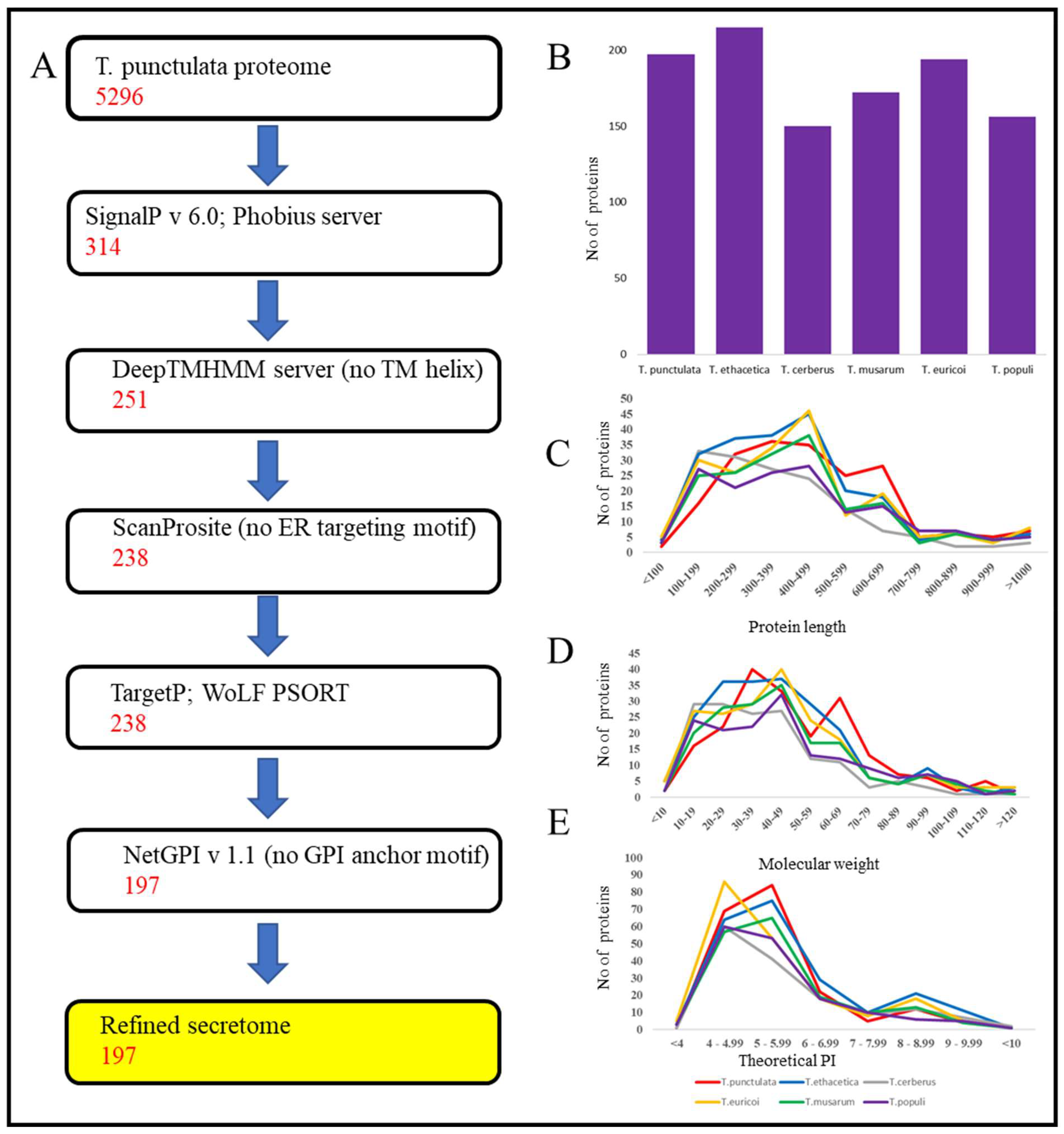
2.2. Prediction of the Secretome

We used a pipeline described previously to predict fungal secretome [31,32]. Briefly, SignalP (version 6.0) was used in combination with Phobius server [33,34]. The sequences, that were predicted to carry a signal peptide by both programs were selected for further screening. To exclude the transmembrane proteins, DeepTMHMM server was used [35]. The endoplasmic reticulum-targeting protein sequences were removed by scanning the sequences for PS00014 ER motif retention against the Prosite database with the ScanProsite web server [36]. The subcellular localization of the proteins was predicted using TargetP and WoLF PSORT servers [37,38]. The proteins harboring glycophosphatidylinositol anchor motifs were predicted using NetGPI (version 1.1) [39].

2.3. Annotation of Secretory Proteins

The refined secretome were scanned against Uniprot, PFAM, InterPro, and Gene3D to retrieve functional annotation of the predicted proteins [40,41,42,43]. The CAZy database and dbCAN web server were used to retrieve the annotation of carbohydrate-degrading enzymes [44,45]. For the effector prediction, the standalone software, EffectorP (version 3.0), combined with the manual inspection was used [46]. In addition, the BlASTP (E value < 1 × 10⁻¹⁰) was used to search against the pathogen–host interaction database (PHI database) to find similarities to known effectors and virulence factors [47]. Proteolytic enzymes were identified using a BlastP search against MEROPS database.

3. Results and Discussion

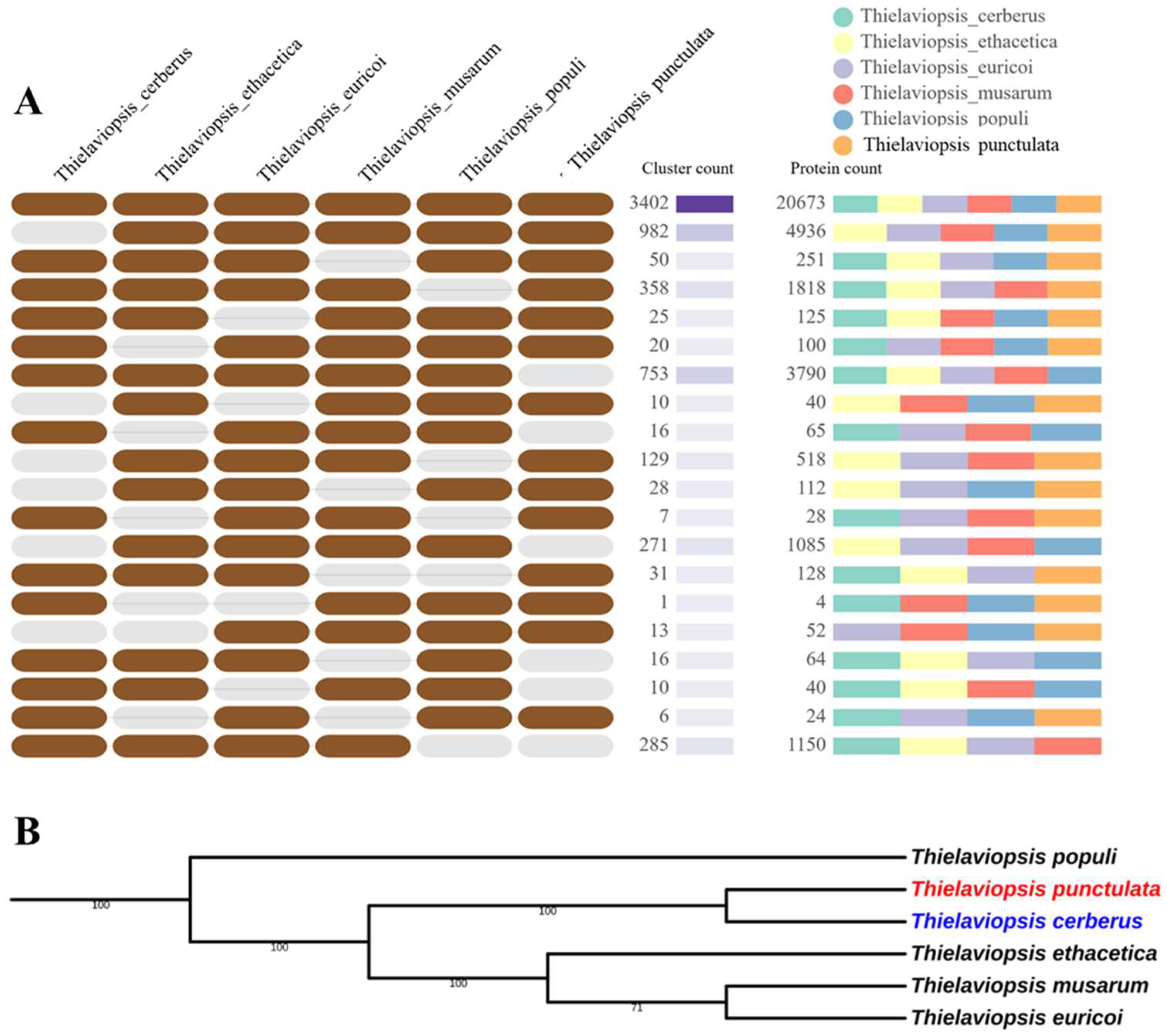
3.1. Prediction of Gene Models and Orthologue Analysis

The draft genome sequence of the Thielaviopsis punctulata (NCBI accession: GCA_000968615.1) and the 5296 predicted gene models were used for the identification of secretome. In addition, for the comparative analysis, we have also predicted the genes and the encoded proteins for 5 closely related species of T. punctulata viz—T. musarum, T. ethacetica, T. euricoi, T. populi, and T. cerberus—by utilizing the draft genome of these species available publicly (Table 1) [30]. The gene models were predicted according to the method described previously [30]. The number of predicted proteins in each species is given in Table 1. The gene models predicted for the closely related species were given in the Supplementary Table S1. The orthologue analysis using the whole proteome revealed that all six species form 7001 clusters, 3719 orthologous clusters (at least contains two species) and 3282 single-copy gene clusters (Figure 1, Supplementary Table S2). The number of singletons (The proteins which did not form any cluster) vary among the species. The highest number of singletons were found for T. cerberus (354), whereas the lowest were found for T. punctulata (50), which suggested that 94.4% of predicted proteins of T. punctulata had orthologue in other species. A phylogenetic tree was constructed using a concatenated alignment of 50 single copy orthologue proteins and revealed that T. cerberus was closely related to T. punctulata. The close genetic similarity between T. punctulata and T. cerberus was shown previously using internal transcribed spacer (ITS), β-tubulin, and transcription elongation factor 1-α DNA markers (Figure 1B) [25].

3.2. Secretome Identification and Analysis

The methodology used to predict the secretome of T. punctulata is illustrated in Figure 2. Using a combination of SignalP and Phobius server, of the 5296 total proteins, 314 proteins were predicted to have a signal peptide at their N-terminal region. Among these 314 proteins, 63 transmembrane proteins were excluded, and the remaining 251 proteins were scanned for an endoplasmic reticulum (ER)-targeting signal to exclude the proteins that remain in the endoplasmic reticulum. Of the 251 proteins, 13 were predicted to have PS00014 ER motif and were excluded from further analysis. The remaining 238 proteins were predicted as “extra-cellularly localized” through TargetP and WoLF PSORT analysis. Next, of the 238 proteins, 41 proteins were predicted to harbor glycophosphatidylinositol anchor motifs using NetGPI (version 1.1), which likely represent surface proteins rather than secreted effectors and were excluded. This resulted in a list of 197 “refined secretome”, which is 3.7% of the whole predicted proteome of T. punctulata (Table 1). Using this method, the secretome of T. musarum, T. ethacetica, T. euricoi, T. populi, and T. cerberus were also predicted. Overall, the number of “refined” secretome in all six species ranged from 150 (T. cerberus) to 215 (T. ethacetica) (Table 1, Figure 2B).

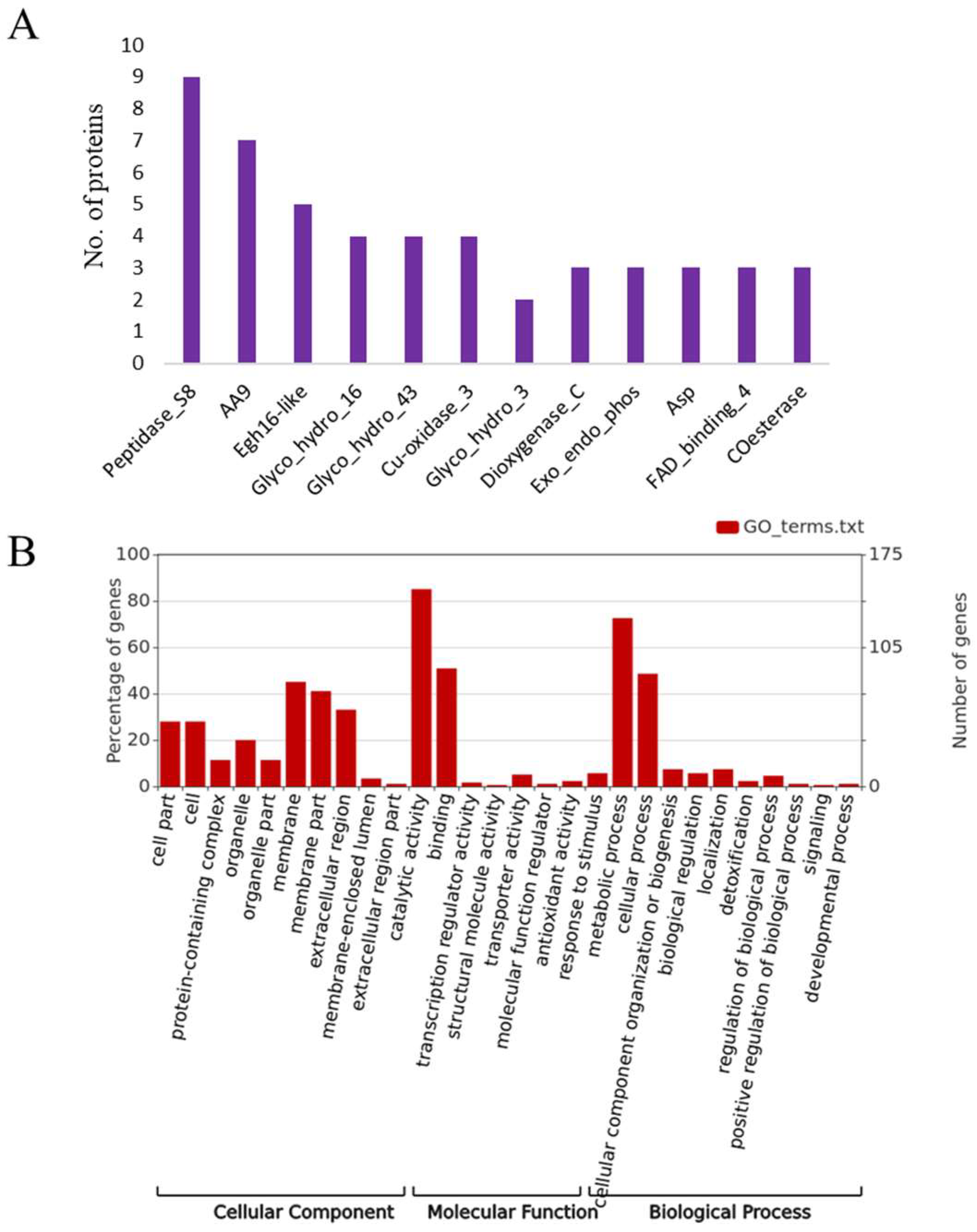
3.3. Structural and Functional Characterization of Secretome

The length of 197 refined secretome of T. punctulata ranged from 78 aa to 1356 aa. Of these, 43% (86) of proteins had a length of 78 aa to 399 aa, which indicated that small secretory proteins were enriched in the secretome of T. punctulata (Figure 2C).
Moreover, the molecular weight (MW) of secretory proteins ranged from 8.0 kDa to 147 kDa, and for most of the secretory proteins, it ranged between 10 and 49 kDa (56%) (Figure 2D). Similarly, the theoretical isoelectric point (pI) of the secretory proteins ranged from 3.8 to 9.4, of which the majorities (>77) pI ranged from 4–5.9 (Figure 2E). A similar pattern of length, PI and MW distribution, was also observed in closely-related species of T. punctulata (Figure 2C–E). The refined secretome of T. punctulata was characterized based on their matches in Uniprot, NCBI fungal reference proteome, Interpro and PFAM, Gene3D. The domain analysis revealed the presence of at least one function domain in 162 proteins. The most represented domains were Peptidase_S8 (PF00082), Auxiliary Activity family 9 (PF03443), Egh16-like (PF11327), Glycosyl hydrolases family 16 (PF00722), Glycosyl hydrolases family 43 (PF04616), Asp (PF00026), etc. (Figure 3A). Based on the sequence homology, gene ontology (GO) terms were assigned to 132 proteins which were further grouped into three major functional categories: biological process (71 proteins), molecular function (116 proteins), and cellular components (39 proteins) (Figure 3B). The biological processes include carbohydrate metabolic process (GO:0005975), polysaccharide catabolic process (GO:0000272), lipid metabolic process (GO:0006629), cellulose catabolic process (GO:0030245), chitin catabolic process (GO:0006032), arabinan catabolic process (GO:0031222), cellular aromatic compound metabolic process (GO:0006725), and xylan catabolic process (GO:0045493) (Figure 3B). The prominent category under the molecular function includes hydrolase activity (GO:0004553), serine-type endopeptidase activity (GO:0004252), cellulose binding (GO:0030248), endo-1,4-beta-xylanase activity (GO:0031176), oxidoreductase activity (GO:0016614), etc. (Figure 3B). The cellular component includes extracellular region (GO:0005576), cell wall (GO:0005618), and membrane (GO:0016020) (Figure 3B).

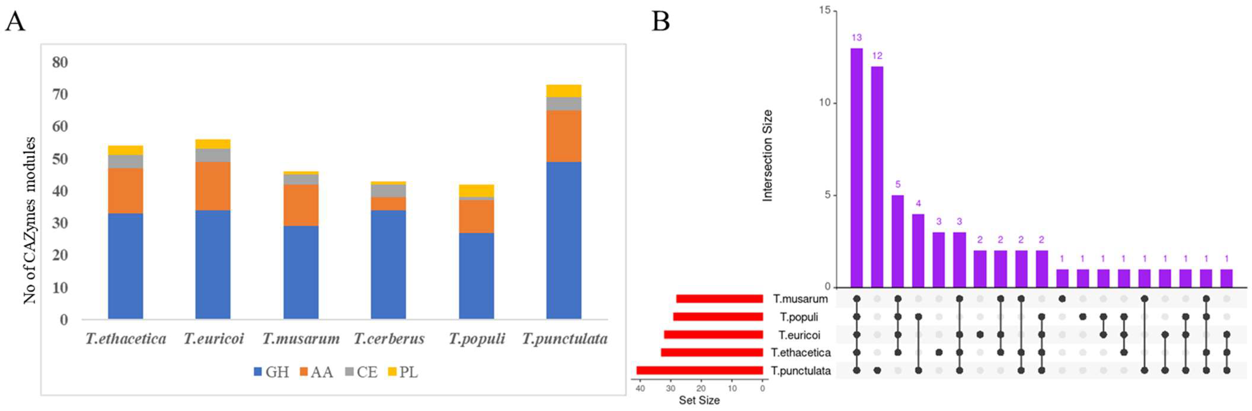
3.4. Carbohydrate Active Enzymes

Carbohydrate active enzymes, also called CAZymes, are a general group of enzymes involved in the biosynthesis and breakdown of carbohydrate and glycoconjugates. They are categorized into glycoside hydrolases (GH), polysaccharide lyases (PL), carbohydrate esterases (CE), auxiliary activity (AA), glycosyltransferasse (GT), and carbohydrate-binding module (CBM) classes [48,49]. To identify the CAZymes in T. punctulata and its closely related species, different sources of information such as blast description, Gene ontology, EC number, PFAM domain, and the results of annotation with CAZy database were combined [48]. Of the 197 refined secretome of T. punctulata, 75 proteins were identified as putative CAZymes, including 47 GH, 19 AA, 4 PL, 4 CE, and 1 GT (Figure 4, Table 2). Of these 75 CAZymes, 31 proteins contain multiple CAZymes modules. Among these, eight proteins possess two or more copies of the same CAZymes module that include two proteins with four copies of AA5 module and six proteins with two copies of GH3, GH7, GH20, GH32, and PL modules, respectively. Overall, 24 proteins contained two or three CA-Zyme modules of different types. In addition, 5 GH, 1 PL, 1 AA9, and 1 AA3 protein also contained a CBM module. CAZymes were also identified from five other species and found more numbers in T. punctulata (Table 2).
Except for the GT family, all other families of CAZymes (GH, CE, PL, and AA) were considered as cell wall-degrading enzymes since they are involved in the breakdown of plant cell wall components such as cellulose, hemi cellulose, pectin, and lignin [50,51]. Cellulose is an organic polysaccharide composed of a linear chain of hundreds of β-linked D-glucose units, and the enzymes involved in the breakdown of cellulose are exo-β-1,4-glucanases, endo-β-1,4-glucanases, β-1,4-glucosidases, cellobiose dehydrogenase, and lytic cellulose monooxygenase [51,52,53,54]. Based on the substrate specificity, of the 75 CAZymes of T. punctulata, 20 were predicted to be involved in the degradation of cellulose including seven endo-β-1,4-glucanases, one exo-β-1,4-glucanase (cellulose 1,4-beta-cellobiosidase (reducing end)), two β-glucosidase, two cellobiose dehydrogenase, and five lytic cellulose monooxygenase (Table 2). The CAZymes families containing endo-β-1,4-glucanases include GH5 (5), GH45 (1), and GH7 (1). One exo-β-1,4-glucanase was found in the GH7 family and five β-glucosidase were found in GH3 (3) and GH131 (2) families, respectively (Table 2). Seven lytic cellulose monooxygenases (EC:1.14.99.54) were found in AA9 and two cellobiose dehydrogenase (EC:1.1.99.18) in AA3 families (Table 2).
Hemicellulose is another major component of the plant cell wall, which include xyloglucans, xylans, mannans and glucomannans, and beta-(1-->3,1-->4)-glucans. The major enzymes involved in hemicellulose degradation are L-arabinanases, D-galactanases, D-mannanases, and D-xylanases [55]. Apart from this, Endo-_-1,4-glucanases with xyloglucanase activity were also identified from several fungal species [51]. Of the 74 CAZymes, 16 proteins were predicted to be involved in the degradation of hemicellulose, which included 14 GH and 2 CE. The GH group included 4 GH43, 2 GH10, 2 GH11, one member from GH38, GH51, GH53, GH93, GH115, and GH125, respectively (Table 2). The GH43 family consists of 3 exo-β-1,3-galactanases (EC:3.2.1.145) and one endo-α-1,5-L-arabinanase (3.2.1.99), which were predicted to be involved in the cleavage of galactans and arabinans, respectively (Table 2) [55]. Both the GH10 and GH11 family encode endo-1,4-β-xylanase (EC:3.2.1.8), which catalyzes endohydrolysis of (1->4)-beta-D-xylosidic linkages in xylans (Table 2) [55]. The GH38, GH51, GH53, GH93, GH115, and GH125 families encode α-mannosidase (involved in the cleavage of mannose), α-L-arabinofuranosidase (involved in the cleavage of arabinans), endo-β-1,4-galactanase (involved in the cleavage of galactans), exo-α-L-1,5-arabinanase (involved in the cleavage of arabinans), xylan α-1,2-glucuronidase (involved in the cleavage of xylans), and exo-α-1,6-mannosidase (involved in the cleavage of mannans), respectively (Table 2) [55]. The two CE members belong to CE5 family encoding acetyl xylan esterase (3.1.1.72), which catalyzes the hydrolysis of acetyl groups from polymeric xylan (Table 2) [56].
Within the refined secretome, members of polysaccharide lyases (PLs), including two pectin lyase (EC:4.2.2.10), one pectate lyase (EC:4.2.2.2), one rhamnogalacturonan endolyase (EC:4.2.2.23), and one GH28 polygalacturonase, were also identified (Table 2). These enzymes are known to degrade pectin (Table 1) [57]. In addition to glycoside hydrolases, members of Auxillary activity (AA) families were identified with the potential to degrade lignin (Table 2) [57]. These include three AA1 laccases (EC:1.10.3.2), one AA2 peroxidase (EC 1.11.1.-), one AA2 versatile peroxidase (EC:1.11.1.16), one aryl alcohol oxidase (EC:1.1.3.7), and one AA5 Oxidase with oxygen as acceptor (EC:1.1.3.-) (Table 2). In addition to these cell wall-degrading enzymes, the refined secretome also contains two starch, one sucrose, one trehalose, three glucans, and three callose-degrading enzymes (Table 2).
The comparison of CAZymes in closely-related species revealed 59 different classes of CAZymes in all species, including T. punctulata (Figure 4, Table 3, Supplementary Table S3). The secretome of all these species were rich in secreted CAZymes families, especially those involved in plant cell wall degradation (PCWD). High numbers of secreted CAZymes involved with PCWD have also been found in the genomes of several Botryosphaeriaceae pathogens [58]. However, the occurrence of these classes varied among Thielaviopsis species. For example, only 11 classes of CAZymes (AA1, AA5, GH10, GH11, GH16, GH17, GH20, GH32, GH43, GH76, and PL1) were found common in the secretome of all species (Figure 4, Table 3, Supplementary Table S3). In addition, some classes of CAZymes were found to be specific for some species, and notably 10 classes of CAZymes (GH64, GH125, GH45, GH132, AA16, GH128, GT4, GH51, GH115, GH38) were found only in the secretome of T. punctulata (Figure 4, Table 3, Supplementary Table S3), suggesting that they might have a species-specific role in black scorch disease of date palm.

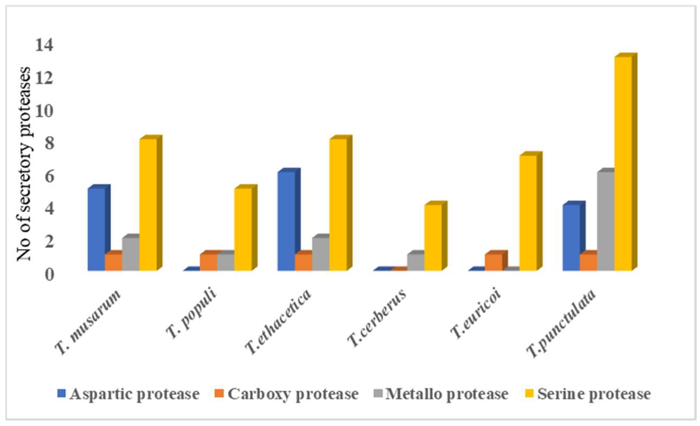
3.5. Secreted Proteases

Several studies have shown that plant pathogenic fungi secrete proteases that degrade plant antimicrobial proteins and protease inhibitors (PIs) to facilitate virulence [59]. The BlastP search against MEROPS database resulted in the identification of 24 putative proteases from the 197 refined secretome, which were classified into several groups based on their catalytic residues (Supplementary Table S4). Among the proteases, serine proteases (13) were dominant, followed by metallo proteases (6), aspartic proteases (4), and carboxy protease (1). The serine proteases included S8, S10, and S28 families. Among these, S8 was found to be dominant (Supplementary Table S4). Members of metallo protease were further classified into M14, M28, M36, and M43 families based on their similarity to the known members from these families. Members of the same classes of proteases were also identified in the closely related species (Supplementary Table S4). Comparison to closely related species revealed that Serine protease S8 was prominent in T. unctulate, whereas S9 was completely absent (Figure 5).

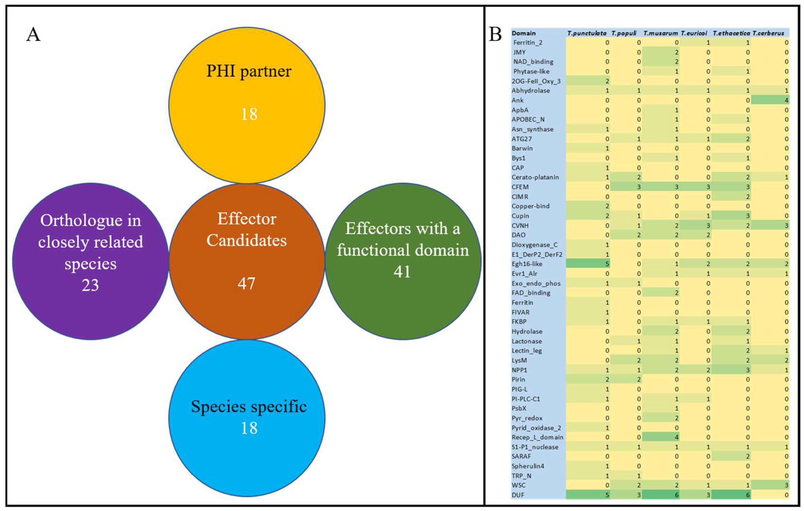
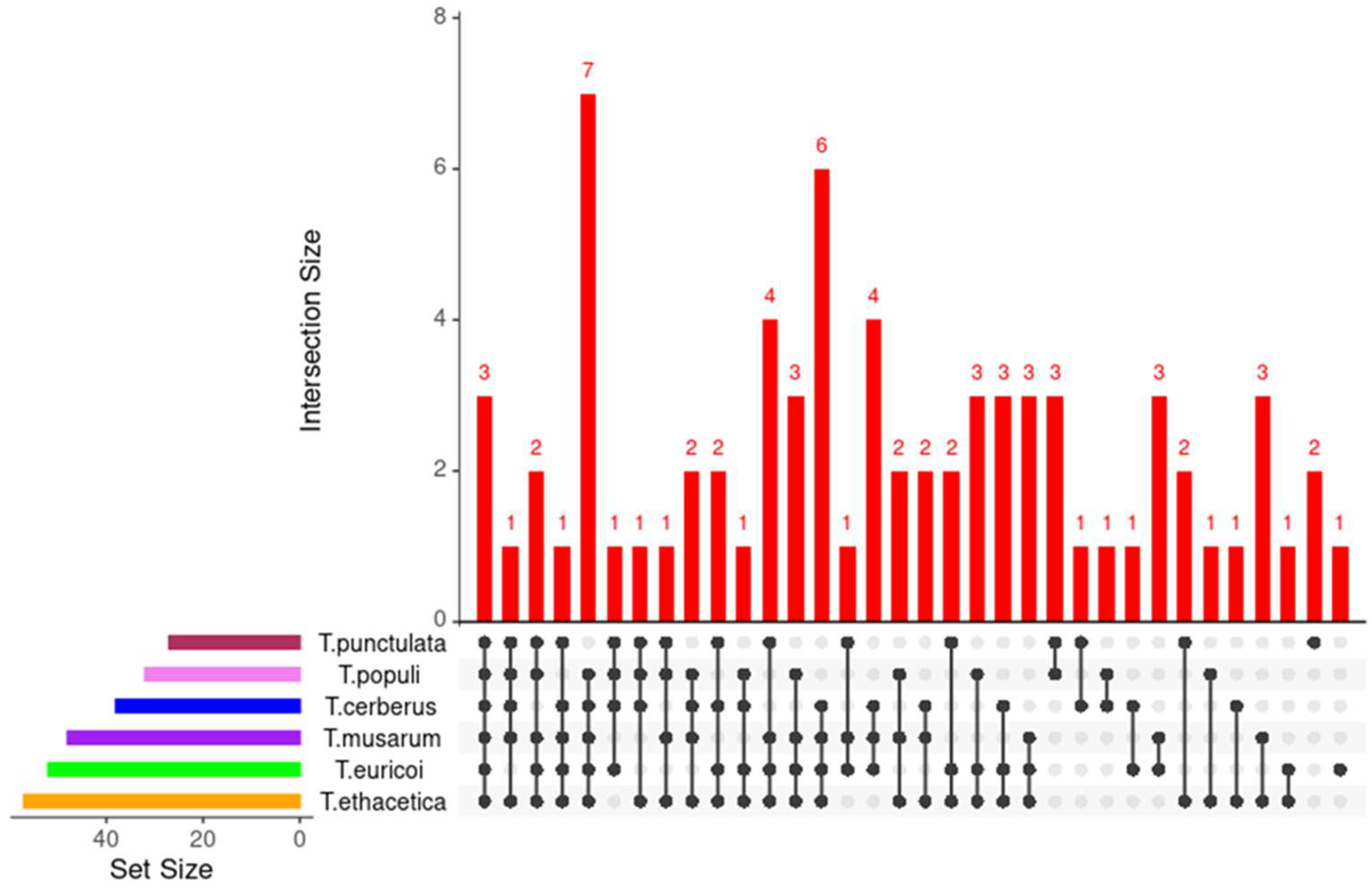
3.6. Putative Effector Proteins

EffectorP, combined with the manual inspection, was employed to identify the putative effector proteins with the following characteristics: a signal peptide for secretion, no trans-membrane domains, fairly small size, and cysteine-rich [9,31,60]. This analysis resulted in the identification of 47 proteins as putative “effector” candidates (Figure 6). The length of these effector candidates varied from 78 to 392 Amino acids, of which 15 candidates were 100 to 200, 16 were 200 to 300, and 14 were 300 to 400 amino acids in length, respectively (Table 4). Two candidates (KKA29758.1 and KKA27537.1) were found with a length of less than 100 amino acids. The number of cysteine residues varied from 2 to 8 in the selected putative effectors, of which 70% were found with more than four cysteine residues (Table 4). Of the 47 putative effector proteins, functional domains were identified in 34 proteins, notably five putative effectors (KKA26926.1, KKA26947.1, KKA27553.1, KKA27672.1, KKA29410.1) possessed an Egh16-like (PF11327) domain. Three putative effectors (KKA26938.1, KKA27913.1, KKA28390.1) possessed copper bind domain (PF00127) (Figure 4, Table 4. A necrosis-inducing protein (NPP1) domain (PF05630) was identified in one protein (KKA27476.1). Seven putative effectors possessed a domain of unknown function (DUF) and 13 had no known functional domain in it (Figure 6, Table 4). Orthologue analysis found that T. punctulata shared 24 effectors with other closely related species, of which 3 proteins were shared with all closely related species (T. musarum, T. euricoi, T. populi, T. ethacetica and T. cerberus) (Figure 7). Two pairs of duplicated effector proteins were identified in the secretome of T. punctulata (KKA28270.1 and KKA26687.1 and KKA27979.1 and KKA29712.1). These paralogs showed 100% identity to each other, indicating a recent duplication. Interestingly, 18 effectors were found to be specific to T. punctulata, and functional analysis of these effectors may reveal more insight into the pathogenicity of T, punctulata on Date palm (Table 4).

3.7. Putative Virulence Factors

To identify the homologs of pathogenicity-associated genes in other phytopathogens, we screened 197 refined secretome, including all the putative effectors against the PHI (Pathogen–host interactions) database [47]. The protein sequences in the PHI database are classified into different categories such as loss of pathogenicity, reduced virulence, unaffected pathogenicity, increased virulence, effector (plant avirulence determinant), lethal, enhanced antagonism, resistant to chemical, and sensitivity to chemicals based on the results of mutation experiments. For example, the “Loss of pathogenicity” group includes proteins for which the mutant strains fail to cause diseases in host compared to the wild type. Based on the PHI annotation, of the 197 secretomes, 49 had PHI homologue, including 27 CAZymes, 7 proteases, and 15 putative effectors (Supplementary Table S5). Of the 27 CAZymes, four were assigned as effector (plant_avirulence_determinant), including three lytic cellulose monooxygenases in AA9 CAZymes family (KKA27328.1, KKA28497.1 and KKA25992.1) and one endo-β-1,4-xylanase in GH11 family (KKA30007.1). Both these enzymes were reported to contribute to the virulence of several plant pathogenic fungi. The homologue of lytic cellulose monooxygenase in Magnaporthe oryzae (MoCDIP) induced cell death when it was expressed in rice plant cells [61]. Similarly, a lytic cellulose monooxygenase gene (PHEC27213) in Podosphaera xanthii was shown to suppress the chitin-triggered immunity in cucurbit host [62]. Endo-β-1,4-xylanase was also shown to involved in the pathogenicity of many fungal pathogens, including Verticillium dahlia, Ustilago maydis, Valsa mali, etc. [63,64,65]. In addition, 13 CAZymes were also assigned as “reduced virulence” according PHI database, which includes three Pectin lyases (PL), two laccases (AA1), two β-glucosidases (GH03), one peroxidase (AA2), Oxidase (AA5), endo-1,4-β-xylanase (GH10), exo-α-1,6-mannosidase (GH25), and licheninase (GH16). The contribution of these genes in virulence was demonstrated for many plant-pathogenic fungi [66,67,68,69,70]. Of the CAZymes, six were assigned as “Unaffected category” (Table S5). Among the 25 proteases identified, two metallo peptidases (KKA26166.1 and KKA27603.1) were assigned as “loss of pathogenicity” and one carboxy peptidase (KKA30601.1) was assigned as “reduced virulence” (Supplementary Table S5). The homologs of these two metallo peptidases were shown to require the pathogenicity of Fusarium oxysporum f. sp. lycopersici and Magnaporthe oryzae on their respective hosts [71,72]. In addition, two proteases were assigned as “unaffected pathogenicity” according to PHI annotation (Supplementary Table S5). From the putative effectors group, PHI partners were identified for 15 proteins, of which one was assigned as “loss of pathogenicity”, which encodes an acetylglucosaminyl phosphatidylinositol deacetylase (KKA29596.1) (Supplementary Table S5). The homologue of this protein in Colletotrichum graminicola, the causal agent of maize anthracnose, was shown to require cell-wall integrity and pathogenicity [73]. Thirteen effectors were assigned as “reduced virulence”, and homologs of these proteins were shown to be required for pathogenicity in many fungal species. Notably, the most enriched group of effectors in T. punctulata were Egh16-like proteins, and their homologue in PHI database (PHI:256) was shown to act as virulence factors in rice blast fungus Magnaporthe grisea [74]. The Egh16 family members were also shown to be involved in the virulence of many pathogenic filamentous fungi. For example, two Egh16-like factors in Erysiphe pisi, EpCSEP087 and EpCSP083, were found to be highly induced during early infection stages on pea, suggesting a critical role in appressorium penetration and pathogenesis [75].

4. Conclusions

In the current study, the potential secretory proteins of Thielaviopsis punctulata were extensively characterized using a well-designed bioinformatics approach. Within the secretome, 74 CAZymes, 25 proteases, and 47 putative effector proteins were identified. For the comparative analysis, the genes models for five closely related species of T. punctulata were predicted and the secretome of each species was also well-characterized. The comparative analysis revealed that all Thielaviopsis species studied possessed species-specific CAZymes families, putative effectors, and several putative virulence factors. The current study will be a valuable source for the studies aimed at understanding the pathogenicity mechanism not only in T. punctulata–date palm interaction, but also in other Thielaviopsis species and their respective hosts.

Supplementary Materials

The following supporting information can be downloaded at: https://www.mdpi.com/article/10.3390/jof9030303/s1, Supplementary Table S1—Predicted gene models of T. musarum, T. ethacetica, T. populi, T. euricoi and T. cerberus. Supplementary Table S2—Single-copy gene clusters in Thielaviopsis species. Supplementary Table S3—CAZymes in the secretome of Thielaviopsis species. Supplementary Table S4—Proteases in the secretome of Thielaviopsis species. Supplementary Table S5—PHI homologs of T. punctulata secretome.

Author Contributions

Conceptualization, Bioinformatics analysis and writing the manuscript—B.V.C. B.V.C., S.M.E.-G., H.S.A. and H.A.-S. have read, edited and agreed to the published version of the manuscript.

Funding

The authors extend their appreciation to the Deputyship for Research and Innovation, Ministry of Education in Saudi Arabia, for funding this research work (Project number INST119).

Institutional Review Board Statement

This study did not require ethical approval.

Informed Consent Statement

Not Applicabale.

Data Availability Statement

Not Applicable.

Conflicts of Interest

The authors declare no conflict of interest.

References

  1. Dean, R.; Van Kan, J.A.L.; Pretorius, Z.A.; Hammond-Kosack, K.E.; Di Pietro, A.; Spanu, P.D.; Rudd, J.J.; Dickman, M.; Kahmann, R.; Ellis, J.; et al. The Top 10 fungal pathogens in molecular plant pathology. Mol. Plant Pathol. 2012, 13, 414–430. [Google Scholar] [CrossRef] [PubMed] [Green Version]
  2. Biju, V.C.; Fokkens, L.; Houterman, P.M.; Rep, M.; Cornelissen, B.J.C. Multiple Evolutionary Trajectories Have Led to the Emergence of Races in Fusarium oxysporum f. sp. lycopersici. Appl. Environ. Microbiol. 2017, 83, e02548-16. [Google Scholar] [CrossRef] [PubMed] [Green Version]
  3. Pontes, J.G.D.M.; Fernandes, L.S.; dos Santos, R.V.; Tasic, L.; Fill, T.P. Virulence Factors in the Phytopathogen–Host Interactions: An Overview. J. Agric. Food Chem. 2020, 68, 7555–7570. [Google Scholar] [CrossRef] [PubMed]
  4. Zhou, J.-M.; Zhang, Y. Plant Immunity: Danger Perception and Signaling. Cell 2020, 181, 978–989. [Google Scholar] [CrossRef] [PubMed]
  5. Ali, G.S.; Reddy, A. PAMP-triggered immunity. Plant Signal. Behav. 2008, 3, 423–426. [Google Scholar] [CrossRef]
  6. de Wit, P.J.G.M.; Mehrabi, R.; van den Burg, H.A.; Stergiopoulos, I. Fungal effector proteins: Past, present and future. Mol. Plant Pathol. 2009, 10, 735–747. [Google Scholar] [CrossRef]
  7. Lo Presti, L.; Lanver, D.; Schweizer, G.; Tanaka, S.; Liang, L.; Tollot, M.; Zuccaro, A.; Reissmann, S.; Kahmann, R. Fungal Effectors and Plant Susceptibility. Annu. Rev. Plant Biol. 2015, 66, 513–545. [Google Scholar] [CrossRef]
  8. Pradhan, A.; Ghosh, S.; Sahoo, D.; Jha, G. Fungal effectors, the double edge sword of phytopathogens. Curr. Genet. 2020, 67, 27–40. [Google Scholar] [CrossRef]
  9. Todd, J.N.A.; Carreón-Anguiano, K.G.; Islas-Flores, I.; Canto-Canché, B. Fungal Effectoromics: A World in Constant Evolution. Int. J. Mol. Sci. 2022, 23, 13433. [Google Scholar] [CrossRef]
  10. Ma, W.; Wang, Y.; McDowell, J. Focus on Effector-Triggered Susceptibility. Mol. Plant-Microbe Interact. 2018, 31, 5. [Google Scholar] [CrossRef]
  11. Hammond-Kosack, K.E.; Jones, J.D. Plant disease resistance genes. Annu. Rev. Plant Biol. 1997, 48, 575–607. [Google Scholar] [CrossRef] [PubMed] [Green Version]
  12. Guo, J.; Cheng, Y. Advances in Fungal Elicitor-Triggered Plant Immunity. Int. J. Mol. Sci. 2022, 23, 12003. [Google Scholar] [CrossRef] [PubMed]
  13. Laugé, R.; de Wit, P. Fungal Avirulence Genes: Structure and Possible Functions. Fungal Genet. Biol. 1998, 24, 285–297. [Google Scholar] [CrossRef] [PubMed]
  14. Xu, X.; Chen, Y.; Li, B.; Zhang, Z.; Qin, G.; Chen, T.; Tian, S. Molecular mechanisms underlying multi-level defense responses of horticultural crops to fungal pathogens. Hortic. Res. 2022, 9, uhac066. [Google Scholar] [CrossRef] [PubMed]
  15. Petit-Houdenot, Y.; Fudal, I. Complex Interactions between Fungal Avirulence Genes and Their Corresponding Plant Resistance Genes and Consequences for Disease Resistance Management. Front. Plant Sci. 2017, 8, 1072. [Google Scholar] [CrossRef] [PubMed] [Green Version]
  16. Balint-Kurti, P.; Balint-Kurti, P. The plant hypersensitive response: Concepts, control and consequences. Mol. Plant Pathol. 2019, 20, 1163–1178. [Google Scholar] [CrossRef] [PubMed] [Green Version]
  17. Takken, F.; Rep, M. The arms race between tomato and Fusarium oxysporum. Mol. Plant Pathol. 2010, 11, 309–314. [Google Scholar] [CrossRef]
  18. Anderson, J.; Gleason, C.A.; Foley, R.C.; Thrall, P.H.; Burdon, J.B.; Singh, K.B. Plants versus pathogens: An evolutionary arms race. Funct. Plant Biol. 2010, 37, 499–512. [Google Scholar] [CrossRef]
  19. Jaswal, R.; Kiran, K.; Rajarammohan, S.; Dubey, H.; Singh, P.K.; Sharma, Y.; Deshmukh, R.; Sonah, H.; Gupta, N.; Sharma, T. Effector Biology of Biotrophic Plant Fungal Pathogens: Current Advances and Future Prospects. Microbiol. Res. 2020, 241, 126567. [Google Scholar] [CrossRef]
  20. Rafiqi, M.; Jelonek, L.; Diouf, A.M.; Mbaye, A.; Rep, M.; Diarra, A. Profile of the in silico secretome of the palm dieback pathogen, Fusarium oxysporum f. sp. albedinis, a fungus that puts natural oases at risk. PLoS ONE 2022, 17, e0260830. [Google Scholar] [CrossRef]
  21. Jansson, H.B.; Lopez Lorca, L.V.; Abdullah, S.K. Diseases of date palms (phoenix dactylifera L.). Basrah J. Date Palm Res. 2010, 9, 1–44. [Google Scholar]
  22. Abdullah, S.; Asensio, L.; Monfort, E.; Gomez-Vidal, S.; Salinas, J.; Lorca, L.; Jansson, H. Incidence of the Two Date Palm Pathogens, Thielaviopsis Paradoxa and T. Punctulata in Soil From Date Palm Plantations in Elx, South-East Spain. J. Plant Prot. Res. 2009, 49. [Google Scholar] [CrossRef]
  23. Al-Raisi, Y.; B’Chir, M.; Al-Mandhari, A.; Deadman, M.; Gowen, S. First report of Ceratocystis radicicola associated with date palm disease in Oman. New Dis. Rep. 2011, 23, 23. [Google Scholar] [CrossRef] [Green Version]
  24. Al-Sharidah, A. Report of Black Scorch Disease on Date Palm Trees in the State of Kuwait. Am. Sci. Res. J. Eng. 2017, 28, 14–17. [Google Scholar]
  25. Alhudaib, K.A.; El-Ganainy, S.M.; Almaghasla, M.I.; Sattar, M.N. Characterization and Control of Thielaviopsis punctulata on Date Palm in Saudi Arabia. Plants 2022, 11, 250. [Google Scholar] [CrossRef] [PubMed]
  26. Al-Naemi, F.A.; Nishad, R.; Ahmed, T.A.; Radwan, O. First Report of Thielaviopsis punctulata Causing Black Scorch Disease on Date Palm in Qatar. Plant Dis. 2014, 98, 1437. [Google Scholar] [CrossRef] [PubMed]
  27. Saeed, E.E.; Sham, A.; El-Tarabily, K.; Abu Elsamen, F.; Iratni, R.; AbuQamar, S.F. Chemical Control of Black Scorch Disease on Date Palm Caused by the Fungal Pathogen Thielaviopsis punctulata in United Arab Emirates. Plant Dis. 2016, 100, 2370–2376. [Google Scholar] [CrossRef] [Green Version]
  28. Al-Naemi, F.A.; Ahmed, T.A.; Nishad, R.; Radwan, O. Antagonistic Effects of Trichoderma harzianum Isolates against Ceratocystis radicicola: Pioneering a Biocontrol Strategy against Black Scorch Disease in Date Palm Trees. J. Phytopathol. 2016, 164, 464–475. [Google Scholar] [CrossRef]
  29. Saeed, E.E.; Sham, A.; Salmin, Z.; Abdelmowla, Y.; Iratni, R.; El-Tarabily, K.; Abu Qamar, S. Streptomyces globosus UAE1, a Potential Effective Biocontrol Agent for Black Scorch Disease in Date Palm Plantations. Front. Microbiol. 2017, 8, 1455. [Google Scholar] [CrossRef] [Green Version]
  30. Wingfield, B.D.; Ades, P.K.; Al-Naemi, F.A.; Beirn, L.A.; Bihon, W.; Crouch, J.A.; De Beer, Z.W.; De Vos, L.; Duong, T.A.; Fields, C.J.; et al. Draft genome sequences of Chrysoporthe austroafricana, Diplodia scrobiculata, Fusarium nygamai, Leptographium lundbergii, Limonomyces culmigenus, Stagonosporopsis tanaceti, and Thielaviopsis punctulata. IMA Fungus 2015, 6, 233–248. [Google Scholar] [CrossRef] [Green Version]
  31. Sonah, H.; Deshmukh, R.; Bélanger, R.R. Computational Prediction of Effector Proteins in Fungi: Opportunities and Challenges. Front. Plant Sci. 2016, 7, 126. [Google Scholar] [CrossRef] [Green Version]
  32. Rafiqi, M.; Jelonek, L.; Akum, N.F.; Zhang, F.; Kogel, K.-H. Effector candidates in the secretome of Piriformospora indica, a ubiquitous plant-associated fungus. Front. Plant Sci. 2013, 4, 228. [Google Scholar] [CrossRef] [Green Version]
  33. Teufel, F.; Armenteros, J.J.A.; Johansen, A.R.; Gíslason, M.H.; Pihl, S.I.; Tsirigos, K.D.; Winther, O.; Brunak, S.; von Heijne, G.; Nielsen, H. SignalP 6.0 predicts all five types of signal peptides using protein language models. Nat. Biotechnol. 2022, 40, 1023–1025. [Google Scholar] [CrossRef] [PubMed]
  34. Käll, L.; Krogh, A.; Sonnhammer, E.L. A Combined Transmembrane Topology and Signal Peptide Prediction Method. J. Mol. Biol. 2004, 338, 1027–1036. [Google Scholar] [CrossRef] [PubMed]
  35. Krogh, A.; Larsson, B.; von Heijne, G.; Sonnhammer, E.L. Predicting transmembrane protein topology with a hidden markov model: Application to complete genomes. J. Mol. Biol. 2001, 305, 567–580. [Google Scholar] [CrossRef] [PubMed] [Green Version]
  36. Gattiker, A.; Gasteiger, E.; Bairoch, A. ScanProsite: A reference implementation of a PROSITE scanning tool. Appl. Bioinform. 2002, 1, 107–108. [Google Scholar]
  37. Horton, P.; Park, K.-J.; Obayashi, T.; Fujita, N.; Harada, H.; Adams-Collier, C.J.; Nakai, K. WoLF PSORT: Protein localization predictor. Nucleic Acids Res. 2007, 35, W585–W587. [Google Scholar] [CrossRef] [Green Version]
  38. Armenteros, J.J.A.; Salvatore, M.; Emanuelsson, O.; Winther, O.; von Heijne, G.; Elofsson, A.; Nielsen, H. Detecting sequence signals in targeting peptides using deep learning. Life Sci. Alliance 2019, 2, e201900429. [Google Scholar] [CrossRef] [Green Version]
  39. Gíslason, M.H.; Nielsen, H.; Armenteros, J.J.A.; Johansen, A.R. Prediction of GPI-anchored proteins with pointer neural networks. Curr. Res. Biotechnol. 2021, 3, 6–13. [Google Scholar] [CrossRef]
  40. Lees, J.; Yeats, C.; Perkins, J.R.; Sillitoe, I.; Rentzsch, R.; Dessailly, B.H.; Orengo, C. Gene3D: A domain-based resource for comparative genomics, functional annotation and protein network analysis. Nucleic Acids Res. 2011, 40, D465–D471. [Google Scholar] [CrossRef]
  41. Blum, M.; Chang, H.-Y.; Chuguransky, S.; Grego, T.; Kandasaamy, S.; Mitchell, A.; Nuka, G.; Paysan-Lafosse, T.; Qureshi, M.; Raj, S.; et al. The InterPro protein families and domains database: 20 years on. Nucleic Acids Res. 2021, 49, D344–D354. [Google Scholar] [CrossRef] [PubMed]
  42. Finn, R.D.; Bateman, A.; Clements, J.; Coggill, P.; Eberhardt, R.Y.; Eddy, S.R.; Heger, A.; Hetherington, K.; Holm, L.; Mistry, J.; et al. Pfam: The protein families database. Nucleic Acids Res. 2014, 42, D222–D230. [Google Scholar] [CrossRef] [PubMed] [Green Version]
  43. The UniProt Consortium. UniProt: The Universal Protein Knowledgebase in 2023. Nucleic Acids Res. 2022, 51, D523–D531. [Google Scholar] [CrossRef]
  44. Yin, Y.; Mao, X.; Yang, J.; Chen, X.; Mao, F.; Xu, Y. dbCAN: A web resource for automated carbohydrate-active enzyme annotation. Nucleic Acids Res. 2012, 40, W445–W451. [Google Scholar] [CrossRef] [PubMed]
  45. Cantarel, B.L.; Coutinho, P.M.; Rancurel, C.; Bernard, T.; Lombard, V.; Henrissat, B. The Carbohydrate-Active EnZymes database (CAZy): An expert resource for Glycogenomics. Nucleic Acids Res. 2009, 37, D233–D238. [Google Scholar] [CrossRef]
  46. Sperschneider, J.; Dodds, P.N. EffectorP 3.0: Prediction of Apoplastic and Cytoplasmic Effectors in Fungi and Oomycetes. Mol. Plant-Microbe Interact. 2022, 35, 146–156. [Google Scholar] [CrossRef]
  47. Winnenburg, R.; Urban, M.; Beacham, A.; Baldwin, T.K.; Holland, S.; Lindeberg, M.; Hansen, H.; Rawlings, C.; Hammond-Kosack, K.E.; Köhler, J. PHI-base update: Additions to the pathogen host interaction database. Nucleic Acids Res. 2007, 36, D572–D576. [Google Scholar] [CrossRef]
  48. Drula, E.; Garron, M.-L.; Dogan, S.; Lombard, V.; Henrissat, B.; Terrapon, N. The carbohydrate-active enzyme database: Functions and literature. Nucleic Acids Res. 2022, 50, D571–D577. [Google Scholar] [CrossRef]
  49. Benini, S. Carbohydrate-Active Enzymes: Structure, Activity, and Reaction Products. Int. J. Mol. Sci. 2020, 21, 2727. [Google Scholar] [CrossRef] [Green Version]
  50. Lairson, L.L.; Henrissat, B.; Davies, G.J.; Withers, S.G. Glycosyltransferases: Structures, Functions, and Mechanisms. Annu. Rev. Biochem. 2008, 77, 521–555. [Google Scholar] [CrossRef] [Green Version]
  51. Kubicek, C.P.; Starr, T.L.; Glass, N.L. Plant Cell Wall–Degrading Enzymes and Their Secretion in Plant-Pathogenic Fungi. Annu. Rev. Phytopathol. 2014, 52, 427–451. [Google Scholar] [CrossRef] [PubMed]
  52. Villares, A.; Moreau, C.; Bennati-Granier, C.; Garajova, S.; Foucat, L.; Falourd, X.; Saake, B.; Berrin, J.-G.; Cathala, B. Lytic polysaccharide monooxygenases disrupt the cellulose fibers structure. Sci. Rep. 2017, 7, 40262. [Google Scholar] [CrossRef] [PubMed] [Green Version]
  53. Phillips, C.M.; Beeson, W.T.; Cate, J.H.; Marletta, M.A. Cellobiose Dehydrogenase and a Copper-Dependent Polysaccharide Monooxygenase Potentiate Cellulose Degradation by Neurospora crassa. ACS Chem. Biol. 2011, 6, 1399–1406. [Google Scholar] [CrossRef] [PubMed]
  54. Horn, S.J.; Vaaje-Kolstad, G.; Westereng, B.; Eijsink, V.G. Novel enzymes for the degradation of cellulose. Biotechnol. Biofuels 2012, 5, 45. [Google Scholar] [CrossRef] [Green Version]
  55. van den Brink, J.; De Vries, R.P. Fungal enzyme sets for plant polysaccharide degradation. Appl. Microbiol. Biotechnol. 2011, 91, 1477–1492. [Google Scholar] [CrossRef] [Green Version]
  56. Margolles-Clark, E.; Tenkanen, M.; Soderlund, H.; Penttila, M. Acetyl Xylan Esterase from Trichoderma reesei Contains an Active-Site Serine Residue and a Cellulose-Binding Domain. JBIC J. Biol. Inorg. Chem. 1996, 237, 553–560. [Google Scholar] [CrossRef]
  57. Pollegioni, L.; Tonin, F.; Rosini, E. Lignin-degrading enzymes. FEBS J. 2015, 282, 1190–1213. [Google Scholar] [CrossRef]
  58. Nagel, J.H.; Wingfield, M.J.; Slippers, B. Increased abundance of secreted hydrolytic enzymes and secondary metabolite gene clusters define the genomes of latent plant pathogens in the Botryosphaeriaceae. BMC Genom. 2021, 22, 589. [Google Scholar] [CrossRef]
  59. Jashni, M.E.; Mehrabi, R.; Collemare, J.; Mesarich, C.; De Wit, P.J.G.M. The battle in the apoplast: Further insights into the roles of proteases and their inhibitors in plant–pathogen interactions. Front. Plant Sci. 2015, 6, 584. [Google Scholar] [CrossRef] [Green Version]
  60. Stergiopoulos, I.; de Wit, P.J. Fungal Effector Proteins. Annu. Rev. Phytopathol. 2009, 47, 233–263. [Google Scholar] [CrossRef] [Green Version]
  61. Chen, S.; Songkumarn, P.; Venu, R.C.; Gowda, M.; Bellizzi, M.; Hu, J.; Liu, W.; Ebbole, D.; Meyers, B.; Mitchell, T.; et al. Identification and Characterization of In planta–Expressed Secreted Effector Proteins from Magnaporthe oryzae That Induce Cell Death in Rice. Mol. Plant-Microbe Interact. 2013, 26, 191–202. [Google Scholar] [CrossRef] [PubMed] [Green Version]
  62. Polonio, Á.; Fernández-Ortuño, D.; Vicente, A.; Pérez-García, A. A haustorial-expressed lytic polysaccharide monooxygenase from the cucurbit powdery mildew pathogen Podosphaera xanthii contributes to the suppression of chitin-triggered immunity. Mol. Plant Pathol. 2021, 22, 580–601. [Google Scholar] [CrossRef] [PubMed]
  63. Moreno-Sánchez, I.; Pejenaute-Ochoa, M.D.; Navarrete, B.; Barrales, R.R.; Ibeas, J.I. Ustilago maydis Secreted Endo-Xylanases Are Involved in Fungal Filamentation and Proliferation on and Inside Plants. J. Fungi 2021, 7, 1081. [Google Scholar] [CrossRef] [PubMed]
  64. Yu, C.; Li, T.; Shi, X.; Saleem, M.; Li, B.; Liang, W.; Wang, C. Deletion of Endo-β-1,4-Xylanase VmXyl1 Impacts the Virulence of Valsa mali in Apple Tree. Front. Plant Sci. 2018, 9, 663. [Google Scholar] [CrossRef] [PubMed] [Green Version]
  65. Liu, L.; Wang, Z.; Li, J.; Wang, Y.; Yuan, J.; Zhan, J.; Wang, P.; Lin, Y.; Li, F.; Ge, X. Verticillium dahliae secreted protein Vd424Y is required for full virulence, targets the nucleus of plant cells, and induces cell death. Mol. Plant Pathol. 2021, 22, 1109–1120. [Google Scholar] [CrossRef] [PubMed]
  66. Lin, S.Y.; Okuda, S.; Ikeda, K.; Okuno, T.; Takano, Y. LAC2 Encoding a Secreted Laccase Is Involved in Appressorial Melanization and Conidial Pigmentation in Colletotrichum orbiculare. Mol. Plant-Microbe Interact. 2012, 25, 1552–1561. [Google Scholar] [CrossRef] [PubMed] [Green Version]
  67. Mir, A.A.; Park, S.-Y.; Sadat, A.; Kim, S.; Choi, J.; Jeon, J.; Lee, Y.-H. Systematic characterization of the peroxidase gene family provides new insights into fungal pathogenicity in Magnaporthe oryzae. Sci. Rep. 2015, 5, 11831. [Google Scholar] [CrossRef] [Green Version]
  68. López-Pérez, M.; Ballester, A.-R.; González-Candelas, L. Identification and functional analysis of Penicillium digitatum genes putatively involved in virulence towards citrus fruit. Mol. Plant Pathol. 2014, 16, 262–275. [Google Scholar] [CrossRef] [Green Version]
  69. Zhang, M.; Sun, C.; Liu, Y.; Feng, H.; Chang, H.; Cao, S.; Li, G.; Yang, S.; Hou, J.; Zhu-Salzman, K.; et al. Transcriptome analysis and functional validation reveal a novel gene, BcCGF1, that enhances fungal virulence by promoting infection-related development and host penetration. Mol. Plant Pathol. 2020, 21, 834–853. [Google Scholar] [CrossRef] [Green Version]
  70. Garfoot, A.L.; Shen, Q.; Wüthrich, M.; Klein, B.S.; Rappleye, C.A. The Eng1 β-Glucanase Enhances Histoplasma Virulence by Reducing β-Glucan Exposure. Mbio 2016, 7, e01388-15. [Google Scholar] [CrossRef] [Green Version]
  71. Jashni, M.K.; Dols, I.H.; Iida, Y.; Boeren, S.; Beenen, H.G.; Mehrabi, R.; Collemare, J.; De Wit, P.J. Synergistic Action of a Metalloprotease and a Serine Protease from Fusarium oxysporum f. sp. lycopersici Cleaves Chitin-Binding Tomato Chitinases, Reduces Their Antifungal Activity, and Enhances Fungal Virulence. Mol. Plant Microbe Interact. 2015, 28, 996–1008. [Google Scholar] [CrossRef] [PubMed] [Green Version]
  72. Saitoh, H.; Fujisawa, S.; Ito, A.; Mitsuoka, C.; Berberich, T.; Tosa, Y.; Asakura, M.; Takano, Y.; Terauchi, R. SPM1 encoding a vacuole-localized protease is required for infection-related autophagy of the rice blast fungus Magnaporthe oryzae. FEMS Microbiol. Lett. 2009, 300, 115–121. [Google Scholar] [CrossRef] [PubMed] [Green Version]
  73. Oliveira-Garcia, E.; Deising, H.B. The Glycosylphosphatidylinositol Anchor Biosynthesis Genes GPI12, GAA1, and GPI8 Are Essential for Cell-Wall Integrity and Pathogenicity of the Maize Anthracnose Fungus Colletotrichum graminicola. Mol. Plant-Microbe Interact. 2016, 29, 889–901. [Google Scholar] [CrossRef] [Green Version]
  74. Xue, C.; Park, G.; Choi, W.; Zheng, L.; Dean, R.A.; Xu, J.-R. Two Novel Fungal Virulence Genes Specifically Expressed in Appressoria of the Rice Blast Fungus. Plant Cell 2002, 14, 2107–2119. [Google Scholar] [CrossRef] [PubMed] [Green Version]
  75. Sharma, G.; Aminedi, R.; Saxena, D.; Gupta, A.; Banerjee, P.; Jain, D.; Chandran, D. Effector mining from the Erysiphe pisi haustorial transcriptome identifies novel candidates involved in pea powdery mildew pathogenesis. Mol. Plant Pathol. 2019, 20, 1506–1522. [Google Scholar] [CrossRef] [PubMed] [Green Version]
Figure 1. Orthologue gene clustering and phylogenetic analysis using predicted proteins of six Thielaviopsis species. (A). Orthologue clusters. Orthologue gene clusters were identified and visualized using the OrthoVenn2 web platform. The e-value cut off 1 × 10⁻¹⁰ was used for the analysis. (B). Phylogenetic tree of six Thielaviopsis species (T. populi, T. ethacetica, T. cerberus, T. euricoi, T. musarum, and T. punctulata) inferred from concatenated alignment of 50 shared orthologue proteins. The relationship was constructed by MEGA11 using the Maximum Likelihood method and JTT matrix-based model (based on 1000 bootstrap replications). The percentage of replicate trees in which the associated taxa clustered together in the bootstrap test are shown next to the branches.
Jof 09 00303 g001
Figure 2. Secretome prediction in Thielaviopsis punctulata and its closely related species. (A) Pipeline for the prediction of secretome. The number of proteins in each step is given for T. punctulata. (B) Number of secretome of all six species analyzed. (C) Length distribution of the secretome. (D) Molecular weight of the secretome. (E) Theoretical PI of the secretome.
Jof 09 00303 g002
Figure 3. Functional domain and Gene ontology of secretome of Thielaviopsis punctulata. (A) Most enriched domains are shown in x axis, and y axis denotes the number proteins with respective domain. (B) Gene ontology annotation of genes based on domains present in the encoded proteins.
Jof 09 00303 g003
Figure 4. Overview of carbohydrate-activating enzymes (CAZymes) in T. punctulata and its closely related species. (A) CAZymes modules in T. punctulata and its closely related species. y axis denotes number different CAZymes modules. (B) Number of CAZY families shared between T. punctulata and its closely related species. Purple bar indicates the intersection. Red bar indicates the data size. Dots and lines connect the species with shared orthologues.
Jof 09 00303 g004
Figure 5. Secretory proteases in T. punctulata and its closely related species.
Jof 09 00303 g005
Figure 6. Putative effector proteins in the secretome of Thielaviopsis punctulata. (A) Overview: EffectorP identified a total of 47 putative effectors. Among these, functional domains were identified in 41 effectors. 18 putative effectors were identified as having a PHI partner, orthologues for 23 were found in the closely related species, and 18 were identified as species-specific. (B) Functional domains in the putative effector proteins of T. punctulata and closely related species. The number of domains of range 0–6 is represented by a golden to green color gradient.
Jof 09 00303 g006
Figure 7. Orthologues of putative effector proteins in Thielaviopsis species. The number of orthologue genes shared by T. punctulata and its closely related species is shown as a red bar. The size of clusters in species is represented by different colors. Dots and lines connect the species with shared orthologues. The figure is generated by UpsetR.
Jof 09 00303 g007
Table 1. Overview of Secretome of T. punctulata and its closely related species.
SpeciesGenome Size (Mb)Total ProteinsRefined Secretome% of SecretomeCazymesProteasesPutative Effectors
T. punctulata28.15296 a1973.71752447
T. ethacetica29.47079 b2153.03541768
T. cerberus28.65591 b1502.6843559
T. musarum28.46801 b1722.52461656
T. euricoi29.67004 b1942.7656862
T. populi23.96220 b1562.5042755
a—NCBI accession: GCA_000968615.1, b—gene prediction in this study.
Table 2. List of CAZymes in the secretome T. punctulata.
CAZy FamilyProtein IdPFAM IdE.C. NumberEnzyme NameSubstrate
AA1KKA28993.1PF07731.171.10.3.2Laccaselignin
KKA29108.1PF07732.181.10.3.2Laccaselignin
KKA30055.1PF07732.181.10.3.2Laccaselignin
AA16KKA29756.1PF03067.18naLytic cellulose monooxygenasecellulose
AA2KKA28039.1PF00141.261.11.1.16versatile peroxidaselignin
KKA29461.1PF01822.22naperoxidaselignin
AA3KKA28521.1PF16010.81.1.99.18Cellobiose dehydrogenasecellulose
KKA30608.1PF16010.81.1.99.18Cellobiose dehydrogenasecellulose
KKA31082.1PF00732.221.1.3.7aryl alcohol oxidaselignin
AA5KKA28638.1PF01822.221.1.3.-Oxidase with oxygen as acceptorlignin
AA7KKA27659.1PF01565.261.1.3.-glucooligosaccharide oxidasecellobiose
KKA30937.1PF01565.261.1.3.-glucooligosaccharide oxidasecellobiose
AA9KKA27328.1PF03443.171.14.99.54lytic cellulose monooxygenasecellulose
KKA28212.1PF03443.171.14.99.54lytic cellulose monooxygenasecellulose
KKA28497.1PF03443.171.14.99.54lytic cellulose monooxygenasecellulose
KKA25992.1PF03443.171.14.99.54endo-β-1,4-glucanasecellulose
KKA25994.1PF03443.171.14.99.54endo-β-1,4-glucanasecellulose
KKA29038.1PF03443.171.14.99.54lytic cellulose monooxygenasecellulose
KKA29219.1PF03443.171.14.99.54lytic cellulose monooxygenasecellulose
CE4KKA26186.1PF01522.243.5.1.41chitin deacetylasechitin
KKA27343.1PF01522.243.5.1.41chitin deacetylasechitin
CE5KKA30377.1PF01083.253.1.1.72acetyl xylan esteraseHemi cellulose (xylan)
KKA30382.1PF01083.253.1.1.72acetyl xylan esteraseHemi cellulose (xylan)
GH03KKA26832.1PF01915.253.2.1.21β-glucosidaseCellulose, Hemi cellulose
KKA30767.1PF01915.253.2.1.21β-glucosidaseCellulose, Hemi cellulose
GH05KKA26007.1PF00150.213.2.1.4endo-β-1,4-glucanasecellulose
KKA26778.1PF00150.213.2.1.4endo-β-1,4-glucanasecellulose
KKA28137.1PF00150.213.2.1.4endo-β-1,4-glucanasecellulose
GH07KKA26295.1PF00840.233.2.1.176cellulose 1,4-beta-cellobiosidasecellulose
KKA28489.1PF00840.233.2.1.4endo-β-1,4-glucanasecellulose
GH10KKA27891.1PF00331.233.2.1.8endo-1,4-β-xylanaseHemi cellulose (xylan)
KKA29568.1PF00331.233.2.1.8endo-1,4-β-xylanaseHemi cellulose (xylan)
GH11KKA29107.1PF00457.203.2.1.8endo-β-1,4-xylanaseHemi cellulose (xylan)
KKA30007.1PF00457.203.2.1.8endo-β-1,4-xylanaseHemi cellulose (xylan)
GH115KKA28239.1PF15979.83.2.1.131xylan α-1,2-glucuronidaseHemi cellulose (xylan)
GH125KKA28305.1PF06824.14-exo-α-1,6-mannosidasemannan (Hemi cellulose)
GH128KKA29105.1PF11790.11-β-1,3-glucanaseβ-glucans
GH13KKA30803.1PF00128.273.2.1.1α-amylasestarch
GH131KKA28951.1PF18271.43.2.1.21endo-β-1,4-glucanasestarch
KKA29646.1PF18271.43.2.1.21endo-β-1,4-glucanasestarch
GH132KKA26122.1PF03856.163.2.1.-Beta-glucosidasestarch
GH15KKA29558.1PF00723.243.2.1.3glucoamylasestarch
GH16KKA26151.1PF00722.243.2.1.73licheninaseStarch
KKA27451.1PF00722.243.2.1.73licheninaseStarch
KKA28499.1PF00722.243.2.1.73licheninaseStarch
KKA30944.1PF00722.243.2.1.181endo-β-1,3-galactanasePectin (Arabinogalactan)
GH18KKA26416.1PF03009.20N Polysaccharides
KKA27515.1PF00704.313.2.1.14chitinasechitin
KKA30054.1PF00704.313.2.1.14chitinasechitin
KKA30697.1PF00704.313.2.1.14chitinasechitin
GH20KKA30299.1PF00728.253.2.1.52β-hexosaminidasePolysaccharides
GH28KKA31208.1PF00295.203.2.1.15polygalacturonasePectin
GH30KKA27339.1PF14587.93.2.1.164endo-β-1,6-galactanasePectin (Arabinogalactan)
KKA29858.1PF02057.183.2.1.164endo-β-1,6-galactanasePectin (Arabinogalactan)
GH32KKA28220.1PF00251.233.2.1.26invertasesucrose
GH37KKA30799.1PF01204.213.2.1.28α,α-trehalaseTrehalose
GH38KKA26248.1PF01532.233.2.1.24α-mannosidaseHemi cellulose (mannan)
GH43KKA27803.1PF04616.173.2.1.99endo-α-1,5-L-arabinanaseHemi cellulose (xylan)
KKA28970.1PF04616.173.2.1.145exo-β-1,3-galactanaseHemi cellulose (xylan)
KKA29859.1PF04616.173.2.1.145exo-β-1,3-galactanaseHemi cellulose (xylan)
KKA30545.1PF04616.173.2.1.145exo-β-1,3-galactanaseHemi cellulose (xylan)
GH45KKA28018.1PF02015.193.2.1.4endo-β-1,4-glucanasecellulose
GH51KKA29147.1PF06964.153.2.1.55α-L-arabinofuranosidaseHemicellulose
GH53KKA29651.1PF07745.163.2.1.89endo-β-1,4-galactanaseHemicellulose
GH55KKA30026.1PF12708.103.2.1.58glucan β-1,3-glucosidasecallose
KKA30509.1PF12708.103.2.1.58glucan β-1,3-glucosidasecallose
GH64KKA27366.1PF16483.83.2.1.39glucan endo-1,3-β-D-glucosidasecallose
GH76KKA26192.1PF03663.173.2.1.101α-1,6-mannanasemannan
GH78KKA27604.1PF17390.53.2.1.40α-L-rhamnosidasemannan
GH93KKA30496.1PF06964.153.2.1.-exo-α-L-1,5-arabinanaseHemicellulose
GT4KKA26489.1PF04488.182.4.1.257α-1,6-mannosyltransferasemannan
PL1KKA26877.1PF00544.224.2.2.10pectin lyasepectin
KKA27238.1PF00544.224.2.2.10pectin lyasepectin
PL3KKA30830.1PF03211.164.2.2.2pectate lyasepectin
PL4KKA28462.1PF09284.134.2.2.23rhamnogalacturonan endolyasepectin
Table 3. Comparison of CAZymes in the secretome of six Thielaviopsis species.
CAZyme ClassClass MembersThielaviopsis Species
T. punctulataT. ethaceticaT. cerberusT. euricoiT. musarumT. populi
Auxiliary ActivitiesAA1311212
AA2200100
AA3330321
AA5111111
AA7200020
AA80111 2
AA9750553
AA11021221
AA12010000
AA16100000
Carbohydrate esterasesCE1011221
CE3010000
CE4211100
CE5212110
Glycoside HydrolasesGH0322222
GH05300011
GH06000100
GH07211010
GH10211221
GH11233321
GH12001000
GH13000001
GH15100001
GH16455533
GH17434344
GH20111111
GH28101000
GH30221110
GH31000010
GH32111111
GH37101000
GH38100000
GH43444532
GH45100000
GH51100000
GH53100001
GH55211101
GH63010110
GH64100000
GH72012223
GH74001100
GH76122222
GH78101000
GH92010111
GH93110010
GH115100000
GH125100000
GH128100000
GH131200001
GH132100000
Glycosyl TransferasesGT4100000
GT8000101
GT32000001
GT34010110
GT61010000
Polysaccharide LyasesPL1221112
PL3110101
PL4100001
Table 4. Putative effectors in the secretome of T. punctulata.
Protein IdLengthCysteinesDomain1NameDomain2Name
KKA28270.12754PF136402OG-FeII_Oxy_3
KKA29775.13924PF12697Abhydrolase_6
KKA30577.13597PF00733Asn_synthase
KKA26543.13165PF00967Barwin
KKA27620.13484PF00188CAP
KKA28836.11334PF07249Cerato-platanin
KKA26938.13566PF00127Copper-bind
KKA28390.11772PF00127Copper-bind
KKA28890.12344PF00190Cupin_1
KKA27979.13693PF00775Dioxygenase_C
KKA28872.12034PF07510DUF1524
KKA26687.12784PF10057DUF2294PF136402OG-FeII_Oxy_3
KKA27329.12373PF10901DUF2690PF07883Cupin_2
KKA26724.12534PF11693DUF2990PF11937DUF3455
KKA26892.12473PF11937DUF3455
KKA27739.11187PF15371DUF4599
KKA29708.12607PF05359DUF748
KKA31236.11794PF02221E1_DerP2_DerF2
KKA26926.13456PF11327Egh16-likePF09716ETRAMP
KKA26947.11716PF11327Egh16-like
KKA27553.11584PF11327Egh16-like
KKA27672.12724PF11327Egh16-like
KKA29410.13818PF11327Egh16-likePF12230PRP21_like_P
KKA25960.13072PF03372Exo_endo_phos
KKA29592.13164PF13668Ferritin_2PF06140Ifi-6-16
KKA29712.12893PF07554FIVARPF00775Dioxygenase_C
KKA26457.12002PF00254FKBP_C
KKA27476.12552PF05630NPP1
KKA29596.12962PF02585PIG-L
KKA28719.12908PF16670PI-PLC-C1
KKA28033.12662PF13883Pyrid_oxidase_2
KKA29951.13045PF02265S1-P1_nuclease
KKA28667.13165PF12138Spherulin4PF15862Coilin_N
KKA29465.11274PF14558TRP_N
KKA26744.12912No domain
KKA27434.11575No domain
KKA27537.1986No domain
KKA27913.11794No domain
KKA28484.13398No domain
KKA29066.12116No domain
KKA29758.1787No domain
KKA30719.11892No domain
KKA30870.11694No domain
KKA30907.11402No domain
KKA30938.13718No domain
KKA31074.11402No domain
KKA31087.11138No domain
Disclaimer/Publisher’s Note: The statements, opinions and data contained in all publications are solely those of the individual author(s) and contributor(s) and not of MDPI and/or the editor(s). MDPI and/or the editor(s) disclaim responsibility for any injury to people or property resulting from any ideas, methods, instructions or products referred to in the content.

