
Candida albicans and Oral Carcinogenesis. A Brief Review
by
 , , and
Michele Di Cosola
1,†, 
Angela Pia Cazzolla
1,†,
Ioannis Alexandros Charitos
2,*,
Andrea Ballini
3,4,*,
Francesco Inchingolo
4,‡ and
and
Luigi Santacroce
4,‡
1
Department of Clinical and Experimental Medicine, Università degli Studi di Foggia, 71122 Foggia, Italy
2
Department of Emergency and Urgency, National Poisoning Centre, Riuniti University Hospital of Foggia, 71122 Foggia, Italy
3
Department of Biosciences, Biotechnologies and Biopharmaceutics, Campus Universitario Ernesto Quagliariello, University of Bari “Aldo Moro”, 70125 Bari, Italy
4
Department of Interdisciplinary Medicine, University of Bari “Aldo Moro” School of Medicine, 70124 Bari, Italy
*
Authors to whom correspondence should be addressed.
†
Co-first authors.
‡
Co-last authors.
J. Fungi 2021, 7(6), 476; https://doi.org/10.3390/jof7060476
Submission received: 2 May 2021
/
Revised: 2 June 2021
/
Accepted: 9 June 2021
/
Published: 12 June 2021
(This article belongs to the Special Issue Fungal Research in Dental Infection, Immunity and Inflammation)
Abstract
:Current medical knowledge and research on patients’ management are still evolving, and several protocols on minimizing risk of infection by Candida spp. among the population have developed. The aim of this work is to review the epidemiological and biomolecular characteristics and the various histopathological carcinogenesis hypothesis mechanisms that can occur during Candida albicans infections. Current evidence from the literature on the role of C. albicans during potentially malignant oral disorders and oral cancer has been sought. Thus, these biomolecular processes can give or contribute to benign lesions, also in precancerous or cancerous situations. Alongside this, the physiological microorganism oral flora (microbiota) can play a crucial role in maintaining oral health during those infections and therefore avoid carcinogenesis.
Keywords:
oral infections; Candida albicans; oncogenesis; oral cancer; inflammation; oral microbiota1. Introduction
Pre-cancerous and cancerous oral lesions are among the most common forms of cancer, mainly in developed countries with a male prevalence mostly around the fifth and sixth decades of life [1]. Some of these lesions are already described in ancient times, such as in the “Iπποκρατικό σώμα” (Corpus Hippocraticum) and later in the manuscripts of the doctors of the Byzantine Roman Empire. Oral cancer, such as squamous cell carcinoma, in more than 50% of cases is preceded by potentially malignant lesions [2,3]. In fact, a characteristic aspect is the presence of white areas in the white and/or red patches (leukoplakia/erythroplakia) on the mucosa, which subsequently evolve into ulcers and/or a lump. The 5-year survival is less than 50% and despite the progress made in terms of early diagnosis and treatment in the last twenty years, it has not significantly improved [2,4]. Potentially malignant lesions, which the WHO defines as precancerous lesions, include leukoplakia, oral lichen planus, and also oral submucosal fibrosis (it is present in Eastern populations where it is customary to chew quid leaf) [5]. These precancerous forms for a differential diagnosis of exclusion from other oral pathologies will be based on the medical history, physical examination, and/or histological examination. In fact, for a pseudo-membranous candidiasis lesion, the main diagnostic criterion will be the clinical objective examination, and instead, for a chronic hyperplastic candidiasis, it will be the histological examination that makes the differential diagnosis from oral leukoplakia [2,6]. Instead, in North America and Europe, high risk human papillomavirus (HPV) infections are responsible for a growing percentage of oropharyngeal cancers among young people; for other infectious agents, this link is still debated (e.g., it has been reported that different species belonging to the Candida spp. (mostly Candida albicans) that produce endogenous nitrosamines from dietary nitrites are present in the oral cavity, especially in saliva) [7,8].
The Candida genus can cause a wide range of pathologies of varying degrees depending on the pathogen and the host’s immune condition. The colonization of the mucous membranes can occur by a change in the microbial population of the microbiota with a preponderant growth of Candida, which can then develop into a disseminated form, in synergy to other oral diseases [9,10]. The most frequently encountered forms are superficial infections affecting the skin (intertrigo) and mucous membranes of the female genital system (vaginitis) and oral cavity (thrush). More rarely, there are deep infections resulting from dissemination that lead to a septic state, that can then lead to multi-organ failure (MOF) [11]. Indeed, invasive candidiasis is a systemic infection that includes two main clinical conditions, Candida bloodstream infection and deep-seated candidiasis, which lead to a severe septic state. Host risk factors associated with candidemia and invasive candidiasis are mainly related to the state of health, such as immune compromission (neutropenia) and/or environment reasons (such as the hospital one). Therefore, prolonged hospitalization in the ICU, the use of broad-spectrum antibiotics, chemotherapy, mucosal colonization, vascular catheters, parenteral nutrition, major surgery (especially that of the gastrointestinal tract), and renal failure (e.g., hemodialysis) have been demonstrated as important risk factors in multivariate analyses [11,12,13]. In addition, some patients have, already upon admission, a higher risk of developing candidemia due to their underlying medical conditions, such as transplant recipients, diabetics, premature newborns, and elderly patients. Candidiasis and candidemia can have an endogenous or exogenous origin. The endogenous origin is a consequence of the increased colonization of Candida spp. of the mucous membranes mainly from the gastrointestinal tract, favored by prolonged exposure to broad-spectrum antibiotics that suppress the natural bacterial flora and increase the growth rate of endogenous Candida on the mucous membranes [13,14]. The translocation of the fungus, which also occurs on intact mucous membranes, is favored in the event of damage to the integrity of the mucosa resulting from surgery or chemotherapy. The exogenous origin is favored by solutions of the continuity of the barrier that are realized in the presence of vascular catheters or for prolonged hospitalization, which increase the possibility of infection due to contact with healthcare personnel or the environment itself [14,15].
2. The Main Features of Candida albicans
2.1. Cell Wall Structure and Virulence Factors
Candida albicans is a saprophytic fungus belonging to the Saccharomycetaceae family found in the human microbiota of the gastrointestinal tract, oral cavity, and vagina [16,17]. The cell wall is a dynamic and fluid structure that constantly changes its composition. It is mainly composed of polysaccharides (phosphorylated mannans, glucans, and chitins), polypeptides, and proteins. The mannan residues are bound to the cell wall via O- and N-glycosylation and the structure of these can vary if conditions change in pH and temperature. β-Glucans are in the wall but more internally than the mannans. Lipids’ cell walls mainly are represented by phospholipids and sterols, and by ergosterol [17,18]. These lipids provide the site of action for the synthesis of enzymes involved in the development of the structure/shape of the cell wall (morphogenesis) and are the target of many antifungals. A lipid alteration can occur during the transformation from yeast to mycelium [17,18,19]. The polypeptides and proteins, by binding to the polysaccharides of the cell wall, create many inter and intra species differences regarding hydrophobic properties, adhesion ability (to host cells or other surfaces), and antigenic structure [16,17]. Therefore, the expression of the macromolecular cell wall components also varies within the same cell in order to give them different functions in different parts. Entry into the host cell then occurs mainly with adhesins (ALS1, ALS5, HWP1, and INT1 are the most important), membrane proteins which bind with fibrinogen or laminins of the extracellular matrix of the cell membrane. The adhesion to the cell surface occurs with electrostatic charges and Van der Waals forces [18]. The hydrophobicity of the fungal cell membrane contributes in an important way to the adhesion to inert substrates and this characteristic could be conferred by the glycosylation of the surface mannoproteins. Mannans present on the surface contribute to virulence by giving hydrophobicity, changing the adherence capabilities to the host cell, and suppressing the immune response [20]. Blastoconidia have a hydrophilic surface, but the formation of a germination tube is associated with the adhesion capabilities of the yeast. Mannoproteins bind red blood cells and induce hemolysis. The hyphae, unlike the blastoconidia, bind to hemoglobin to use it as a source of iron and for this reason have many more receptors for hemoglobin itself. The cell wall has three types of molecules for adhesion: (a) a glycoprotein belonging to the 2-integrin family, (b) a protein portion of a glycoprotein, and (c) a polysaccharide portion of a mannoprotein [21,22,23]. It is known that the protein fragment iC3b, which is part of the complement system component of the immune system, is produced when the complement factor I cleaves C3b. Complement receptors on white blood cells can bind iC3b and cannot bind to factor B, thus preventing amplification of the complement cascade via the alternative pathway. Furthermore, the complement factor I can split iC3b into C3d [21]. The glycoprotein binds the arginine-glycine-aspartate (RGD) sequence, which is common in many glycoprotein matrices of host organisms (it is expressed on the hyphae surface) acting as a receptor, thus mimicking complement iC3b and C3d. Finally, the polysaccharide portion of the mannoprotein binds a host receptor, which is currently still under study [24]. Important virulence factors are represented by enzymes which induce yeast to lysate the cell membrane, such as phospholipase, lipase, SAP (secreted aspartyl proteases), and hydrolytic and adhesion enzymes [22,23]. There are different isoforms of SAP (from SAP1 to SAP10) that produce different types of clinical effects (e.g., SAP1–3 causes superficial cutaneous or mucosal infections in experimental models, while SAP 4, 5, and 6 are important for systemic candidiasis). The production of phospholipase is essential for virulence because it models the lipid substrate of the membrane by means of the lipid hydrolysis of the host cell. Phospholipase B is produced during the infection process and is limited by pH changes [24,25]. Finally, another virulence factor would be the formation of biofilms that develop on the surfaces of objects (catheters, etc.). The formation of biofilms can occur in a first phase in which the cells adhere to the surface of the foreign body and form a layer of cells and increase the synthesis and production of hyphal wall proteins; then, an active cell growth occurs, an extracellular matrix is produced, and hyphae are formed which stabilize the biofilm. The hyphal is the final stage, as in some conditions it will remain as a yeast form due to the presence on quorum sensing molecules, such as farnesol, to allow the dissemination of Candida albicans. The biofilm provides protection to yeast cells against the host’s immune system and resistance to antifungal drugs [26,27,28].
2.2. The Genome
The genome is diploid with asexual replication. However, it has been detected that there exists a process activation of divergence, appointed with the term “(para)sexuality cycle”, thus without evidence of meiosis, and involving mating, recombination, and genomic reduction. This condition increases its ability to recombine and adapt to various environments, helping its growth and proliferation [29,30]. In fact, the CUG and CTG codons (that encode for leucine) in Candida spp. encode for serine. This led to the change in the serine tRNA loop, which could be the cause of its thermos tolerance [30,31]. C. albicans has eight chromosomes, numbered from 1 to 7, plus a highly variable chromosome called R. Previous molecular epidemiological studies have shown that a single group of genetically related C. albicans may predominate in a given patient population, that is the gene clad A. Thus, this widely represented genotypic cluster among C. albicans representing the so called general-purpose-genotype cluster (GPG). In a subsequent comparative study, the data showed that cluster A constitutes a generic, geographically widespread genotype and is a predominant etiological agent of all forms of candidiasis in the patients studied and belongs to the GPG [32,33]. The next step was to find out the correlation between GPG and the ALS7 gene (agglutinin-like sequence; protein with a key role in yeast adherence). The ALS7 gene was found to have two modulable regions [34]. The first contains sequences called tandem repeat domain and the second region contains VA/TSES sequences (of the coded amino acid sequence). Based on the analysis of these sequences, 49 genotypes of C. albicans were identified. The GPG genotype was found to be predominantly associated with strains that contained a high repeat count of both tandem and VA/TSES. Many other genes and open reading frames related to the GPG genotype, such as FUR1, YHB4, and YWP1, were subsequently identified. In a study in neonatal intensive care units, the mortality rate was 45.5% among infants infected with the GPG + strain [35,36].
2.3. The Immune Response
Candida can turn into a pathogen when the immune system is compromised. All components of the immune system are involved in the recognition and thus in the defense against C. albicans. In healthy subjects, innate immunity reacts by identifying yeast cells, and cell-mediated immunity and cytokines have the task of protecting the mucous membranes, while the role of humoral immunity is not entirely clear [37]. Monocytes on their membrane have receptors called toll-like receptor (TLR), and to a lesser extent by neutrophils, which recognize specific sites of the membrane structure of C. albicans antigen, expressing high concentrations of pattern recognition receptors (PRR), which are also used for the recognition of the fungi. The neutrophils, in addition to the TLRs, show a strong expression of complement receptor 3 (CR3) and of receptors that bind the Fc region of immunoglobulins (Fcγ) [37,38]. Membrane proteins, such as mannans and mannoproteins, that increase cytokines are recognized. Those in turn inhibit in a non-specific way the immunity mediated in response to the antigen, as well as disadvantaging the maturation of dendritic cells [39]. The various receptors bind to the PAMP regions (pathogen-associated molecular patterns), leading to the production of cytokines, and to the phagocytosis of the fungus. In vitro evidence reported of how the glucans, released following a deep fungal infection, stimulate the production of leukocytes and pro-inflammatory factors, such as cytokines and chemokines’ molecular patterns that lead to a series of metabolic processes and enzymatic cascades that also stimulate the production of cytokines, to the phagocytosis of the fungi. Following the opsonization, the yeast is eliminated through oxidative and non-oxidative mechanisms present in the cell [40,41]. Finally, thanks to an interaction between commensal and/or environmental microorganisms and the immune system, a tolerance and “good neighbor” status (eubiosis) occurs in normal healthy individuals. To make it all happen, some commensal fungi have invented tolerance mechanisms for the host’s immune system in order to establish colonization. It has been noted that there is a high concentration of specific regulatory T cells (for C. albicans) in the blood and that is five to ten times higher than the specific effector T cells for the same fungus. It was noted in mice that the indoleamine 2,3-dioxygenase of the fungi led to the development of tolerogenic dendritic cells of the intestinal Peyer’s patch. Hence, this leads to the degradation of tryptophan, which will activate the hydrocarbon receptor aryl (AhR) in T cells to thus promote tolerant regulatory T cells [40,41].
3. The Role of Oral Dysbiosis
In healthy individuals, the Candida spp. is found in mucous membranes, such as the gastrointestinal tract, mouth, nose, reproductive organs, skin, etc. It is a member of the resident human microbiota. Bacteria, virus, and fungi colonize all parts of the mouth, and are components of the permanent microorganisms that connect and communicate with each other creating a biomembrane. They affect their host in many ways and are responsible for diseases in the oral cavity or other parts of the body (Table 1) [42,43].
Traditional cultivation methods isolate only 60% of oral bacteria. The culture of oral material provides a variety of microorganisms, including those that are necessarily aerobic and anaerobic. These organisms have a wide range of metabolic capabilities, including the ability to reconstruct sugars and proteins and complex substrates found in the oral cavity [44]. Today, one of the most important databases about the oral human microbiome is the Human Oral Microbiome Database (eHOMD). The Streptococcus predominates in the oral cavity and S. hemolytic A is more often isolated (presence rates are: 28% dental plaque, 46% saliva, 45% tongue, and 29% gingival cleft) and fungi as part of the oral microbiome make up a small percentage. The predominant species is C. albicans, and other fungal genera in the oral cavity are Cladosporium, Saccharomyces, Aspergillus, Fusarium, and Cryptococcus. Several studies suggest that the dysbiosis that leads to an overpopulation of Candida spp. increases the risk to malignant cell transformation. Indeed, in oral neoplasms, there is a deterioration of the composition of the fungi with an over-representation of the genera C. albicans, Hannaella, and Gibberella in oral neoplasms [42,43,44]. In this case, it is possible that the profile of the oral microbiome can function as a diagnostic biomarker in the prevention or diagnosis of those malignant diseases, such as the oral cancer’s one. The germs of normal flora interact with each other and with their host through complex networks. Usually, these interactions lead to a “harmonious” coexistence (eubiosis) and when balance is disturbed (dysbiosis), various diseases may develop [42,45,46,47,48,49]. The oral microbiome plays an essential role in maintaining health in the oral cavity and in the general physiology of the human body. It acts as a barrier to the installation of other germs, some of which may be potentially pathogenic as they help increase the natural and acquired immunity of the host. This is due to the interactions between microbes and between microbes and host. The presence of microorganisms such as Streptococcus salivarius K12 (secretes a bactericidal inhibitor that inhibits the growth of Gram-negative species associated with periodontitis and halitosis) and others in the normal microflora of the mouth inhibits the colonization by other germs, some of which may be pathogenic [50,51]. Indeed, the use of bacterium therapy with probiotics helps immune defenses and in the concept of eubiosis of the microbiota [51,52]. The consumption of large amounts of carbohydrates causes the production of acids that the saliva no longer could regulate, leading to a continuous pH decrease. This in turn changes the composition of the microbiome in the mouth because it favors microorganisms that thrive in acidic pH [53].
C. albicans is the only yeast of clinical relevance to have different morphological aspects, such as blastoconidia, elongated pseudohyphae, real hyphae, and in special conditions, chlamydospores. Candida spp. cause a wide range of acute and chronic infections, especially in patients with weakened immune systems, and thus can become pathogenic under specific conditions (opportunism) causing candidiasis [20,21,22,23]. Through the intestine, it can reach the blood where it releases its toxins, causing candidemia with symptoms of abdominal swelling, slowing of digestion, intestinal disorders (constipation or diarrhea), food intolerances, fatigue, irritability, insomnia, memory loss, headache, and depression [15,28]. Oral candidiasis is a common occasional fungal infection of the oral mucosa caused by excessive growth of Candida species, with the most common species being C. albicans [17,37]. Factors contributing to its onset include dysfunction of the salivary glands, certain medications, artificial prosthetics, and a high-carbohydrate diet, while cigarette smoking, diabetes, cancer, and immunosuppression appear to be of particular importance in oral candidiasis. The treatment of excessive growth of Candida does not seek to eliminate C. albicans from the individual, but to restore its correct and balanced ecological relationship between human and microorganisms. In the saliva, there are various antimicrobial substances, such as lactoferrin, amylase, glycosylated proline-rich protein (PRP), lysozyme, and special antibodies against Candida, interacting with the balance of the mucous membranes and controlling oral candidiasis and preventing excessive growth in its population. Reduced functionality of the salivary glands can cause oral candidiasis [15,54,55]. Many medications, such as inhalation steroids due to the suppression of cellular immunity and phagocytosis, have shown to increase the risk of oral candidiasis. The local immunity of the mucosa is restored to normal after the termination of the steroids by inhalation. Broad-spectrum antibiotics destroy the local oral flora in favor of Candida proliferation. Additional dental components contribute to Candida infection because they create a microenvironment conducive to its development with anaerobic and low pH, and Candida’s growth in saliva and its attachment to oral epithelial cells is boosted by a carbohydrate-rich diet [28,43,54,55]. There was also a significant percentage of other fungi, such as Aspergillus, Penicillium, Schizophyllum, Rhodotorula, and Gibberella. Scientists eventually failed to find a significant difference in the overall composition of fungi between periodontally affected and healthy areas, although there was a clear correlation between caries, tooth loss due to to the apoptotic effect induced by TNF-related apoptosis-inducing ligand (TRAIL), and increased prevalence of Candida spp. [56,57].
Interactions have recently been observed between C. albicans and Porphyromonas gingivalis, which have shown that cohesion caused by specific proteins causes significant changes in gene expression by P. gingivalis, which could be used to increase infectivity. This means that interactions between different groups of microorganisms can trigger specific oral diseases such as the precancerous conditions. All the above show that the etiological flora of periodontal disease is not yet revealed and that several fungi can be a parameter responsible for the onset and progression of periodontal infection (Figure 1) [58,59].
4. The Current Clinical Evidence about C. albicans’ Role in Oral Cancer
4.1. Epidemiological Findings
The genus Candida has about 200 species, of which twenty are most often isolated during infection, such as C. albicans and non-albicans Candida (e.g., C. tropicalis, C. glabrata, C. parapsilosis, C. krusei, C. guilliermondii, and others). However, the C. albicans species is the most common found and the frequency of non-C. albicans species is constantly increasing, while in the study of single patient groups, a slight variation in the distribution of these species is observed [60]. It has been noted that unlike adults, pediatric patients are more likely to develop candidiasis than C. albicans and C. parapsilosis. Several epidemic cases have been reported, especially in high-risk wards, such as geriatric, hematology, centers for the treatment of severe burns, intensive care units (ICU), and transplantation units. Newborns are often contaminated at the time of delivery by Candida present in the mother’s vagina (particularly common during the third trimester of pregnancy and over 56% of women in labor are affected) [60,61]. Candida bloodstream infection and deep-seated candidiasis may lead to a severe septic state and have a mortality that can range up to a maximum of 50% [62].
The presence of oral candidiasis (with an incidence from 7% to 52%) has been observed in patients with neoplastic pathology (such as head and neck malignancies, hematopoietic neoplasms, and others) and undergoing chemotherapy and/or radiotherapy [63,64]. Indeed, in a study with 100 patients who were affected by oral cavity squamous cell carcinoma, the presence of C. albicans was found with an incidence of 84% compared to the other species of fungi found [65,66]. In a 25-year observational study following 520 cases of oral leukoplakia, it was concluded that there was a link between Candida-infected oral leukoplakia and malignant neoplasms. Candida infection presented in 13.5% of oral leukoplakia cases and the malignant transformation rate of these was 28.7% (20 out of 70 patients) [67,68]. Another study showed an association between Candida and malignant transformation of oral leukoplakia of 257 patients. Cultures showed that 31% of patients were positive for Candida and two of these patients subsequently developed carcinoma with a malignant transformation rate of 2.5% [69].
In a case-control biopsy study of 78 patients with oral leukoplakia, the results showed that Candida infection was present for 39.7%, and 28 of these patients showed dysplasia (90.3%: 4 cases with mild dysplasia, 14 with moderate dysplasia, and 10 with severe dysplasia). Afterwards, two cases had a malignant transformation [70]. In a preliminary study (in vitro), chronic hyperplastic candidiasis or leukoplakia with Candida super infection can significantly promote cancer. However, the presence of oral Candida can be a sign of immunodeficiency and therefore it is important to investigate [71]. In several studies, it has been noted that the presence of Candida in oral squamous cell carcinomas is associated with a worse prognosis and the incidence rate of Candida infection reported in oral squamous cell carcinoma of the head and neck region varies from 25% to 74.7% [65,72].
4.2. C. albicans and Oral Malignant Transformation Condition
C. albicans infection can contribute to the carcinogenesis process like known carcinogenic substances even in animal models. In some cases of dysplasia there is a significant association between candidiasis and the increasing degree of dysplasia in oral potentially precancerous disorders, and between oral squamous cell carcinoma and oral potentially precancerous disorders, but there is no significant correlation in epithelial dysplasia in mucous membranes affected by oral potentially precancerous disorders [73,74,75,76,77,78,79,80]. In some case reports with oral leukoplakia, C. albicans infection is significantly associated and furthermore this association is more marked if the oral leukoplakia is localized to the tongue, and in dysplastic and neoplastic forms of oral leukoplakia. In a systematic review conducted on the influence of Candida infection and leukoplakia on a malignant transformation, it showed that Candida may play a role in dysplastic and malignant transformations [75]. Finally, the formation of invasive hyphae of C. albicans occurs based on the production of IL1β, which activates the production of proinflammatory cytokines. The molecular investigation data have also identified, in C. albicans genotype A, significant colonization in oral squamous cell carcinoma lesions, postulating that genotypic diversity may affect the carcinogenic process [76,77,78,79,80].
5. The Biomolecular Mechanisms of C. albicans-Induced Oncogenesis
There are several hypothetical molecular mechanisms, that according to each Candida, can cause dysplasia and malignant neoformations in the oral epithelium and can be mainly from [43,54,55,56,57,58]:
- An over-expression of P53, Ki-67 labeling index, and Prostaglandin-endoperoxide synthase 2 (COX-2) are some of the additional mechanisms by which Candida can affect malignant transformation into oral leukoplakia. P53 and Ki-67, which are markers of cell proliferation, have overexpression that is well established in malignant lesions, and COX-2, which is markedly increased in inflammation states and is associated with the release of prostaglandins, thus influencing cell proliferation, cell death, and tumor invasion [67,84,85,86].
- Acid aspartyl proteinase appears to be more present in oral lesions and therefore also in those with leukoplakia than in healthy subjects [67]. The production of acid aspartyl-proteinase are putative virulence factors in candidiasis, and are why an acidic pH exists, thus degrading the sub endothelial extracellular matrix, as well as laminin 332 and E-cadherin. This induces dysplastic alterations and thus begins the C. albicans dissemination in the systemic circulation and therefore in the organs [48,85,86]. On the other hand, in a model of hyphal invasion (localized or uniform) of Candida, there is no difference between oral potentially precancerous disorders and oral squamous cell carcinoma. These biomolecular mechanisms highlight the ability of Candida to influence malignant and cellular changes in oral leukoplakia [86].
- Oral Candida infection is a cause of up-regulation in proinflammatory cytokines (interleukin (IL)-1α, IL-1β, IL-6, IL-8, IL-18, tumor necrosis factor (TNF)-α, IFN-γ, and GM-CSF), that influences the metabolic pathways and induces directly an endothelial dysfunction, playing a role in immune-related mechanisms with cancer development [48,62,63].
- C. albicans can produce acetaldehyde (carcinogen due to mutagenic qualities in DNA) from precursors found in the oral cavity (metabolizing ethanol and glucose in high quantities, especially when associated with smoking and alcohol consumption) [67,72,84]. Thus, Candida can produce large quantities of acetaldehyde and acetyl-CoA synthetase (more in smokers) in cases of potentially malignant disorders and in oral carcinomas (concentrations of acetaldehyde and acetyl-CoA synthetase increase) compared to healthy individuals and those with ectodermal dystrophy and autoimmune polyendocrinopathy (with candidiasis) [72,85,86,87,88,89,90,91]. However, the increase in the mutagenic amounts of acetaldehyde is more marked even in occupationally exposed workers to carcinogen [92] and people with poor oral hygiene, than in healthy subjects, via the oral microbiota (Streptococcus viridans and resident fungi such as Candida) that can convert ethanol into acetaldehyde (possess the enzyme alcohol-dehydrogenase) [93]. Indeed, the levels of acetaldehyde produced by Candida increase in proportion to the increase in alcohol consumption [93,94].
- In oral squamous cell carcinoma, the reduction of β-defensins favors Candida superinfections. In chronic hyperplastic candidiasis, C. albicans is the predominant species and is associated with high concentrations of alcohol dehydrogenase enzyme and P53 that suggests a dysplastic potential factor [95]. In fact, there is evidence that Candida’s epithelial invasion can cause hyperplastic conditions (Figure 2) [67,72,96].
- The candidalysin (or 31-amino acid α-helical amphipathic peptide) is a cytolytic toxin of C. albicans. It is encoded by the ECE1 gene initially associated with fungal filamentation ability (release the toxin from the hypha) and host cell adhesion. Initially, ECE1 encodes 271 amino acid pre-proproteins that are cleaved by Kex8p enzyme into eight smaller peptides (Ece1-I to Ece1- VIII). Ece1-III6-93 is an epithelial immune activator and collaborates with the cytolytic activity of C. albicans [97]. Likewise, candidalysin is an inducer for NF-κB and MAPK pathways. Candidalysin has been reported to excite granulocyte macrophage colony-stimulating factor GM-CSF, an essential molecule in carcinogenesis. After the macrophage death, the C. albicans can escape, survive, and outgrow other macrophages. On the other hand, it induces epithelial damage and elicits host inflammatory processes because it is a trigger for NLR family pyrin domain containing protein 3 (NLRP3) [98].
6. Conclusions
From the literature, it would appear that C. albicans plays an important role in the risk for precancerous lesions of the oral mucosa as well as for moderate and severe dysplasia (in the dysplastic and malignant transformation of oral lesions). Furthermore, the ability of Candida to produce carcinogens such as nitrosamine and acetaldehyde, and the induction of proinflammatory cytokines, can be risk factors in the development of oral cancer. In fact, the potential role of yeast in oncogenic processes in the oral cavity remains the subject of much debate. There is evidence that Candida is the direct cause of oral cancer probably only when the infection is chronic, deep, and associated with risk factors such as tobacco and alcohol, or others.
Author Contributions
Concept and design: L.S.; Acquisition, analysis, and interpretation of data: M.D.C., A.P.C., I.A.C. and A.B.; Drafting of the manuscript: M.D.C., I.A.C. and L.S.; Critical revision of the manuscript for important intellectual content: A.B. and F.I.; Administrative, technical, or material support: F.I. and L.S.; Supervision: L.S. All authors have read and agreed to the published version of the manuscript.
Funding
No public funds or private grants were available for this work.
Institutional Review Board Statement
Not applicable.
Informed Consent Statement
Not applicable.
Data Availability Statement
Not applicable.
Conflicts of Interest
The authors declare no conflict of interest.
References
- Irani, S. Pre-Cancerous Lesions in the Oral and Maxillofacial Region: A Literature Review with Special Focus on Etiopathogenesis. Iran. J. Pathol. 2016, 11, 303–322. [Google Scholar]
- Sujir, N.; Ahmed, J.; Pai, K.; Denny, C.; Shenoy, N. Challenges in Early Diagnosis of Oral Cancer: Cases Series. Acta Stomatol. Croat. 2019, 53, 174–180. [Google Scholar] [CrossRef] [PubMed]
- Inchingolo, F.; Santacroce, L.; Ballini, A.; Topi, S.; DiPalma, G.; Haxhirexha, K.; Bottalico, L.; Charitos, I.A. Oral Cancer: A Historical Review. Int. J. Environ. Res. Public Health 2020, 17, 3168. [Google Scholar] [CrossRef] [PubMed]
- van der Waal, I.; Schepman, K.P.; van der Meij, E.H.; Smeele, L.I. Oral leukoplakia: A Clinicopathological review. Oral Oncol. 1997, 33, 291–301. [Google Scholar] [CrossRef]
- Warnakulasuriya, S.; Johnson, N.W.; Van Der Waal, I. Nomenclature and classification of potentially malignant disorders of the oral mucosa. J. Oral Pathol. Med. 2007, 36, 575–580. [Google Scholar] [CrossRef] [PubMed]
- Coronado-Castellote, L.; Jiménez-Soriano, Y. Clinical and microbiological diagnosis of oral candidiasis. J. Clin. Exp. Dent. 2013, 5, e279. [Google Scholar] [CrossRef] [PubMed] [Green Version]
- Mehanna, H.; Beech, T.; Nicholson, T.; El-Hariry, I.; McConkey, C.; Paleri, V.; Roberts, S. Prevalence of human papillomavirus in oropharyngeal and nonoropharyngeal head and neck cancer-systematic review and meta-analysis of trends by time and region. Head Neck 2013, 35, 747–755. [Google Scholar] [CrossRef]
- Santacroce, L.; Di Cosola, M.; Bottalico, L.; Topi, S.; Charitos, I.; Ballini, A.; Inchingolo, F.; Cazzolla, A.; Dipalma, G. Focus on HPV Infection and the Molecular Mechanisms of Oral Carcinogenesis. Viruses 2021, 13, 559. [Google Scholar] [CrossRef] [PubMed]
- Romo, J.A.; Kumamoto, C.A. On Commensalism of Candida. J. Fungi 2020, 6, 16. [Google Scholar] [CrossRef] [Green Version]
- Ballini, A.; Scattarella, A.; Carlaio, R.G.; Papa, F.; Perillo, L.; Romanazzo, T.; Bux, M.V.; Nardi, G.M.; Dituri, A.; Cantore, S.; et al. Surgical treatment of gingival overgrowth with 10 years of follow-up. Head Face Med. 2010, 6, 19. [Google Scholar] [CrossRef] [Green Version]
- Charitos, I.A.; Topi, S.; Castellaneta, F.; D’Agostino, D. Current Issues and Perspectives in Patients with Possible Sepsis at Emergency Departments. Antibiotics 2019, 8, 56. [Google Scholar] [CrossRef] [Green Version]
- Yapar, N. Epidemiology and risk factors for invasive candidiasis. Ther. Clin. Risk Manag. 2014, 10, 95–105. [Google Scholar] [CrossRef] [PubMed] [Green Version]
- Arya, N.R.; Rafiq, N.B. Candidiasis. [Updated 2020 Nov 20]. In: StatPearls [Internet]. Treasure Island (FL): StatPearls Publishing. Available online: https://www.ncbi.nlm.nih.gov/books/NBK560624 (accessed on 25 April 2021).
- Tortorano, A.M.; Peman, J.; Bernhardt, H.; Klingspor, L.; Kibbler, C.C.; Faure, O.; Biraghi, E.; Canton, E.; Zimmermann, K.; Seaton, S.; et al. ECMM Working Group on Candidemia. Epidemiology of candidemia in Europe: Results of 28-month European Confederation of Medical Mycology (ECMM) hospital-based surveillance study. Eur. J. Clin. Microbiol. Infect. Dis. 2004, 23, 317–322. [Google Scholar] [CrossRef] [PubMed]
- Man, A.; Ciurea, C.N.; Pasaroiu, D.; Savin, A.-I.; Toma, F.; Sular, F.; Santacroce, L.; Mare, A. New perspectives on the nutritional factors influencing growth rate of Candida albicans in diabetics. An in vitro study. Memórias do Instituto Oswaldo Cruz 2017, 112, 587–592. [Google Scholar] [CrossRef]
- Ruiz-Herrera, J.; Elorza, M.V.; Valentin, E.; Sentandreu, R. Molecular organization of the cell wall of Candida albicans and its relation to pathogenicity. FEMS Yeast Res 2006, 6, 14–29. [Google Scholar] [CrossRef] [PubMed] [Green Version]
- Mishra, P.; Bolard, J.; Prasad, R. Emerging role of lipids of Candida albicans, a pathogenic dimorphic yeast. Biochim. Biophys. Acta (BBA) Lipids Lipid Metab. 1992, 1127, 1–14. [Google Scholar] [CrossRef]
- Cotter, G.; Kavanagh, K. Adherence mechanisms of Candida albicans. Br. J. Biomed. Sci. 2000, 57, 241–249. [Google Scholar]
- Shibata, N.; Okawa, Y. Structure of Fungal Cell Wall Polysaccharides. Nippon. Ishinkin Gakkai Zasshi 2006, 47, 179–184. [Google Scholar] [CrossRef] [Green Version]
- Wang, K.; Luo, Y.; Zhang, W.; Xie, S.; Yan, P.; Liu, Y.; Li, Y.; Ma, X.; Xiao, K.; Fu, H.; et al. Diagnostic value of Candida mannan antigen and anti-mannan IgG and IgM antibodies for Candida infection. Mycoses 2019, 63, 181–188. [Google Scholar] [CrossRef] [PubMed]
- Dunkelberger, J.R.; Song, W.-C. Complement and its role in innate and adaptive immune responses. Cell Res. 2010, 20, 34–50. [Google Scholar] [CrossRef] [Green Version]
- Fukazawa, Y.; Kagaya, K. Molecular bases of adhesion of Candida albicans. Med Mycol. 1997, 35, 87–99. [Google Scholar] [CrossRef] [PubMed] [Green Version]
- Nelson, R.D.; Shibata, N.; Podzorski, R.P.; Herron, M.J. Candida mannan: Chemistry, suppression of cell-mediated immunity, and possible mechanisms of action. Clin. Microbiol. Rev. 1991, 4, 1–19. [Google Scholar] [CrossRef]
- Hostetter, M.K. Linkage of adhesion, morphogenesis, and virulence in Candida albicans. J. Lab. Clin. Med. 1998, 132, 258–263. [Google Scholar] [CrossRef]
- Naglik, J.R.; Challacombe, S.J.; Hube, B. Candida albicans Secreted Aspartyl Proteinases in Virulence and Pathogenesis. Microbiol. Mol. Biol. Rev. 2003, 67, 400–428. [Google Scholar] [CrossRef] [PubMed] [Green Version]
- Taylor, B.N.; Hannemann, H.; Sehnal, M.; Biesemeier, A.; Schweizer, A.; Röllinghoff, M.; Schröppel, K. Induction of SAP7 Correlates with Virulence in an Intravenous Infection Model of Candidiasis but Not in a Vaginal Infection Model in Mice. Infect. Immun. 2005, 73, 7061–7063. [Google Scholar] [CrossRef] [Green Version]
- Kolotila, M.P.; Diamond, R.D. Effects of neutrophils and in vitro oxidants on survival and phenotypic switching of Candida albicans WO-1. Infect. Immun. 1990, 58, 1174–1179. [Google Scholar] [CrossRef] [PubMed] [Green Version]
- Nobile, C.J.; Johnson, A.D. Candida albicansBiofilms and Human Disease. Annu. Rev. Microbiol. 2015, 69, 71–92. [Google Scholar] [CrossRef] [PubMed] [Green Version]
- Zhang, N.; Magee, B.B.; Magee, P.T.; Holland, B.R.; Rodrigues, E.; Holmes, A.; Cannon, R.D.; Schmid, J. Selective Advantages of a Parasexual Cycle for the Yeast Candida albicans. Genetics 2015, 200, 1117–1132. [Google Scholar] [CrossRef] [PubMed] [Green Version]
- Forche, A.; Alby, K.; Schaefer, D.; Johnson, A.D.; Berman, J.; Bennett, R.J. The Parasexual Cycle in Candida albicans Provides an Alternative Pathway to Meiosis for the Formation of Recombinant Strains. PLoS Biol. 2008, 6, e110. [Google Scholar] [CrossRef] [Green Version]
- Reedy, J.L.; Floyd, A.M.; Heitman, J. Mechanistic plasticity of sexual reproduction and meiosis in the Candida pathogenic species complex. Curr. Biol. 2009, 19, 891–899. [Google Scholar] [CrossRef] [Green Version]
- Doi, M.; Homma, M.; Chindamporn, A.; Tanaka, K. Estimation of chromosome number and size by pulsed-field gel electrophoresis (PFGE) in medically important Candida species. J. Gen. Microbiol. 1992, 138, 2243–2251. [Google Scholar] [CrossRef] [Green Version]
- Schmid, J.; Herd, S.; Hunter, P.; Cannon, R.; Yasin, M.S.M.; Samad, S.; Carr, M.; Parr, D.; McKinney, W.; Schousboe, M.; et al. Evidence for a general-purpose genotype in Candida albicans, highly prevalent in multiple geographical regions, patient types and types of infection. Microbiology 1999, 145, 2405–2413. [Google Scholar] [CrossRef] [Green Version]
- Zhang, N.; Upritchard, J.E.; Holland, B.R.; Fenton, L.E.; Ferguson, M.M.; Cannon, R.; Schmid, J. Distribution of mutations distinguishing the most prevalent disease-causing Candida albicans genotype from other genotypes. Infect. Genet. Evol. 2009, 9, 493–500. [Google Scholar] [CrossRef]
- Zhang, N.; Harrex, A.L.; Holland, B.R.; Fenton, L.E.; Cannon, R.; Schmid, J. Sixty Alleles of the ALS7 Open Reading Frame in Candida albicans: ALS7 Is a Hypermutable Contingency Locus. Genome Res. 2003, 13, 2005–2017. [Google Scholar] [CrossRef] [Green Version]
- Ropars, J.; Maufrais, C.; Diogo, D.; Marcet-Houben, M.; Perin, A.; Sertour, N.; Mosca, K.; Permal, E.; Laval, G.; Bouchier, C.; et al. Gene flow contributes to diversification of the major fungal pathogen Candida albicans. Nat. Commun. 2018, 9, 1–10. [Google Scholar] [CrossRef] [PubMed] [Green Version]
- Černáková, L.; Rodrigues, C.F. Microbial interactions and immunity response in oral Candida species. Futur. Microbiol. 2020, 15, 1653–1677. [Google Scholar] [CrossRef] [PubMed]
- Wang, Y.; Jönsson, F. Expression, Role, and Regulation of Neutrophil Fcγ Receptors. Front. Immunol. 2019, 10, 1958. [Google Scholar] [CrossRef] [PubMed]
- Kruppa, M.; Greene, R.R.; Noss, I.; Lowman, D.W.; Williams, D.L.C. albicans increases cell wall mannoprotein, but not mannan, in response to blood, serum and cultivation at physiological temperature. Glycobiology 2011, 21, 1173–1180. [Google Scholar] [CrossRef] [Green Version]
- Wheeler, M.L.; Limon, J.J.; Underhill, D.M. Immunity to Commensal Fungi: Detente and Disease. Annu. Rev. Pathol. Mech. Dis. 2017, 12, 359–385. [Google Scholar] [CrossRef] [PubMed]
- Tortorano, A.M.; Prigitano, A.; Biraghi, E.; Viviani, M.A. The European Confederation of Medical Mycology (ECMM) survey of candidaemia in Italy: In vitro susceptibility of 375 Candida albicans isolates and biofilm production. J. Antimicrob. Chemother. 2005, 56, 777–779. [Google Scholar] [CrossRef] [PubMed]
- Arweiler, N.B.; Netuschil, L. The Oral Microbiota. Adv. Exp. Med. Biol. 2016, 902, 45–60. [Google Scholar] [CrossRef]
- Ghannoum, M.A.; Jurevic, R.J.; Mukherjee, P.K.; Cui, F.; Sikaroodi, M.; Naqvi, A.; Gillevet, P.M. Characterization of the oral fungal microbiome (mycobiome) in healthy individuals. PLoS Pathog 2010, 6, e1000713. [Google Scholar] [CrossRef] [Green Version]
- Santacroce, L.; Sardaro, N.; Topi, S.; Pettini, F.; Bottalico, L.; Cantore, S.; Cascella, G.; Del Prete, R.; DiPalma, G.; Inchingolo, F. The pivotal role of oral microbiota in health and disease. J. Biol. Regul. Homeost. Agents. 2020, 34, 733–737. [Google Scholar]
- Ballini, A.; DiPalma, G.; Isacco, C.G.; Boccellino, M.; Di Domenico, M.; Santacroce, L.; Nguyễn, K.C.; Scacco, S.; Calvani, M.; Boddi, A.; et al. Oral Microbiota and Immune System Crosstalk: A Translational Research. Biology 2020, 9, 131. [Google Scholar] [CrossRef]
- Saini, R.; Cantore, S.; Saini, S.R.; Mastrangelo, F.; Ballini, A.; Santacroce, L. Efficacy of Fluorescence Technology vs. Conventional Oral Examination for the Early Detection of Oral Pre-Malignant Lesions. A Clinical Comparative Study. Endocr. Metab. Immune Disord. Drug Targets 2019, 19, 852–858. [Google Scholar] [CrossRef]
- Yang, S.-F.; Huang, H.-D.; Fan, W.-L.; Jong, Y.-J.; Chen, M.-K.; Huang, C.-N.; Kuo, Y.-L.; Chung, W.-H.; Su, S.-C. Compositional and functional variations of oral microbiota associated with the mutational changes in oral cancer. Oral. Oncol. 2018, 77, 1–8. [Google Scholar] [CrossRef]
- Lim, Y.; Totsika, M.; Morrison, M.; Punyadeera, C. Oral Microbiome: A New Biomarker Reservoir for Oral and Oropharyngeal Cancers. Theranostics 2017, 7, 4313–4321. [Google Scholar] [CrossRef] [PubMed]
- Chen, J.; Domingue, J.C.; Sears, C.L. Microbiota dysbiosis in select human cancers: Evidence of association and causality. Semin. Immunol. 2017, 32, 25–34. [Google Scholar] [CrossRef] [PubMed]
- Jansen, P.M.; Abdelbary, M.M.H.; Conrads, G. A concerted probiotic activity to inhibit periodontitis-associated bacteria. PLoS ONE 2021, 16, e0248308. [Google Scholar] [CrossRef] [PubMed]
- Santacroce, L.; Charitos, I.A.; Bottalico, L. A successful history: Probiotics and their potential as antimicrobials. Expert Rev. Anti-infective Ther. 2019, 17, 635–645. [Google Scholar] [CrossRef]
- Fux, C.A.; Stoodley, P.; Hall-Stoodley, L.; Costerton, J.W. Bacterial biofilms: A diagnostic and therapeutic challenge. Expert Rev. Anti-infective Ther. 2003, 1, 667–683. [Google Scholar] [CrossRef]
- Takahashi, N.; Nyvad, B. The role of bacteria in the caries process: Ecological perspectives. J. Dent. Res. 2011, 90, 294–303. [Google Scholar] [CrossRef]
- Patil, S.; Rao, R.S.; Majumdar, B.; Anil, S. Clinical Appearance of Oral Candida Infection and Therapeutic Strategies. Front. Microbiology 2015, 6, 1391. [Google Scholar] [CrossRef] [Green Version]
- Kabir, M.A.; Ahmad, Z. Candida Infections and Their Prevention. ISRN Prev. Med. 2013, 2013, 1–13. [Google Scholar] [CrossRef] [Green Version]
- Peters, B.A.; Wu, J.; Hayes, R.B.; Ahn, J. The oral fungal mycobiome: Characteristics and relation to periodontitis in a pilot study. BMC Microbiol. 2017, 17, 1–11. [Google Scholar] [CrossRef]
- Mori, G.; Brunetti, G.; Colucci, S.; Ciccolella, F.; Coricciati, M.; Pignataro, P.; Oranger, A.; Ballini, A.; Farronato, D.; Mastrangelo, F.; et al. Alteration of activity and survival of osteoblasts obtained from human periodontitis patients: Role of TRAIL. J. Biol. Regul. Homeost. Agents 2007, 21, 105–114. [Google Scholar]
- Sztukowska, M.N.; Dutton, L.C.; Delaney, C.; Ramsdale, M.; Ramage, G.; Jenkinson, H.F.; Nobbs, A.H.; Lamont, R.J. Community Development between Porphyromonas gingivalis and Candida albicans Mediated by InlJ and Als3. mBio 2018, 9. [Google Scholar] [CrossRef] [PubMed] [Green Version]
- Bottalico, L.; Tatullo, M.; Marrelli, M.; Santacroce, L. Lights and shadows of dental implants: Focus on mucositis and perimplantitis and their biological markers. J. Biol. Regul. Homeost. Agents 2016, 30, 859–861. [Google Scholar] [PubMed]
- Kollia, K.; Velegraki, A. Candidemia. Arch. Hell. Med. 2008, 25, 41–59. [Google Scholar]
- Rodríguez-Cerdeira, C.; Martínez-Herrera, E.; Carnero-Gregorio, M.; López-Barcenas, A.; Fabbrocini, G.; Fida, M.; El-Samahy, M.; González-Cespón, J.L. Pathogenesis and Clinical Relevance of Candida Biofilms in Vulvovaginal Candidiasis. Front. Microbiol. 2020, 11. [Google Scholar] [CrossRef]
- Delaloye, J.; Calandra, T. Invasive candidiasis as a cause of sepsis in the critically ill patient. Virulence 2014, 5, 161–169. [Google Scholar] [CrossRef] [Green Version]
- Jayachandran, A.L.; Katragadda, R.; Thyagarajan, R.; Vajravelu, L.; Manikesi, S.; Kaliappan, S.; Jayachandran, B. Oral Candidiasis among Cancer Patients Attending a Tertiary Care Hospital in Chennai, South India: An Evaluation of Clinicomycological Association and Antifungal Susceptibility Pattern. Can. J. Infect. Dis. Med Microbiol. 2016, 2016, 1–6. [Google Scholar] [CrossRef] [PubMed] [Green Version]
- Mäkinen, A.I.; Mäkitie, A.; Meurman, J.H. Candida prevalence in saliva before and after oral cancer treatment. Surgeon 2021. [Google Scholar] [CrossRef] [PubMed]
- Mäkinen, A.; Nawaz, A.; Mäkitie, A.; Meurman, J.H. Role of Non-Albicans Candida and Candida Albicans in Oral Squamous Cell Cancer Patients. J. Oral Maxillofac. Surg. 2018, 76, 2564–2571. [Google Scholar] [CrossRef] [Green Version]
- Chung, L.-M.; Liang, J.-A.; Lin, C.-L.; Sun, L.-M.; Kao, C.-H. Cancer risk in patients with candidiasis: A nationwide population-based cohort study. Oncotarget 2017, 8, 63562–63573. [Google Scholar] [CrossRef] [Green Version]
- Warnakulasuriya, S.; Ariyawardana, A. Malignant transformation of oral leukoplakia: A systematic review of observational studies. J. Oral Pathol. Med. 2016, 45, 155–166. [Google Scholar] [CrossRef]
- Bánóczy, J.; Sugár, L. Progressive and regressive changes in Hungarian oral leukoplakias in the course of longitudinal studies. Community Dent. Oral Epidemiology 1975, 3, 194–197. [Google Scholar] [CrossRef]
- Silverman, S., Jr.; Gorsky, M.; Lozada, F. Oral leukoplakia and malignant transformation. A follow-up study of 257 patients. Cancer 1984, 53, 563–568. [Google Scholar]
- Abdulrahim, M.H.; McManus, B.A.; Flint, S.R.; Coleman, D.C. Genotyping Candida albicans from Candida Leukoplakia and Non-Candida Leukoplakia Shows No Enrichment of Multilocus Sequence Typing Clades but Enrichment of ABC Genotype C in Candida Leukoplakia. PLoS ONE 2013, 8, e73738. [Google Scholar] [CrossRef] [Green Version]
- Chi, A.C.; Day, T.A.; Neville, B.W. Oral cavity and oropharyngeal squamous cell carcinoma-an update. CA A Cancer J. Clin. 2015, 65, 401–421. [Google Scholar] [CrossRef]
- Bombeccaria, G.P.; Spadaria, F.; Rossia, M.; Porrinia, M.; Bosottia, M.; Giannì, A.B. Biology of Candida spp. in potentially malignant disorders and carcinoma of the oral cavity. Dental Cadmos 2016, 84, 624–634. [Google Scholar] [CrossRef]
- Hongal, B.P.; Kulkarni, V.V.; Joshi, P.S.; Karande, P.P.; Shroff, A.S.; Deshmukh, R.S. Prevalence of fungal hyphae in potentially malignant lesions and conditions-does its occurrence play a role in epithelial dysplasia? J. Oral Maxillofac. Pathol. 2015, 19, 10–17. [Google Scholar] [CrossRef] [PubMed] [Green Version]
- Nakazawa, T.; Waguri, N.; Nozawa, A.; Wakui, I.; Kobayashi, I. A case of multiple gastric ulcers associated with Candida infection. Nihon Shokakibyo Gakkai Zasshi 1997, 94, 480–486. [Google Scholar] [PubMed]
- Alnuaimi, A.D.; Wiesenfeld, D.; O’Brien-Simpson, N.M.; Reynolds, E.C.; McCullough, M.J. Oral Candida colonization in oral cancer patients and its relationship with traditional risk factors of oral cancer: A matched case-control study. Oral Oncol. 2015, 51, 139–145. [Google Scholar] [CrossRef]
- Birman, E.G.; Kignel, S.; Da Silveira, F.R.; Paula, C.R. Candida albicans: Frequency and characterization in oral cancer (Stage I) from smokers and drinkers. Rev. Iberoam. Micol. 1997, 14, 101–103. [Google Scholar] [PubMed]
- Rosa, D.D.; Pasqualotto, A.C.; Denning, D.W. Chronic mucocutaneous candidiasis and oesophageal cancer. Med Mycol. 2008, 46, 85–91. [Google Scholar] [CrossRef] [Green Version]
- Astekar, M.; Roy, S.K.; Sapra, G.; Chitlangia, R.K.; Raj, N. Evaluation of candidal species among individuals with oral potentially malignant disorders and oral squamous cell carcinoma. J. Oral Maxillofac. Pathol. 2019, 23, 302. [Google Scholar] [CrossRef]
- Walsh, T.; Liu, J.L.; Brocklehurst, P.; Glenny, A.-M.; Lingen, M.; Kerr, A.R.; Ogden, G.; Warnakulasuriya, S.; Scully, C. Clinical assessment to screen for the detection of oral cavity cancer and potentially malignant disorders in apparently healthy adults. Cochrane Database Syst. Rev. 2013. [Google Scholar] [CrossRef] [PubMed] [Green Version]
- Santacroce, L.; Carlaio, R.G.; Bottalico, L. Does it make sense that diabetes is reciprocally associated with periodontal disease? Endocr. Metab. Immune Disord. Drug Targets 2010, 10, 57–70. [Google Scholar] [CrossRef]
- Krogh, P.; Hald, B.; Holmstrup, P. Possible mycological etiology of oral mucosal cancer: Catalytic potential of infecting Candida albicans and other yeasts in production of N-nitrosobenzylmethylamine. Carcinogenesis 1987, 8, 1543–1548. [Google Scholar] [CrossRef]
- Isacco, C.G.; Ballini, A.; De Vito, D.; Nguyen, K.C.D.; Cantore, S.; Bottalico, L.; Quagliuolo, L.; Boccellino, M.; Di Domenico, M.; Santacroce, L.; et al. Rebalancing the Oral Microbiota as an Efficient Tool in Endocrine, Metabolic and Immune Disorders. Endocr. Metab. Immune Disord. Drug Targets 2021, 21, 777–784. [Google Scholar] [CrossRef]
- Gayathri, K.; Balachander, N.; Malathi, L.; Sankari, S.L. Candida in potentially malignant oral disorders. J. Pharm. Bioallied Sci. 2015, 7, 164–174. [Google Scholar] [CrossRef]
- Alnuaimi, A.D.; Ramdzan, A.N.; Wiesenfeld, D.; O’Brien-Simpson, N.M.; Kolev, S.D.; Reynolds, E.C.; McCullough, M.J. Candida virulence and ethanol-derived acetaldehyde production in oral cancer and non-cancer subjects. Oral. Dis. 2016, 22, 805–814. [Google Scholar] [CrossRef]
- Bakri, M.M.; Hussaini, H.M.; Holmes, A.; Cannon, R.; Rich, A.M. Revisiting the association between candidal infection and carcinoma, particularly oral squamous cell carcinoma. J. Oral Microbiol. 2010, 2, 2. [Google Scholar] [CrossRef]
- Sitheeque, M.; Samaranayake, L. Chronic Hyperplastic Candidosis/Candidiasis (Candidal Leukoplakia). Crit. Rev. Oral Biol. Med. 2003, 14, 253–267. [Google Scholar] [CrossRef]
- Muzio, L.L.; Bucci, P.; Canfora, M.; Pannone, G.; Santacroce, L.; Bucci, T.; Staibano, S. [Physiopathology of beta and gamma catenin expression in the oral epithelium]. Minerva Stomatol. 1998, 47, 583–588. [Google Scholar]
- Santacroce, L.; Monea, A.; Marrelli, M.; Man, A. Oral Candidiasis and Inflammatory Response: A Potential Synergic Contribution to the Onset of Type-2 Diabetes Mellitus. Australas. Med. J. 2017, 10, 550–556. [Google Scholar] [CrossRef]
- Meurman, J.H.; Uittamo, J. Oral micro-organisms in the etiology of cancer. Acta Odontol. Scand. 2008, 66, 321–326. [Google Scholar] [CrossRef]
- Scully, C.; Bagan, J. Oral squamous cell carcinoma overview. Oral Oncol. 2009, 45, 301–308. [Google Scholar] [CrossRef]
- Marttila, E.; Uittamo, J.; Rusanen, P.; Lindqvist, C.; Salaspuro, M.; Rautemaa, R. Acetaldehyde production and microbial colonization in oral squamous cell carcinoma and oral lichenoid disease. Oral Surg. Oral Med. Oral Pathol. Oral Radiol. 2013, 116, 61–68. [Google Scholar] [CrossRef]
- Lovreglio, P.; Bukvic, N.; Fustinoni, S.; Ballini, A.; Drago, I.; Foà, V.; Guanti, G.; Soleo, L. Lack of genotoxic effect in workers exposed to very low doses of 1,3-butadiene. Arch. Toxicol. 2006, 80, 378–381. [Google Scholar] [CrossRef]
- Gainza-Cirauqui, M.L.; Nieminen, M.T.; Novak Frazer, L.; Aguirre-Urizar, J.M.; Moragues, M.D.; Rautemaa, R. Production of carcinogenic acetaldehyde by Candida albicans from patients with potentially malignant oral mucosal disorders. J. Oral. Pathol. Med. 2013, 42, 243–249. [Google Scholar] [CrossRef]
- O’Grady, I.; Anderson, A.; O’Sullivan, J. The interplay of the oral microbiome and alcohol consumption in oral squamous cell carcinomas. Oral Oncol. 2020, 110, 105011. [Google Scholar] [CrossRef]
- Phan, Q.T.; Belanger, P.H.; Filler, S.G. Role of Hyphal Formation in Interactions of Candida albicans with Endothelial Cells. Infect. Immun. 2000, 68, 3426–3430. [Google Scholar] [CrossRef] [Green Version]
- Gupta, B.; Johnson, N.W. Systematic Review and Meta-Analysis of Association of Smokeless Tobacco and of Betel Quid without Tobacco with Incidence of Oral Cancer in South Asia and the Pacific. PLoS ONE 2014, 9, e113385. [Google Scholar] [CrossRef]
- Moyes, D.; Wilson, D.; Richardson, J.P.; Mogavero, S.; Tang, S.X.; Wernecke, J.; Höfs, S.; Gratacap, R.L.; Robbins, J.; Runglall, M.; et al. Candidalysin is a fungal peptide toxin critical for mucosal infection. Nat. Cell Biol. 2016, 532, 64–68. [Google Scholar] [CrossRef] [Green Version]
- Satiman, E.A.F.E.N.; Ahmad, H.; Ramzi, A.B.; Wahab, R.A.; Kaderi, M.A.; Harun, W.H.A.W.; Dashper, S.; McCullough, M.; Arzmi, M.H. The role of Candida albicans candidalysin ECE1 gene in oral carcinogenesis. J. Oral Pathol. Med. 2020, 49, 835–841. [Google Scholar] [CrossRef]
Figure 1.
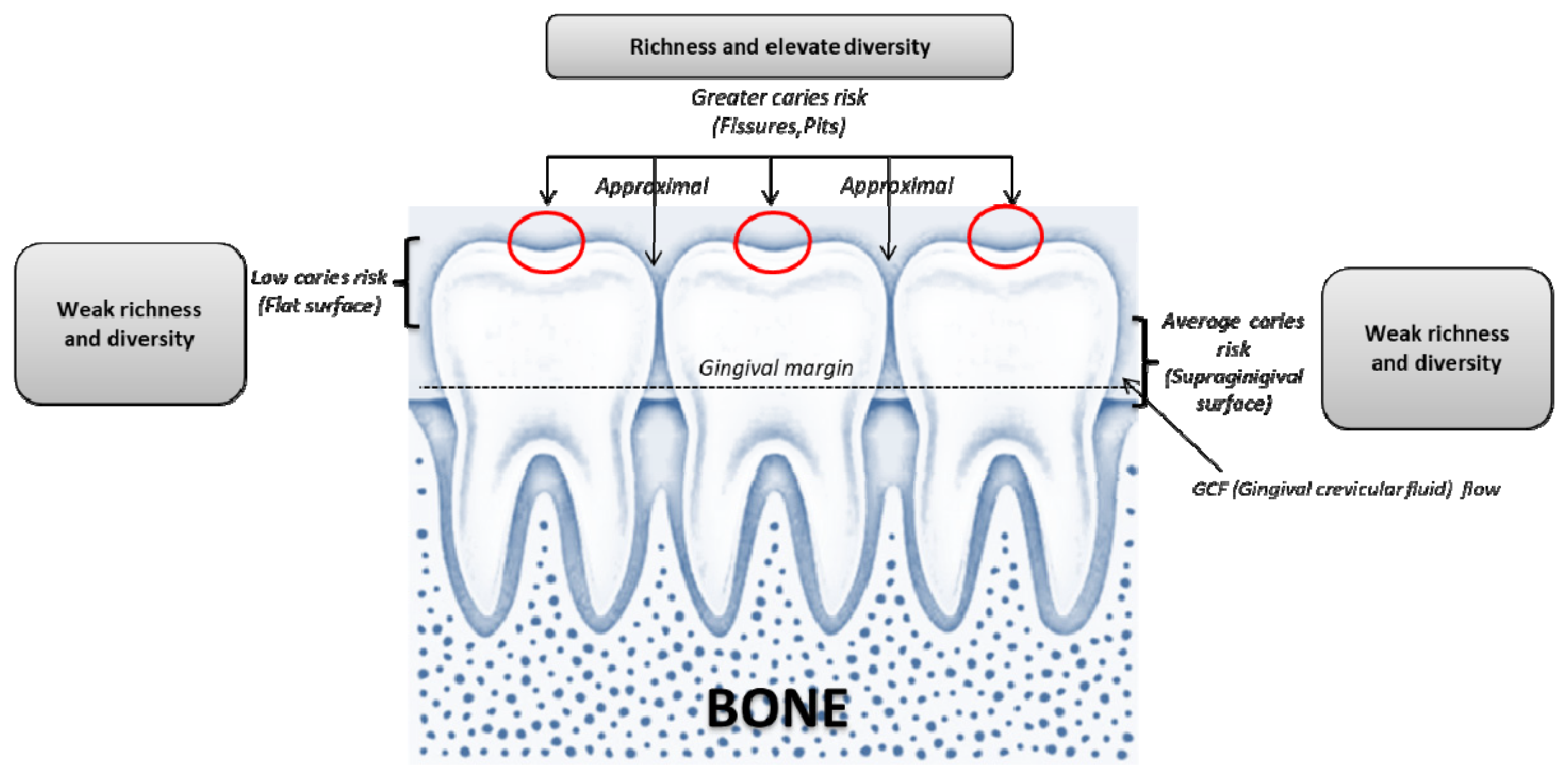
The risk associated with the areas that may be affected by caries and periodontal disease. Surfaces and locations with the highest variety and richness of oral microbial communities are more sensitive to caries and in the genital areas of periodontal disease. When tooth decay or periodontitis develops, the acidic environment reduces the variety and richness of local microbes.
Figure 1.
The risk associated with the areas that may be affected by caries and periodontal disease. Surfaces and locations with the highest variety and richness of oral microbial communities are more sensitive to caries and in the genital areas of periodontal disease. When tooth decay or periodontitis develops, the acidic environment reduces the variety and richness of local microbes.

Figure 2.
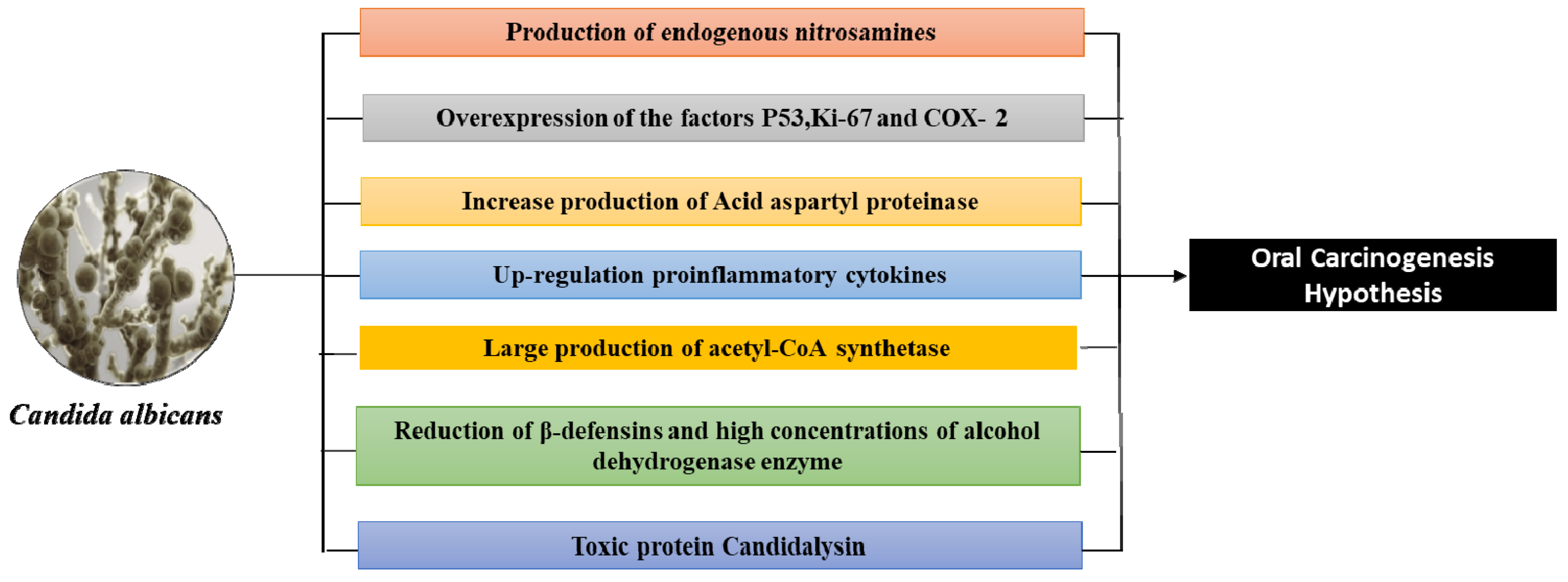
The main hypothetical molecular mechanisms by which C. albicans can cause precancerous and malignant neoformations in the oral cavity.
Figure 2.
The main hypothetical molecular mechanisms by which C. albicans can cause precancerous and malignant neoformations in the oral cavity.


Table 1.
The main diseases associated with dysbiosis of the human microbiota.
| Oral Microbiome and Associated Diseases | |
|---|---|
| Oral | Systemic |
|
|
Publisher’s Note: MDPI stays neutral with regard to jurisdictional claims in published maps and institutional affiliations. |
© 2021 by the authors. Licensee MDPI, Basel, Switzerland. This article is an open access article distributed under the terms and conditions of the Creative Commons Attribution (CC BY) license (https://creativecommons.org/licenses/by/4.0/).
Share and Cite
MDPI and ACS Style
Di Cosola, M.; Cazzolla, A.P.; Charitos, I.A.; Ballini, A.; Inchingolo, F.; Santacroce, L. Candida albicans and Oral Carcinogenesis. A Brief Review. J. Fungi 2021, 7, 476. https://doi.org/10.3390/jof7060476
AMA Style
Di Cosola M, Cazzolla AP, Charitos IA, Ballini A, Inchingolo F, Santacroce L. Candida albicans and Oral Carcinogenesis. A Brief Review. Journal of Fungi. 2021; 7(6):476. https://doi.org/10.3390/jof7060476
Chicago/Turabian StyleDi Cosola, Michele, Angela Pia Cazzolla, Ioannis Alexandros Charitos, Andrea Ballini, Francesco Inchingolo, and Luigi Santacroce. 2021. "Candida albicans and Oral Carcinogenesis. A Brief Review" Journal of Fungi 7, no. 6: 476. https://doi.org/10.3390/jof7060476
Note that from the first issue of 2016, this journal uses article numbers instead of page numbers. See further details here.