Article

Combined Culture and DNA Metabarcoding Analysis of Cyanobacterial Community Structure in Response to Coral Reef Health Status in the South China Sea

1
Third Institute of Oceanography, Ministry of Natural Resources, Xiamen 361005, China
2
Observation and Research Station of Coastal Wetland Ecosystem in Beibu Gulf, Ministry of Natural Resources, Beihai 536015, China
3
Fujian Provincial Key Laboratory of Marine Ecological Conservation and Restoration, Xiamen 361005, China
4
Faculty of Science, Botany & Microbiology Department, Al-Azhar University (Girls Branch), Cairo 11751, Egypt
5
Institute of Marine Drugs, Guangxi University of Chinese Medicine, Nanning 530200, China
6
College of Harbor and Coastal Engineering, Jimei University, Xiamen 361021, China
*
Authors to whom correspondence should be addressed.
These authors contributed equally to this work.
J. Mar. Sci. Eng. 2022, 10(12), 1984; https://doi.org/10.3390/jmse10121984
Submission received: 8 November 2022 / Revised: 2 December 2022 / Accepted: 7 December 2022 / Published: 13 December 2022
(This article belongs to the Special Issue The Impact of Changes in the Marine Environment on Marine Organisms)

Abstract

:
Cyanobacteria occupy an extraordinarily diverse array of ecological niches in coral reefs because they play multifaceted roles, including primary carbon and nitrogen fixation, calcification, nutrient cycling, and oxygen production, as well as coral reef degradation through skeletal biocorrosion and polymicrobial diseases. In this study, cyanobacterial diversity in sediment, water, and coral tissues were explored in relation to coral health status (slightly, moderately, and severely damaged) of coral reefs at Weizhou Island, South China Sea. Microscopy of taxa morphological characteristics was combined with 16S rRNA gene metabarcoding. Fifteen and forty-three cyanobacterial genera were identified based on universal prokaryotic 16S rRNA gene primers and cyanobacteria-specific 16S rRNA gene primers metabarcoding, respectively, indicating a more sophisticated efficiency of the latter. In addition, three out of seven cyanobacterial strains that were isolated and identified based on morphology and phylogeny could not be detected using either molecular method. Therefore, culture-based combined cyanobacteria-specific 16S rRNA gene metabarcoding are highly recommended in future routine surveys. There was a clear distinction in cyanobacterial assemblage composition among locations with different coral health statuses, with degraded reefs exhibiting approximately a 1.25-fold increase in species compared to healthy habitats. In addition, the spreading of potentially toxic cyanobacteria, such as Nostoc and Lyngbya, in the degraded reef implies putative links to reef degradation. This study provides novel insights into the taxonomical diversity of cyanobacteria in tropical coral reefs. Metabarcoding is recommended as an effective tool for revealing cyanobacterial diversity patterns and thereby providing critical information for the effective management of coral reef ecosystems.

1. Introduction

Tropical coral reefs provide essential habitat and resources for numerous marine organisms, as well as support human livelihoods, fisheries, and tourism [1]. However, corals are vulnerable to climatic change and human activity impacts, such as nutrient overloading from agricultural, urban, and domestic sources [2,3]. Many studies have reported a reduction in coral cover associated with a rapid increase in the expansion of benthic algae, referred to as a coral–algal phase shift [4,5]. In particular, benthic cyanobacteria are often early colonizers of dead coral [6,7]. It became evident that some cyanobacteria exhibit additional tactics rather than space occupation to minimize coral recruitment and consequently coral community recovery [6,8]. Thus, understanding the diversity of cyanobacteria may be important in managing coral reef ecosystems.
Cyanobacteria are important components of coral reef ecosystems where they contribute to reef-building through the formation of microbial mats and carbonate microbialites [9]. They are important principal producers that can fix carbon (oxyphototrophy) as well as nitrogen (diazotrophy), and contribute to supporting the nutrient circulation and energy exchanges of coral reefs [10,11,12]. Moreover, cyanobacteria also contribute to reef degradation by participating in skeletal biocorrosion or by causing polymicrobial diseases, such as black band disease [13]. Mortality from coral reef diseases is potentially accelerated by the expansion of cyanobacterial biomass on reefs, which therefore represents a significant threat to the survival of important reef-building coral species, alongside the general threats faced from sedimentation, local pollution, physical degradation, and global stressors [13,14,15]. In addition, some cyanobacteria are responsible for shellfish, fish, and mammal poisoning events, and can be bioaccumulated through the food chain, with adverse effects on coral reef ecosystems and public health [16,17,18]. Some marine strains of cyanobacteria also show medicinal properties, and have pronounced anti-inflammatory activity, or show preferential apoptogenic activity against neuroblastoma, proliferative, or cancer cells [19,20,21]. Cyanobacteria have high ecological value and potential potent toxin implications, and are important in drug development and production of biological cell compounds. Therefore, significant attention has been paid to the role of cyanobacterial communities in the coral reef ecosystem [6,11,13].
Traditionally, isolation and culturing are important processes for the identification of cyanobacterial taxa based on morphological and molecular characterization [22,23]. The criteria for taxonomic classification of cyanobacteria have radically changed in the last few decades after the application of data obtained from electron microscope studies and after the application of phylogenetic analyses, mainly derived from molecular sequencing [24]. However, it is challenging to obtain axenic cultures of species that are ecologically specialized, such as endosymbionts and epiphytes [25,26]. In addition, picocyanobacteria, or those with less distinguishable morphological features, show limited resolution under light microscopy [27]. High-throughput sequencing has provided another reliable and powerful tool for determining the diversity of cyanobacteria [27,28]. The majority of DNA metabarcoding studies on marine prokaryotes, including cyanobacterial communities, are based on the V4 domains in the 16S rRNA gene [29,30]. However, universal prokaryotic 16S rRNA gene primers based metabarcoding analysis usually shows low diversity cyanobacteria or just the Synechococcus domain, rather than the complete cyanobacterial community [31,32]. Cyanobacteria-specific 16S rRNA gene primers based metabarcoding have been widely applied to freshwater cyanobacteria with successful results [33,34]. However, the use of these cyanobacteria-specific 16S rRNA primers remains limited in marine ecosystems.
The first survey of marine cyanobacteria in the South China Sea dates back to the 1960s. Prof. Maosen Hua of the Institute of Oceanography, Chinese Academy of Sciences, conducted a relatively extensive study of cyanobacteria in the Xisha Islands, and his work has been published in the book Checklist of Marine Biota of China Seas (2008) [35]. Huang and Ding [36] investigated the diversity of cyanobacterial species along the coast of China, and listed the 96 species of cyanobacteria in the South China Sea. Coral reefs are proven to be rich in cyanobacterial diversity [37]. However, knowledge of cyanobacterial distribution and ecology in the coral reefs of the South China Sea is still very limited. The aim of the present study was to characterize taxonomic compositions of cyanobacterial populations using morphological and next-generation molecular tools, and to investigate the changes in cyanobacterial diversity and species assemblage composition in response to coral reef health status.

2. Materials and Methods
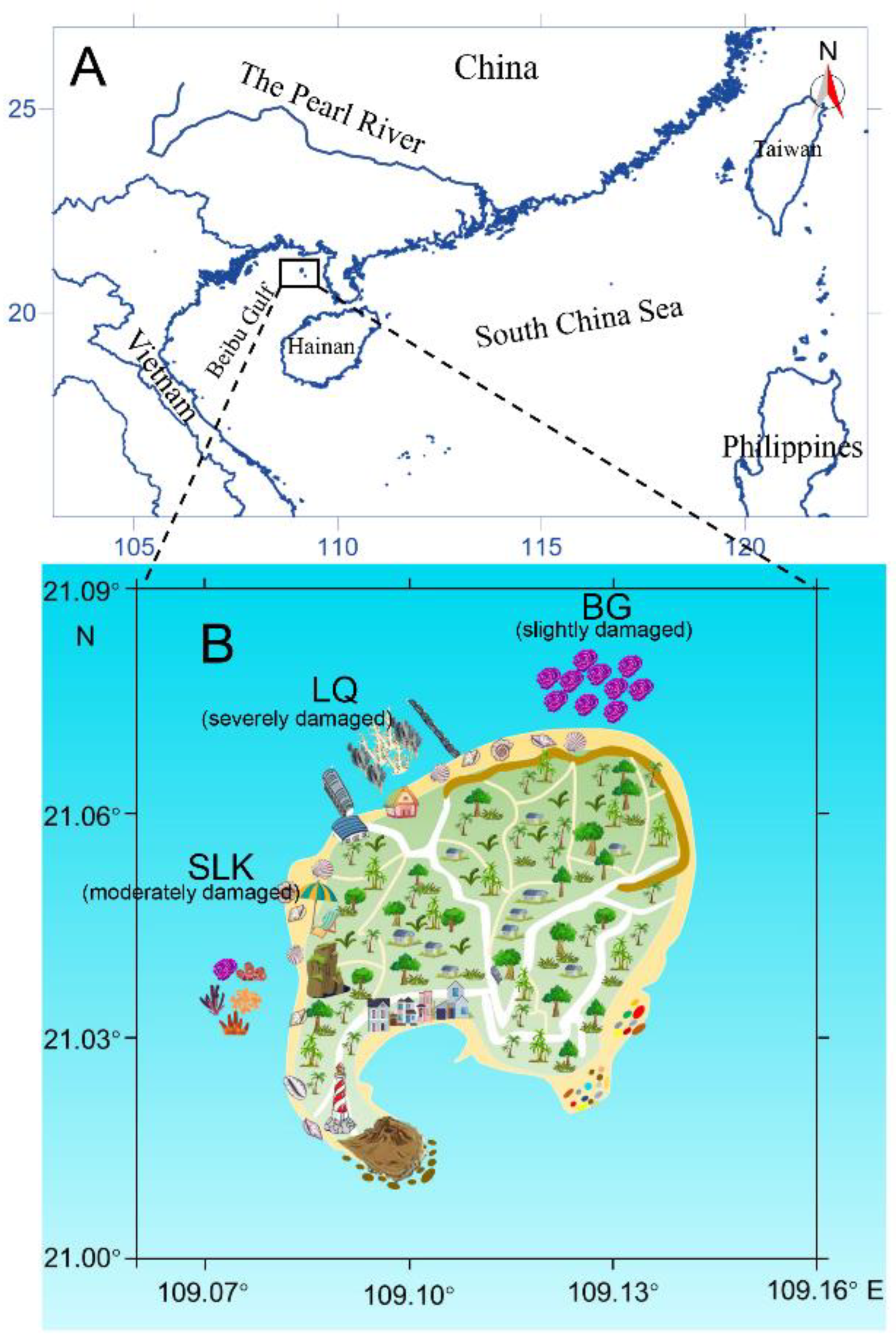
2.1. Study Area

The Beibu Gulf (17–22° N, 105–110° E; Figure 1A) covers a total area of 130,000 km2 with an average water depth of 38 m and a 1629 km coastline [38], located in the northwestern part of the South China Sea (Figure 1). The climate is southwest monsoon in the Indian Ocean, which brings high temperatures and strong rainfall to the region. As a result, it occupies a significant geographical location that is rich in fishery resources. The Weizhou Island is the largest and youngest volcanic island in the north of Beibu Gulf, which probably originates from a mantle plume that rose 50–32 million years ago. The Weizhou Island’s north–south length is 6.5 km, the east–west length is 6 km, and the coastline is 15.6 km, with coral reefs having been established around the island. The volcanic Weizhou Island is a natural source of nutrients such as N and P, and trace metals such as iron and manganese with surface runoff, which fuels the entire marine ecosystem nearby [39].
Due to climatic and anthropogenic disturbances, with the continuous development of tourism resources, coral reef biodiversity in Weizhou Island has been rapidly decreasing, and the ecological functions of the coral reef ecosystem are severely degenerating [40]. According to topography, circulation, nutrient concentration, and anthropogenic influences, Weizhou Island can be divided into three zones: the north sector (site BG in Figure 1B), less disturbed by human activity; the northwest sector (site LQ in Figure 1B), overloaded with impacts from cruise ships, refineries, sewage treatment plants, etc.; and the southwest sector (site SLK in Figure 1B), which is slightly affected by tourism development. The north, northwest, and southwest sectors exhibited 80%, 7%, and 53% coral cover with 54.5%, 3.3%, and 42.7% living corals, respectively [41].

2.2. Seawater, Coral Tissue, and Reef Sediment Sampling

Three coral reef sites around Weizhou Island; BG, LQ, and SLK; with different health statuses representing the slightly damaged, severely damaged, and moderately damaged coral reefs, respectively (Figure 2, [41]), were chosen for the assessment of cyanobacterial community composition. All sites were classified as sandy reefs with some patches of Pavona corals, especially site BG [41]. Seawater, coral tissue, and upper centimeters of sandy sediments were collected by Dr. Amro Abd Elgawad in June 2020 and by scuba divers in April 2021. One liter of seawater immediately above coral reefs was collected and filtered through 0.22 μm polycarbonate membrane (Millipore, Bedford, MA, USA). Small random coral fragments (~1 cm3) chiseled from the bottom part of coral colonies were sampled, and the upper centimeter reef sediment with or without benthic cyanobacterial mats (BCMs) was also collected. Triplicate of each substrate type was collected in June 2020, and a single sample of each substrate type was collected in April 2021. All filter membranes, coral colonies, and sediment samples were preserved in 1 mL DNA lysis buffer (10 mM Tris-HCl, pH 8.0; 100 mM EDTA, pH 8.0; 0.5% w/v SDS). Samples were snap-frozen in liquid nitrogen and brought back to the laboratory. In the laboratory, all samples were stored at −80 °C until DNA extraction.

2.3. Environmental DNA Extraction and Metabarcoding Sequencing

The DNA isolation and PCR preparation stages were carried out in a laminar flow hood, cleaned with bleach and sterilized under ultraviolet radiation for at least 30 min before use. Genomic DNA was extracted using a CTAB protocol combined with Zymo DNA Clean & Concentrator kit (Zymo, Irvine, USA) as described by [42]. For samples of June 2020, the primers 341F (5′-CCTACGGGNGGCWGCAG-3′)/806R (5′-GGACTACHVGGGTATCTAAT-3′) [43,44] targeting the V3–V4 domain of universal prokaryotic 16S rRNA gene were used. The high throughput sequencing results showed a low proportion and diversity of cyanobacteria (See the result Section 3.1 and Figure 3A). Thus, for samples of April 2021, the cyanobacteria-specific primers CYA359F (5′-GGGGAATYTTCCGCAATGGG-3′)/CYA781R (5′-ACTACWGGGGTATCTAATCCC-3′) [33,45] targeting partial V4 domain of 16S rRNA gene were used. PCR reactions were carried out using 1 × ExTag buffer, 50 µM deoxynucleotide mixture, 0.2 μM of each primer, 1.25 U of ExTaq DNA Polymerase (Takara, Tokyo, Japan), and 10 ng of template genomic DNA in 50 μL total volume reactions. The PCR program on the thermal cycler follows the protocols described in Caporaso et al. [43] and Monchamp et al. [33] for universal prokaryotic and cyanobacteria-specific primers, respectively. PCR was replicated three times in independent runs and the products of the same sample were pooled. The pooled amplicons were purified using the AxyPrep DNA gel extraction kit (Axygen, Union, CA, USA) following the manufacturer’s instructions. The purified amplicons were quantified using the ABI Step One Plus real-time PCR system (Life Technologies, Foster City, CA, USA), assessed and sequenced on an Illumina HiSeq 2500 platform (Illumina, San Diego, CA, USA) using a paired-end (2 × 250 bp) HiSeq 2500 Reagent Kit following manufacturer’s instructions.

2.4. Sequence Data Processing

Paired-end Illumina 16S rRNA gene sequences were processed using R. The sequences were quality filtered, merged, dereplicated, and chimeras were removed using the DADA2 workflow [46] to determine amplicon sequence variants (ASVs). Quality filtered reads were assigned to ASVs at a 100% sequence similarity threshold. Representative sequences from each of the ASVs were annotated by the SILVA Release128, http://www.arb-silva.de, [47] and Greengene [48] rRNA database using BLASTn. For the universal prokaryotes primers, the representative sequence of each of the ASVs was taxonomy assigned with a confidence threshold of 97% sequence similarity and 98% coverage with the two databases [29,49]. For the cyanobacteria-specific primers, the representative sequence of each of the ASVs was taxonomy assigned with a confidence threshold of 85% sequence similarity and 98% coverage with the two databases [33,50]. Each of the ASVs was annotated again for verification through blast in the NCBI database; the most recent verified sequences supported by the literature will be updated. Data for the original paired-end reads have been deposited at GenBank under the project PRJNA891132 with the accession number SAMN31310599–SAMN31310634.

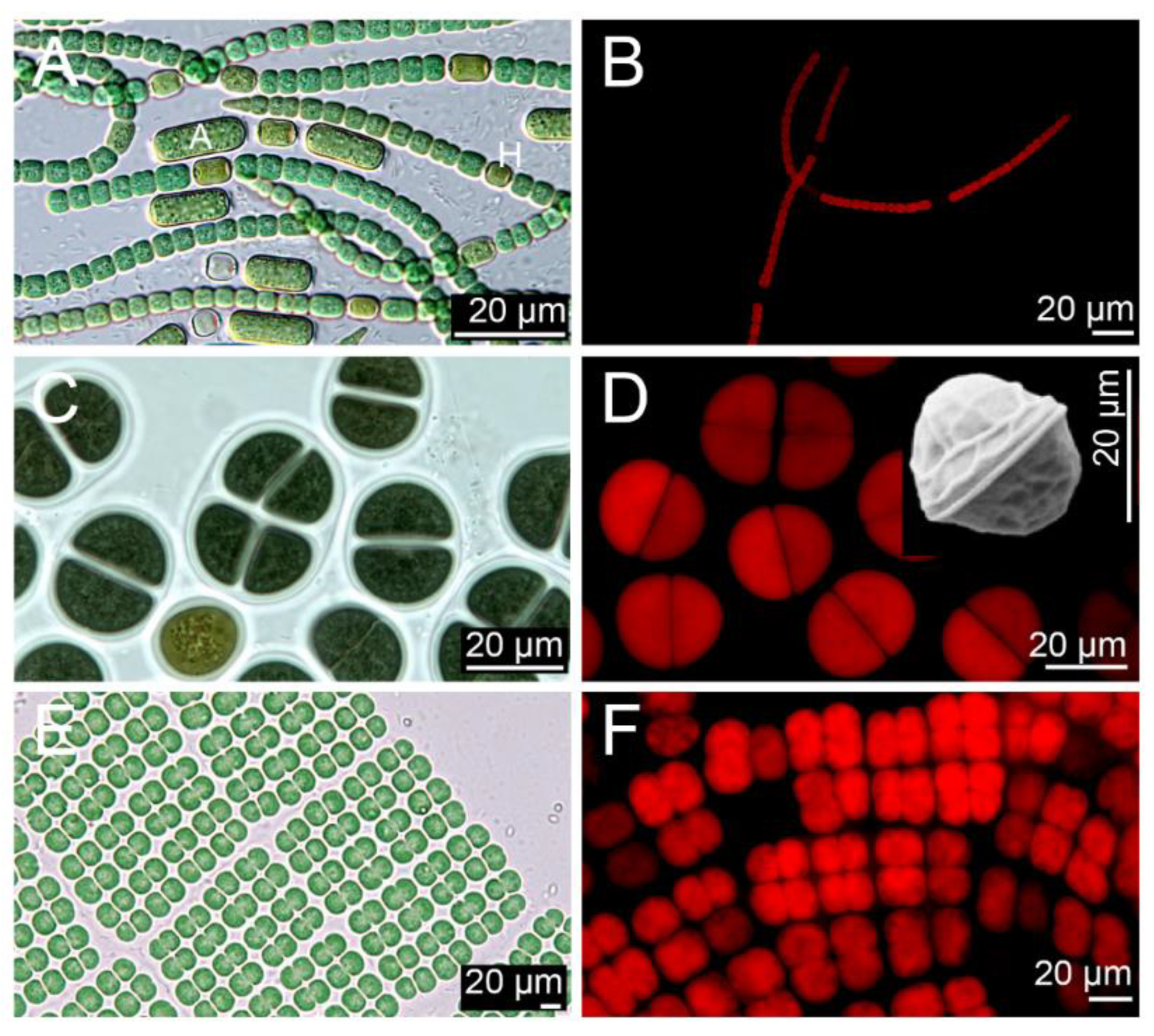
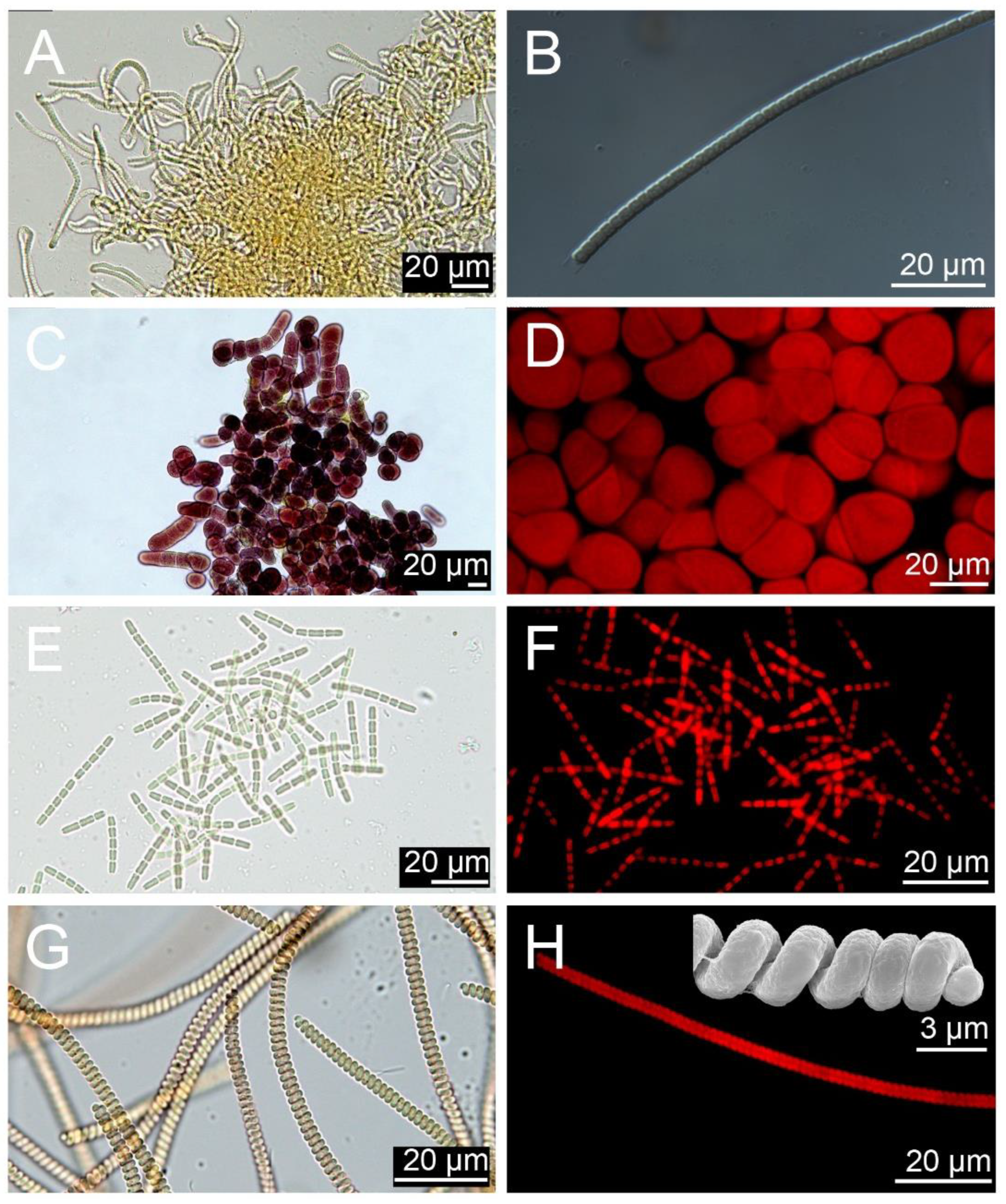
2.5. Clone Strains and Microscopy

Seawater, coral tissue, and upper centimeter of sandy sediments were collected by scuba divers as described before. Each sample was transferred into a 5 L polycarbonate bottle with filtered seawater and stirred vigorously to detach the epibenthic cyanobacteria. The suspension materials were subsequently sieved through 120 μm and 10 μm filters. The 10–120 μm fractions were rinsed with sterile f/2-Si medium [51]. The samples were incubated at 25 °C, 90 μmol photons·m−2·s−1 from cool-white tubes, and under a light: dark cycle of 12 h: 12 h (hereafter, called “standard culture conditions”) for two weeks. Single cells/filamentous were isolated from this material with a micropipette under an inverted microscope Eclipse TS100 (Nikon, Tokyo, Japan) into a 96-well tissue culture plate containing 330 μL f/2-Si medium, and examined every week with the inverted microscope. The clonal cultures were transferred to 50 mL polystyrene tissue culture flasks and maintained under the standard culture conditions. The light and chloroplast auto-fluorescence microscopy, as well as scanning electron microscopy, were carried out following the standard protocols described in Pei et al. [52].
Genomic DNA of clone strains was extracted using the CTAB protocol as described before. Partial 16S rRNA gene sequence was amplified using the 27F (5′-AGA GTT TGA TCC TGG CTC AG-3′)/1492R (5′-TAC GAC TTA ACC CCA ATC GC-3′) primers [53] or CYA106F (5′-CGG ACG GGT GAG TAA CGC GTG A-3′)/CYA781R (5′-GAC TAC TGG GGT ATC TAA TCC CAT T-3′) following the protocols described in Nübel et al. [45]. Newly 16S rRNA gene sequences aligned with related sequences from GenBank using MAFFT v7.110 [54]. Maximum likelihood (ML) analyses were calculated with RaxML v7.2.6 [55] on the T-REX web online server [56] with the best-fitting model selected by jModelTest 2 [57].

3. Results

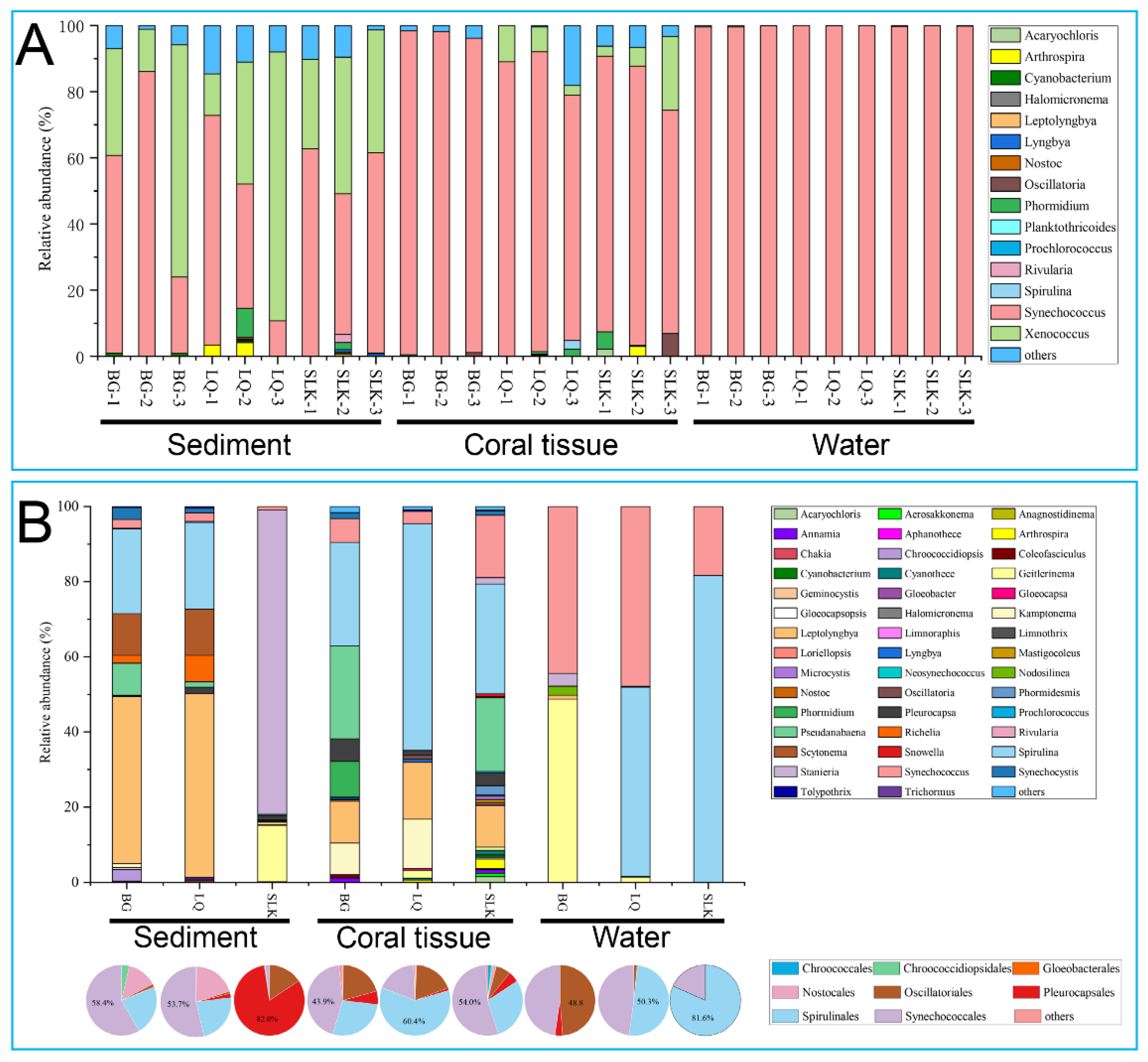
3.1. Diversity of Cyanobacteria Based on Universal Prokaryotic 16S rRNA Gene Primers

The sequencing depth of all samples based on universal prokaryotic 16S rRNA gene primers is summarized in Table S1. A total of 3,519,202 reads were derived from all samples, and 1,087,132 unique sequences were obtained after filtering/denoising and chimera removal (Table S1). A total of 60,800 unique amplicon sequence variants (ASVs) were obtained with sizes ranging from 244 to 476 bp (mean = 455 ± 17 bp) in length. Finally, 23,147 ASVs (38.07%) passed the filtering threshold annotation for procaryotic organism. Among them, 392 ASVs (1.69%) belonging to cyanobacteria were retained for downstream analysis. The average relative abundances of cyanobacteria in the sediments, coral tissue, and seawater compartments were 4.63%, 5.31%, and 10.70%, respectively (Table S1). Fifteen different cyanobacterial genera were identified (Table 1, Figure 3A). The dominant genera across all sites/samples were Synechococcus, Trichormus, and Phormidium with 96.1, 2.6, and 0.2% of the total cyanobacterial reads, respectively. The 15 genera were classified into six orders; Chroococcales, Nostocales, Oscillatoriales, Pleurocapsales, Spirulinales, and Synechococcales (Table 1). Regarding sites, in relation to different coral health statues and coral coverage, the dominant genera in the sediment were Synechococcus (55.3, 32.9, and 54.6% at sites BG, LQ, and SLK, respectively), Trichormus (39.1, 35.6, and 35.3% at sites BG, LQ, and SLK, respectively), and Phormidium (0, 6.9, and 8.5% at sites BG, LQ, and SLK, respectively). The dominant genera in coral tissue samples were Synechococcus (97.2, 85.5, and 81.3% at sites BG, LQ, and SLK, respectively), Trichormus (2.3, 35.6, and 8.1% at sites BG, LQ, and SLK, respectively), and Phormidium (0.2, 1.1, and 1.1% at sites BG, LQ, and SLK, respectively). The dominant genera in the water samples were Synechococcus (99.5, 99.9, and 99.7% at sites BG, LQ, and SLK, respectively), Trichormus (0.3, 0.03, and 0.08% at sites BG, LQ, and SLK, respectively), and Prochlorococcus (0, 0.01, and 0.1% at sites BG, LQ, and SLK, respectively).

3.2. Diversity of Cyanobacteria Based on Cyanobacteria-Specific 16S rRNA Gene Primers

Cyanobacterial genera based on cyanobacteria-specific 16S rRNA gene primer metabarcording are listed in Table S2. Samples gave a total of 625,759 reads, with 363,026 unique sequences (Table S1). Out of these unique sequences, a total of 1276 ASVs had the sizes of 235 to 407 bp (mean = 391 ± 12 bp). In addition, 1206 ASVs (94.51%) passed the filtering threshold annotation for procaryotes of which 156 ASVs (12.94%) belonging to cyanobacteria were subjected to downstream analysis. The cyanobacterial relative abundances in the upper centimeter of sandy sediments, coral tissue, and seawater were 26.27, 11.78, and 35.64%, respectively. A total of 43 cyanobacterial genera were detected (Table 1, Figure 3B). Spirulina, Synechococcus, Leptolyngbya, Stanieria, and Geitlerinema were the dominant genera across all sites/samples with 58.1, 30.8, 3.4, 3.2, and 1.4%, respectively. The 43 genera belong to eight orders; Chroococcales, Chroococcidiopsidales, Gloeobacterales, Nostocales, Oscillatoriales, Pleurocapsales, Spirulinales, and Synechococcales (Table 1, Figure 3B). Site-wise, Stanieria (0.2, 0.2, and 80.9% at sites BG, LQ, and SLK, respectively), Leptolyngbya (44.4, 48.7, and 0.7% at sites BG, LQ, and SLK, respectively) and Spirulina (22.6, 23.1, and 0.4% at sites BG, LQ, and SLK, respectively) was the dominant genera in the sediment. While in coral tissue samples, the dominant genera were Spirulina (27.6, 60.4, and 29.2% at sites BG, LQ, and SLK, respectively), Pseudanabaena (24.7, 0, and 19.7% at sites BG, LQ, and SLK, respectively) and Leptolyngbya (11.0, 15.1, and 11% at sites BG, LQ, and SLK, respectively). Regarding the water samples, Spirulina (0, 50.3, and 81.6% at sites BG, LQ, and SLK, respectively), Synechococcus (44.4, 47.9, and 18.4% at sites BG, LQ, and SLK, respectively) and Geitlerinema (48.8, 1.4, and 0% at sites BG, LQ, and SLK, respectively) were the most dominant genera.

3.3. Diversity of Cyanobacterial Phenotypes

Based on morphology and phylogeny, seven species were cultured and identified as Anabaena sp., Chroococcus sp., Merismopedia sp., Phormidesmis sp., Pleurocapsa sp., Pseudanabaena sp., and Spirulina sp. (Table 2, Figure 4 and Figure 5).
Anabaena sp. strain TIOX110 (Figure 4A,B), dark-green-colored to brownish vegetative cells, grew as slight sinuous filaments. Cells showed beaded appearance from square to spherical of diameter ranging from 3.78 to 5.24 μm. The last cell in the filament showed a characteristic pointed end with 8 to 50 cells on average in one filament. Heterocysts were oval to elongate barrel in shape, and 1.0–1.92-fold compared to the vegetative cells. Intercalary heterocysts were mostly observed, and some terminal heterocysts were also associated with broken filaments. Akinetes were observed with a length of 2.37–5.36-fold compared to vegetative cells, and slightly more in breadth. The akinetes were generally distant away from the heterocyst. ML phylogeny based on 16S rRNA sequences of Anabaena is illustrated in Figure S1.
Chroococcus sp. strain TIOX101 (Figure 4C,D) formed macroscopic dark-green mats on the bottom of the culture medium flasks: 2–4 celled colonies, 15.71–27.35 μm in width, surrounded with mucilage. Individual cells of the colonies were hemispherical in shape, 7.28–12.80 μm in width, with homogeneous content, olive-green when young, granulated with many polyhydroxybutyrate granules and yellowish when old. Cell division occurs in two planes by binary fission, and neither nanocytes nor baeocytes were observed. ML phylogeny based on 16S rRNA sequences of Chroococcus is illustrated in Figure S2.
Merismopedia sp. strain TIOX109 (Figure 4E,F) was blue-green, with elliptical or hemispherical vegetative cells 10.19–22.70 μm wide and 5.82–12.80 μm long. Cell content was homogeneous, with many polyhydroxybutyrate granules. Four cells were regularly arranged, gathered in a flat layer with colorless fragile mucilaginous envelopes outside the cells. Cell division occurred in perpendicular planes by binary fission. This Merismopedia strain was lost, and retrieving a 16S rRNA sequence was failed.
Phormidesmis sp. strain TIOX96 (Figure 5A,B) was yellowish-brown, with long and straight filaments up to 2.33–3.78 μm in width. Trichomes were cylindrical, usually distinctly constricted at the cross-walls, sometimes slightly longer or shorter rather than wide, without diversified terminal cells. Thick, lamellated, or colored sheaths were observed. At the end of cultivation, the cultured strain formed gelatinous mats on the substrate. The mL phylogeny based on 16S rRNA sequences of Phormidesmis is illustrated in Figure S3.
Pleurocapsa sp. strain TIOX102 (Figure 5C,D) formed macroscopic blackish mats on the bottom of the culture medium flasks. Pseudofilaments were moderately long, and uniseriate. Sheaths were thin, colorless, and tightly attached to the cell walls. Cells were of various shapes and sizes, with 15.71–27.61 μm width and granular cytoplasm, and various planes of cell division. ML phylogeny based on 16S rRNA sequences of Pleurocapsa is illustrated in Figure S4.
Pseudanabaena sp. strain TIOX98 (Figure 5E,F) formed a thin layer on the bottom of the culture medium flasks with dark blue-green motile filaments without oscillation or rotation. Trichomes were straight, short, and distinctly constricted at cross-walls. Cells were 1.46–2.04 μm in width and 1.46–2.91 μm in length. Cell content was differentiated into chromatoplasm and centroplasm. Heterocytes and akinetes were absent, and apical cells were round without polar calyptras or aerotopes. ML phylogeny based on 16S rRNA sequences of Pseudanabaena is illustrated in Figure S5.
Spirulina sp. TIOX113 (Figure 5G,H) showed regularly helically coiled trichomes, with no visible crosswalks in light or scanning microscopy and no visible sheaths. Coiling was counter-clockwise, and motility was detected with rotation along the helix axis. The helix shape was closed, with a trichome width of 2.91 to 3.78 μm and helix width of 2.91 to 3.78 μm. The trichome coils were closed. ML phylogeny based on 16S rRNA sequences of Spirulina is illustrated in Figure S6.

4. Discussion

4.1. Comparison of Different Primer Datasets

Traditional surveys of cyanobacteria that have relied mainly on culture combined light microscopy morphology or the macroscopic appearance of benthic cyanobacterial mats (BCMs) have shown a limited number of cyanobacterial species compositions [37,59]. This highlights the significance of applying DNA metabarcoding tools, which have proven powerful for improving the understanding of cyanobacteria biodiversity in coral reefs [31,49]. The hyper-variable V4 regions of the 16S rRNA have been widely used for assessing the microbial prokaryote diversity of marine environmental samples with next-generation sequencing technologies [29,30,60]. Cyanobacteria-specific primers have been previously applied to investigate biodiversity patterns and risk-oriented monitoring of cyanotoxin blooms [33,61]. In the present study, a total of 15 and 43 cyanobacterial genera were revealed from three coral reef locations at Weizhou Island based on universal prokaryotic 16S rRNA gene primers datasets and cyanobacteria-specific 16S rRNA gene primers datasets, respectively (Figure 3). This indicates that the DNA metabarcoding approach using cyanobacteria-specific 16S rRNA gene primers detected a significantly higher number of cyanobacterial (including several harmful species) than universal prokaryotic 16S rRNA gene primers. Although, we could not ignore the differences brought about by different sampling times. However, universal 16S rRNA gene primers based metabarcoding result in low diversity and abundance of cyanobacterial have been widely reported ([31,49], this study), even for the samples collected from BCMs with no significant improvement [62]. It is proposed that the cyanobacteria-specific 16S rRNA gene primers datasets had a higher tendency toward cyanobacteria phylotype richness and community structure. Their application is therefore recommended for future metabarcoding-based cyanobacteria diversity surveys to achieve a more accurate determination of cyanobacteria diversity. It is also worth noting that the genera Planktothricoides and Xenococcus were detected in universal prokaryotic 16S rRNA gene primers datasets, but not in cyanobacteria-specific 16S rRNA gene primers datasets (Table 1). This implies that cyanobacteria-specific 16S rRNA gene primers also have limitations. In addition, four out of seven cyanobacteria genera (Pleurocapsa, Spirulina, Phormidesmis, and Pseudanabaena) were cultured and identified by morphology and molecular phylogeny means, and were consistently detected using both universal and cyanobacteria-specific 16S rRNA gene primers (Table 1). However, the other three cultured genera, Anabaena, Chroococcus, and Merismopedia, could not be detected using either molecular method. This inconsistency between metabarcoding and microscopic tools has been reported in previous studies [27,63]. The main reasons is the database for now are far from complete, with many uncultured and unnamed sequences. In this study, many picocyanobacterial and thin filamentous cyanobacteria (e.g., Synechococcus, Leptolyngbya, and Phormidium) were detected by metabarcoding (Table 1). These genera are difficult to identify by microscopy, as they lack conspicuous morphological features [64,65]. The advantage of morphological results enabled the comparison between the past and current cyanobacterial diversity, although the microscopic identifications did not always agree with the molecular data for cyanobacteria. For these reasons, different combined techniques are recommended for investigating cyanobacterial diversity in order to minimize the limitations associated with individual techniques [32,63]. Overall, culture-based monitoring remains essential in routine survey, while cyanobacteria-specific prokaryotic 16S rRNA gene primers based on high-throughput sequencing enable estimates of a more realistic and sensitive picture of both cyanobacterial phylotype richness and assemblage structure.

4.2. Cyanobacterial Diversity at Weizhou Island

Weizhou Island harbored a rich diversity of cyanobacteria revealed by next-generation sequences and microscopy tools. In total, 48 genera were detected based on metabarcoding and culture-based surveys (Table 1). To the best of the authors’ knowledge, many of these genera are reported here for the first time at Weizhou Island, including the harmful toxin producer genera Nostoc, Microcystis, Geitlerinema, Lyngbya, Oscillatoria, and Leptolyngbya.
Weizhou Island is subject to both anthropogenic activities as well as climatic disturbances, such as petroleum processing, tourism development, shipping, ocean warming, and heavy rainfall [40,66]. The present results showed a clear distinction in the composition of cyanobacterial assemblages among different coral health status sites. Twenty-eight genera were detected at site BG, containing mostly healthy coral reefs, and approximately 1.25 times more genera were detected (36 and 34 genera at sites LQ and SLK, respectively) at severely damaged and moderately damaged coral reef habitats (Table 3). This result supports the notion that the degradation of a reef area is likely to increase benthic algal occupation including cyanobacteria, thus enabling cyanobacterial expansion over dead corals and competition for space [6,7]. Over the past two decades, BCMs have become the dominant component on many reefs worldwide [6,7], and will continue to increase in abundance under the environmental disturbances associated with eutrophication, as well as ocean warming [4,5]. Specially, many BCMs have been reported on coral reefs located nearby urban areas in the Caribbean, Red Sea, and South China Sea [67,68]. High nutrient inputs from municipal sewage could increase coral mortality, as well as stimulate the development of phototrophic algae on reefs [66]. In the severely damaged habitats of site LQ, BCMs covered large areas of dead coral reefs (Figure 2C,D), which may be strongly related to the increased nutrient runoff from domestic water.

4.3. Biological Nitrogen-Fixing Cyanobacteria

Coral reefs are one of the most highly productive ecosystems on earth, despite existing in nutrient-poor waters [69]. Dissolved inorganic nitrogen is usually the limitation for phototrophic organisms in coral reefs [70,71]. Biological nitrogen fixation by cyanobacteria is an important source of nitrogen, and nitrogen-fixing cyanobacteria have been recommended to play a key role in the nitrogen cycle of the coral reef ecosystem [72,73]. The present study was unsuccessful at detecting nitrogen-fixing cyanobacteria from the healthiest coral reef site BG, regardless of whether using high throughput-sequencing or culture-based techniques. It may be concluded that nitrogen-fixing cyanobacteria may be rare in the healthiest coral reefs at Weizhou Island. In contrast, two nitrogen-fixing cyanobacteria taxa, Lyngbya and Nostoc, were identified based on high-throughput sequences at both sites LQ and SLK, where the coral reefs were severely and moderately damaged (Table 3), respectively. In addition, strains of Anabaena capable of fixing N2 were cultured from corals tissue from the site LQ (Table 2, Figure 4A,B). It has been reported that excess nutrients can favor the production of algae at the expense of reef-building corals [5,7]. The coral reefs at sites LQ and SLK have been exposed to significant sewage discharge due to urbanization, which is responsible for the continuous degradation of the coral communities in both areas (Figure 2, [40]). Damaged coral reefs can be subsequently colonized by microalgae, especially cyanobacteria [6,7]. Although it is usually hypothesized that nitrogen fixation by cyanobacteria may be suppressed by high (in-) organic nutrients [74], there was no apparent negative correlation between the diversity of nitrogen-fixing cyanobacteria and potential high nutrients due to domestic sewage discharge in the present study. The reason for these results warrants further in-depth investigation. Microbial mats dominated by Anabaena, Calothrix, Lyngbya, Nostoc, and Oscillatoria show a high nitrogen fixation rate in coral reefs in Sanya Bay, South China Sea [68]. Oscillatoria were detected at all three sites; BG, LQ, and SLK in the present study (Table 3). Oscillatoria-dominated BCMs in the Southern Caribbean exhibit nitrogen fixation [73,75]. Whether Oscillatoria contributes to the nitrogen supply at the three sites of Weizhou Island was not confirmed by the present study, mainly because it was the only potential nitrogen-fixing cyanobacteria detected at site BG, with mostly healthy coral reefs.

4.4. Harmful Cyanobacterial Species

Due to climatic changes and eutrophication, bloom-forming and toxin-producing cyanobacteria have rapidly increased in marine ecosystems worldwide, and have severely impacted the functioning of the ecosystems, ultimately threatening reef organisms and even humans [7,18]. Marine cyanobacteria have been reported to be responsible for toxic events associated with shellfish, fish, and mammals [76,77,78]. Generally, the cyanotoxins include endotoxins, dermatoxins, hepatotoxins, and neurotoxins, among others. Leptolyngbya, Lyngbya, Microcystis, Oscillatoria, and Synechococcus produce toxic secondary metabolites known as cyanotoxins and were detected at Weizhou Island (Table 1). The heterocystous cyanobacteria Nostoc have been reported to produce cyanotoxin β-Methylamino-L-alanine (BMAA), which is capable of causing several neuronal human diseases such as amyotrophic lateral sclerosis (ALS) [18,79]. Nostoc was detected in the present study using both universal prokaryotic 16S rRNA gene primers and cyanobacteria-specific 16S rRNA gene primers, implying the potential BMAA contamination risk at Weizhou Island. Mortality from coral diseases that are likely propelled by increasing biomass of benthic cyanobacteria, such as black band disease, may increasingly drive the trajectory and velocity of reef degradation [13,14]. Some cyanobacteria, together with the sulfur-reducing bacteria, can cause black band disease and lead to coral death due to anaerobic and increased hydrogen sulfide conditions [80,81]. Three potential pathogenic cyanobacteria of black band disease, Geitlerinema, Leptolyngbya, and Phormidium were detected at all three sites of Weizhou Island (Table 2), suggesting both healthy and degraded corals are under risk of black band disease. In conclusion, considering that many potential harmful cyanobacterial species were detected in Weizhou Island coral reefs, it is important to estimate such cyanobacterial species density, and quantitative cyanotoxin analysis is urgently required in routine harmful algae monitoring and management programs in the South China Sea.

5. Conclusions

During this study, cyanobacterial diversity in sediment, water, and coral tissues were explored in relation to slightly, moderately, and severely damaged coral reefs at Weizhou Island, South China Sea. Fifteen and forty-three cyanobacterial genera were identified based on universal prokaryotic 16S rRNA gene primers and cyanobacteria-specific 16S rRNA gene primers metabarcoding, respectively, indicating a more sophisticated efficiency of the latter. Seven Cyanobacterial species were cultured and identified as Anabaena sp., Chroococcus sp., Merismopedia sp., Phormidesmis sp., Pleurocapsa sp., Pseudanabaena sp., and Spirulina sp. based on morphology and phylogeny. However, three out of the seven culture cyanobacterial strains could not be detected using molecular methods. Therefore, culture-based combined cyanobacteria-specific 16S rRNA gene metabarcoding are highly recommended in future routine survey. There was a clear distinction in cyanobacterial assemblage composition among locations with different coral health statuses, with degraded reefs exhibiting approximately a 1.25-fold increase in species compared to healthy habitats. In addition, the spreading of potentially toxic cyanobacteria, such as, Nostoc and Lyngbya, in the degraded reef, implies putative links to reef degradation.

Supplementary Materials

The following supporting information can be downloaded at: https://www.mdpi.com/article/10.3390/jmse10121984/s1.

Author Contributions

Conceptualization, Z.L.; formal analysis, S.H. and X.L. (Xiangyuan Lin); funding acquisition, X.Z. and Z.L.; investigation, X.L. (Xinming Liu); software, L.P.; writing—original draft, J.K., H.F.M., X.Z. and Z.L.; writing—review & editing, J.K., H.F.M., X.L. (Xinming Liu), X.Z. and Z.L. All authors have read and agreed to the published version of the manuscript.

Funding

This research was funded by the Scientific Research Foundation of the Third Institute of Oceanography, MNR (2020017, 2020002), the National Key Research and Development Program of China (2019YFE0124700), the Natural Science Foundation of Fujian, China (2022J06029), and the Youth Innovation Project of Xiamen (3502Z20206095).

Institutional Review Board Statement

Not applicable.

Informed Consent Statement

Not applicable.

Data Availability Statement

Data for the original paired-end reads have been deposited at GenBank (https://www.ncbi.nlm.nih.gov/genbank/) under the project PRJNA891132 with the accession number SAMN31310599–SAMN31310634.

Acknowledgments

The authors are thankful to Amro Abd Elgawad for samples collection and photographing of benthic cyanobacterial mats in Weizhou Island.

Conflicts of Interest

The authors declare no conflict of interest.

References

  1. Darling, E.S.; McClanahan, T.R.; Maina, J.; Gurney, G.G.; Graham, N.A.J.; Januchowski-Hartley, F.; Cinner, J.E.; Mora, C.; Hicks, C.C.; Maire, E.; et al. Social–environmental drivers inform strategic management of coral reefs in the anthropocene. Nat. Ecol. Evol. 2019, 3, 1341–1350. [Google Scholar] [CrossRef]
  2. Hughes, T.P.; Kerry, J.T.; Baird, A.H.; Connolly, S.R.; Dietzel, A.; Eakin, C.M.; Heron, S.F.; Hoey, A.S.; Hoogenboom, M.O.; Liu, G.; et al. Global warming transforms coral reef assemblages. Nature 2018, 556, 492–496. [Google Scholar] [CrossRef]
  3. Guo, J.; Yu, K.; Wang, Y.; Xu, D.; Huang, X.; Zhao, M.; Yang, H.; Zhang, R. Nutrient distribution in coral reef degraded areas within sanya bay, South China Sea. J. Coast. Res. 2017, 33, 1148–1160. [Google Scholar] [CrossRef]
  4. McManus, J.W.; Polsenberg, J.F. Coral–algal phase shifts on coral reefs: Ecological and environmental aspects. Prog. Oceanogr. 2004, 60, 263–279. [Google Scholar] [CrossRef]
  5. Adam, T.C.; Burkepile, D.E.; Holbrook, S.J.; Carpenter, R.C.; Claudet, J.; Loiseau, C.; Thiault, L.; Brooks, A.J.; Washburn, L.; Schmitt, R.J. Landscape-scale patterns of nutrient enrichment in a coral reef ecosystem: Implications for coral to algae phase shifts. Ecol. Appl. 2021, 31, e2227. [Google Scholar] [CrossRef] [PubMed]
  6. De Bakker, D.M.; Van Duyl, F.C.; Bak, R.P.M.; Nugues, M.M.; Nieuwland, G.; Meesters, E.H. 40 Years of benthic community change on the Caribbean reefs of Curaçao and Bonaire: The rise of slimy cyanobacterial mats. Coral Reefs 2017, 36, 355–367. [Google Scholar] [CrossRef] [Green Version]
  7. Reverter, M.; Jackson, M.; Daraghmeh, N.; von Mach, C.; Milton, N. 11-yr of coral community dynamics in reefs around Dahab (Gulf of Aqaba, Red Sea): The collapse of urchins and rise of macroalgae and cyanobacterial mats. Coral Reefs 2020, 39, 1605–1618. [Google Scholar] [CrossRef]
  8. Ilsa, B.K.; Linda, J.W.; Mikel, A.B.; Valerie, J.P.; Raphael, R.-W.; Kevin, S.B. Inhibition of coral recruitment by macroalgae and cyanobacteria. Mar. Ecol. Prog. Ser. 2006, 323, 107–117. [Google Scholar] [CrossRef] [Green Version]
  9. Seckbach, J.; Oren, A. Microbial Mats: Modern and Ancient Microorganisms in Stratified Systems, 2010th ed.; Springer Science & Business Media: Berlin/Heidelberg, Germany, 2010; Volume 14. [Google Scholar]
  10. Bowers, H.A.; Ryan, J.P.; Hayashi, K.; Woods, A.L.; Marin, R.; Smith, G.J.; Hubbard, K.A.; Doucette, G.J.; Mikulski, C.M.; Gellene, A.G.; et al. Diversity and toxicity of Pseudo-nitzschia species in Monterey Bay: Perspectives from targeted and adaptive sampling. Harmful Algae 2018, 78, 129–141. [Google Scholar] [CrossRef]
  11. Cissell, E.C.; McCoy, S.J. Shotgun metagenomic sequencing reveals the full taxonomic, trophic, and functional diversity of a coral reef benthic cyanobacterial mat from Bonaire, Caribbean Netherlands. Sci. Total Environ. 2021, 755, 142719. [Google Scholar] [CrossRef]
  12. Ciebiada, M.; Kubiak, K.; Daroch, M. Modifying the cyanobacterial metabolism as a Key to efficient biopolymer production in photosynthetic microorganisms. Int. J. Mol. Sci. 2020, 21, 7204. [Google Scholar] [CrossRef] [PubMed]
  13. Meyer, J.L.; Gunasekera, S.P.; Scott, R.M.; Paul, V.J.; Teplitski, M. Microbiome shifts and the inhibition of quorum sensing by black band disease cyanobacteria. ISME J. 2016, 10, 1204–1216. [Google Scholar] [CrossRef] [PubMed] [Green Version]
  14. Cissell, E.C.; Eckrich, C.E.; McCoy, S.J. Cyanobacterial mats as benthic reservoirs and vectors for coral black band disease pathogens. Ecol. Appl. 2022, 32, e2692. [Google Scholar] [CrossRef] [PubMed]
  15. Porter, J.W.; Meier, O.W. Quantification of loss and change in Floridian reef coral populations. Am. Zool. 2015, 32, 625–640. [Google Scholar] [CrossRef] [Green Version]
  16. Vasconcelos, V. Eutrophication, toxic cyanobacteria and cyanotoxins: When ecosystems cry for help. Limnetica 2006, 25, 425–432. [Google Scholar] [CrossRef]
  17. Regueiras, A.; Pereira, S.; Costa, M.S.; Vasconcelos, V. Differential toxicity of cyanobacteria Isolated from marine sponges towards echinoderms and crustaceans. Toxins 2018, 10, 297. [Google Scholar] [CrossRef] [Green Version]
  18. Sini, P.; Dang, T.B.C.; Fais, M.; Galioto, M.; Padedda, B.M.; Lugliè, A.; Iaccarino, C.; Crosio, C. Cyanobacteria, cyanotoxins, and neurodegenerative diseases: Dangerous liaisons. Int. J. Mol. Sci. 2021, 22, 8726. [Google Scholar] [CrossRef]
  19. Assunção, J.; Amaro, H.M.; Lopes, G.; Tavares, T.; Malcata, F.X.; Guedes, A.C. Exploration of marine genus Chroococcidiopsis sp.: A valuable source for antioxidant industry? J. Appl. Phycol. 2021, 33, 2169–2187. [Google Scholar] [CrossRef]
  20. Huang, I.S.; Zimba, P.V. Cyanobacterial bioactive metabolites—A review of their chemistry and biology. Harmful Algae 2019, 83, 42–94. [Google Scholar] [CrossRef]
  21. Du, X.; Liu, H.; Yuan, L.; Wang, Y.; Ma, Y.; Wang, R.; Chen, X.; Losiewicz, M.D.; Guo, H.; Zhang, H. The diversity of cyanobacterial toxins on structural characterization, distribution and Identification: A systematic review. Toxins 2019, 11, 530. [Google Scholar] [CrossRef]
  22. Siegesmund, M.A.; Johansen, J.R.; Karsten, U.; Friedl, T. Coleofasciculus gen. nov. (cyanobacteria): Morphological and molecular criteria for revision of the genus Microcoleus gomont. J. Phycol. 2008, 44, 1572–1585. [Google Scholar] [CrossRef]
  23. Malone, C.F.d.S.; Genuário, D.B.; Vaz, M.G.M.V.; Fiore, M.F.; Sant’Anna, C.L. Monilinema gen. nov., a homocytous genus (Cyanobacteria, Leptolyngbyaceae) from saline–alkaline lakes of Pantanal wetlands, Brazil. J. Phycol. 2021, 57, 473–483. [Google Scholar] [CrossRef]
  24. Komárek, J. A polyphasic approach for the taxonomy of cyanobacteria: Principles and applications. Eur. J. Phycol. 2016, 51, 346–353. [Google Scholar] [CrossRef] [Green Version]
  25. Temraleeva, A.D.; Dronova, S.A.; Moskalenko, S.V.; Didovich, S.V. Modern methods for isolation, purification, and cultivation of soil cyanobacteria. Microbiology 2016, 85, 389–399. [Google Scholar] [CrossRef]
  26. Alvarenga, D.O.; Fiore, M.F.; Varani, A.M. A metagenomic approach to cyanobacterial genomics. Front. Microbiol. 2017, 8, 809. [Google Scholar] [CrossRef] [Green Version]
  27. Li, X.; Huo, S.; Zhang, J.; Ma, C.; Xiao, Z.; Zhang, H.; Xi, B.; Xia, X. Metabarcoding reveals a more complex cyanobacterial community than morphological identification. Ecol. Indic. 2019, 107, 105653. [Google Scholar] [CrossRef]
  28. Curren, E.; Yoshida, T.; Kuwahara, V.S.; Leong, S.C.Y. Rapid profiling of tropical marine cyanobacterial communities. Reg. Stud. Mar. Sci. 2019, 25, 100485. [Google Scholar] [CrossRef]
  29. Marcelino, V.R.; van Oppen, M.J.H.; Verbruggen, H. Highly structured prokaryote communities exist within the skeleton of coral colonies. ISME J. 2018, 12, 300–303. [Google Scholar] [CrossRef] [PubMed] [Green Version]
  30. Bouvy, M.; Bélières, A.; Carré, C.; Got, P.; Pagano, M.; Agogué, H.; Bec, B.; Roques, C.; Bigot, L.; Chabanet, P.; et al. Do microbial planktonic communities reflect the ecological changes of Glorieuses coral reefs (Iles Eparses, Western Indian Ocean)? Mar. Pollut. Bull. 2022, 174, 113218. [Google Scholar] [CrossRef] [PubMed]
  31. Nuryadi, H.; Nguyen, T.T.M.; Ito, M.; Okada, N.; Wakaoji, S.; Maruyama, T.; Nakano, Y.; Fujimura, H.; Takeyama, H.; Suda, S. A metabarcoding survey for seasonal picophytoplankton composition in two coral reefs around Sesoko Island, Okinawa, Japan. J. Appl. Phycol. 2018, 30, 3179–3186. [Google Scholar] [CrossRef]
  32. MacKeigan, P.W.; Garner, R.E.; Monchamp, M.-È.; Walsh, D.A.; Onana, V.E.; Kraemer, S.A.; Pick, F.R.; Beisner, B.E.; Agbeti, M.D.; da Costa, N.B.; et al. Comparing microscopy and DNA metabarcoding techniques for identifying cyanobacteria assemblages across hundreds of lakes. Harmful Algae 2022, 113, 102187. [Google Scholar] [CrossRef] [PubMed]
  33. Monchamp, M.E.; Spaak, P.; Domaizon, I.; Dubois, N.; Bouffard, D.; Pomati, F. Homogenization of lake cyanobacterial communities over a century of climate change and eutrophication. Nat. Ecol. Evol. 2018, 2, 317–324. [Google Scholar] [CrossRef] [PubMed] [Green Version]
  34. Cao, X.; Xu, X.; Bian, R.; Wang, Y.; Yu, H.; Xu, Y.; Duan, G.; Bi, L.; Chen, P.; Gao, S.; et al. Sedimentary ancient DNA metabarcoding delineates the contrastingly temporal change of lake cyanobacterial communities. Water Res. 2020, 183, 116077. [Google Scholar] [CrossRef] [PubMed]
  35. Liu, R. Checklist of Marine Biota of China Seas, 2008th ed.; Science Press, Academia Sinica: Beijing, China, 2008. [Google Scholar]
  36. Huang, B.; Ding, L. The catalogue of marine cyanobacteria of China. Guangxi Sci. 2014, 21, 580–586. [Google Scholar]
  37. Zubia, M.; Vieira, C.; Palinska, K.A.; Roué, M.; Gaertner, J.-C.; Zloch, I.; Grellier, M.; Golubic, S. Benthic cyanobacteria on coral reefs of Moorea Island (French Polynesia): Diversity response to habitat quality. Hydrobiologia 2019, 843, 61–78. [Google Scholar] [CrossRef]
  38. Gao, J.; Wu, G.; Ya, H. Review of the circulation in the Beibu Gulf, South China Sea. Cont. Shelf Res. 2017, 138, 106–119. [Google Scholar] [CrossRef]
  39. Li, F.; Lai, C. Investigations on Red Tide in the Sea Area of Guangxi and Countermeasures. J. Environ. Sci. Manag. 2007, 32, 76–109, (in Chineses with an English abstract). [Google Scholar]
  40. Chen, T.; Li, S.; Zhao, J.; Feng, Y. Uranium-thorium dating of coral mortality and community shift in a highly disturbed inshore reef (Weizhou Island, northern South China Sea). Sci. Total Environ. 2021, 752, 141866. [Google Scholar] [CrossRef]
  41. Mohamed, H.F.; Abd-Elgawad, A.; Cai, R.; Luo, Z.; Xu, C. The bacterial signature offers vision into the machinery of coral fitness across high-latitude coral reef in the South China Sea. Environ. Microbiol. Rep. 2022. [Google Scholar] [CrossRef]
  42. Yuan, J.; Li, M.; Lin, S. An Improved DNA extraction method for efficient and quantitative recovery of phytoplankton diversity in natural assemblages. PLoS ONE 2015, 10, e0133060. [Google Scholar] [CrossRef] [PubMed]
  43. Caporaso, J.G.; Lauber, C.L.; Walters, W.A.; Berg-Lyons, D.; Huntley, J.; Fierer, N.; Owens, S.M.; Betley, J.; Fraser, L.; Bauer, M. Ultra-high-throughput microbial community analysis on the Illumina HiSeq and MiSeq platforms. ISME J. 2012, 6, 1621–1624. [Google Scholar] [CrossRef] [PubMed] [Green Version]
  44. Muyzer, G.; Dewaal, E.C.; Uitterlinden, A.G. Profiling of complex microbial populations by denaturing gradient gel electrophoresis analysis of polymerase chain reaction-amplified genes coding for 16S rRNA. Appl. Environ. Microbiol. 1993, 59, 695–700. [Google Scholar] [CrossRef] [PubMed]
  45. Nübel, U.; Garcia-Pichel, F.; Muyzer, G. PCR primers to amplify 16S rRNA genes from cyanobacteria. Appl. Environ. Microbiol. 1997, 63, 3327–3332. [Google Scholar] [CrossRef] [PubMed] [Green Version]
  46. Callahan, B.J.; McMurdie, P.J.; Rosen, M.J.; Han, A.W.; Johnson, A.J.A.; Holmes, S.P. DADA2: High-resolution sample inference from Illumina amplicon data. Nat. Methods 2016, 13, 581–583. [Google Scholar] [CrossRef] [PubMed] [Green Version]
  47. Quast, C.; Pruesse, E.; Yilmaz, P.; Gerken, J.; Schweer, T.; Yarza, P.; Peplies, J.; Glöckner, F.O. The SILVA ribosomal RNA gene database project: Improved data processing and web-based tools. Nucleic Acids Res. 2012, 41, D590–D596. [Google Scholar] [CrossRef] [PubMed]
  48. DeSantis, T.Z.; Hugenholtz, P.; Larsen, N.; Rojas, M.; Brodie, E.L.; Keller, K.; Huber, T.; Dalevi, D.; Hu, P.; Andersen, G.L. Greengenes, a Chimera-Checked 16S rRNA Gene Database and Workbench Compatible with ARB. Appl. Environ. Microbiol. 2006, 72, 5069–5072. [Google Scholar] [CrossRef] [Green Version]
  49. Marcelino, V.R.; Verbruggen, H. Multi-marker metabarcoding of coral skeletons reveals a rich microbiome and diverse evolutionary origins of endolithic algae. Sci. Rep. 2016, 6, 31508. [Google Scholar] [CrossRef] [Green Version]
  50. Monchamp, M.-E.; Walser, J.-C.; Pomati, F.; Spaak, P. Sedimentary DNA reveals cyanobacterial community diversity over 200 years in two Perialpine Lakes. Appl. Environ. Microbiol. 2016, 82, 6472–6482. [Google Scholar] [CrossRef] [Green Version]
  51. Guillard, R.R.L.; Ryther, J.H. Studies of marine planktonic diatoms. I. Cyclotella nana Hustedt, and Detonula confervacea (Cleve) Gran. Can. J. Microbiol. 1962, 8, 229–239. [Google Scholar] [CrossRef]
  52. Pei, L.; Hu, W.; Wang, P.; Kang, J.; Mohamed, H.F.; Wang, C.; Liu, L.; Luo, Z. Morphologic and phylogenic characterization of two bloom-forming planktonic Prorocentrum (Dinophyceae) species and their potential distribution in the China Sea. Algal Res. 2022, 66, 102788. [Google Scholar] [CrossRef]
  53. Lane, D. 16S/23S rRNA sequencing. In Nucleic Acid Techniques in Bacterial Systematics; Stackebrandt, E., Goodfellow, M., Eds.; John Wiley and Sons: Chichester, UK, 1991; pp. 115–175. [Google Scholar]
  54. Katoh, K.; Standley, D.M. MAFFT multiple sequence alignment software version 7: Improvements in performance and usability. Mol. Biol. Evol. 2013, 30, 772–780. [Google Scholar] [CrossRef] [PubMed] [Green Version]
  55. Stamatakis, A. RAxML-VI-HPC: Maximum likelihood-based phylogenetic analyses with thousands of taxa and mixed models. Bioinformatics 2006, 22, 2688–2690. [Google Scholar] [CrossRef] [Green Version]
  56. Boc, A.; Diallo, A.B.; Makarenkov, V. T-REX: A web server for inferring, validating and visualizing phylogenetic trees and networks. Nucleic Acids Res. 2012, 40, W573–W579. [Google Scholar] [CrossRef] [PubMed] [Green Version]
  57. Darriba, D.; Taboada, G.L.; Doallo, R.; Posada, D. jModelTest 2: More models, new heuristics and parallel computing. Nat. Methods 2012, 9, 772. [Google Scholar] [CrossRef] [Green Version]
  58. Meriluoto, J.; Spoof, L.; Codd, G.A. Handbook of Cyanobacterial Monitoring and Cyanotoxin Analysis; John Wiley & Sons: Hoboken, NJ, USA, 2017. [Google Scholar] [CrossRef]
  59. Nuryadi, H.; Suda, S. Revealing the species diversity of Neolyngbya (Cyanobacteria, Oscillatoriales) from subtropical coastal regions of Okinawa, Japan, with descriptions of Neolyngbya intertidalis sp. nov. and Neolyngbya latusa sp. nov. Phycol. Res. 2022, 70, 69–80. [Google Scholar] [CrossRef]
  60. Ip, Y.C.A.; Chang, J.J.M.; Oh, R.M.; Quek, Z.B.R.; Chan, Y.K.S.; Bauman, A.G.; Huang, D. Seq’ and ARMS shall find: DNA (meta)barcoding of Autonomous Reef Monitoring Structures across the tree of life uncovers hidden cryptobiome of tropical urban coral reefs. Mol. Ecol. 2022. [Google Scholar] [CrossRef] [PubMed]
  61. Casero, M.C.; Velázquez, D.; Medina-Cobo, M.; Quesada, A.; Cirés, S. Unmasking the identity of toxigenic cyanobacteria driving a multi-toxin bloom by high-throughput sequencing of cyanotoxins genes and 16S rRNA metabarcoding. Sci. Total Environ. 2019, 665, 367–378. [Google Scholar] [CrossRef] [PubMed]
  62. Biessy, L.; Wood, S.A.; Chinain, M.; Roué, M.; Smith, K.F. Exploring benthic cyanobacterial diversity and co-occurring potentially harmful dinoflagellates in six islands of the South Pacific. Hydrobiologia 2021, 848, 2815–2829. [Google Scholar] [CrossRef]
  63. Salmaso, N.; Vasselon, V.; Rimet, F.; Vautier, M.; Elersek, T.; Boscaini, A.; Donati, C.; Moretto, M.; Pindo, M.; Riccioni, G.; et al. DNA sequence and taxonomic gap analyses to quantify the coverage of aquatic cyanobacteria and eukaryotic microalgae in reference databases: Results of a survey in the Alpine region. Sci. Total Environ. 2022, 834, 155175. [Google Scholar] [CrossRef]
  64. Komarek, J.; Johansen, R.J.; Smarda, J.; Strunecky, O. Phylogeny and taxonomy of Synechococcus-like cyanobacteria. Fottea 2020, 20, 171–191. [Google Scholar] [CrossRef]
  65. Dvořák, P.; Poulíčková, A.; Hašler, P.; Belli, M.; Casamatta, D.A.; Papini, A. Species concepts and speciation factors in cyanobacteria, with connection to the problems of diversity and classification. Biodivers. Conserv. 2015, 24, 739–757. [Google Scholar] [CrossRef]
  66. Ning, Z.; Yu, K.; Wang, Y.; Huang, X.; Han, M.; Zhang, J. Carbon and nutrient dynamics of permeable carbonate and silicate sands adjacent to coral reefs around Weizhou Island in the northern South China Sea. Estuar. Coast. Shelf Sci. 2019, 225, 106229. [Google Scholar] [CrossRef]
  67. Brocke, H.J.; Polerecky, L.; de Beer, D.; Weber, M.; Claudet, J.; Nugues, M.M. Organic matter degradation drives benthic cyanobacterial mat abundance on Caribbean coral reefs. PLoS ONE 2015, 10, e0125445. [Google Scholar] [CrossRef] [PubMed] [Green Version]
  68. Dong, J.; Zhang, Y.; Wang, Y.; Zhang, S.; Wang, H. Spatial and seasonal variations of Cyanobacteria and their nitrogen fixation rates in Sanya Bay, South China Sea. Sci. Mar. 2008, 72, 239–251. [Google Scholar] [CrossRef]
  69. Hughes, D.J.; Alderdice, R.; Cooney, C.; Kühl, M.; Pernice, M.; Voolstra, C.R.; Suggett, D.J. Coral reef survival under accelerating ocean deoxygenation. Nat. Clim. Chang. 2020, 10, 296–307. [Google Scholar] [CrossRef]
  70. Liang, J.; Yu, K.; Wang, Y.; Huang, X.; Huang, W.; Qin, Z.; Wang, G.; Su, H.; Chen, B.; Wu, Z. Diazotroph diversity associated with scleractinian corals and Its relationships with environmental variables in the South China Sea. Front. Physiol. 2020, 11, 615. [Google Scholar] [CrossRef]
  71. Lesser, M.P.; Morrow, K.M.; Pankey, S.M.; Noonan, S.H.C. Diazotroph diversity and nitrogen fixation in the coral Stylophora pistillata from the Great Barrier Reef. ISME J. 2018, 12, 813–824. [Google Scholar] [CrossRef]
  72. Charpy, L.; Palinska, K.A.; Casareto, B.; Langlade, M.J.; Suzuki, Y.; Abed, R.M.M.; Golubic, S. Dinitrogen-fixing cyanobacteria in microbial mats of two shallow coral reef ecosystems. Microb. Ecol. 2010, 59, 174–186. [Google Scholar] [CrossRef] [Green Version]
  73. Brocke, H.J.; Piltz, B.; Herz, N.; Abed, R.M.M.; Palinska, K.A.; John, U.; Haan, J.d.; de Beer, D.; Nugues, M.M. Nitrogen fixation and diversity of benthic cyanobacterial mats on coral reefs in Curaçao. Coral Reefs 2018, 37, 861–874. [Google Scholar] [CrossRef] [Green Version]
  74. El-Khaled, Y.C.; Roth, F.; Tilstra, A.; Rädecker, N.; Karcher, D.B.; Kürten, B.; Jones, B.H.; Voolstra, C.R.; Wild, C. In situ eutrophication stimulates dinitrogen fixation, denitrification, and productivity in Red Sea coral reefs. Mar. Ecol. Prog. Ser. 2020, 645, 55–66. [Google Scholar] [CrossRef]
  75. den Haan, J.; Visser, P.M.; Ganase, A.E.; Gooren, E.E.; Stal, L.J.; van Duyl, F.C.; Vermeij, M.J.A.; Huisman, J. Nitrogen fixation rates in algal turf communities of a degraded versus less degraded coral reef. Coral Reefs 2014, 33, 1003–1015. [Google Scholar] [CrossRef]
  76. Souza, N.R.; Metcalf, J.S. Cyanobacterial toxins and their effects on human and animal health. In Handbook of Algal Science, Technology and Medicine; Konur, O., Ed.; Academic Press: London, UK; Cambridge, MA, USA, 2020; pp. 561–574. [Google Scholar]
  77. Kalaitzidou, M.; Papageorgiou, K.; Theodoridis, A.; Economou, V.; Filiousis, G.; Kritas, S.; Angelidis, P.; Petridou, E. Experimental Exposure of the Mediterranean Mussels Mytilus galloprovincialis to Potentially Toxic Cyanobacteria (Synechocystis sp.) and Detection of Microcystins. Acta Vet. Eurasia 2021, 47, 10–19. [Google Scholar] [CrossRef]
  78. Nagle, D.G.; Paul, V.J. Chemical defense of a marine cyanobacterial bloom. J. Exp. Mar. Biol. Ecol. 1998, 225, 29–38. [Google Scholar] [CrossRef]
  79. Banack, S.A.; Johnson, H.E.; Cheng, R.; Cox, P.A. Production of the Neurotoxin BMAA by a Marine Cyanobacterium. Mar. Drugs 2007, 5, 180–196. [Google Scholar] [CrossRef] [Green Version]
  80. Sato, Y.; Civiello, M.; Bell, S.C.; Willis, B.L.; Bourne, D.G. Integrated approach to understanding the onset and pathogenesis of black band disease in corals. Environ. Microbiol. 2016, 18, 752–765. [Google Scholar] [CrossRef]
  81. Sato, Y.; Ling, E.Y.S.; Turaev, D.; Laffy, P.; Weynberg, K.D.; Rattei, T.; Willis, B.L.; Bourne, D.G. Unraveling the microbial processes of black band disease in corals through integrated genomics. Sci. Rep. 2017, 7, 40455. [Google Scholar] [CrossRef]
Figure 1. Map of sampling locations. (A) Location of Weizhou Island in the Beibu Gulf, South China Sea. (B) Map of Weizhou Island, showing the sampling locations (sites BG, LQ and SLK).
Jmse 10 01984 g001
Figure 2. Macroscopic view of the benthic cyanobacterial mats (BCMs). (A,B) BCMs at site BG. (C,D) BCMs at site LQ. (E,F) BCMs at site SLK.
Jmse 10 01984 g002
Figure 3. Results of the cyanobacterial community diversity analyses from sequencing reads. (A) Stacked bar chart displaying the results of the relative abundances of genera based on universal 16S rRNA gene primers for each site. (B) Stacked bar chart displaying the results of the relative abundances of genera based on cyanobacteria-specific 16S rRNA gene primers for each site. Classification of cyanobacterial genera into different orders from each site. Percentages indicate the dominant order(s) of cyanobacteria. BG, LQ, and SLK; with different health statuses representing the slightly damaged, severely damaged and moderately damaged coral reefs, respectively.
Jmse 10 01984 g003
Figure 4. Light microscopy (LM), fluorescence, and scanning electron microscopy (SEM) images of cultured cyanobacteria strains. (A) Anabaena sp. strain TIOX110 showing heterocyst (H) and akinetes (A). (B) Anabaena sp. showing the chlorophyll autofluorescence. (C) Chroococcus sp. TIOX101. (D) Chroococcus sp. showing the chlorophyll autofluorescence and SEM of the cell. (E) Merismopedia sp. TIOX109. (F) Merismopedia sp. showing the chlorophyll autofluorescence.
Jmse 10 01984 g004
Figure 5. Light microscopy (LM), fluorescence, and scanning electron microscopy (SEM) images of cultured cyanobacterial strains. (A) Phormidesmis sp. TIOX96. (B) Phormidesmis sp. showing the chlorophyll autofluorescence and SEM of cell. (C) Pleurocapsa sp. TIOX102. (D) Pleurocapsa sp. showing the chlorophyll autofluorescence. (E) Pseudanabaena sp. TIOX98. (F) Pseudanabaena sp. showing the chlorophyll autofluorescence. (G) Spirulina sp. TIOX113. (H) Spirulina sp. showing the chlorophyll autofluorescence and SEM of a cell.
Jmse 10 01984 g005
Table 1. List of cyanobacterial genera based on universal and cyanobacteria-specific 16S rRNA gene primer metabarcording, as well as morphotypes observed in the present study, including potential toxic and nitrogen-fixing species.
OrderFamilyUniversal Prokaryotic PrimersCyanobacteria-specific PrimersCultured StrainsPotential Toxin [58]Nitrogen Fixation Cyanobacteria
ChroococcalesAphanothecaceae Aphanothece
Chroococcaceae Chroococcus
CyanobacteriaceaeCyanobacteriumCyanobacterium
Geminocystaceae Geminocystis
Microcystaceae Gloeocapsa
Microcystis Microcystin/Anatoxin-a
ChroococcidiopsidalesChroococcidiopsidaceae Chroococcidiopsis
Gloeocapsopsis
GloeobacteralesGloeobacteraceae Gloeobacter
NostocalesHapalosiphonaceae Mastigocoleus
Nostocaceae AnabaenaMicrocystin/Cylindrospermopsins/Saxitoxins/Anatoxin-a/BMAA
NostocNostoc BMAA/MicrocystinsNostoc
Trichormus Microcystins
Rivulariaceae Rivularia Microcystins
Scytonemataceae Chakia
Scytonema Lyngbyatoxin/Saxitoxins
Symphyonemataceae Loriellopsis
Tolypothrichaceae Tolypothrix Microcystins
OscillatorialesColeofasciculaceae Anagnostidinema
Coleofasciculus
Geitlerinema Microcystin/Saxitoxins/BBD
Cyanothecaceae Cyanothece
Microcoleaceae Annamia Microcystins
ArthrospiraArthrospira Microcystins/Anatoxin-a
Kamptonema
Planktothricoides
Oscillatoriaceae Aerosakkonema
LyngbyaLyngbya Cylindrospermopsins/Saxitoxins/Lyngbyatoxins/Barbamides/BMAALyngbya
OscillatoriaOscillatoria Microcystins/Cylindrospermopsins/Anatoxin-a/Lyngbyatoxins/BMAA
PhormidiumPhormidium Microcystins/Anatoxin-a
Sirenicapillariaceae Limnoraphis
PleurocapsalesDermocarpellaceae Stanieria
Hyellaceae PleurocapsaPleurocapsa
XenococcaceaeXenococcus
SpirulinalesSpirulinaceaeSpirulinaSpirulinaSpirulinaMicrocystins
SynechococcalesAcaryochloridaceaeAcaryochlorisAcaryochloris
Coelosphaeriaceae Snowella
LeptolyngbyaceaeLeptolyngbyaLeptolyngbya Microcystin/BMAA/BBD
Neosynechococcus
PhormidesmisPhormidesmis
Merismopediaceae Synechocystis Microcystin/BMAA/LPS
MerismopediaMicrocystin
ProchlorococcaceaeProchlorococcusProchlorococcus
HalomicronemaHalomicronema
Nodosilinea
Limnothrix Microcystin/Saxitoxins
PseudanabaenaPseudanabaenaPseudanabaenaMicrocystin/Anatoxin-a
SynechococcaceaeSynechococcusSynechococcus icrocystin/BMAA
Table 2. Cyanobacterial strains cultured in the present study, including the collection date, coordinates, sampling site, and GenBank accession numbers.
SpeciesStrainsCollection DateLatitude
(N)
Longtitude
(E)
SiteSourceGenBank
No. (16S)
Anabaena sp.TIOX1102020.06109.100321.0644LQSedimentOP942405
Chroococcus sp.TIOX1012020.06109.125021.0831BGSedimentOP942406
Merismopedia sp.TIOX1092020.06109.100321.0644LQWater-
Phormidesmis sp.TIOX962020.06109.125021.0831BGWaterOP942407
Phormidesmis sp.TIOX952020.06109.125021.0831BGWater-
Phormidesmis sp.TIOX892020.08109.125021.0831BGWater-
Pleurocapsa sp.TIOX1022020.06109.083521.0400SLKSedimentOP942408
Pseudanabaena sp.TIOX982021.04109.100321.0644LQWaterOP942409
Spirulina sp.TIOX1132021.04109.100321.0644LQWaterOP942410
Table 3. Cyanobacterial genera detected at the three different sites of Weizhou Island.
Site BGSite LQSite SLK
AnnamiaAnnamiaAnnamia
ChroococcidiopsisChroococcidiopsisChroococcidiopsis
Coleofasciculus
CyanobacteriumCyanobacteriumCyanobacterium
GeitlerinemaGeitlerinemaGeitlerinema
Gloeocapsopsis
Kamptonema
LeptolyngbyaLeptolyngbyaLeptolyngbya
Limnoraphis Limnoraphis
Loriellopsis Loriellopsis
Lyngbya Lyngbya
Nodosilinea Nodosilinea
OscillatoriaOscillatoriaOscillatoria
PhormidesmisPhormidesmisPhormidesmis
PhormidiumPhormidiumPhormidium
Planktothricoides
PleurocapsaPleurocapsaPleurocapsa
PseudanabaenaPseudanabaenaPseudanabaena
RicheliaRicheliaRivularia
ScytonemaScytonema
SpirulinaSpirulinaSpirulina
StanieriaStanieriaStanieria
SynechococcusSynechococcusSynechococcus
SynechocystisSynechocystisSynechocystis
TolypothrixTolypothrix
XenococcusXenococcusXenococcus
AcaryochlorisAcaryochloris
AerosakkonemaAerosakkonema
Anagnostidinema
ArthrospiraArthrospira
Chakia
CyanotheceCyanothece
Geminocystis
Gloeobacter
Gloeocapsa
HalomicronemaHalomicronema
Kamptonema
Limnothrix
Microcystis
Lyngbya
NostocNostoc
ProchlorococcusProchlorococcus
Aphanothece
Mastigocoleus
Neosynechococcus
Snowella
Trichormus
Publisher’s Note: MDPI stays neutral with regard to jurisdictional claims in published maps and institutional affiliations.

