
Effect of Silymarin Supplementation in Lung and Liver Histological Modifications during Exercise Training in a Rodent Model

 ,
,  , , , , and
Abstract
:1. Introduction
2. Materials and Methods
2.1. Experimental Design
2.2. Exercise-Training (ET) Protocol
2.3. Sample Collection
2.4. Microscopy Analyses
3. Results
3.1. Histological Analysis
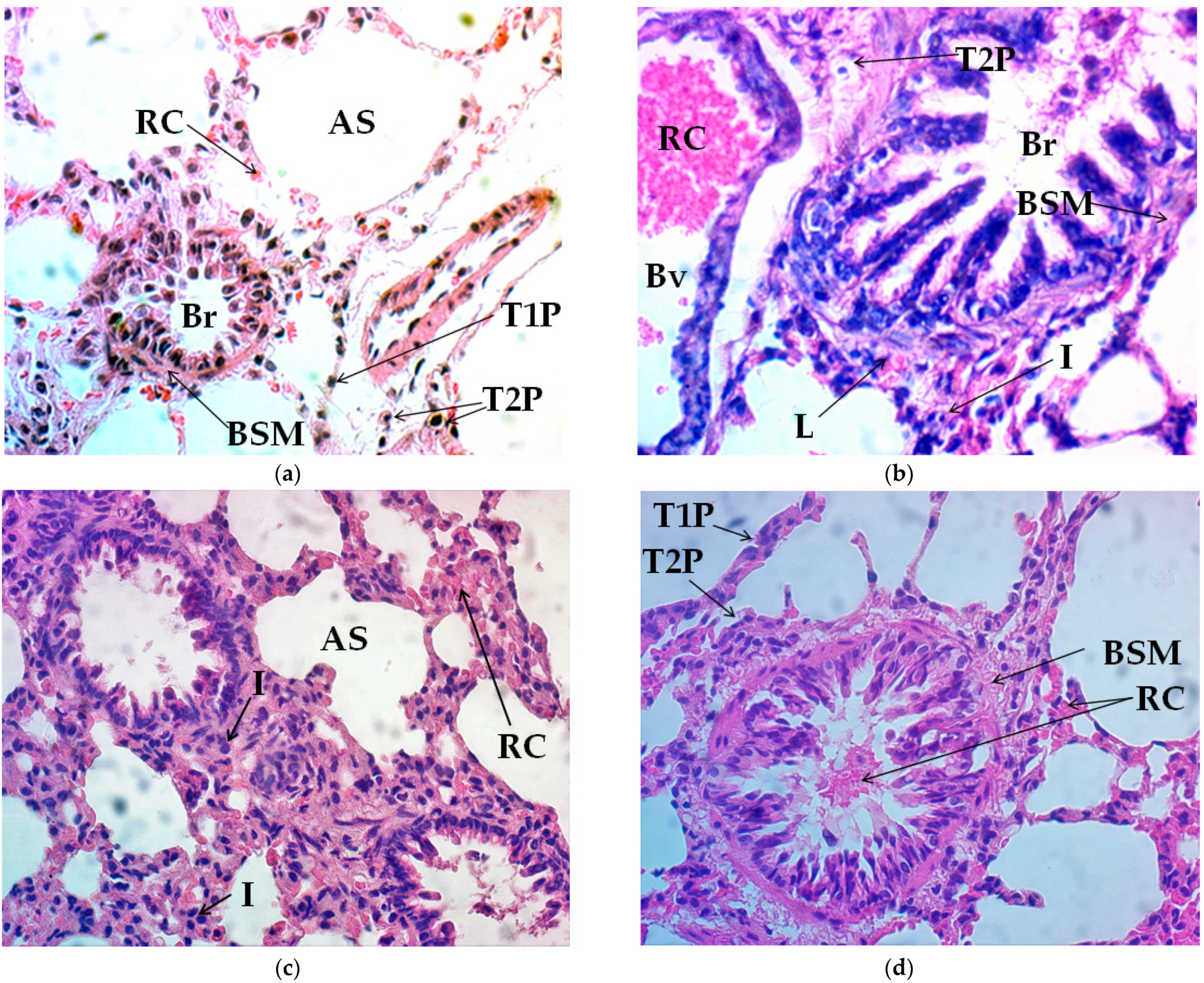
3.1.1. Lung Tissue
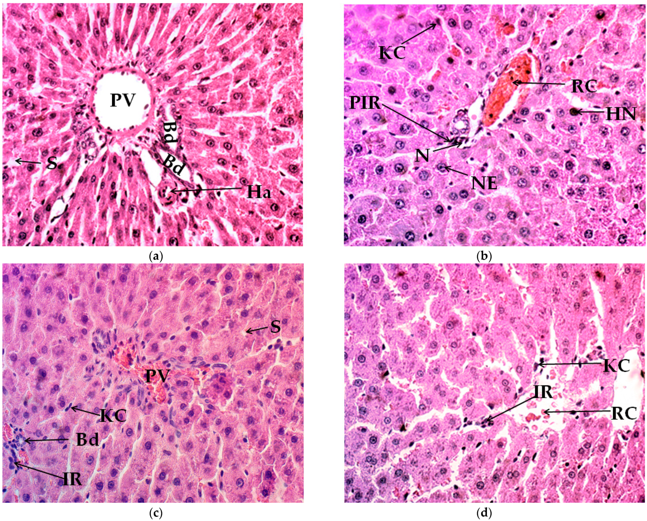
3.1.2. Liver Tissue
4. Discussion
5. Conclusions
Author Contributions
Funding
Institutional Review Board Statement
Acknowledgments
Conflicts of Interest
References
- MacInnis, M.J.; Gibala, M.J. Physiological adaptations to interval training and the role of exercise intensity. J. Physiol. 2017, 595, 2915–2930. [Google Scholar] [CrossRef] [Green Version]
- Hughes, D.C.; Ellefsen, S.; Baar, K. Adaptations to endurance and strength training. Cold Spring Harb. Perspect. Med. 2018, 8, a029769. [Google Scholar] [CrossRef]
- Di Meo, S.; Napolitano, G.; Venditti, P. Mediators of physical activity protection against ROS-linked skeletal muscle Damage. Int. J. Mol. Sci. 2019, 20, 3024. [Google Scholar] [CrossRef] [PubMed] [Green Version]
- Federico, A.; Dallio, M.; Loguercio, C. Silymarin/silybin and chronic liver disease: A marriage of many years. Molecules 2017, 22, 191. [Google Scholar] [CrossRef] [Green Version]
- Vargas-Mendoza, N.; Ángeles-Valencia, M.; Madrigal-Santillán, E.O.; Morales-Martínez, M.; Tirado-Lule, J.M.; Solano-Urrusquieta, A.; Madrigal-Bujaidar, E.; Álvarez-González, I.; Fregoso-Aguilar, T.; Morales-González, Á.; et al. Effect of silymarin supplementation on physical performance, muscle and myocardium histological changes, bodyweight, and food consumption in rats subjected to regular exercise training. Int. J. Mol. Sci. 2020, 21, 7724. [Google Scholar] [CrossRef] [PubMed]
- Roman, M.A.; Rossiter, H.B.; Casaburi, R. Exercise, ageing and the lung. Eur. Respir. J. 2016, 48, 1471–1486. [Google Scholar] [CrossRef] [PubMed] [Green Version]
- Gonzalez, J.T.; Fuchs, C.J.; Betts, J.A.; van Loon, L.J. Liver glycogen metabolism during and after prolonged endurance-type exercise. Am. J. Physiol. Endocrinol. Metab. 2016, 311, E543–E553. [Google Scholar] [CrossRef]
- Trefts, E.; Williams, A.S.; Wasserman, D.H. Exercise and the regulation of hepatic metabolism. Prog. Mol. Biol. Transl. Sci. 2015, 135, 203–225. [Google Scholar] [CrossRef] [PubMed] [Green Version]
- Daigle, C.C.; Chalupa, D.C.; Gibb, F.R.; Morrow, P.E.; Oberdörster, G.; Utell, M.J.; Frampton, M.W. Ultrafine particle deposition in humans during rest and exercise. Inhal. Toxicol. 2003, 15, 539–552. [Google Scholar] [CrossRef]
- Wittkopp, S.; Staimer, N.; Tjoa, T.; Gillen, D.; Daher, N.; Shafer, M.; Schauer, J.J.; Sioutas, C.; Delfino, R.J. Mitochondrial genetic background modifies the relationship between traffic-related air pollution exposure and systemic biomarkers of inflammation. PLoS ONE 2013, 8, e64444. [Google Scholar] [CrossRef] [PubMed] [Green Version]
- Heinonen, I.; Kalliokoski, K.K.; Hannukainen, J.C.; Duncker, D.J.; Nuutila, P.; Knuuti, J. Organ-specific physiological responses to acute physical exercise and long-term training in humans. Physiology 2014, 29, 421–436. [Google Scholar] [CrossRef]
- Ross, R.; Soni, S.; Houle, S.A. Negative energy balance induced by exercise or diet: Effects on visceral adipose tissue and liver fat. Nutrients 2020, 12, 891. [Google Scholar] [CrossRef] [PubMed] [Green Version]
- Kerksick, C.M.; Wilborn, C.D.; Roberts, M.D.; Smith-Ryan, A.; Kleiner, S.M.; Jäger, R.; Collins, R.; Cooke, M.; Davis, J.N.; Galvan, E.; et al. ISSN exercise & sports nutrition review update: Research & recommendations. J. Int. Soc. Sports Nutr. 2018, 15, 38. [Google Scholar] [CrossRef] [PubMed] [Green Version]
- Maughan, R.J.; Burke, L.M.; Dvorak, J.; Larson-Meyer, D.E.; Peeling, P.; Phillips, S.M.; Rawson, E.S.; Walsh, N.P.; Garthe, I.; Geyer, H.; et al. IOC consensus statement: Dietary supplements and the high-performance athlete. Int. J. Sport Nutr. Exerc. Metab. 2018, 28, 104–125. [Google Scholar] [CrossRef] [PubMed] [Green Version]
- Wang, P.; Li, C.G.; Qi, Z.; Cui, D.; Ding, S. Acute exercise induced mitochondrial H2O2 production in mouse skeletal muscle: Association with p66Shc and FOXO3a signaling and antioxidant enzymes. Oxid. Med. Cell. Longev. 2015, 2015, 536456. [Google Scholar] [CrossRef] [PubMed]
- Cardiff, R.D.; Miller, C.H.; Munn, R.J. Manual hematoxylin and eosin staining of mouse tissue sections. Cold Spring Harb. Protoc. 2014, 2014, 655–658. [Google Scholar] [CrossRef] [PubMed]
- Leslie, K.O.; Yousem, S.A.; Colby, T.V. Lungs. In Histology for Pathologist, 4th ed.; Mills, S.E., Ed.; Lippincott Williams & Wilkins: Philadelphia, PA, USA, 2012. [Google Scholar]
- Morales-González, J.A.; Gutiérrez-Salinas, J.; Yáñez, L.; Villagómez-Rico, C.; Badillo-Romero, J.; Hernández-Muñoz, R. Morphological and biochemical effects of a low ethanol dose on rat liver regeneration: Role of route and timing of administration. Dig. Dis. Sci. 1999, 44, 1963–1974. [Google Scholar] [CrossRef]
- Bijak, M. Silybin, a major bioactive component of milk thistle (Silybum marianum L. Gaernt.)—Chemistry, bioavailability, and metabolism. Molecules 2017, 22, 1942. [Google Scholar] [CrossRef] [Green Version]
- Vargas-Mendoza, N.; Madrigal-Santillán, E.; Morales-González, A.; Esquivel-Soto, J.; Esquivel-Chirino, C.; García-Luna, Y.G.-R.M.; Gayosso-de-Lucio, J.A.; Morales-González, J.A. Hepatoprotective effect of silymarin. World J. Hepatol. 2014, 6, 144–149. [Google Scholar] [CrossRef]
- Loguercio, C.; Festi, D. Silybin and the liver: From basic research to clinical practice. World J. Gastroenterol. 2011, 17, 2288–2301. [Google Scholar] [CrossRef]
- Surai, P.F. Silymarin as a Natural antioxidant: An overview of the current evidence and perspectives. Antioxidants 2015, 4, 204–247. [Google Scholar] [CrossRef] [Green Version]
- Choi, E.J.; Kim, E.K.; Jeoung, N.H.; Kim, S.H. Effect of silymarin on gluconeogenesis and lactate production in exercising rats. Food Sci. Biotechnol. 2016, 25, 119–124. [Google Scholar] [CrossRef]
- Haddad, M.; Sharma, S. Physiology, lung. In StatPearls; StatPearls Publishing LLC: Treasure Island, FL, USA, 2021. [Google Scholar]
- Stickland, M.K.; Lindinger, M.I.; Olfert, I.M.; Heigenhauser, G.J.; Hopkins, S.R. Pulmonary gas exchange and acid-base balance during exercise. Compr. Physiol. 2013, 3, 693–739. [Google Scholar] [CrossRef]
- Hsia, C.C. Recruitment of lung diffusing capacity: Update of concept and application. Chest 2002, 122, 1774–1783. [Google Scholar] [CrossRef] [Green Version]
- Tedjasaputra, V.; Bouwsema, M.M.; Stickland, M.K. Effect of aerobic fitness on capillary blood volume and diffusing membrane capacity responses to exercise. J. Physiol. 2016, 594, 4359–4370. [Google Scholar] [CrossRef] [PubMed] [Green Version]
- Tedjasaputra, V.; van Diepen, S.; Collins, S.; Michaelchuk, W.M.; Stickland, M.K. Assessment of pulmonary capillary blood volume, membrane diffusing capacity, and intrapulmonary arteriovenous anastomoses during exercise. J. Vis. Exp. JoVE 2017, 120, 54949. [Google Scholar] [CrossRef] [PubMed] [Green Version]
- Lovering, A.T.; Duke, J.W.; Elliott, J.E. Intrapulmonary arteriovenous anastomoses in humans—Response to exercise and the environment. J. Physiol. 2015, 593, 507–520. [Google Scholar] [CrossRef] [PubMed] [Green Version]
- Chimenti, L.; Morici, G.; Paternò, A.; Bonanno, A.; Siena, L.; Licciardi, A.; Veca, M.; Guccione, W.; Macaluso, F.; Bonsignore, G.; et al. Endurance training damages small airway epithelium in mice. Am. J. Respir. Crit. Care Med. 2007, 175, 442–449. [Google Scholar] [CrossRef] [PubMed]
- Greenwald, R.; Ferdinands, J.M.; Teague, W.G. Ionic determinants of exhaled breath condensate pH before and after exercise in adolescent athletes. Pediatric Pulmonol. 2009, 44, 768–777. [Google Scholar] [CrossRef]
- Denguezli, M.; Ben Chiekh, I.; Ben Saad, H.; Zaouali-Ajina, M.; Tabka, Z.; Abdelkrim, Z. One-year endurance training: Effects on lung function and airway inflammation. J. Sports Sci. 2008, 26, 1351–1359. [Google Scholar] [CrossRef] [PubMed]
- Fernández-Luna, Á.; Gallardo, L.; Plaza-Carmona, M.; García-Unanue, J.; Sánchez-Sánchez, J.; Felipe, J.L.; Burillo, P.; Ara, I. Respiratory function and changes in lung epithelium biomarkers after a short-training intervention in chlorinated vs. ozone indoor pools. PLoS ONE 2013, 8, e68447. [Google Scholar] [CrossRef] [Green Version]
- Pedersen, L.; Lund, T.K.; Barnes, P.J.; Kharitonov, S.A.; Backer, V. Airway responsiveness and inflammation in adolescent elite swimmers. J. Allergy Clin. Immunol. 2008, 122, 322–327. [Google Scholar] [CrossRef]
- Araneda, O.F.; Carbonell, T.; Tuesta, M. Update on the mechanisms of pulmonary inflammation and oxidative imbalance induced by exercise. Oxid. Med. Cell Longev. 2016, 2016, 4868536. [Google Scholar] [CrossRef] [Green Version]
- Sonnenbichler, J.; Zetl, I. Biochemical effects of the flavonolignane silibinin on RNA, protein and DNA synthesis in rat livers. Prog. Clin. Biol. Res. 1986, 213, 319–331. [Google Scholar] [PubMed]
- Sonnenbichler, J.; Zetl, I. Mechanism of action of silibinin. V. Effect of silibinin on the synthesis of ribosomal RNA, mRNA and tRNA in rat liver in vivo. Hoppe-Seyler’s Z. Fur Physiol. Chem. 1984, 365, 555–566. [Google Scholar] [CrossRef]
- Yavari, A.; Javadi, M.; Mirmiran, P.; Bahadoran, Z. Exercise-induced oxidative stress and dietary antioxidants. Asian J. Sports Med. 2015, 6, e24898. [Google Scholar] [CrossRef] [PubMed] [Green Version]
- Paschalis, V.; Theodorou, A.A.; Kyparos, A.; Dipla, K.; Zafeiridis, A.; Panayiotou, G.; Vrabas, I.S.; Nikolaidis, M.G. Low vitamin C values are linked with decreased physical performance and increased oxidative stress: Reversal by vitamin C supplementation. Eur. J. Nutr. 2016, 55, 45–53. [Google Scholar] [CrossRef] [PubMed]
- Hemilä, H. Vitamin C may alleviate exercise-induced bronchoconstriction: A meta-analysis. BMJ Open 2013, 3, e002416. [Google Scholar] [CrossRef] [Green Version]
- Colbert, L.H.; Visser, M.; Simonsick, E.M.; Tracy, R.P.; Newman, A.B.; Kritchevsky, S.B.; Pahor, M.; Taaffe, D.R.; Brach, J.; Rubin, S.; et al. Physical activity, exercise, and inflammatory markers in older adults: Findings from the Health, Aging and Body Composition Study. J. Am. Geriatr. Soc. 2004, 52, 1098–1104. [Google Scholar] [CrossRef]
- Wen, B.; Yu, L.; Fang, Y.; Wang, X. Dose-effect relationship between vitamin C and paraquat poisoning rats. Zhong Nan Da Xue Xue Bao. Yi Xue Ban = J. Cent. South Univ. Med Sci. 2016, 41, 1323–1327. [Google Scholar] [CrossRef]
- Yadegari, M.; Riahy, S.; Mirdar, S.; Hamidian, G.; Afkhami, S.M.; Saeidi, A.; Rhibi, F.; Ben Abderrahman, A.; Hackney, A.C.; Zouhal, H. The TNF-α, P53 protein response and lung respiratory changes to exercise, chronic hypoxia and Adiantum capillus-veneris supplementation. Adv. Respir. Med. 2019, 87, 226–234. [Google Scholar] [CrossRef] [Green Version]
- Ahmed, R.F.; Moussa, R.A.; Eldemerdash, R.S.; Zakaria, M.M.; Abdel-Gaber, S.A. Ameliorative effects of silymarin on HCl-induced acute lung injury in rats; role of the Nrf-2/HO-1 pathway. Iran. J. Basic Med Sci. 2019, 22, 1483–1492. [Google Scholar] [CrossRef] [PubMed]
- Zhao, F.; Shi, D.; Li, T.; Li, L.; Zhao, M. Silymarin attenuates paraquat-induced lung injury via Nrf2-mediated pathway in vivo and in vitro. Clin. Exp. Pharmacol. Physiol. 2015, 42, 988–998. [Google Scholar] [CrossRef] [PubMed]
- Podder, B.; Kim, Y.S.; Zerin, T.; Song, H.Y. Antioxidant effect of silymarin on paraquat-induced human lung adenocarcinoma A549 cell line. Food Chem. Toxicol. Int. J. Publ. Br. Ind. Biol. Res. Assoc. 2012, 50, 3206–3214. [Google Scholar] [CrossRef]
- Tonelli, C.; Chio, I.I.C.; Tuveson, D.A. Transcriptional regulation by Nrf2. Antioxid. Redox Signal. 2018, 29, 1727–1745. [Google Scholar] [CrossRef] [Green Version]
- He, F.; Ru, X.; Wen, T. NRF2, a Transcription factor for stress response and beyond. Int. J. Mol. Sci. 2020, 21, 4777. [Google Scholar] [CrossRef] [PubMed]
- Cuthbertson, D.J.; Shojaee-Moradie, F.; Sprung, V.S.; Jones, H.; Pugh, C.J.; Richardson, P.; Kemp, G.J.; Barrett, M.; Jackson, N.C.; Thomas, E.L.; et al. Dissociation between exercise-induced reduction in liver fat and changes in hepatic and peripheral glucose homoeostasis in obese patients with non-alcoholic fatty liver disease. Clin. Sci. 2016, 130, 93–104. [Google Scholar] [CrossRef] [Green Version]
- Brouwers, B.; Schrauwen-Hinderling, V.B.; Jelenik, T.; Gemmink, A.; Sparks, L.M.; Havekes, B.; Bruls, Y.; Dahlmans, D.; Roden, M.; Hesselink, M.K.C.; et al. Exercise training reduces intrahepatic lipid content in people with and people without nonalcoholic fatty liver. Am. J. Physiol. Endocrinol. Metab. 2018, 314, E165–E173. [Google Scholar] [CrossRef]
- Katsagoni, C.N.; Georgoulis, M.; Papatheodoridis, G.V.; Panagiotakos, D.B.; Kontogianni, M.D. Effects of lifestyle interventions on clinical characteristics of patients with non-alcoholic fatty liver disease: A meta-analysis. Metab. Clin. Exp. 2017, 68, 119–132. [Google Scholar] [CrossRef] [Green Version]
- Hallsworth, K.; Fattakhova, G.; Hollingsworth, K.G.; Thoma, C.; Moore, S.; Taylor, R.; Day, C.P.; Trenell, M.I. Resistance exercise reduces liver fat and its mediators in non-alcoholic fatty liver disease independent of weight loss. Gut 2011, 60, 1278–1283. [Google Scholar] [CrossRef]
- Kwak, M.S.; Kim, D. Non-alcoholic fatty liver disease and lifestyle modifications, focusing on physical activity. Korean J. Intern. Med. 2018, 33, 64–74. [Google Scholar] [CrossRef] [Green Version]
- Hashida, R.; Kawaguchi, T.; Bekki, M.; Omoto, M.; Matsuse, H.; Nago, T.; Takano, Y.; Ueno, T.; Koga, H.; George, J.; et al. Aerobic vs. resistance exercise in non-alcoholic fatty liver disease: A systematic review. J. Hepatol. 2017, 66, 142–152. [Google Scholar] [CrossRef] [PubMed]
- Dixon, L.J.; Barnes, M.; Tang, H.; Pritchard, M.T.; Nagy, L.E. Kupffer cells in the liver. Compr. Physiol. 2013, 3, 785–797. [Google Scholar] [CrossRef] [PubMed] [Green Version]
- Chawla, A.; Nguyen, K.D.; Goh, Y.P. Macrophage-mediated inflammation in metabolic disease. Nat. Reviews. Immunol. 2011, 11, 738–749. [Google Scholar] [CrossRef] [PubMed] [Green Version]
- Yazdani, H.O.; Kaltenmeier, C.; Morder, K.; Moon, J.; Traczek, M.; Loughran, P.; Zamora, R.; Vodovotz, Y.; Li, F.; Wang, J.H.; et al. Exercise training decreases hepatic injury and metastases through changes in immune response to liver ischemia/reperfusion in mice. Hepatology 2021, 73, 2494–2509. [Google Scholar] [CrossRef]
- Baradaran, A.; Samadi, F.; Ramezanpour, S.S.; Yousefdoust, S. Hepatoprotective effects of silymarin on CCl(4)-induced hepatic damage in broiler chickens model. Toxicol. Rep. 2019, 6, 788–794. [Google Scholar] [CrossRef] [PubMed]
- Dockalova, H.; Zeman, L.; Horky, P. Influence of milk thistle (Silybum marianum) seed cakes on biochemical values of equine plasma subjected to physical exertion. Animals 2021, 11, 210. [Google Scholar] [CrossRef] [PubMed]


| Group | Alveolar Size | Type 1 Pneumocytes | Type 2 Pneumocytes | Vascularization | Inflammation | Bronchial muscle | Connective Tissue | Alveolar Wall Thickness | Pleural Tissue Thickness |
|---|---|---|---|---|---|---|---|---|---|
| Control | +/++ | ++++ | ++ | ++ | + | + | + | ++ | + |
| ET | +++ | ++++ | + | +++ | ++/+++ | +++ | ++ | ++/+++ | ++ |
| ET+Aa | +++ | ++++ | ++ | +++ | ++ | +++ | + | ++ | + |
| ET+Sm | ++++ | ++++ | ++ | ++++ | + | ++++ | + | + | + |
| Group | Inflammatory Response | Kupffer Cells | Vascularization | Steatosis | Mitosis | Apoptosis/Necrosis | Hyperchromatic Nuclei | Nuclear Enlargement |
|---|---|---|---|---|---|---|---|---|
| Control | 0/+ | ++ | + | +++ | 0 | 0 | 0/+ | 0/+ |
| ET | 0/+ | ++ | +++ | ++ | 0 | 0 | + | ++ |
| Et+Aa | + | +++ | ++++ | + | 0 | + | 0 | 0 |
| Et+Sm | 0/+ | +++ | ++++ | + | 0 | 0 | 0 | 0 |
Publisher’s Note: MDPI stays neutral with regard to jurisdictional claims in published maps and institutional affiliations. |
© 2021 by the authors. Licensee MDPI, Basel, Switzerland. This article is an open access article distributed under the terms and conditions of the Creative Commons Attribution (CC BY) license (https://creativecommons.org/licenses/by/4.0/).
Share and Cite
Vargas-Mendoza, N.; Angeles-Valencia, M.; Morales-González, Á.; Morales-Martínez, M.; Madrigal-Bujaidar, E.; Álvarez-González, I.; Fregoso-Aguilar, T.; Delgado-Olivares, L.; Madrigal-Santillán, E.O.; Morales-González, J.A. Effect of Silymarin Supplementation in Lung and Liver Histological Modifications during Exercise Training in a Rodent Model. J. Funct. Morphol. Kinesiol. 2021, 6, 72. https://doi.org/10.3390/jfmk6030072
Vargas-Mendoza N, Angeles-Valencia M, Morales-González Á, Morales-Martínez M, Madrigal-Bujaidar E, Álvarez-González I, Fregoso-Aguilar T, Delgado-Olivares L, Madrigal-Santillán EO, Morales-González JA. Effect of Silymarin Supplementation in Lung and Liver Histological Modifications during Exercise Training in a Rodent Model. Journal of Functional Morphology and Kinesiology. 2021; 6(3):72. https://doi.org/10.3390/jfmk6030072
Chicago/Turabian StyleVargas-Mendoza, Nancy, Marcelo Angeles-Valencia, Ángel Morales-González, Mauricio Morales-Martínez, Eduardo Madrigal-Bujaidar, Isela Álvarez-González, Tomás Fregoso-Aguilar, Luis Delgado-Olivares, Eduardo Osiris Madrigal-Santillán, and José A. Morales-González. 2021. "Effect of Silymarin Supplementation in Lung and Liver Histological Modifications during Exercise Training in a Rodent Model" Journal of Functional Morphology and Kinesiology 6, no. 3: 72. https://doi.org/10.3390/jfmk6030072