
Two-Dimensional Graphitic Carbon Nitride (g-C3N4) Nanosheets and Their Derivatives for Diagnosis and Detection Applications
 ,
,  ,
,  and
and
Abstract
:1. Introduction
2. g-C3N4-Based Materials: Properties
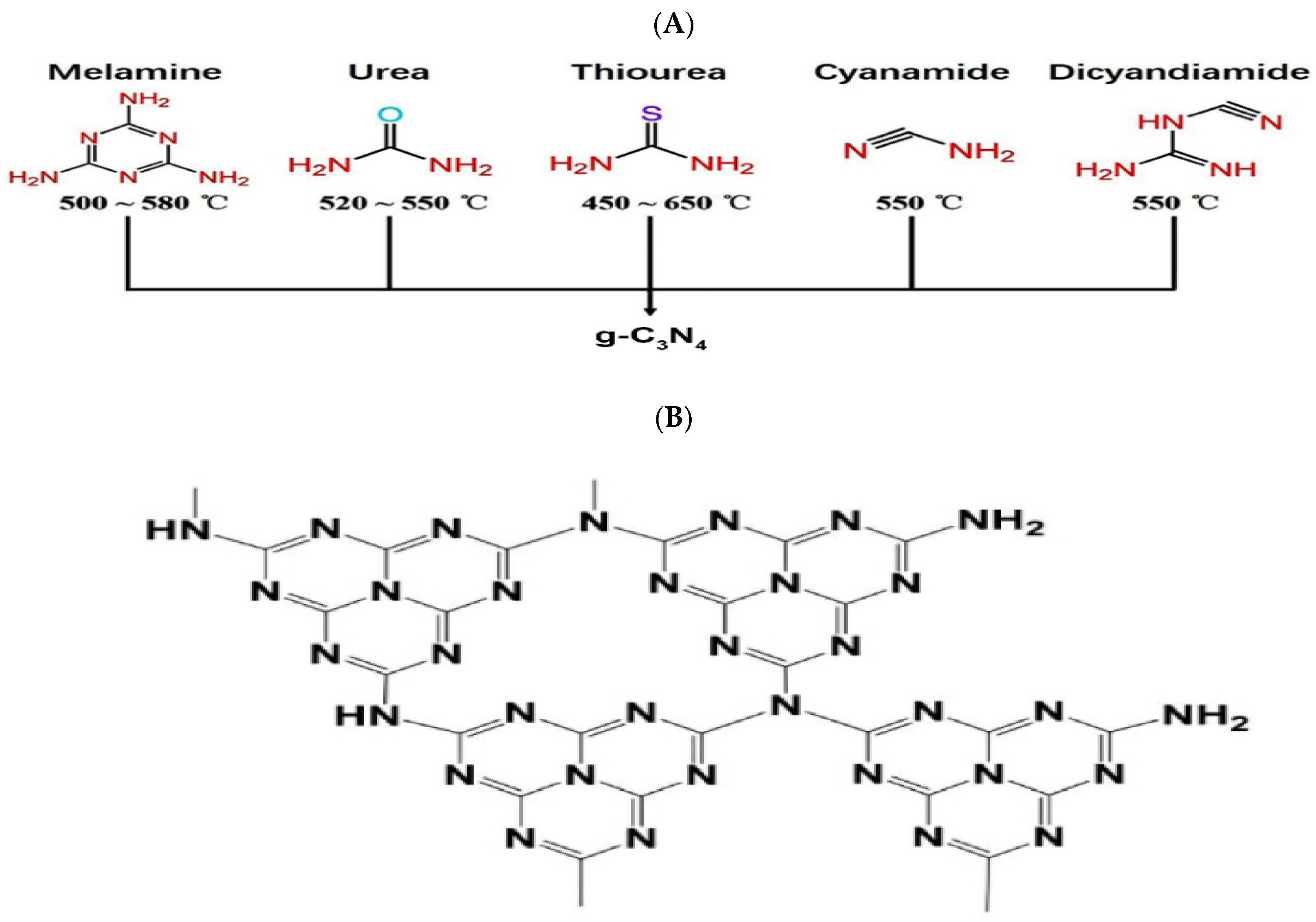
3. g-C3N4-Based Materials: Synthesis Methods
3.1. Synthesis of g-C3N4 Nanosheets
3.2. Synthesis of g-C3N4-Based Composites
4. g-C3N4-Based Biosensors
4.1. g-C3N4-Based Surface Plasmon Resonance (SPR) Biosensors
4.2. g-C3N4-Based Electrochemical Biosensors
4.3. g-C3N4-Based Photoelectrochemical (PEC) Biosensors
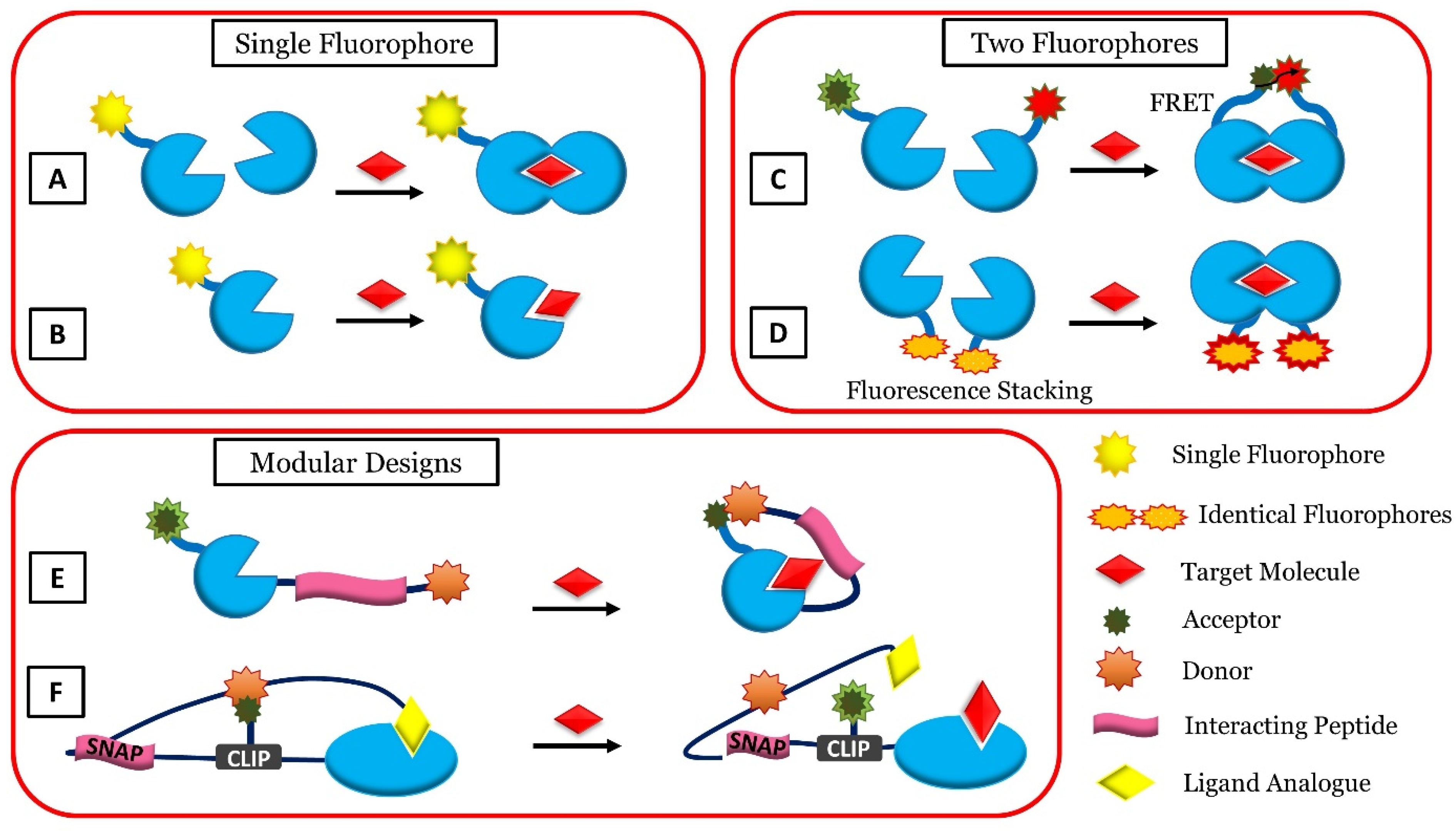
4.4. g-C3N4-Based Fluorescent Biosensors
4.5. g-C3N4-Based Electrochemiluminescent (ECL) Biosensors
5. Conclusions and Future Perspectives
Author Contributions
Funding
Data Availability Statement
Conflicts of Interest
Abbreviations
| CV | Cyclic Voltammetry |
| EC | Electrochemical Biosensor |
| ECL | Electrochemiluminescent Biosensor |
| FCS | Fluorescence Correlation Spectroscopy |
| FI | Fluorescence Intensity |
| FLIM | Fluorescence Lifetime Imaging |
| FRET | Forster Resonance Energy Transfer |
| g-C3N4 | Graphite Phase Carbon Nitride |
| PCL | Photochemoluminescence Biosensor |
| SPR | Surface Plasmon Resonance |
| SPW | Surface Plasmon Wave |
References
- Srinivas, P.R.; Kramer, B.S.; Srivastava, S. Trends in Biomarker Research for Cancer Detection. Lancet Oncol. 2001, 2, 698–704. [Google Scholar] [CrossRef]
- Samadi, A.; Pourmadadi, M.; Yazdian, F.; Rashedi, H.; Navaei-Nigjeh, M.; Eufrasio-da-silva, T. Ameliorating Quercetin Constraints in Cancer Therapy with PH-Responsive Agarose-Polyvinylpyrrolidone -Hydroxyapatite Nanocomposite Encapsulated in Double Nanoemulsion. Int. J. Biol. Macromol. 2021, 182, 11–25. [Google Scholar] [CrossRef] [PubMed]
- Ludwig, J.A.; Weinstein, J.N. Biomarkers in Cancer Staging, Prognosis and Treatment Selection. Nat. Rev. Cancer 2005, 5, 845–856. [Google Scholar] [CrossRef] [PubMed]
- Azimi, S.; Farahani, A.; Sereshti, H. Plasma-functionalized highly aligned CNT-based biosensor for point of care determination of glucose in human blood plasma. Electroanalysis 2020, 32, 394–403. [Google Scholar] [CrossRef]
- Bohunicky, B.; Mousa, S.A. Biosensors: The New Wave in Cancer Diagnosis. Nanotechnol. Sci. Appl. 2011, 4, 1–10. [Google Scholar] [CrossRef] [Green Version]
- Smith, D.S.; Humphrey, P.A.; Catalona, W.J. The Early Detection of Prostate Carcinoma with Prostate Specific Antigen The Washington University Experience. Cancer 1997, 80, 1852–1856. [Google Scholar] [CrossRef]
- Samadi, A.; Yazdian, F.; Navaei-Nigjeh, M.; Rashedi, H. Nanocomposite Hydrogels: A Promising Approach for Developing Stimuli-Responsive Platforms and Their Application in Targeted Drug Delivery. J. Shahid Sadoughi Univ. Med. Sci. 2021, 29, 3877–3897. [Google Scholar] [CrossRef]
- Yazdani, M.; Tavakoli, O.; Khoobi, M.; Wu, Y.S.; Faramarzi, M.A.; Gholibegloo, E.; Farkhondeh, S. Beta-Carotene/Cyclodextrin-Based Inclusion Complex: Improved Loading, Solubility, Stability, and Cytotoxicity. J. Incl. Phenom. Macrocycl. Chem. 2022, 102, 55–64. [Google Scholar] [CrossRef]
- Salehipour, M.; Rezaei, S.; Rezaei, M.; Yazdani, M.; Mogharabi-Manzari, M. Opportunities and Challenges in Biomedical Applications of Metal–Organic Frameworks. J. Inorg. Organomet. Polym. Mater. 2021, 31, 4443–4462. [Google Scholar] [CrossRef]
- Pourmadadi, M.; Dinani, H.S.; Tabar, F.S.; Khassi, K.; Janfaza, S.; Tasnim, N.; Hoorfar, M. Properties and Applications of Graphene and Its Derivatives in Biosensors for Cancer Detection: A Comprehensive Review. Biosensors 2022, 12, 269. [Google Scholar] [CrossRef]
- Samadi, A.; Haseli, S.; Pourmadadi, M.; Rashedi, H.; Yazdian, F.; Navaei-Nigjeh, M. Curcumin-Loaded Chitosan-Agarose-Montmorillonite Hydrogel Nanocomposite for the Treatment of Breast Cancer. In Proceedings of the 2020 27th National and 5th International Iranian Conference on Biomedical Engineering (ICBME), Tehran, Iran, 26 November 2020; pp. 148–153. [Google Scholar]
- Haseli, S.; Pourmadadi, M.; Samadi, A.; Yazdian, F.; Abdouss, M.; Rashedi, H.; Navaei-Nigjeh, M. A Novel PH-Responsive Nanoniosomal Emulsion for Sustained Release of Curcumin from a Chitosan-Based Nanocarrier: Emphasis on the Concurrent Improvement of Loading, Sustained Release, and Apoptosis Induction. Biotechnol. Prog. 2022, 38, e3280. [Google Scholar] [CrossRef] [PubMed]
- Pourmadadi, M.; Ahmadi, M.; Abdouss, M.; Yazdian, F.; Rashedi, H.; Navaei-Nigjeh, M.; Hesari, Y. The Synthesis and Characterization of Double Nanoemulsion for Targeted Co-Delivery of 5-Fluorouracil and Curcumin Using PH-Sensitive Agarose/Chitosan Nanocarrier. J. Drug Deliv. Sci. Technol. 2022, 70, 102849. [Google Scholar] [CrossRef]
- Rajabzadeh-Khosroshahi, M.; Pourmadadi, M.; Yazdian, F.; Rashedi, H.; Navaei-Nigjeh, M.; Rasekh, B. Chitosan/Agarose/Graphitic Carbon Nitride Nanocomposite as an Efficient PH-Sensitive Drug Delivery System for Anticancer Curcumin Releasing. J. Drug Deliv. Sci. Technol. 2022, 74, 103443. [Google Scholar] [CrossRef]
- Rahmani, E.; Pourmadadi, M.; Ghorbanian, S.A.; Yazdian, F.; Rashedi, H.; Navaee, M. Preparation of a PH-Responsive Chitosan-Montmorillonite-Nitrogen-Doped Carbon Quantum Dots Nanocarrier for Attenuating Doxorubicin Limitations in Cancer Therapy. Eng. Life Sci. 2022, 22, 634–649. [Google Scholar] [CrossRef] [PubMed]
- Heydari Foroushani, P.H.; Rahmani, E.; Alemzadeh, I.; Vossoughi, M.; Pourmadadi, M.; Rahdar, A.; Díez-Pascual, A.M. Curcumin Sustained Release with a Hybrid Chitosan-Silk Fibroin Nanofiber Containing Silver Nanoparticles as a Novel Highly Efficient Antibacterial Wound Dressing. Nanomaterials 2022, 12, 3426. [Google Scholar] [CrossRef] [PubMed]
- Sugumaran, S.; Jamlos, M.F.; Ahmad, M.N.; Bellan, C.S.; Schreurs, D. Nanostructured Materials with Plasmonic Nanobiosensors for Early Cancer Detection: A Past and Future Prospect. Biosens. Bioelectron. 2018, 100, 361–373. [Google Scholar] [CrossRef]
- Pourmadadi, M.; Yazdian, F.; Hojjati, S.; Khosravi-Darani, K. Detection of Microorganisms Using Graphene-Based Nanobiosensors. Food Technol. Biotechnol. 2021, 59, 496–506. [Google Scholar] [CrossRef]
- Holzinger, M.; Le Goff, A.; Cosnier, S. Nanomaterials for Biosensing Applications: A Review. Front. Chem. 2014, 2, 63. [Google Scholar] [CrossRef] [Green Version]
- Pandey, P.; Datta, M.; Malhotra, B.D. Prospects of Nanomaterials in Biosensors. Anal. Lett. 2008, 41, 159–209. [Google Scholar] [CrossRef]
- Pourmadadi, M.; Nouralishahi, A.; Shalbaf, M.; Shabani Shayeh, J.; Nouralishahi, A. An electrochemical aptasensor fordetection of Prostate-specific antigen-based oncarbon quantum dots-gold nanoparticles. Biotechnol. Appl. Biochem. 2022, 1–9. [Google Scholar] [CrossRef]
- Dinani, H.S.; Pourmadadi, M.; Yazdian, F.; Rashedi, H.; Ebrahimi, S.A.S.; Shayeh, J.S.; Ghorbani, M. Fabrication of Au/Fe3O4 /RGO Based Aptasensor for Measurement of MiRNA-128, a Biomarker for Acute Lymphoblastic Leukemia (ALL). Eng. Life Sci. 2022, 22, 519–534. [Google Scholar] [CrossRef] [PubMed]
- Pourmadadi, M.; Ahmadi, M.J.; Dinani, H.S.; Ajalli, N.; Dorkoosh, F. Theranostic Applications of Stimulus-Responsive Systems Based on Fe2O3. Pharm. Nanotechnol. 2022, 10, 90–112. [Google Scholar] [CrossRef] [PubMed]
- Golnaz, M.; Javad, S.; Meysam, S.O.; Mehrab, P.; Fatemeh, Y.; Lobat, T. An electrochemical aptasensor for detection of prostate-specific antigen using reduced graphene gold nanocomposite and cu/carbon quantum dots. Biotechnol. Appl. Biochem. 2021, 1–10. [Google Scholar] [CrossRef]
- Zavareh, H.S.; Pourmadadi, M.; Moradi, A.; Yazdian, F.; Omidi, M. Chitosan/Carbon Quantum Dot/Aptamer Complex as a Potential Anticancer Drug Delivery System towards the Release of 5-Fluorouracil. Int. J. Biol. Macromol. 2020, 165, 1422–1430. [Google Scholar] [CrossRef] [PubMed]
- Pourmadadi, M.; Shayeh, J.S.; Arjmand, S.; Omidi, M.; Fatemi, F. An Electrochemical Sandwich Immunosensor of Vascular Endothelial Growth Factor Based on Reduced Graphene Oxide/Gold Nanoparticle Composites. Microchem. J. 2020, 159, 105476. [Google Scholar] [CrossRef]
- Pourmadadi, M.; Shayeh, J.S.; Omidi, M.; Yazdian, F.; Alebouyeh, M.; Tayebi, L. A Glassy Carbon Electrode Modified with Reduced Graphene Oxide and Gold Nanoparticles for Electrochemical Aptasensing of Lipopolysaccharides from Escherichia coli Bacteria. Microchim. Acta 2019, 186, 2–9. [Google Scholar] [CrossRef] [PubMed]
- Aayanifard, Z.; Alebrahim, T.; Pourmadadi, M.; Yazdian, F.; Dinani, H.S.; Rashedi, H.; Omidi, M. Ultra PH-Sensitive Detection of Total and Free Prostate-Specific Antigen Using Electrochemical Aptasensor Based on Reduced Graphene Oxide/Gold Nanoparticles Emphasis on TiO2/Carbon Quantum Dots as a Redox Probe. Eng. Life Sci. 2021, 21, 739–752. [Google Scholar] [CrossRef]
- Behboudi, H.; Mehdipour, G.; Safari, N.; Pourmadadi, M.; Saei, A.; Omidi, M.; Tayebi, L.; Rahmandoust, M. Carbon Quantum Dots in Nanobiotechnology. In Nanomaterials for Advanced Biological Applications; Springer: Berlin/Heidelberg, Germany, 2019; pp. 145–179. [Google Scholar]
- Kazemi, S.; Pourmadadi, M.; Yazdian, F.; Ghadami, A. The Synthesis and Characterization of Targeted Delivery Curcumin Using Chitosan-Magnetite-Reduced Graphene Oxide as Nano-Carrier. Int. J. Biol. Macromol. 2021, 186, 554–562. [Google Scholar] [CrossRef]
- Malmir, S.; Karbalaei, A.; Pourmadadi, M.; Hamedi, J.; Yazdian, F.; Navaee, M. Antibacterial Properties of a Bacterial Cellulose CQD-TiO2 Nanocomposite. Carbohydr. Polym. 2020, 234, 115835. [Google Scholar] [CrossRef]
- Zamani, M.; Pourmadadi, M.; Seyyed Ebrahimi, S.A.; Yazdian, F.; Shabani Shayeh, J. A Novel Labeled and Label-Free Dual Electrochemical Detection of Endotoxin Based on Aptamer-Conjugated Magnetic Reduced Graphene Oxide-Gold Nanocomposite. J. Electroanal. Chem. 2022, 908, 116116. [Google Scholar] [CrossRef]
- Azadmanesh, F.; Pourmadadi, M.; Zavar Reza, J.; Yazdian, F.; Omidi, M.; Haghirosadat, B.F. Synthesis of a Novel Nanocomposite Containing Chitosan as a Three-Dimensional Printed Wound Dressing Technique: Emphasis on Gene Expression. Biotechnol. Prog. 2021, 37, e3132. [Google Scholar] [CrossRef] [PubMed]
- Behboodi, H.; Pourmadadi, M.; Omidi, M.; Rahmandoust, M.; Siadat, S.O.R.; Shayeh, J.S. Cu-CDs as Dual Optical and Electrochemical Nanosensor for ΒME Detection. Surf. Interfaces 2022, 29, 101710. [Google Scholar] [CrossRef]
- Tabar, F.S.; Pourmadadi, M.; Rashedi, H.; Yazdian, F. Design of Electrochemical Nanobiosensor in the Diagnosis of Prostate Specific Antigen (PSA) Using Nanostructures. In Proceedings of the 2020 27th National and 5th International Iranian Conference on Biomedical Engineering (ICBME), Tehran, Iran, 26–27 November 2020; pp. 35–40. [Google Scholar]
- Abolghasemzade, S.; Pourmadadi, M.; Rashedi, H.; Yazdian, F.; Kianbakht, S.; Navaei-Nigjeh, M. PVA Based Nanofiber Containing CQDs Modified with Silica NPs and Silk Fibroin Accelerates Wound Healing in a Rat Model. J. Mater. Chem. B 2021, 9, 658–676. [Google Scholar] [CrossRef] [PubMed]
- Kalajahi, S.T.; Mofradnia, S.R.; Yazdian, F.; Rasekh, B.; Neshati, J.; Taghavi, L.; Pourmadadi, M.; Haghirosadat, B.F. Inhibition Performances of Graphene Oxide/Silver Nanostructure for the Microbial Corrosion: Molecular Dynamic Simulation Study. Environ. Sci. Pollut. Res. 2022, 29, 49884–49897. [Google Scholar] [CrossRef]
- Dinani, H.S.; Pourmadadi, M.; Rashedi, H.; Yazdian, F. Fabrication of Nanomaterial-Based Biosensor for Measurement of a MicroRNA Involved in Cancer. In Proceedings of the 2020 27th National and 5th International Iranian Conference on Biomedical Engineering (ICBME), Tehran, Iran, 26–27 November 2020; pp. 47–54. [Google Scholar]
- Xiong, M.; Rong, Q.; Meng, H.M.; Zhang, X.B. Two-Dimensional Graphitic Carbon Nitride Nanosheets for Biosensing Applications. Biosens. Bioelectron. 2017, 89, 212–223. [Google Scholar] [CrossRef]
- Duan, F.; Zhang, S.; Yang, L.; Zhang, Z.; He, L.; Wang, M. Bifunctional Aptasensor Based on Novel Two-Dimensional Nanocomposite of MoS2 Quantum Dots and g-C3N4 Nanosheets Decorated with Chitosan-Stabilized Au Nanoparticles for Selectively Detecting Prostate Specific Antigen. Anal. Chim. Acta 2018, 1036, 121–132. [Google Scholar] [CrossRef]
- Naderian, N.; Pourmadadi, M.; Rashedi, H.; Yazdian, F. Design of a Novel Nanobiosensor for the Diagnosis of Acute Lymphoid Leukemia (ALL) by Measurement of MiRNA-128. In Proceedings of the 2020 27th National and 5th International Iranian Conference on Biomedical Engineering (ICBME), Tehran, Iran, 26–27 November 2020; pp. 41–46. [Google Scholar]
- Sakthivel, A.; Chandrasekaran, A.; Sadasivam, M.; Manickam, P.; Alwarappan, S. Sulphur Doped Graphitic Carbon Nitride as a Dual Biosensing Platform for the Detection of Cancer Biomarker CA15-3. J. Electrochem. Soc 2021, 168, 017507. [Google Scholar] [CrossRef]
- Ismael, M. A Review on Graphitic Carbon Nitride (g-C3N4) Based Nanocomposites: Synthesis, Categories, and Their Application in Photocatalysis. J. Alloys Compd. 2020, 846, 156446. [Google Scholar] [CrossRef]
- Wang, H.; Qi, C.; He, W.; Wang, M.; Jiang, W.; Yin, H.; Ai, S. A Sensitive Photoelectrochemical Immunoassay of N6-Methyladenosine Based on Dual-Signal Amplification Strategy: Ru Doped in SiO2 Nanosphere and Carboxylated g-C3N4. Biosens. Bioelectron. 2018, 99, 281–288. [Google Scholar] [CrossRef]
- Feng, L.; He, F.; Liu, B.; Yang, G.; Gai, S.; Yang, P.; Li, C.; Dai, Y.; Lv, R.; Lin, J. G-C3N4 Coated Upconversion Nanoparticles for 808 Nm Near-Infrared Light Triggered Phototherapy and Multiple Imaging. Chem. Mater. 2016, 28, 7935–7946. [Google Scholar] [CrossRef]
- Zhang, X.; Xie, X.; Wang, H.; Zhang, J.; Pan, B.; Xie, Y. Enhanced Photoresponsive Ultrathin Graphitic-Phase C3N 4 Nanosheets for Bioimaging. J. Am. Chem. Soc. 2013, 135, 18–21. [Google Scholar] [CrossRef] [PubMed]
- Zhang, M.; Wang, Q.; Xu, Y.; Guo, L.; Lai, Z.; Li, Z. Graphitic Carbon Nitride Quantum Dots as Analytical Probe for Viewing Sialic Acid on the Surface of Cells and Tissues. Anal. Chim. Acta 2020, 1095, 204–211. [Google Scholar] [CrossRef] [PubMed]
- Chen, L.; Huang, D.; Ren, S.; Dong, T.; Chi, Y.; Chen, G. Preparation of Graphite-like Carbon Nitride Nanoflake Film with Strong Fluorescent and Electrochemiluminescent Activity. Nanoscale 2013, 5, 225–230. [Google Scholar] [CrossRef] [PubMed]
- Ong, W.J.; Tan, L.L.; Ng, Y.H.; Yong, S.T.; Chai, S.P. Graphitic Carbon Nitride (g-C3N4)-Based Photocatalysts for Artificial Photosynthesis and Environmental Remediation: Are We a Step Closer to Achieving Sustainability? Chem. Rev. 2016, 116, 7159–7329. [Google Scholar] [CrossRef]
- Vasiljević, J.; Jerman, I.; Simončič, B. Graphitic Carbon Nitride as a New Sustainable Photocatalyst for Textile Functionalization. Polymers 2021, 13, 2568. [Google Scholar] [CrossRef]
- Alwin, E.; Kočí, K.; Wojcieszak, R.; Zieliński, M.; Edelmannová, M.; Pietrowski, M. Influence of High Temperature Synthesis on the Structure of Graphitic Carbon Nitride and Its Hydrogen Generation Ability. Materials 2020, 13, 2756. [Google Scholar] [CrossRef]
- Thomas, A.; Fischer, A.; Goettmann, F.; Antonietti, M.; Müller, J.O.; Schlögl, R.; Carlsson, J.M. Graphitic Carbon Nitride Materials: Variation of Structure and Morphology and Their Use as Metal-Free Catalysts. J. Mater. Chem. 2008, 18, 4893–4908. [Google Scholar] [CrossRef] [Green Version]
- Tian, C.; Zhao, H.; Sun, H.; Xiao, K.; Keung Wong, P. Enhanced Adsorption and Photocatalytic Activities of Ultrathin Graphitic Carbon Nitride Nanosheets: Kinetics and Mechanism. Chem. Eng. J. 2020, 381, 122760. [Google Scholar] [CrossRef]
- Du, X.; Kleitz, F.; Li, X.; Huang, H.; Zhang, X.; Qiao, S.Z. Disulfide-Bridged Organosilica Frameworks: Designed, Synthesis, Redox-Triggered Biodegradation, and Nanobiomedical Applications. Adv. Funct. Mater. 2018, 28, 1707325. [Google Scholar] [CrossRef] [Green Version]
- Dante, R.C.; Trakulmututa, J.; Meejoo-Smith, S.; Sirisit, N.; Martín-Ramos, P.; Chamorro-Posada, P.; Rutto, D.; Dante, D.G. A Solid-State Glucose Sensor Based on Cu and Fe–Doped Carbon Nitride. Mater. Chem. Phys. 2021, 258, 124023. [Google Scholar] [CrossRef]
- Ma, T.Y.; Tang, Y.; Dai, S.; Qiao, S.Z. Proton-Functionalized Two-Dimensional Graphitic Carbon Nitride Nanosheet: An Excellent Metal-/Label-Free Biosensing Platform. Small 2014, 10, 2382–2389. [Google Scholar] [CrossRef] [PubMed]
- Zheng, Y.; Lin, L.; Wang, B.; Wang, X. Graphitic Carbon Nitride Polymers toward Sustainable Photoredox Catalysis. Angew. Chem.-Int. Ed. 2015, 54, 12868–12884. [Google Scholar] [CrossRef] [PubMed]
- Liao, G.; He, F.; Li, Q.; Zhong, L.; Zhao, R.; Che, H.; Gao, H.; Fang, B. Emerging Graphitic Carbon Nitride-Based Materials for Biomedical Applications. Prog. Mater. Sci. 2020, 112, 100666. [Google Scholar] [CrossRef]
- Wang, A.J.; Li, H.; Huang, H.; Qian, Z.S.; Feng, J.J. Fluorescent Graphene-like Carbon Nitrides: Synthesis, Properties and Applications. J. Mater. Chem. C 2016, 4, 8146–8160. [Google Scholar] [CrossRef]
- Reddy, K.R.; Reddy, C.V.; Nadagouda, M.N.; Shetti, N.P.; Jaesool, S.; Aminabhavi, T.M. Polymeric Graphitic Carbon Nitride (g-C3N4)-Based Semiconducting Nanostructured Materials: Synthesis Methods, Properties and Photocatalytic Applications. J. Environ. Manag. 2019, 238, 25–40. [Google Scholar] [CrossRef]
- Yu, H.; Shi, R.; Zhao, Y.; Waterhouse, G.I.N.; Wu, L.Z.; Tung, C.H.; Zhang, T. Smart Utilization of Carbon Dots in Semiconductor Photocatalysis. Adv. Mater. 2016, 28, 9454–9477. [Google Scholar] [CrossRef]
- Hatamie, A.; Marahel, F.; Sharifat, A. Green Synthesis of Graphitic Carbon Nitride Nanosheet (g-C3N4) and Using It as a Label-Free Fluorosensor for Detection of Metronidazole via Quenching of the Fluorescence. Talanta 2018, 176, 518–525. [Google Scholar] [CrossRef]
- Wang, L.; Wang, C.; Hu, X.; Xue, H.; Pang, H. Metal/Graphitic Carbon Nitride Composites: Synthesis, Structures, and Applications. Chem.-Asian J. 2016, 11, 3305–3328. [Google Scholar] [CrossRef]
- Vinoth, S.; Ramaraj, R.; Pandikumar, A. Facile Synthesis of Calcium Stannate Incorporated Graphitic Carbon Nitride Nanohybrid Materials: A Sensitive Electrochemical Sensor for Determining Dopamine. Mater. Chem. Phys. 2020, 245, 122743. [Google Scholar] [CrossRef]
- Imran, H.; Manikandan, P.N.; Dharuman, V. Highly Selective and Rapid Non-Enzymatic Glucose Sensing at Ultrathin Layered Nb Doped C3N4 for Extended Linearity Range. Microchem. J. 2021, 160, 105774. [Google Scholar] [CrossRef]
- Vinoth, S.; Sampathkumar, P.; Giribabu, K.; Pandikumar, A. Ultrasonically Assisted Synthesis of Barium Stannate Incorporated Graphitic Carbon Nitride Nanocomposite and Its Analytical Performance in Electrochemical Sensing of 4-Nitrophenol. Ultrason. Sonochem. 2020, 62, 104855. [Google Scholar] [CrossRef] [PubMed]
- Lewandowski, C.M. Surface Plasmon Resonance (SPR) Biosensor Development. Eff. Br. Mindfulness Interv. Acute Pain Exp. Exam. Individ. Differ. 2015, 1, 43–48. [Google Scholar]
- Nie, W.; Wang, Q.; Zou, L.; Zheng, Y.; Liu, X.; Yang, X.; Wang, K. Low-Fouling Surface Plasmon Resonance Sensor for Highly Sensitive Detection of MicroRNA in a Complex Matrix Based on the DNA Tetrahedron. Anal. Chem. 2018, 90, 12584–12591. [Google Scholar] [CrossRef]
- Ho, A.H.P.; Kim, D.; Somekh, M.G. Handbook of Photonics for Biomedical Engineering; Springer: Berlin/Heidelberg, Germany, 2017; pp. 1–947. [Google Scholar] [CrossRef]
- Yao, Y.; Yi, B.; Xiao, J.; Li, Z.H. Surface Plasmon Resonance Biosensors and Its Application. In Proceedings of the 2007 1st International Conference on Bioinformatics and Biomedical Engineering, Wuhan, China, 6–8 July 2007; pp. 1043–1046. [Google Scholar] [CrossRef]
- Maurya, J.B.; Prajapati, Y.K. A Comparative Study of Different Metal and Prism in the Surface Plasmon Resonance Biosensor Having MoS2-Graphene. Opt. Quantum Electron. 2016, 48, 1–12. [Google Scholar] [CrossRef]
- Miyazaki, C.M.; Shimizu, F.M.; Ferreira, M. Surface Plasmon Resonance (SPR) for Sensors and Biosensors; Elsevier Inc.: Amsterdam, The Netherlands, 2017; ISBN 9780323497794. [Google Scholar]
- Lin, C.; Chen, S. Design of High-Performance Au-Ag-Dielectric-Graphene Based Surface Plasmon Resonance Biosensors Using Genetic Algorithm. J. Appl. Phys. 2019, 125, 113101. [Google Scholar] [CrossRef]
- Homola, J. Surface Plasmon Resonance Sensors for Detection of Chemical and Biological Species. Chem. Rev. 2008, 108, 462–493. [Google Scholar] [CrossRef]
- Boozer, C.; Kim, G.; Cong, S.; Guan, H.W.; Londergan, T. Looking towards Label-Free Biomolecular Interaction Analysis in a High-Throughput Format: A Review of New Surface Plasmon Resonance Technologies. Curr. Opin. Biotechnol. 2006, 17, 400–405. [Google Scholar] [CrossRef]
- Gao, Y.; Liu, M.; Zhang, Y.; Liu, Z.; Yang, Y.; Zhao, L. Recent Development on Narrow Bandgap Conjugated Polymers for Polymer Solar Cells. Polymers 2017, 9, 39. [Google Scholar] [CrossRef] [Green Version]
- Topkaya, S.N.; Azimzadeh, M.; Ozsoz, M. Electrochemical Biosensors for Cancer Biomarkers Detection: Recent Advances and Challenges. Electroanalysis 2016, 28, 1402–1419. [Google Scholar] [CrossRef]
- Taniselass, S.; Arshad, M.K.M.; Gopinath, S.C.B. Graphene-Based Electrochemical Biosensors for Monitoring Noncommunicable Disease Biomarkers. Biosens. Bioelectron. 2019, 130, 276–292. [Google Scholar] [CrossRef]
- Gan, T.; Shi, Z.; Sun, J.; Liu, Y. Simple and Novel Electrochemical Sensor for the Determination of Tetracycline Based on Iron/Zinc Cations-Exchanged Montmorillonite Catalyst. Talanta 2014, 121, 187–193. [Google Scholar] [CrossRef]
- Cho, I.H.; Kim, D.H.; Park, S. Electrochemical Biosensors: Perspective on Functional Nanomaterials for on-Site Analysis. Biomater. Res. 2020, 24, 1–12. [Google Scholar] [CrossRef] [Green Version]
- Li, Y.P.; Cao, H.B.; Liu, C.M.; Zhang, Y. Electrochemical Reduction of Nitrobenzene at Carbon Nanotube Electrode. J. Hazard. Mater. 2007, 148, 158–163. [Google Scholar] [CrossRef] [PubMed]
- Patnaik, S.; Martha, S.; Acharya, S.; Parida, K.M. An Overview of the Modification of G-C3N4 with High Carbon Containing Materials for Photocatalytic Applications. Inorg. Chem. Front. 2016, 3, 336–347. [Google Scholar] [CrossRef]
- Fang, T.; Yang, X.; Zhang, L.; Gong, J. Ultrasensitive Photoelectrochemical Determination of Chromium(VI) in Water Samples by Ion-Imprinted/Formate Anion-Incorporated Graphitic Carbon Nitride Nanostructured Hybrid. J. Hazard. Mater. 2016, 312, 106–113. [Google Scholar] [CrossRef] [PubMed] [Green Version]
- Wang, X.; Maeda, K.; Thomas, A.; Takanabe, K.; Xin, G.; Carlsson, J.M.; Domen, K.; Antonietti, M. A Metal-Free Polymeric Photocatalyst for Hydrogen Production from Water under Visible Light. Nat. Mater. 2009, 8, 76–80. [Google Scholar] [CrossRef]
- Zhao, Q.; Wu, W.; Wei, X.; Jiang, S.; Zhou, T.; Li, Q.; Lu, Q. Graphitic Carbon Nitride as Electrode Sensing Material for Tetrabromobisphenol-A Determination. Sens. Actuators B Chem. 2017, 248, 673–681. [Google Scholar] [CrossRef]
- Kathiresan, V.; Rajarathinam, T.; Lee, S.; Kim, S.; Lee, J.; Thirumalai, D.; Chang, S.C. Cost-Effective Electrochemical Activation of Graphitic Carbon Nitride on the Glassy Carbon Electrode Surface for Selective Determination of Serotonin. Sensors 2020, 20, 6083. [Google Scholar] [CrossRef]
- Zhan, T.; Tian, X.; Ding, G.; Liu, X.; Wang, L.; Teng, H. Quaternarization Strategy to Ultrathin Lamellar Graphitic C3N4 Ionic Liquid Nanostructure for Enhanced Electrochemical 2,4-Dichlorophenol Sensing. Sens. Actuators B Chem. 2019, 283, 463–471. [Google Scholar] [CrossRef]
- Miao, J.; Li, X.; Li, Y.; Dong, X.; Zhao, G.; Fang, J.; Wei, Q.; Cao, W. Dual-Signal Sandwich Electrochemical Immunosensor for Amyloid β-Protein Detection Based on Cu–Al2O3-g–C3N4–Pd and UiO-66@PANI-MB. Anal. Chim. Acta 2019, 1089, 48–55. [Google Scholar] [CrossRef]
- Ansari, S.; Ansari, M.S.; Devnani, H.; Satsangee, S.P.; Jain, R. CeO2/g-C3N4 Nanocomposite: A Perspective for Electrochemical Sensing of Anti-Depressant Drug. Sens. Actuators B Chem. 2018, 273, 1226–1236. [Google Scholar] [CrossRef]
- Li, C.; Xu, J.; Wu, Y.; Zhang, Y.; Zhang, C.; Lei, W.; Hao, Q. G-C3N4 Nanofibers Doped Poly(3,4-Ethylenedioxythiophene) Modified Electrode for Simultaneous Determination of Ascorbic Acid and Acetaminophen. J. Electroanal. Chem. 2018, 824, 52–59. [Google Scholar] [CrossRef]
- Zhou, D.; Wang, M.; Dong, J.; Ai, S. A Novel Electrochemical Immunosensor Based on Mesoporous Graphitic Carbon Nitride for Detection of Subgroup J of Avian Leukosis Viruses. Electrochim. Acta 2016, 205, 95–101. [Google Scholar] [CrossRef]
- Yan, K.; Yang, Y.; Zhang, J. A Self-Powered Sensor Based on Molecularly Imprinted Polymer-Coupled Graphitic Carbon Nitride Photoanode for Selective Detection of Bisphenol A. Sens. Actuators B Chem. 2018, 259, 394–401. [Google Scholar] [CrossRef]
- Sun, A.L.; Qi, Q.A. Silver-Functionalized g-C3N4 Nanohybrids as Signal-Transduction Tags for Electrochemical Immunoassay of Human Carbohydrate Antigen 19-9. Analyst 2016, 141, 4366–4372. [Google Scholar] [CrossRef]
- Afzali, M.; Shafiee, M.R.M.; Parhizkar, J. Au Nanorods/ g-C3N4 Composite Based Biosensor for Electrochemical Detection of Chronic Lymphocytic Leukemia. Nanomed. Res. J. 2020, 5, 32–43. [Google Scholar] [CrossRef]
- Chen, X.; Ke, X.X.; Liu, Y.; Weerasooriya, R.; Li, H.; Wu, Y.C. Photocatalytically Induced Au/Mpg-C3N4 Nanocomposites for Robust Electrochemical Detection of Cr(VI) in Tannery Wastewater. J. Environ. Chem. Eng. 2020, 9, 104642. [Google Scholar] [CrossRef]
- Xiao, F.; Li, H.; Yan, X.; Yan, L.; Zhang, X.; Wang, M.; Qian, C.; Wang, Y. Graphitic Carbon Nitride/Graphene Oxide(g-C3N4/GO) Nanocomposites Covalently Linked with Ferrocene Containing Dendrimer for Ultrasensitive Detection of Pesticide. Anal. Chim. Acta 2020, 1103, 84–96. [Google Scholar] [CrossRef]
- Xu, Y.; Lei, W.; Su, J.; Hu, J.; Yu, X.; Zhou, T.; Yang, Y.; Mandler, D.; Hao, Q. A High-Performance Electrochemical Sensor Based on g-C3N4-E-PEDOT for the Determination of Acetaminophen. Electrochim. Acta 2018, 259, 994–1003. [Google Scholar] [CrossRef]
- Kesavan, G.; Chen, S.M. Highly Sensitive Electrochemical Sensor Based on Carbon-Rich Graphitic Carbon Nitride as an Electrocatalyst for the Detection of Diphenylamine. Microchem. J. 2020, 159, 105587. [Google Scholar] [CrossRef]
- Zou, J.; Wu, S.; Liu, Y.; Sun, Y.; Cao, Y.; Hsu, J.P.; Shen Wee, A.T.; Jiang, J. An Ultra-Sensitive Electrochemical Sensor Based on 2D g-C3N4/CuO Nanocomposites for Dopamine Detection. Carbon N. Y. 2018, 130, 652–663. [Google Scholar] [CrossRef]
- Ponnaiah, S.K.; Prakash, P.; Muthupandian, S. Ultrasonic Energy-Assisted in-Situ Synthesis of Ru0/PANI/g-C3N4 Nanocomposite: Application for Picomolar-Level Electrochemical Detection of Endocrine Disruptor (Bisphenol-A) in Humans and Animals. Ultrason. Sonochem. 2019, 58, 104629. [Google Scholar] [CrossRef] [PubMed]
- Sun, Y.; Jiang, J.; Liu, Y.; Wu, S.; Zou, J. A Facile One-Pot Preparation of Co3O4/g-C3N4 Heterojunctions with Excellent Electrocatalytic Activity for the Detection of Environmental Phenolic Hormones. Appl. Surf. Sci. 2018, 430, 362–370. [Google Scholar] [CrossRef]
- Karthika, A.; Suganthi, A.; Rajarajan, M. An In-Situ Synthesis of Novel V2O5/G-C3N4/PVA Nanocomposite for Enhanced Electrocatalytic Activity toward Sensitive and Selective Sensing of Folic Acid in Natural Samples. Arab. J. Chem. 2020, 13, 3639–3652. [Google Scholar] [CrossRef]
- Kokulnathan, T.; Wang, T.-J. Vanadium Carbide-Entrapped Graphitic Carbon Nitride Nanocomposites: Synthesis and Electrochemical Platforms for Accurate Detection of Furazolidone. ACS Appl. Nano Mater. 2020, 3, 2554–2561. [Google Scholar] [CrossRef]
- Balasubramanian, P.; Annalakshmi, M.; Chen, S.M.; Chen, T.W. Sonochemical Synthesis of Molybdenum Oxide (MoO3) Microspheres Anchored Graphitic Carbon Nitride (g-C3N4) Ultrathin Sheets for Enhanced Electrochemical Sensing of Furazolidone. Ultrason. Sonochem. 2019, 50, 96–104. [Google Scholar] [CrossRef]
- Yola, M.L.; Atar, N. Amperometric Galectin-3 Immunosensor-Based Gold Nanoparticle-Functionalized Graphitic Carbon Nitride Nanosheets and Core-Shell Ti-MOF@COFs Composites. Nanoscale 2020, 12, 19824–19832. [Google Scholar] [CrossRef]
- Zeng, G.; Duan, M.; Xu, Y.; Ge, F.; Wang, W. Platinum (II)-Doped Graphitic Carbon Nitride with Enhanced Peroxidase-like Activity for Detection of Glucose and H2O2. Spectrochim. Acta-Part A Mol. Biomol. Spectrosc. 2020, 241, 118649. [Google Scholar] [CrossRef]
- Tian, K.J.; Liu, H.; Dong, Y.P.; Chu, X.F.; Wang, S.B. Amperometric Detection of Glucose Based on Immobilizing Glucose Oxidase on G-C3N4 Nanosheets. Colloids Surf. A Physicochem. Eng. Asp. 2019, 581, 123808. [Google Scholar] [CrossRef]
- Liu, L.; Wang, M.; Wang, C. In-Situ Synthesis of Graphitic Carbon Nitride/Iron Oxide−copper Composites and Their Application in the Electrochemical Detection of Glucose. Electrochim. Acta 2018, 265, 275–283. [Google Scholar] [CrossRef]
- Amiri, M.; Salehniya, H.; Habibi-Yangjeh, A. Graphitic Carbon Nitride/Chitosan Composite for Adsorption and Electrochemical Determination of Mercury in Real Samples. Ind. Eng. Chem. Res. 2016, 55, 8114–8122. [Google Scholar] [CrossRef]
- Ganjali, M.R.; Rahmani, A.R.; Shokoohi, R.; Farmany, A.; Khazaei, M. A Highly Sensitive and Selective Electrochemical Mercury(II) Sensor Based on Nanoparticles of Hg(II)-Imprinted Polymer and Graphitic Carbon Nitride (g-C3N4). Int. J. Electrochem. Sci. 2019, 14, 6420–6430. [Google Scholar] [CrossRef]
- Mahmoudian, M.R.; Basirun, W.J.; Alias, Y.; MengWoi, P. Investigating the Effectiveness of G-C3N4 on Pt /g-C3N4/ Polythiophene Nanocomposites Performance as an Electrochemical Sensor for Hg2+ Detection. J. Environ. Chem. Eng. 2020, 8, 104204. [Google Scholar] [CrossRef]
- Zhang, J.; Zhu, Z.; Di, J.; Long, Y.; Li, W.; Tu, Y. A Sensitive Sensor for Trace Hg2+ Determination Based on Ultrathin G-C3N4 Modified Glassy Carbon Electrode. Electrochim. Acta 2015, 186, 192–200. [Google Scholar] [CrossRef]
- Sun, C.; Zhang, M.; Fei, Q.; Wang, D.; Sun, Z.; Geng, Z.; Xu, W.; Liu, F. Graphite-like g-C3N4-F127-Au Nanosheets Used for Sensitive Monitoring of Heat Shock Protein 90. Sens. Actuators B Chem. 2018, 256, 160–166. [Google Scholar] [CrossRef]
- Dai, G.; Xie, J.; Li, C.; Liu, S. Flower-like Co3O4/Graphitic Carbon Nitride Nanocomposite Based Electrochemical Sensor and Its Highly Sensitive Electrocatalysis of Hydrazine. J. Alloys Compd. 2017, 727, 43–51. [Google Scholar] [CrossRef]
- Mohammad, A.; Khan, M.E.; Cho, M.H. Sulfur-Doped-Graphitic-Carbon Nitride (S-g-C3N4) for Low Cost Electrochemical Sensing of Hydrazine. J. Alloys Compd. 2020, 816, 152522. [Google Scholar] [CrossRef]
- Afshari, M.; Dinari, M.; Momeni, M.M. The Graphitic Carbon Nitride/Polyaniline/Silver Nanocomposites as a Potential Electrocatalyst for Hydrazine Detection. J. Electroanal. Chem. 2019, 833, 9–16. [Google Scholar] [CrossRef]
- Dai, G.; Xie, J.; Li, C.; Liu, S. A Highly Sensitive Non-Enzymatic Sensor Based on a Cu/MnO2/g-C3N4-Modified Glassy Carbon Electrode for the Analysis of Hydrogen Peroxide Residues in Food Samples. Aust. J. Chem. 2017, 70, 1118–1126. [Google Scholar] [CrossRef]
- Mohammad, A.; Khan, M.E.; Yoon, T.; Hwan Cho, M. Na,O-Co-Doped-Graphitic-Carbon Nitride (Na,O-g-C3N4) for Nonenzymatic Electrochemical Sensing of Hydrogen Peroxide. Appl. Surf. Sci. 2020, 525, 146353. [Google Scholar] [CrossRef]
- Gomez, C.G.; Silva, A.M.; Strumia, M.C.; Avalle, L.B.; Rojas, M.I. The Origin of High Electrocatalytic Activity of Hydrogen Peroxide Reduction Reaction by a G-C3N4/HOPG Sensor. Nanoscale 2017, 9, 11170–11179. [Google Scholar] [CrossRef] [PubMed]
- Fu, R.; Yu, P.; Wang, M.; Sun, J.; Chen, D.; Jin, C.; Li, Z. The Research of Lead Ion Detection Based on RGO/g-C3N4 Modified Glassy Carbon Electrode. Microchem. J. 2020, 157, 105076. [Google Scholar] [CrossRef]
- Wang, M.; Liu, C.; Zhang, X.; Fan, Z.; Xu, J.; Tong, Z. In Situ Synthesis of CsTi2NbO7@g-C3N4 Core-Shell Heterojunction with Excellent Electrocatalytic Performance for the Detection of Nitrite. J. Mater. Res. 2018, 33, 3936–3945. [Google Scholar] [CrossRef]
- Vinoth, S.; Mary Rajaitha, P.; Pandikumar, A. In-Situ Pyrolytic Processed Zinc Stannate Incorporated Graphitic Carbon Nitride Nanocomposite for Selective and Sensitive Electrochemical Determination of Nitrobenzene. Compos. Sci. Technol. 2020, 195, 108192. [Google Scholar] [CrossRef]
- Rana, A.; Killa, M.; Yadav, N.; Mishra, A.; Mathur, A.; Kumar, A.; Khanuja, M.; Narang, J.; Pilloton, R. Graphitic Carbon Nitride as an Amplification Platform on an Electrochemical Paper-Based Device for the Detection of Norovirus-Specific DNA. Sensors 2020, 20, 2070. [Google Scholar] [CrossRef] [Green Version]
- Zhu, X.; Kou, F.; Xu, H.; Han, Y.; Yang, G.; Huang, X.; Chen, W.; Chi, Y.; Lin, Z. Label-Free Ochratoxin A Electrochemical Aptasensor Based on Target-Induced Noncovalent Assembly of Peroxidase-like Graphitic Carbon Nitride Nanosheet. Sens. Actuators B Chem. 2018, 270, 263–269. [Google Scholar] [CrossRef]
- Wang, B.; Ye, C.; Zhong, X.; Chai, Y.; Chen, S.; Yuan, R. Electrochemical Biosensor for Organophosphate Pesticides and Huperzine-A Detection Based on Pd Wormlike Nanochains/Graphitic Carbon Nitride Nanocomposites and Acetylcholinesterase. Electroanalysis 2016, 28, 304–311. [Google Scholar] [CrossRef]
- Alizadeh, T.; Nayeri, S.; Hamidi, N. Graphitic Carbon Nitride (g-C3N4)/Graphite Nanocomposite as an Extraordinarily Sensitive Sensor for Sub-Micromolar Detection of Oxalic Acid in Biological Samples. RSC Adv. 2019, 9, 13096–13103. [Google Scholar] [CrossRef] [Green Version]
- Yan, Y.; Jamal, R.; Yu, Z.; Zhang, R.; Zhang, W.; Ge, Y.; Liu, Y.; Abdiryim, T. Composites of Thiol-Grafted PEDOT with N-Doped Graphene or Graphitic Carbon Nitride as an Electrochemical Sensor for the Detection of Paracetamol. J. Mater. Sci. 2020, 55, 5571–5586. [Google Scholar] [CrossRef]
- Zou, J.; Deng, W.; Jiang, J.; Arramel; He, X.; Li, N.; Fang, J.; Hsu, J.P. Built-in Electric Field-Assisted Step-Scheme Heterojunction of Carbon Nitride-Copper Oxide for Highly Selective Electrochemical Detection of p-Nonylphenol. Electrochim. Acta 2020, 354, 136658. [Google Scholar] [CrossRef]
- Zhou, X.; Yang, L.; Tan, X.; Zhao, G.; Xie, X.; Du, G. A Robust Electrochemical Immunosensor Based on Hydroxyl Pillar[5]Arene@AuNPs@g-C3N4 Hybrid Nanomaterial for Ultrasensitive Detection of Prostate Specific Antigen. Biosens. Bioelectron. 2018, 112, 31–39. [Google Scholar] [CrossRef] [PubMed]
- Ding, L.L.; Ge, J.P.; Zhou, W.Q.; Gao, J.P.; Zhang, Z.Y.; Xiong, Y. Nanogold-Functionalized g-C3N4 Nanohybrids for Sensitive Impedimetric Immunoassay of Prostate-Specific Antigen Using Enzymatic Biocatalytic Precipitation. Biosens. Bioelectron. 2016, 85, 212–219. [Google Scholar] [CrossRef] [PubMed]
- Selvarajan, S.; Suganthi, A.; Rajarajan, M. Fabrication of G-C3N4/NiO Heterostructured Nanocomposite Modified Glassy Carbon Electrode for Quercetin Biosensor. Ultrason. Sonochem. 2018, 41, 651–660. [Google Scholar] [CrossRef]
- Mahmoudian, M.R.; Alias, Y.; Meng Woi, P.; Yousefi, R.; Basirun, W.J. An Electrochemical Sensor Based on Pt/g-C3N4/Polyaniline Nanocomposite for Detection of Hg2+. Adv. Powder Technol. 2020, 31, 3372–3380. [Google Scholar] [CrossRef]
- Rajaji, U.; Chen, T.W.; Chinnapaiyan, S.; Chen, S.M.; Govindasamy, M. Two-Dimensional Binary Nanosheets (Bi2Te3@g-C3N4): Application toward the Electrochemical Detection of Food Toxic Chemical. Anal. Chim. Acta 2020, 1125, 220–230. [Google Scholar] [CrossRef]
- Li, M.; He, B. Ultrasensitive Sandwich-Type Electrochemical Biosensor Based on Octahedral Gold Nanoparticles Modified Poly (Ethylenimine) Functionalized Graphitic Carbon Nitride Nanosheets for the Determination of Sulfamethazine. Sens. Actuators B Chem. 2021, 329, 129158. [Google Scholar] [CrossRef]
- Chen, J.; Liu, Y.; Zhao, G.C. A Novel Photoelectrochemical Biosensor for Tyrosinase and Thrombin Detection. Sensors 2016, 16, 135. [Google Scholar] [CrossRef] [Green Version]
- Devadoss, A.; Sudhagar, P.; Terashima, C.; Nakata, K.; Fujishima, A. Photoelectrochemical Biosensors: New Insights into Promising Photoelectrodes and Signal Amplification Strategies. J. Photochem. Photobiol. C Photochem. Rev. 2015, 24, 43–63. [Google Scholar] [CrossRef]
- Abolhasan, R.; Mehdizadeh, A.; Rashidi, M.R.; Aghebati-Maleki, L.; Yousefi, M. Application of Hairpin DNA-Based Biosensors with Various Signal Amplification Strategies in Clinical Diagnosis. Biosens. Bioelectron. 2019, 129, 164–174. [Google Scholar] [CrossRef]
- Forster, R.J.; Bertoncello, P.; Keyes, T.E. Electrogenerated Chemiluminescence. Annu. Rev. Anal. Chem. 2009, 2, 359–385. [Google Scholar] [CrossRef] [Green Version]
- Zhao, W.W.; Xu, J.J.; Chen, H.Y. Photoelectrochemical Immunoassays. Anal. Chem. 2018, 90, 615–627. [Google Scholar] [CrossRef]
- Victorious, A.; Saha, S.; Pandey, R.; Didar, T.F.; Soleymani, L. Affinity-Based Detection of Biomolecules Using Photo-Electrochemical Readout. Front. Chem. 2019, 7, 617. [Google Scholar] [CrossRef]
- Zou, X.; Sun, Z.; Hu, Y.H. G-C3N4-Based Photoelectrodes for Photoelectrochemical Water Splitting: A Review. J. Mater. Chem. A 2020, 8, 21474–21502. [Google Scholar] [CrossRef]
- Zang, Y.; Ju, Y.; Hu, X.; Zhou, H.; Yang, Z.; Jiang, J.; Xue, H. WS2 Nanosheets-Sensitized CdS Quantum Dots Heterostructure for Photoelectrochemical Immunoassay of Alpha-Fetoprotein Coupled with Enzyme-Mediated Biocatalytic Precipitation. Analyst 2018, 143, 2895–2900. [Google Scholar] [CrossRef] [PubMed]
- Pang, X.; Pan, J.; Gao, P.; Wang, Y.; Wang, L.; Du, B.; Wei, Q. A Visible Light Induced Photoelectrochemical Aptsensor Constructed by Aligned ZnO@CdTe Core Shell Nanocable Arrays/Carboxylated g-C3N4 for the Detection of Proprotein Convertase Subtilisin/Kexin Type 6 Gene. Biosens. Bioelectron. 2015, 74, 49–58. [Google Scholar] [CrossRef] [PubMed]
- Sui, C.; Li, F.; Wu, H.; Yin, H.; Zhang, S.; Waterhouse, G.I.N.; Wang, J.; Zhu, L.; Ai, S. Photoelectrochemical Biosensor for 5hmC Detection Based on the Photocurrent Inhibition Effect of ZnO on MoS2/C3N4 Heterojunction. Biosens. Bioelectron. 2019, 142, 111516. [Google Scholar] [CrossRef] [PubMed]
- Mao, L.; Xue, X.; Xu, X.; Wen, W.; Chen, M.M.; Zhang, X.; Wang, S. Heterostructured CuO-g-C3N4 Nanocomposites as a Highly Efficient Photocathode for Photoelectrochemical Aflatoxin B1 Sensing. Sens. Actuators B Chem. 2021, 329, 129146. [Google Scholar] [CrossRef]
- Wang, F.X.; Ye, C.; Mo, S.; Liao, L.L.; Zhang, X.F.; Ling, Y.; Lu, L.; Luo, H.Q.; Li, N.B. A Novel “Signal-on” Photoelectrochemical Sensor for Ultrasensitive Detection of Alkaline Phosphatase Activity Based on a TiO2/g-C3N4 Heterojunction. Analyst 2018, 143, 3399–3407. [Google Scholar] [CrossRef]
- Sui, C.; Zhou, Y.; Wang, M.; Yin, H.; Wang, P.; Ai, S. Aptamer-Based Photoelectrochemical Biosensor for Antibiotic Detection Using Ferrocene Modified DNA as Both Aptamer and Electron Donor. Sens. Actuators B Chem. 2018, 266, 514–521. [Google Scholar] [CrossRef]
- Kang, Q.; Wang, X.; Ma, X.; Kong, L.; Zhang, P.; Shen, D. Sensitive Detection of Ascorbic Acid and Alkaline Phosphatase Activity by Double-Channel Photoelectrochemical Detection Design Based on g-C3N4/TiO2 Nanotubes Hybrid Film. Sens. Actuators B Chem. 2016, 230, 231–241. [Google Scholar] [CrossRef]
- Sun, B.; Dong, J.; Cui, L.; Feng, T.; Zhu, J.; Liu, X.; Ai, S. A Dual Signal-on Photoelectrochemical Immunosensor for Sensitively Detecting Target Avian Viruses Based on AuNPs/g-C3N4 Coupling with CdTe Quantum Dots and in Situ Enzymatic Generation of Electron Donor. Biosens. Bioelectron. 2019, 124–125, 1–7. [Google Scholar] [CrossRef] [PubMed]
- Wu, T.; Zhang, Y.; Wei, D.; Wang, X.; Yan, T.; Du, B.; Wei, Q. Label-Free Photoelectrochemical Immunosensor for Carcinoembryonic Antigen Detection Based on g-C3N4 Nanosheets Hybridized with Zn0.1Cd0.9S Nanocrystals. Sens. Actuators B Chem. 2018, 256, 812–819. [Google Scholar] [CrossRef]
- Zhang, K.; Lv, S.; Zhou, Q.; Tang, D. CoOOH Nanosheets-Coated g-C3N4/CuInS2 Nanohybrids for Photoelectrochemical Biosensor of Carcinoembryonic Antigen Coupling Hybridization Chain Reaction with Etching Reaction. Sens. Actuators B Chem. 2020, 307, 127631. [Google Scholar] [CrossRef]
- Liu, X.P.; Chen, J.S.; Mao, C.J.; Jin, B.K. A Label-Free Photoelectrochemical Immunosensor for Carcinoembryonic Antigen Detection Based on a g-C3N4/CdSe Nanocomposite. Analyst 2021, 146, 146–155. [Google Scholar] [CrossRef]
- Pang, X.; Cui, C.; Su, M.; Wang, Y.; Wei, Q.; Tan, W. Construction of Self-Powered Cytosensing Device Based on ZnO Nanodisks@g-C3N4 Quantum Dots and Application in the Detection of CCRF-CEM Cells. Nano Energy 2018, 46, 101–109. [Google Scholar] [CrossRef] [PubMed]
- Peng, B.; Lu, Y.; Luo, J.; Zhang, Z.; Zhu, X.; Tang, L.; Wang, L.; Deng, Y.; Ouyang, X.; Tan, J.; et al. Visible Light-Activated Self-Powered Photoelectrochemical Aptasensor for Ultrasensitive Chloramphenicol Detection Based on DFT-Proved Z-Scheme Ag2CrO4/g-C3N4/Graphene Oxide. J. Hazard. Mater. 2021, 401, 123395. [Google Scholar] [CrossRef] [PubMed]
- Li, F.; Yin, H.; Chen, Y.; Wang, S.; Li, J.; Zhang, Y.; Li, C.; Ai, S. Preparation of P-g-C3N4-WS2 Nanocomposite and Its Application in Photoelectrochemical Detection of 5-Formylcytosine. J. Colloid Interface Sci. 2020, 561, 348–357. [Google Scholar] [CrossRef] [PubMed]
- Yuan, C.; He, Z.; Chen, Q.; Wang, X.; Zhai, C.; Zhu, M. Selective and Efficacious Photoelectrochemical Detection of Ciprofloxacin Based on the Self-Assembly of 2D/2D g-C3N4/Ti3C2 Composites. Appl. Surf. Sci. 2021, 539, 148241. [Google Scholar] [CrossRef]
- Cao, Y.; Wang, L.; Wang, C.; Hu, X.; Liu, Y.; Wang, G. Sensitive Detection of Glyphosate Based on a Cu-BTC MOF/g-C3N4 Nanosheet Photoelectrochemical Sensor. Electrochim. Acta 2019, 317, 341–347. [Google Scholar] [CrossRef]
- Li, Z.; Dong, W.; Du, X.; Wen, G.; Fan, X. A Novel Photoelectrochemical Sensor Based on G-C3N4@CdS QDs for Sensitive Detection of Hg2+. Microchem. J. 2020, 152, 104259. [Google Scholar] [CrossRef]
- Cai, Z.; Rong, M.; Zhao, T.; Zhao, L.; Wang, Y.; Chen, X. Solar-Induced Photoelectrochemical Sensing for Dopamine Based on TiO2 Nanoparticles on g-C3N4 Decorated Graphene Nanosheets. J. Electroanal. Chem. 2015, 759, 32–37. [Google Scholar] [CrossRef]
- Liu, P.; Huo, X.; Tang, Y.; Xu, J.; Liu, X.; Wong, D.K.Y. A TiO2 Nanosheet-g-C3N4 Composite Photoelectrochemical Enzyme Biosensor Excitable by Visible Irradiation. Anal. Chim. Acta 2017, 984, 86–95. [Google Scholar] [CrossRef] [PubMed]
- Çakıroğlu, B.; Demirci, Y.C.; Gökgöz, E.; Özacar, M. A Photoelectrochemical Glucose and Lactose Biosensor Consisting of Gold Nanoparticles, MnO2 and g-C3N4 Decorated TiO2. Sens. Actuators B Chem. 2019, 282, 282–289. [Google Scholar] [CrossRef]
- Zhang, X.Y.; Liu, S.G.; Zhang, W.J.; Wang, X.H.; Han, L.; Ling, Y.; Li, N.B.; Luo, H.Q. Photoelectrochemical Platform for Glucose Sensing Based on G-C3N4/ZnIn2S4 Composites Coupled with Bi-Enzyme Cascade Catalytic in-Situ Precipitation. Sens. Actuators B Chem. 2019, 297, 126818. [Google Scholar] [CrossRef]
- Chen, D.; Jiang, D.; Du, X.; Zhou, L.; Huang, L.; Qian, J.; Liu, Q.; Hao, N.; Li, Y.; Wang, K. Engineering Efficient Charge Transfer Based on Ultrathin Graphite-like Carbon Nitride/WO3 Semiconductor Nanoheterostructures for Fabrication of High-Performances Non-Enzymatic Photoelectrochemical Glucose Sensor. Electrochim. Acta 2016, 215, 305–312. [Google Scholar] [CrossRef]
- Zhang, F.; Zhang, P.; Wu, Q.; Xiong, W.; Kang, Q.; Shen, D. Impedance Response of Photoelectrochemical Sensor and Size-Exclusion Filter and Catalytic Effects in Mn3(BTC)2/g-C3N4/TiO2 Nanotubes. Electrochim. Acta 2017, 247, 80–88. [Google Scholar] [CrossRef]
- Wang, Y.; Cheng, Y.; Wu, N.; Zhang, Z. Graphitic Carbon Nitride/Poly(3-Hexylthiophene) Nanocomposites for the Photoelectrochemical Detection of H2O2 in Living Cells. ACS Appl. Nano Mater. 2020, 3, 8598–8603. [Google Scholar] [CrossRef]
- Wang, H.; Zhang, Q.; Yin, H.; Wang, M.; Jiang, W.; Ai, S. Photoelectrochemical Immunosensor for Methylated RNA Detection Based on G-C3N4/CdS Quantum Dots Heterojunction and Phos-Tag-Biotin. Biosens. Bioelectron. 2017, 95, 124–130. [Google Scholar] [CrossRef]
- Wang, H.; Liu, P.; Jiang, W.; Li, X.; Yin, H.; Ai, S. Photoelectrochemical Immunosensing Platform for M. SssI Methyltransferase Activity Analysis and Inhibitor Screening Based on g-C3N4 and CdS Quantum Dots. Sens. Actuators B Chem. 2017, 244, 458–465. [Google Scholar] [CrossRef]
- Li, X.; Yuan, Y.; Pan, X.; Zhang, L.; Gong, J. Boosted Photoelectrochemical Immunosensing of Metronidazole in Tablet Using Coral-like g-C3N4 Nanoarchitectures. Biosens. Bioelectron. 2019, 123, 7–13. [Google Scholar] [CrossRef]
- Ouyang, X.; Tang, L.; Feng, C.; Peng, B.; Liu, Y.; Ren, X.; Zhu, X.; Tan, J.; Hu, X. Au/CeO2/g-C3N4 Heterostructures: Designing a Self-Powered Aptasensor for Ultrasensitive Detection of Microcystin-LR by Density Functional Theory. Biosens. Bioelectron. 2020, 164, 112328. [Google Scholar] [CrossRef] [PubMed]
- Wang, M.; Yin, H.; Zhou, Y.; Sui, C.; Wang, Y.; Meng, X.; Waterhouse, G.I.N.; Ai, S. Photoelectrochemical Biosensor for MicroRNA Detection Based on a MoS2/g-C3N4/Black TiO2 Heterojunction with Histostar@AuNPs for Signal Amplification. Biosens. Bioelectron. 2019, 128, 137–143. [Google Scholar] [CrossRef] [PubMed]
- Dong, Y.X.; Cao, J.T.; Wang, B.; Ma, S.H.; Liu, Y.M. Exciton-Plasmon Interactions between CdS@g-C3N4 Heterojunction and Au@Ag Nanoparticles Coupled with DNAase-Triggered Signal Amplification: Toward Highly Sensitive Photoelectrochemical Bioanalysis of MicroRNA. ACS Sustain. Chem. Eng. 2017, 5, 10840–10848. [Google Scholar] [CrossRef]
- Ma, Y.; Dong, Y.X.; Wang, B.; Ren, S.W.; Cao, J.T.; Liu, Y.M. CdS:Mn-Sensitized 2D/2D Heterostructured g-C3N4-MoS2 with Excellent Photoelectrochemical Performance for Ultrasensitive Immunosensing Platform. Talanta 2020, 207, 120288. [Google Scholar] [CrossRef]
- Dang, X.; Song, Z.; Zhao, H. Signal Amplified Photoelectrochemical Assay Based on Polypyrrole/g-C3N4/WO3 Inverse Opal Photonic Crystals Triple Heterojunction Assembled through Sandwich-Type Recognition Model. Sens. Actuators B Chem. 2020, 310, 127888. [Google Scholar] [CrossRef]
- Dang, X.; Zhang, X.; Zhao, H. Signal Amplified Photoelectrochemical Sensing Platform with G-C3N4/Inverse Opal Photonic Crystal WO3 Heterojunction Electrode. J. Electroanal. Chem. 2019, 840, 101–108. [Google Scholar] [CrossRef]
- Mak, W.C.; Beni, V.; Turner, A.P.F. Lateral-Flow Technology: From Visual to Instrumental. TrAC-Trends Anal. Chem. 2016, 79, 297–305. [Google Scholar] [CrossRef]
- Zhu, H.; Fan, J.; Du, J.; Peng, X. Fluorescent Probes for Sensing and Imaging within Specific Cellular Organelles. Acc. Chem. Res. 2016, 49, 2115–2126. [Google Scholar] [CrossRef]
- Tan, G.R.; Wang, M.; Hsu, C.Y.; Chen, N.; Zhang, Y. Small Upconverting Fluorescent Nanoparticles for Biosensing and Bioimaging. Adv. Opt. Mater. 2016, 4, 984–997. [Google Scholar] [CrossRef]
- Han, J.; Burgess, K. Fluorescent Indicators for Intracellular PH. Chem. Rev. 2010, 110, 2709–2728. [Google Scholar] [CrossRef]
- Wang, H.; Wang, D.; Wang, Q.; Li, X.; Schalley, C.A. Nickel(Ii) and Iron(Iii) Selective off-on-Type Fluorescence Probes Based on Perylene Tetracarboxylic Diimide. Org. Biomol. Chem. 2010, 8, 1017–1026. [Google Scholar] [CrossRef] [PubMed]
- Kaczmarski, J.A.; Mitchell, J.A.; Spence, M.A.; Vongsouthi, V.; Jackson, C.J. Structural and Evolutionary Approaches to the Design and Optimization of Fluorescence-Based Small Molecule Biosensors. Curr. Opin. Struct. Biol. 2019, 57, 31–38. [Google Scholar] [CrossRef] [PubMed]
- Nawrot, W.; Drzozga, K.; Baluta, S.; Cabaj, J.; Malecha, K. A Fluorescent Biosensors for Detection Vital Body Fluids’ Agents. Sensors 2018, 18, 2357. [Google Scholar] [CrossRef] [PubMed] [Green Version]
- Dodani, S.C.; He, Q.; Chang, C.J. A Turn-on Fluorescent Sensor for Detecting Nickel in Living Cells. J. Am. Chem. Soc. 2009, 131, 18020–18021. [Google Scholar] [CrossRef] [PubMed] [Green Version]
- Bauch, M.; Toma, K.; Toma, M.; Zhang, Q.; Dostalek, J. Plasmon-enhanced fluorescence biosensors: A review. Plasmonics 2014, 9, 781–799. [Google Scholar] [CrossRef] [Green Version]
- Serrano-Andrés, L.; Serrano-Pérez, J.J. Calculation of Excited States: Molecular Photophysics and Photochemistry on Display. In Handbook of Computational Chemistry; Springer: Berlin/Heidelberg, Germany, 2012; pp. 483–560. [Google Scholar] [CrossRef]
- Girigoswami, K.; Akhtar, N. Nanobiosensors and Fluorescence Based Biosensors: An Overview. Int. J. Nano Dimens. 2019, 10, 1–17. [Google Scholar]
- Tao, H.; Fan, Q.; Ma, T.; Liu, S.; Gysling, H.; Texter, J.; Guo, F.; Sun, Z. Two-Dimensional Materials for Energy Conversion and Storage. Prog. Mater. Sci. 2020, 111, 100637. [Google Scholar] [CrossRef]
- Akada, K.; Terasawa, T.O.; Imamura, G.; Obata, S.; Saiki, K. Control of Work Function of Graphene by Plasma Assisted Nitrogen Doping. Appl. Phys. Lett. 2014, 104, 131602. [Google Scholar] [CrossRef]
- Paquin, F.; Rivnay, J.; Salleo, A.; Stingelin, N.; Silva, C. Multi-Phase Semicrystalline Microstructures Drive Exciton Dissociation in Neat Plastic Semiconductors. J. Mater. Chem. C 2015, 3, 10715–10722. [Google Scholar] [CrossRef]
- Lee, E.Z.; Jun, Y.S.; Hong, W.H.; Thomas, A.; Jin, M.M. Cubic Mesoporous Graphitic Carbon(IV) Nitride: An All-in-One Chemosensor for Selective Optical Sensing of Metal Ions. Angew. Chemie-Int. Ed. 2010, 49, 9706–9710. [Google Scholar] [CrossRef]
- Kadam, A.N.; Moniruzzaman, M.; Lee, S.W. Dual Functional S-Doped g-C3N4 Pinhole Porous Nanosheets for Selective Fluorescence Sensing of Ag+ and Visible-Light Photocatalysis of Dyes. Molecules 2019, 24, 450. [Google Scholar] [CrossRef] [PubMed] [Green Version]
- Wang, S. g-C3N4 Nanosheets as “on-off-on” Selective Fluorescence Biosensor to Detect Ascorbic Acid via Redox Reaction. J. Alloys Compd. 2019, 770, 952–958. [Google Scholar] [CrossRef]
- Obregón, S.; Vázquez, A.; Ruíz-Gómez, M.A.; Rodríguez-González, V. SBA-15 Assisted Preparation of Mesoporous g-C3N4 for Photocatalytic H2 Production and Au3+ Fluorescence Sensing. Appl. Surf. Sci. 2019, 488, 205–212. [Google Scholar] [CrossRef]
- Rong, M.; Lin, L.; Song, X.; Wang, Y.; Zhong, Y.; Yan, J.; Feng, Y.; Zeng, X.; Chen, X. Fluorescence Sensing of Chromium (VI) and Ascorbic Acid Using Graphitic Carbon Nitride Nanosheets as a Fluorescent “Switch”. Biosens. Bioelectron. 2015, 68, 210–217. [Google Scholar] [CrossRef]
- Shiravand, G.; Ghasemi, J.B.; Badiei, A.; Mohammadi Ziarani, G. A Dual-Emission Fluorescence Probe for Simultaneous Quantification of CN− and Cr2O72− Ions Based on Modified g-C3N4. J. Photochem. Photobiol. A Chem. 2020, 389, 112261. [Google Scholar] [CrossRef]
- Guo, X.; Wang, Y.; Wu, F.; Ni, Y.; Kokot, S. Preparation of Protonated, Two-Dimensional Graphitic Carbon Nitride Nanosheets by Exfoliation, and Their Application as a Fluorescent Probe for Trace Analysis of Copper(II). Microchim. Acta 2016, 183, 773–780. [Google Scholar] [CrossRef]
- Salehnia, F.; Hosseini, M.; Ganjali, M.R. A Fluorometric Aptamer Based Assay for Cytochrome C Using Fluorescent Graphitic Carbon Nitride Nanosheets. Microchim. Acta 2017, 184, 2157–2163. [Google Scholar] [CrossRef]
- Wang, S.; Lu, Q.; Yan, X.; Yang, M.; Ye, R.; Du, D.; Lin, Y. “On-Off-On” Fluorescence Sensor Based on g-C3N4 Nanosheets for Selective and Sequential Detection of Ag+ and S2-. Talanta 2017, 168, 168–173. [Google Scholar] [CrossRef]
- Yang, C.; Wang, X.; Liu, H.; Ge, S.; Yan, M.; Yu, J.; Song, X. An Inner Filter Effect Fluorescent Sensor Based on G-C3N4 Nanosheets/Chromogenic Probe for Simple Detection of Glutathione. Sens. Actuators B Chem. 2017, 248, 639–645. [Google Scholar] [CrossRef]
- Lv, J.; Feng, S.; Ding, Y.; Chen, C.; Zhang, Y.; Lei, W.; Hao, Q.; Chen, S.M. A High-Performance Fluorescent Probe for Dopamine Detection Based on g-C3N4 Nanofibers. Spectrochim. Acta-Part A Mol. Biomol. Spectrosc. 2019, 212, 300–307. [Google Scholar] [CrossRef]
- Guo, X.; Huang, J.; Wang, M.; Wang, L. A Dual-Emission Water-Soluble g-C3N4@AuNCs-Based Fluorescent Probe for Label-Free and Sensitive Analysis of Trace Amounts of Ferrous (II) and Copper (II) Ions. Sens. Actuators B Chem. 2020, 309, 127766. [Google Scholar] [CrossRef]
- Liu, H.; Wang, H.; Zhang, L.; Sang, Y.; Pu, F.; Ren, J.; Qu, X. Fe(Ⅲ)-Oxidized Graphitic Carbon Nitride Nanosheets as a Sensitive Fluorescent Sensor for Detection and Imaging of Fluoride Ions. Sens. Actuators B Chem. 2020, 321, 128630. [Google Scholar] [CrossRef]
- Bagheri, N.; Dastborhan, M.; Khataee, A.; Hassanzadeh, J.; Kobya, M. Synthesis of G-C3N4@CuMOFs Nanocomposite with Superior Peroxidase Mimetic Activity for the Fluorometric Measurement of Glucose. Spectrochim. Acta-Part A Mol. Biomol. Spectrosc. 2019, 213, 28–36. [Google Scholar] [CrossRef] [PubMed]
- Zhang, X.L.; Zheng, C.; Guo, S.S.; Li, J.; Yang, H.H.; Chen, G. Turn-on Fluorescence Sensor for Intracellular Imaging of Glutathione Using g-C3N4 Nanosheet-MnO2 Sandwich Nanocomposite. Anal. Chem. 2014, 86, 3426–3434. [Google Scholar] [CrossRef]
- Guo, Z.; Li, B.; Zhang, Y.; Zhao, Q.; Zhao, J.; Li, L.; Feng, L.; Wang, M.; Meng, X.; Zuo, G. Acid-Treated Graphitic Carbon Nitride Nanosheets as Fluorescence Probe for Detection of Hemin. ChemistrySelect 2019, 4, 8178–8182. [Google Scholar] [CrossRef]
- Guo, Z.; Zhao, Q.; Zhang, Y.; Li, B.; Li, L.; Feng, L.; Wang, M.; Meng, X.; Zuo, G. A Novel “Turn-on” Fluorescent Sensor for Hydrogen Peroxide Based on Oxidized Porous g-C3N4 Nanosheets. J. Biomed. Mater. Res.-Part B Appl. Biomater. 2020, 108, 1077–1084. [Google Scholar] [CrossRef]
- Arabi, M.S.; Karami, C.; Taher, M.A.; Ahmadi, E. Fluorescence Detection of Laccases Activity by the Photoinduced Electron Transfer (PET) Process. J. Biol. Inorg. Chem. 2020, 25, 151–159. [Google Scholar] [CrossRef]
- Hu, S.; Ouyang, W.; Guo, L.; Lin, Z.; Jiang, X.; Qiu, B.; Chen, G. Facile Synthesis of Fe3O4/g-C3N4/HKUST-1 Composites as a Novel Biosensor Platform for Ochratoxin A. Biosens. Bioelectron. 2017, 92, 718–723. [Google Scholar] [CrossRef]
- Chan, M.H.; Liu, R.S.; Hsiao, M. Graphitic Carbon Nitride-Based Nanocomposites and Their Biological Applications: A Review. Nanoscale 2019, 11, 14993–15003. [Google Scholar] [CrossRef]
- Yoo, S.M.; Jeon, Y.M.; Heo, S.Y. Electrochemiluminescence Systems for the Detection of Biomarkers: Strategical and Technological Advances. Biosensors 2022, 12, 738. [Google Scholar] [CrossRef]
- Zheng, X.; Hua, X.; Qiao, X.; Xia, F.; Tian, D.; Zhou, C. Simple and Signal-off Electrochemiluminescence Immunosensor for Alpha Fetoprotein Based on Gold Nanoparticle-Modified Graphite-like Carbon Nitride Nanosheet Nanohybrids. RSC Adv. 2016, 6, 21308–21316. [Google Scholar] [CrossRef]
- Zhang, M.; Chen, Z.; Qin, H.; Yang, X.; Cao, W.; Liu, S. G-C3N4-Heme Bound to Amyloid β Peptides: In-Situ Generation of the Secondary Co-Reactant for Dual-Enhanced Electrochemiluminescence Assay of Amyloid β Detection. Electrochim. Acta 2020, 361, 137096. [Google Scholar] [CrossRef]
- Fang, J.; Zhao, G.; Dong, X.; Li, X.; Miao, J.; Wei, Q.; Cao, W. Ultrasensitive Electrochemiluminescence Immunosensor for the Detection of Amyloid-β Proteins Based on Resonance Energy Transfer between g-C3N4 and Pd NPs Coated NH2-MIL-53. Biosens. Bioelectron. 2019, 142, 111517. [Google Scholar] [CrossRef] [PubMed]
- Wu, L.; Sha, Y.; Li, W.; Wang, S.; Guo, Z.; Zhou, J.; Su, X.; Jiang, X. One-Step Preparation of Disposable Multi-Functionalized g-C3N4 Based Electrochemiluminescence Immunosensor for the Detection of CA125. Sens. Actuators B Chem. 2016, 226, 62–68. [Google Scholar] [CrossRef]
- Fan, Y.; Tan, X.; Ou, X.; Lu, Q.; Chen, S.; Wei, S. A Novel “on-off” Electrochemiluminescence Sensor for the Detection of Concanavalin A Based on Ag-Doped g-C3N4. Electrochim. Acta 2016, 202, 90–99. [Google Scholar] [CrossRef]
- Liu, M.; Zhang, B.; Zhang, M.; Hu, X.; Chen, W.; Fang, G.; Wang, S. A Dual-Recognition Molecularly Imprinted Electrochemiluminescence Sensor Based on g-C3N4 Nanosheets Sensitized by Electrodeposited RGO-COOH for Sensitive and Selective Detection of Tyramine. Sens. Actuators B Chem. 2020, 311, 127901. [Google Scholar] [CrossRef]
- Li, M.; Wang, C.; Liu, D. A Novel “off-on” Electrochemiluminescence Sensor Based on Highly Efficient Resonance Energy Transfer in C-g-C3N4/CuO Nanocomposite. Anal. Chim. Acta 2020, 1138, 30–37. [Google Scholar] [CrossRef]
- Fu, X.; Feng, J.; Tan, X.; Lu, Q.; Yuan, R.; Chen, S. Electrochemiluminescence Sensor for Dopamine with a Dual Molecular Recognition Strategy Based on Graphite-like Carbon Nitride Nanosheets/3,4,9,10-Perylenetetracarboxylic Acid Hybrids. RSC Adv. 2015, 5, 42698–42704. [Google Scholar] [CrossRef]
- Lu, Q.; Zhang, J.; Liu, X.; Wu, Y.; Yuan, R.; Chen, S. Enhanced Electrochemiluminescence Sensor for Detecting Dopamine Based on Gold Nanoflower@graphitic Carbon Nitride Polymer Nanosheet-Polyaniline Hybrids. Analyst 2014, 139, 6556–6562. [Google Scholar] [CrossRef]
- Zhou, C.; Chen, Y.; Shang, P.; Chi, Y. Strong Electrochemiluminescent Interactions between Carbon Nitride Nanosheet-Reduced Graphene Oxide Nanohybrids and Folic Acid, and Ultrasensitive Sensing for Folic Acid. Analyst 2016, 141, 3379–3388. [Google Scholar] [CrossRef]
- Yin, J.; Chen, X.; Chen, Z. Quenched Electrochemiluminescence Sensor of ZnO@g-C3N4 Modified Glassy Carbon Electrode for Fipronil Determination. Microchem. J. 2019, 145, 295–300. [Google Scholar] [CrossRef]
- Fan, Z.; Lin, Z.; Wang, Z.; Wang, J.; Xie, M.; Zhao, J.; Zhang, K.; Huang, W. Dual-Wavelength Electrochemiluminescence Ratiometric Biosensor for NF-ΚB P50 Detection with Dimethylthiodiaminoterephthalate Fluorophore and Self-Assembled DNA Tetrahedron Nanostructures Probe. ACS Appl. Mater. Interfaces 2020, 12, 11409–11418. [Google Scholar] [CrossRef] [PubMed]
- Wang, Y.Z.; Hao, N.; Feng, Q.M.; Shi, H.W.; Xu, J.J.; Chen, H.Y. A Ratiometric Electrochemiluminescence Detection for Cancer Cells Using G-C3N4 Nanosheets and Ag-PAMAM-Luminol Nanocomposites. Biosens. Bioelectron. 2016, 77, 76–82. [Google Scholar] [CrossRef] [PubMed]
- Ma, H.; Liu, Y.; Zhao, Y.; Li, L.; Zhang, Y.; Wu, D.; Wei, Q. Ultrasensitive Immunoassay of Insulin Based on Highly Efficient Electrochemiluminescence Quenching of Carboxyl-Functionalized g-C3N4 through Coreactant Dual-Consumption by NiPd-DNAzyme. J. Electroanal. Chem. 2018, 818, 168–175. [Google Scholar] [CrossRef]
- Zhou, M.; Pu, Y.; Wu, Q.; Wang, P.; Liu, T.; Zhang, M. 2D Hexagonal SnS2 Nanoplates as Novel Co-Reaction Accelerator for Construction of Ultrasensitive g-C3N4-Based Electrochemiluminescent Biosensor. Sens. Actuators B Chem. 2020, 319, 128298. [Google Scholar] [CrossRef]
- Shao, H.; Lin, H.; Lu, J.; Hu, Y.; Wang, S.; Huang, Y.; Guo, Z. Potential-Resolved Faraday Cage-Type Electrochemiluminescence Biosensor for Simultaneous Determination of MiRNAs Using Functionalized g-C3N4 and Metal Organic Framework Nanosheets. Biosens. Bioelectron. 2018, 118, 247–252. [Google Scholar] [CrossRef]
- Li, L.; Zhao, Y.; Li, X.; Ma, H.; Wei, Q. Label-Free Electrochemiluminescence Immunosensor Based on Ce-MOF@g-C3N4/Au Nanocomposite for Detection of N-Terminal pro-B-Type Natriuretic Peptide. J. Electroanal. Chem. 2019, 847, 113222. [Google Scholar] [CrossRef]
- Xu, H.; Zhu, X.; Dong, Y.; Wu, H.; Chen, Y.; Chi, Y. Highly Sensitive Electrochemiluminescent Sensing Platform Based on Graphite Carbon Nitride Nanosheets for Detection of Pyrophosphate Ion in the Synovial Fluid. Sens. Actuators B Chem. 2016, 236, 8–15. [Google Scholar] [CrossRef]
- Wu, L.; Hu, Y.; Sha, Y.; Li, W.; Yan, T.; Wang, S.; Li, X.; Guo, Z.; Zhou, J.; Su, X. An “in-Electrode”-Type Immunosensing Strategy for the Detection of Squamous Cell Carcinoma Antigen Based on Electrochemiluminescent AuNPs/g-C3N4 Nanocomposites. Talanta 2016, 160, 247–255. [Google Scholar] [CrossRef]
- Liu, X.; Wang, Q.; Chen, J.; Chen, X.; Yang, W. Ultrasensitive Electrochemiluminescence Biosensor for the Detection of Tumor Exosomes Based on Peptide Recognition and Luminol-AuNPs@g-C3N4 Nanoprobe Signal Amplification. Talanta 2021, 221, 121379. [Google Scholar] [CrossRef]
- Liu, J.L.; Jiang, J.; Zhang, J.Q.; Chai, Y.Q.; Xiao, Q.; Yuan, R. The Combination of Ternary Electrochemiluminescence System of G-C3N4 Nanosheet/TEA/Cu@Cu2O and G-Quadruplex-Driven Regeneration Strategy for Ultrasensitive Bioanalysis. Biosens. Bioelectron. 2020, 152, 112006. [Google Scholar] [CrossRef] [PubMed]
- Cheng, J.L.; Liu, X.P.; Chen, J.S.; Mao, C.J.; Jin, B.K. Highly Sensitive Electrochemiluminescence Biosensor for VEGF165 Detection Based on a G-C3N4/PDDA/CdSe Nanocomposite. Anal. Bioanal. Chem. 2020, 412, 3073–3081. [Google Scholar] [CrossRef] [PubMed]


| Method | Interface | Biomarker | LOD | Dynamic Range | Ref. |
|---|---|---|---|---|---|
| Electrochemistry | IL-CNNS | 2,4-Dichlorophenol | 0.0062 μM | 0.02–160 μM | [87] |
| Electrochemistry | Cu-Al2O3-g-C3N4-Pd | amyloid β-protein | 3.3 fg/mL | 10 fg/mL–100 ng/mL | [88] |
| Electrochemistry | CeO2/g-C3N4 | anti-depressant drug Agomelatine (AG) | 0.96 ng/mL | 1–20 ng/mL | [89] |
| Electrochemistry | PEDOT/h-CN | ascorbic acid (AA) acetaminophen (AP) | 1.51 μM 0.49 μM | 4–20, 20–1800 μM 1–10, 10–50 μM | [90] |
| Electrochemistry | MoS2QDs@g-C3N4@CS-AuNPs | PSA | 0.71 pg/mL | - | [40] |
| Electrochemistry | mpg-C3N4 | Avian Leukosis Viruses | 120 TCID50/mL | - | [91] |
| Electrochemistry | MIP/g-C3N4/FTO | bisphenol A | 23 μmol L−1 | 5–200 μmol L−1 | [92] |
| Electrochemistry | Ag/g-C3N4 | CA 19-9 | 1.2 mU mL−1 | 5.0 mU mL−1–50 U mL−1 | [93] |
| Electrochemistry | Au/ g-C3N4 | chronic lymphocytic leukemia | 20 pM | 0.6 nM–6.4 nM | [94] |
| Electrochemistry | Au/mpg-C3N4 | Cr(VI) | 14 ppb | 100–1000 ppb | [95] |
| Electrochemistry | g-C3N4/GO | pesticide | 8.3 nM | 0.045–213 μM | [96] |
| Electrochemistry | g-C3N4-E-PEDOT | acetaminophen | 0.034 μM | 0.01–2.0, 2.0–100 μM | [97] |
| diasadiElectrochemistry | C-g-C3N4 | diphenylamine | 0.009 μM | 0.008–682 μM | [98] |
| Electrochemistry | g-C3N4/CuO | dopamine | 1 × 10⁻¹⁰ mol L−1 | 2 × 10⁻⁹–7.11 × 10⁻⁵ mol L−1 | [99] |
| Electrochemistry | Ru0 /PANI@g-C3N4 | Bisphenol-A | 0.18 nM | 0.01–1.1 μM | [100] |
| Electrochemistry | Co3O4/g-C3N4 | environmental phenolic hormones | 3.3 × 10⁻⁹ mol L−1 | 1.0 × 10⁻⁸–1.2 × 10⁻⁵ mol L−1 | [101] |
| Electrochemistry | V2O5/g-C3N4/PVA | folic acid | 0.0017 μM | 0.01–60 μM | [102] |
| Electrochemistry | VC/g-CN NSs | Furazolidone | 0.5 nM | 0.004−141 μM | [103] |
| Electrochemistry | g-C3N4/MoO3 | Furazolidone | 1.4 nM | 0.01–228 μM | [104] |
| Electrochemistry | g-C3N4@Au NPs | galectin-3 | 25.0 fg mL−1 | 0.0001–20.0 ng mL−1 | [105] |
| Electrochemistry | Pt2+@g-C3N4 | glucose | 10 μM | 13–2000 μM | [106] |
| Electrochemistry | g-C3N4 | glucose | 5 μM | 50 μM–2 mM | [107] |
| Electrochemistry | g-C3N4/Fe2O3-Cu | glucose | 0.3 μM | 0.6 μM-2.0 mM | [108] |
| Electrochemistry | g-C3N4−CH | Hg(II) | 0.010 μmol L−1 | 1.00−80.0, μmol L−1 0.100−5.00 μmol L−1 | [109] |
| Electrochemistry | g-C3N4 and Hg(II)-imprinted polymer | Hg(II) | 0.018 nmol L−1 | 0.06–25 nmol L−1 | [110] |
| Electrochemistry | Pt /g-C3N4/ Polythiophene | Hg2+ | 0.009 nM | 1–500 nM | [111] |
| Electrochemistry | Utg-C3N4 | Hg(II) | 0.023 µg/L | 0.1–15.0 µg/L | [112] |
| Electrochemistry | g-C3N4-F127-Au NSs | HSP90 | 2.67 µg/mL | 3.5 µg/mL–2.43 mg/mL | [113] |
| Electrochemistry | Co3O4/g-C3N4 | hydrazine | 1 µM | 5–1000 µM | [114] |
| Electrochemistry | S-g-C3N4/FTO | hydrazine | 0.06 µM | 60 µM–475 µM | [115] |
| Electrochemistry | PANI/g-C3N4/AgNPs | hydrazine | 300 μM | 5–300 mM | [116] |
| Electrochemistry | Cu/MnO2/g-C3N4 | hydrogen peroxide | 0.85 µM | 10–20,000, 20,000–400,000 µM | [117] |
| Electrochemistry | Na,O-g-C3N4 | hydrogen peroxide | 0.05 µM | 1 µM–50 µM | [118] |
| Electrochemistry | g-C3N4/HOPG | hydrogen peroxide | 0.12 μM | 0.12–120 μM | [119] |
| Electrochemistry | rGO/g-C3N4 | Pb(II) | 1.07 × 10⁻¹² mol/L | - | [120] |
| Electrochemistry | CsTi2NbO7@g-C3N4 | nitrite | 2.63 × 10⁻⁵ mol/L | 0.0999–3.15 mmol/L | [121] |
| Electrochemistry | ZSO-gCN | nitrobenzene | 2.2 μM | 30–100 μM | [122] |
| Electrochemistry | Ox-g-C3N4 | Norovirus-Specific DNA | 100 fM | - | [123] |
| Electrochemistry | g-CNNS | ochratoxin A | 0.073 nM | - | [124] |
| Electrochemistry | AChE/CS/Pd WLNCs/g-C3N4 | acetylthiocholine (ATCl) | 0.67 nM | 0.002–2.46 μM | [125] |
| Electrochemistry | g-C3N4 | oxalic acid | 0.75 × 10⁻⁶ mol L−1 | (1–1000) × 10−6 mol L−1 | [126] |
| Electrochemistry | g-C3N4/PEDOT-MeSH | paracetamol | 1 μM | 0.4–1280 μM | [127] |
| Electrochemistry | g-C3N4 /CuO | p-nonylphenol | 1.2 × 10⁻⁸ mol·L−1 | 3.0 × 10⁻⁸–5.1 × 10⁻⁶ mol·L−1 | [128] |
| Electrochemistry | HP5@AuNPs@g-C3N4 | PSA | 0.12 pg mL−1 | 0.0005–10.00 ng mL−1 | [129] |
| Electrochemistry | AuNP/g-C3N4 | PSA | 5.2 pg mL−1 | 0.01–30 ng mL−1 | [130] |
| Electrochemistry | g-C3N4/NiO | quercetin | 0.002 μM | 0.010–230 μM | [131] |
| Electrochemistry | Pt/g-C3N4/Polyaniline | Hg2+ | 0.014 nM | 1–500 nM | [132] |
| Electrochemistry | Bi2Te3@g-C3N4 BNs | ractopamine (RAC) | 1.77 nM | 0.015–456.4 μM | [133] |
| Electrochemistry | AuOct-PEI-C3N4 | sulfamethazine | 6.9 × 10⁻⁵ ng·mL−1 | 0.0001–100 ng·mL−1 | [134] |
| Method | Interface | Biomarker | LOD | Dynamic Range | Ref. |
|---|---|---|---|---|---|
| PEC | ZnO@CdTe nanocable arrays/carboxylated g-C3N4 | Proprotein convertase subtilisin/kexin type 6 (PCSK6) | 2 pg/mL | 10 pg/mL–20.0 ng/mL | [143] |
| PEC | ZnO/MoS2/g-C3N4 | 5-hydroxymethylcytosine (5hmC) | 2.6 pM | 0.01–200 nM | [144] |
| PEC | CuO-g-C3N4 | aflatoxin B1 | 6.8 pg mL−1 | 0.01 ng mL−1–1 μg mL−1 | [145] |
| PEC | TiO2/g-C3N4 | alkaline phosphatase | 0.03 U/L | - | [146] |
| PEC | g-C3N4 | chloramphenicol | 0.22 pM | 1 pM–100 nM | [147] |
| PEC | g-C3N4/TiO2 | ascorbic acid alkaline phosphatase | 0.3 nM 0.1 mU/L | 1 nM–10 μM 0.3 mU/L–1 U/L | [148] |
| PEC | AuNPs/g-C3N4 | avian viruses | 85 TCID50/mL | - | [149] |
| PEC | Zn 0.1 Cd 0.9S/g-C3N4 | Carcinoembryonic Antigen | 1.4 pg·mL−1 | 0.005 ng·mL−1–20 ng·mL−1 | [150] |
| PEC | g-C3N4/CuInS2 | Carcinoembryonic Antigen | 5.2 pg mL−1 | 0.02−40 ng mL−1 | [151] |
| PEC | g-C3N4/CdSe | Carcinoembryonic Antigen | 0.21 ng mL−1 | 10 ng mL−1–100 µg mL−1 | [152] |
| PEC | ZnO NDs@g-C3N4 QDs | CCRF-CEM cell | 20 cell/mL | 20–20,000 cell/mL | [153] |
| PEC | Ag2CrO4/g-C3N4/GO | chloramphenicol | 0.29 pM | 0.5 pM–50 nM | [154] |
| PEC | P-g-C3N4-WS2 | 5- formylcytosine | 3.8 pM | 0.01–200 nM | [155] |
| PEC | g-C3N4/Ti3C2 | ciprofloxacin | 0.13 nM | 0.4–1000 nM | [156] |
| PEC | Cu-BTC MOF/g-C3N4 | glyphosate | 1.3 × 10⁻¹³ mol L−1 | 1.0 × 10⁻¹²–1.0 × 10⁻⁸ mol L−1 and 1.0 × 10⁻⁸–1.0× 10⁻³ mol L−1 | [157] |
| PEC | g-C3N4@CdS QDs | Hg2+ | 12 nM | 20–550 nM | [158] |
| PEC | TiO2/g-C3N4/ graphene | dopamine | 0.02 μM | 0.1 to 50 μM | [159] |
| PEC | GOx|g-C3N4-TiO2|ITO | glucose oxidase | 0.01 mM | 0.05–16 mM | [160] |
| PEC | GOx-β-Gal@Au NPs-g-C3N4- MnO2-TiO2/ITO | Glucose and Lactose | 0.23 mM | 0.008–2.50 mM | [161] |
| PEC | g-C3N4/ZnIn2S4 | glucose | 0.28 μM | 1–10,000 μM | [162] |
| PEC | utg-C3N4/WO3/ITO | glucose | 0.0001 mM | 0.01–7.12 mM | [163] |
| PEC | Mn3(BTC)2/g-C3N4/TiO2 | H2O2 | 0.001 μM | 0.003–10 μM | [164] |
| PEC | g-C3N4/P3HT | H2O2 | 0.38 μM | 1.0–800 μM | [165] |
| PEC | g-C3N4/CdS quantum dots | methylated RNA | 3.53 pM | 0.01-10 nM | [166] |
| PEC | g-C3N4/CdS quantum dots | DNA MTase | 0.316 U/mL | 1–80 U/mL | [167] |
| PEC | cg-C3N4 | Metronidazole | 0.005 µM | 0.01–100 µM | [168] |
| PEC | Au/CeO2/g-C3N4 | Microcystin-LR | 0.01 pM | 0.05–105 pM | [169] |
| PEC | MoS2/g-C3N4/black TiO2 | microRNA | 0.13 fM | 0.5 fM–5000 fM | [170] |
| PEC | CdS@g-C3N4 | MicroRNA | 0.05 fM | 0.1 fM–1.0 nM | [171] |
| PEC | g-C3N4-MoS2@CdS:Mn | myoglobin | 0.42 pg mL−1 | 1.0 pg mL−1–50 ng mL−1 | [172] |
| PEC | PPy/g-C3N4/WO3 IOPCs | Oxytetracycline (OTC( | 0.004 nM | 0.01–5 nM | [173] |
| PEC | g-C3N4/WO3 IOPCs | Oxytetracycline (OTC( | 0.12 nM | 1 nM–230 nM | [174] |
| Method | Interface | Biomarker | LOD | Dynamic Range | Ref. |
|---|---|---|---|---|---|
| Fluorescent | S-Doped g-C3N4 Pinhole Porous Nanosheets | Ag+ | 57 nM | 0 to 1000 nM | [190] |
| Fluorescent | g-C3N4 | ascorbic acid | 5.3nM | 0–26.67 nM | [191] |
| Fluorescent | mpg-C3N4 | Au3+ | 1.1 μM | - | [192] |
| Fluorescent | g-C3N4 | chromium (VI) | 0.15 μM | 0.6 μM–300 μM | [193] |
| Fluorescent | g-C3N4 | CN− Cr2O7 2− | 1.5 µM 18 nM | - - | [194] |
| Fluorescent | g-C3N4 | copper(II) | 8 pM | 0.01–0.4 nM | [195] |
| Fluorescent | g-C3N4 | cytochrome C | 2.6 nM | 16–140 nM | [196] |
| Fluorescent | g-C3N4 | Ag+ S2− | 4.2 nM 3.5 nM | 0–40 nmol /L 0–30 nmol/L | [197] |
| Fluorescent | g-C3N4 nanosheets/chromogenic | glutathione | 0.01 μM | 0.05 M L−1–1.0 M L−1 | [198] |
| Fluorescent | g-C3N4 | dopamine | 0.017 μM | 0–20 μM | [199] |
| Fluorescent | WS-g-C3N4@AuNCs | Fe2+ Cu2+ | 1.73 nmol L−1 3.63 nmol L−1 | - | [200] |
| Fluorescent | Fe-g-CNO | Fluoride Ions | 1 × 10⁻⁶ M | - | [201] |
| Fluorescent | g-C3N4@CuMOFs | glucose | 59 nM | 0.1–22 μM | [202] |
| Fluorescent | g-C3N4−MnO2 | Glutathione | 0.2 μM | - | [203] |
| Fluorescent | g-C3N4 | Hemin | 0.15 μM | 0.5–25 μM | [204] |
| Fluorescent | g-C3N4 | H2O2 | 0.07 μM | 0.1–100 μM | [205] |
| Fluorescent | g-C3N4–Dopa | laccase activity | 2 U L−1 | 0–430 U L−1 | [206] |
| Fluorescent | g-C3N4 | metronidazole | 0.008 μg ml−1 | 0.01–0.10 μg ml−1 | [62] |
| Fluorescent | Fe3O4/g-C3N4/HKUST-1 | ochratoxin A | 2.57 ng/mL | 5.0–160.0 ng/mL | [207] |
| Method | Interface | Biomarker | LOD | Dynamic Range | Ref. |
|---|---|---|---|---|---|
| ECL | Au-g-C3N4 NHs | alpha fetoprotein | 0.0005 ng mL−1 | 0.001–5 ng mL−1 | [210] |
| ECL | g-C3N4 | amyloid β peptides | 3.25 fM | 10 fM–0.1 μM | [211] |
| ECL | g-C3N4@Au NPs coated Pd NPs@NH2-MIL-53 | amyloid β peptides | 3.4 fg·mL−1 | 10 fg·mL−1–50 ng·mL−1 | [212] |
| ECL | Fe3O4@g-C3N4 | CA125 | 0.4 mU·mL−1 | 0.001–5 U·mL−1 | [213] |
| ECL | Ag-doped g-C3N4 | concanavalin A | 0.0003 ng·mL−1 | 0.001–50 ng·mL−1 | [214] |
| ECL | g-C3N4 | tyramine | 1.79 nmol L−1 | 1 × 10⁻⁸ −1 × 10⁻³ mol L−1 | [215] |
| ECL | C-g-C3N4/CuO | dopamine | 8.2 nM | 10 nM–1 mM | [216] |
| ECL | g-C3N4 NSs–PTCA | dopamine | 2.4 pM | 6.0 pM–30.0 nM | [217] |
| ECL | AuNF@g-C3N4–PAN | dopamine | 1.7 × 10⁻⁹ M | 5.0 × 10⁻⁹–1.6 × 10⁻⁶ M | [218] |
| ECL | g-C3N4 NSs-rGO/S2O8 2− | folic acid | 62 pM | 0.1–90 nM | [219] |
| ECL | ZnO@g-C3N4 | fipronil | 1.5 nmol L−1 | 5–1000 nmol L−1 | [220] |
| ECL | Au-g-C3N4 | Nuclear factor-kappa B | 5.8 pM | - | [221] |
| ECL | g-C3N4 nanosheets and Ag-PAMAM-luminol | HL-60 cancer cells | 150 cells | 200–9000 cells·mL−1 | [222] |
| ECL | C- g-C3N4 | insulin | 33 fg·mL−1 | 0.1 pg·mL−1–20.0 ng·mL−1 | [223] |
| ECL | C60/g-C3N4 NS | melamine | 1.3 × 10⁻¹³ M | 2.7 × 10⁻¹¹–1.9 × 10⁻⁸ M | [188] |
| ECL | g-C3N4/K2S2O8 | methotrexate (MTX) | 0.27 pM | 1 pM–10 μM | [224] |
| ECL | g-C3N4@AuNPs | miRNAs | 0.3 fM | 1 fM–10 pM | [225] |
| ECL | Ce-MOF@g-C3N4/Au | N-terminal pro-B-type natriuretic peptide | 3.59 pg mL−1 | 0.005–20 ng mL−1 | [226] |
| ECL | g-C3N4 NSs | Pyrophosphate Ion | 75 pM | 2.0–800 nM | [227] |
| ECL | AuNPs/g-C3N4 | squamous cell carcinoma antigen (SCCA) | 0.4 pg·mL−1 | 0.001–10 ng·mL−1 | [228] |
| ECL | Lum-AuNPs@g-C3N4 | tumor exosomes | 39 particles μL−1 | - | [229] |
| ECL | g-C3N4 NS/TEA/Cu@Cu2O | microRNA-21 | 48 aM | - | [230] |
| ECL | g-C3N4/PDDA/CdSe | VEGF165 | 0.68 pg mL−1 | 2 pg mL−1–2 ng mL−1 | [231] |
Publisher’s Note: MDPI stays neutral with regard to jurisdictional claims in published maps and institutional affiliations. |
© 2022 by the authors. Licensee MDPI, Basel, Switzerland. This article is an open access article distributed under the terms and conditions of the Creative Commons Attribution (CC BY) license (https://creativecommons.org/licenses/by/4.0/).
Share and Cite
Pourmadadi, M.; Rajabzadeh-Khosroshahi, M.; Saeidi Tabar, F.; Ajalli, N.; Samadi, A.; Yazdani, M.; Yazdian, F.; Rahdar, A.; Díez-Pascual, A.M. Two-Dimensional Graphitic Carbon Nitride (g-C3N4) Nanosheets and Their Derivatives for Diagnosis and Detection Applications. J. Funct. Biomater. 2022, 13, 204. https://doi.org/10.3390/jfb13040204
Pourmadadi M, Rajabzadeh-Khosroshahi M, Saeidi Tabar F, Ajalli N, Samadi A, Yazdani M, Yazdian F, Rahdar A, Díez-Pascual AM. Two-Dimensional Graphitic Carbon Nitride (g-C3N4) Nanosheets and Their Derivatives for Diagnosis and Detection Applications. Journal of Functional Biomaterials. 2022; 13(4):204. https://doi.org/10.3390/jfb13040204
Chicago/Turabian StylePourmadadi, Mehrab, Maryam Rajabzadeh-Khosroshahi, Fatemeh Saeidi Tabar, Narges Ajalli, Amirmasoud Samadi, Mahsa Yazdani, Fatemeh Yazdian, Abbas Rahdar, and Ana M. Díez-Pascual. 2022. "Two-Dimensional Graphitic Carbon Nitride (g-C3N4) Nanosheets and Their Derivatives for Diagnosis and Detection Applications" Journal of Functional Biomaterials 13, no. 4: 204. https://doi.org/10.3390/jfb13040204