Next Article in Journal
Early Spinal Injury Stabilization in Multiple-Injured Patients: Do All Patients Benefit?
Next Article in Special Issue
The Predictive Value of the Cervical Consistency Index to Predict Spontaneous Preterm Birth in Asymptomatic Twin Pregnancies at the Second-Trimester Ultrasound Scan: A Prospective Cohort Study
Previous Article in Journal
Fecal Microbiota Transplantation: Screening and Selection to Choose the Optimal Donor
Previous Article in Special Issue
Middle Cerebral Artery Doppler Velocimetry for the Diagnosis of Twin Anemia Polycythemia Sequence: A Systematic Review
 
 
jcm-logo
Article Menu

Article Menu

Article

Post-Laser Twin Anemia Polycythemia Sequence: Diagnosis, Management, and Outcome in an International Cohort of 164 Cases

by
Lisanne S.A. Tollenaar
1,*,
Enrico Lopriore
2,
Stefano Faiola
3,
Mariano Lanna
3,
Julien Stirnemann
4,
Yves Ville
4,
Liesbeth Lewi
5,
Roland Devlieger
5,
Anne Sophie Weingertner
6,
Romain Favre
6,
Sebastian R. Hobson
7,
Greg Ryan
7,
Carlota Rodo
8,
Silvia Arévalo
8,
Philipp Klaritsch
9,
Patrick Greimel
9,
Kurt Hecher
10,
Manuela Tavares de Sousa
10,
Asma Khalil
11,
Basky Thilaganathan
11,
Eric P. Bergh
12,
Ramesha Papanna
12,
Glenn J. Gardener
13,
Andrew Carlin
14,
Elisa Bevilacqua
14,
Victorya A. Sakalo
15,
Kirill V. Kostyukov
15,
Mert O. Bahtiyar
16,
Abigail Wilpers
16,
Mark D. Kilby
17,
Eleonor Tiblad
18,
Dick Oepkes
1,
Johanna M. Middeldorp
1,
Monique C. Haak
1,
Frans J.C.M. Klumper
1,
Joost Akkermans
1 and
Femke Slaghekke
1
add Show full author list remove Hide full author list
1
Department of Obstetrics, Division of Fetal therapy, Leiden University Medical Center, 2333 ZA Leiden, The Netherlands
2
Department of Pediatrics, Division of Neonatology, Leiden University Medical Center, 2333 ZA Leiden, The Netherlands
3
Fetal Therapy Unit “U. Nicolini”, Vittore Buzzi Children’s Hospital, University of Milan, 20154 Milan, Italy
4
Department of Obstetrics and Maternal-Fetal Medicine, Hôpital Necker Enfants Malades, AP-HP, 75015 Paris, France
5
Department of Obstetrics and Gynecology, University Hospitals Leuven, 3000 Leuven, Belgium
6
Department of Obstetrics and Gynecology, Strasbourg University Hospital, CEDEX, 67000 Strasbourg, France
7
Fetal Medicine Unit, Department of Obstetrics & Gynecology, Mount Sinai Hospital, University of Toronto, Toronto, ON M5G 1X5, Canada
8
Maternal Fetal Medicine Unit, Department of Obstetrics, Vall d’Hebron University Hospital, 08035 Barcelona, Spain
9
Division of Obstetrics and Maternal Fetal Medicine, Department of Obstetrics and Gynecology, Medical University of Graz, 8036 Graz, Austria
10
Department of Obstetrics and Fetal Medicine, University Medical Center Hamburg-Eppendorf, 20251 Hamburg, Germany
11
Fetal Medicine Unit, St George University Hospital NHS Foundation Trust, London SW17 0RE, UK
12
The Fetal Center, Department of Obstetrics, Children’s Memorial Hermann Hospital, Gynecology and Reproductive Sciences, UT Health, McGovern Medical School, University of Texas, Houston, TX 77030, USA
13
Department of Maternal Fetal Medicine, Mater Mothers’ Hospital, South Brisbane, QLD 4101, Australia
14
Department of Obstetrics and Gynecology, University Hospital Brugmann, Université Libre de Bruxelles, 1020 Brussels, Belgium
15
Acad. V.I. Kulakov Research Center of Obstetrics, Gynecology, and Perinatology, Ministry of Health of the Russian Federation, 495 Moscow, Russia
16
Department of Obstetrics, Gynecology and Reproductive Sciences, Yale School of Medicine, New Haven, CT 06510, USA
17
Fetal Medicine Centre, Birmingham Women’s and Children’s Foundation Trust, University of Birmingham, Birmingham B4 6NH, UK
18
Center for Fetal Medicine, Karolinska University Hospital, 171 76 Stockholm, Sweden
*
Author to whom correspondence should be addressed.
J. Clin. Med. 2020, 9(6), 1759; https://doi.org/10.3390/jcm9061759
Submission received: 21 April 2020 / Revised: 19 May 2020 / Accepted: 25 May 2020 / Published: 5 June 2020
(This article belongs to the Special Issue Improving Perinatal Outcomes in Twin and Multiple Pregnancy)

Abstract

:
The aim of this study was to investigate the management and outcome in the post-laser twin anemia polycythemia sequence (TAPS). Data of the international TAPS Registry, collected between 2014 and 2019, were used for this study. The primary outcomes were perinatal mortality and severe neonatal morbidity. Secondary outcomes included a risk factor analysis for perinatal mortality and severe neonatal morbidity. A total of 164 post-laser TAPS pregnancies were included, of which 92% (151/164) were diagnosed antenatally and 8% (13/164) postnatally. The median number of days between laser for TTTS and detection of TAPS was 14 (IQR: 7–28, range: 1–119). Antenatal management included expectant management in 43% (62/151), intrauterine transfusion with or without partial exchange transfusion in 29% (44/151), repeated laser surgery in 15% (24/151), selective feticide in 7% (11/151), delivery in 6% (9/151), and termination of pregnancy in 1% (1/151). The median gestational age (GA) at birth was 31.7 weeks (IQR: 28.6–33.7; range: 19.0–41.3). The perinatal mortality rate was 25% (83/327) for the total group, 37% (61/164) for donors, and 14% (22/163) for recipients (p < 0.001). Severe neonatal morbidity was detected in 40% (105/263) of the cohort and was similar for donors (43%; 51/118) and recipients (37%; 54/145), p = 0.568. Independent risk factors for spontaneous perinatal mortality were antenatal TAPS Stage 4 (OR = 3.4, 95%CI 1.4-26.0, p = 0.015), TAPS donor status (OR = 4.2, 95%CI 2.1–8.3, p < 0.001), and GA at birth (OR = 0.8, 95%CI 0.7–0.9, p = 0.001). Severe neonatal morbidity was significantly associated with GA at birth (OR = 1.5, 95%CI 1.3–1.7, p < 0.001). In conclusion, post-laser TAPS most often occurs within one month after laser for TTTS, but may develop up to 17 weeks after initial surgery. Management is mostly expectant, but varies greatly, highlighting the lack of consensus on the optimal treatment and heterogeneity of the condition. Perinatal outcome is poor, particularly due to the high rate of perinatal mortality in donor twins.

1. Introduction

The twin anemia polycythemia sequence (TAPS) can arise from chronic unbalanced feto-fetal transfusion through minuscule placental anastomoses in monochorionic twins, leading to anemia in the TAPS donor and polycythemia in the TAPS recipient [1]. Unlike twin-twin transfusion syndrome (TTTS), TAPS develops in the absence of the twin oligohydramnios-polyhydramnios sequence. The iatrogenic form of TAPS, post-laser TAPS, can develop in 2–16% after laser surgery for TTTS due to the presence of minuscule residual anastomoses [2,3,4]. The rate of post-laser TAPS can be reduced by using the Solomon technique instead of the selective laser technique (3% vs. 16%, respectively) [2]. With this approach, the entire placental vascular equator is laser photocoagulated, thereby blocking all anastomoses, even the miniscule ones that may not be visualized. Although the rate of residual anastomoses has significantly dropped after the implementation of the Solomon technique, post-laser TAPS may still occur [2,5,6]. Options to manage post-laser TAPS include expectant management, preterm delivery, intrauterine transfusion (IUT) with or without a partial exchange transfusion (PET), fetoscopic laser surgery, and selective feticide [7]. The best treatment has not been established. The technical feasibility of a second intrauterine intervention may be limited due to complications that have arisen from the initial laser procedure for TTTS, such as amnion-chorion separation or preterm premature rupture of the membranes (PPROM). For informed decision making regarding the preferred intervention, it is crucial to have insight into perinatal outcome of post-laser TAPS twins. Due to the rarity of the complication, perinatal outcome is insufficiently investigated, and available information is based on small cohort studies. To expand our knowledge on TAPS, we set up the TAPS Registry, an international collaboration aimed at collecting data on diagnosis, treatment, and outcome in TAPS twins.
In the current study, the data from this TAPS Registry were used to (1) characterize diagnosis, treatment modalities, and outcome in post-laser TAPS twins, (2) to compare perinatal outcome between TAPS donors and recipients, and (3) to investigate potential risk factors for adverse perinatal outcome.

2. Experimental Section

The TAPS Registry, established in 2014, was a web-based registry for anonymous data collection (www.tapsregistry.org). Fetal therapy centers across the world were invited to participate. Participating centers were provided with personal credentials to enter the data of their TAPS cases into the online registry. Between 2014 and 2019, seventeen specialized fetal therapy centers contributed to data collection (see Appendix A).
All monochorionic twin pregnancies diagnosed with post-laser TAPS were considered eligible for this study. Pregnancies with spontaneous TAPS were excluded and were described in a separate study [8].
Antenatal diagnosis of TAPS was based on discordant middle cerebral artery peak systolic velocity (MCA-PSV) measures, with an increased MCA-PSV (>1.5 multiples of the median (MoM)) in the TAPS donor, indicative of fetal anemia, combined with a decreased MCA-PSV measure (<1.0 MoM) in the TAPS recipient, indicative of fetal polycythemia [9]. Postnatal diagnosis was reached by the presence of an inter-twin hemoglobin difference > 8.0 g/dL combined with at least one of the following: a reticulocyte count ratio > 1.7 or the presence of only minuscule vascular anastomoses (diameter < 1 mm) detected through color dye injection of the placenta [10,11]. Cases that were diagnosed with TAPS within one week after laser surgery for TTTS were excluded from the study, unless TAPS persisted. We did this as a large MCA-PSV discrepancy shortly after laser is likely to be a result of hemodynamic re-equilibration and is not based on TAPS [12].
The following information was obtained from local medical records: gravidity, parity, location of the placenta, moment of diagnosis (antenatal or postnatal), gestational age (GA) at diagnosis, TAPS stage at diagnosis, and the presence of additional ultrasound findings including "starry-sky liver” in the recipient and/or a difference in placental echogenicity. The severity of antenatal TAPS was determined according to the previously published staging system by Slaghekke et al. [13]. For post-laser TAPS specifically, the following data regarding preceding TTTS were collected: Quintero stage, GA at laser, laser technique, operator’s opinion on completeness of the laser procedure, and TTTS-donor-recipient role. In addition, the antenatal management for TAPS was recorded, including expectant management, delivery (defined as a delivery within 7 days after diagnosis), IUT (± PET), fetoscopic laser surgery, selective feticide, and termination of pregnancy (TOP). Furthermore, information on placental color dye injection was collected, including classification of the type (arterio-venous (AV), veno-arterial (VA), arterio-arterial (AA), veno-venous (VV)), and the number and size of anastomoses. Perinatal outcome measures included: TAPS donor/recipient status, birth weight, hemoglobin and reticulocyte values, treatment with blood transfusion or partial exchange transfusion on Day 1, the presence of severe neonatal morbidity and/or severe cerebral injury, and the occurrence of perinatal mortality.
Primary outcomes were perinatal mortality and severe neonatal morbidity. Perinatal mortality was defined as fetal demise or neonatal death within 28 days after birth. In the context of selective feticide or TOP, a distinction was made between spontaneous fetal demise and intended fetal demise. Severe neonatal morbidity was defined as the presence of at least one of the following, diagnosed within 28 days after birth or prior to discharge: respiratory distress syndrome requiring mechanical ventilation and surfactant, patent ductus arteriosus requiring treatment, necrotizing enterocolitis ≥ Stage 2 [14], retinopathy of prematurity ≥ Stage 3 [15], amniotic band syndrome, ischemic limb injury, or severe cerebral injury. Severe cerebral injury was diagnosed in case of one of the following abnormalities was identified on cerebral imaging: intraventricular hemorrhage ≥ Stage 3 [16], ventricular dilatation (including post-hemorrhagic ventricular dilatation) [17], cystic periventricular leukomalacia ≥ Grade 2 [18], porencephalic or parenchymal cysts, arterial infarction, or other severe cerebral lesions associated with an adverse outcome.
Secondary outcomes included diagnosis- and therapy-related characteristics, hematological and placental characteristics, and a risk factor analysis for spontaneous perinatal mortality and severe neonatal morbidity. Cases with intended fetal demise in the context of selective feticide or termination of pregnancy were excluded for the risk factor analysis for spontaneous perinatal mortality. Since TAPS cases may be managed according to different strategies in one pregnancy, management-group assignment was based on the first treatment that was performed. The following parameters were investigated in the univariate risk analysis: GA at diagnosis of TAPS, GA at laser for TTTS, days between laser for TTTS and development of TAPS, Quintero stage, antenatal TAPS stage, persistence of TTTS-TAPS donor-recipient status, type of antenatal management, and GA at birth (in weeks). For antenatal TAPS stage, the highest recorded antenatal TAPS stage was selected. In case of TAPS Stage 5, the highest TAPS stage before Stage 5 was used. For the risk factor analysis for severe neonatal morbidity, two more parameters were added: severe growth restriction defined as birth weight < 3rd centile and the presence of postnatal TAPS.
The following additional outcomes were determined: inter-twin hemoglobin difference (highest hemoglobin value–lowest hemoglobin value), reticulocyte count ratio (highest reticulocyte value (‰)/lowest reticulocyte value (‰)), the presence of severe growth restriction (defined as a birth weight < 3rd centile according to Hoftiezer [19]), postnatal TAPS stage (according to Slaghekke [7]), and the configuration of anastomosis type per TAPS placenta.
Statistical analyses were carried out using SPSS Version 25.0 (IBM, Armonk, NY, USA). Data are presented as the mean ± standard deviation (SD) or as median and interquartile range (IQR) and/or range (minimum-maximum), as appropriate. A p-value < 0.05 was considered statistically significant. Differences between donors and recipients were calculated using the paired t-test for normally distributed continuous outcomes. To account for the fact that observations between co-twins are not independent, the Generalized Estimated Equation module was executed for analyses per fetus or neonate. Potential risk factors were checked for correlation using Spearman’s rank test (R). A correlation coefficient R > (−) 0.7 was considered to indicate a strong relationship between the factors. Potential risk factors for perinatal mortality and severe neonatal morbidity were assessed in a univariate logistic regression model. A multivariate logistic regression model was applied to the variables that showed significant association in the univariate analysis. Results are expressed as odds ratios (OR) with 95% confidence intervals (CI).

3. Results

Of the 422 TAPS cases entered into the TAPS Registry, two-hundred forty-nine (59%) were spontaneous TAPS and were excluded from the study. The remaining 173 (41%) were post-laser TAPS and eligible for the study. In eight post-laser TAPS cases, TAPS was diagnosed within one week after laser for TTTS. As TAPS did not persist one week after laser surgery for TTTS, these cases were excluded. One case was excluded based on TAPS being diagnosed at Stage 5. A total of 164 post-laser TAPS cases were included in the analysis for the current study.
TAPS was diagnosed antenatally in 92% (151/164) and only postnatally in 8% (13/164) of the group (Table 1). Laser for TTTS (prior to post-laser TAPS) was performed using the Solomon technique in 37% (60/164) and the selective technique in 63% (104/164). The operating surgeon assumed that the laser for TTTS was complete in 81% (126/156) of post-laser TAPS cases, in 74% (43/58) of cases treated with the Solomon technique, and in 85% (83/98) of cases treated with the selective technique. Reasons for incomplete laser were: poor visibility (n = 11), fetal position (n = 7), placental position (n = 5), bleeding (n = 2), velamentous anastomoses (n = 1), vomiting of patient (n = 1), and not specified (n = 3). In half of the antenatally detected TAPS cases, TAPS developed within 14 days (IQR: 7–28; range 1–119) after laser treatment for TTTS. Figure 1 depicts time between laser for TTTS and the diagnosis of post-laser TAPS for all antenatally diagnosed cases. In 45% (73/161) of cases, TTTS donors became TAPS donors. Antenatal management included expectant management in 43% (62/151), IUT (± PET) in 29% (44/151), laser reintervention in 15% (24/151), selective feticide in 7% (11/151), delivery in 6% (9/151), and TOP in 1% (1/151) (Table 2).
In total, 51% (84/164) of post-laser TAPS placentas were injected with color dye (Table 3). Of the 84 injected placentas, ten cases were treated with laser reintervention, and 74 cases were not treated with laser reintervention. In placentas not treated with laser reintervention, the median total number of anastomoses was one (IQR: 0–1), and 55% (40/74) had only AV anastomoses running in one direction. AA and VV anastomoses were detected in 8% (6/74) and 7% (5/74) of the group, respectively. In ten TAPS placentas, no residual anastomoses were found after color dye injection: five placentas belonged to cases with spontaneous resolution of TAPS after expectant management or treatment with IUT (±PET), and the other five belonged to cases with TAPS confirmed both antenatally and postnatally. Residual anastomoses were found in 40% (4/10) of post-laser TAPS cases treated with laser reintervention. Three cases with residual anastomoses had only one minuscule AV anastomosis, and the fourth had one single minuscule AA anastomosis. Out of the four cases with residual anastomoses, two cases had confirmed postnatal TAPS, one case had a double fetal demise after reintervention with laser, and one case showed an inter-twin hemoglobin difference of 8 g/dL (borderline TAPS) and had a low reticulocyte count ratio, but required a blood transfusion and partial exchange transfusion on day 1.
Table 4 provides information on the perinatal outcome of post-laser TAPS twins, separated for TAPS donors and recipients. Median GA at birth was 31.7 weeks (IQR: 28.6–33.7, range 19.0–41.3). TAPS donors had significantly lower mean birth weights than TAPS recipients, 1346 g ± 525 g vs. 1533 g ± 588 g p < 0.001. Fetal demise occurred in 17% (56/327) of the group, either spontaneously in 10% (33/327) or intentionally in 7% (23/327). TAPS donors had a higher risk of fetal demise than TAPS recipients, both for spontaneous fetal demise (15% (25/164) vs. 5% (8/163); p < 0.001) and intended fetal demise (11% (18/164) vs, 3% (5/163); p = 0.007). The rate of neonatal mortality was 10% (27/271) and was higher in TAPS donors than in TAPS recipients, 15% (18/121) vs. 6% (9/150), respectively (p = 0.008). Overall, perinatal mortality (including intended demise) occurred in 25% (83/327) of TAPS twins, in 37% (61/164) of TAPS donors, and 14% (22/163) of TAPS recipients, respectively (p < 0.001). Severe neonatal morbidity was diagnosed in 40% (105/263) of liveborn twins and was similar for TAPS donors (43%; 51/118) and TAPS recipients (37%; 54/145), p = 0.568. Severe cerebral injury was identified in 11% (28/263) of liveborn twins, in 11% (13/118) of TAPS donors, and in 10% (15/145) of TAPS recipients (p = 0.916). Severe cerebral injury was diagnosed based on intraventricular hemorrhage ≥ Grade 3 (n = 15), cystic periventricular leukomalacia ≥ Grade 2 (n = 5), (post-hemorrhagic) ventricular dilatation (n = 6), porencephalic or parenchymal cysts (n = 3), arterial infarction (n = 1), and other severe lesions associated with adverse outcome (n = 1). Ischemic limb injury did not occur in this cohort of post-laser TAPS twins.
In cases that were diagnosed with TAPS at birth (72%; 76/106), the median inter-twin hemoglobin difference was 12.6 g/dL (IQR: 10.3–15.1, range: 8.2–21.7), and the median reticulocyte count ratio was 2.7 (IQR: 2.3–3.9). At birth, 68% (52/76) of donors needed a blood transfusion to treat anemia, and 49% (37/76) of recipients needed a PET to treat polycythemia. In total, 33% (25/76) had Stage 1 postnatal TAPS, 34% (26/76) Stage 2, 20% (15/76) Stage 3, 11% (8/76) Stage 4, and 1% (1/76) Stage 5 postnatal TAPS.
Univariate risk factor analysis showed that spontaneous perinatal mortality was significantly associated with Quintero Stage 3 (OR = 3.6, 95%CI 1.1–12.0, p = 0.039), antenatal TAPS Stage 4 (OR = 6.1, 95%CI 1.8–20.4, p = 0.003), TAPS donor status (OR = 3.7, 95%CI 2.2–6.3, p < 0.001), and GA at birth (OR = 0.8, 95%CI 0.7-1.0, p = 0.006). There was no strong correlation between the risk factors (Quintero stage–TAPS donor status (R < 0.001, p = 1.000); Quintero stage–antenatal TAPS stage (R = 0.078, p = 0.187); TAPS donor status–antenatal TAPS stage (R = −0.005, p = 0.931); Quintero stage–GA at birth (R = −0.022, p = 0.701); TAPS donor status–GA at birth (R < 0.001, p = 0.998); antenatal TAPS stage–GA at birth (R = −0.005, p = 0.931)), so all parameters were included in the multivariate analysis. In the multivariate analysis, antenatal TAPS Stage 4 (OR = 6.1, 95%CI 1.4–26.0, p = 0.015), TAPS donor status (OR = 4.2, 95%CI 2.1–8.3, p < 0.001), and GA at birth (OR = 0.8, 95%CI 0.7–0.9, p = 0.001) were identified as independent risk factors for spontaneous perinatal mortality. Univariate risk factor analysis showed that severe neonatal morbidity was significantly associated with GA at birth (OR = 1.5, 95%CI 1.3–1.8, p < 0.001) and Quintero Stage 2 (OR = 3.2 95%CI 1.1–9.7, p = 0.038). Both parameters were included in multivariate risk factor analysis as no correlation was found between the two (R = −0.022, p = 0.701). Multivariate risk factor analysis revealed that only GA at birth was an independent risk factor for severe neonatal morbidity (OR = 1.5, 95%CI 1.3–1.7, p < 0.001). More details on the risk analyses for perinatal mortality and severe neonatal morbidity are presented in Table A2 and Table A3 of Appendix B.

4. Discussion

This was the first large international study investigating management and outcome in post-laser TAPS twins. Our study showed that post-laser TAPS generally developed within one month after laser for TTTS, but could be detected up to 17 weeks after laser intervention. Management for post-laser TAPS was mostly expectant, but varied considerably, highlighting the lack of consensus for optimal treatment. In this cohort, perinatal outcome was poor, particularly due to high perinatal mortality rates in TAPS donors. This study provided important information for clinicians involved in the care for TTTS twins treated with laser surgery and might contribute to a better understanding of post-laser TAPS.
This is the first study that gives a clear overview of the time of onset of post-laser TAPS and and shows that there is a wide range in timing of presentation. This variation might be attributed to two factors. First, reversal of the donor-recipient role could result in a slower development of post-laser TAPS. Possibly, former TTTS recipients that became TAPS donors may be protected against anemia for a longer period of time due to the excess of blood they received during TTTS. In contrast, former TTTS donors that became TAPS donors might suffer sooner from anemia due to their relatively hypovolemic state. Alternatively, TAPS might develop later in cases with compensating blood supply, allowed by VA, AA, or VV anastomoses. Our results showed that half of the post-laser TAPS cases presented within two weeks after laser surgery for TTTS. Although all cases in this study had signs of ongoing TAPS after the first week, spontaneous normalization of a large MCA-PSV discordancy after laser surgery has also been reported in previous literature [12,20]. Importantly, a large MCA-PSV difference shortly after laser might in some cases be the result of fetal hemodynamic re-equilibration after intervention, rather than the onset of post-laser TAPS due to the presence of a patent anastomosis. Consequently, intervening directly within 1-2 weeks after laser surgery in these cases could lead to unnecessary treatment since there is no ongoing transfusion. Therefore, close follow-up ultrasound examination to identify persistence or progression of a MCA-PSV discrepancy after the first weeks after laser surgery is recommended to confirm the diagnosis of post-laser TAPS and prevent unnecessary intervention and exposure to iatrogenic risks.
Our study demonstrated that 81% of the surgeons initially thought that the laser for TTTS was complete. This “low index of suspicion” causes TAPS to often occur unexpectedly and shows that operator-reported completeness cannot be relied upon. Interestingly, approximately a third of post-laser TAPS twins were treated for TTTS with the Solomon technique. This illustrated that, although the Solomon technique has been proven to decrease the incidence of post-laser TAPS [2], clinicians should remain vigilant for the development of this complication even after a complete Solomon line was thought to be achieved. In agreement with the current recommendations of the twin guideline of the International Society of Ultrasound in Obstetrics and Gynecology [21], we strongly underline the importance of strict routine MCA-PSV Doppler follow-up examination in TTTS twins treated with laser surgery during the entire pregnancy to check for the presence of post-laser TAPS.
We found high rates of perinatal mortality, particularly in the TAPS donor, reflecting the detrimental impact of fetal anemia on perinatal survival. Remarkably, TAPS donors only showed increased risk for perinatal mortality. After birth, donors and recipients had similar rates of severe neonatal morbidity, suggesting that neonatal health is more strongly related to the degree of prematurity than TAPS donor-recipient status. Importantly, this study demonstrated that GA at birth is a strong risk factor for both perinatal mortality and severe neonatal morbidity in post-laser TAPS twins. Compared to post-laser TAPS survivors that were previously investigated in the Solomon trial [22], we reported similar rates of severe neonatal morbidity (38% vs. 39%) and higher rates of perinatal mortality (18% vs. 26%). Of note, long-term outcome was not investigated in this study, and therefore, a difference in long-term neurodevelopmental impairment between donors and recipients cannot be precluded.
Post-laser TAPS twins showed an overall more detrimental outcome than spontaneous TAPS twins [8]. The exact cause of this difference in outcome is not entirely clear, but it is likely to be multifactorial. The first and most obvious explanation is that post-laser TAPS twins have previously experienced TTTS, a severe condition itself [23]. Therefore, their fetal condition might already be compromised when they start developing TAPS. Given the fact that half of the post-laser TAPS cases occur within the first two weeks after laser, twins have had only limited time to recover, making them more prone to decompensation. A second explanation could be found in the angioarchitecture of placentas of post-laser TAPS twins. This study represents the biggest cohort of injected post-laser TAPS placentas and confirms previous findings that post-laser TAPS placentas often show only one or a few placental anastomoses [24]. Additionally, we found that most cases only had unidirectional AV anastomoses, without compensating flow from VA, AA, or VV anastomoses in the opposite direction. This might lead to an accelerated deterioration of TAPS, resulting more rapidly in abnormal Doppler blood flows, hydrops, and fetal death. Interestingly, a minority of post-laser TAPS placentas did not show residual anastomoses, in spite of the presence of confirmed postnatal TAPS. This could be explained by suboptimal color dye or by the presence of deep, hidden anastomoses [4,25]. Third, the choice of antenatal management might have also influenced the condition of post-laser TAPS twins. Our results showed that treatment for post-laser TAPS was diverse, but that the majority of the group was managed expectantly or received IUT (± PET); two treatment strategies that are not definitive in nature and allow the condition to progress. Possibly, laser was considered more challenging or not feasible in cases that already underwent laser, due to expected reduced visibility, membrane separation, iatrogenic monoamnionicity, PPROM, or because of the same reasons that caused the laser to be incomplete in the first place, such as the position of the placenta. Illustratively, we found a high rate (40%) of residual anastomoses in twins treated with laser reintervention. A detailed evaluation of differences in perinatal outcome between the various management strategies will be presented in a separate study [26].
As with all registries, this study was fully dependent on local registrations of post-laser TAPS cases. In many countries, TTTS cases are sent back to the referring hospital after laser procedure, leaving care in less experienced hands. As the diagnosis of post-laser TAPS is only reached by adequate MCA-PSV Doppler examination, hemoglobin and reticulocyte measures, and placental-injection studies, it is likely that some post-laser TAPS cases have been missed. Nonetheless, this study represented the largest cohort of post-laser TAPS twins to date and was able to provide valuable insights into management and outcome in post-laser TAPS.

5. Conclusions

To conclude, post-laser TAPS could occur at any time after laser for TTTS, is managed heterogeneously, and is associated with poor outcome, particularly in donor twins. Our findings necessitate further research into the best treatment option for TAPS. To investigate the best treatment for TAPS adequately, an international randomized controlled trial is needed.

Author Contributions

L.S.A.T., E.L., F.S., J.A., and D.O. were involved with the conceptualization and methodology of this study, J.A. designed the online database, L.S.A.T., E.L., S.F., M.L., J.S., Y.V., L.L., R.D., A.S.W., R.F., S.R.H., G.R., C.R., S.A., P.K., P.G., K.H., M.T.d.S., A.K., B.T., E.P.B., R.P., G.J.G., A.C., E.B., V.A.S., K.V.K., M.O.B., A.W., M.D.K., E.T., D.O., J.M.M., M.C.H., F.J.C.M.K., J.A. and F.S. provided data of eligible patients, L.S.A.T. performed data curation, L.S.A.T. and E.L. conducted formal analysis of the study data.; L.S.A.T. wrote the original draft of the manuscript; Reviewing and editing of the manuscript was done by L.S.A.T., E.L., S.F., L.L., A.W., G.R., C.R., P.K., M.T.d.S., A.K., E.P.B., G.J.G., A.C., K.V.K., M.O.B., M.D.K., E.T., J.A., and F.S.; F.S., and E.L. supervised the project; L.S.A.T., F.S., and E.L. were involved with the projects’ administration; All authors read and agreed to the published version of the manuscript.

Funding

This research received no external funding.

Conflicts of Interest

The authors declare no conflict of interest.

Appendix A

Table A1. Participating fetal therapy centers and their number of contributed post-laser TAPS cases.
CenterCountryPost-Laser TAPS Cases
Leiden University Medical CenterThe Netherlands66
Children’s Hospital V. Buzzi MilanItaly17
Necker-Enfants Malades Hospital ParisFrance16
Leuven University HospitalBelgium13
Center Medico-Chirurgical Obstetrical StrasbourgFrance10
Mount Sinai Hospital TorontoCanada10
Saint George’s Hospital LondonUnited Kingdom5
Birmingham Women’s and Children’s NHS Foundation TrustUnited Kingdom5
Medical University of GrazAustria3
Mater Hospital BrisbaneAustralia4
University of Texas McGovern Medical School at HoustonUnited States of America4
V.I. Kulakov National Medical Research Center of Obstetrics, Gynecology and Perinatology MoscowRussia4
Karolinska University Hospital StockholmSweden3
Hospital Universitari Vall d’Hebron BarcelonaSpain2
Brugmann University HospitalBelgium1
University Medical Center Hamburg-EppendorfGermany1

Appendix B

Table A2. Univariate and multivariate risk analysis for spontaneous perinatal mortality in the post-laser twin anemia polycythemia sequence.
Death †
(n = 60/304)
Alive †
(n = 244/304)
Univariate Analysis OR (95% CI)SEpMultivariate Analysis OR (95% CI)SEp
GA at diagnosis TAPS23.3 ± 3.323.7 ± 3.71.0 (0.9–1.1)0.10.623
GA at laser TTTS20.1 ± 2.420.8 ± 3.30.9 (0.8–1.0)0.10.233
Days between TTTS laser and TAPS20.9 ± 18.421.8 ± 23.11.0 (1.0–1.0)0.00.829
Quintero stage for TTTS ‡
13/35 (9)32/35 (91)- * - *
227/140 (19)113/140 (81)2.5 (0.7–8.5)0.60.1391.7 (0.4–6.9)0.70.439
327/111 (24)84/111(76)3.6 (1.1–12.0)0.60.0393.5 (0.9–13.2)0.70.069
42/10 (20)8/10 (80)2.8 (0.2–32.4)1.30.4183.4 (0.3–33.6)1.20.298
Antenatal TAPS stage
16/47 (13)41/47 (87)- * - *
212/122 (10)110/122 (90)0.9 (0.3–3.2)0.60.8640.8 (0.2–3.2)0.70.790
314/56 (25)50/56 (75)2.6 (0.7–9.1)0.60.1362.6 (0.6–10.3)0.70.182
420/46 (44)26/46 (55)6.1 (1.8–20.4)0.60.0036.1 (1.4–26.0)0.70.015
TAPS recipient17/158 (11)146/158 (89)- * - *
TAPS donor43/146 (30)103/146 (70)3.7 (2.2–6.3)0.3<0.0014.2 (2.1–8.3)0.3<0.001
Persistence of TTTS-TAPS donor-role, no §25/162 (15)137/162 (85)- *
Persistence of TTTS-TAPS donor-role, yes §34/137 (25)103/137 (77)1.8 (1.0–3.5)0.30.061
Antenatal therapy
Expectant management27/124 (22)97/124 (78)- *
Delivery4/18 (22)14/18 (78)1.4 (0.3–4.2)0.60.622
IUT (± PET)16/81 (20)72/81 (82)0.9 (0.4–1.9)0.40.893
Laser surgery9/44 (21)38/44 (79)1.0 (0.3–2.8)0.50.942
Selective feticide (co-twin)0/11 (0)11/11 (100)0.3 (0.0–2.9)1.10.330
GA at birth29.5 ± 5.031.6 ± 3.10.8 (0.7–1.0)0.10.0060.8 (0.7–0.9)0.10.001
Values are the odds ratios (OR) (95% confidence intervals (CI)), the standard error (SE), and the p-value. * Set to zero as the reference. † Twenty-three of 327 cases were excluded based on selective feticide or termination of pregnancy. ‡ Five cases had a missing Quintero stage value.§ In 3 cases, persistence of TTS-TAPS donor-role was unknown. ¶ SPSS was not able to calculate OR for groups in which the event (spontaneous mortality) did not occur; therefore, a correction for non-occurring events was applied, and with this correction, an unaffected child was changed into an affected child, for all groups. TAPS, twin anemia polycythemia sequence; GA, gestational age; TTTS, twin-twin transfusion syndrome; IUT, intrauterine transfusion; PET, partial exchange transfusion.
Table A3. Univariate and multivariate risk analysis for spontaneous perinatal mortality in the post-laser twin anemia polycythemia sequence.
SNM †
(139/429)
No SNM †
(290/429)
Univariate Analysis OR (95% CI)SEpMultivariate Analysis OR (95% CI)SEp
GA at diagnosis TAPS24.5 ± 3.123.4 ± 4.00.9 (0.8–1.0)0.10.090
GA at laser TTTS21.1 ± 2.620.6 ± 3.60.9 (0.8–1.0)0.10.261
Days between TTTS laser and TAPS22.3 ± 21.922.1 ± 23.91.0 (1.0–1.0)0.00.987
Quintero stage for TTTS
18/34 (24)26/34 (76)- * - *
264/123 (52)59/123 (48)3.2 (1.1–9.7)0.60.0382.8 (0.9–9.0)0.60.066
328/89 (32)61/89 (68)1.3 (0.4–4.2)0.60.6171.3 (0.4–3.9)0.60.690
43/10 (30)3/10 (70)1.3 (0.2–9.3)1.00.7821.1 (0.1–10.0)1.10.959
Antenatal TAPS stage
118/45 (40)30/45 (60)- *
241/112 (37)71/112 (63)1.0 (0.4–2.5)0.50.970
316/45 (36)29/45 (64)0.7 (0.2–2.2)0.60.570
417/32 (53)15/32 (47)1.6 (0.5–5.0)0.60.381
TAPS recipient54/145(37)91/145 (63)- *
TAPS donor51/118 (43)67/118 (57)1.1 (0.8–1.5)0.20.568
Persistence of TTTS-TAPS donor-role, no47/139 (34)92/139 (66)- *
Persistence of TTTS-TAPS donor-role, yes58/120 (48)62/120 (52)1.6 (0.9–3.0)0.30.116
Antenatal therapy
Expectant management34/100 (34)66/100 (66)- *
Delivery9/17 (53)8/17 (47)2.1 (0.6–8.1)0.70.270
IUT (± PET)34/72 (47)38/72 (53)1.7 (0.8–3.7)0.40.158
Laser surgery12/37 (32)25/37 (68)1.1 (0.4–2.9)0.50.851
Selective feticide3/11 (27)8/11 (73)0.7 (0.2–3.2)0.70.720
GA at birth29.4 ± 2.632.9 ± 2.81.5 (1.3–1.7)0.1<0.0011.5 (1.3–1.7)0.1<0.001
Severe growth restriction, no81/200 (41)119/200 (60)- *
Severe growth restriction, yes24/62 (39)38/62 (61)1.3 (0.8–2.2)0.30.351
Postnatal TAPS, no23/58 (40)35/58 (60)- *
Postnatal TAPS, yes64/151 (42)87/151(58)1.1 (0.5–2.5)0.40.800
Values are the odds ratios (OR) (95% confidence intervals (CI)), the standard error (SE), and the p-value. * Set to zero as the reference. † Nine missing values (6 cases with unknown neonatal outcome and 3 neonates that died shortly after birth). SNM, severe neonatal morbidity; TAPS, twin anemia polycythemia sequence; GA, gestational age; IUT, intrauterine transfusion; PET, partial exchange transfusion.

References

  1. Lopriore, E.; Middeldorp, J.M.; Oepkes, D.; Kanhai, H.H.; Walter, F.J.; Vandenbussche, F.P. Twin anemia-polycythemia sequence in two monochorionic twin pairs without oligo-polyhydramnios sequence. Placenta 2007, 28, 47–51. [Google Scholar] [CrossRef] [PubMed]
  2. Slaghekke, F.; Lopriore, E.; Lewi, L.; Middeldorp, J.M.; van Zwet, E.W.; Weingertner, A.S.; Klumper, F.J.; Dekoninck, P.; Devlieger, R.; Kilby, M.D.; et al. Fetoscopic laser coagulation of the vascular equator versus selective coagulation for twin-to-twin transfusion syndrome: An open-label randomised controlled trial. Lancet 2014, 383, 2144–2151. [Google Scholar] [CrossRef]
  3. Lewi, L.; Jani, J.; Blickstein, I.; Huber, A.; Gucciardo, L.; Van Mieghem, T.; Done, E.; Boes, A.S.; Hecher, K.; Gratacos, E.; et al. The outcome of monochorionic diamniotic twin gestations in the era of invasive fetal therapy: A prospective cohort study. Am. J. Obstet. Gynecol. 2008, 199, 514. [Google Scholar] [CrossRef] [PubMed]
  4. Lewi, L.; Jani, J.; Cannie, M.; Robyr, R.; Ville, Y.; Hecker, K.; Gratacos, E.; Vandecruys, H.; Vandecaveye, V.; Dymarkowski, S.; et al. Intertwin anastomoses in monochorionic placentas after fetoscopic laser coagulation for twin-to-twin transfusion syndrome: Is there more than meets the eye? Am. J. Obstet. Gynecol. 2006, 194, 790–795. [Google Scholar] [CrossRef]
  5. Knijnenburg, P.J.C.; Slaghekke, F.; Tollenaar, L.S.A.; van Klink, J.M.; Zhao, D.P.; Middeldorp, J.M.; Haak, M.C.; Klumper, F.J.; Oepkes, D.; Lopriore, E. Incidence of and Risk Factors for Residual Anastomoses in Twin-Twin Transfusion Syndrome Treated with Laser Surgery: A 15-Year Single-Center Experience. Fetal. Diagn. Ther. 2019, 45, 13–20. [Google Scholar] [CrossRef]
  6. Slaghekke, F.; Lewi, L.; Middeldorp, J.M.; Weingertner, A.S.; Klumper, F.J.; Dekoninck, P.; Devlieger, R.; Lanna, M.M.; Deprest, J.; Favre, R.; et al. Residual anastomoses in twin-twin transfusion syndrome after laser: The Solomon randomized trial. Am. J. Obstet. Gynecol. 2014, 211, 285 e1–285 e7. [Google Scholar] [CrossRef] [PubMed] [Green Version]
  7. Tollenaar, L.S.; Slaghekke, F.; Middeldorp, J.M.; Klumper, F.J.; Haak, M.C.; Oepkes, D.; Lopriore, E. Twin Anemia Polycythemia Sequence: Current Views on Pathogenesis, Diagnostic Criteria, Perinatal Management, and Outcome. Twin Res. Hum. Genet. 2016, 19, 222–233. [Google Scholar] [CrossRef] [PubMed] [Green Version]
  8. Tollenaar, L.S.A.; Slaghekke, F.; Lewi, L.; Colmant, C.; Lanna, M.M.; Weingertner, A.S.; Ryan, G.; Arevalo, S.; Tavares de Sousa, M. Spontaneous Twin Anemia Polycythemia Sequence: Management and Outcome in a Large International Cohort of 249 Cases. Submitted.
  9. Slaghekke, F.; Pasman, S.; Veujoz, M.; Middeldorp, J.M.; Lewi, L.; Devlieger, R.; Favra, R.; Lopriore, E.; Oepkes, D. Middle cerebral artery peak systolic velocity to predict fetal hemoglobin levels in twin anemia-polycythemia sequence. Ultrasound Obstet. Gynecol. 2015, 46, 432–436. [Google Scholar] [CrossRef] [Green Version]
  10. Lopriore, E.; Slaghekke, F.; Oepkes, D.; Middeldorp, J.M.; Vandenbussche, F.P.; Walther, F.J. Hematological characteristics in neonates with twin anemia-polycythemia sequence (TAPS). Prenat. Diagn. 2010, 30, 251–255. [Google Scholar]
  11. Lopriore, E.; Slaghekke, F.; Middeldorp, J.M.; Klumper, F.J.; Van Lith, J.M.; Walther, F.J.; Oepkes, D. Accurate and simple evaluation of vascular anastomoses in monochorionic placentas using colored dye. J. Vis. Exp. 2011, 55, e3208. [Google Scholar] [CrossRef] [PubMed] [Green Version]
  12. Trieu, N.T.; Weingertner, A.S.; Guerra, F.; Dautun, D.; Kohler, M.; Vayssiere, C.; Nisand, I.; Favre, R. Evaluation of the measurement of the middle cerebral artery peak systolic velocity before and after placental laser coagulation in twin-to-twin transfusion syndrome. Prenat. Diagn. 2012, 32, 127–130. [Google Scholar] [CrossRef] [PubMed]
  13. Slaghekke, F.; Kist, W.J.; Oepkes, D.; Pasman, S.A.; Middeldorp, J.M.; Klumper, F.J.; Walther, F.J.; Vandenbussche, F.P.; Lopriore, E. Twin anemia-polycythemia sequence: Diagnostic criteria, classification, perinatal management and outcome. Fetal Diagn. Ther. 2010, 27, 181–190. [Google Scholar] [CrossRef] [PubMed]
  14. Bell, M.J.; Ternberg, J.L.; Feigin, R.D.; Keating, J.P.; Marshall, R.; Barton, L.; Brotherton, T.; Neonatal necrotizing enterocolitis. Therapeutic decisions based upon clinical staging. Ann. Surg. 1978, 187, 1–7. [Google Scholar] [CrossRef] [PubMed]
  15. An international classification of retinopathy of prematurity. The Committee for the Classification of Retinopathy of Prematurity. Arch. Ophthalmol. 1984, 74, 127–133. [Google Scholar]
  16. Volpe, J.J. Intraventricular hemorrhage and brain injury in the premature infant. Diagn. Progn. Prev. Clin. Perinatol. 1989, 16, 387–411. [Google Scholar] [CrossRef]
  17. Levene, M.I. Measurement of the growth of the lateral ventricles in preterm infants with real-time ultrasound. Arch. Dis. Child. 1981, 56, 900–904. [Google Scholar] [CrossRef] [Green Version]
  18. De Vries, L.S.; Eken, P.; Dubowitz, L.M. The spectrum of leukomalacia using cranial ultrasound. Behav. Brain Res. 1992, 49, 1–6. [Google Scholar] [CrossRef]
  19. Hoftiezer, L.; Hof, M.H.P.; Dijs-Elsinga, J.; Hogeveen, M.; Hukkelhoven, C.; van Lingen, R.A. From population reference to national standard: New and improved birthweight charts. Am. J. Obstet. Gynecol. 2019, 220, 383. [Google Scholar] [CrossRef]
  20. Lopriore, E.; Hecher, K.; Vandenbussche, F.P.; van den Wijngaard, J.P.; Klumper, F.J.; Oepkes, D. Fetoscopic laser treatment of twin-to-twin transfusion syndrome followed by severe twin anemia-polycythemia sequence with spontaneous resolution. Am. J. Obstet. Gynecol. 2008, 198, e4–e7. [Google Scholar] [CrossRef]
  21. Khalil, A.; Rodgers, M.; Baschat, A.; Bhide, A.; Gratacos, E.; Hecher, K.; Kilby, M.D.; Lewi, L.; Nicolaides, K.H.; Oepkes, D.; et al. ISUOG Practice Guidelines: Role of ultrasound in twin pregnancy. Ultrasound Obstet. Gynecol. 2016, 47, 247–263. [Google Scholar]
  22. Slaghekke, F.; van Klink, J.M.; Koopman, H.M.; Middeldorp, J.M.; Oepkes, D.; Lopriore, E. Neurodevelopmental outcome in twin anemia-polycythemia sequence after laser surgery for twin-twin transfusion syndrome. Ultrasound Obstet. Gynecol. 2014, 44, 316–321. [Google Scholar] [CrossRef]
  23. Senat, M.V.; Deprest, J.; Boulvain, M.; Paupe, A.; Winer, N.; Ville, Y. Endoscopic laser surgery versus serial amnioreduction for severe twin-to-twin transfusion syndrome. N. Engl. J Med. 2004, 351, 136–144. [Google Scholar] [CrossRef]
  24. De, Villiers, S.F.; Slaghekke, F.; Middeldorp, J.M.; Walther, F.J.; Oepkes, D.; Lopriore, E. Placental characteristics in monochorionic twins with spontaneous versus post-laser twin anemia-polycythemia sequence. Placenta 2013, 34, 456–459. [Google Scholar] [CrossRef] [PubMed]
  25. Lopriore, E.; Oepkes, D.; van den Wijngaard, J.P.; van Gemert, M.J.; Middeldorp, J.M.; Vandenbussche, F.P. Twin anemia-polycythemia sequence (TAPS) without a cause. Prenat. Diagn. 2008, 28, 559–560. [Google Scholar] [CrossRef] [PubMed]
  26. Tollenaar, L.S.A.; Slaghekke, F.; Lewi, L.; Ville, Y.; Lanna, M.; Weingertner, A.; Ryan, G.; Arevalo, S.; Khalil, A.; Brock, C.O.; et al. Treatment and outcome in 370 cases with spontaneous or post-laser twin anemia polycythemia sequence managed in 17 different fetal therapy centers. Ultrasound Obstet. Gynecol. 2020. [Google Scholar] [CrossRef]
Figure 1. Weeks between fetoscopic laser surgery for twin-twin transfusion syndrome and diagnosis of post-laser twin anemia polycythemia sequence.
Jcm 09 01759 g001
Table 1. Baseline characteristics for the post-laser twin anemia polycythemia sequence.
Post-Laser TAPS
(N = 164 Pregnancies, 328 Fetuses)
Gravidity2 (1–3)
Parity0 (0–1)
Antenatal diagnosis of TAPS151/164 (92)
Location of placenta
Anterior72/164 (44)
Posterior86/164 (52)
Other †6/164 (4)
TTTS stage ‡
Q120/159 (13)
Q275/159 (47)
Q359/159 (37)
Q45/159 (3)
Solomon technique for TTTS laser60/164 (37)
GA at laser for TTTS20.6 (18.0–23.0)
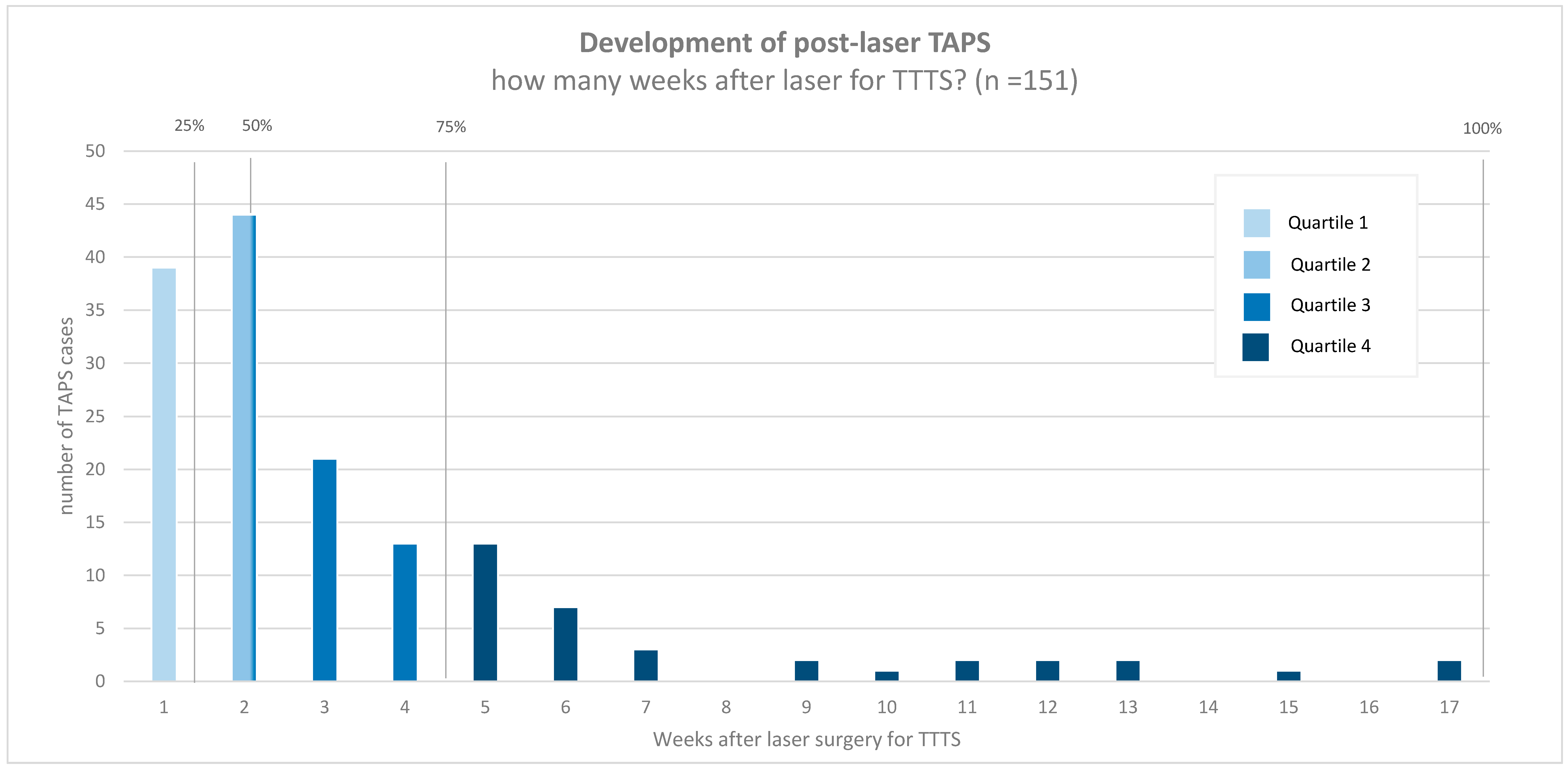
Time between laser and post-laser TAPS (days)14 (7–28)
Persistence of TTTS-TAPS donor-recipient role §73/161 (45)
Laser for TTTS complete (surgeon’s opinion) ¶126/156 (81)
Data are presented as n/N (%) or the median (IQR). † Other types of placental position included: partly anterior and partly posterior (n = 1) and partly fundal (n = 1) lateral left (n = 2) and lateral right (n = 2). ‡ Five missing values. § Three missing values. ¶ Eight missing values. TAPS, twin anemia polycythemia sequence; TTTS, twin-twin transfusion syndrome; Q, Quintero; GA, gestational age.
Table 2. Diagnosis- and management-related characteristics for the post-laser twin anemia polycythemia sequence.
Post-Laser TAPS
(N = 164 Pregnancies, 328 Fetuses)
GA at diagnosis23.1 (20.4–25.9)
TAPS stage at diagnosis
143/151 (28)
269/151 (48)
327/151 (17)
412/151 (8)
Highest TAPS stage
122/151 (15)
262/151(40)
326/151 (16)
422/151 (14)
519/151 (15)
Presence of additional ultrasound markers †
Starry-sky liver (recipient)56/141 (40)
Difference in placental echogenicity35/151 (23)
Antenatal management
Expectant management62/151 (43)
Delivery9/151 (6)
IUT (± PET)44/151 (29)
Laser surgery24/151 (15)
Selective feticide11/151 (7)
Termination of pregnancy1/151 (1)
Female ‡162/308 (53)
Cesarean §206/326 (63)
Data are the median (IQR) or n/N (%). Except for “female” and “cesarean”, all other parameters are presented for antenatally diagnosed post-laser TAPS cases only (N = 151).† In 10 cases, the presence of a starry-sky liver was not assessed. ‡ Twenty missing values. § Two missing values.TAPS, twin anemia polycythemia; GA, gestational age; IUT, intrauterine transfusion; PET, partial exchange transfusion.
Table 3. Characteristics for placentas not treated with laser reintervention for the post-laser twin anemia polycythemia sequence.
Injected Post-Laser
TAPS Placentas
(N = 74)
Total number of anastomoses1 (1–2)
Number of AV anastomoses1 (0–1)
Number of VA anastomoses0 (0–1)
Number of AA anastomoses0 (0–0)
Number of VV anastomoses0 (0–0)
Presence of anastomoses
Presence of AV/VA anastomoses58/74 (80)
Presence of AA anastomoses6/74 (8)
Presence of VV anastomoses5/74 (7)
Type of anastomoses per placenta
No anastomoses10/74 (14)
AV (one direction)40/74 (54)
AVs (both directions)14/74 (19)
AV/VA and AA3/74 (4)
AV/VA and VV2/74 (3)
Only AA2/74 (3)
Only VV2/74 (3)
AV/VA, AA and VV1/74 (1)
All anastomoses diameter < 1 mm62/64 (97)
Data are the median (IQR) or n/N (%). TAPS, twin anemia polycythemia; TTTS, twin-twin transfusion syndrome; AV, arterio-venous; VA, veno-arterial; AA, arterio-arterial; VV, veno-venous.
Table 4. Perinatal outcome in the post-laser twin anemia polycythemia sequence.
Post-Laser TAPS
(N = 164 Pregnancies; 328 Fetuses)
TAPS Donors
(N = 164 Fetuses)
TAPS Recipients
(N = 164 Fetuses) †
p-Value
GA at birth31.7 (28.6–33.7; 19.0–41.3)
Fetal demise †56/327 (17)43/164 (26)13/163 (8)<0.001
Spontaneous33/327 (10)25/164 (15)8/163 (5)<0.001
Intended23/327 (7)18/164 (11)5/163 (3)0.007
Neonatal mortality27/271 (10)18/121 (15)9/150 (6)0.008
Perinatal mortality (overall) †83/327 (25)61/164 (37)22/163 (14)<0.001
Perinatal mortality (spontaneous) †60/327 (18)43/164 (26)17/163 (10)<0.001
Severe neonatal morbidity ‡105/263 (40)51/118 (43)54/145 (37)0.568
Respiratory distress syndrome88/263(34)42/118 (36)46/145 (32)0.945
Patent ductus arteriosus20/263 (8)11/118 (9)9/145 (6)0.278
Necrotizing enterocolitis6/263 (2)4/118 (3)2/145 (1)0.275
Retinopathy of prematurity9/263 (3)6/118 (5)3/145 (2)0.165
Amniotic band syndrome3/263 (1)2/118 (2)1/145 (1)0.451
Severe cerebral injury28/263 (11)13/118 (11)15/145 (10)0.916
Birth weight (g) §1390 ± 5671346 ± 5251533 ± 588<0.001
Severe growth restriction (bw < p3) §62/270 (23)31/121 (26)31/149 (21)0.267
Mild growth restriction (bw < p10) §122/270 (45)60/121 (50)61/149 (41)0.061
Data are presented as the means ± SD, medians (IQR), or n/N (%). † One missing value (1 post-laser TAPS recipient with unknown perinatal outcome). ‡ Nine missing values (same as “†” plus 4 cases with unknown neonatal morbidity info and 4 cases that died shortly after birth). § Two missing values (same as “†” plus one case with missing birth weight). TAPS, twin anemia polycythemia sequence; GA, gestational age; bw, birth weight.

