Article

Decreasing Trends of Secondary Primary Colorectal Cancer among Women with Uterine Cancer: A Population-Based Analysis

1
Division of Gynecologic Oncology, Department of Obstetrics and Gynecology, University of Southern California, Los Angeles, CA 90033, USA
2
Norris Comprehensive Cancer Center, University of Southern California, Los Angeles, CA 90033, USA
3
Department of Obstetrics and Gynecology, Tokai University School of Medicine, Kanagawa 259-1193, Japan
4
Department of Obstetrics and Gynecology, Niigata University School of Medicine, Niigata 951-8510, Japan
5
Division of Medical Oncology, Department of Medicine, New York University, New York, NY 10016, USA
6
Division of Gynecologic Oncology, Department of Obstetrics and Gynecology, Columbia University College of Physicians and Surgeons, New York, NY 10032, USA
*
Author to whom correspondence should be addressed.
J. Clin. Med. 2019, 8(5), 714; https://doi.org/10.3390/jcm8050714
Submission received: 15 April 2019 / Revised: 6 May 2019 / Accepted: 14 May 2019 / Published: 20 May 2019
(This article belongs to the Section Obstetrics & Gynecology)

Abstract

:
The current study examined trends, characteristics, and outcomes of women with uterine cancer who had secondary colorectal cancer. This is a retrospective study utilizing the Surveillance, Epidemiology, and End Results Program between 1973–2013. Among uterine cancer (n = 246,272) and colorectal cancer (n = 421,312) cohorts, women with both diagnoses were identified, and clinico-pathological factors and survival were extracted and analyzed. There were 6862 women with both cancer diagnoses, representing 2.8% of the uterine cancer cohort and 1.6% of the colorectal cancer cohort. Among 123,940 women with uterine cancer survivors, the number with postcedent colorectal cancer decreased from 5.3% to 0.7% between 1981–2008 (relative risk reduction 87.0% p < 0.001). Similarly, of 141,801 women with colorectal cancer survivors, the number with postcedent uterine cancer decreased from 1.7% to 0.5% between 1973–2008 (relative risk reduction 71.6%, p < 0.001). In the uterine cancer cohort, women with antecedent/synchronous colorectal cancer had more high-grade tumors and advanced-stage disease resulting in poorer survival, whereas those who had postcedent colorectal cancer had more low-grade tumors and early-stage disease resulting in superior survival compared to those without secondary colorectal cancer (all, p < 0.05). In conclusion, the development of postcedent colorectal cancer following uterine cancer has decreased in recent years in the United States.

Graphical Abstract

1. Introduction

Colorectal cancer and uterine cancer are the third and fourth most common female malignancies in the United States, respectively [1]. In 2019, nearly 62,000 women are estimated to be diagnosed with uterine cancer and more than 67,000 women are estimated to be diagnosed with colorectal cancer [1]. The incidence of uterine cancer has been increasing over past decades, with obesity and aging as key risk factors for uterine cancer development [2,3]. It has also been established that women with uterine cancer are at increased risk of developing secondary colorectal cancer [4,5,6,7,8,9].
This increased risk of colorectal cancer in women with uterine cancer is likely due to similar epidemiological and genetic risk factors for the two malignancies and the fact that treatments for uterine cancer such as pelvic irradiation may also increase the risk of colorectal cancer [10,11,12]. In addition, approximately 3–5% of all women with uterine cancer have Lynch syndrome, a familial cancer syndrome involving a DNA mismatch repair gene deficiency, which increases the risk of both uterine cancer and colorectal cancer [13,14].
Given recent demographic trends in the U.S. population that may impact the incidence of both uterine and colorectal cancers, namely increasing age and obesity rates as well as increasing utilization of colorectal cancer screening and surveillance in at-risk populations [15,16,17,18], it would be of value to examine population-based statistics of secondary colorectal cancer in women with uterine cancer. Moreover, the survival of women with uterine cancer who develop secondary colorectal cancer has not been examined completely [8]. The objective of the study was to examine the trends, characteristics, and outcomes of women with uterine cancer who had secondary colorectal cancer by utilizing a population-based tumor registry in the United States.

2. Materials and Methods

2.1. Study Population and Eligibility

This retrospective study examined the National Cancer Institute’s Surveillance, Epidemiology, and End Results program. This database, launched in 1973, is the largest population-based tumor registry in the United States, covering approximately 34.6% of the population in the latest version [19]. Patient identification, data entry, and rigorous quality control for this database are performed by registered and trained personnel by the National Cancer Registrars Association and the program [20]. The Institutional Review Board at the University of Southern California exempted this study due to the use of publicly available de-identified data.
The cases between 1973–2013 were obtained from the SEER*Stat 8.3.2 (IMS Inc., Calverton, MD, USA). First, two datasets were generated, one for uterine cancer and one for colorectal cancer, limited to malignancy and female sex. Cases with the same study identification number assigned by the program in each dataset were then identified and linked, and the multiple entry cases were excluded to retain only the primary entry. These cases identified in both uterine and colorectal cancer databases were considered to be secondary primary cancer (SPC) cases as described previously [21,22].

2.2. Clinical Information

From the uterine cancer database, the following variables were abstracted: (i) patient demographics (age, year and month at diagnosis, race, marital status, and registration area), (ii) tumor characteristics (cancer stage, histologic subtype, tumor grade, tumor size, and lymph node status), (iii) treatment types (hysterectomy and radiotherapy), and (iv) survival outcome (cause-specific survival (CSS) and overall survival (OS)).

2.3. Study Definition

The chronologic time sequence was examined between the uterine and colorectal cancer diagnoses, and when the time interval between the two diagnoses was ≤2 months, the cases were considered to be synchronous SPC as previously described [23]. SPC diagnosed >2 months after the index cancer was considered postcedent SPC, whereas SPC diagnosed >2 months prior to the index cancer was considered antecedent SPC.
Recorded cancer stages were based on the American Joint Committee on Cancer surgical-pathological staging classification schema [24]. The International Classification of Diseases for Oncology third edition site/histology validation and the World Health Organization histological classification codes were used to group histologic subtypes as described before [21,22,25]. The grouping and categorization of clinico-pathological factors for this study were based on prior studies [21,22,25]. CSS was defined as the time between uterine cancer diagnosis and death from uterine cancer. OS was defined as the time between uterine cancer diagnosis and death from any cause. Cases without survival events were censored at the last follow-up. Cause of death is linked with state mortality records and the National Death Index [26].

2.4. Study Aim

The primary objective of the study was to examine temporal trends of secondary colorectal cancer in women with uterine cancer. The secondary objective was to examine clinico-pathological characteristics and outcomes of women with uterine cancer who developed secondary colorectal cancer. The timing of colorectal cancer diagnosis relative to uterine cancer diagnosis (antecedent, synchronous, and postcedent) among those who had two cancer diagnoses was examined and compared to those without secondary colorectal cancer.

2.5. Statistical Consideration
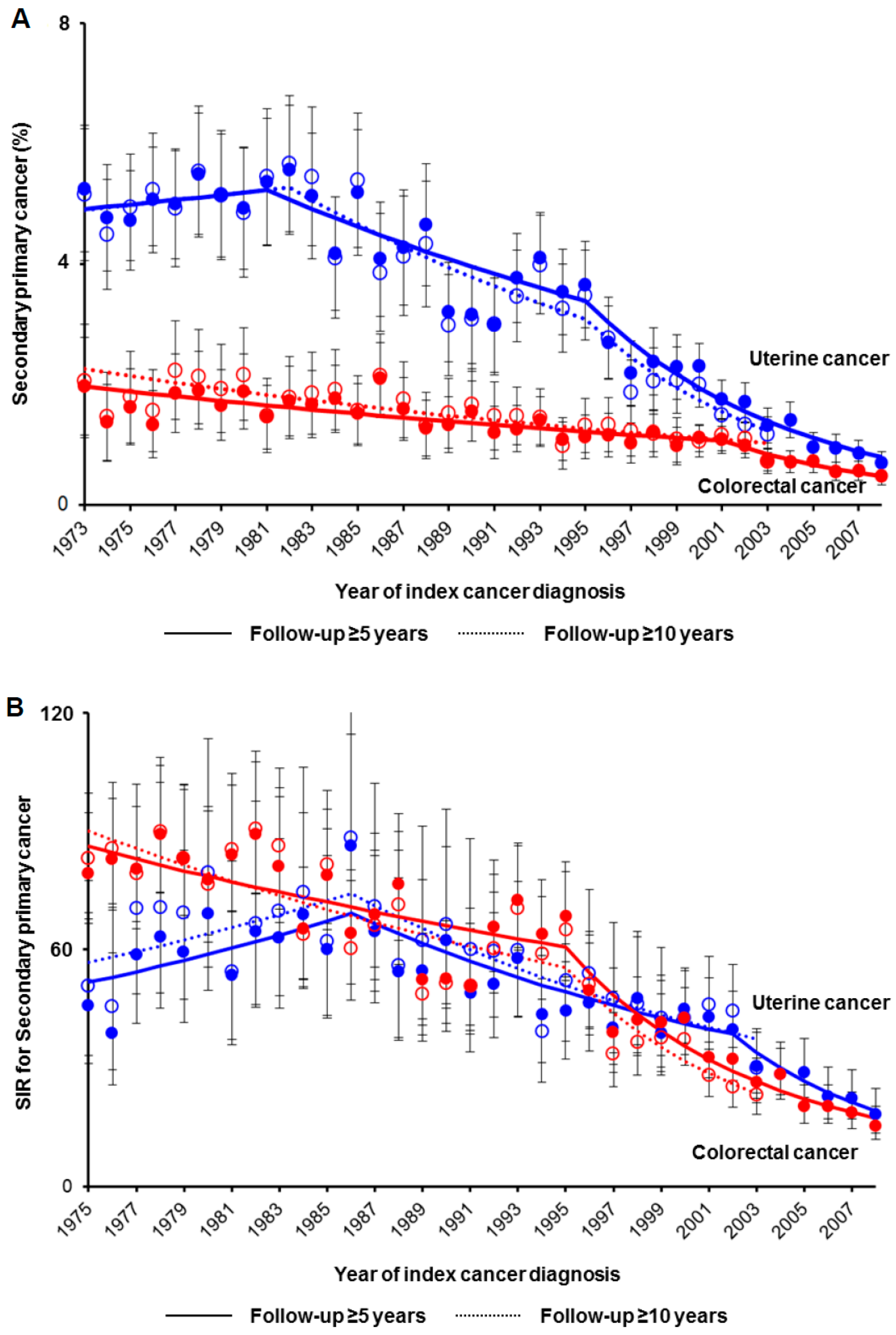
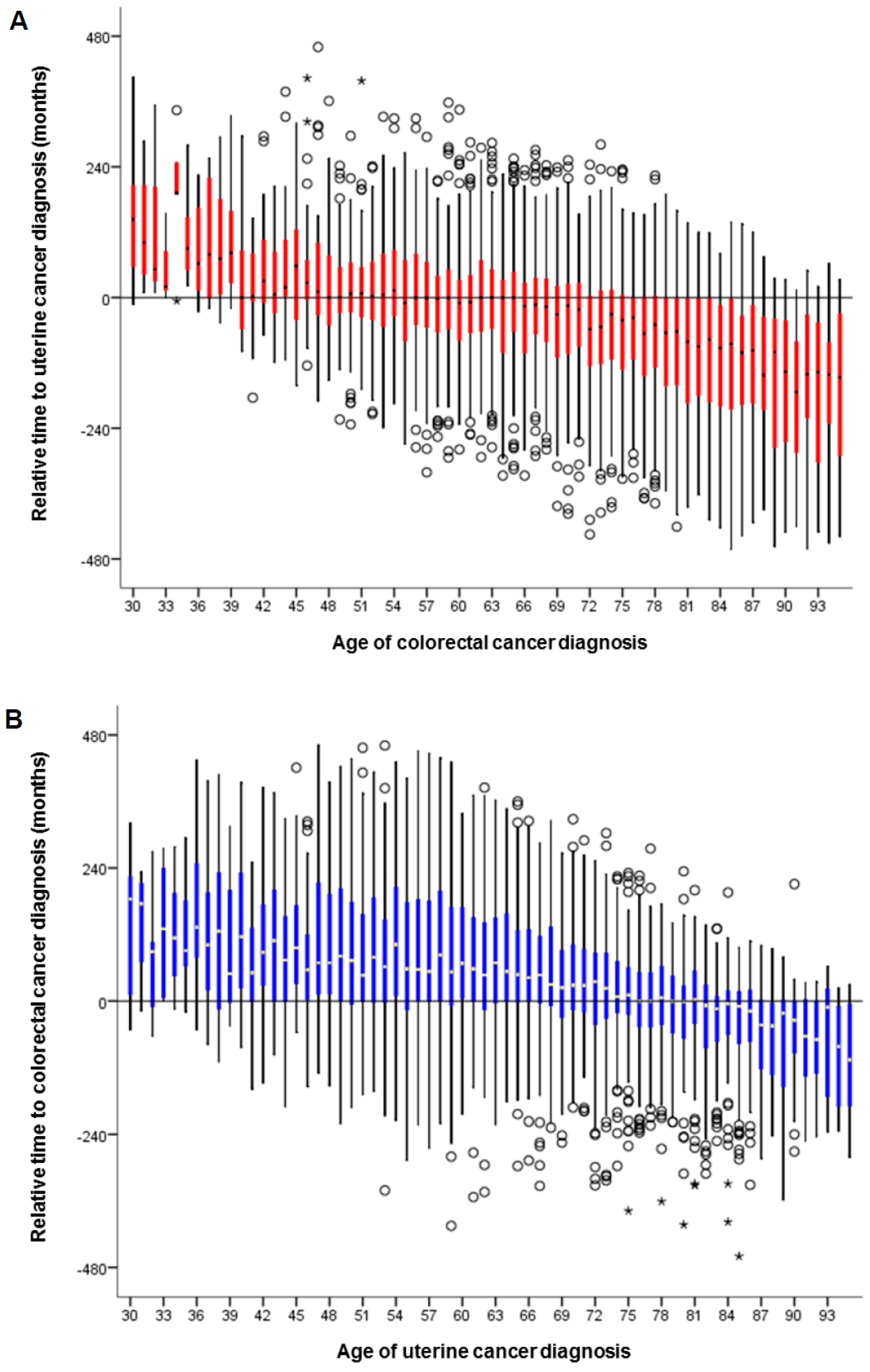
Continuous variables were assessed for normality with the Kolmogorov–Smirnov test, and differences were assessed by the one-way ANOVA or Kruskal–Wallis H test as appropriate. Differences in ordinal or categorical variables were assessed by the chi-square test. Cox proportional hazard regression models were used to estimate hazard ratio (HR) with 95% confidence interval (CI) for multivariable analysis [27]. Patient demographics, tumor characteristics, and treatment types were entered in the final model.
Joinpoint Trend Software (version 4.4.0.0, National Cancer Institute, Bethesda, MD, USA) was used to identify the temporal changes in the trend analysis [28]. Time increments were grouped by every one calendar year to provide percent frequencies with CIs or every one year of age to provide mean values with standard errors of collected variables. The linear segmented regression test was used for the analysis, and log-transformation was performed to determine annual percent change of the slope with 95% CI [29].
For a sensitivity analysis, incidence of postcedent colorectal cancer was examined among uterine cancer women without antecedent or synchronous colorectal cancer who did not die of uterine cancer and had a follow-up of ≥5 (and ≥10) years, termed as cancer survivors in this study. Among the survivors, trends in standardized incidence ratio (SIR) for uterine cancer and colorectal cancer were also examined based on observed and expected events per standardized population [30]. This is based on the rationale that the diagnosis of postcedent SPC is dependent on follow-up time, and a short follow-up interval may result in lead-time bias for SPC [31]. Trends in SPC-specific mortality were examined among survivors. The study population with uterine cancer was also limited to only endometrial tumors, as the majority of uterine cancer is epithelial, particularly endometrioid-type. Moreover, temporal trends of secondary uterine cancer were examined in the colorectal cancer cohort.
All analyses were based on two-tailed hypotheses, and a p < 0.05 was considered statistically significant. Statistical Package for Social Sciences (SPSS, version 24.0, IBM Corp, Armonk, NY, USA) was used for the statistical analysis. The STROBE guidelines were outlined for performance of this observational study [32].

3. Results

The study schema is shown in Figure S1. There were 6862 women with both cancer diagnoses, representing 2.8% (95% CI 2.7–2.9) of the uterine cancer cohort (n = 246,272) and 1.6% (95% CI 1.6–1.7) of the colorectal cohort (n = 421,312). Among 6811 women with uterine cancer in whom time to secondary colorectal cancer was available, postcedent colorectal cancer after uterine cancer was most common (n = 4093 (60.1%), median time interval 7.6 years (interquartile range (IQR), 3.4–14.1)), followed by antecedent colorectal cancer prior to uterine cancer (n = 2103 (30.9%), median time interval 4.9 years (IQR, 2.2–9.5)) and synchronous uterine and colorectal cancers (n = 615, 9.0%).
In the uterine cancer cohort, women with antecedent colorectal cancer had similar characteristics to those with synchronous colorectal cancer and were more likely to be older than those without secondary colorectal cancer (p < 0.001; Table 1). Women with postcedent colorectal cancer were similar to those without secondary colorectal cancer and were more likely to be white, married and to have received radiotherapy (all, p < 0.001). Uterine tumors in the postcedent colorectal cancer group were more likely to be endometrioid type, early-stage, and low-grade but less likely to have nodal involvement, whereas tumors in the antecedent and synchronous colorectal cancer groups were more likely to have high-grade histology and be advanced-stage compared to tumors without secondary colorectal cancer (all, p < 0.01).
Age at uterine cancer diagnosis was similar between those with and without secondary colorectal cancer until the mid-1980s in the uterine cancer cohort (mean, 59.2 versus 60.8 in 1973; p = 0.15; Figure 1A); subsequently, women with secondary colorectal cancer were diagnosed at older ages compared to those without secondary colorectal cancer (66.9 versus 62.5 in 2013; p < 0.001). In the colorectal cancer cohort (Figure 1B), women who had secondary uterine cancer were younger compared to those without secondary uterine cancer until the early-1990s (62.8 versus 68.8 in 1973; p = 0.005); however, afterwards, women who had secondary uterine cancer were older compared to those without secondary uterine cancer (69.5 versus 67.9 in 2013; p = 0.057).
The number of women with uterine cancer who had secondary colorectal cancer decreased from 4.5% to 1.2% between 1992–2013 (relative risk reduction (RRR) 74.0%; p < 0.001; Figure 2A). When stratified by the chronology of colorectal cancer diagnoses, the number of women with uterine cancer with synchronous colorectal cancer decreased from 0.5% to 0.2% between 1979–2013 (RRR 56.2%, p < 0.001). Contrastingly, there was an increase in the number of women with uterine cancer with antecedent colorectal cancer from 0.6% to 0.9% between 2001–2013 (45% relative risk increase (RRI), p = 0.014). The number of women with colorectal cancer who had secondary uterine cancer increased from 0.8% to 2.0% between 1973–1986 (1.5-fold RRI, p < 0.001; Figure 2B). When stratified by the chronology of diagnoses, the number of colorectal cancer women who had antecedent uterine cancer increased from 0.8% to 1.3% between 2001–2013 (66% RRI, p < 0.001).
Trends of postcedent colorectal cancer were examined among 123,940 uterine cancer who survived ≥5 years (median follow-up 11.7 years, IQR 8.0–17.9; Figure S2). The number of uterine cancer survivors who developed postcedent colorectal cancer decreased from 5.3% to 0.7% between 1981–2008 (RRR 87.0% p < 0.001; Figure 3A). Similar results were observed among the 75,624 women with uterine cancer who survived ≥10 years (median follow-up 16.0 years, IQR 12.4–17.1), and RRR was 79.2% between 1982–2003 (p < 0.001). When standardized by the colorectal cancer incidence in the general population (Figure 3B), postcedent colorectal cancer decreased among ≥5-year uterine cancer survivors between 1975–2008 (RRR 80.5%, p < 0.005) and among ≥10-year uterine cancer survivors between 1975–2003 (RRR 72.0%, p < 0.001). The decreased incidence of postcedent colorectal cancer was particularly observed in the group diagnosed with colorectal cancer ≥5 years after uterine cancer diagnosis (Figure S3).
Among 141,801 women with colorectal cancer who survived ≥5 years (median follow-up 10.3 years, IQR 7.3–14.3; Figure S2), the number of colorectal cancer survivors who developed postcedent uterine cancer decreased from 1.7% to 0.5% between 1973–2008 (RRR 71.6%, p < 0.001; Figure 3A). Similarly, among 73,582 women who survived ≥10 years (median follow-up 14.1 years, IQR 11.8–19.1), the RRR of postcedent uterine cancer was 58.2% between 1973–2003 (p < 0.001). When standardized by the uterine cancer incidence in the general population (Figure 3B), postcedent uterine cancer decreased among ≥5-year colorectal cancer survivors between 1986–2008 (RRR 78.7%, p < 0.001) and among ≥10-year uterine cancer survivors between 1986–2003 (RRR 66.2%, p < 0.001). The decreased incidence of postcedent uterine cancer was also observed particularly in the group diagnosed with uterine cancer ≥5 years after colorectal cancer diagnosis (Figure S3).
The age-specific trends of secondary colorectal cancer among uterine cancer women were examined, stratified by the chronology of the colorectal cancer diagnoses (Figure S4A). After age 60, the number of women with uterine cancer with secondary colorectal cancer increased (p < 0.01). The peak age for antecedent colorectal cancer was 88 compared to 75 for postcedent colorectal cancer (both, p < 0.001). A trend of increasing synchronous colorectal cancer was observed after age 54 (p < 0.001). Age-specific trends of secondary uterine cancer among women with colorectal cancer were also examined (Figure S4B). The peak age of secondary uterine cancer diagnosis overall was 66 (p < 0.05), contrasting to the peak age of diagnosis for antecedent uterine cancer only, which was 79 years (p < 0.005). A trend of decreasing synchronous and postcedent uterine cancer diagnosis was observed after the mid-60s (66 and 65, respectively; p < 0.001).
The relative time interval between the two cancer diagnoses was examined among the 6811 women with both diagnoses. Uterine cancer diagnosis was more likely to antecede colorectal cancer diagnosis after age 40 (Figure 4A), whereas colorectal cancer was more likely to antecede uterine cancer after age 76 (Figure 4B) (both, p < 0.001). The median absolute time interval between the two diagnoses was 5.8 years (IQR, 2–11.5). The absolute time interval between the two diagnoses was shorter when uterine cancer was diagnosed at an older age, whereas the time interval was longer when colorectal cancer was diagnosed at older age (both, p < 0.001; Figure S5).
In the uterine cancer cohort, the median follow-up of censored cases was 6.6 years, and there were 110,219 all-cause deaths including 44,811 uterine cancer deaths during the follow-up. Irrespective of the chronology of secondary colorectal cancer diagnosis, uterine cancer women who had secondary colorectal cancer had superior CSS compared to those who did not have secondary colorectal cancer on multivariable analysis (adjusted-HR: 0.82 for antecedent, 0.77 for synchronous, and 0.26 for postcedent, all p < 0.01; Table 2).
In contrast, women who had antecedent or synchronous colorectal cancer had poorer OS (adjusted-HR: 1.26 for antecedent, and 1.55 for synchronous, both p < 0.001) whereas those who had postcedent colorectal cancer had superior OS (adjusted-HR: 0.91, p < 0.001) compared to the non-SPC group. The number of uterine cancer survivors who died of colorectal cancer decreased from 2.2% to <0.1% between 1982–2008, and the number of colorectal cancer survivors who died of uterine cancer decreased from 0.3% to <0.1% between 1997–2006 (both, p < 0.001; Figure S6).
Histology-specific analyses were performed in 227,275 women with endometrial cancer and in 177,897 women with endometrioid-type endometrial cancer. In both cohorts, patient characteristics were similar to the entire study cohort, and postcedent colorectal cancer also decreased among women with long-term follow-up during the study period (Tables S1 and S2 and Figures S7 and S8). Like the whole cohort, CSS was also superior in women with secondary colorectal cancer compared to those without secondary colorectal cancer, whereas OS was inferior in women with antecedent or synchronous colorectal cancer in the two subgroups (Table 2).

4. Discussion

This study found that secondary colorectal cancer among uterine cancer is not rare, and ~3% of women with uterine cancer developed secondary colorectal cancer. Previously, studies have shown an increased risk of secondary colorectal cancer in women with uterine cancer; however these studies did not evaluate trends in timing or chronology of the two cancer diagnoses [4,5,6,7,8,9]. Our study is unique in showing that there is a recent decrease in the number of women with uterine cancer who had secondary colorectal cancer, which is attributable to a decrease in postcedent colorectal cancer following uterine cancer.
Various causalities may explain the recent decrease in postcedent colorectal cancer after uterine cancer diagnosis. First, successful implementation of colorectal cancer screening strategies in the United States may be identifying more pre-cancerous lesions and reducing the incidence of subsequent colorectal cancer [17,18], and this may also apply to women with uterine cancer. In 2015, nearly two thirds of U.S. adults aged 50–75 received colorectal cancer screening, and the population-based incidence of colorectal cancer has decreased from 57 to 37 per 100,000 between 1992–2015 (RRR 35.1%) [33,34,35]. Because diagnosis of uterine cancer is likely to be antecedent to colorectal cancer among those eventually diagnosed with both malignancies, as shown both in this study and others [4], age-based colorectal cancer screening may be warranted in those with uterine cancer even without Lynch syndrome.
Second, increasing utilization of genetic testing for women with both uterine and colorectal cancers may be responsible for decreasing rates of postcedent uterine cancer and colorectal cancer cases. Universal Lynch syndrome screening has been suggested for women with both colorectal and uterine cancers, given high prevalence in this unique population. A recent nationwide survey of genetic counselors showed significant increases in the utilization of routine Lynch syndrome screening for newly diagnosed colorectal and uterine cancer patients [36]. In women with Lynch syndrome, surveillance strategies such as frequent colonoscopies to identify pre-cancerous lesions or possible chemoprevention with aspirin may also have contributed to the decrease in postcedent colorectal cancer [37]. It would be of interest to examine whether the decreasing trends in postcedent uterine cancer after colorectal cancer in women with Lynch syndrome may be due to increasing utilization of genetic testing and recommendations for risk-reducing hysterectomy [14,38].
Third, changes in treatment patterns for uterine cancer may have led to the decrease in postcedent colorectal cancer after uterine cancer. There has been a decrease in the utilization of whole pelvic radiotherapy with a concomitant increase in vaginal brachytherapy in surgically treated women with uterine cancer [39]. As pelvic irradiation may increase the risk of colorectal cancer [11], the decrease in whole pelvic radiotherapy use may have indirectly resulted in decreasing postcedent colorectal cancer incidence after uterine cancer.
Another salient finding of this study is that there has been a recent increase in antecedent SPC in both uterine and colorectal cancers. Parallel to this trend, women in the SPC group became older, likely reflecting our aging society (~15% of the North American population was ≥65 years in 2015), resulting in more occasions to develop another malignancy during follow-up [15].
Our study demonstrated that patient demographics, tumor characteristic, and survival of women with uterine cancer are largely different based on the chronology of secondary colorectal cancer, potentially implying different disease characteristics or patient factors in antecedent and synchronous (more aggressive) versus postcedent (less aggressive) SPCs. Women with postcedent colorectal cancer were younger, which is likely responsible for the superior OS in this group compared to other types of SPC. Additionally, whether Lynch syndrome may be associated with postcedent colorectal cancer is of interest and deserves further exploration, especially given that Lynch syndrome-related uterine cancer is more likely to be diagnosed at a young age and have favorable histology [14].
Limitations of this study include that this is a retrospective study and there may be missing confounders for analysis. For instance, no information was available for patient’s geographic relocation between the data-covered and uncovered areas, thus some cases of SPC may not be captured by the database, resulting in misclassification. Information for genetic testing is also not available in the datasets, and we do not know whether mutations of genes predispositions co-occur in both cancers or are mutually exclusive and whether such mutations have any contribution towards chronology of SPC.
Another limitation of our study may be inadequate duration of follow-up to capture SPC, which may have contributed to the decreased numbers of postcedent SPC diagnoses in more recent years. A recent large-scale analysis of nearly three million malignancies found that tumor registries with shorter operating times (<10 years) are less likely to capture SPC compared to registries spanning greater lengths of time [31]. The current database includes over 40 years of operation, and we further examined patients who had long-term follow-up for analysis (Figure S2). Moreover, we observed similar decreasing trends of postcedent SPC in the two cohorts when examined with a one-year latency exclusion period in a post-hoc analysis (data not shown) [40]. Thus, both the use of this database, which has been previously validated and spans several decades, and the consistency of our results in long-term cancer survivors, support our findings of a recent decrease in postcedent colorectal cancer in women with uterine cancer as well as decreasing incidence of postcedent uterine cancer in women with colorectal cancer.

5. Conclusions

In summary, our data likely reflect demographic changes both in the U.S. population as well as in women with uterine cancer and colorectal cancer. In particular, the incidence of secondary colorectal cancer has decreased among women with uterine cancer, especially colorectal cancer diagnosed after uterine cancer. Similar trends of decreasing postcedent uterine cancer among colorectal cancer women were also observed. These results are encouraging for care providers and patients, and further study to validate these findings in a different population is warranted.

Supplementary Materials

The following are available online at https://www.mdpi.com/2077-0383/8/5/714/s1: Figure S1: CONSORT diagram for study schema, Figure S2: Median follow-up time among cancer survivors, Figure S3: Trends of postcedent secondary primary cancer among cancer survivors per year stratified chronology, Figure S4: Trends in secondary primary cancer per age, Figure S5: Absolute time interval between the two cancer diagnoses per age at the index cancer diagnosis, Figure S6: Secondary primary cancer-specific survival among survivors, Figure S7: Secondary colorectal cancer among endometrial cancer, Figure S8: Secondary colorectal cancer among endometrioid type endometrial cancer, Table S1: Patient demographics (endometrial cancer cohort), Table S2: Patient demographics for endometrioid type endometrial cancer based on timing of secondary colorectal cancer, Table S3: Temporal trends of age at the index cancer diagnosis based on presence of secondary primary cancer (for Figure 1A,B), Table S4: Temporal trends for secondary primary cancer per year (for Figure 2A,B), Table S5: Temporal trends for secondary primary cancer in long-term follow-up cases (for Figure 3A), Table S6: Temporal trends of SIR for secondary primary cancer (for Figure 3B), Table S7: Temporal trends for secondary primary cancer per SPC timing (for Supplemental Figure S3), Table S8: Temporal trends for secondary primary cancer per age (for Figure 4A,B), Table S9: Temporal trends for secondary primary cancer-specific survival among survivors (for Supplemental Figure S6), Table S10: Temporal trends for secondary colorectal cancer among endometrial cancer (for Supplemental Figure S7A), Table S11: Temporal trends for postcedent colorectal cancer in long-term follow-up cases with endometrial cancer (for Supplemental Figure S7B), Table S12: Temporal trends for secondary colorectal cancer among endometrioid type endometrial cancer (for Supplemental Figure S8A), Table S13: Temporal trends for postcedent colorectal cancer in long-term follow-up cases with endometrioid type endometrial cancer (for Supplemental Figure S8B).

Author Contributions

Conceptualization: K.M.; data curation: H.M.; formal analysis: K.M.; funding acquisition: K.M. and L.D.R.; investigation: all authors; methodology: K.M.; project administration: K.M.; resources: H.M.; software: H.M. and K.M.; supervision: K.M. and J.D.W.; validation: K.M.; visualization: K.M.; writing—original draft: K.M.; writing—review & editing: all authors.

Funding

Ensign Endowment for Gynecologic Cancer Research (K.M.).

Conflicts of Interest

Consultant, Clovis Oncology and Tesaro (J.D.W.); consultant, Tempus Lab (L.D.R.); honorarium, Chugai, book editorial, Springer, and meeting attendance expense, VBL Therapeutics (K.M.); none for others.

References

  1. Siegel, R.L.; Miller, K.D.; Jemal, A. Cancer statistics, 2019. CA Cancer J. Clin. 2019, 69, 7–34. [Google Scholar] [CrossRef]
  2. Wright, J.D.; Barrena Medel, N.I.; Sehouli, J.; Fujiwara, K.; Herzog, T.J. Contemporary management of endometrial cancer. Lancet 2012, 379, 1352–1360. [Google Scholar] [CrossRef]
  3. Morice, P.; Leary, A.; Creutzberg, C.; Abu-Rustum, N.; Darai, E. Endometrial cancer. Lancet 2015, 387, 1094–1108. [Google Scholar] [CrossRef]
  4. Lu, K.H.; Dinh, M.; Kohlmann, W.; Watson, P.; Green, J.; Syngal, S.; Bandipalliam, P.; Chen, L.M.; Allen, B.; Conrad, P.; et al. Gynecologic cancer as a “sentinel cancer” for women with hereditary nonpolyposis colorectal cancer syndrome. Obstet. Gynecol. 2005, 105, 569–574. [Google Scholar] [CrossRef]
  5. Weinberg, D.S.; Newschaffer, C.J.; Topham, A. Risk for colorectal cancer after gynecologic cancer. Ann. Intern. Med. 1999, 131, 189–193. [Google Scholar] [CrossRef]
  6. Brown, A.P.; Neeley, E.S.; Werner, T.; Soisson, A.P.; Burt, R.W.; Gaffney, D.K. A population-based study of subsequent primary malignancies after endometrial cancer: Genetic, environmental, and treatment-related associations. Int. J. Radiat. Oncol. Biol. Phys. 2010, 78, 127–135. [Google Scholar] [CrossRef] [PubMed]
  7. Phipps, A.I.; Chan, A.T.; Ogino, S. Anatomic subsite of primary colorectal cancer and subsequent risk and distribution of second cancers. Cancer 2013, 119, 3140–3147. [Google Scholar] [CrossRef] [PubMed]
  8. Lim, M.C.; Won, Y.J.; Lim, J.; Seo, S.S.; Kang, S.; Yoo, C.W.; Kim, J.Y.; Oh, J.H.; Bristow, R.E.; Park, S.Y. Second primary colorectal cancer among endometrial cancer survivor: Shared etiology and treatment sequelae. J. Cancer Res. Clin. Oncol. 2018, 144, 845–854. [Google Scholar] [CrossRef] [PubMed]
  9. Singh, H.; Nugent, Z.; Demers, A.; Czaykowski, P.M.; Mahmud, S.M. Risk of colorectal cancer after diagnosis of endometrial cancer: A population-based study. J. Clin. Oncol. 2013, 31, 2010–2015. [Google Scholar] [CrossRef] [PubMed]
  10. Brenner, H.; Kloor, M.; Pox, C.P. Colorectal cancer. Lancet 2014, 383, 1490–1502. [Google Scholar] [CrossRef]
  11. Onsrud, M.; Cvancarova, M.; Hellebust, T.P.; Trope, C.G.; Kristensen, G.B.; Lindemann, K. Long-term outcomes after pelvic radiation for early-stage endometrial cancer. J. Clin. Oncol. 2013, 31, 3951–3956. [Google Scholar] [CrossRef]
  12. Rombouts, A.J.M.; Hugen, N.; van Beek, J.J.P.; Poortmans, P.M.P.; de Wilt, J.H.W.; Nagtegaal, I.D. Does pelvic radiation increase rectal cancer incidence?—A systematic review and meta-analysis. Cancer Treat Rev. 2018, 68, 136–144. [Google Scholar] [CrossRef]
  13. Moreira, L.; Balaguer, F.; Lindor, N.; de la Chapelle, A.; Hampel, H.; Aaltonen, L.A.; Hopper, J.L.; Le Marchand, L.; Gallinger, S.; Newcomb, P.A.; et al. Identification of Lynch syndrome among patients with colorectal cancer. JAMA 2012, 308, 1555–1565. [Google Scholar] [CrossRef]
  14. ACOG Practice Bulletin No. 147: Lynch syndrome. Obstet. Gynecol. 2014, 124, 1042–1054.
  15. An Aging World: 2015. International Population Reports. United States Census Bureau. Available online: https://www.census.gov/content/dam/Census/library/publications/2016/demo/p95-16-1.pdf (accessed on 14 April 2019).
  16. Health, United States, 2016. Centers for Disease Control and Prevention. Available online: https://www.cdc.gov/nchs/data/hus/hus16.pdf#053 (accessed on 14 April 2019).
  17. Colorectal Cancer Screening, Incidence, and Mortality—United States, 2002–2010. Centers for Disease Control and Prevention. Available online: https://www.cdc.gov/mmwr/preview/mmwrhtml/mm6026a4.htm (accessed on 14 April 2019).
  18. NIH State-of-the-Science Conference: Enhancing Use and Quality of Colorectal Cancer Screening. NIH Consensus Development Program Office of Disease Prevention. Available online: https://consensus.nih.gov/2010/colorectalstatement.htm (accessed on 14 April 2019).
  19. The Surveillance, Epidemiology, and End Results (SEER) Program of the National Cancer Institute. Available online: https://seer.cancer.gov/ (accessed on 15 April 2019).
  20. National Cancer Registrars Association. Available online: http://www.ncra-usa.org/i4a/pages/index.cfm?pageid=1%3e (accessed on 15 April 2019).
  21. Matsuo, K.; Machida, H.; Blake, E.A.; Holman, L.L.; Rimel, B.J.; Roman, L.D.; Wright, J.D. Trends and outcomes of women with synchronous endometrial and ovarian cancer. Oncotarget 2018, 9, 28757–28771. [Google Scholar] [CrossRef]
  22. Matsuo, K.; Machida, H.; Stone, R.L.; Soliman, P.T.; Thaker, P.H.; Roman, L.D.; Wright, J.D. Risk of Subsequent Ovarian Cancer After Ovarian Conservation in Young Women with Stage I Endometrioid Endometrial Cancer. Obstet. Gynecol. 2017, 130, 403–410. [Google Scholar] [CrossRef]
  23. AIRTUM Working Group. Italian cancer figures, report 2013: Multiple tumours. Epidemiol. Prev. 2013, 37, 1–152. [Google Scholar]
  24. Edge, S.B.; Byrd, D.R.; Compton, C.C.; Fritz, A.G.; Greene, F.L.; Trotti, A. AJCC Cancer Staging Manual, 7th ed.; Springer: New York, NY, USA, 2010. [Google Scholar]
  25. Matsuo, K.; Machida, H.; Shoupe, D.; Melamed, A.; Muderspach, L.I.; Roman, L.D.; Wright, J.D. Ovarian Conservation and Overall Survival in Young Women with Early-Stage Low-Grade Endometrial Cancer. Obstet. Gynecol. 2016, 128, 761–770. [Google Scholar] [CrossRef]
  26. National Death Index. Available online: https://www.cdc.gov/nchs/ndi/ (accessed on 14 April 2019).
  27. Cox, D.R. Regression Models and Life-Tables. J. R. Stat. Soc. Series B Stat. Methodol. 1972, 34, 187–220. [Google Scholar] [CrossRef]
  28. National Cancer Institute Joinpoint Trend Analysis Software. Available online: http://surveillance.cancer.gov/joinpoint (accessed on 14 April 2019).
  29. Kim, H.J.; Fay, M.P.; Feuer, E.J.; Midthune, D.N. Permutation tests for joinpoint regression with applications to cancer rates. Stat. Med. 2000, 19, 335–351. [Google Scholar] [CrossRef]
  30. Cancer stat facts. National Cancer Institute Surveillance Epidemiology End Result Program. Available online: https://seer.cancer.gov/statfacts/ (accessed on 14 April 2019).
  31. Rosso, S.; De Angelis, R.; Ciccolallo, L.; Carrani, E.; Soerjomataram, I.; Grande, E.; Zigon, G.; Brenner, H. Multiple tumours in survival estimates. Eur. J. Cancer 2009, 45, 1080–1094. [Google Scholar] [CrossRef]
  32. Von Elm, E.; Altman, D.G.; Egger, M.; Pocock, S.J.; Gotzsche, P.C.; Vandenbroucke, J.P. Strengthening the Reporting of Observational Studies in Epidemiology (STROBE) statement: Guidelines for reporting observational studies. BMJ 2007, 335, 806–808. [Google Scholar] [CrossRef]
  33. Colorectal cancer screening. National Cncer Institute Cancer Trends Progress Reports. Available online: https://progressreport.cancer.gov/detection/colorectal_cancer (accessed on 14 April 2019).
  34. Bibbins-Domingo, K.; Grossman, D.C.; Curry, S.J.; Davidson, K.W.; Epling, J.W., Jr.; Garcia, F.A.R.; Gillman, M.W.; Harper, D.M.; Kemper, A.R.; Krist, A.H.; et al. Screening for Colorectal Cancer: US Preventive Services Task Force Recommendation Statement. JAMA 2016, 315, 2564–2575. [Google Scholar]
  35. Cancer stat facts: Colorectal cancer. National Cancer Institute Surveillance, Epidemiology, and End Results Program. Available online: https://seer.cancer.gov/statfacts/html/colorect.html (accessed on 14 April 2019).
  36. Cohen, S.A. Current Lynch syndrome tumor screening practices: A survey of genetic counselors. J. Genet. Couns. 2013, 23, 38–47. [Google Scholar] [CrossRef]
  37. Lindor, N.M.; Petersen, G.M.; Hadley, D.W.; Kinney, A.Y.; Miesfeldt, S.; Lu, K.H.; Lynch, P.; Burke, W.; Press, N. Recommendations for the care of individuals with an inherited predisposition to Lynch syndrome: A systematic review. JAMA 2006, 296, 1507–1517. [Google Scholar] [CrossRef]
  38. Yurgelun, M.B.; Mercado, R.; Rosenblatt, M.; Dandapani, M.; Kohlmann, W.; Conrad, P.; Blanco, A.; Shannon, K.M.; Chung, D.C.; Terdiman, J.; et al. Impact of genetic testing on endometrial cancer risk-reducing practices in women at risk for Lynch syndrome. Gynecol. Oncol. 2012, 127, 544–551. [Google Scholar] [CrossRef] [Green Version]
  39. Matsuo, K.; Machida, H.; Ragab, O.M.; Takiuchi, T.; Pham, H.Q.; Roman, L.D. Extent of pelvic lymphadenectomy and use of adjuvant vaginal brachytherapy for early-stage endometrial cancer. Gynecol. Oncol. 2017, 144, 515–523. [Google Scholar] [CrossRef]
  40. Curtis, R.E.; Freedman, D.M.; Ron, E.; Ries, L.A.G.; Hacker, D.G.; Edwards, B.K.; Tucker, M.A.; Fraumeni, J.F. New Malignancies among Cancer Survivors: SEER Cancer Registries, 1973–2000; National Cancer Institute (NCI): Bethesda, MD, USA, 2006; pp. 9–14.
Figure 1. Trends in age at diagnosis. Age at diagnosis of the index cancer is shown for (A) uterine cancer cohort and (B) colorectal cancer cohort. Dots represent actual mean values. Bars represent standard errors. Colored lines represent modeled lines. Raw results of temporal trends are shown in Supplemental Table S3.
Jcm 08 00714 g001
Figure 2. Trends in secondary primary cancer per year. Temporal trends are shown for (A) secondary colorectal cancer among uterine cancer and (B) secondary uterine cancer among colorectal cancer based on the timing of secondary primary cancer. Dots represent actual observed values. Bars represent standard confidence intervals. Colored lines represent modeled lines. Raw results of temporal trends are shown in Supplemental Table S4.
Jcm 08 00714 g002
Figure 3. Trends of postcedent secondary primary cancer among cancer survivors per year. Among women who were alive at a follow-up ≥5 years (solid lines) or ≥10 years (dashed lines), (A) temporal trends of the incidence of postcedent secondary cancer and (B) standardized incidence ratio were examined. Dots represent actual observed values. Bars represent standard confidence intervals. Colored lines represent modeled lines. Blue lines represent the uterine cancer cohort, and red lines represent the colorectal cancer cohort, respectively. Raw results of temporal trends are shown in Supplemental Tables S5 and S6. Median follow-up times across the study period are shown in Supplemental Figure S2. Abbreviation: SPC, secondary primary cancer; and SIR, standardized incidence ratio.
Jcm 08 00714 g003
Figure 4. Relative time to secondary primary cancer per age. Among 6811 women who had the two cancer diagnoses, relative time to secondary primary cancer is displayed per age at the index cancer diagnosis. (A) Relative time to uterine cancer diagnosis is shown per age of colorectal cancer diagnosis. The first age that the median relative time interval became zero was 40 years. (B) Relative time to colorectal cancer diagnosis is shown per age of uterine cancer diagnosis. The first age that the median relative time interval became zero was 76 years.
Jcm 08 00714 g004
Table 1. Patient demographics for uterine cancer based on timing of secondary colorectal cancer.
CharacteristicNo CRCAntecedent-CRCSynchronous-CRCPostcedent-CRCp-Value
Number n = 239,410n = 2103n = 615n = 4093
Age62.9 (± 12.6)70.5 (± 12.4)68.5 (± 12.4)63.1 (± 11.5)<0.001
Year <0.001
Before 198020,564 (8.6%)59 (2.8%)51 (8.3%)883 (21.6%)
1980–198927,218 (11.4%)238 (11.3%)80 (13.0%)978 (23.9%)
1990–199939,864 (16.7%)448 (21.3%)121 (19.7%)987 (24.1%)
2000–2009100,853 (42.1%)814 (38.7%)252 (41.0%)1125 (27.5%)
2010 or later50,911 (21.3%)544 (25.9%)111 (18.0%)120 (2.9%)
Race/ethnicity <0.001
White184,497 (77.1%)1661 (79.0%)499 (81.1%)3445 (84.2%)
Black18,661 (7.8%)153 (7.3%)58 (9.4%)238 (5.8%)
Hispanic18,993 (7.9%)137 (6.5%)32 (5.2%)170 (4.2%)
Asian12,611 (5.3%)121 (5.8%)21 (3.4%)189 (4.6%)
Others *4648 (1.9%)31 (1.5%)5 (0.8%)51 (1.2%)
Area <0.001
West123,311 (51.5%)1021 (48.5%)260 (42.3%)1966 (48.0%)
Central55,898 (23.3%)584 (27.8%)168 (27.3%)1195 (29.2%)
East60,201 (25.1%)498 (23.7%)187 (30.4%)932 (22.8%)
Marital status <0.001
Single35,108 (14.7%)205 (9.7%)87 (14.1%)475 (11.6%)
Married123,912 (51.8%)945 (44.9%)293 (47.6%)2290 (55.9%)
Others *80,390 (33.6%)953 (45.3%)235 (38.2%)1328 (32.4%)
Histology types <0.001
Endometrioid172,784 (72.2%)1416 (67.3%)416 (67.6%)3250 (79.4%)
Serous13,975 (5.8%)173 (8.2%)55 (8.9%)221 (5.4%)
Clear cell2938 (1.2%)45 (2.1%)13 (2.1%)41 (1.0%)
Carcinosarcoma10,664 (4.5%)159 (7.6%)28 (4.6%)84 (2.1%)
Sarcoma10,376 (4.3%)44 (2.1%)20 (3.3%)69 (1.7%)
Mixed7318 (3.1%)95 (4.5%)17 (2.8%)45 (1.1%)
Others21,357 (8.9%)171 (8.1%)66 (10.7%)383 (9.4%)
Stage <0.001
I150,210 (62.7%)1263 (60.1%)326 (53.0%)2525 (61.7%)
II10,034 (4.2%)119 (5.7%)33 (5.4%)172 (4.2%)
III20,446 (8.5%)221 (10.5%)64 (10.4%)219 (5.4%)
IV18,163 (7.6%)165 (7.8%)65 (10.6%)111 (2.7%)
Unknown40,557 (16.9%)335 (15.9%)127 (20.7%)1066 (26.0%)
Grade <0.001
183,271 (34.8%)538 (25.6%)174 (28.3%)1514 (37.0%)
262,105 (25.9%)559 (26.6%)163 (26.5%)1307 (31.9%)
351,833 (21.7%)609 (29.0%)149 (24.2%)666 (16.3%)
Unknown42,201 (17.6%)397 (18.9%)129 (21.0%)606 (14.8%)
Tumor size <0.001
≤2 cm21,820 (9.1%)205 (9.7%)47 (7.6%)284 (6.9%)
>2 cm81,395 (34.0%)745 (35.4%)185 (30.1%)893 (21.8%)
Unknown136,194 (56.9%)1153 (54.8%)383 (62.3%)2916 (71.2%)
Pelvic lymph node <0.001
Not involved167,161 (91.6%)1444 (91.7%)402 (92.0%)2439 (95.6%)
Involved15,236 (8.4%)131 (8.3%)35 (8.0%)112 (4.4%)
Lymph node ratio (%) 20.0 (9.1-50.0)33.3 (12.5-57.1)31.3 (17.0-100)18.6 (10.0-38.2)0.004
Hysterectomy <0.001
No19,763 (8.3%)306 (14.6%)64 (10.4%)102 (2.5%)
Yes187,201 (78.2%)1657 (78.8%)451 (73.3%)2744 (67.0%)
Unknown32,446 (13.6%)140 (6.7%)100 (16.3%)1247 (30.5%)
Radiotherapy <0.001
None165,520 (69.1%)1534 (72.9%)470 (76.4%)2486 (60.7%)
External beam48,940 (20.4%)358 (17.0%)108 (17.6%)1117 (27.3%)
Implants13,082 (5.5%)130 (6.2%)19 (3.1%)138 (3.4%)
Both6861 (2.9%)32 (1.5%)6 (1.0%)290 (7.1%)
Unknown5007 (2.1%)49 (2.3%)12 (2.0%)62 (1.5%)
Number (percent per column), median (interquartile range), or mean (standard deviation) is shown. Significant p-values are emboldened. Cases without known time interval to secondary colorectal cancer were not included. * including unknown cases. among staged cases with available results. Abbreviation: CRC, secondary colorectal cancer.
Table 2. Associations of secondary colorectal cancer and uterine cancer survival based on histology types.
Cause-Specific SurvivalOverall Survival
CharacteristicHR (95% CI)p-ValueHR (95% CI)p-Value
All histologySecondary colorectal cancer
No1 1
Yes (any timing)0.69 (0.67–0.72)<0.0011.01 (0.99–1.03)0.08
Secondary colorectal cancer
No1 1
Antecedent0.82 (0.74–0.91)<0.0011.26 (1.19–1.33)<0.001
Synchronous0.77 (0.63–0.93)0.0081.55 (1.41–1.71)<0.001
Postcedent0.26 (0.23–0.30)<0.0010.91 (0.88–0.94)<0.001
EndometrialSecondary colorectal cancer
No1 1
Yes (any timing)0.70 (0.67–0.73)<0.0011.02 (1.01–1.04)0.005
Secondary colorectal cancer
No1 1
Antecedent0.82 (0.74–0.92)0.0011.26 (1.19–1.33)<0.001
Synchronous0.81 (0.66–0.99)0.0461.67 (1.5–1.85)<0.001
Postcedent0.28 (0.24–0.32)<0.0010.93 (0.90–0.97)<0.001
EndometrioidSecondary colorectal cancer
No1 1
Yes (any timing)0.68 (0.63–0.70)<0.0011.04 (1.02–1.06)<0.001
Secondary colorectal cancer
No1 1
Antecedent0.78 (0.67–0.91)0.0021.32 (1.23–1.42)<0.001
Synchronous0.65 (0.50–0.86)0.0021.55 (1.37–1.75)<0.001
Postcedent0.29 (0.25–0.34)<0.0010.99 (0.95–1.03)0.55
Multivariable analysis with Cox proportional hazard regression models for the results. Covariates adjusted for the association of secondary colorectal cancer and uterine cancer survival included patient age (continuous), Year at diagnosis (<1980, 1980–1989, 1990–1999, 2000–2009, and ≥2010), registry area (West, Central, and East), race/ethnicity (White, Black, Hispanic, Asian, and others), marital status (single, married, and others), histology (endometrioid, serous, clear cell, carcinosarcoma, sarcoma, others), cancer stage (I, II, III, IV, and unknown), tumor grade (1, 2, 3, and unknown), and tumor size (≤2, >2 cm, and unknown), hysterectomy (no, yes, and unknown), and radiotherapy (no, yes, and unknown). Abbreviations: HR, hazard ratio; and CI, confidence interval.

