Article

Prognostic Value of Cardiac Troponin and Risk Assessment in Pediatric Supraventricular Tachycardia

1
Department of Emergency Medicine, Chang Gung Memorial Hospital, Linkou Branch, Taoyuan City 333, Taiwan
2
College of Medicine, National Yang Ming University, Taipei City 112, Taiwan
3
Chang Gung Medical Education Research Center, Chang Gung Memorial Hospital, Taoyuan City 333, Taiwan
4
Division of Medical Education, Graduate Institute of Clinical Medical Sciences, College of Medicine, Chang Gung University, Taoyuan City 333, Taiwan
5
College of Medicine, Chang Gung University, Taoyuan City 333, Taiwan
*
Author to whom correspondence should be addressed.
J. Clin. Med. 2021, 10(16), 3638; https://doi.org/10.3390/jcm10163638
Submission received: 7 July 2021 / Revised: 12 August 2021 / Accepted: 13 August 2021 / Published: 17 August 2021
(This article belongs to the Section Emergency Medicine)

Abstract

:
Cardiac troponin I (cTnI) elevation is common in an acute episode of supraventricular tachycardia (SVT). However, there is limited evidence regarding the prognostic value of cTnI and the predictors of SVT recurrence in pediatric patients. We screened the electronic medical records of all pediatric patients presenting to the emergency departments at five Taiwanese hospitals from 1 January 2010 to 31 May 2021. Our primary outcomes were the occurrence of major adverse cardiac events (MACEs) during the follow-up period and 30-day SVT recurrence. A total of 112 patients were included in our study. Of these, 29 (25.9%) patients had positive cTnI values. Patients with cTnI elevation had significantly more complaints of dyspnea (27.6% vs. 7.2%, p = 0.008) and gastrointestinal discomfort (24.1% vs. 4.8%, p = 0.006). There were significantly more intensive care unit admissions (41.4% vs. 16.9%, p = 0.007) among the cTnI-positive group. One MACE was found in the cTnI-negative group. For 30-day SVT recurrence, the cTnI-positive group had a higher recurrence rate, without a statistically significant difference (20.7% vs. 7.2%, p = 0.075). Multivariable logistic regression analysis showed hypotension as an independent predictor of 30-day SVT recurrence (OR = 4.98; Cl 1.02–24.22; p = 0.047). Troponin had low value for predicting the outcomes of pediatric patients with SVT. The only significant predictor for recurrent SVT was initial hypotension.

1. Introduction

Supraventricular tachycardia (SVT) is the most common group of arrhythmias in the pediatric population, with an incidence of up to 1 in 250 individuals [1]. There are various symptoms in pediatric patients, ranging from gastrointestinal, respiratory, to psychosomatic presentations, which are somewhat different from those of adult patients [2,3,4,5]. The diagnosis of SVT can be made by electrocardiogram and electrophysiologic studies by mapping the electrical conduction systems and identifying aberrant pathways [2].
Cardiac enzymes, such as troponin, play important roles in the diagnosis and treatment of acute coronary syndrome (ACS) [6]. A number of studies have shown that elevated troponin levels are observed in other acute heart and vessel diseases, such as aortic dissection, pericarditis, myocarditis, and pulmonary embolism [6,7,8,9]. The value of troponin in predicting prognosis in SVT patients remains inconclusive. Although some research has demonstrated an increased risk for future adverse outcomes in specific populations, there have been few studies on the utility of troponin and recurrence risk in pediatric patients with SVT [10,11,12]. The aims of our study were to evaluate the utility of troponin in pediatric patients with SVT and to identify the predictors of SVT recurrence.

2. Materials and Methods

2.1. Study Design and Setting

This was a retrospective, multiple-center, observational study. The study was conducted at five Taiwanese hospitals, including two tertiary care centers and three regional hospitals, with a total capacity of 9000 beds and 500,000 annual emergency department (ED) visits. The study period was between 1 January 2010 and 31 May 2021. This study was approved by the Chang Gung Medical Foundation Institutional Review Board (IRB no. 202100905B0) and qualified for a waiver of informed consent.

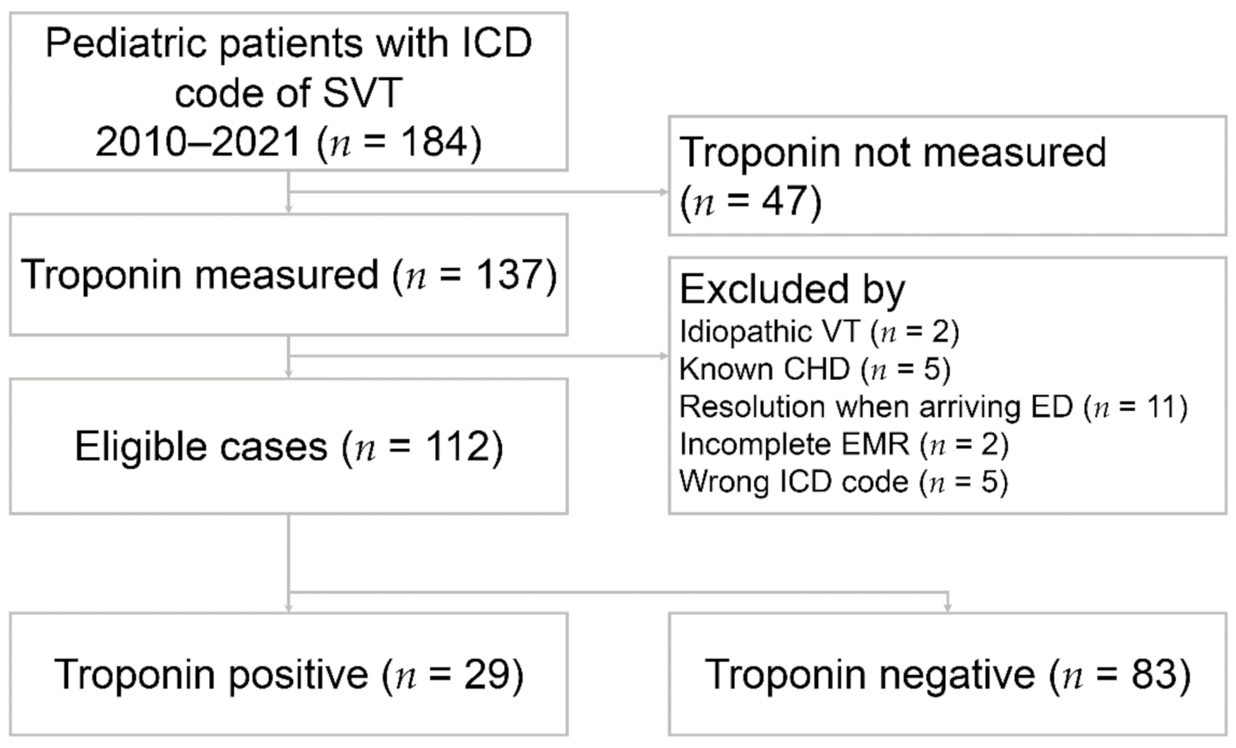
2.2. Patient Selection and Data Collection

We first identified all pediatric patients aged 0–18 years with International Classification of Diseases (ICD)-9 code 427.0 and ICD-10 code I471 of supraventricular tachycardia (SVT) who presented to the ED through a computerized search from the electrical medical record (EMR) system. The patients selected by EMR were reviewed for 12-lead electrocardiogram and documentation by attending physicians. Patients with a confirmed diagnosis of SVT and troponin tests were included. Patients with known congenital heart disease, incomplete medical records, arrhythmias other than SVT, or resolution of SVT when arriving at the ED were excluded through a detailed chart review (Figure 1). SVT was defined as regular narrow complex tachycardia but did not include atrial fibrillation and flutter. The diagnosis of SVT was confirmed by 12-lead electrocardiogram and documentation in the ED notes of attending physicians.
We collected the relevant variables, including age, sex, vital signs, symptoms and signs, laboratory results, electrocardiograms, chest radiographs, length of hospital stay, underlying diseases, medication therapies, whether patients received radiofrequency ablation, and ED disposition. A peak troponin value was defined as the highest value if patients underwent follow-up troponin testing. Further cardiac investigations, including echocardiography and electrophysiological (EP) studies, were also documented.
The primary outcomes were the occurrence of major adverse cardiac events (MACEs) during the follow-up period and 30-day SVT recurrence. MACE was defined as ischemic stroke, admission due to acute decompensated heart failure, acute coronary syndrome, revascularization, coronary artery bypass grafting (CABG), and all-cause mortality.

2.3. Measurement of Cardiac Troponin I

The cardiac troponin I (cTnI) levels were measured with a UniCel DxI 800 immunoassay analyzer (Beckman Coulter DxC880i, Danaher Corporation, Brea, California, United States). The assay has a minimum detectable concentration of <0.01 ng/mL, and the cutoff level (coefficient of variation of ≤10%) for positivity is 0.04 ng/mL.

2.4. Statistical Analysis

Descriptive data are presented as the means ± SDs or as numbers (percentages). Categorical data were analyzed with chi-square or Fisher’s exact tests, as appropriate, and continuous variables were analyzed using independent Student’s t-tests for normally distributed continuous variables and Mann–Whitney U-tests for skewed continuous variables. Multivariable logistic regression was performed for outcome risk assessment with stepwise backward selection of variables, with a p-value cutoff of 0.1. As a result of normal variations of baseline blood pressure and heart rate in pediatric patients, hypotension was defined as systolic blood pressure lower than 2× age (years) + 70 mmHg, and heart rate was transformed to an age-adjusted z score [13]. All p-values were two-sided, and the significance level was set at 0.05. All statistical analyses were performed using SPSS software (version 13.0 for Windows; SPSS Inc., Chicago, IL, USA).

3. Results

3.1. Patient Characteristics

During the study periods, a total of 112 patients were included in our study. Of these, 29 (25.9%) patients had a positive cardiac troponin I (cTnI) value, and 83 (74.1%) had a negative value. Age and male proportion were similar between the two groups. The unadjusted peak heart rate was significantly higher in the cTnI-positive group (219 vs. 200.8 beats per min, p = 0.037), but no significant difference was found after adjusting the heart rate with the z score (6.5 vs. 6.3, p = 0.537). For symptoms, patients with cTnI elevation had significantly more complaints of dyspnea (27.6% vs. 7.2%, p = 0.008) and gastrointestinal discomfort (24.1% vs. 4.8%, p = 0.006). There was no difference in the medical treatment of SVT between the two groups. Among the cTnI-positive group, there were significantly more intensive care unit admissions (41.4% vs. 16.9%, p = 0.007). The length of hospital stay was longer in the cTnI-positive group, without a significant difference (48.3 vs. 28.4 h, p = 0.19). The detailed patient characteristics are summarized in Table 1.
For laboratory investigations, the results were mostly within normal ranges. None of our patients with hemoglobin testing received blood transfusions. Electrolyte levels, including sodium, potassium, calcium, and magnesium, were almost normal. Only one patient in the cTnI-negative group had a free thyroxine level of 4.75 ng/dL (normal range: 0.7–1.48 ng/dL) and was diagnosed with primary hyperthyroidism. The full laboratory results are reported in Table 2.
Mechanisms of SVT were divided into atrioventricular reentrant tachycardia (AVRT), atrioventricular nodal reentrant tachycardia (AVNRT), and atrial tachycardia (AT) according to EP studies, while Wolff–Parkinson–White syndrome (WPW syndrome) was classified on the basis of electrocardiogram findings with the characteristic delta wave of ventricular pre-excitation. No significant difference was noted between the two groups (Table 3). Echocardiography was performed in most patients (N = 95, 81.2%). Among patients with parameter data (N = 56), left atrial diameter was non-significantly larger in the cTnI-negative group (23.20 vs. 20.56 mm, p = 0.093). Two (7.4%) patients in the cTnI-positive group had abnormal findings: one had a small muscle ridge over the middle portion of the interventricular septum without left ventricular outflow tract obstruction, and the other had a stationary right atrium cardiac mass. Five (7.4%) patients in the cTnI-negative group had abnormal findings: one had pre-existing Pompe disease-related hypertrophic cardiomyopathy, one had a ventricular septal defect, one had congestive heart failure with moderate mitral regurgitation and tricuspid regurgitation, and two had mitral valve prolapse.

3.2. Clinical Outcomes

During the follow-up period (40 ± 37 months), one major adverse cardiac event (MACE) was found in the cTnI-negative group. The patient was hospitalized due to acute decompensated heart failure six months after the first presentation of SVT at our ED. All patients survived until the last follow-up time. For 30-day SVT recurrence, the cTnI-positive group had a higher recurrence rate, without a statistically significant difference (20.7% vs. 7.2%, p = 0.075) (Table 4).
The age distribution of 30-day SVT recurrence is shown in Figure 2. No 30-day SVT recurrence was found in patients aged 13 years or older, which had a sensitivity of 100% and specificity of 60%. The results of univariate and multivariable logistic regression analyses showed hypotension as an independent predictor of 30-day SVT recurrence (OR = 4.98; Cl 1.02–24.22; p = 0.047) (Table 5) (Figure 3).

4. Discussion

We have presented, to the best of our knowledge, the largest cohort study evaluating the prognostic value of troponin and comprehensive laboratory investigations in pediatric patients with supraventricular tachycardia (SVT). This study also offers the first description of hypotension as a predictor for 30-day SVT recurrence.
A prior adult systemic review and meta-analysis investigated the prevalence of troponin elevation in patients presenting with SVT and revealed a pooled proportion of 32% (12–46%) [11]. One pediatric study illustrated a prevalence of 29% in patients with SVT at the time of the index ED presentation, which showed a similar proportion of elevated troponin levels [5]. It is not uncommon that troponin elevation occurs in the absence of coronary artery disease after SVT presentation [10,11,14,15,16]. However, the utility of troponin in patients with SVT is still debated [17].
As recommended by consensus guidelines, troponin is used as a biomarker for acute myocardial infarction (MI) diagnosis, accompanied by symptoms and electrocardiographic (ECG) findings [18]. However, limited evidence reveals the diagnostic value of troponin in SVT. One study conducted by Moore J.P. et al. analyzed the predictors of troponin elevation in pediatric SVT patients, and 13 patients with elevated troponin levels were all alive at the time of the last follow-up [5]. Nevertheless, there were no outcome variables, for instance, with rehospitalization, cardiac intervention, and SVT recurrence shown in their study. Our study indicated that pediatric SVT patients with positive troponin values had similar outcomes compared with patients with normal troponin levels. Troponin also had a poor predictive value for 30-day SVT recurrence. Troponin provides little prognostic significance and may contribute to unnecessary admissions, longer length of stay, and higher health care costs.
It remains unclear which mechanisms contribute to the elevation of troponin in SVT patients. In the adult literature, possible precipitating factors include concurrent microvascular coronary disease, occult cardiomyopathy, or demand ischemia-related myocyte injury [19,20,21]. Given the low prevalence rates of coronary artery disease and cardiomyopathy in pediatric patients, the pathophysiology of elevated troponin levels in SVT might differ from that in adult patients. The increase in heart rate during SVT results in shortening of diastole with decreased oxygen supply, and simultaneous increased oxygen consumption may lead to subendocardial ischemia. Another feature of troponin elevation during tachycardia is myocardial stretch, which was identified to be associated with myocyte functional injury and cell death [22]. A close relationship has been demonstrated between an increase in troponin and B-type natriuretic peptide (BNP) in patients with various tachyarrhythmias [23]. These reports implied that increased levels of troponin could probably be attributed to the physiology of SVT and were not associated with major cardiovascular events in pediatric patients.
It has been demonstrated that congenital heart disease (CHD) is related to infants with SVT. Studies have shown prevalence rates of 30% of CHD in SVT infants with WPW syndrome and 20% in those without WPW syndrome [1,24,25]. However, one study conducted by L’Italien, K., et al. revealed that the prevalence of CHD in patients aged two or older presenting with SVT was similar to that in the general population, and only one patient in the cohort required immediate intervention [1]. Our study showed similar results, and no patients needed to receive emergent operations. Although left atrial diameter was non-significantly larger in the cTnI-negative group, it may be less meaningful given the combined influence of weight, height, and age on cardiac size and the limited parameter data of our patients [26]. Immediate echocardiography seems unnecessary in the acute setting.
Blood tests, including hemoglobin, electrolytes, and thyroid function tests, were common in patients who underwent SVT. Anemia was recognized as a possible risk factor for SVT, and it should be considered if other symptoms of anemia exist [27]. Abnormal potassium and magnesium levels were reported to lead to arrhythmias, but no direct evidence supports the relationship between electrolyte imbalance and SVT [11,27,28]. Thyroid function tests were recommended in SVT patients by the UK National Service Framework guidelines [11,27]. Although there is no well-established evidence regarding the association between thyroid dysfunction and SVT, a few case reports have demonstrated that either hyperthyroidism or hypothyroidism may lead to SVT by inducing sympathetic-vagal imbalance [29,30,31].
There have been few documented risk factors for SVT recurrence in pediatric patients in previous studies. Hypotension on initial presentation was found to be an independent predictor for recurrent SVT 30 days after adjustment for associated variables in our study. Another interesting finding was that none of the patients aged 13 or older had SVT recurrence within 30 days. Although one previous study reported that patients with an onset age younger than one year were at decreased risk of recurrence at the final follow-up, no other research mentioned age as an indicator for predicting SVT recurrence [32]. These findings may help physicians determine the best course of action in the ED. For patients at risk of SVT recurrence, close outpatient department follow-up and further cardiac investigations, such as EP studies, are appropriate.
This study has several limitations, which need to be mentioned. It was a retrospective study, and selection bias may exist. Some clinical variables, such as detailed symptoms and definite onset time of SVT, could be missed if not recorded in the EMR. Second, given that not every patient underwent further EP studies, the exact mechanism of SVT in one-half of the patients was unknown. Finally, the small number of patients in our study may limit the overall power and hamper our ability to draw clear-cut conclusions, although, to our knowledge, this was the largest study on the topic.

5. Conclusions

Troponin had low value for predicting outcomes of pediatric patients with SVT. The only significant predictor for recurrent SVT was initial hypotension. Emergent blood tests and echocardiography may not be necessary in the ED and could be performed at follow-up.

Author Contributions

Conceptualization, C.-C.Y. and C.-K.W.; Data curation, H.-T.Y.; Formal analysis, C.-C.Y.; Investigation, C.-C.Y. and C.-J.N.; Resources, C.-H.C.; Supervision, S.-Y.C.; Writing—original draft, C.-C.Y. All authors have read and agreed to the published version of the manuscript.

Funding

This research received no external funding.

Institutional Review Board Statement

The study was conducted according to the guidelines of the Declaration of Helsinki, and approved by the Institutional Review Board of Chang Gung Medical Foundation (IRB no. 202100905B0, Date of Approval: 26 June 2021).

Informed Consent Statement

This study qualified for a waiver of informed consent.

Data Availability Statement

The data in this study are available from the corresponding author upon reasonable request.

Conflicts of Interest

The authors declare no competing interest.

References

  1. L’Italien, K.; Conlon, S.; Kertesz, N.; Bezold, L.; Kamp, A. Usefulness of Echocardiography in Children with New-Onset Supraventricular Tachycardia. J. Am. Soc. Echocardiogr. 2018, 31, 1146–1150. [Google Scholar] [CrossRef]
  2. Quattrocelli, A.; Lang, J.; Davis, A.; Pflaumer, A. Age makes a difference: Symptoms in pediatric supraventricular tachycardia. J. Arrhythmia 2018, 34, 565–571. [Google Scholar] [CrossRef]
  3. Yetkin, E.; Ozturk, S.; Cuglan, B.; Turhan, H. Clinical presentation of paroxysmal supraventricular tachycardia: Evaluation of usual and unusual symptoms. Cardiovasc. Endocrinol. Metab. 2020, 9, 153–158. [Google Scholar] [CrossRef]
  4. Wood, K.A.; Drew, B.J.; Scheinman, M.M. Frequency of Disabling Symptoms in Supraventricular Tachycardia. Am. J. Cardiol. 1997, 79, 145–149. [Google Scholar] [CrossRef]
  5. Moore, J.P.; Arcilla, L.; Wang, S.; Lee, M.S.; Shannon, K.M. Characterization of Cardiac Troponin Elevation in the Setting of Pediatric Supraventricular Tachycardia. Pediatr. Cardiol. 2015, 37, 392–398. [Google Scholar] [CrossRef]
  6. Bagai, A.; Alexander, K.P.; Berger, J.; Senior, R.; Sajeev, C.; Pracon, R.; Mavromatis, K.; Lopez-Sendón, J.L.; Gosselin, G.; Diaz, A.; et al. Use of troponin assay 99th percentile as the decision level for myocardial infarction diagnosis. Am. Heart J. 2017, 190, 135–139. [Google Scholar] [CrossRef]
  7. Peacock, W.F.; De Marco, T.; Fonarow, G.; Diercks, D.B.; Wynne, J.; Apple, F.S.; Wu, A.H. Cardiac Troponin and Outcome in Acute Heart Failure. N. Engl. J. Med. 2008, 358, 2117–2126. [Google Scholar] [CrossRef] [PubMed] [Green Version]
  8. Korff, S.; Katus, H.A.; Giannitsis, E. Differential diagnosis of elevated troponins. Heart 2006, 92, 987–993. [Google Scholar] [CrossRef] [PubMed] [Green Version]
  9. Yousufuddin, M.; Abdalrhim, A.D.; Wang, Z.; Murad, M.H. Cardiac troponin in patients hospitalized with acute decompensated heart failure: A systematic review and meta-analysis. J. Hosp. Med. 2016, 11, 446–454. [Google Scholar] [CrossRef] [PubMed] [Green Version]
  10. Ghersin, I.; Zahran, M.; Azzam, Z.S.; Suleiman, M.; Bahouth, F. Prognostic value of cardiac troponin levels in patients presenting with supraventricular tachycardias. J. Electrocardiol. 2020, 62, 200–203. [Google Scholar] [CrossRef] [PubMed]
  11. Fernando, H.; Adams, N.; Mitra, B. Review article: The utility of troponin and other investigations in patients presenting to the emergency department with supraventricular tachycardia. Emerg. Med. Australas. 2019, 31, 35–42. [Google Scholar] [CrossRef] [Green Version]
  12. Bukkapatnam, R.N.; Robinson, M.; Turnipseed, S.; Tancredi, D.; Amsterdam, E.; Srivatsa, U.N. Relationship of myocardial ischemia and injury to coronary artery disease in patients with supraventricular tachycardia. Am. J. Cardiol. 2010, 106, 374–377. [Google Scholar] [CrossRef]
  13. Bratincsák, A.; Kimata, C.; Limm-Chan, B.N.; Vincent, K.P.; Williams, M.R.; Perry, J.C. Electrocardiogram Standards for Children and Young Adults Using Z -Scores. Circ. Arrhythmia Electrophysiol. 2020, 13, 008253. [Google Scholar] [CrossRef] [PubMed]
  14. Murer, M.; Cuculi, F.; Toggweiler, S.; Weberndoerfer, V.; Young, M.; Kobza, R. Elevated high-sensitivity troponin does not indicate the presence of coronary artery disease in patients presenting with supraventricular tachycardia. Cardiol. J. 2017, 24, 642–648. [Google Scholar] [CrossRef] [PubMed] [Green Version]
  15. Noorvash, D.; Ramos, R.; Hatch, L.; Muck, A.; Olson, A.S. Assessment of the Utility of Ordering a Troponin in Low- and Intermediate-Risk Patients Presenting to the Emergency Department with Supraventricular Tachycardia: A Retrospective Chart Review. J. Emerg. Med. 2018, 55, 1–6. [Google Scholar] [CrossRef] [PubMed]
  16. Sayadnik, M.; Shafiee, A.; Jenab, Y.; Jalali, A.; Sadeghian, S. Predictors of High-Sensitivity Cardiac Troponin T Elevation in Patients with Acute Paroxysmal Supra-ventricular Tachycardia and Ischemic Heart Disease. Tex. Heart Inst. J. 2017, 44, 306–311. [Google Scholar] [CrossRef] [PubMed] [Green Version]
  17. Allen, R.; deSouza, I.S. Troponin Testing in Patients with Supraventricular Tachycardia—Are We Overtesting? A Teachable Moment. JAMA Intern. Med. 2021, 181, 842–843. [Google Scholar] [CrossRef]
  18. Maresca, B.; Manzione, A.; Moioli, A.; Salerno, G.; Cardelli, P.; Punzo, G.; Barberi, S.; Menè, P. Prognostic value of high-sensitive cardiac troponin I in asymptomatic chronic hemodialysis patients. J. Nephrol. 2019, 33, 129–136. [Google Scholar] [CrossRef]
  19. Chow, G.V.; Hirsch, G.A.; Spragg, D.D.; Cai, J.X.; Cheng, A.; Ziegelstein, R.C.; Marine, J.E. Prognostic Significance of Cardiac Troponin I Levels in Hospitalized Patients Presenting with Supraventricular Tachycardia. Medicine 2010, 89, 141–148. [Google Scholar] [CrossRef]
  20. Ben Yedder, N.; Roux, J.F.; Paredes, F.A. Troponin Elevation in Supraventricular Tachycardia: Primary Dependence on Heart Rate. Can. J. Cardiol. 2011, 27, 105–109. [Google Scholar] [CrossRef]
  21. Healey, J.S. Troponin Elevation in Patients with Supraventricular Tachycardia: What Does It Mean? Can. J. Cardiol. 2011, 27, 110–111. [Google Scholar] [CrossRef]
  22. Taskapan, M.C.; Taskapan, H.; Ulutas, O.; Orhan, M.; Sahin, I. Relationships between Brain Natriuretic Peptide, Troponin I and QT Dispersion in Asymptomatic Dialysis Patients. Ren. Fail. 2007, 29, 221–225. [Google Scholar] [CrossRef] [PubMed]
  23. Higgins, J.P.; Higgins, J.A. Elevation of cardiac troponin I indicates more than myocardial ischemia. Clin. Invest. Med. 2003, 26, 133–147. [Google Scholar] [PubMed]
  24. Snyder, C.S.; Fenrich, A.L.; Friedman, R.A.; Rosenthal, G.; Kertesz, N.J. Usefulness of echocardiography in infants with supraventricular tachycardia. Am. J. Cardiol. 2003, 91, 1277–1279. [Google Scholar] [CrossRef]
  25. Chandra, M.S.; Kerber, R.E.; Brown, D.D.; Funk, D.C. Echocardiography in Wolff-Parkinson-White syndrome. Circulation 1976, 53, 943–946. [Google Scholar] [CrossRef] [PubMed] [Green Version]
  26. Dallaire, F.; Bigras, J.-L.; Prsa, M.; Dahdah, N. Bias Related to Body Mass Index in Pediatric Echocardiographic Z Scores. Pediatr. Cardiol. 2014, 36, 667–676. [Google Scholar] [CrossRef]
  27. Ashok, A.; Cabalag, M.; Taylor, D.M. Usefulness of laboratory and radiological investigations in the management of supraventricular tachycardia. Emerg. Med. Australas. 2017, 29, 394–399. [Google Scholar] [CrossRef]
  28. Bonato, F.O.B.; Canziani, M.E.F. Ventricular arrhythmia in chronic kidney disease patients. Braz. J. Nephrol. 2017, 39, 186–195. [Google Scholar] [CrossRef]
  29. Mak, W.W.; Nurazni, R.A.R.; Badrulnizam, L.B.M. Severe hypothyroidism presenting with supraventricular tachycardia. Med. J. Malays. 2018, 73, 349–350. [Google Scholar]
  30. Abbasoğlu, A.; Ecevit, A.; Tuğcu, A.U.; Kınık, S.T.; Tarcan, A. Neonatal thyrotoxicosis with severe supraventricular tachycardia: Case report and review of the literature. J. Pediatr. Endocrinol. Metab. 2015, 28, 463–466. [Google Scholar] [CrossRef]
  31. Vanin, L.N.; Smetnev, A.S.; Sokolov, S.F.; Kotova, G.A.; Masenko, V.P. Study of thyroid function in patients with paroxysmal supraventricular tachycardia. Kardiologiia 1989, 29, 71–74. [Google Scholar] [PubMed]
  32. Riggs, T.W.; Byrd, J.A.; Weinhouse, E. Recurrence risk of supraventricular tachycardia in pediatric patients. Cardiology 1999, 91, 25–30. [Google Scholar] [CrossRef] [PubMed]
Figure 1. Flow chart of patient selection. ICD: International Classification of Diseases; SVT: supraventricular tachycardia; VT: ventricular tachycardia; CHD: congenital heart disease; ED: emergency department; EMR: electrical medical record.
Jcm 10 03638 g001
Figure 2. Distribution of 30-day SVT recurrence rate by age group. Infant: 28 d—<1 y; Toddler: 1—<3 y; Preschool: 3—<6 y; School: 6—<13 y; Adolescent: 13—≤18 y.
Jcm 10 03638 g002
Figure 3. Multivariable predictors of 30-day SVT recurrence.
Jcm 10 03638 g003
Table 1. Patient characteristics according to cTnI levels.
VariablecTnI(+) (n = 29)cTnI(–) (n = 83)p Value
Age (yr)8.6 ± 5.410.3 ± 4.70.099
Male13 (44.8%)37 (44.6%)1.000
Previous SVT10 (34.5%)30 (36.1%)1.000
Vital signs
 Peak heart rate (beats/min)219.0 ± 42.0200.8 ± 28.50.037
 Peak heart rate (z score)6.5 ± 1.66.3 ± 1.50.537
 Systolic blood pressure (mmHg)101.5 ± 18.9108.9 ± 18.50.084
 Diastolic blood pressure (mmHg)64.1 ± 12.070.8 ± 14.00.027
 Hypotension6 (21.4%)7 (9.2%)0.106
 Fever 2 (6.9%)11 (13.3%)0.509
Symptoms
 Palpitation23 (79.3%)69 (83.1%)0.779
 Chest pain7 (24.1%)18 (21.7%)0.799
 Dyspnea8 (27.6%)6 (7.2%)0.008
 Dizziness2 (6.9%)8 (9.6%)1.000
 Syncope0 (0%)2 (2.4%)1.000
 Respiratory7 (24.1%)12 (14.5%)0.257
 Gastrointestinal7 (24.1%)4 (4.8%)0.006
SVT duration since ED (min)60.5 ± 80.046.8 ± 78.70.424
Length of stay (hr)48.3 ± 111.528.4 ± 47.00.190
Treatment
 Without medication §3 (10.3%)11 (13.4%)1.000
 Adenosine use25 (86.2%)72 (86.7%)1.000
 Verapamil use1 (3.4%)4 (4.8%)1.000
 Amiodarone use5 (17.2%)10 (12.0%)0.530
Radiofrequency ablation 16 (34.0%)1 (10.0%)0.247
ED Disposition
 Ordinary medical ward6 (20.7%)21 (25.3%)0.802
 Intensive care unit12 (41.4%)14 (16.9%)0.007
 Discharge11 (37.9%)48 (57.8%)0.084
Count data are expressed as number (percentage) and continuous values are expressed as mean ± SD. cTnI: cardiac troponin I; SVT: supraventricular tachycardia; ED: emergency department. Defined as body temperature > 38 °C in the triage, § Defined as spontaneous resolution or Valsalva maneuver, Performed within five years after patients discharge.
Table 2. Analysis of laboratory findings in patients with or without elevated cTnI levels.
VariablecTnI(+) (n = 29)cTnI(–) (n = 83)p Value
Laboratory exam
 White cell count (103/uL) (n = 107)12.45 ± 5.41 10.05 ± 3.570.01
 Hemoglobin (g/dL) (n = 107)13.48 ± 1.4113.56 ± 1.340.775
 Platelet (103/uL) (n = 107)316.1 ± 89.1285.9 ± 68.00.06
 Blood urea nitrogen (mg/dL) (n = 41)12.8 ± 2.813.2 ± 10.50.908
 Creatinine (mg/dL) (n = 74)0.56 ± 0.200.69 ± 1.170.622
 Sodium (mEq/L) (n = 94)139.6 ± 2.2139.4 ± 2.20.733
 Potassium (mEq/L) (n = 93)4.12 ± 0.563.95 ± 0.400.109
 Calcium (mg/dL) (n = 70)9.47 ± 0.409.38 ± 0.520.488
 Magnesium (mEq/L) (n = 39)1.85 ± 0.181.80 ± 0.200.499
 AST (U/L) (n = 70)32.50 ± 16.0738.18 ± 46.410.596
 ALT (U/L) (n = 62) 21.69   ±   16.49 31.26 ± 48.140.441
 Initial cTnI (ng/mL) (n = 112)0.172 ± 0.163 0.015 ± 0.010<0.001
 Peak cTnI (n = 112)0.256 ± 0.299 0.017 ± 0.008<0.001
 TSH (uIU/mL) (n = 23)2.37 ± 1.412.31 ± 1.520.926
 Free thyroxine (ng/dL) (n = 20)1.26 ± 0.171.57 ± 1.150.436
 BNP (pg/mL) (n = 19)173.4 ± 185.3 163.3 ± 232.9 0.927
 C-Reactive Protein (mg/L) (n = 24)5.07 ± 9.23 1.13 ± 1.640.105
Continuous values are expressed as mean ± SD. cTnI: cardiac troponin I; AST: aspartate aminotransferase; ALT: alanine aminotransferase; TSH: thyroid-stimulating hormone; BNP: B-type natriuretic peptide; Increased value.
Table 3. Comparisons of SVT mechanisms and echocardiographic findings in patients with or without elevated cTnI levels.
VariablecTnI(+) (n = 29)cTnI(–) (n = 83)p Value
SVT mechanism
 AVRT §4 (13.8%)3 (3.6%)0.073
 AVNRT 2 (6.9%)14 (16.9%)0.232
 AT 0 (0%)2 (2.4%)1.000
 WPW8 (27.6%)12 (14.5%)0.157
 Not classified by EP study15 (51.7%)52 (62.7%)0.38
Echocardiography (n = 27)(n = 68)
 Normal25 (92.6%)59 (86.8%)0.723
 Incidental 0 (0%)4 (5.9%)0.575
 Abnormal2 (7.4%)5 (7.4%)1.000
 LVEF(%)71.83 ± 5.1870.66 ± 8.910.605
 LA diameter (mm)20.56 ± 6.2623.20 ± 4.780.093
 LVEDD (mm)34.28 ± 10.7738.22 ± 8.950.160
 LVESD (mm)20.67 ± 5.9622.36 ± 5.550.307
 Aortic root diameter (mm)20.17 ± 5.8521.11 ± 4.310.506
Count data are expressed as number (percentage) and continuous values are expressed as mean ± SD. AVRT: atrioventricular reentrant tachycardia; AVNRT: atrioventricular nodal reentrant tachycardia; AT: atrial tachycardia; WPW: Wolff–Parkinson–White syndrome; EP: electrophysiologic; LVEF: left ventricular ejection fraction; LVEDD: left ventricular end-diastolic diameter; LVESD: left ventricular end-systolic diameter. Parameter data available in 56 patients (n = 18 in cTn+; n = 38 in cTn−). § Not including WPW syndrome. Incidental findings, not classified as abnormal echocardiography, included patent foramen ovale and left-sided aortic arch.
Table 4. Clinical outcomes of patients with positive or negative cTnI.
VariablecTnI(+) (n = 29)cTnI(–) (n = 83)p Value
MACE0 (0%)1 (0.01%)1.000
30-day SVT recurrence6 (20.7%)6 (7.2%)0.075
Count data are expressed as number (percentage). MACE: major adverse cardiovascular event.
Table 5. Univariate and multivariable logistic regression results for 30-day SVT recurrence.
UnivariateMultivariable
OR (95%CI)p ValueOR (95%CI)p Value
Age 0.86 (0.75, 0.98)0.0220.87 (0.75, 1.001)0.051
Male1.27 (0.38, 4.22)0.693
Peak heart rate (z score)0.99 (0.66, 1.49)0.965
Hypotension5.33 (1.31, 21.8)0.0204.98 (1.02, 24.22)0.047 *
cTnI(+)3.35 (0.99, 11.4)0.0532.52 (0.62, 10.19)0.195
* p value < 0.05.
Publisher’s Note: MDPI stays neutral with regard to jurisdictional claims in published maps and institutional affiliations.

