Article

Cardiorespiratory Functioning in Youth with Persistent Post-Concussion Symptoms: A Pilot Study

1
Department of Psychiatry, University of California, Los Angeles, CA 90095, USA
2
UCLA Steve Tisch BrainSPORT Program, Los Angeles, CA 90095, USA
3
Department of Radiology, Wake Forest School of Medicine, Winston-Salem, NC 27101, USA
4
Department of Psychology, University of California, Los Angeles, CA 90095, USA
5
Departments of Neurology and Neuropsychiatry, David Geffen School of Medicine at UCLA, Los Angeles, CA 90095, USA
6
Department of Psychiatry, Psychology Service, University of California, San Diego, CA 92093, USA
7
VA San Diego Healthcare System, San Diego, CA 92161, USA
8
UCLA Mattel Children’s Hospital, Los Angeles, CA 90095, USA
9
Department of Pediatrics, David Geffen School of Medicine at UCLA, Los Angeles, CA 90095, USA
10
Department of Neurosurgery, David Geffen School of Medicine at UCLA, Los Angeles, CA 90095, USA
*
Author to whom correspondence should be addressed.
J. Clin. Med. 2021, 10(4), 561; https://doi.org/10.3390/jcm10040561
Submission received: 16 December 2020 / Revised: 22 January 2021 / Accepted: 30 January 2021 / Published: 3 February 2021
(This article belongs to the Special Issue Children Behavior and Psychophysiology)

Abstract

:
Dysregulation of the autonomic nervous system (ANS) may play an important role in the development and maintenance of persistent post-concussive symptoms (PPCS). Post-injury breathing dysfunction, which is influenced by the ANS, has not been well-studied in youth. This study evaluated cardiorespiratory functioning at baseline in youth patients with PPCS and examined the relationship of cardiorespiratory variables with neurobehavioral outcomes. Participants were between the ages of 13–25 in two groups: (1) Patients with PPCS (concussion within the past 2–16 months; n = 13) and (2) non-injured controls (n = 12). Capnometry was used to obtain end-tidal CO2 (EtCO2), oxygen saturation (SaO2), respiration rate (RR), and pulse rate (PR) at seated rest. PPCS participants exhibited a reduced mean value of EtCO2 in exhaled breath (M = 36.3 mmHg, SD = 2.86 mmHg) and an altered inter-correlation between EtCO2 and RR compared to controls. Neurobehavioral outcomes including depression, severity of self-reported concussion symptoms, cognitive catastrophizing, and psychomotor processing speed were correlated with cardiorespiratory variables when the groups were combined. Overall, results from this study suggest that breathing dynamics may be altered in youth with PPCS and that cardiorespiratory outcomes could be related to a dimension of neurobehavioral outcomes associated with poorer recovery from concussion.

1. Introduction

Persistent post-concussion symptoms (PPCS), defined by most as symptoms lasting longer than 2 to 3 months post-injury, affect a significant minority of patients who sustain a concussion [1,2,3,4,5,6]. Multimodal rehabilitation models for PPCS may be beneficial, particularly those that include interventions such as exercise, physiotherapy, and/or cognitive behavioral treatment [7,8,9,10,11]. However, the specific interventions that render multimodal treatments successful in patients with PPCS are either difficult to isolate or have yet to be fully elucidated. Although biopsychosocial frameworks and other studies describe personal, injury, and environmental factors that interact with recovery [12,13,14,15], a cohesive model for the mechanisms of PPCS has not yet been established.
Post-concussion symptoms are not specific to concussion and also occur in the general population [16,17]. The role of the autonomic nervous system (ANS) in the development and maintenance of post-concussion symptoms has emerged as a potentially important component of PPCS etiological frameworks [18,19,20]. The ANS is responsible for maintenance of internal homeostasis through the regulating actions of its two branches, the sympathetic nervous system (SNS) and parasympathetic nervous system (PNS). The ANS is involved in conscious and unconscious regulation of critical bodily responses including changes in heart rate, blood pressure, breathing dynamics, gastrointestinal response, oculomotor adjustment, and thermoregulation, among others. There is strong overlap between symptoms associated with dysregulated ANS responses in other medical conditions (e.g., chronic pain, migraine, lupus) and those most commonly reported in concussion patients (e.g., headache, fatigue, dizziness, brain fog) [21,22,23,24,25]. Brain regions and networks critical to the central control of ANS branches, specifically the dynamic responses of the SNS and PNS, are highly vulnerable to the biomechanical forces of brain injury, even on the milder end of the severity spectrum [26,27,28]. In a study of patients with PPCS aged 13–18 years, autonomic dysfunction was identified in 70.6% of the 36 participants tested for orthostatic intolerance within 6 months post-injury [29]. Other evidence of impaired autonomic regulation of cardiovascular responses has been reliably detected in patients during acute and chronic phases of injury, primarily through measurement of heart rate variability [30,31,32,33,34].
Cardiorespiratory functioning, especially breathing dynamics, can also provide insight into ANS functioning and may be particularly applicable to the study of concussion and PPCS. Capnometry is a relatively rapid and non-invasive tool that permits the measurement of carbon dioxide concentration in exhaled breath. End-tidal CO2 (EtCO2), the CO2 concentration obtained at the end of an expiratory phase, has been linked to several neurobehavioral processes relevant to concussion recovery. In particular, the presence of hypocapnia (lower than normal levels of EtCO2) is commonly reported following or sometimes preceding panic attacks [35,36,37]. Periods of hyperventilation, leading to hypocapnia, can result in acute respiratory alkalosis and symptoms such as dizziness, light-headedness, confusion, and syncope [38,39,40], which are also common to concussion. Differences in EtCO2 concentrations at baseline can be detected in patients with a variety of medical conditions including migraine and chronic pain, suggesting ongoing alterations in autonomic regulation. In individuals with chronic anxiety, difficulties with breathing are often reported and can take the form of pathological breathlessness or hyperventilation [37,41,42].
The relationship between cardiorespiratory function and symptom experience following concussion is understudied. At present, one study demonstrated that adult patients may exhibit altered EtCO2 at baseline compared to controls [43], but more evidence is needed to determine whether changes in ANS dysfunction are associated with the etiology of PPCS, especially in youth patients with PPCS. As such, the purpose of this pilot study was to examine cardiorespiratory functioning, as measured by capnometry, in youth patients with PPCS and its relationship with neurobehavioral outcomes (e.g., neurocognitive functioning, mood, sleep) compared to non-injured controls.

2. Experimental Section

2.1. Study Design and Setting

Data were obtained from the pre-intervention baseline of a prospective case-control study. The goal of the trial was to examine the effect of psychological intervention on youth patients with persistent post-concussion symptoms. Participants included concussion patients and age-matched non-injured controls recruited from the community. Data collection occurred between June 2018 and February 2020. Approval was secured from the appropriate Institutional Review Board and informed consent and assent (if under the age of 18) were obtained for all participants.

2.2. Participants

Participants in the concussion group were recruited from university-affiliated concussion clinics in Los Angeles, CA, USA and non-injured control participants were recruited from the local community setting, chiefly through a university. Eligibility criteria for the concussion group were as follows: between 13–25 years of age at the time of assessment, history of concussion diagnosed by a medical provider, within 2 to 16 months post-injury at the time of assessment, and experiencing persistent concussion symptoms along with evidence of behavioral avoidance due to symptoms (i.e., avoiding full participation in typical, pre-injury activities for fear of symptom exacerbation). Behavioral avoidance was assessed through a screening measure developed for this project, called the Return to Activity Avoidance Inventory (RAAvI) (see Supplementary Materials, Figure S1). The RAAvI measure required participants to identify three activities they were actively avoiding due to concern about symptom provocation or exacerbation. Participants then rated their level of avoidance on a Likert scale (0 to 6 where 0 = likely to avoid the activity none of the time and 6 = likely to avoid the activity all of the time) and their perceived level of emotional and/or physical distress if they were to participate in the activity, also on a Likert scale (0 to 6 where 0 = likely to experience no physical or emotional distress and 6 = likely to experience extreme physical or emotional distress). Participants were selected if they were able to identify at least one activity that they actively avoided (1 or higher on Likert rating).
Participants were excluded if they had any comorbid neurological conditions (including history of stroke, seizure disorder, moderate to severe traumatic brain injury, anoxia), severe cardiovascular conditions, history of psychosis, current substance abuse or dependence, severe symptoms of depression and/or anxiety at the time of the assessment (as determined by self-report questionnaires), or were unable to speak English. Participants who were prescribed psychotropic medication for treatment of concussion-related symptoms (e.g., headache and migraine) were permitted to be enrolled in the study as long as they were not prescribed serotonin-norepinephrine reuptake inhibitors (e.g., desvenlafaxine, venlafaxine, duloxetine, etc.) given their impact on autonomic function. Control participants were eligible for the study if they were able to speak English and were between 13–25 years old. They were excluded if they had a history of concussion within the past 12 months or were experiencing persistent post-concussive symptoms from a previous injury. The neurological and psychiatric exclusion criteria described above were also applied to controls, but there were no restrictions on current medications.

2.3. Instrumentation and Outcome Measures

General demographic and background information was obtained via self-report questionnaire and included age, sex, education, as well as self and family medical/psychiatric history. Neurobehavioral outcomes were acquired via standardized self-report questionnaires and neurocognitive performance was obtained through paper-and-pencil testing administered by trained research staff. All data were collected during a single session and assessments were administered in a fixed order.

2.3.1. Neurobehavioral Measures

Standardized self-report measures were administered to evaluate post-concussion neurobehavioral function, including symptom burden, mood, sleep quality, and pain catastrophizing. The adolescent form (ages 13–18) of the Post-Concussion Symptom Inventory (PCSI-SR13; [44]) was used, which is a 21-item graded checklist. All participants were asked to rate their current symptoms (covering the day of and the day before the assessment). Symptom of depression and anxiety were evaluated using the raw scores from the Beck Depression Inventory, 2nd Edition (BDI-II; [45]) and the Beck Anxiety Inventory (BAI; [46]). The BDI-II is a 21-item scale that asks participants to evaluate how they have been feeling over the past two weeks. Similarly, the BAI is a 21-item graded scale that asks participants to rate how much they have been bothered by anxiety symptoms over the past week. Higher scores on both the BDI-II and BAI suggest greater symptoms of depression and anxiety, respectively. The global composite score on the Pittsburgh Sleep Quality Index (PSQI; [47]) was used to characterize sleep quality. Participants answered based on their sleep experience over the past month, and the global composite was calculated using the 7 components scores of sleep quality (i.e., subjective sleep quality, sleep latency, sleep duration, sleep efficiency, sleep disturbance, use of sleep medication, and daytime dysfunction), with higher scores indicating worse sleep quality. The Pain Catastrophizing Scale (PCS; [48]) is a 13-item measure of cognitive catastrophizing that is associated with the perception and experience of pain. Higher raw scores on the PCS indicate a more maladaptive cognitive coping style in response to pain. Participants were administered an abbreviated battery of the Repeatable Battery for the Assessment of Neuropsychological Status (RBANS; [49]). Subtests were selected to focus on cognitive domains typically affected by concussion, including attention (Digit Span and Coding), learning and memory (List Learning and List Recall), and verbal fluency (Semantic Fluency).

2.3.2. Capnometry Instrumentation and Assessment

The CapnostreamTM 35 portable capnometer (Medtronic Inc., Minneapolis, MN, USA) was used to measure the primary outcome of interest, EtCO2, as well as other cardiorespiratory metrics including pulse rate (PR, number of heart beats per minute), respiration rate (RR, number of breaths taken per minute), and oxygen saturation (SaO2, percentage of oxygen bound to hemoglobin). EtCO2 and RR were measured via nasal cannula and PR and SaO2 were collected via pulse oximeter, both of which were connected to the device. Capnometry allows for non-invasive, real-time monitoring of cardiorespiratory outcomes and data were sampled at a rate of fs = 1. Data were obtained after the neurobehavioral assessments were administered and a brief (5–10 min) break was provided to participants. Cardiorespiratory values were averaged over a 5-min continuous recording period during which participants remained in an upright, seated position and were instructed to breathe normally and avoid talking. Capnometry units were initially calibrated prior to the study and no re-calibration took place during data collection in accordance with manufacturer’s instructions.

2.3.3. Statistical Analyses

Data were inspected for normality and outlier cardiorespiratory responses were removed only if likely to be discrepant, non-biologic values (i.e., 0). Two participants were excluded (one in each group) due to nasal cannula dysfunction. Demographic comparisons between groups were analyzed via chi-square analyses, Fisher’s exact test, and parametric t-tests. Statistical procedures included parametric (independent samples) and non-parametric (Mann-Whitney) t-tests to compare groups on cardiorespiratory, demographic, and neurobehavioral variables (i.e., EtCO2, PR, RR). Separate one-way analyses of covariance (ANCOVA) were included to evaluate potential confounding effects of age and sex on cardiorespiratory variables. Pearson bi-variate and Spearman rank order correlations were used to examine the relationships between variables, and correlation coefficients were compared using Fisher’s z transformation. Data were analyzed using IBM SPSS Statistics (IBM Corp., Armonk, NY, USA), version 26.0 [50].

3. Results

A total of 13 PPCS participants and 12 controls were recruited from UCLA outpatient concussion clinics and surrounding community. Participant demographics are presented in Table 1. Overall, the two groups were broadly similar when sex-distribution and medical history were compared. The PPCS group was two years younger on average, t(23) = 2.31, p = 0.03. Additionally, all participants in the PPCS group reported having sustained at least one previous concussion, whereas no participant in the control group reported prior history of concussion, t(23) = −6.40, p < 0.001.
The PPCS group demonstrated a significantly higher current neurobehavioral burden than the control group (see Table 2). The PPCS group exhibited higher pain catastrophizing scores and higher self-reported symptoms. On both the PCSI and PSQI, the PPCS group reported more sleep problems, but the groups differed significantly only on the PCSI sleep subscale. Although not statistically significant, the PPCS group trended toward a greater emotional burden on the BDI-II and the emotional subscale of the PSCI. Although PPCS patients were selected because they exhibited activity avoidance due to symptoms, they did not report clinically or statistically higher levels of anxiety symptoms. Along with differences on the self-report measures, the PPCS group demonstrated significantly poorer neurocognitive performance on subtests of the RBANS measuring learning and attention (List Learning Total Score), semantic verbal fluency (Semantic Fluency), and verbal recall (List Recall). Effect sizes for these differences were large (Cohen’s d range, 0.80–1.43).

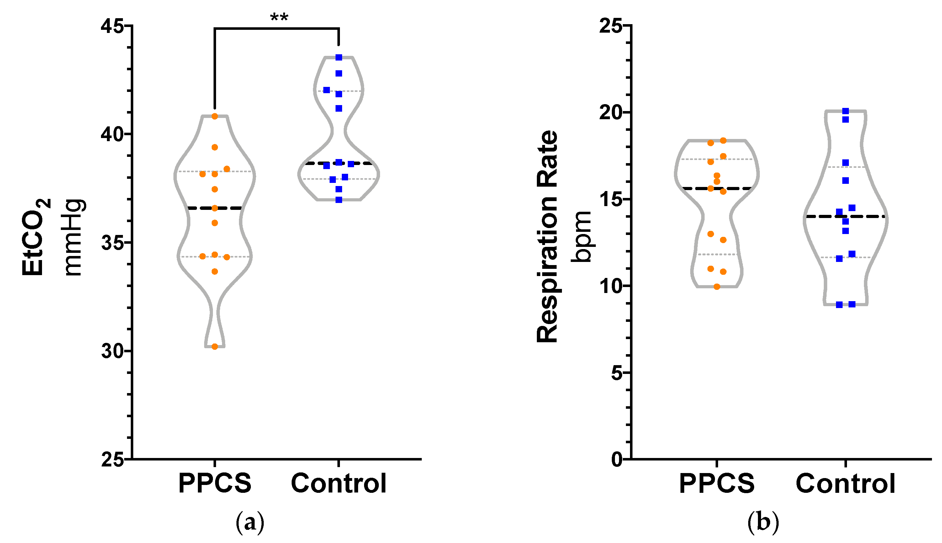
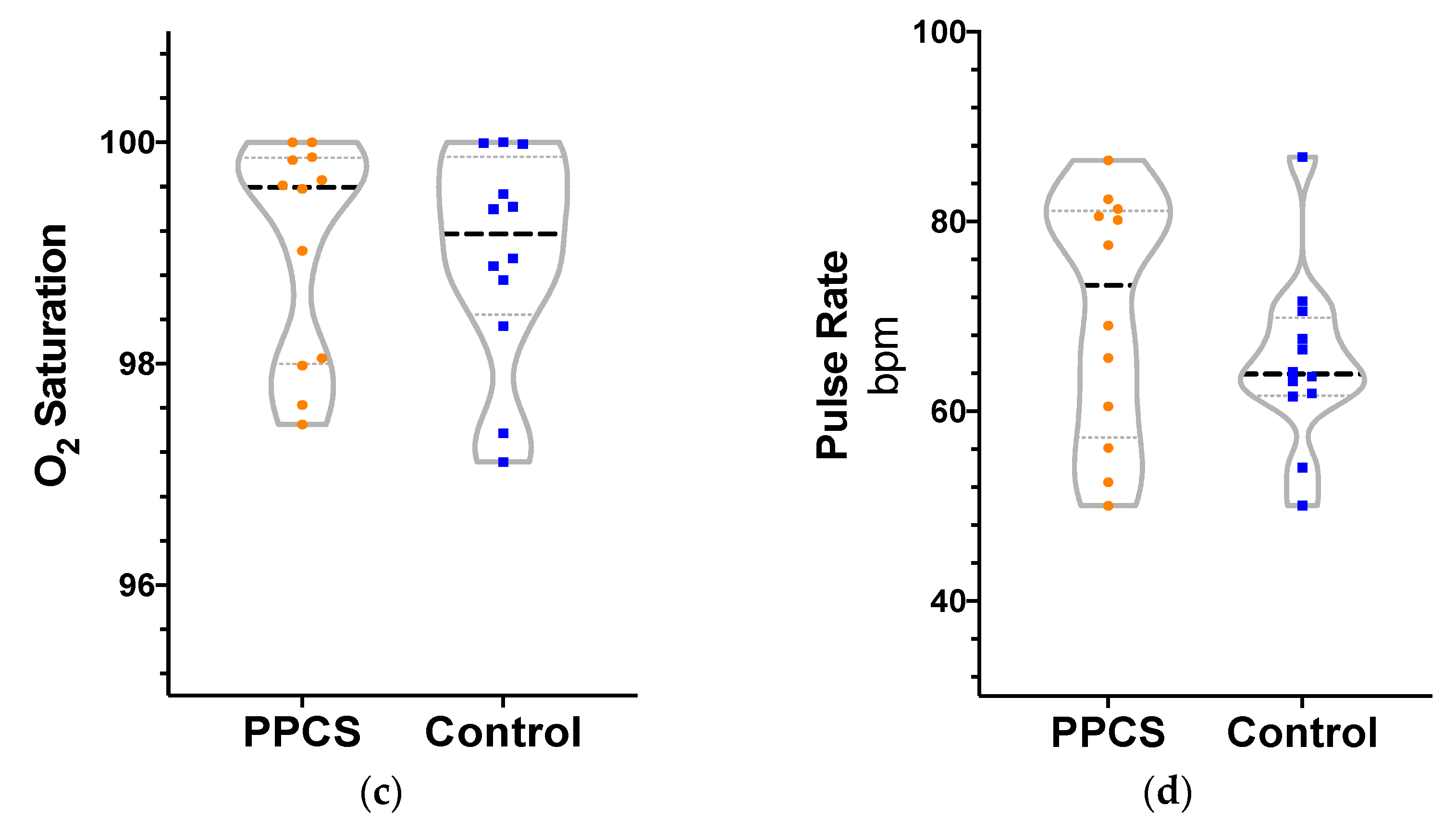
3.1. Cardiorespiratory Variables by Group

Independent samples t-test revealed significantly lower EtCO2 in PPCS patients (M = 36.30, SD = 2.86) compared to controls (M = 39.80, SD = 2.30), t(23) = 3.36, p = 0.003, d = 1.35. There were no significant statistical differences between groups for RR, PR, or SaO2, although there was greater variability in the pulse rate of the PPCS group (see Figure 1). Separate one-way ANCOVAs were employed with age as a covariate given that age was significantly different between groups. Quade’s rank analysis of covariance was used for SaO2. There were no statistically significant main effects of age on the cardiorespiratory variables, with the exception of RR, F(1, 24) = 11.86, p = 0.002, η2 = 0.35. Older age was associated with lower RR (b = −0.77). The inclusion of age as a covariate did not change the pattern of significant findings by group reported above. In separate ANCOVAs, sex was not a significant covariate for any of the cardiorespiratory variables. For the PPCS group, bi-variate correlations of days since injury and cardiorespiratory outcomes produced no statistically significant relationships.
When the relationships between EtCO2 and the other cardiorespiratory variables were examined, the control group demonstrated a strong, negative correlation between EtCO2 and SaO2 using Spearman’s rank-order correlation, r(22) = −0.71, p = 0.009. However, for the PPCS group, there were no significant intercorrelations between EtCO2 and the other cardiorespiratory variables, including SaO2, r(22) = −0.20, p = −0.54.
The individual inter-correlations between EtCO2, SaO2, PR, and RR were computed and then compared by group. Results demonstrated that the intercorrelation between EtCO2 and RR were significantly different between groups (independent samples t-test), t(23) = −2.22, p = 0.04, such that the control group participants were more likely to exhibit a negative relationship (M = −0.25, SD = 0.28) than participants in the PPCS group (M = 0.01, SD = 0.30). No other inter-correlation values were significantly different between the groups.

3.2. Cardiorespiratory Variables and Neurobehavioral Outcomes

The relationships between cardiorespiratory variables and neurobehavioral outcomes were also examined via Pearson and Spearman rank-order correlations (see Table 3). Correlations were examined both by group and by the overall combined sample to better assess the dimensionality of cardiorespiratory and neurobehavioral variables and to provide estimates of effect. When the groups were combined, EtCO2 was significantly associated with pain catastrophizing, self-reported symptoms, and depression; lower EtCO2 was related to greater symptom burden on these scales. For SaO2, total self-reported PCSI symptoms and depression were strongly correlated suggesting higher baseline SaO2 is associated with greater symptom burden, but only in the PPCS group. RR was positively correlated with age, depression, and psychomotor processing speed performance (i.e., Coding) when the groups were combined. Lastly, PR was positively correlated with pain catastrophizing, anxiety, depression, and psychomotor processing speed when the groups were combined. Although there was trending variation in the pattern of correlations between cardiorespiratory and demographic/neurobehavioral outcomes when the groups were examined independently, these differences were not statistically different by group when correlation coefficients were compared using Fisher’s z transformation.

4. Discussion

The results of this pilot study indicate that baseline cardiorespiratory response may be altered in youth patients with PPCS who exhibit behavioral avoidance compared to control participants. At baseline, PPCS participants exhibited a significantly reduced mean value of EtCO2 in exhaled breath (M = 36.3 mmHg, SD = 2.86 mmHg), falling on the lower end of the typical ranges (38.7 ± 2.7 mmHg) identified by a large study of ventilatory characteristics of young, healthy adults [51]. Other studies identify 35 mmHg as a clinical cut point of EtCO2 for hypocapnia in emergency or surgical settings [52,53], but EtCO2 values below 37 mmHg have also been used as evidence of mild hypocapnia [54,55]. Taken together, results from this study suggest the possibility of a mild degree of breathing dysregulation in our sample (62% of PPCS vs. 8% of Controls with EtCO2 < 37 mmHg).
Previous literature has shown that imbalances in arterial carbon dioxide tension leading to hypocapnia are generally due to hyperventilation, an umbrella term that includes deep and/or rapid breathing. In our study, the number of breaths per minute (RR) was not different between the groups, suggesting that the decrease in EtCO2 observed in the PPCS patients is not simply attributable to breathing faster at baseline. Instead, the relationship between RR and EtCO2 in PPCS patients is different from controls, potentially indicating that another mechanism such as tidal volume (the depth or amount of air inhaled during a normal breath), which was not measured by this study, may be responsible for the lower EtCO2 in PPCS patients. Given the small sample size, there are other factors that may have influenced these results, including medication effects, environmental exposure to smoke or pollutants, and timing and breath holding, among others that were not measured. Future research is needed to confirm these results and assess other factors that may mediate or moderate the relationship between PPCS and EtCO2.
Our findings of possible mild hypocapnia in PPCS patients conflicts with findings from Siedlecki et al. of mild hypercapnia in PPCS patients. The discrepancy between the two studies may be accounted for by methodological differences in sample characteristics and measurement parameters. Siedlecki et al.’s study reported on a smaller sample of slightly older PPCS patients (16–29 years old; n = 5). Additionally, the present study reported average EtCO2 over a 5-min baseline assessment compared to baseline data obtained for 20 s in Siedlecki et al.’s work. Lastly, our study specifically selected for PPCS patients who presented with an activity avoidance component, which increased the likelihood that they might exhibit cardiorespiratory effects of increased arousal and anxiety.
Typical cardiovascular reactions to stress are characterized by cardiovascular sympathetic responses (i.e., increased heart rate, tidal volume, and breath frequency) and a reciprocal decrease in parasympathetic tone to help the body meet the resource demands of an internal or external challenge [56]. For patients with autonomic dysregulation, especially those with an anxiety disorder, chronic arousal alters breathing dynamics both at rest and in response to stress [57,58]. Breathing dysregulation is generally accompanied by corresponding increase in ANS-mediated physical, cognitive, and mood symptoms (e.g., headache, balance problems, dizziness, intrusive thoughts, panic, etc.). Consistent with this literature, the present study demonstrated an association between certain neurobehavioral outcomes, namely depression symptoms, self-reported concussion symptoms, cognitive catastrophizing, and slower psychomotor processing speed, and cardiorespiratory variables when the groups were examined together. Neurobehavioral measures are dimensional, and combining groups effectively increases the variance and may account for the stronger magnitude of correlations when the groups were examined together. Lower EtCO2 and higher PR at rest appeared to be most strongly and consistently related to worse neurobehavioral outcomes in our sample. The groups did not statistically differ in their relationships between the cardiorespiratory variables and neurobehavioral outcomes. Our findings suggest that cardiorespiratory function overall could be related to neurobehavioral outcomes associated with poor recovery from concussion. Moreover, our findings overlap with symptom and physiological evidence of autonomic dysregulation in other medical, psychiatric, and research populations.
Self-reported anxiety was not strongly related to the cardiorespiratory outcomes measured in this study, but pain catastrophizing, a type of negative cognitive response to anticipated or experienced pain, was correlated. Another study examining transcutaneous partial pressure of arterial carbon dioxide in patients with chronic neck pain also found that pain catastrophizing was more strongly related to changes in respiration than self-reported anxiety [59]. Many studies have demonstrated a link between EtCO2 and anxiety, particularly panic disorders [55,57,60]. Subjects who experienced a high frequency of internalizing or self-directed thoughts also tended to exhibit decreased EtCO2 [61]. Interestingly, female youth and adolescents with conduct disorder (an externalizing disorder) did not exhibit anomalies in the autonomic nervous system unless they were also diagnosed with a co-morbid internalizing disorder, in which case there was evidence of reduced parasympathetic activity in cardiovascular responses [62]. In concussion, several psychological factors including pain catastrophizing, cogniphobia (fear of cognitive exertion due to headache), and fear avoidance interfere with recovery from injury [63,64]. Pain catastrophizing in particular mediates the relationship between anxiety and post-concussion symptoms [65]. The present study is the first to demonstrate a possible link between pain catastrophizing and changes in cardiorespiratory function in PPCS patients.
Potential mechanisms or models for how PPCS could lead to altered cardiorespiratory patterns at baseline are most effectively viewed through the neurobiopsychosocial lens, a complex interaction of pre-morbid and post-injury biological, psychiatric, personality, and environmental factors. The pathophysiology of acute concussion is known to impact central ANS control resulting in autonomic abnormalities affecting cardiovascular and cerebrovascular function that may remain far into the chronic post-injury period for some patients and contribute to persistent symptomatology [27,30,66,67,68]. Within a few days post-injury, patients with mild traumatic brain injury have demonstrated impaired ability to modulate cerebral blood flow (i.e., cerebral vasoreactivity) in response to CO2 changes from respiratory challenge [68]. Greater impairment in cerebral vasoreactivity has also been associated with increased post-concussive symptom burden [69], highlighting the potential reciprocal impact of altered CO2 dynamics on recovery after brain injury. Heart rate variability (HRV), an index of autonomic function that is more robustly studied in patients with concussion, is associated with symptom burden and concussion recovery [70]. HRV is closely linked to breathing, with as much as 70% of the variation in HRV accounted for by respiratory dynamics [71,72]. Pöyhönen et al. [72] identified independent contributions of EtCO2 and respiration rate to the high-frequency power spectrum in HRV, which is an index of vagal activity. Taken together, this study provides additional methodological perspective and data for examining cardiorespiratory function as an indicator of autonomic functioning in patients with PPCS.
Imbalance in the responsivity of sympathetic and, especially, parasympathetic cardiovascular responses may lead to a pattern of dysregulated autonomic adaptation to stress, which is then reinforced through avoidance for some patients. In concussion clinics, patients often report behavioral avoidance (i.e., avoiding or reducing participation in social, physical, or cognitive activities) as a way to manage concussion-induced symptoms (sometimes encouraged by their well-meaning treating clinicians). However, this approach is now known to be a double-edged sword and can drive persistent symptoms instead. Said another way, ANS dysfunction initiated by concussion may become self-propagating for some patients with avoidant coping styles whereby the feedback between ANS dysfunction and neuropsychological perturbations may drive further or more persistent ANS dysfunction. The results of this study highlight cardiorespiratory function as a potential target for future investigation to better understand its role in post-injury autonomic regulation. Indeed, hypocapnia on its own has the ability to prolong and initiate systemic pathology by impacting cellular metabolism, blood oxygenation, and neurotransmitter systems [73]. Dysregulated breathing can also trigger anxiety, particularly panic symptoms, in otherwise healthy participants [74].
Additional research is needed on cardiorespiratory dynamics following concussion, particularly to uncover the specific differences in respiration following PPCS, especially with respect to tidal volume. Our study was not designed to interrogate a causal relationship between breathing dynamics and neurobehavioral functioning post-concussion, and it is likely that no straightforward relationship exists given how tightly coupled and reciprocally interactive they are. It is plausible that the PPCS group may have experienced lower EtCO2 prior to their injury, since reduced EtCO2 has also been linked to personality differences [60], specifically on the neuroticism scale of the NEO Personality Inventory, (the tendency to worry and experience negative emotions) [75]. Negative affectivity is associated with greater interoceptive attention and negative interpretation of physiological sensations [76], both of which could predispose individuals with this personality characteristic to more problematic concussion recoveries. Sex differences were not found in this study likely due to limited sample size, but previous work has identified slightly higher resting EtCO2 in men compared to women [75]. Given the discrepancies in concussion symptom reporting between sexes, future studies should examine sex differences in breathing following injury.
Hypocapnia is a treatable phenomenon and future efforts might examine the impact of experimentally altering breathing to determine its more precise impacts on neurobehavioral outcomes after concussion. EtCO2 can also be used to index physiological response to a stressor or intervention [51]. Controlled breathing practices such as capnometry-assisted respiratory training (CART) have demonstrated efficacy in improving breathing dysregulation [77], autonomic modulation [78], pain responsivity [79]. CART can be easily combined with cognitive-behavioral treatment or psychoeducation interventions following concussion. Physical exercise is also a common intervention target for autonomic disruption in other conditions such as postural orthostatic tachycardia syndrome (POTS) and approaches that combine controlled breathing and physical exercise, may be especially effective in increasing autonomic control [80,81,82].
Several limitations should be acknowledged in this work. Firstly, the generalizability of these findings is tempered by the small sample size and evidence of heterogeneity within the sample. Findings should be expanded and replicated before they are used to guide clinical practice as the clinical utility of these results is yet to be seen. Additionally, the sample specifically selected individuals with evidence of behavioral avoidance; the cardiorespiratory findings may not extend to patients with PPCS who do not report avoidant behavior or to older adults given that EtCO2 may impacted by age. Other demographic factors, such as socioeconomic status, family and social environment, community setting, race, and ethnicity, were not collected as part of this study, but should be included in future efforts to describe ANS functioning post-concussion, because ANS function can be impacted by environmental and social determinants of health that may interact with the results presented [83,84]. Lastly, the present study utilized correlational relationships, which cannot offer evidence of directionality or causation between measures. In future studies, additional metrics such as tidal or minute volume are important to collect alongside EtCO2, RR, and PR to better understand the etiology of hypocapnia in PPCS patients. Longitudinal assessment and experimental study designs that include paced breathing and/or controlled CO2 manipulation would also produce more robust information about breathing dynamics and their impact on longer-term outcomes in this population.

Supplementary Materials

The following are available online at https://www.mdpi.com/2077-0383/10/4/561/s1, Figure S1: Return to Activity Avoidance Inventory (RAAvI).

Author Contributions

A.S., A.T., M.C. (Michelle Craske), T.B., M.C. (Meeryo Choe), C.G., and R.A.; methodology, A.S., A.T., M.C. (Michelle Craske), T.B., M.C. (Meeryo Choe), C.G., and R.A.; formal analysis, A.S., C.S., and K.B.; investigation, A.S., C.S., and A.T.; resources, C.G. and R.A.; data curation, A.S. and C.S.; writing—original draft preparation, A.S. and M.S.; writing—review and editing, A.S., C.S., A.T., M.S., M.C. (Michelle Craske), K.B., T.B., M.C. (Meeryo Choe) C.G., and R.A.; supervision, M.C. (Michelle Craske), T.B., M.C. (Meeryo Choe), C.G., and R.A.; project administration, A.S., C.S., and A.T.; funding acquisition, C.G. and R.A. All authors have read and agreed to the published version of the manuscript.

Funding

This research was funded by the UCLA Brain Injury Research Center, UCLA Steve Tisch BrainSPORT Program, UCLA Easton Labs for Brain Injury, and Stan and Patty Silver, NINDS NS09229, and the Della Martin Foundation.

Institutional Review Board Statement

The study was conducted according to the guidelines of the Declaration of Helsinki, and approved by the Medical Institutional Review Board of The University of California, Los Angeles (protocol code 17-000346, approved 5 June 2017).

Informed Consent Statement

Informed consent was obtained from all subjects involved in the study.

Data Availability Statement

The data that support the findings of this study are available from the corresponding author, A.S., upon reasonable request.

Acknowledgments

We would like to acknowledge the following research assistants and contributors: Meghna Patel, Holly Kular, Millie Kirchberg, Caroline Carrara, Feodora Kosasih, and Rodney Ahdoot.

Conflicts of Interest

There are no author conflicts of interest to disclose. The funders had no role in the design of the study; in the collection, analyses, or interpretation of data; in the writing of the manuscript, or in the decision to publish the results.

References

  1. Carroll, L.J.; Cassidy, J.D.; Peloso, P.M.; Borg, J.; Von Holst, H.; Holm, L.; Paniak, C.; Pépin, M. Prognosis for mild traumatic brain injury: Results of the who collaborating centre task force on mild traumatic brain injury. J. Rehabil. Med. 2004, 36, 84–105. [Google Scholar] [CrossRef] [PubMed] [Green Version]
  2. Losoi, H.; Silverberg, N.D.; Wäljas, M.; Turunen, S.; Rosti-Otajärvi, E.; Helminen, M.; Luoto, T.M.; Julkunen, J.; Öhman, J.; Iverson, G.L. Recovery from Mild Traumatic Brain Injury in Previously Healthy Adults. J. Neurotrauma 2016, 33, 766–776. [Google Scholar] [CrossRef] [PubMed]
  3. Norrie, J.; Heitger, M.; Leathem, J.; Anderson, T.; Jones, R.; Flett, R. Mild traumatic brain injury and fatigue: A prospective longitudinal study. Brain Inj. 2010, 24, 1528–1538. [Google Scholar] [CrossRef] [PubMed]
  4. Åhman, S.; Saveman, B.-I.; Styrke, J.; Björnstig, U.; Stålnacke, B.-M. Long-term follow-up of patients with mild traumatic brain injury: A mixed-method study. J. Rehabil. Med. 2013, 45, 758–764. [Google Scholar] [CrossRef] [PubMed] [Green Version]
  5. Barlow, K.M.; Crawford, S.; Stevenson, A.; Sandhu, S.S.; Belanger, F.; Dewey, D. Epidemiology of Postconcussion Syndrome in Pediatric Mild Traumatic Brain Injury. Pediatrics 2010, 126, e374–e381. [Google Scholar] [CrossRef]
  6. Babcock, L.; Byczkowski, T.; Wade, S.L.; Ho, M.; Mookerjee, S.; Bazarian, J.J. Predicting Postconcussion Syndrome After Mild Traumatic Brain Injury in Children and Adolescents Who Present to the Emergency Department. JAMA Pediatr. 2013, 167, 156–161. [Google Scholar] [CrossRef] [Green Version]
  7. Janak, J.C.; Cooper, D.B.; Bowles, A.O.; Alamgir, A.H.; Cooper, S.P.; Gabriel, K.P.; Pérez, A.; Orman, J.A. Completion of Multidisciplinary Treatment for Persistent Postconcussive Symptoms Is Associated with Reduced Symptom Burden. J. Head Trauma Rehabil. 2017, 32, 1–15. [Google Scholar] [CrossRef]
  8. Grabowski, P.; Wilson, J.; Walker, A.; Enz, D.; Wang, S. Multimodal impairment-based physical therapy for the treatment of patients with post-concussion syndrome: A retrospective analysis on safety and feasibility. Phys. Ther. Sport 2017, 23, 22–30. [Google Scholar] [CrossRef]
  9. Thastum, M.M.; Rask, C.U.; Naess-Schmidt, E.T.; Jensen, J.S.; Frederiksen, O.-V.; Tuborgh, A.; Svendsen, S.W.; Nielsen, J.F.; Schröder, A. Design of an early intervention for persistent post-concussion symptoms in adolescents and young adults: A feasibility study. Neurorehabilitation 2018, 43, 155–167. [Google Scholar] [CrossRef]
  10. Dobney, D.M.; Grilli, L.; Kocilowicz, H.; Beaulieu, C.; Straub, M.; Friedman, D.; Gagnon, I. Evaluation of an active rehabilitation program for concussion management in children and adolescents. Brain Inj. 2017, 31, 1753–1759. [Google Scholar] [CrossRef]
  11. Gagnon, I.; Grilli, L.; Friedman, D.; Iverson, G.L. A pilot study of active rehabilitation for adolescents who are slow to recover from sport-related concussion. Scand. J. Med. Sci. Sports 2016, 26, 299–306. [Google Scholar] [CrossRef] [PubMed]
  12. Iverson, G.L. A biopsychosocial conceptualization of poor outcome from mTBI. In PTSD Mild Traumatic Brain Injury; The Guilford Press: New York, NY, USA, 2012; pp. 37–60. [Google Scholar]
  13. Kenzie, E.S.; Parks, E.L.; Bigler, E.D.; Lim, M.M.; Chesnutt, J.C.; Wakeland, W. Concussion as a Multi-Scale Complex System: An Interdisciplinary Synthesis of Current Knowledge. Front. Neurol. 2017, 8, 513. [Google Scholar] [CrossRef] [Green Version]
  14. McCrea, M.; Broshek, D.K.; Barth, J.T. Sports concussion assessment and management: Future research directions. Brain Inj. 2015, 29, 276–282. [Google Scholar] [CrossRef] [PubMed]
  15. Wäljas, M.; Iverson, G.L.; Lange, R.T.; Hakulinen, U.; Dastidar, P.; Huhtala, H.; Liimatainen, S.; Hartikainen, K.M.; Öhman, J. A Prospective Biopsychosocial Study of the Persistent Post-Concussion Symptoms following Mild Traumatic Brain Injury. J. Neurotrauma 2015, 32, 534–547. [Google Scholar] [CrossRef]
  16. Voormolen, D.C.; Cnossen, M.C.; Polinder, S.; Gravesteijn, B.Y.; Von Steinbuechel, N.; Real, R.G.; Haagsma, J.A. Prevalence of post-concussion-like symptoms in the general population in Italy, The Netherlands and the United Kingdom. Brain Inj. 2019, 33, 1078–1086. [Google Scholar] [CrossRef] [PubMed] [Green Version]
  17. Asken, B.M.; Snyder, A.R.; Smith, M.S.; Zaremski, J.L.; Bauer, R.M. Concussion-like symptom reporting in non-concussed adolescent athletes. Clin. Neuropsychol. 2017, 31, 138–153. [Google Scholar] [CrossRef]
  18. Hutchison, M.G.; Mainwaring, L.; Senthinathan, A.; Churchill, N.; Thomas, S.; Richards, D. Psychological and Physiological Markers of Stress in Concussed Athletes Across Recovery Milestones. J. Head Trauma Rehabil. 2017, 32, E38–E48. [Google Scholar] [CrossRef]
  19. Leddy, J.J.; Kozlowski, K.; Fung, M.; Pendergast, D.R.; Willer, B. Regulatory and autoregulatory physiological dysfunction as a primary characteristic of post concussion syndrome: Implications for treatment. Neurorehabilitation 2007, 22, 199–205. [Google Scholar] [CrossRef]
  20. Sung, C.-W.; Lee, H.-C.; Chiang, Y.-H.; Chiu, W.-T.; Chu, S.-F.; Ou, J.-C.; Tsai, S.-H.; Liao, K.-H.; Lin, C.-M.; Lin, J.-W.; et al. Early dysautonomia detected by heart rate variability predicts late depression in female patients following mild traumatic brain injury. Psychophysiology 2016, 53, 455–464. [Google Scholar] [CrossRef]
  21. Howard, L.; Dumkrieger, G.; Chong, C.; Ross, K.; Berisha, V.; Schwedt, T. Symptoms of autonomic dysfunction in persistent post-traumatic headache versus migraine. In Headache; Wiley: New York, NY, USA, 2018; Volume 58, pp. 62–63. [Google Scholar]
  22. Shechter, A.; Stewart, W.F.; Silberstein, S.D.; Lipton, R.B. Migraine and autonomic nervous system function: A population-based, case-control study. Neurology 2002, 58, 422–427. [Google Scholar] [CrossRef]
  23. Iverson, G.L.; McCracken, L.M. ‘Postconcussive’ symptoms in persons with chronic pain. Brain Inj. 1997, 11, 783–790. [Google Scholar] [CrossRef] [PubMed]
  24. Boris, J.R. Postural orthostatic tachycardia syndrome in children and adolescents. Auton. Neurosci. Basic Clin. 2018, 215, 97–101. [Google Scholar] [CrossRef] [PubMed]
  25. Aydemir, M.; Yazisiz, V.; Basarici, I.; Avci, A.B.; Erbasan, F.; Belgi, A.; Terzioglu, E. Cardiac autonomic profile in rheumatoid arthritis and systemic lupus erythematosus. Lupus 2009, 19, 255–261. [Google Scholar] [CrossRef] [PubMed]
  26. Leddy, J.; Baker, J.G.; Haider, M.N.; Hinds, A.; Willer, B. A Physiological Approach to Prolonged Recovery from Sport-Related Concussion. J. Athl. Train. 2017, 52, 299–308. [Google Scholar] [CrossRef] [PubMed] [Green Version]
  27. Esterov, D.; Greenwald, B.D. Autonomic Dysfunction after Mild Traumatic Brain Injury. Brain Sci. 2017, 7, 100. [Google Scholar] [CrossRef]
  28. Polak, P.; Leddy, J.J.; Dwyer, M.G.; Willer, B.; Zivadinov, R. Diffusion tensor imaging alterations in patients with postconcussion syndrome undergoing exercise treatment: A pilot longitudinal study. J. Head Trauma Rehabil. 2015, 30, E32–E42. [Google Scholar] [CrossRef] [PubMed]
  29. Heyer, G.L.; Fischer, A.; Wilson, J.; MacDonald, J.; Cribbs, S.; Ravindran, R.; Pommering, T.L.; Cuff, S. Orthostatic intolerance and autonomic dysfunction in youth with persistent postconcussion symptoms: A head-upright tilt table study. Clin. J. Sport Med. 2016, 26, 40–45. [Google Scholar] [CrossRef]
  30. Hilz, M.J.; Defina, P.A.; Anders, S.; Koehn, J.; Lang, C.J.; Pauli, E.; Flanagan, S.R.; Schwab, S.; Marthol, H. Frequency Analysis Unveils Cardiac Autonomic Dysfunction after Mild Traumatic Brain Injury. J. Neurotrauma 2011, 28, 1727–1738. [Google Scholar] [CrossRef]
  31. Bishop, S.; Dech, R.; Baker, T.; Butz, M.; Aravinthan, K.; Neary, J.P. Parasympathetic baroreflexes and heart rate variability during acute stage of sport concussion recovery. Brain Inj. 2017, 31, 247–259. [Google Scholar] [CrossRef]
  32. LaFountaine, M.F.; Heffernan, K.S.; Gossett, J.D.; Bauman, W.A.; De Meersman, R.E. Transient suppression of heart rate complexity in concussed athletes. Auton. Neurosci. Basic Clin. 2009, 148, 101–103. [Google Scholar] [CrossRef]
  33. Tan, G.; Fink, B.; Dao, T.K.; Hebert, R.; Farmer, L.S.; Sanders, A.; Pastorek, N.; Gevirtz, R. Associations among Pain, PTSD, mTBI, and Heart Rate Variability in Veterans of Operation Enduring and Iraqi Freedom: A Pilot Study. Pain Med. 2009, 10, 1237–1245. [Google Scholar] [CrossRef] [PubMed] [Green Version]
  34. Abaji, J.P.; Curnier, D.; Moore, R.D.; Ellemberg, D. Persisting Effects of Concussion on Heart Rate Variability during Physical Exertion. J. Neurotrauma 2016, 33, 811–817. [Google Scholar] [CrossRef] [PubMed]
  35. Roberson-Nay, R.; Beadel, J.R.; Gorlin, E.I.; Latendresse, S.J.; Teachman, B.A. Examining the latent class structure of CO2 hypersensitivity using time course trajectories of panic response systems. J. Behav. Ther. Exp. Psychiatry 2015, 47, 68–76. [Google Scholar] [CrossRef] [PubMed] [Green Version]
  36. Griez, E.; Schruers, K. Experimental pathophysiology of panic. J. Psychosom. Res. 1998, 45, 493–503. [Google Scholar] [CrossRef]
  37. Roth, W.T. Physiological markers for anxiety: Panic disorder and phobias. Int. J. Psychophysiol. 2005, 58, 190–198. [Google Scholar] [CrossRef] [PubMed]
  38. Krapf, R.; Beeler, I.; Hertner, D.; Hulter, H.N. Chronic Respiratory Alkalosis. N. Engl. J. Med. 1991, 324, 1394–1401. [Google Scholar] [CrossRef] [PubMed]
  39. Rafferty, G.F.; Saisch, S.; Gardner, W. Relation of hypocapnic symptoms to rate of fall of end-tidal PCO2 in normal subjects. Respir. Med. 1992, 86, 335–340. [Google Scholar] [CrossRef]
  40. Blau, J.N.; Wiles, C.M.; Solomon, F.S. Unilateral somatic symptoms due to hyperventilation. BMJ 1983, 286, 1108. [Google Scholar] [CrossRef] [Green Version]
  41. Wilhelm, F.H.; Gevirtz, R.; Roth, W.T. Respiratory Dysregulation in Anxiety, Functional Cardiac, and Pain Disorders: Assessment, phenomenology, and treatment. Behav. Modif. 2001, 25, 513–545. [Google Scholar] [CrossRef] [Green Version]
  42. Paulus, M.P. The breathing conundrum-interoceptive sensitivity and anxiety. Depress. Anxiety 2013, 30, 315–320. [Google Scholar] [CrossRef] [Green Version]
  43. Siedlecki, P.; Sanzo, P.; Zerpa, C.; Newhouse, I. End-tidal carbon dioxide levels in patients with post-concussion syndrome during neurocognitive and physical tasks compared to a normative control group. Brain Inj. 2018, 32, 1824–1833. [Google Scholar] [CrossRef] [PubMed]
  44. Sady, M.D.; Vaughan, C.G.; Gioia, G.A. Psychometric Characteristics of the Postconcussion Symptom Inventory in Children and Adolescents. Arch. Clin. Neuropsychol. 2014, 29, 348–363. [Google Scholar] [CrossRef] [PubMed] [Green Version]
  45. Li, S.-T.; Golub, E.; Katz, E.P. Electrostatic side chain complementarity in collagen fibrils. J. Mol. Biol. 1975, 98, 835–839. [Google Scholar] [CrossRef]
  46. Beck, A.T.; Epstein, N.; Brown, G.; Steer, R.A. An Inventory for Measuring Clinical Anxiety: Psychometric Properties. J. Consult. Clin. Psychol. 1988, 56, 893–897. [Google Scholar] [CrossRef]
  47. Buysse, D.J.; Reynolds, C.F.; Monk, T.H.; Berman, S.R.; Kupfer, D.J. The Pittsburgh sleep quality index: A new instrument for psychiatric practice and research. Psychiatry Res. 1989, 28, 193–213. [Google Scholar] [CrossRef]
  48. Sullivan, M.J.L.; Bishop, S.R.; Pivik, J. The Pain Catastrophizing Scale: Development and Validation. Psychol. Assess. 1995, 7, 524–532. [Google Scholar] [CrossRef]
  49. Schatz, P. Repeatable Battery for the Assessment of Neuropsychological Status. In Encyclopedia of Clinical Neuropsychology; The Psychological Corporation: San Antonio, TX, USA, 2018. [Google Scholar] [CrossRef]
  50. IBM Corp. SPSS Statistics, Version 26.0; IBM Software Business Analytics: Armonk, NY, USA, 2019. [Google Scholar]
  51. Coffman, K.E.; Cheuvront, S.N.; Salgado, R.M.; Kenefick, R.W. Biological variation of resting measures of ventilation and gas exchange in a large healthy cohort. Eur. J. Appl. Physiol. 2019, 119, 2033–2040. [Google Scholar] [CrossRef]
  52. Soleimanpour, H.; Taghizadieh, A.; Niafar, M.; Rahmani, F.; Golzari, S.E.J.; Esfanjani, R.M. Predictive Value of Capnography for Suspected Diabetic Ketoacidosis in the Emergency Department. West. J. Emerg. Med. 2013, 14, 590–594. [Google Scholar] [CrossRef]
  53. Dony, P.; Dramaix, M.; Boogaerts, J.G. Hypocapnia measured by end-tidal carbon dioxide tension during anesthesia is associated with increased 30-day mortality rate. J. Clin. Anesth. 2017, 36, 123–126. [Google Scholar] [CrossRef]
  54. Corne, S.; Webster, K.; Younes, M. Hypoxic Respiratory Response during Acute Stable Hypocapnia. Am. J. Respir. Crit. Care Med. 2003, 167, 1193–1199. [Google Scholar] [CrossRef]
  55. Meuret, A.E.; Wilhelm, F.H.; Ritz, T.; Roth, W.T. Feedback of end-tidal pCO2 as a therapeutic approach for panic disorder. J. Psychiatr. Res. 2008, 42, 560–568. [Google Scholar] [CrossRef] [PubMed] [Green Version]
  56. Porges, S.W. Cardiac vagal tone: A physiological index of stress. Neurosci. Biobehav. Rev. 1995, 19, 225–233. [Google Scholar] [CrossRef]
  57. Wilhelm, F.H.; Trabert, W.; Roth, W.T. Physiologic instability in panic disorder and generalized anxiety disorder. Biol. Psychiatry 2001, 49, 596–605. [Google Scholar] [CrossRef]
  58. Papp, L.A.; Martinez, J.M.; Klein, D.F.; Coplan, J.D.; Norman, R.G.; Cole, R.; De Jesus, M.J.; Ross, D.; Goetz, R.; Gorman, J.M. Respiratory psychophysiology of panic disorder: Three respiratory challenges in 98 subjects. Am. J. Psychiatry 1997, 154, 1557–1565. [Google Scholar] [PubMed]
  59. Dimitriadis, Z.; Kapreli, E.; Strimpakos, N.; Oldham, J. Hypocapnia in patients with chronic neck pain: Association with pain, muscle function, and psychologic states. Am. J. Phys. Med. Rehabil. 2013, 92, 746–754. [Google Scholar] [CrossRef] [Green Version]
  60. Masaoka, Y.; Homma, I. Anxiety and respiratory patterns: Their relationship during mental stress and physical load. Int. J. Psychophysiol. 1997, 27, 153–159. [Google Scholar] [CrossRef]
  61. Blom, E.H.; Serlachius, E.; Chesney, M.A.; Olsson, E.M.G. Adolescent girls with emotional disorders have a lower end-tidal CO2 and increased respiratory rate compared with healthy controls. Psychophysiology 2014, 51, 412–418. [Google Scholar] [CrossRef] [Green Version]
  62. Oldenhof, H.; Prätzlich, M.; Ackermann, K.; Baker, R.; Batchelor, M.; Baumann, S.; Bernhard, A.; Clanton, R.; Dikeos, D.; Dochnal, R.; et al. Baseline autonomic nervous system activity in female children and adolescents with conduct disorder: Psychophysiological findings from the FemNAT-CD study. J. Crim. Justice 2019, 65, 101564. [Google Scholar] [CrossRef]
  63. Silverberg, N.; Iverson, G.; Panenka, W. Accepted Abstracts from the International Brain Injury Association’s 12th World Congress on Brain Injury. Brain Inj. 2017, 31, 719–1017. [Google Scholar]
  64. Silverberg, N.D.; Panenka, W.J.; Iverson, G.L. Fear Avoidance and Clinical Outcomes from Mild Traumatic Brain Injury. J. Neurotrauma 2018, 35, 1864–1873. [Google Scholar] [CrossRef]
  65. Greenberg, J.; Mace, R.A.; Funes, C.J.; Silverberg, N.D.; Iverson, G.L.; Caplan, D.N.; Vranceanu, A.-M. Pain Catastrophizing and Limiting Behavior Mediate the Association between Anxiety and Postconcussion Symptoms. J. Psychosom. Res. 2020, 61, 49–55. [Google Scholar] [CrossRef] [PubMed]
  66. Hilz, M.J.; Aurnhammer, F.; Flanagan, S.R.; Intravooth, T.; Wang, R.; Hösl, K.M.; Pauli, E.; Koehn, J. Eyeball Pressure Stimulation Unveils Subtle Autonomic Cardiovascular Dysfunction in Persons with a History of Mild Traumatic Brain Injury. J. Neurotrauma 2015, 32, 1796–1804. [Google Scholar] [CrossRef] [PubMed]
  67. Williamson, J.B.; Heilman, K.M.; Porges, E.; Lamb, D.G.; Porges, S.W. A possible mechanism for PTSD symptoms in patients with traumatic brain injury: Central autonomic network disruption. Front. Neuroeng. 2013, 6, 13. [Google Scholar] [CrossRef] [PubMed] [Green Version]
  68. Len, T.K.; Neary, J.P.; Asmundson, G.J.G.; Candow, D.G.; Goodman, D.G.; Bjornson, B.H.; Bhambhani, Y.N. Serial monitoring of CO2 reactivity following sport concussion using hypocapnia and hypercapnia. Brain Inj. 2013, 27, 346–353. [Google Scholar] [CrossRef]
  69. AlBalawi, T.; Hamner, J.W.; Lapointe, M.; Meehan, W.P.; Tan, C.O. The Relationship between Cerebral Vasoreactivity and Post-Concussive Symptom Severity. J. Neurotrauma 2017, 34, 2700–2705. [Google Scholar] [CrossRef]
  70. Bishop, S.A.; Dech, R.T.; Guzik, P.; Neary, J.P. Heart rate variability and implication for sport concussion. Clin. Physiol. Funct. Imaging 2018, 38, 733–742. [Google Scholar] [CrossRef]
  71. Houtveen, J.H.; Rietveld, S.; de Geus, E.J.C. Contribution of tonic vagal modulation of heart rate, central respiratory drive, respiratory depth, and respiratory frequency to respiratory sinus arrhythmia during mental stress and physical exercise. Psychophysiology 2002, 39, 427–436. [Google Scholar] [CrossRef]
  72. Pöyhönen, M.; Syväoja, S.; Hartikainen, J.; Ruokonen, E.; Takala, J. The effect of carbon dioxide, respiratory rate and tidal volume on human heart rate variability. Acta Anaesthesiol. Scand. 2003, 48, 93–101. [Google Scholar] [CrossRef]
  73. Laffey, J.G.; Kavanagh, B.P. Hypocapnia. N. Engl. J. Med. 2002, 347, 43–53. [Google Scholar] [CrossRef]
  74. Colasanti, A.; Salamon, E.; Schruers, K.; Van Diest, R.; Van Duinen, M.; Griez, E. Carbon Dioxide-Induced Emotion and Respiratory Symptoms in Healthy Volunteers. Neuropsychopharmacology 2008, 33, 3103–3110. [Google Scholar] [CrossRef] [Green Version]
  75. Dhokalia, A.; Parsons, D.J.; Anderson, D.E. Resting End-Tidal CO2 Association with Age, Gender, and Personality. Psychosom. Med. 1998, 60, 33–37. [Google Scholar] [CrossRef] [PubMed]
  76. Watson, D.; Pennebaker, J.W. Health Complaints, Stress, and Distress: Exploring the Central Role of Negative Affectivity. Psychol. Rev. 1989, 96, 234–254. [Google Scholar] [CrossRef] [PubMed]
  77. Courtney, R. The functions of breathing and its dysfunctions and their relationship to breathing therapy. Int. J. Osteopat. Med. 2009, 12, 78–85. [Google Scholar] [CrossRef]
  78. Oneda, B.; Ortega, K.C.; Gusmao, J.L.; Araujo, T.G.; Mion, D. Sympathetic nerve activity is decreased during device-guided slow breathing. Hypertens. Res. 2010, 33, 708–712. [Google Scholar] [CrossRef] [PubMed]
  79. Yuksel, H.; Cayir, Y.; Kosan, Z.; Tastan, K. Effectiveness of breathing exercises during the second stage of labor on labor pain and duration: A randomized controlled trial. J. Integr. Med. 2017, 15, 456–461. [Google Scholar] [CrossRef]
  80. Toschi-Dias, E.; Tobaldini, E.; Solbiati, M.; Costantino, G.; Sanlorenzo, R.; Doria, S.; Irtelli, F.; Mencacci, C.; Montano, N. Sudarshan Kriya Yoga improves cardiac autonomic control in patients with anxiety-depression disorders. J. Affect. Disord. 2017, 214, 74–80. [Google Scholar] [CrossRef]
  81. Jyotsna, V.P.; Ambekar, S.; Singla, R.; Joshi, A.; Dhawan, A.; Kumar, N.; Gupta, N.; Sreenivas, V.; Deepak, K.K. Prospective randomized controlled intervention trial: Comprehensive Yogic Breathing Improves Cardiac autonomic functions and Quality of life in Diabetes. Indian J. Endocrinol. Metab. 2012, 16, S489–S491. [Google Scholar] [CrossRef]
  82. Teng, H.-C.; Yeh, M.-L.; Wang, M.-H. Walking with controlled breathing improves exercise tolerance, anxiety, and quality of life in heart failure patients: A randomized controlled trial. Eur. J. Cardiovasc. Nurs. 2018, 17, 717–727. [Google Scholar] [CrossRef]
  83. Lampert, R.; Ickovics, J.; Horwitz, R.; Lee, F. Depressed autonomic nervous system function in African Americans and individuals of lower social class: A potential mechanism of race- and class-related disparities in health outcomes. Am. Heart J. 2005, 150, 153–160. [Google Scholar] [CrossRef]
  84. Fuller-Rowell, T.E.; Williams, D.R.; Love, G.D.; McKinley, P.S.; Sloan, R.P.; Ryff, C.D. Race Differences in Age-Trends of Autonomic Nervous System Functioning. J. Aging Health 2013, 25, 839–862. [Google Scholar] [CrossRef] [Green Version]
Figure 1. Truncated violin graphs for cardiorespiratory variables by group. (a) EtCO2 (b) Respiration rate (c) Oxygen saturation (d) Pulse rate. The lines represent quartile ranges (gray) and median values (black). bpm = beats per minute (respiration rate) or breaths per minute (pulse rate).
Jcm 10 00561 g001aJcm 10 00561 g001b
Table 1. Demographic information and injury characteristics by group.
PPCSControlp-Value
Demographics(n = 13)(n = 12)
Sex (n)
Male (n)460.33
Female (n)96
Age (years)16.15 (1.86)18.50 (3.12)0.03 *
Education completed (years)9.77 (2.01)12.58 (2.97)0.01 *
Medical History
Migraine (yes)321
Depression (yes)111
Anxiety (yes)510.16
AD/HD (yes)111
Learning disorder (yes)101
Taking medication (yes)420.64
Previously diagnosed concussions2.00 (1.08)0 (0.0)<0.001 **
Days since most recent injury138.85 (75.76)----
Note: Values are presented in mean (SD) form unless noted otherwise. * p-value < 0.05; ** p-value < 0.01. AD/HD = Attention-Deficit/ Hyperactivity Disorder.
Table 2. Neurobehavioral outcomes by group.
PPCSControlp-Value
Neurobehavioral Outcomes(n = 13)(n = 12)
Pain Catastrophizing Scale25.15 (9.66)12.33 (8.25)0.002 **
PCSI
Physical12.08 (8.77)3.42 (5.25)0.008 **
Emotional8.62 (5.84)4.67 (5.84).05
Cognitive12.85 (8.03)3.58 (5.48)0.004 **
Sleep/Fatigue5.31 (3.99)2.08 (2.35)0.03 *
Total39.38 (23.68)13.75 (16.86)0.002 **
Pre-Injury Total5.38 (6.80)----
Beck Anxiety Inventory10.25 (7.57)7.58 (6.76)0.35
Beck Depression Inventory, 2nd Ed.14.08 (7.32)7.67 (8.60)0.06
Pittsburgh Sleep Quality Index8.58 (3.66)6.33 (3.92)0.16
Neurocognitive Outcomes
RBANS
List Learning Total Score27.38 (4.23)32.17 (2.33)0.002 **
Semantic Fluency17.92 (2.87)21.92 (3.34)0.004 **
Coding52.85 (8.00)59.33 (9.08)0.07
List Recall6.38 (1.81)8.42 (0.90)0.002 **
Note: Values are presented in mean (SD) form unless noted otherwise. * p-value < 0.05; ** p-value < 0.01; PCSI = Post-Concussion Symptom Inventory, RBANS = Repeatable Battery for the Assessment of Neuropsychological Status.
Table 3. Correlation coefficients of neurobehavioral outcomes and cardiorespiratory variables.
EtCO2 (mmHg)SaO2 (%)RR (Breaths per Minute)PR (Beats per Minute)
PPCSControlCombinedPPCSControlCombinedPPCSControlCombinedPPCSControlCombined
Neurobehavioral Outcomes(n = 13)(n = 12)(n = 12)(n = 12)(n = 13)(n = 12)(n = 13)(n = 12)
Pain Catastrophizing Scale−0.09−0.16−0.42 *0.340.020.15−0.070.50.220.380.58 *0.50 *
Rumination0.03−0.22−0.400.07−0.180.010.000.520.270.380.62 *0.51 *
Magnification−0.120.03−0.210.5−0.040.24−0.290.340.010.160.330.26
Helplessness−0.14−0.13−0.45 *0.380.130.20.040.370.230.420.45.47 *
Total−0.09−0.16−0.42 *0.340.020.15−0.070.50.220.380.58 *0.50 *
PCSI
Physical−0.07−0.34−0.42 *0.430.260.350.05−0.22−0.02−0.07−0.020.06
Emotional−0.35−0.22−0.42 *0.69 *0.180.38−0.020.310.18−0.010.540.27
Cognitive−0.20−0.16−0.45 *0.58 *0.140.380.200.100.18−0.170.230.09
Sleep/Fatigue−0.05−0.43−0.380.29−0.180.200.250.140.22−0.280.430.03
Total−0.17−0.26−0.46 *0.70 *0.070.380.150.090.15−0.120.320.14
Pre-Injury Total0.26----0.24----−0.13----−0.24----
Beck Anxiety Inventory−0.31−0.18−0.320.46−0.030.16−0.120.220.080.470.430.47 *
Beck Depression Inventory, 2nd Ed.−0.53−0.35−0.55 **0.73 *0.160.350.370.420.41 *0.67 *0.530.61 **
Pittsburgh Sleep Quality Index0.100.31−0.010.42−0.090.080.360.290.32−0.190.410.14
Neurocognitive Outcomes
RBANS
List Learning Total Score0.050.130.380.03−0.03−0.02−0.340.05−0.20−0.160.32−0.16
Semantic Fluency−0.120.280.36−0.07−0.51−0.320.38−0.42−0.120.18−0.51−0.23
Coding−0.150.330.26−0.41−0.01−0.20−0.33−0.60 *−0.48 *−0.15−0.65 *−0.42 *
List Recall0.050.030.37−0.180.15−0.16−0.1−0.38−0.210.23−0.04−0.02
Note: Value are correlation coefficients (R). * p < 0.05, ** p < 0.01; EtCO2 = End-tidal carbon dioxide, SaO2 = Percent oxygen saturation, PR = Pulse rate, RBANS = Repeatable Battery for the Assessment of Neuropsychological Status, RR = Respiration rate.
Publisher’s Note: MDPI stays neutral with regard to jurisdictional claims in published maps and institutional affiliations.

