
Doxycycline Attenuates Doxorubicin-Induced Cardiotoxicity by Improving Myocardial Energy Metabolism in Rats

 , , , ,
Abstract
:1. Introduction
2. Material and Methods
2.1. Study Design
2.2. Echocardiogram
2.3. Isolated Heart Study
2.4. Western Blot: TIMP-4 and Collagen I Protein Expression
2.5. Zymography: MMP-2 Activity
2.6. Histology
2.7. Myocardial Energy Metabolism
2.8. Oxidative Stress
2.9. Statistical Analyses
3. Results
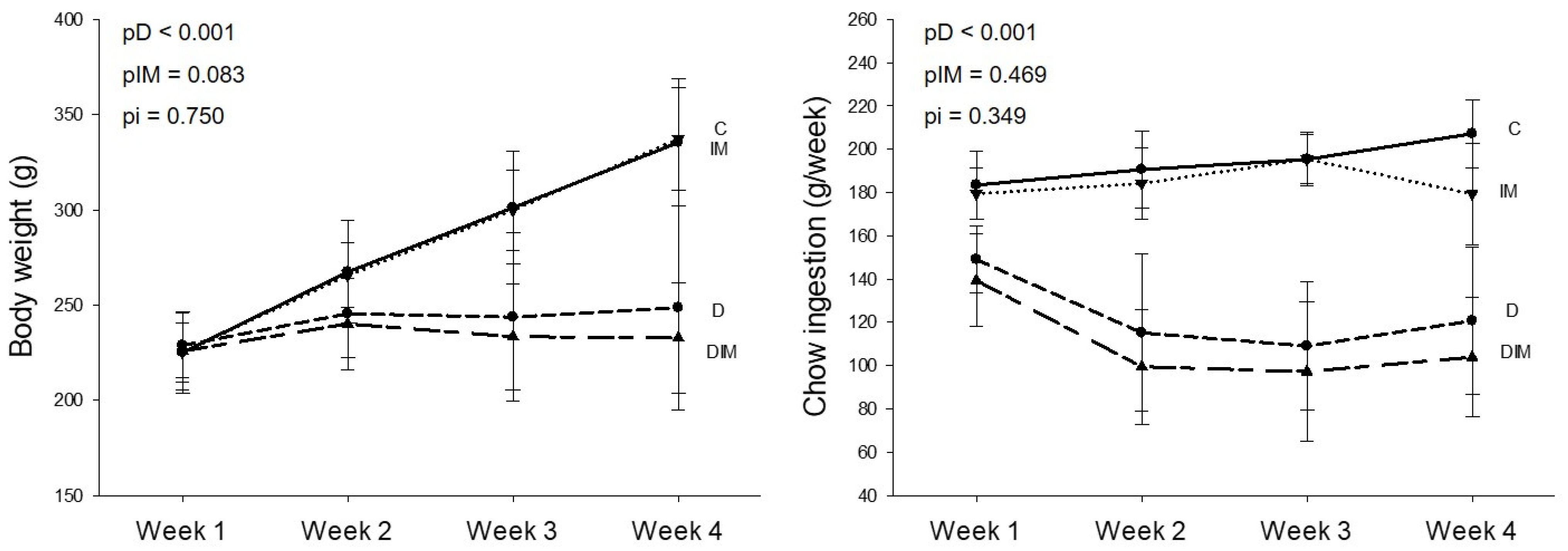
3.1. Body Weight and Fluid and Chow Intake
3.2. Echocardiogram
3.3. Isolated Heart Study
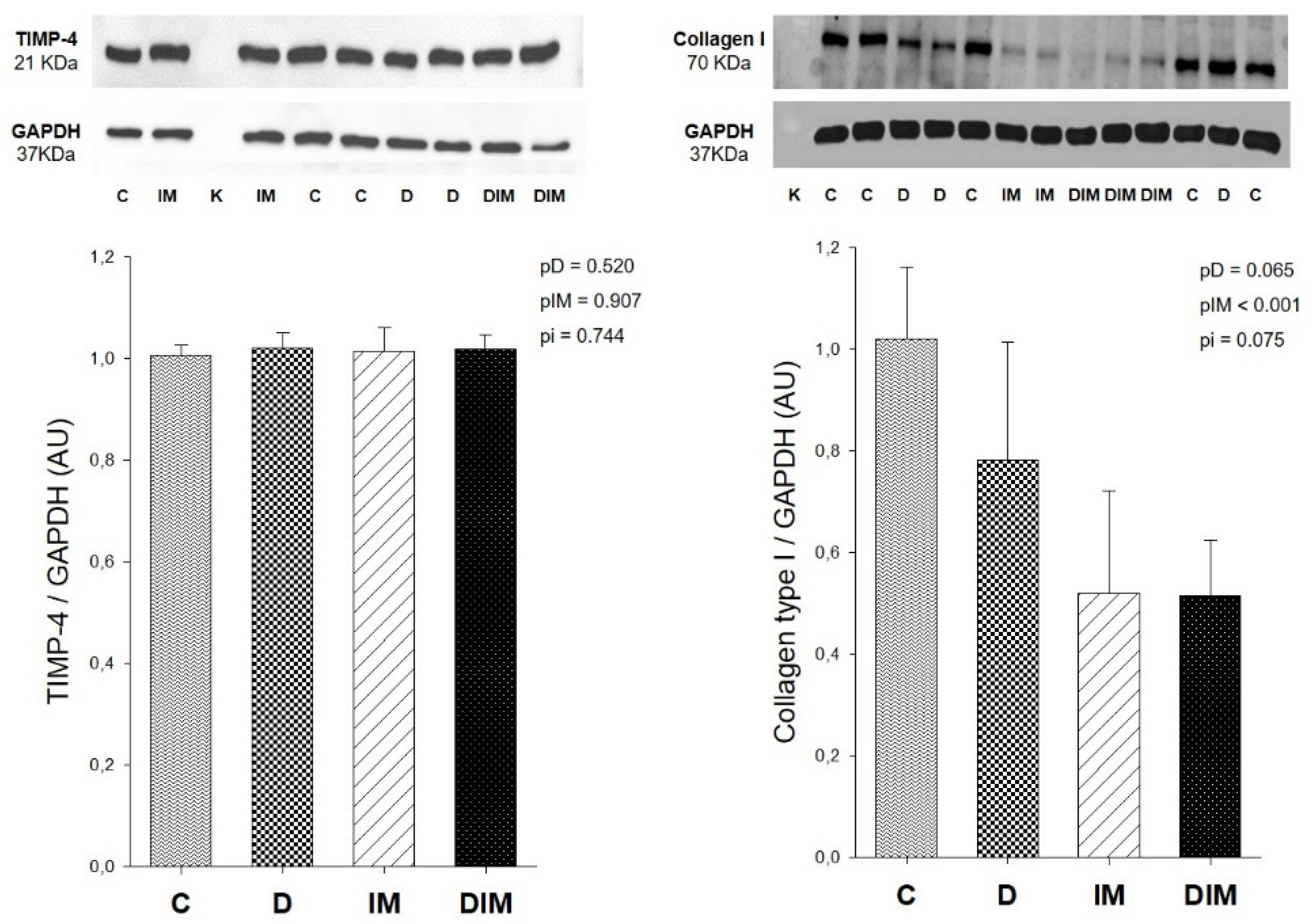
3.4. Collagen Type I and TIMP-4 Protein Expression
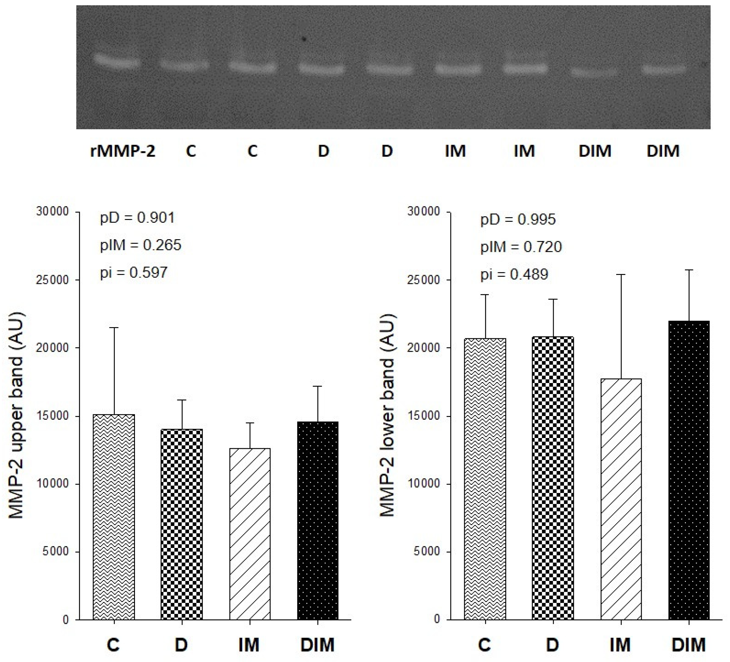
3.5. MMP-2 Activity
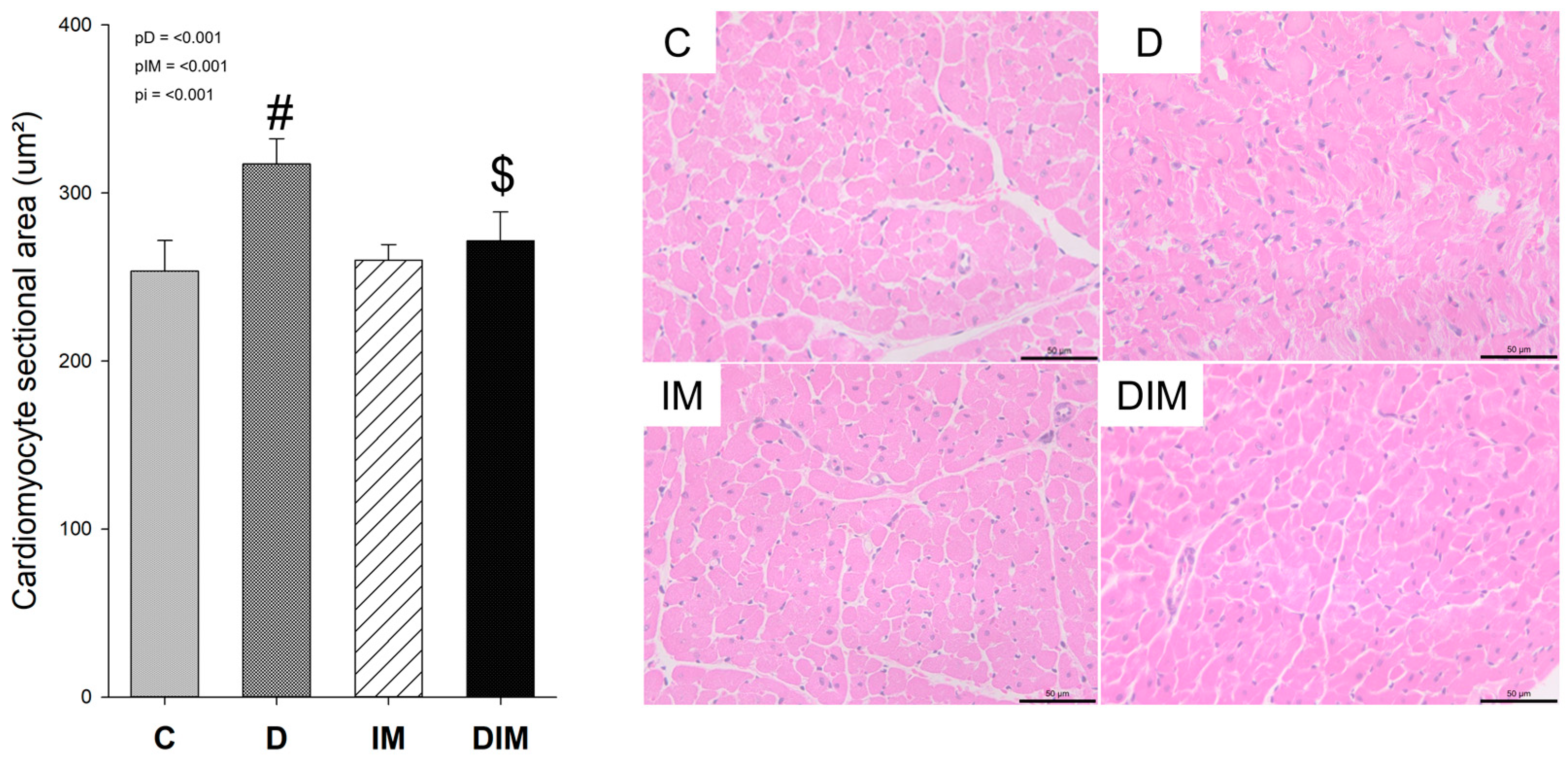
3.6. Histology
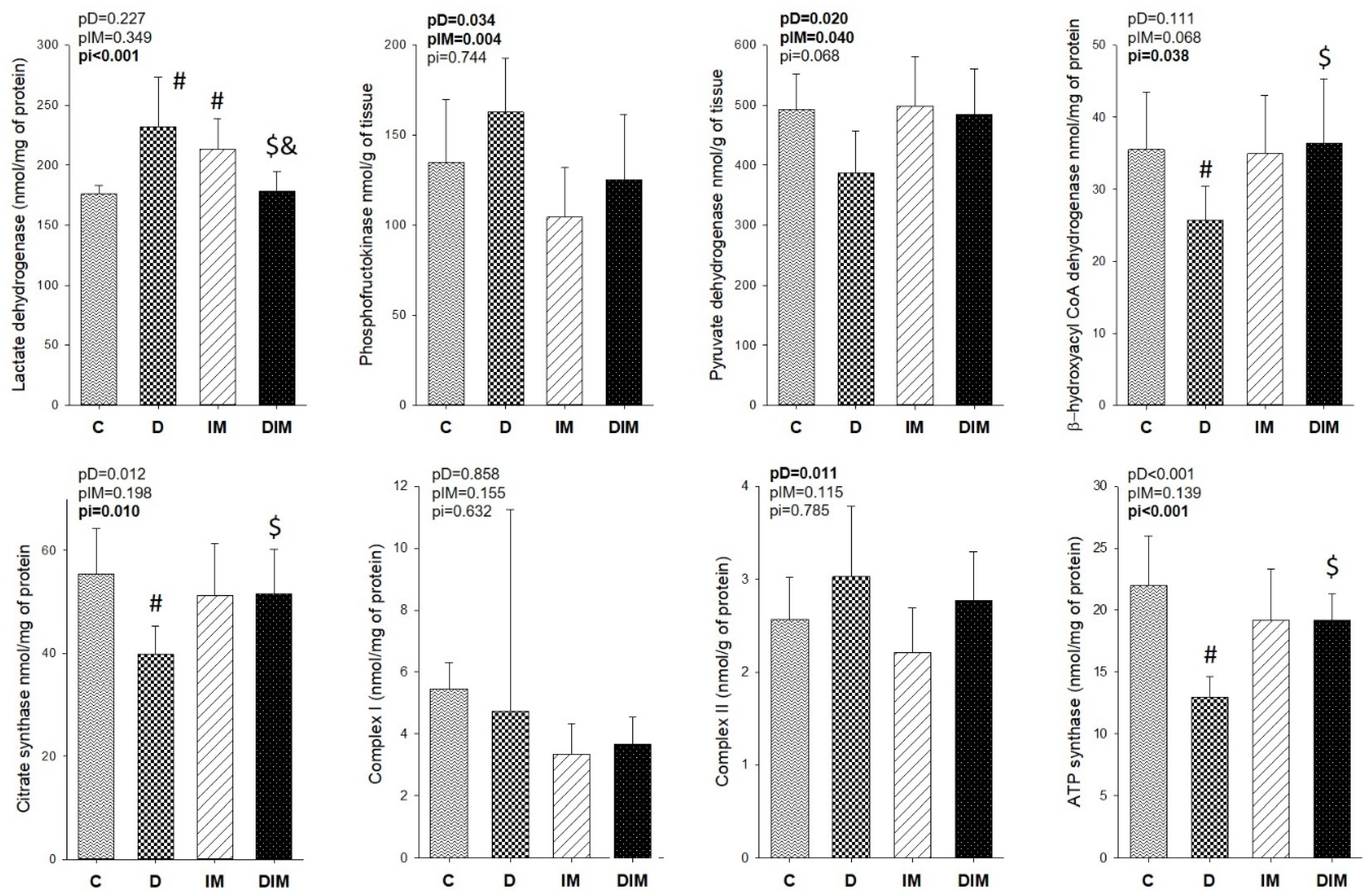
3.7. Myocardial Energy Metabolism
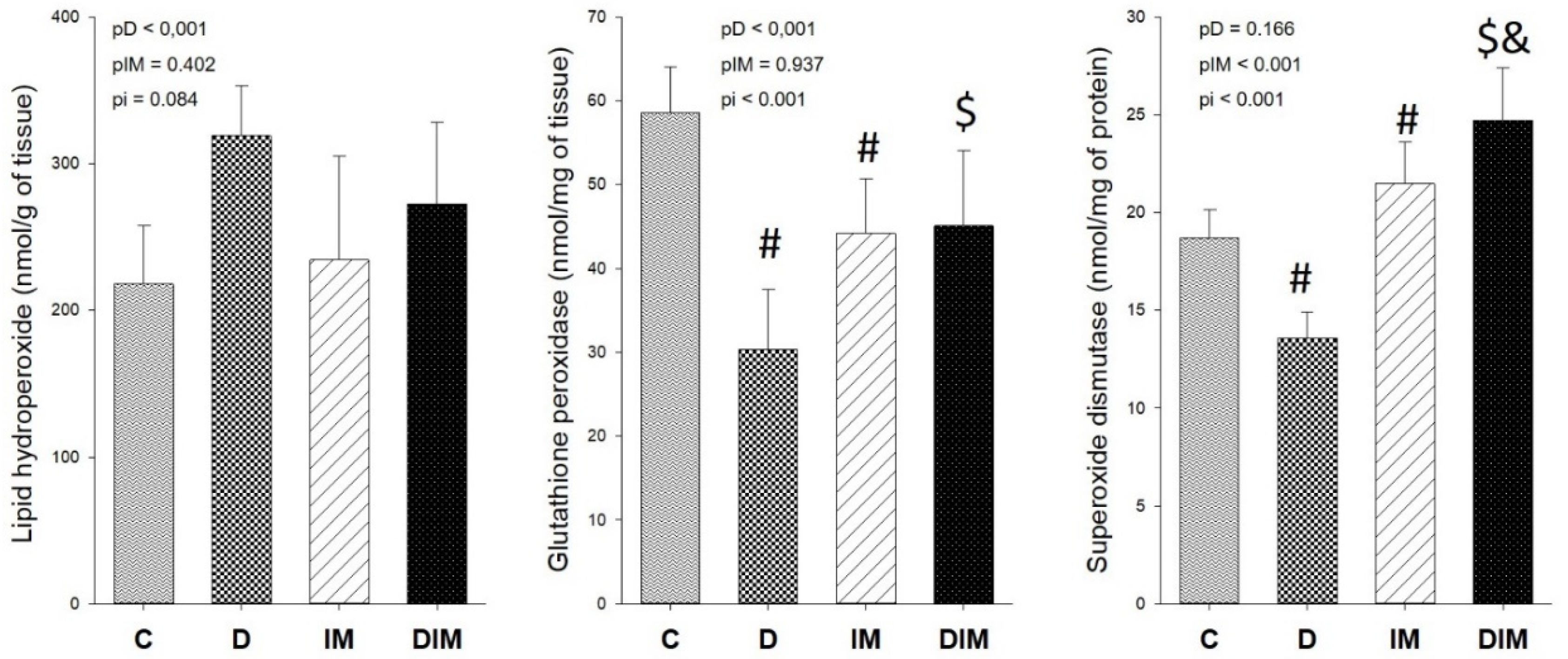
3.8. Oxidative Stress
4. Discussion
5. Conclusions
Author Contributions
Funding
Institutional Review Board Statement
Informed Consent Statement
Data Availability Statement
Conflicts of Interest
References
- Rawat, P.S.; Jaiswal, A.; Khurana, A.; Bhatti, J.S.; Navik, U. Doxorubicin-Induced Cardiotoxicity: An Update on the Molecular Mechanism and Novel Therapeutic Strategies for Effective Management. Biomed. Pharmacother. 2021, 139, 111708. [Google Scholar] [CrossRef] [PubMed]
- Avagimyan, A.; Kakturskiy, L.; Heshmat-Ghahdarijani, K.; Pogosova, N.; Sarrafzadegan, N. Anthracycline Associated Disturbances of Cardiovascular Homeostasis. Curr. Probl. Cardiol. 2021, 47, 100909. [Google Scholar] [CrossRef] [PubMed]
- Vejpongsa, P.; Yeh, E.T.H. Prevention of Anthracycline-Induced Cardiotoxicity: Challenges and Opportunities. J. Am. Coll. Cardiol. 2014, 64, 938–945. [Google Scholar] [CrossRef] [PubMed] [Green Version]
- Chatterjee, K.; Zhang, J.; Honbo, N.; Karliner, J.S. Doxorubicin Cardiomyopathy. Cardiology 2010, 115, 155–162. [Google Scholar] [CrossRef] [PubMed]
- Ribeiro, A.P.D.; Pereira, A.G.; Todo, M.C.; Fujimori, A.S.S.; dos Santos, P.P.; Dantas, D.; Fernandes, A.A.; Zanati, S.G.; Hassimotto, N.M.A.; Zornoff, L.A.M.; et al. Pera Orange (Citrus sinensis) and Moro Orange (Citrus sinensis (L.) Osbeck) Juices Attenuate Left Ventricular Dysfunction and Oxidative Stress and Improve Myocardial Energy Metabolism in Acute Doxorubicin-Induced Cardiotoxicity in Rats. Nutrition 2021, 91–92, 111350. [Google Scholar] [CrossRef]
- Ghigo, A.; Li, M.; Hirsch, E. New Signal Transduction Paradigms in Anthracycline-Induced Cardiotoxicity. Biochim. Biophys. Acta 2016, 1863, 1916–1925. [Google Scholar] [CrossRef]
- Meeran, M.F.N.; Azimullah, S.; Mamoudh, H.H.; Sharma, C.; Kumar, S.; Goyal, S.N.; Ojha, S. Nerolidol, a Sesquiterpene from the Essential Oils of Aromatic Plants, Attenuates Doxorubicin-Induced Chronic Cardiotoxicity in Rats. J. Agric. Food Chem. 2021, 69, 7334–7343. [Google Scholar] [CrossRef]
- Johansson, N. Matrix Metalloproteinases in Tumor Invasion. Cell. Mol. Life Sci. CMLS 2000, 57, 5–15. [Google Scholar] [CrossRef]
- Tampa, M.; Georgescu, S.R.; Mitran, M.I.; Mitran, C.I.; Matei, C.; Caruntu, A.; Scheau, C.; Nicolae, I.; Matei, A.; Caruntu, C.; et al. Current Perspectives on the Role of Matrix Metalloproteinases in the Pathogenesis of Basal Cell Carcinoma. Biomolecules 2021, 11, 903. [Google Scholar] [CrossRef]
- de Carvalho, P.B.; Gonçalves, A.d.F.; Alegre, P.H.C.; Azevedo, P.S.; Roscani, M.G.; Bergamasco, C.M.; Modesto, P.N.; Fernandes, A.A.; Minicucci, M.F.; Paiva, S.A.R.; et al. Pamidronate Attenuates Oxidative Stress and Energetic Metabolism Changes but Worsens Functional Outcomes in Acute Doxorubicin-Induced Cardiotoxicity in Rats. Cell. Physiol. Biochem. Int. J. Exp. Cell. Physiol. Biochem. Pharmacol. 2016, 40, 431–442. [Google Scholar] [CrossRef]
- Polegato, B.F.; Minicucci, M.F.; Azevedo, P.S.; Carvalho, R.F.; Chiuso-Minicucci, F.; Pereira, E.J.; Paiva, S.A.R.; Zornoff, L.A.M.; Okoshi, M.P.; Matsubara, B.B.; et al. Acute Doxorubicin-Induced Cardiotoxicity Is Associated with Matrix Metalloproteinase-2 Alterations in Rats. Cell. Physiol. Biochem. 2015, 35, 1924–1933. [Google Scholar] [CrossRef] [PubMed]
- Bai, P.; Mabley, J.G.; Liaudet, L.; Virág, L.; Szabó, C.; Pacher, P. Matrix Metalloproteinase Activation Is an Early Event in Doxorubicin-Induced Cardiotoxicity. Oncol. Rep. 2004, 11, 505–508. [Google Scholar] [CrossRef] [PubMed]
- Kandasamy, A.D.; Chow, A.K.; Ali, M.A.M.; Schulz, R. Matrix Metalloproteinase-2 and Myocardial Oxidative Stress Injury: Beyond the Matrix. Cardiovasc. Res. 2010, 85, 413–423. [Google Scholar] [CrossRef] [Green Version]
- Castro, M.M.; Kandasamy, A.D.; Youssef, N.; Schulz, R. Matrix Metalloproteinase Inhibitor Properties of Tetracyclines: Therapeutic Potential in Cardiovascular Diseases. Pharmacol. Res. 2011, 64, 551–560. [Google Scholar] [CrossRef] [PubMed]
- Castro, M.M.; Tanus-Santos, J.E.; Gerlach, R.F. Matrix Metalloproteinases: Targets for Doxycycline to Prevent the Vascular Alterations of Hypertension. Pharmacol. Res. 2011, 64, 567–572. [Google Scholar] [CrossRef] [Green Version]
- Ogut, D.; Reel, B.; Korkmaz, C.G.; Arun, M.Z.; Micili, S.C.; Ergur, B.U. Doxycycline Down-Regulates Matrix Metalloproteinase Expression and Inhibits NF-ΚB Signaling in LPS-Induced PC3 Cells. Folia Histochem. Cytobiol. 2016, 54, 171–180. [Google Scholar] [CrossRef] [Green Version]
- Parente, J.M.; Blascke de Mello, M.M.; da Silva, P.H.L.; Omoto, A.C.M.; Pernomian, L.; de Oliveira, I.S.; Mahmud, Z.; Fazan, R.; Arantes, E.C.; Schulz, R.; et al. MMP Inhibition Attenuates Hypertensive Eccentric Cardiac Hypertrophy and Dysfunction by Preserving Troponin I and Dystrophin. Biochem. Pharmacol. 2021, 193, 114744. [Google Scholar] [CrossRef]
- Stechmiller, J.; Cowan, L.; Schultz, G. The Role of Doxycycline as a Matrix Metalloproteinase Inhibitor for the Treatment of Chronic Wounds. Biol. Res. Nurs. 2010, 11, 336–344. [Google Scholar] [CrossRef]
- Xu, X.; Abdalla, T.; Bratcher, P.E.; Jackson, P.L.; Sabbatini, G.; Wells, J.M.; Lou, X.-Y.; Quinn, R.; Blalock, J.E.; Clancy, J.P.; et al. Doxycycline Improves Clinical Outcomes during Cystic Fibrosis Exacerbations. Eur. Respir. J. 2017, 49, 1601102. [Google Scholar] [CrossRef] [Green Version]
- Garcia, R.A.; Go, K.V.; Villarreal, F.J. Effects of Timed Administration of Doxycycline or Methylprednisolone on Post-Myocardial Infarction Inflammation and Left Ventricular Remodeling in the Rat Heart. Mol. Cell. Biochem. 2007, 300, 159–169. [Google Scholar] [CrossRef]
- Villarreal, F.J.; Griffin, M.; Omens, J.; Dillmann, W.; Nguyen, J.; Covell, J. Early Short-Term Treatment with Doxycycline Modulates Postinfarction Left Ventricular Remodeling. Circulation 2003, 108, 1487–1492. [Google Scholar] [CrossRef] [PubMed] [Green Version]
- Cerisano, G.; Buonamici, P.; Parodi, G.; Santini, A.; Moschi, G.; Valenti, R.; Migliorini, A.; Colonna, P.; Bellandi, B.; Gori, A.M.; et al. Early Changes of Left Ventricular Filling Pattern after Reperfused ST-Elevation Myocardial Infarction and Doxycycline Therapy: Insights from the TIPTOP Trial. Int. J. Cardiol. 2017, 240, 43–48. [Google Scholar] [CrossRef] [PubMed]
- Chan, B.Y.H.; Roczkowsky, A.; Cho, W.J.; Poirier, M.; Sergi, C.; Keschrumrus, V.; Churko, J.M.; Granzier, H.; Schulz, R. MMP Inhibitors Attenuate Doxorubicin Cardiotoxicity by Preventing Intracellular and Extracellular Matrix Remodelling. Cardiovasc. Res. 2021, 117, 188–200. [Google Scholar] [CrossRef]
- O’Dell, J.R.; Elliott, J.R.; Mallek, J.A.; Mikuls, T.R.; Weaver, C.A.; Glickstein, S.; Blakely, K.M.; Hausch, R.; Leff, R.D. Treatment of Early Seropositive Rheumatoid Arthritis: Doxycycline plus Methotrexate versus Methotrexate Alone. Arthritis Rheum. 2006, 54, 621–627. [Google Scholar] [CrossRef] [PubMed]
- Brown, D.L.; Desai, K.K.; Vakili, B.A.; Nouneh, C.; Lee, H.-M.; Golub, L.M. Clinical and Biochemical Results of the Metalloproteinase Inhibition with Subantimicrobial Doses of Doxycycline to Prevent Acute Coronary Syndromes (MIDAS) Pilot Trial. Arterioscler. Thromb. Vasc. Biol. 2004, 24, 733–738. [Google Scholar] [CrossRef] [Green Version]
- Antonio, R.C.; Ceron, C.S.; Rizzi, E.; Coelho, E.B.; Tanus-Santos, J.E.; Gerlach, R.F. Antioxidant Effect of Doxycycline Decreases MMP Activity and Blood Pressure in SHR. Mol. Cell. Biochem. 2014, 386, 99–105. [Google Scholar] [CrossRef]
- Clemens, D.L.; Duryee, M.J.; Hall, J.H.; Thiele, G.M.; Mikuls, T.R.; Klassen, L.W.; Zimmerman, M.C.; Anderson, D.R. Relevance of the Antioxidant Properties of Methotrexate and Doxycycline to Their Treatment of Cardiovascular Disease. Pharmacol. Ther. 2020, 205, 107413. [Google Scholar] [CrossRef]
- Clemens, D.L.; Duryee, M.J.; Sarmiento, C.; Chiou, A.; McGowan, J.D.; Hunter, C.D.; Schlichte, S.L.; Tian, J.; Klassen, L.W.; O’Dell, J.R.; et al. Novel Antioxidant Properties of Doxycycline. Int. J. Mol. Sci. 2018, 19, 4078. [Google Scholar] [CrossRef] [Green Version]
- Lai, H.-C.; Yeh, Y.-C.; Ting, C.-T.; Lee, W.-L.; Lee, H.-W.; Wang, L.-C.; Wang, K.-Y.; Wu, A.; Liu, T.-J. Doxycycline Suppresses Doxorubicin-Induced Oxidative Stress and Cellular Apoptosis in Mouse Hearts. Eur. J. Pharmacol. 2010, 644, 176–187. [Google Scholar] [CrossRef]
- Liu, Y.; Ramamurthy, N.; Marecek, J.; Lee, H.M.; Chen, J.L.; Ryan, M.E.; Rifkin, B.R.; Golub, L.M. The Lipophilicity, Pharmacokinetics, and Cellular Uptake of Different Chemically-Modified Tetracyclines (CMTs). Curr. Med. Chem. 2001, 8, 243–252. [Google Scholar] [CrossRef]
- Mathias, L.M.B.S.; Alegre, P.H.C.; dos Santos, I.D.O.F.; Bachiega, T.; Figueiredo, A.M.; Chiuso-Minicucci, F.; Fernandes, A.A.; Bazan, S.G.Z.; Minicucci, M.F.; Azevedo, P.S.; et al. Euterpe Oleracea Mart. (Açai) Supplementation Attenuates Acute Doxorubicin-Induced Cardiotoxicity in Rats. Cell. Physiol. Biochem. Int. J. Exp. Cell. Physiol. Biochem. Pharmacol. 2019, 53, 388–399. [Google Scholar] [CrossRef] [PubMed] [Green Version]
- Tyagi, S.C.; Matsubara, L.; Weber, K.T. Direct Extraction and Estimation of Collagenase(s) Activity by Zymography in Microquantities of Rat Myocardium and Uterus. Clin. Biochem. 1993, 26, 191–198. [Google Scholar] [CrossRef]
- Desai, V.G.; Weindruch, R.; Hart, R.W.; Feuers, R.J. Influences of Age and Dietary Restriction on Gastrocnemius Electron Transport System Activities in Mice. Arch. Biochem. Biophys. 1996, 333, 145–151. [Google Scholar] [CrossRef] [PubMed]
- Bass, A.; Brdiczka, D.; Eyer, P.; Hofer, S.; Pette, D. Metabolic Differentiation of Distinct Muscle Types at the Level of Enzymatic Organization. Eur. J. Biochem. 1969, 10, 198–206. [Google Scholar] [CrossRef] [PubMed]
- Fischer, J.C.; Ruitenbeek, W.; Berden, J.A.; Trijbels, J.M.; Veerkamp, J.H.; Stadhouders, A.M.; Sengers, R.C.; Janssen, A.J. Differential Investigation of the Capacity of Succinate Oxidation in Human Skeletal Muscle. Clin. Chim. Acta 1985, 153, 23–36. [Google Scholar] [CrossRef]
- Jiang, Z.Y.; Woollard, A.C.; Wolff, S.P. Lipid Hydroperoxide Measurement by Oxidation of Fe2+ in the Presence of Xylenol Orange. Comparison with the TBA Assay and an Iodometric Method. Lipids 1991, 26, 853–856. [Google Scholar] [CrossRef]
- Ewing, J.F.; Janero, D.R. Microplate Superoxide Dismutase Assay Employing a Nonenzymatic Superoxide Generator. Anal. Biochem. 1995, 232, 243–248. [Google Scholar] [CrossRef]
- Pereira, B.; Costa-Rosa, L.F.B.P.; Bechara, E.J.H.; Newsholme, P.; Curi, R. Changes in the TBARs Content and Superoxide Dismutase, Catalase and Glutathione Peroxidase Activities in the Lymphoid Organs and Skeletal Muscles of Adrenodemedullated Rats. Braz. J. Med. Biol. Res. 1998, 31, 827–833. [Google Scholar] [CrossRef] [Green Version]
- Figueiredo, V.C.; McCarthy, J.J. Targeting Cancer via Ribosome Biogenesis: The Cachexia Perspective. Cell. Mol. Life Sci. 2021. [Google Scholar] [CrossRef]
- Carvalho, C.; Santos, R.X.; Cardoso, S.; Correia, S.; Oliveira, P.J.; Santos, M.S.; Moreira, P.I. Doxorubicin: The Good, the Bad and the Ugly Effect. Curr. Med. Chem. 2009, 16, 3267–3285. [Google Scholar] [CrossRef]
- Wojtacki, J.; Lewicka-Nowak, E.; Leśniewski-Kmak, K. Anthracycline-Induced Cardiotoxicity: Clinical Course, Risk Factors, Pathogenesis, Detection and Prevention—Review of the Literature. Med. Sci. Monit. 2000, 6, 411–420. [Google Scholar] [PubMed]
- Gu, Y.; Walker, C.; Ryan, M.E.; Payne, J.B.; Golub, L.M. Non-Antibacterial Tetracycline Formulations: Clinical Applications in Dentistry and Medicine. J. Oral Microbiol. 2012, 4, 336–344. [Google Scholar] [CrossRef] [PubMed] [Green Version]
- Valentín, S.; Morales, A.; Sánchez, J.L.; Rivera, A. Safety and Efficacy of Doxycycline in the Treatment of Rosacea. Clin. Cosmet. Investig. Dermatol. 2009, 2, 129–140. [Google Scholar] [PubMed] [Green Version]
- Scott, I.U.; Jackson, G.R.; Quillen, D.A.; Larsen, M.; Klein, R.; Liao, J.; Holfort, S.; Munch, I.C.; Gardner, T.W. Effect of Doxycycline vs Placebo on Retinal Function and Diabetic Retinopathy Progression in Patients with Severe Nonproliferative or Non-High-Risk Proliferative Diabetic Retinopathy. JAMA Ophthalmol. 2014, 132, 535–543. [Google Scholar] [CrossRef] [PubMed]
- Chen, S.-H.; Lin, Y.-J.; Wang, L.-C.; Tsai, H.-Y.; Yang, C.-H.; Teng, Y.-T.; Hsu, S.-M. Doxycycline Ameliorates the Severity of Experimental Proliferative Vitreoretinopathy in Mice. Int. J. Mol. Sci. 2021, 22, 11670. [Google Scholar] [CrossRef]
- Rok, J.; Karkoszka, M.; Rzepka, Z.; Respondek, M.; Banach, K.; Beberok, A.; Wrześniok, D. Cytotoxic and Proapoptotic Effect of Doxycycline—An in Vitro Study on the Human Skin Melanoma Cells. Toxicol. Vitr. 2020, 65, 104790. [Google Scholar] [CrossRef]
- Saikali, Z.; Singh, G. Doxycycline and Other Tetracyclines in the Treatment of Bone Metastasis. Anticancer Drugs 2003, 14, 773–778. [Google Scholar] [CrossRef]
- Duivenvoorden, W.C.; Hirte, H.W.; Singh, G. Use of Tetracycline as an Inhibitor of Matrix Metalloproteinase Activity Secreted by Human Bone-Metastasizing Cancer Cells. Invasion Metastasis 1997, 17, 312–322. [Google Scholar]
- Iwasaki, H.; Inoue, H.; Mitsuke, Y.; Badran, A.; Ikegaya, S.; Ueda, T. Doxycycline Induces Apoptosis by Way of Caspase-3 Activation with Inhibition of Matrix Metalloproteinase in Human T-Lymphoblastic Leukemia CCRF-CEM Cells. J. Lab. Clin. Med. 2002, 140, 382–386. [Google Scholar] [CrossRef]
- Platt, B.N.; Jacobs, C.A.; Conley, C.E.W.; Stone, A.V. Tetracycline Use in Treating Osteoarthritis: A Systematic Review. Inflamm. Res. 2021, 70, 249–259. [Google Scholar] [CrossRef]
- Ma, Z.; Zhang, K.; Wang, Y.; Wang, W.; Yang, Y.; Liang, X.; Zhang, Y.; Li, G. Doxycycline Improves Fibrosis-Induced Abnormalities in Atrial Conduction and Vulnerability to Atrial Fibrillation in Chronic Intermittent Hypoxia Rats. Med. Sci. Monit. 2020, 26, e918883. [Google Scholar] [CrossRef] [PubMed]
- Zhang, K.; Zhao, L.; Ma, Z.; Wang, W.; Li, X.; Zhang, Y.; Yuan, M.; Liang, X.; Li, G. Doxycycline Attenuates Atrial Remodeling by Interfering with MicroRNA-21 and Downstream Phosphatase and Tensin Homolog (PTEN)/Phosphoinositide 3-Kinase (PI3K) Signaling Pathway. Med. Sci. Monit. 2018, 24, 5580–5587. [Google Scholar] [CrossRef] [PubMed]
- Cerisano, G.; Buonamici, P.; Valenti, R.; Sciagrà, R.; Raspanti, S.; Santini, A.; Carrabba, N.; Dovellini, E.V.; Romito, R.; Pupi, A.; et al. Early Short-Term Doxycycline Therapy in Patients with Acute Myocardial Infarction and Left Ventricular Dysfunction to Prevent the Ominous Progression to Adverse Remodelling: The TIPTOP Trial. Eur. Heart J. 2014, 35, 184–191. [Google Scholar] [CrossRef] [PubMed] [Green Version]
- Gaignebet, L.; Kańduła, M.M.; Lehmann, D.; Knosalla, C.; Kreil, D.P.; Kararigas, G. Sex-Specific Human Cardiomyocyte Gene Regulation in Left Ventricular Pressure Overload. Mayo Clin. Proc. 2020, 95, 688–697. [Google Scholar] [CrossRef] [PubMed]
- Marian, A.J.; Braunwald, E. Hypertrophic Cardiomyopathy: Genetics, Pathogenesis, Clinical Manifestations, Diagnosis, and Therapy. Circ. Res. 2017, 121, 749–770. [Google Scholar] [CrossRef]
- Hall, M.L.; Ogle, B.M. Cardiac Extracellular Matrix Modification as a Therapeutic Approach. Card. Extracell. Matrix 2018, 1098, 131–150. [Google Scholar] [CrossRef]
- Zhu, H.; Sun, X.; Wang, D.; Hu, N.; Zhang, Y. Doxycycline Ameliorates Aggregation of Collagen and Atrial Natriuretic Peptide in Murine Post-Infarction Heart. Eur. J. Pharmacol. 2015, 754, 66–72. [Google Scholar] [CrossRef]
- Hakamy, S.; Assidi, M.; Jafri, M.A.; Nedjadi, T.; Alkhatabi, H.; Al-Qahtani, A.; Al-Maghrabi, J.; Sait, K.; Al-Qahtani, M.; Buhmeida, A.; et al. Assessment of Prognostic Value of Tissue Inhibitors of Metalloproteinase 3 (TIMP3) Protein in Ovarian Cancer. Libyan J. Med. 2021, 16, 1937866. [Google Scholar] [CrossRef]
- Spinale, F.G. Myocardial Matrix Remodeling and the Matrix Metalloproteinases: Influence on Cardiac Form and Function. Physiol. Rev. 2007, 87, 1285–1342. [Google Scholar] [CrossRef]
- Fan, D.; Takawale, A.; Lee, J.; Kassiri, Z. Cardiac Fibroblasts, Fibrosis and Extracellular Matrix Remodeling in Heart Disease. Fibrogenes. Tissue Repair 2012, 5, 15. [Google Scholar] [CrossRef] [Green Version]
- Meng, X.-M.; Nikolic-Paterson, D.J.; Lan, H.Y. TGF-β: The Master Regulator of Fibrosis. Nat. Rev. Nephrol. 2016, 12, 325–338. [Google Scholar] [CrossRef] [PubMed]
- Nakagawa, T.; Kakizoe, Y.; Iwata, Y.; Miyasato, Y.; Mizumoto, T.; Adachi, M.; Izumi, Y.; Kuwabara, T.; Suenaga, N.; Narita, Y.; et al. Doxycycline Attenuates Cisplatin-Induced Acute Kidney Injury through Pleiotropic Effects. Am. J. Physiol. Renal Physiol. 2018, 315, F1347–F1357. [Google Scholar] [CrossRef] [PubMed]
- Krakauer, T.; Buckley, M. Doxycycline Is Anti-Inflammatory and Inhibits Staphylococcal Exotoxin-Induced Cytokines and Chemokines. Antimicrob. Agents Chemother. 2003, 47, 3630–3633. [Google Scholar] [CrossRef] [PubMed] [Green Version]
- Navarro-Triviño, F.; Pérez-López, I.; Ruíz-Villaverde, R. Doxycycline, an Antibiotic or an Anti-Inflammatory Agent? The Most Common Uses in Dermatology. Actas Dermo-Sifiliogr. Engl. Ed. 2020, 111, 561–566. [Google Scholar] [CrossRef] [PubMed]
- Riba, A.; Deres, L.; Eros, K.; Szabo, A.; Magyar, K.; Sumegi, B.; Toth, K.; Halmosi, R.; Szabados, E. Doxycycline Protects against ROS-Induced Mitochondrial Fragmentation and ISO-Induced Heart Failure. PLoS ONE 2017, 12, e0175195. [Google Scholar] [CrossRef]
- Altoé, L.S.; Alves, R.S.; Miranda, L.L.; Sarandy, M.M.; Bastos, D.S.S.; Gonçalves-Santos, E.; Novaes, R.D.; Gonçalves, R.V. Doxycycline Hyclate Modulates Antioxidant Defenses, Matrix Metalloproteinases, and COX-2 Activity Accelerating Skin Wound Healing by Secondary Intention in Rats. Oxidative Med. Cell. Longev. 2021, 2021, 4681041. [Google Scholar] [CrossRef]
- Henehan, M.; Montuno, M.; De Benedetto, A. Doxycycline as an Anti-Inflammatory Agent: Updates in Dermatology. J. Eur. Acad. Dermatol. Venereol. 2017, 31, 1800–1808. [Google Scholar] [CrossRef]


| Variables | C (n = 18) | D (n = 19) | IM (n = 17) | DIM (n = 20) | pD | pIM | pi |
|---|---|---|---|---|---|---|---|
| HR (bpm) | 302 ± 62.60 | 261 ± 56.80 | 302 ± 52.50 | 270 ± 83.8 | 0.020 | 0.762 | 0.764 |
| LVSD (mm) | 2.65 ± 0.45 | 3.51 ± 0.64 # | 2.82 ± 0.18 | 2.90 ± 0.49 $ | <0.001 | 0.050 | <0.001 |
| LVDD (mm) | 6.60 ± 0.48 | 6.49 ± 0.76 | 6.80 ± 0.53 | 6.40 + 0.43 | 0.054 | 0.701 | 0.278 |
| LADD (mm) | 4.50 ± 0.18 | 4.96 ± 0.46 # | 4.62 ± 0.16 | 4.38 ± 0.30 $& | 0.006 | 0.056 | 0.006 |
| Aorta (mm) | 3.58 ± 0.16 | 3.44 ± 0.16 | 3.62 ± 0.14 | 3.47 ± 0.18 | <0.001 | 0.434 | 0.821 |
| LADD/aorta | 1.26 ± 0.06 | 1.44 ± 0.11 # | 1.28 ± 0.04 | 1.26 ± 0.08 $ | <0.001 | <0.001 | <0.001 |
| LVMI (g/kg) | 1860 ± 275 | 2131 ± 345 | 1956 ± 198 | 2391 ± 230 | <0.001 | 0.006 | 0.196 |
| LVFS | 0.60 ± 0.05 | 0.46 ± 0.07 # | 0.58 ± 0.03 | 0.54 ± 0.06 $ | <0.001 | 0.009 | <0.001 |
| E wave (cm/s) | 80.2 ± 16.00 | 66.4 ± 11.60 | 79.9 ± 8.99 | 75.3 ± 13.00 | 0.003 | 0.148 | 0.120 |
| A wave (cm/s) | 52.5 ± 16.30 | 47.0 ± 18.20 | 54.3 ± 12.80 | 47.7 ± 13.10 | 0.218 | 0.488 | 0.155 |
| IVRT (ms) | 23.8 ± 2.62 | 35.6 ± 6.42 | 22.7 ± 1.36 | 29.3 ± 6.42 | 0.006 | 0.294 | 0.160 |
| E’m (cm/s) | 5.75 ± 0.63 | 3.84 ± 1.23 # | 5.35 ± 0.80 | 4.81 ± 1.78 $ | <0.001 | 0.320 | 0.019 |
| A’m (cm/s) | 3.72 ± 0.33 | 4.07 ± 0.87 | 3.31 ± 0.72 | 4.33 ± 0.69 & | <0.001 | 0.638 | 0.040 |
| E/E’m | 13.9 ± 1.84 | 18.4 ± 4.92 | 15.1 ± 1.64 | 17.0 ± 4.07 | <0.001 | 0.872 | 0.109 |
| S’m (cm/s) | 5.74 ± 0.28 | 4.22 ± 0.79 # | 5.58 ± 0.35 | 4.79 ± 0.86 $& | 0.006 | 0.160 | 0.006 |
| Variables | C (n = 8) | D (n = 5) | IM (n = 6) | DIM (n = 8) | pD | pIM | pi |
|---|---|---|---|---|---|---|---|
| V0 | 75.0 ± 22.2 | 56.0 ± 16.4 | 67.5 ± 18.9 | 61.3 ± 24.6 | 0.147 | 0.895 | 0.456 |
| +dP/dt | 5219 ± 743.3 | 4100 ± 1413 | 3833 ± 1211 | 3641 ± 1407 | 0.177 | 0.062 | 0.335 |
| −dP/dt | 3891 ± 362.5 | 3175 ± 1134 | 3063 ± 1078 | 2719 ± 903.2 | 0.136 | 0.074 | 0.593 |
| SP | 201 ± 24.0 | 174.5 ± 43.4 | 164 ± 39.6 | 156 ± 37.0 | 0.230 | 0.071 | 0.540 |
Publisher’s Note: MDPI stays neutral with regard to jurisdictional claims in published maps and institutional affiliations. |
© 2022 by the authors. Licensee MDPI, Basel, Switzerland. This article is an open access article distributed under the terms and conditions of the Creative Commons Attribution (CC BY) license (https://creativecommons.org/licenses/by/4.0/).
Share and Cite
Dantas, D.; Pereira, A.G.; Fujimori, A.S.S.; Ribeiro, A.P.D.; de Almeida Silva, C.C.V.; Monte, M.G.; Corrêa, C.R.; Fernandes, A.A.; Bazan, S.G.Z.; Azevedo, P.S.; et al. Doxycycline Attenuates Doxorubicin-Induced Cardiotoxicity by Improving Myocardial Energy Metabolism in Rats. J. Cardiovasc. Dev. Dis. 2022, 9, 254. https://doi.org/10.3390/jcdd9080254
Dantas D, Pereira AG, Fujimori ASS, Ribeiro APD, de Almeida Silva CCV, Monte MG, Corrêa CR, Fernandes AA, Bazan SGZ, Azevedo PS, et al. Doxycycline Attenuates Doxorubicin-Induced Cardiotoxicity by Improving Myocardial Energy Metabolism in Rats. Journal of Cardiovascular Development and Disease. 2022; 9(8):254. https://doi.org/10.3390/jcdd9080254
Chicago/Turabian StyleDantas, Danielle, Amanda Gomes Pereira, Anderson Seiji Soares Fujimori, Ana Paula Dantas Ribeiro, Carol Cristina Vágula de Almeida Silva, Marina Gaiato Monte, Camila Renata Corrêa, Ana Angélica Fernandes, Silmeia Garcia Zanati Bazan, Paula Schmidt Azevedo, and et al. 2022. "Doxycycline Attenuates Doxorubicin-Induced Cardiotoxicity by Improving Myocardial Energy Metabolism in Rats" Journal of Cardiovascular Development and Disease 9, no. 8: 254. https://doi.org/10.3390/jcdd9080254