Article

Antioxidant Enzymes and Heat Shock Protein Genes from Liposcelis bostrychophila Are Involved in Stress Defense upon Heat Shock

1
Key Laboratory of Entomology and Pest Control Engineering, College of Plant Protection, Southwest University, Chongqing 400715, China
2
Academy of Agricultural Sciences, Southwest University, Chongqing 400715, China
*
Author to whom correspondence should be addressed.
Insects 2020, 11(12), 839; https://doi.org/10.3390/insects11120839
Submission received: 2 November 2020 / Revised: 24 November 2020 / Accepted: 26 November 2020 / Published: 27 November 2020
(This article belongs to the Section Insect Physiology, Reproduction and Development)

Abstract

:

Simple Summary

Liposcelis bostrychophila is one of the most serious pests of stored commodities among the psocids. Controlling psocids mainly relies on chemical insecticides and heat stress. In fact, L. bostrychophila has developed high levels of resistance or tolerance to heat treatment in grain storage systems. In this study, we evaluated the changes in malondialdehyde (MDA) concentration after different high temperatures. The result showed that MDA is increased slightly overall, but a drastic increase is detected at 42.5 °C for exposure of different times. To further explore the principles of L. bostrychophila in response to heat stress, we tested the changes of superoxide dismutase (SOD), catalase (CAT), peroxidases (POD) and glutathione-S-transferases (GST) activities under different heat treatments and identified four inducible LbHsp70 genes and one LbHsp110 gene. Enzyme activities and transcript levels changed drastically after different heat treatments. These findings contribute to our understanding of the mechanism of L. bostrychophila responding to heat stress and provide baseline information for further understanding the excellent targets of L. bostrychophila.

Abstract

Psocids are a new risk for global food security and safety because they are significant worldwide pests of stored products. Among these psocids, Liposcelis bostrychophila has developed high levels of resistance or tolerance to heat treatment in grain storage systems, and thus has led to investigation of molecular mechanisms underlying heat tolerance in this pest. In this study, the time-related effects of thermal stress treatments at relatively high temperatures on the activities of antioxidant enzymes, including superoxide dismutase (SOD), catalase (CAT), peroxidases (POD), glutathione-S-transferases (GST) and malondialdehyde (MDA), of L. bostrychophila were determined. Thermal stress resulted that L. bostrychophila had a significantly higher MDA concentration at 42.5 °C, which indicated that the heat stress increased lipid peroxidation (LPO) contents and oxidative stress in this psocid pest. Heat stress also resulted in significant elevation of SOD, CAT and GST activities but decreased POD activity. Our data indicates that different antioxidant enzymes contribute to defense mechanisms, counteracting oxidative damage in varying levels. POD play minor roles in scavenging deleterious LPO, while enhanced SOD, CAT and GST activities in response to thermal stress likely play a more important role against oxidative damage. Here, we firstly identified five LbHsps (four LbHsp70s and one LbHsp110) from psocids, and most of these LbHsps (except LbHsp70-1) are highly expressed at fourth instar nymph and adults, and LbHsp70-1 likely presents as a cognate form of HSP due to its non-significant changes of expression. Most LbHsp70s (except LbHsp70-4) are significantly induced at moderate high temperatures (<40 °C) and decreased at extreme high temperatures (40–45 °C), but LbHsp110-1 can be significantly induced at all high temperatures. Results of this study suggest that the LbHsp70s and LbHsp110 genes are involved in tolerance to thermal stress in L. bostrychophila, and antioxidant enzymes and heat shock proteins may be coordinately involved in the tolerance to thermal stress in psocids.

1. Introduction

Temperature is one of the most important environmental factors that affect life history, behavioral and physiological traits and community composition in insects [1]. As heterothermic animals, insects have evolved a variety of behavioral and physiological adaptations to avoid temperature impairments [2,3]. Thermal stress tolerance is essential for completion of insect life cycles, and insects may exhibit alterations in behavior, morphology, life history and physiological characteristics when exposed to thermal stress. Enhanced antioxidant enzymes activities and the production of heat shock proteins (HSPs) have been widely considered to be the most important strategies that help insects generate tolerance to temperature stress [4,5,6,7].
Thermal stress can lead to oxidative damage and oxidative stress, which involves elevated intracellular levels of reactive oxygen species (ROS) that can damage lipids, proteins and DNA [8]. Avoiding ROS damage, antioxidant enzymes are involved in the oxidative damage response [9,10]. Generally, there is a balance between ROS production and antioxidant processes. However, thermal stress can disrupt this balance and lead to synthesis of additional ROS, and thus result in LPO via cell lipids disruption [11,12]. LPO could be determined indirectly by measuring malondialdehyde (MDA) concentration [13]. In insects, the primary antioxidative enzymes mainly include superoxide dismutase (SOD), catalase (CAT), peroxidase (POD) and glutathione S-transferases (GSTs) [14,15,16]. SOD converts superoxide anion (O2) into oxygen (O2) and hydrogen peroxide (H2O2), while CAT and POD break down H2O2 into H2O and O2. GST removes the harmful products of LPO or hydroperoxides from cells [13,15]. The remarkable increase of the antioxidant enzymes’ activities is a sign of oxidative stress, as well as an indicator of strong capability for combating oxidative stress through eliminating ROS in cells [1,17].
Heat shock proteins are highly conserved proteins that are synthesized as stress proteins when an organism is exposed to stresses, e.g., extreme temperatures, disease, toxins, hypoxia, ionizing and ultraviolet (UV) radiations. HSPs are a first line of defense against stressful damage, as well as playing important roles in molecular and physiological functions, i.e., DNA repair, oogenesis, embryo development and diapause [18]. According to their molecular weights and sequence similarities, HSPs can be divided into five families, including small Hsps (sHsps), Hsp60s, Hsp70s, Hsp90s and Hsp110s [19]. Among these HSPs families, Hsp70s is one of the most heat-inducible and evolutionally conserved proteins in terms of structure and function [20]. Hsp70 may also prevent apoptosis by interfering with the signaling events that trigger the apoptotic process [21]. Two major conserved domains are usually associated with Hsp70 function, i.e., nucleotide-binding domain (NBD) and substrate-binding domain (SBD) [22].
Hsp110s have close relationships with Hsp70s, but they generally exhibit a longer C-terminal extension. Hsp110s are essentially described as chaperones preventing the aggregation of denatured proteins [23]. Hsp110 genes act as a nucleotide exchange factor, releasing peptide substrate from Hsp70 in an ATP-dependent manner. Recently, a study showed that Hsp110 genes can be induced by heat stress in animals, and moreover, they showed different responses to thermal stress in different species from the same genus [24]. Hsp110 genes may be heat-inducible only in the more thermo-tolerant species, and this trait allows the differential expansion of one species relative to another closely related species under current climate change scenarios. Hsp110s are sometimes still mentioned as a subgroup of the Hsp70 family due to their function as a nucleotide exchange factor for Hsp70, often in complex with the co-chaperone Hsp40. However, recent structural studies suggest that they belong to different families because of their functional differences with Hsp70s [18].
The genus Liposcelis psocids, also named booklice, is a group of important stored-product pests worldwide, and L. bostrychophila is one of the most serious pests of stored commodities among the psocids [25]. Recently, L. bostrychophila and L. entomophila has developed high levels of resistance or tolerance to heat treatment in grain storage systems [26,27]. Meanwhile, different Liposcelis species and development stages usually differ in heat tolerance. For example, the egg stage is the most tolerant (heating to 46 °C for >35 h can control eggs of L. bostrychophila). In contrast, L. decolor is the most tolerant species at 46–51 °C and L. paeta is most tolerant below 46 °C, but most susceptible at 47–51 °C [28]. Presently, the physiological and molecular mechanisms behind the thermal tolerance in psocids is not fully understood. One study showed that differences in heat tolerance may be due partly to physiological protection afforded by the generation of small heat-inducible proteins, HIP23 and HIP27 in L. entomophila [27].
In this study, the time-related effects (durations of 2, 4 and 8 h) of thermal stress treatments of L. bostrychophila at comparatively high (37.5, 40, 42.5 and 45 °C) temperatures on the activities of antioxidant enzymes, including SOD, CAT, GST, POD as well as LPO levels (MDA), were determined. Besides the antioxidant enzymes, the expression profiles of five LbHsps under different heat stress (35, 37.5, 40, 42.5 and 45 °C) and development stages were also investigated. The aim of this study was to determine how variations in temperature affect antioxidant enzyme activities and HSPs in response to oxidative stress in psocids. Such changes may explain why L. bostrychophila has a very high-temperature plasticity. Studies like this can provide new insights into the molecular mechanisms underlying heat tolerance in psocids and novel avenues for promoting the application of heat treatments of this pest.

2. Materials and Methods

2.1. Insect Rearing

The laboratory colony of L. bostrychophila was originally collected from grain storage facilities in Beibei, Chongqing, China, in 2002. L. bostrychophila was reared with an artificial diet consisting of whole-wheat flour, yeast powder and skimmed milk (10:1:1) in and incubator at 27 ± 0.5 °C and 75–80% relative humidity with a scotophase of 24 h.

2.2. Insect Collection, Thermal Stress and Protein Extraction

The sampling of different developmental stages was performed according to Wang’s method [26]. Based on the life history of L. bostrychophila at 27.5 °C, 5, 11, 15, 19, 22 and 26 days were used for collecting the eggs, first-, second-, third- and fourth-stadium nymphs, and adults, respectively. All the insects were collected under a microscope using soft brushes, and collected samples were stored at −80 °C for future use.
For antioxidant enzyme assays and LbHsps expression analysis, adults weighing approximately 0.1 g were transferred into plastic dishes. Four dishes with psocids were exposed at each target temperature including 37.5, 40, 42.5 and 45 °C. The duration of each temperature treatment was 2, 4 and 8 h for antioxidant enzyme assays. Psocids kept at a temperature of 27.5 °C served as a control. Because the measurements of each assay for the control (27.5 °C) did not vary significantly in a very short term (<8 h), the data for the control were pooled as one in the analysis. To evaluate the expression levels of LbHsps under different high-temperature treatments, one hundred newly emerged adults were exposed at a target temperature for 1 h. The treatments were achieved by using a programmable temperature controller (Panasonic MIR-154-PC, Kadoma, Osaka, Japan). After treatments, the living adults were frozen immediately in liquid nitrogen and stored at −80 °C until assay. Each treatment was replicated three times. The treated adults were first homogenized in 120 mL ice-cold 0.05 M pH 7.0 phosphate buffers. The crude homogenates were centrifuged at 4 °C and 10,000× g for 10 min. The supernatants were collected and stored on ice until assayed. The protein concentration was determined using the Bradford method [29], with bovine serum albumin as standard.

2.3. Enzyme Activity and MDA Assay

The activity of SOD (Item number: A001-3-2), CAT (Item number: A007-1-1), POD (Item number: A084-1-1), GST (Item number: A004) and MDA (Item number: A003-1-2) was determined using commercial assay kits (Nanjing Jiancheng Bioengineering Institute, Jiangsu, China) following the manufacturer’s instructions, with some slight modifications. The absorbance was read in a microplate spectrophotometer (XMark™, Bio-Rad, Hercules, CA, USA). MDA concentration and enzyme activities were calculated according to the methods that have been reported in H. graminicola [30].

2.4. RNA Isolation and cDNA Cloning of LbHsps

Total RNA was extracted using TRIzol Reagent (Invitrogen Life Technologies, Carlsbad, CA, USA) according to the manufacturer’s protocols. RNA quality, purity and concentration were evaluated by measuring the absorbance at OD260/280 using a NanoVue spectrophotometer (GE Healthcare Biosciences, Uppsala, Sweden). The cDNA for cloning and qPCR was synthesized using 1 μg of total RNA with the PrimeScript® RT reagent Kit (Takara Biotechnology Dalian Co., Ltd., Dalian, China) following the manufacturer’s protocol.
Five heat shock protein genes were identified based on the L. bostrychophila transcriptome database [31]. The transcripts, which contain the full-length open reading frames (ORFs), were verified by PCRs with specific primers (Table 1). PCRs were conducted using PrimeSTAR high-fidelity DNA polymerase (Takara, Dalian, China), in accordance with the following protocol: 95 °C for 3 min, followed by 34 cycles of 95 °C for 30 s, 60 °C for 30 s, and 72 °C for 2 min and then with a final extension at 72 °C for 10 min. The amplified products were purified by electrophoresis on agarose gels (1.0–1.5%) and cloned into the pGEM-T Easy vector (Promega, Fitchburg, MA, USA). After confirmation by PCR using primers M13F and M13R, the positive clones containing target genes were isolated and sequenced (BGI, Beijing, China).

2.5. Gene Characterization and Phylogenetic Analysis

Sequence alignments were carried out using DNAMAN software (DNAMAN 5.2.2, Lynnon BioSoft, Quebec, QC, Canada), and ORFs were identified using the ORF Finder (https://www.ncbi.nlm.nih.gov/orffinder/). The molecular weight and isoelectric point of the amino acid sequences were computed by ExPASy Server (http://www.expasy.org/tools/protparam.html). The conserved motifs presented in deduced amino acid sequences were identified using InterPro (http://www.ebi.ac.uk/interpro/scan.html) or BLASTP in NCBI. A phylogenetic tree based on the amino acid sequences of LbHSPs and other insect HSPs was constructed with MEGA 7.0 software using the neighbor-joining method, and bootstrap values were calculated on 1000 replications [32].

2.6. Quantitative Real-Time PCR (qPCR)

The expression profiles of the target genes at different developmental stages and high-temperature exposures were conducted by qPCR. All primers for qPCR were designed in Primer 3.0 (http://frodo.wi.mit.edu/) (Table 1). The β-actin gene (FJ447483) was used to normalize results of target gene expression and to correct for sample-to-sample variation. qPCR was carried out in a Mx3000P thermal cycler (Stratagene, La Jolla, CA, USA) with 20 µL reaction volumes containing 1 µL of template cDNA, 10 µL Go Taq® qPCR Master Mix, 1 µL each of forward and reverse primers (0.2 mM), 0.2 µL Rox and 6.8 µL ddH2O. Thermal cycling conditions were: 95 °C for 2 min, followed by 40 cycles of at 95 °C for 15 s and 60 °C for 30 s. A final melt-curve step (ramping from 60 to 95 °C in 0.6 °C steps every 5 s) at the end was conducted to ensure the consistency and specificity of the amplified product. All of these experiments involved three biological replications. Relative gene expression data were calculated according to the 2–ΔΔCT method [33]. All qPCR assays were validated in compliance with the Minimum Information for Publication of Quantitative Real-Time PCR Experiments (MIQE) guidelines [34].

2.7. Statistical Analysis

Differences in antioxidant enzyme activity assays and LbHsps expression levels among different developmental stages and high-temperature treatments were analyzed by two-way (temperature and treatment duration) or one-way analysis of variance (ANOVA) followed by Tukey’s tests in SPSS 19.0 (SPSS, Inc., Chicago, IL, USA). All the data were expressed as mean ± standard error (SE), and the level of significance was set at p < 0.05.

3. Results

3.1. Changes in MDA

The effects of different heat stresses on MDA concentration of L. bostrychophila are presented in Table 2. MDA concentrations in L. bostrychophila were significantly affected by treatment temperatures (F4,44 = 366.523; p < 0.001) and durations (F2,44 = 115.127; p < 0.001). The results showed that the highest concentration of MDA with increasing temperature was formed at 42.5 °C (7.763 nmol·mg−1 protein for 2 h, 17.505 nmol·mg−1 protein for 4 h and 5.720 nmol·mg−1 protein for 8 h). When heat stress lasted 4 h, the concentration of MDA was significantly increased with increasing temperatures. However, for exposure of 2 and 8 h, MDA concentration was not significantly enhanced (except at 42.5 °C) compared to controls.

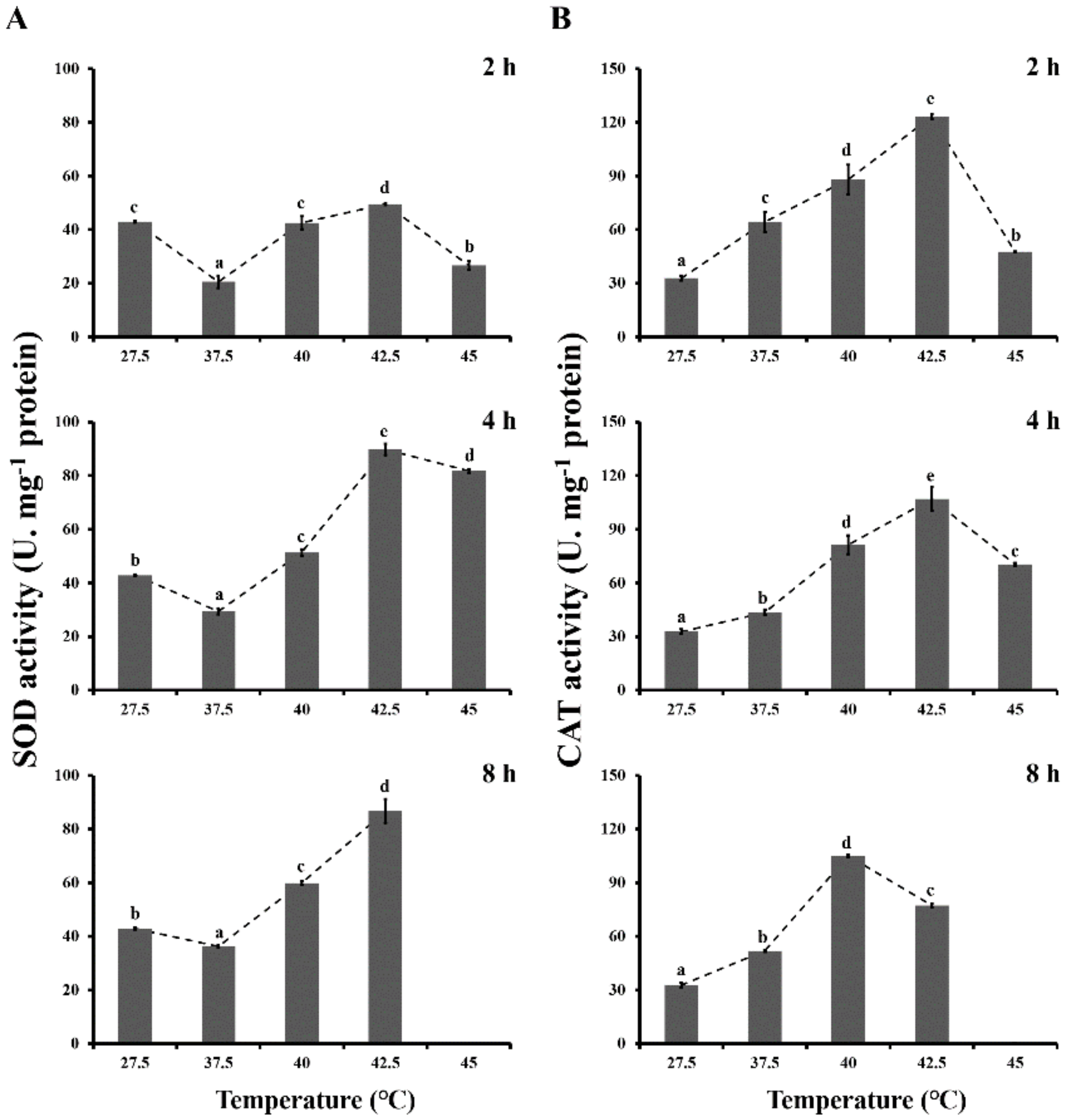
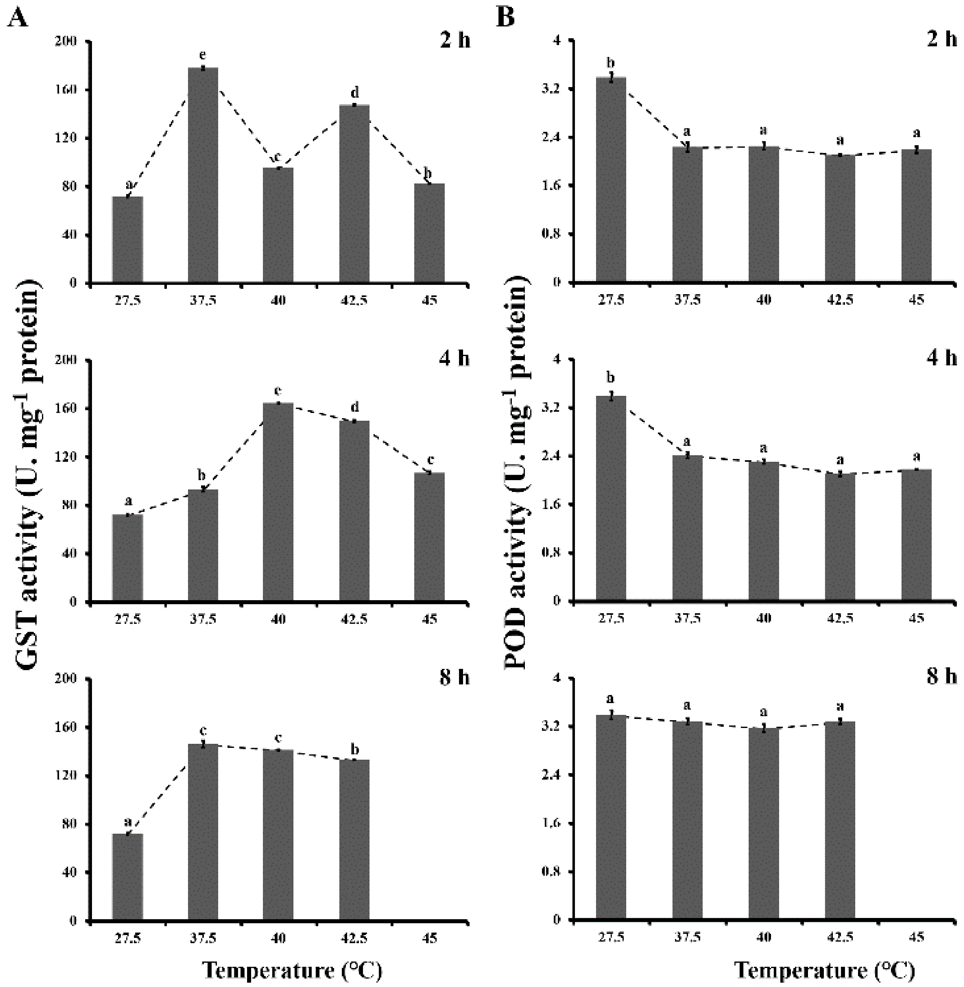
3.2. Antioxidant Enzyme Activities

The SOD activities of L. bostrychophila under heat thermal stresses are presented in Figure 1A. SOD activities were significantly affected by exposure to high-temperature treatments (F4,44 = 338.253; p < 0.001) and various durations (F2, 44 = 228.889; p < 0.001). In some high-temperature treatments (40 and 42.5 °C), SOD activities gradually increased as the temperature increased for durations of 4 and 8 h when compared with the control (27.5 °C). Meanwhile, the highest activity was found at 42.5 °C (49.456 U·mg−1 protein for 2 h, 89.805 U·mg−1 protein for 4 h and 86.716 U·mg−1 protein for 8 h). However, SOD activities decreased significantly at 37.5 °C in all treatment durations.
Changes in CAT activities of L. bostrychophila after the heat shock for different durations are presented in Figure 1B. CAT activities were significantly affected by the high temperatures (F4,44 = 235.305; p < 0.001) and durations (F2,44 = 34.430; p < 0.001). Compared to control (27.5 °C), a significant upregulation of CAT activities was detected during all heat shock treatments for three durations (2, 4 and 8 h). When exposure lasted 2 and 4 h, CAT activities reached a maximum at 42.5 °C (123.096 U·mg−1 protein for 2 h, 106.972 U·mg−1 protein for 4 h), whereas the highest activity was found at 40 °C for exposure of 8 h (105.010 U·mg−1 protein).
The effects of different heat stress on GST activities of L. bostrychophila are presented in Figure 2A. The statistical analysis showed that GST activities were significantly affected by treatment temperatures (F4,44 = 3393.354; p < 0.001) and durations (F2,44 = 381.410; p < 0.001). GST activities increased significantly during all heat shock treatments for three durations (2, 4 and 8 h) in comparison with the control (27.5 °C). The highest activities were found for exposure of 2 and 8 h at 37.5 °C and for exposure of 4 h at 40 °C (177.715 U·mg−1 protein for 2 h, 164.335 U·mg−1 protein for 4 h and 146.171 U·mg−1 protein for 8 h). GST activities increased in various degrees for 2 h duration by 147.5% at 37.5 °C, 32.3% at 40 °C, 105.0% at 42.5 °C and 14.6% at 45 °C respectively, when compared with the control.
POD activities of L. bostrychophila under different thermal stress are presented in Figure 2B. POD activity was found to have a significant difference among the different treatment temperatures (F4,44 = 479.776; p < 0.001) and durations (F2,44 = 15.803; p < 0.001). However, for exposure of 2 and 4 h heat shock treatments, the POD activities were significantly decreased compared to the control, but there was no obvious change among themselves.

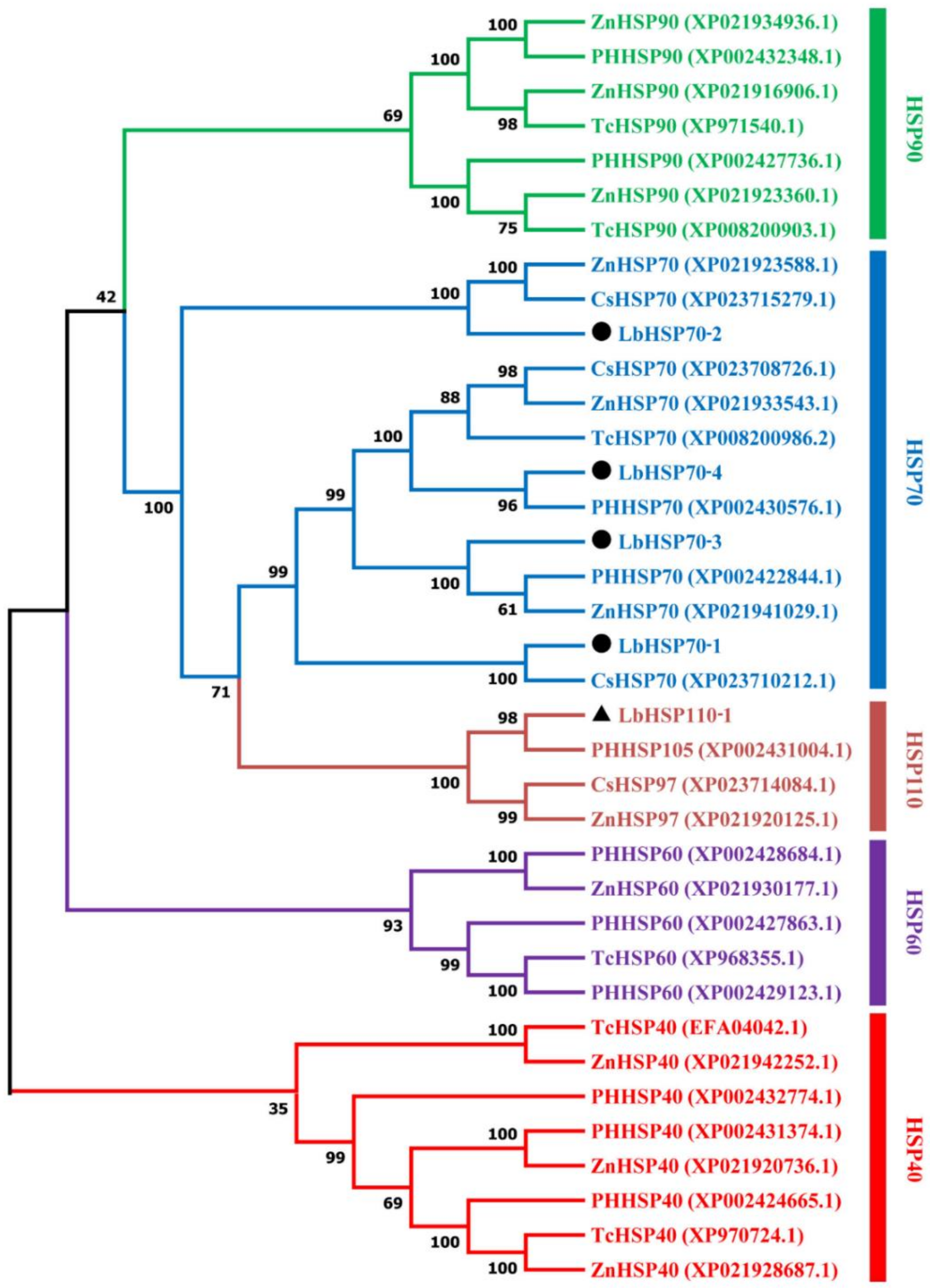
3.3. Sequence Analysis and Phylogenetic Tree

In this study, four genes encoding Hsp70s and one gene encoding Hsp110 were identified in L. bostrychophila, namely, LbHsp70-1, LbHsp70-2, LbHsp70-3, LbHsp70-4 and LbHsp110-1. For four LbHsp70 genes, the length of the deduced amino acid sequences varied from 505 to 690 amino acids, and the predicted molecular weight ranged from 125.2 to 167.4 kDa with the theoretical isoelectric points of 4.95–5.06. For LbHsp110 gene, the length of the deduced amino acid sequence is 829 amino acids, and the predicted molecular weight is 201.3 kDa with the theoretical isoelectric point of 4.91 (Supplementary Table S1). Multiple alignments of amino acid sequences of LbHsps were conducted (Supplementary Figures S1 and S2). The similarity of amino acid sequences among four LbHsp70 genes is 50.90%, and the similarity of amino acid sequences between LbHsp110 gene and four LbHsp70 genes is 38.77%. The deduced amino acid sequences of these genes contained signature sequences and motifs typical of the Hsp70 family and Hsp110 family (Supplementary Figures S1 and S2). The C-termini of three LbHsp70s contain the conserved EEVD motif, which enables Hsp70 to interact with chaperones in the cytoplasm [35]. The N-termini of the LbHsp70s contain a conserved sequence with an ATP-GTP binding site, which is associated with conformational switching and ATPase activity. The constructed phylogeny tree showed that five LbHsp genes fall into two clades (Figure 3). Phylogenetic analysis with other well-documented HSP proteins clearly classified LbHsp70-1, LbHsp70-2, LbHsp70-3 and LbHsp70-4 into the Hsp70 clade, and LbHsp110-1 was located in the Hsp110 clade.

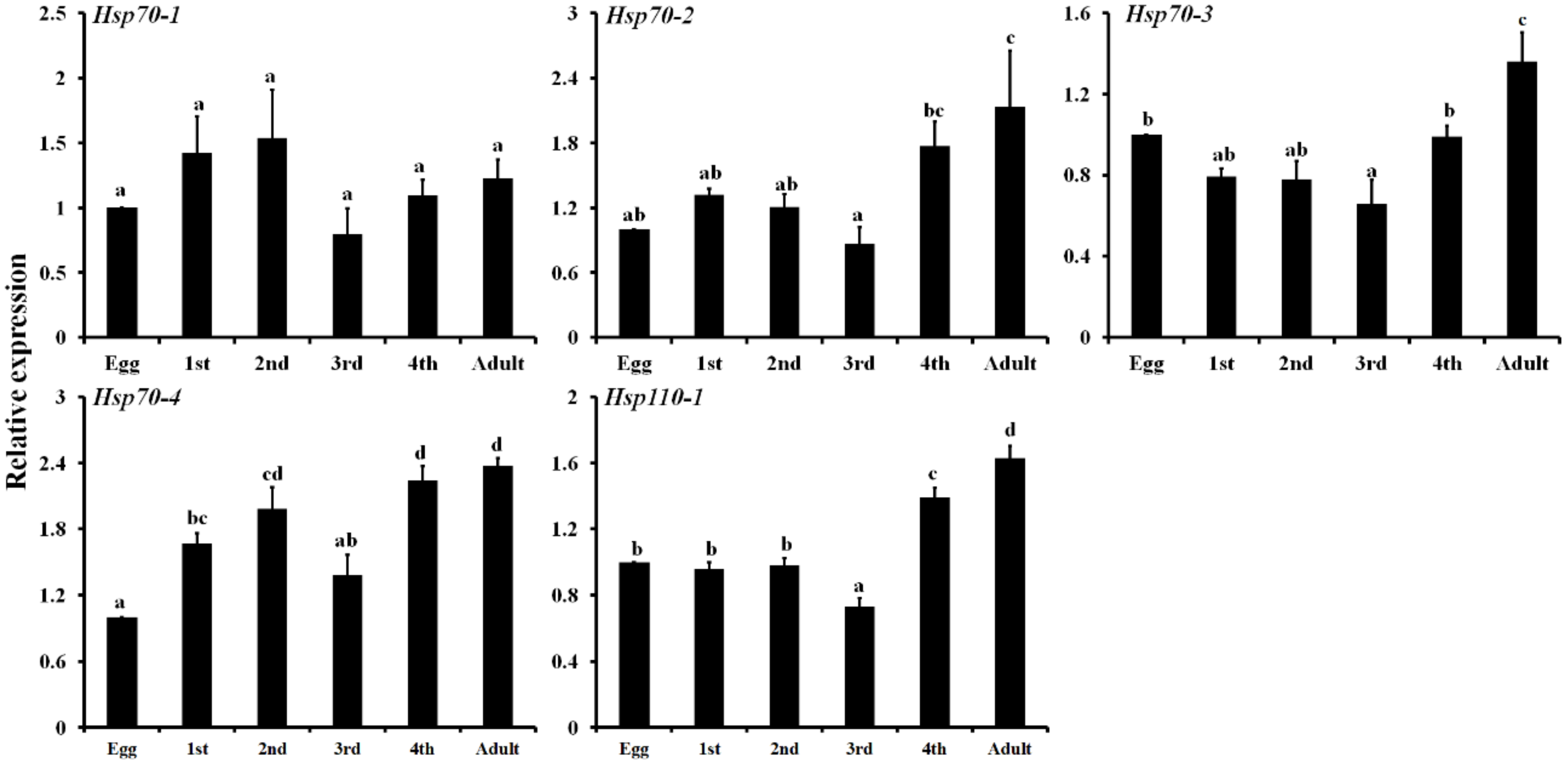
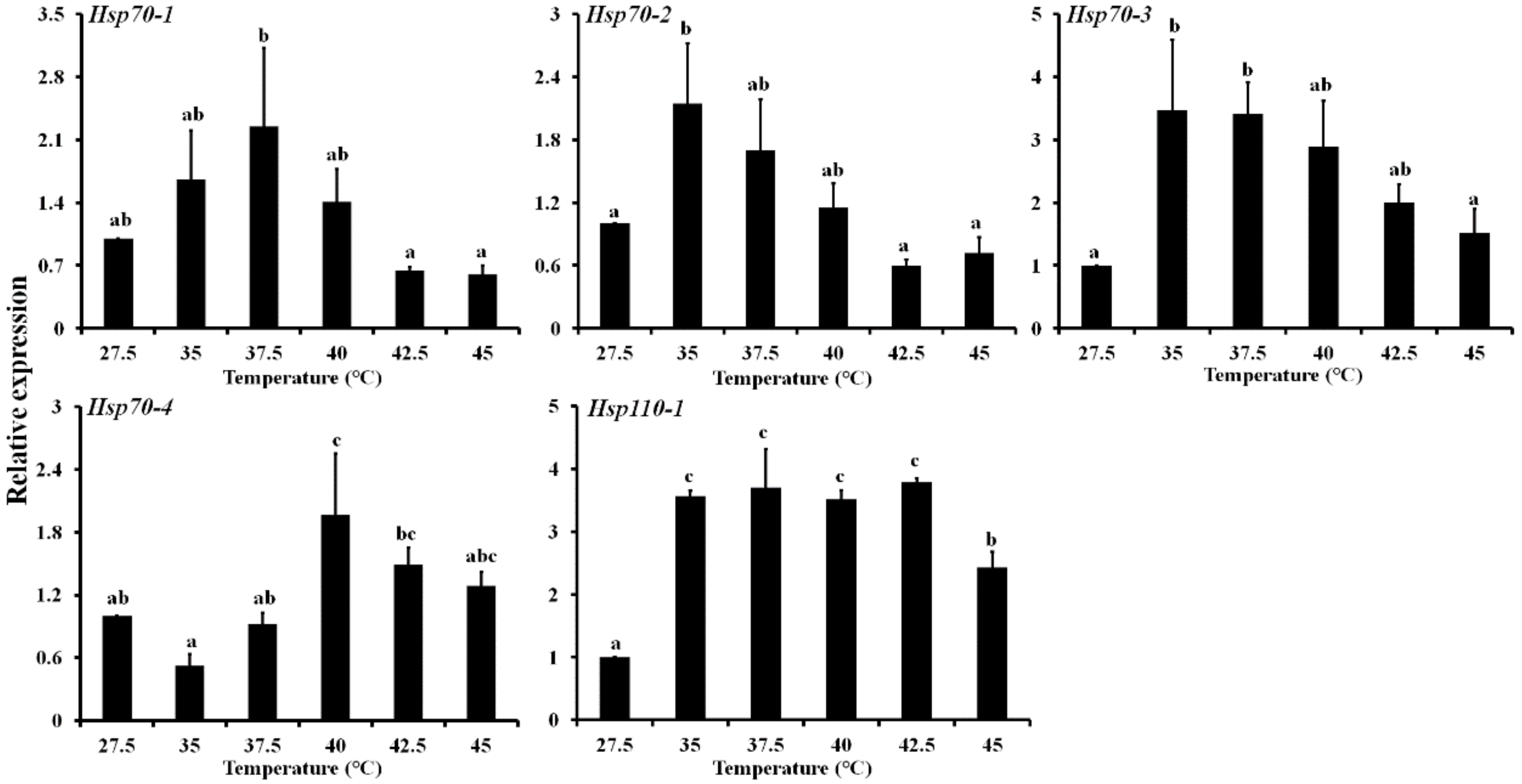
3.4. Expression Profiling of Five LbHsps

The expression patterns of LbHsps at different development stages (eggs, first to fourth-instar nymph, adults) were investigated by qPCR. The results showed that all five genes were consistently expressed for all the tested stages, and expression of these genes varied in various developmental stages (Figure 4). All the genes were highly expressed in adults except the LbHsp70-1. Generally, the relative expressions of five LbHsp genes in the egg and the third nymph stage are lower than that in other stages. For LbHsp70-4, the transcriptional level of the three nymph (first, second, and fourth) stages and adult stages were significantly higher than that in the egg stage, with an increase of 1.67-, 1.99-, 2.24- and 2.37-fold, respectively. The mRNA abundances of the five LbHsps were also evaluated in response to high-temperature stresses by qPCR (Figure 5). These LbHsp genes were upregulated in different degrees by heat treatments below 42.5 °C, and then the abundances of these genes declined at higher temperatures. For LbHsp110-1, the mRNA expression levels were significantly increased by 3.57-, 3.70-, 3.51-, 3.79- and 2.42-fold respectively, in comparison with the control (Figure 5).

4. Discussion

Many studies of insect–thermal stress interaction have revealed that insects have evolved complex protective mechanisms to protect themselves against high temperatures, and antioxidant enzymes and heat shock proteins are the most well-known effectors in this process [6,9]. Insects are usually equipped with a comprehensive antioxidant defense system to relieve oxidative stress and alleviate the effect of damaged macromolecules produced by thermal stress.
MDA is a biological marker of oxidative stress. MDA can be used to determine the degree of lipid peroxidation, because it is a major oxidation product of peroxidized polyunsaturated fatty acids [36,37]. In the current study, MDA concentration is increased slightly overall, but a drastic increase is detected at 42.5 °C for exposure of different times. These results indicated that high-temperature stress led to increased damage of lipids by ROS in this psocid. The normal level of MDA might be due to enhanced activities of antioxidative enzymes or other defense pathways, which can scavenge excess lipid peroxidation. This study demonstrated that in L. bostrychophila, higher temperature stress was accompanied by lipid peroxidation and other responses to oxidative stress, and the similar results were also reported in P. japonica and H. graminicola [1,30].
SOD plays an important role in antioxidant defense against ROS, reducing levels of superoxide radical, which is induced by high temperatures [38]. In this study, a significant increase of SOD activities was observed in L. bostrychophila adults exposed to high temperature (>37.5 °C) at 4 and 8 h, and it reached maximum values at 42.5 °C. Moreover, SOD activities increase gradually along with the prolongation of exposure time. These results suggest that SOD activity might be an adaptive response of psocids to overcome high-temperature-induced superoxide anion toxicity. Our results are partially consistent with the findings in P. japonica, M. separata, B. dorsalis and N. cucumeris exposed to heat stress [1,39,40,41]. Thus, we infer that SOD plays an important role in the response to short-term and long-term phasic thermal stress in L. bostrychophila.
CAT is the principal H2O2 scavenging enzyme, and SOD and CAT work together in stepwise oxygen reduction [42], but CAT removes H2O2 only at high concentrations and has little effect at low concentrations [43]. In this study, CAT activities in L. bostrychophila adults are higher than controls under different high-temperature stress. Interestingly, with the increase of exposure temperature, CAT activities reach the highest activity at 40 or 42.5 °C, and then reduced slightly at 42.5 or 45 °C. The reasons for these changes of CAT activities are unclear, but may be associated with changes of SOD activities. The similar phenomenon was also found in some previous studies of P. japonica and B. dorsalis [1,17].
GST is a group of multifunctional dimer enzymes that can catalyze the combination of glutathione and a variety of endogenous and heterologous compounds to discharge toxins. It has the function of protecting cells from oxidative damage, isomerization and intercellular transport [44]. The elevated GST activities suggest that GST in L. bostrychophila is involved in the inactivation of toxic lipid peroxidation products accumulated due to oxidative damage by heat shock stress. Similar findings are also reported in P. japonica, P. citri, M. separate, C. suppressalis and A. mylitta [1,9,39,42,45]. H2O2 is also broken down by POD, which can metabolize lipid peroxidation products together with GST [17]. In this study, the POD activities exhibited varied decreased changes under different high-temperature stress at 2 and 4 h exposures. We speculate that other peroxidase enzymes, i.e., CAT, may remove most of these hydrogen peroxides produced by SOD, and thus CAT and GSTs may play a more important role in removing excess ROS than POD when L. bostrychophila is exposed to extreme temperature stress.
Previous studies demonstrated that HSPs play important roles in thermal tolerance of insects [19]. Generally, Hsp genes are developmentally regulated in insects, and the role of HSPs in the regulation of insect growth and development has become an important area of inquiry. In fact, Hsps act within networks, and the functions of individual proteins within an Hsp family often differ with developmental stages, subcellular location and environmental conditions. Here, we show that five LbHsps have different expression levels in different developmental stages. These LbHsp genes are usually highly expressed in fourth nymph and adults, whereas their expressions are lowest at the third nymph stage. Other studies also showed that Hsp70s genes could participate in the regulation of growth and development in some insects [46,47]. For example, the expression of Hsp70 III from T. castaneum varied among developmental stages, suggesting that TcHsp70 III is developmentally regulated [48]. Therefore, we inferred that these Hsp proteins may participate in the development process of L. bostrychophila, and the function of these Hsps may be associated with the ability of defense against stress in the life history of this pest.
Many studies have shown that genes encoding Hsp70 and Hsp20 are generally more susceptible to thermal stress than other Hsps [20]. Generally, the Hsp70 genes were expressed at low levels in normal conditions but were induced differently in response to heat stress [49]. The expression of LbHsp70-4 was significantly increased only at 40 °C, but the other LbHsp70s were significantly induced at moderate high temperature (<40 °C) and decreased at extreme high temperature (40–45 °C). Consistent with this result, in Sitodiplosis mosellana, the expression of SmosHsp70 first increased and then decreased with increasing temperature treatment [50]. These similar expression patterns of Hsp90, Hsp70, Hsp60 and small Hsps were also observed in A. gifuensis, M. cinxia and F. occidentalis [6,49,51]. It should be noted that LbHsp110-1 can be significantly induced at all high temperatures tested (Figure 5). To date, relatively little is known about the expression and the physiological function of insect HSP110s, while numerous studies of Hsp110 have been conducted in mammals and yeast. Previous studies showed that Hsp110 members are co-chaperones of mammalian and yeast Hsp70 chaperones and act as nucleotide exchange factors (NEF) during the ATP hydrolysis cycle [52,53]. Recent studies also showed that Hsp110 genes can be induced by heat stress and play important roles in animal reproduction [24,54].
Differential heat shock tolerance among the stored product psocids Liposcelis species was earlier reported [27,28]. In a previous study, L. entomophila possesses higher relative tolerance to heat shock stress compared to L. reticulatus because it expresses two small Hsps (i.e., HIP 23 and HIP 27). However, the expression of Hsp70 was not observed under heat stress in the above psocids [27]. The reasons for non-detection of Hsp70 expression in the above-mentioned psocids by using Western blot analysis is mainly due to the insensitive technology using a monoclonal antibody or possible breakdown of Hsp70. Combined with the former research results, we conclude that Hsp110 and sHsps may greatly help Liposcelis psocids to tolerate heat, and thus lead to evolution of heat resistance in this pest. We also tentatively infer that LbHsp70s play an important role in L. bostrychophila adults in adapting to thermal stress. The current study suggests that heat tolerance in psocids might lead to their more common occurrence in grain stored worldwide, and the response to heat tolerance in L. bostrychophila might facilitate its development of thermal resistance to heat treatments.

5. Conclusions

The present study confirmed that thermal stress disturbs the redox balance in L. bostrychophila, which leads to oxidative stress. To overcome this stress, antioxidant enzymes, i.e., SOD, CAT and GST, and heat shock proteins are involved in antioxidant response to thermal stress, and thus enable L. bostrychophila adults to efficiently deal with ROS induced by thermal stresses. Further, we firstly identified four LbHsp70 genes and one LbHsp110 gene from psocid, L. bostrychophila. These LbHsps are expressed at all development stages with similar expression patterns to some extent. Different expression profiles of LbHsp70s and LbHsp110 in response to high-temperature stresses reveal that Hsp70 and Hsp110 genes are functionally involved in the resistance or tolerance to thermal stresses in this pest.

Supplementary Materials

The following are available online at https://www.mdpi.com/2075-4450/11/12/839/s1, Figure S1: Multiple amino acid sequence alignments of four Hsp70s from Liposcelis bostrychophila, Figure S2: Multiple alignments of the deduced amino acid sequences of Hsp110s from Liposcelis bostrychophila and other insect species, Table S1: Molecular properties of five Liposcelis bostrychophila heat shock protein genes.

Author Contributions

Conceptualization, Z.Q.M., D.D.W. and J.J.W.; Experiments, Z.Q.M., Y.Q.T., P.Y.G. and T.X.J.; analyzed the data, Z.Q.M., W.H., D.D.W. and T.X.J.; writing, Z.Q.M., and D.D.W. All authors have read and agreed to the published version of the manuscript.

Funding

This work was financially supported partially by the National Natural Science Foundation of China (31972276), and the Fundamental Research Funds for the Central Universities, China (XDJK2018B041).

Conflicts of Interest

The authors declare no conflict of interest. The funding agency had no role in the design of the study; in the collection, analyses, or interpretation of data; in the writing of the manuscript, or in the decision to publish the results.

References

  1. Zhang, S.; Fu, W.; Li, N.; Zhang, F.; Liu, T.X. Antioxidant responses of Propylaea japonica (Coleoptera: Coccinellidae) exposed to high temperature stress. J. Insect Physiol. 2015, 73, 47–52. [Google Scholar] [CrossRef]
  2. Wang, H.S.; Kang, L. Effect of cooling rates on the cold hardiness and cryoprotectant profiles of locust eggs. Cryobiology 2005, 51, 220–229. [Google Scholar] [CrossRef]
  3. Storey, K. Organic Solutes in Freezing Tolerance. Comp. Biochem. Physiol. A Physiol. 1997, 117, 319–326. [Google Scholar] [CrossRef]
  4. Franks, S.J.; Hoffmann, A.A. Genetics of climate change adaptation. Annu. Rev. Genet. 2012, 46, 185–208. [Google Scholar] [CrossRef]
  5. King, A.M.; MacRae, T.H. Insect heat shock proteins during stress and diapause. Annu. Rev. Entomol. 2015, 60, 59–75. [Google Scholar] [CrossRef]
  6. Kang, Z.W.; Liu, F.H.; Liu, X.; Yu, W.B.; Tan, X.L.; Zhang, S.Z.; Tian, H.G.; Liu, T.X. The potential coordination of the heat shock proteins and antioxidant enzyme genes of Aphidius gifuensis in response to thermal stress. Front. Physiol. 2017, 8, 12. [Google Scholar] [CrossRef] [Green Version]
  7. Arias, M.B.; Poupin, M.J.; Lardies, M.A. Plasticity of life-cycle, physiological thermal traits and Hsp70 gene expression in an insect along the ontogeny: Effect of temperature variability. J. Therm. Biol. 2011, 36, 355–362. [Google Scholar] [CrossRef]
  8. Lopez-Martinez, G.; Elnitsky, M.A.; Benoit, J.B.; Lee, R.E.; Denlinger, D.L. High resistance to oxidative damage in the Antarctic midge Belgica antarctica, and developmentally linked expression of genes encoding superoxide dismutase, catalase and heat shock proteins. Insect Biochem. Mol. Biol. 2008, 38, 796–804. [Google Scholar] [CrossRef] [PubMed]
  9. Yang, L.H.; Huang, H.; Wang, J.J. Antioxidant responses of citrus red mite, Panonychus citri (McGregor) (Acari: Tetranychidae), exposed to thermal stress. J. Insect Physiol. 2010, 56, 1871–1876. [Google Scholar] [CrossRef] [PubMed]
  10. Chen, H.; Solangi, G.S.; Guo, J.; Wan, F.; Zhou, Z. Antioxidant responses of ragweed leaf beetle Ophraella communa (Coleoptera: Chrysomelidae) exposed to thermal stress. Front. Physiol. 2018, 9, 808. [Google Scholar] [CrossRef] [PubMed] [Green Version]
  11. Lalouette, L.; Williams, C.M.; Hervant, F.; Sinclair, B.J.; Renault, D. Metabolic rate and oxidative stress in insects exposed to low temperature thermal fluctuations. Comp. Biochem. Physiol. A Mol. Integr. Physiol. 2011, 158, 229–234. [Google Scholar] [CrossRef] [PubMed]
  12. Schieber, M.; Chandel, N.S. ROS function in redox signaling and oxidative stress. Curr Biol. 2014, 24, R453–R462. [Google Scholar] [CrossRef] [PubMed] [Green Version]
  13. Meng, J.Y.; Zhang, C.Y.; Zhu, F.; Wang, X.P.; Lei, C.L. Ultraviolet light-induced oxidative stress: Effects on antioxidant response of Helicoverpa armigera adults. J. Insect Physiol. 2009, 55, 588–592. [Google Scholar] [CrossRef] [PubMed]
  14. Felton, G.W.; Summers, C.B. Antioxidant systems in insects. Arch. Insect Biochem. Physiol. 1995, 29, 187–197. [Google Scholar] [CrossRef]
  15. Dubovskiy, I.M.; Martemyanov, V.V.; Vorontsova, Y.L.; Rantala, M.J.; Gryzanova, E.V.; Glupov, V.V. Effect of bacterial infection on antioxidant activity and lipid peroxidation in the midgut of Galleria mellonella L. larvae (Lepidoptera, Pyralidae). Comp. Biochem. Physiol. C Toxicol. Pharmacol. 2008, 148, 1–5. [Google Scholar] [CrossRef]
  16. Wang, Y.; Oberley, L.; Murhammer, D. Antioxidant defense systems of two lipidopteran insect cell lines. Free Radic. Biol. Med. 2001, 30, 1254–1262. [Google Scholar] [CrossRef]
  17. Jia, F.X.; Dou, W.; Hu, F.; Wang, J.J. Effects of thermal stress on lipid peroxidation and antioxidant enzyme activities of oriental fruit fly, Bactrocera dorsalis (Diptera: Tephritidae). Fla. Entomol. 2011, 94, 956–963. [Google Scholar] [CrossRef]
  18. Dubrez, L.; Causse, S.; Bonan, N.B.; Dumetier, B.; Garrido, C. Heat shock proteins: Chaperoning DNA repair. Oncogene 2020, 39, 516–529. [Google Scholar] [CrossRef]
  19. Parsell, D.; Lindquist, S. The function of heat-shock proteins in stress tolerance: Degradation and reactivation of damaged proteins. Annu. Rev. Genet. 1993, 27, 437–496. [Google Scholar] [CrossRef]
  20. Zhao, L.J.; Jones, W.A. Expression of heat shock protein genes in insect stress responses. Invert. Surviv. J. 2012, 9, 93–101. [Google Scholar] [CrossRef]
  21. Beere, H.M.; Wolf, B.B.; Cain, K.; Mosser, D.D.; Mahboubi, A.; Kuwana, T.; Tailor, P.; Morimoto, R.I.; Cohen, G.M.; Green, D.R. Heat-shock protein 70 inhibits apoptosis by preventing recruitment of procaspase-9 to the Apaf-1 apoptosome. Nat. Cell Biol. 2000, 2, 469–475. [Google Scholar] [CrossRef] [PubMed]
  22. Liu, T.; Han, Y.; Liu, Y.; Zhao, H. Genome wide identification and analysis of heat-shock proteins 70/110 to reveal their potential functions in Chinese soft-shelled turtle Pelodiscus sinensis. Ecol. Evol. 2019, 9, 6968–6985. [Google Scholar] [CrossRef] [PubMed] [Green Version]
  23. Mattoo, R.U.; Sharma, S.K.; Priya, S.; Finka, A.; Goloubinoff, P. Hsp110 is a bona fide chaperone using ATP to unfold stable misfolded polypeptides and reciprocally collaborate with Hsp70 to solubilize protein aggregates. J. Biol. Chem. 2013, 288, 21399–21411. [Google Scholar] [CrossRef] [PubMed] [Green Version]
  24. Jones, L.M.; Eves-van den Akker, S.; Hawle, P.V.; Atkinson, H.J.; Urwin, P.E. Duplication of Hsp-110 is implicated in differential success of globodera species under climate change. Mol. Biol. Evol. 2018, 35, 2401–2413. [Google Scholar] [CrossRef] [Green Version]
  25. Nayak, M.K.; Collins, P.J.; Throne, J.E.; Wang, J.J. Biology and management of psocids infesting stored products. Annu. Rev. Entomol. 2014, 59, 279–297. [Google Scholar] [CrossRef] [Green Version]
  26. Wang, J.J.; Tsai, J.H.; Zhao, Z.M.; Li, L.S. Development and reproduction of the psocid Liposcelis bostrychophila (Psocoptera: Liposcelididae) as a function of temperature. Ann. Entomol. Soc. Am. 2000, 93, 261–270. [Google Scholar] [CrossRef]
  27. Guedes, R.N.C.; Zhu, K.Y.; Opit, G.P.; Throne, J.E. Differential heat shock tolerance and expression of heat-inducible proteins in two stored-product psocids. J. Econ. Entomol. 2008, 101, 1974–1982. [Google Scholar] [CrossRef]
  28. Beckett, S.J.; Morton, R. The mortality of three species of Psocoptera, Liposcelis bostrychophila Badonnel, Liposcelis decolor Pearman and Liposcelis paeta Pearman, at moderately elevated temperatures. J. Stored Prod. Res. 2003, 39, 103–115. [Google Scholar] [CrossRef]
  29. Bradford, M.M. A rapid and sensitive method for the quantitation of microgram quantities of protein utilizing the principle of protein-dye binding. Anal. Biochem. 1976, 72, 248–254. [Google Scholar] [CrossRef]
  30. Zhao, Y.; Li, Y.Y.; He, M.; Yun, Y.L.; Peng, Y. Antioxidant responses of the pest natural enemy Hylyphantes graminicola (Araneae: Linyphiidae) exposed to short-term heat stress. J. Therm. Biol. 2020, 87, 6. [Google Scholar] [CrossRef]
  31. Dou, W.; Shen, G.M.; Niu, J.Z.; Ding, T.B.; Wei, D.D.; Wang, J.J. Mining genes involved in insecticide resistance of Liposcelis bostrychophila Badonnel by transcriptome and expression profile analysis. PLoS ONE 2013, 8, e79878. [Google Scholar] [CrossRef] [PubMed] [Green Version]
  32. Kumar, S.; Stecher, G.; Tamura, K. MEGA7: Molecular evolutionary genetics analysis version 7.0 for bigger datasets. Mol. Biol. Evol. 2016, 33, 1870–1874. [Google Scholar] [CrossRef] [PubMed] [Green Version]
  33. Livak, K.J.; Schmittgen, T.D. Analysis of relative gene expression data using Real-Time quantitative PCR and the 2−ΔΔCT method. Methods 2001, 25, 402–408. [Google Scholar] [CrossRef] [PubMed]
  34. Bustin, S.A.; Benes, V.; Garson, J.A.; Hellemans, J.; Huggett, J.; Kubista, M.; Mueller, R.; Nolan, T.; Pfaffl, M.W.; Shipley, G.L.; et al. The MIQE guidelines: Minimum information for publication of Quantitative Real-Time PCR experiments. Clin. Chem. 2009, 55, 611–622. [Google Scholar] [CrossRef] [Green Version]
  35. Daugaard, M.; Rohde, M.; Jäättelä, M. The heat shock protein 70 family: Highly homologous proteins with overlapping and distinct functions. FEBS Lett. 2007, 581, 3702–3710. [Google Scholar] [CrossRef] [PubMed] [Green Version]
  36. Rael, L.T.; Thomas, G.W.; Craun, M.L.; Curtis, C.G.; Bar-Or, R.; Bar-Or, D. Lipid peroxidation and the thiobarbituric acid assay: Standardization of the assay when using saturated and unsaturated fatty acids. J. Biochem. Mol. Biol. 2004, 37, 749–752. [Google Scholar] [CrossRef] [PubMed]
  37. Del Rio, D.; Stewart, A.J.; Pellegrini, N. A review of recent studies on malondialdehyde as toxic molecule and biological marker of oxidative stress. Nutr. Metab. Cardiovas. 2005, 15, 316–328. [Google Scholar] [CrossRef]
  38. Park, M.S.; Jo, P.G.; Choi, Y.K.; An, K.W.; Choi, C.Y. Characterization and mRNA expression of Mn-SOD and physiological responses to stresses in the Pacific oyster Crassostrea gigas. Mar. Biol. Res. 2009, 5, 451–461. [Google Scholar] [CrossRef]
  39. Ali, A.; Rashid, M.A.; Huang, Q.Y.; Wong, C.; Lei, C.L. Response of antioxidant enzymes in Mythimna separata (Lepidoptera: Noctuidae) exposed to thermal stress. Bull. Entomol. Res. 2017, 107, 382–390. [Google Scholar] [CrossRef]
  40. Cai, P.M.; Wang, Y.; Yi, C.D.; Zhang, Q.W.; Xia, H.M.; Li, J.; Zhang, H.H.; Yang, J.Q.; Ji, Q.G.; Chen, J.H. Effects of temperature on the activity of antioxidant enzymes in larvae of Bactrocera dorsalis (Diptera: Tephritidae) parasitized by Diachasmimorpha longicaudata (Hymenoptera: Braconidae): Optimizing the mass rearing of this braconid by varying the temperature. Eur. J. Entomol. 2019, 116, 1–9. [Google Scholar] [CrossRef] [Green Version]
  41. Zhang, G.H.; Liu, H.; Wang, J.J.; Wang, Z.Y. Effects of thermal stress on lipid peroxidation and antioxidant enzyme activities of the predatory mite, Neoseiulus cucumeris (Acari: Phytoseiidae). Exp. Appl. Acarol. 2014, 64, 73–85. [Google Scholar] [CrossRef] [PubMed]
  42. Cui, Y.; Du, Y.; Lu, M.; Qiang, C. Antioxidant responses of Chilo suppressalis (Lepidoptera: Pyralidae) larvae exposed to thermal stress. J. Therm. Biol. 2011, 36, 292–297. [Google Scholar] [CrossRef]
  43. Felton, G.W.; Duffey, S.S. Protective action of midgut catalase in lepidopteran larvae against oxidative plant defenses. J. Chem. Ecol. 1991, 17, 1715–1732. [Google Scholar] [CrossRef] [PubMed]
  44. Board, P.G.; Menon, D. Glutathione transferases, regulators of cellular metabolism and physiology. BBA-Gen. Subj. 2013, 1830, 3267–3288. [Google Scholar] [CrossRef]
  45. Jena, K.; Kumar Kar, P.; Kausar, Z.; Babu, C.S. Effects of temperature on modulation of oxidative stress and antioxidant defenses in testes of tropical tasar silkworm Antheraea mylitta. J. Therm. Biol. 2013, 38, 199–204. [Google Scholar] [CrossRef]
  46. Lu, K.; Chen, X.; Liu, W.; Zhang, Z.; Wang, Y.; You, K.; Li, Y.; Zhang, R.; Zhou, Q. Characterization of heat shock protein 70 transcript from Nilaparvata lugens (Stål): Its response to temperature and insecticide stresses. Pestic. Biochem. Phys. 2017, 142, 102–110. [Google Scholar] [CrossRef]
  47. Sun, Y.; Sheng, Y.; Bai, L.; Zhang, Y.; Xiao, Y.; Xiao, L.; Tan, Y.; Shen, Y. Characterizing heat shock protein 90 gene of Apolygus lucorum (Meyer-Dur) and its expression in response to different temperature and pesticide stresses. Cell Stress Chaperones 2014, 19, 725–739. [Google Scholar] [CrossRef] [Green Version]
  48. Mahroof, R.; Yan Zhu, K.; Neven, L.; Subramanyam, B.; Bai, J. Expression patterns of three heat shock protein 70 genes among developmental stages of the red flour beetle, Tribolium castaneum (Coleoptera: Tenebrionidae). Comp. Biochem. Physiol. A Mol. Integr. Physiol. 2005, 141, 247–256. [Google Scholar] [CrossRef]
  49. Luo, S.; Ahola, V.; Shu, C.; Xu, C.; Wang, R. Heat shock protein 70 gene family in the Glanville fritillary butterfly and their response to thermal stress. Gene 2015, 556, 132–141. [Google Scholar] [CrossRef]
  50. Cheng, W.; Li, D.; Wang, Y.; Liu, Y.; Zhu-Salzman, K. Cloning of heat shock protein genes (hsp70, hsc70 and hsp90) and their expression in response to larval diapause and thermal stress in the wheat blossom midge, Sitodiplosis mosellana. J. Insect Physiol. 2016, 95, 66–77. [Google Scholar] [CrossRef]
  51. Lu, M.X.; Li, H.B.; Zheng, Y.T.; Shi, L.; Du, Y.Z. Identification, genomic organization and expression profiles of four heat shock protein genes in the western flower thrips, Frankliniella occidentalis. J. Therm. Biol. 2016, 57, 110–118. [Google Scholar] [CrossRef] [PubMed]
  52. Raviol, H.; Sadlish, H.; Rodriguez, F.; Mayer, M.P.; Bukau, B. Chaperone network in the yeast cytosol: Hsp110 is revealed as an Hsp70 nucleotide exchange factor. EMBO J. 2006, 25, 2510–2518. [Google Scholar] [CrossRef] [PubMed] [Green Version]
  53. Schuermann, J.P.; Jiang, J.; Cuellar, J.; Llorca, O.; Wang, L.; Gimenez, L.E.; Jin, S.; Taylor, A.B.; Demeler, B.; Morano, K.A. Structure of the Hsp110: Hsc70 nucleotide exchange machine. Mol. Cell 2008, 31, 232–243. [Google Scholar] [CrossRef] [PubMed] [Green Version]
  54. Held, T.; Barakat, A.Z.; Mohamed, B.A.; Paprotta, I.; Meinhardt, A.; Engel, W.; Adham, I.M. Heat-shock protein HSPA4 is required for progression of spermatogenesis. Reproduction 2011, 142, 133–144. [Google Scholar] [CrossRef] [PubMed] [Green Version]
Figure 1. Effects of different thermal stresses on superoxide dismutase (SOD) and catalase (CAT) activities (U·mg−1 protein) in Liposcelis bostrychophila. (A) SOD activity. (B) CAT activity. The temperature of 27.5 °C served as a control. Each value represents the mean (±SE) of three replications. Different letters topped on the bar designated significant difference at p < 0.05. No data was available at 45 °C of 8 h exposure due to the death of tested insects.
Insects 11 00839 g001
Figure 2. Effects of different thermal stresses on glutathione-S-transferases (GST) and peroxidases (POD) activities (U·mg−1 protein) in Liposcelis bostrychophila. (A) GST activity. (B) POD activity. The temperature of 27.5 °C served as a control. Each value represents the mean (±SE) of three replications. Different letters topped on the bar designated significant difference at p < 0.05. No data was available at 45 °C of 8 h exposure due to the death of tested insects.
Insects 11 00839 g002
Figure 3. Neighbor-joining phylogenetic tree of LbHsps with other insects. The LbHsp70s are labeled with a black circle and LbHsp110-1 with a black triangle. Numbers on the branches are the bootstrap values obtained from 1000 replicates. Sequences were downloaded from NCBI. The GenBank accession numbers are shown after the sequence names. The abbreviation of different insect species: Cs: Cryptotermes secundus, PH: Pediculus humanus corporis, Tc: Tribolium castaneum, Zn: Zootermopsis nevadensis.
Insects 11 00839 g003
Figure 4. Developmental expression pattern of the five Hsps genes in Liposcelis bostrychophila. Each value represents the mean (±SE) of three replications. Different letters above the error bar for each gene show significant difference (p < 0.05).
Insects 11 00839 g004
Figure 5. Different thermal stresses induction on the expression profiles of the five Hsps genes in Liposcelis bostrychophila. Bar graph presents values as mean (±SE) of three replications. Different letters above the error bar for each gene show significant difference (p < 0.05).
Insects 11 00839 g005
Table 1. Primers used for cloning and real-time quantitative PCR (qPCR) in this study.
ExperimentsPrimer NamesPrimer Sequences (5′-3′)Product LengthEfficiency (%)R2
Full-length confirmationLbHsp70-1FATCGTCAGGATCATTTAGTGGCT2137 bp--
LbHsp70-1RCGTTTTCGCACAATAAATTCACG
LbHsp70-2FCGCGTTGTTAAAATATGGCTGCTC1541 bp--
LbHsp70-2RTTACCCACCTCGTCAGGAAATGGA
LbHsp70-3FATAGTTCCTGAGAAATTAACCGAA2012 bp--
LbHsp70-3RTCCAGAAGAGGCGTTTAATCAACT
LbHsp70-4FTTAAAATATGATCCGTTATAGGAT1996 bp--
LbHsp70-4RTCACCCTAATTTCAGTCTTTACAA
LbHsp110-1FGTGTGGGTGTTTTAATTTTGTTAT2532 bp--
LbHsp110-1RAACGTTATTTATGATTTATTCTGT
qPCRq LbHsp70-1FGGGAGAAGATGCCGATCCAG113 bp107.50.991
q LbHsp70-1RAACCTTGGTTTTCGCTTGCC
q LbHsp70-2FGAAGCTGCTGTTGGTGGAAA148 bp105.80.981
q LbHsp70-2RCTGCTGCACATGGTTCATCA
q LbHsp70-3FTTGGGTGGAGAGGACTTCGA163 bp105.10.995
q LbHsp70-3RTGCTGGCTTGAGTTGACGAT
q LbHsp70-4FGGGAAAGAACCGAGTCGAGG255 bp103.40.993
q LbHsp70-4RGACTTGGATGGTGACGGTGT
q LbHsp110-1FGGGCAGGAGGAATCGAAACA102 bp109.70.995
q LbHsp110-1RCGGCAGCTACGCCCATTATA
β-actin-FCACGGTATCGTCACCAACTG207 bp98.40.998
β-actin-RAGACAATACGGCTTGGATGG
Table 2. Malondialdehyde (MDA) concentration (±standard error (SE)) (nmol mg−1 protein) of Liposcelis bostrychophila exposed to heat shock stresses.
Temperature (°C)MDA Concentration (nmol mg−1 Protein)
2 h4 h8 h
27.50.795 ± 0.087 a 10.795 ± 0.087 a0.795 ± 0.087 a
37.51.237 ± 0.147 a2.116 ± 0.295 b1.711 ± 0.085 a
401.022 ± 0.107 a2.263 ± 0.376 b1.170 ± 0.498 a
42.57.763 ± 0.332 b17.50 ± 0.263 c5.720 ± 1.088 b
450.846 ± 0.159 a2.943 ± 0.289 bNA
1 Means within a column followed by the different letters are significantly different (p < 0.05) in analysis of variance (ANOVA). NA indicates that no data are available, because all insects died when the treatment last 8 h at 45 °C.
Publisher’s Note: MDPI stays neutral with regard to jurisdictional claims in published maps and institutional affiliations.

