Article

Celosia trigyna Linn (Cucurbitaceae) Annihilate Human Breast, Colon, and Lung Cancer Cells: Combination of Cheap Template for Anticancer Screening

1
Department of Pharmacognosy and Natural Products, Afe Babalola University, Ado Ekiti 360102, Nigeria
2
Cancer and Reproductive Research Group, College of Pharmacy, Igbinedion University, Benin 23401, Nigeria
3
CBIOS Research Center for Biosciences and Health Technologies, Lusófona University, 1749-024 Lisbon, Portugal
4
Department of Pharmacognosy, University of Benin, Benin 300103, Nigeria
5
LAQV/REQUIMTE, Laboratório de Microbiologia, Departamento de Ciências Biológicas, Faculdade de Farmácia da Universidade do Porto, 4050-313 Porto, Portugal
*
Authors to whom correspondence should be addressed.
Int. J. Transl. Med. 2022, 2(4), 574-585; https://doi.org/10.3390/ijtm2040043
Submission received: 6 October 2022 / Revised: 22 November 2022 / Accepted: 24 November 2022 / Published: 30 November 2022
(This article belongs to the Special Issue Trends of Translational Medicine for Oncology)

Abstract

:
Celosia trigyna is a well-known vegetable used in the preparation of many indigenous soups in Southwestern Nigeria. The aim of this study was to evaluate the anticancer property of C. trigyna of crude and solvent fractions using antioxidant, cytotoxic bench-top bioassays, and cancer cell line experiments. Cytotoxicity was carried out using Raniceps ranninus, Saccharomyces cerevisiae, and Sorghum bicolor models, as well as cytotoxicity studies against human breast (MCF), colon (HCT116), and lung (H460) cancer cell lines; radical scavenging potential against DPPH was likewise performed. A concentration of nondependent cytotoxicity against S. cerevisiae was observed in CTA, with the lowest inhibition of organism growth at 31.2 µg/mL (26.40 ± 1.92%) and highest activity at 250 µg/mL (56.00 ± 2.12%). Concentration-dependent inhibition was observed in CTA with 84.80 ± 1.97% at 250 µg/mL, which is significantly different from values observed in DMSO (negative control) at 33.84 ± 1.03% at p < 0.01. Moreover, 100% motility of R. ranninus (tadpoles) was recorded for all concentrations (20–40 µg/mL) in CT and CTA, with significantly different p < 0.05 from values obtained for the vehicle (distilled water). Concentration-dependent DPPH radical scavenging potential was likewise noted both in CT and CTA at 20–100 µg/mL. The lowest inhibition was observed at 20 µg/mL (41.35% and 32.31%), while the highest was noted at 100 µg/mL (63.26% and 41.73%) for CT and CTA, respectively. CT showed cytotoxic effects against all cancer cell lines examined, with CTA exhibiting improved activity compared to CT against human lung (H460), breast (MCF-7), and colon (HCT116) cancer cell lines, with IC50 51.69 ± 5.13, 39.16 ± 9.21, and 38.52 ± 7.65, respectively. Findings from this research experimentally justify the ethnomedicinal claim of usage of C. trigyna in the treatment of cancer in southwestern Nigeria.

1. Introduction

Drug research and development have recently relied on medicinal plants because of their arsenal of promising compounds that might be good therapeutic candidates [1]. The active principle(s) present in various morphological organs or cells of medicinal plants possess a direct or indirect therapeutic influence on living cells and, hence, are used as medicinal agents in the treatment of diverse animal and human diseases, such as cancer [1,2]. Cancer is a class of diseases involving the development of carcinoma cells, usually known for their ability to extend, or spread from one region of the body to another [3,4,5,6,7], in contrast with benign tumors, which are localized to one part of the body with no tendency to spread to other parts [8,9]. Approximately, 90.5 million people in the world have been reported to have cancer in 2015. This is associated with about 14.1 million cases yearly. Recently, 8.8 million cases have been reported with cancer in developing nations (15.7% of deaths) [7,10,11]. The management options include chemotherapy, surgery, radiation therapy, hormonal therapy, palliative care, and targeted therapy [8,12,13,14]. In addition, the treatment choice is a function of the location, type, and stage of cancer, as well as the patient preferences and health [8,13,14,15,16,17,18]. Moreover, the high cost associated with cancer drugs, as well as treatment options, coupled with the unavailability of anticancer agents among locals and corresponding associated treatments and their toxic secondary effects, pave way for the promising natural compounds and/or associated medicinal plants with an ethnomedical history of usage in treatment or management of cancer [3,4,5,19]. In this background, Celosia trigyna (CT), a common vegetable (in preparation of different local soups) in Western Nigeria, with known ethnomedicinal history of usage in the treatment or management of tumor-related ailments in Southwestern Nigeria [20,21], was chosen. Celosia trigyna (CT) has been reported to belong to the family of Amaranthaceae [22]. Nigeria has been the major source of this species in West Africa. The plant is a fast-growing, erect, coarse, simple, or branched and smooth annual herb, 0.4–2 m in height, with many ascending branches and brilliant colored bedding plants. Stem and branches of the herb are strongly ridged and often sulcate, and quite glabrous. Leaves are alternate, lanceolate oblong to narrow linear, entire, 4 to 14 cm long, acute to obtuse, shortly mucronate with the excurrent midrib, glabrous, with bitter taste and odour, and are light green; lamina of the leaves from the center of the main stem are 2–15 × 0.1–3.2 cm, tapering below into an indistinctly demarcated, slender petiole; upper and branch leaves are smaller, markedly reducing; leaf axils are often with small-leaved sterile shoots [22]. Different bioactive compounds have been reported from CT, hence the vast role in the management of several disorders, such as fever, diarrhea, piles, bleeding nose, mouth sores, itching, and cancer [20,22,23]. This research aims to experimentally justify the ethnomedical claim of the usage of CT in the management of cancer and cancer-related ailments using simple bench-top assays, as well as different cancer cell lines.

2. Materials and Methods

2.1. Chemicals and Reagents

The chemicals and reagents used during the course of this research include the following: sodium hydrogen carbonate, 10 mM minimal essential medium (MEM), nonessential amino acid solution (Invitrogen, Waltham, MA, USA), MEM Eagle supplemented with 2 mM L-glutamine and Earle’s balanced salt solution (MEM/EBSS; Hyclone, Logan, UT, USA), 100 mM sodium pyruvate (Hyclone), FBS (PAA Laboratories), 10 mg/mL bovine insulin in 25 mM HEPES, pH 8.2 (Sigma, St. Louis, MO, USA), 2.5% (w/v), 0.5% (w/v) phenol red solution (Sigma), trypsin solution (Invitrogen), 0.48 mM versene-EDTA, 0.4% (w/v) trypan blue in 0.81% (w/v) NaCl and 0.61% (w/v) KH2PO4 (Sigma), doxorubicin (Sigma) or ellipticine (Sigma), dimethyl sulfoxide (DMSO; Sigma), 10% (w/v) TCA, 1% (v/v) acetic acid, 0.057% (w/v) SRB (Fluka) in 1% (v/v) acetic acid, ferric chloride solution, 10 mM unbuffered Tris base solution, vitamin C, DPPH, methanol, chloroform, ethyl acetate, hexane (BDH chemicals, Poole, England), distilled water, and Tween-80 (Riedel-de Hean, Seelze, Germany).

2.2. Plant Collection and Authentication

Celosia trigyna (whole plant) was collected in Ikire, Osun State, Nigeria. The identity of the plant was confirmed and the voucher specimen was deposited at the herbarium section of the University of Benin, with herbarium number UBH-R633. The plant sample was air-dried in the laboratory for 5 days at room temperature, followed by oven-drying at 40 °C. Grinding to powder form was carried out using a laboratory electric mill. The powdered sample was kept in an air-tight container until required.

2.3. Plant Extraction

Extraction of about 1.38 kg of the powdered leaves of C. trigyna was performed with dry methanol (5.5 L) using Soxhlet apparatus at the temperature of 68 °C, followed by concentration using a rotary evaporator. The resulting extract was weighed and kept in the refrigerator at 4 °C.

2.4. Phytochemical Screening of Plant Material

Phytochemical screening of the plant was carried out to test for the presence or otherwise of phenols, flavonoids, saponins, alkaloids, and terpenoids using standard methodologies [24,25].

2.5. Solvent Partitioning of the Extract

About 62.28 g of C. trigyna extract were suspended in a methanol:water (0.5:1) solution and partitioned with chloroform (3 × 500 mL). The obtained chloroform and aqueous fractions were concentrated using a rotary evaporator. A very low yield of the chloroform fraction (0.45 g) was obtained, while the aqueous fraction weighed 57.40 g.

2.6. Biological Assay of Crude Extracts

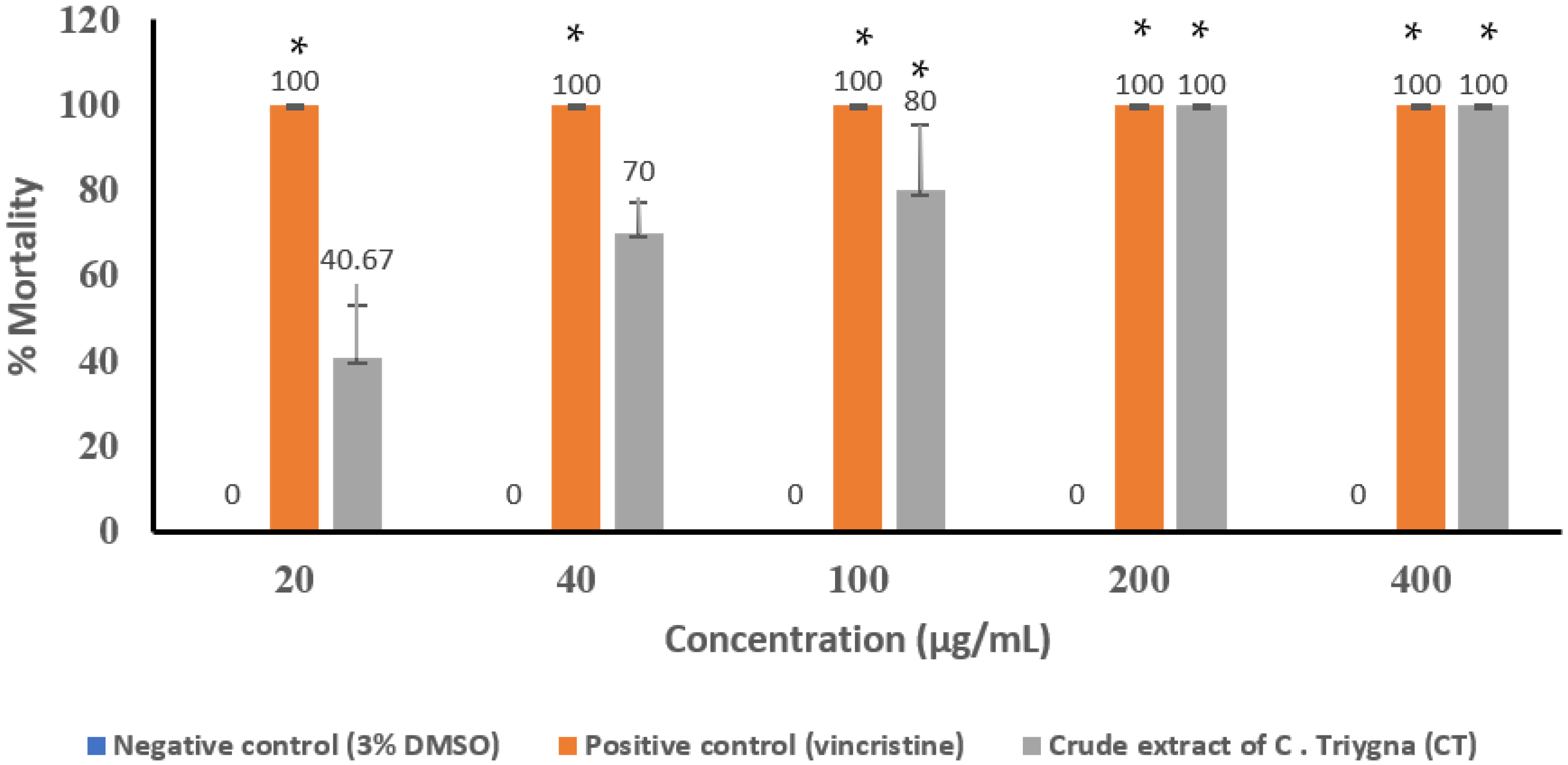
2.6.1. Determination of Cytotoxic Effects Using Tadpoles (Raniceps ranninus)

Raniceps ranninus cytotoxicity assay model was used, as reported by [26]. Ten R. ranninus of relatively similar age and sizes were carefully used and placed into differently labeled beakers containing 49 mL of distilled water and 30 mL of the natural water (collected from the source of the organism). The aqueous mixture was made up to 50 mL with 20, 40, 100, 200, and 400 µg/mL of the CT in 5% DMSO. About 50 mL containing 1 mL of 5% DMSO in distilled water was used as negative control and the assay was performed in triplicate. Mortality was observed for 24 h and used to determine the toxicity of the extract [26,27].

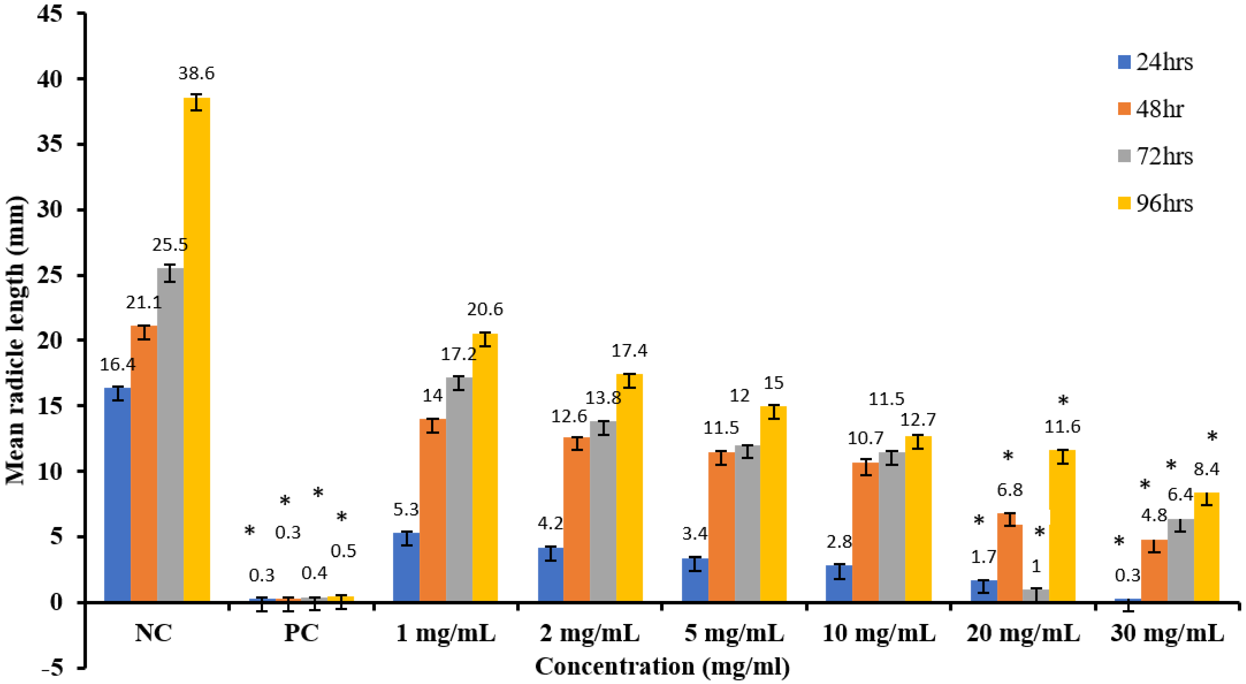
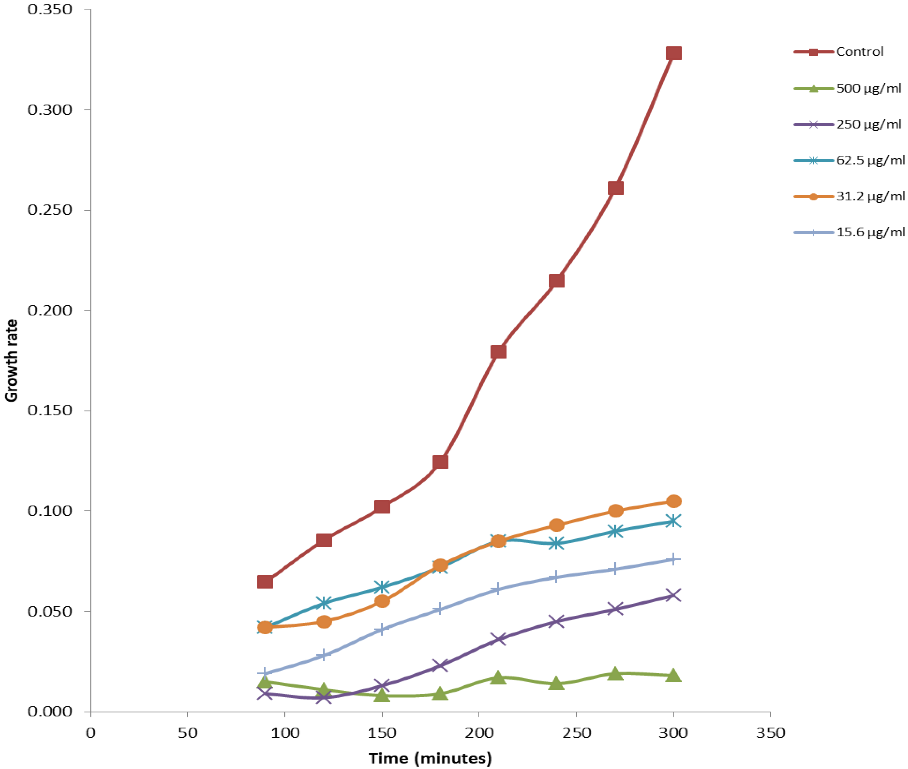
2.6.2. Antiproliferative Studies Using Sorghum bicolor

Sorghum bicolor cytotoxicity bioassay was carried out using the method described by Ayinde et al., 2010, and Ikpefan et al., 2020 [26,27]. The viable seeds of S. bicolor were sterilized using 95% ethanol for 1 min and rinsed with distilled water and dried prior to usage. A volume of 10 mL of different concentrations of CT and CTA (1–30 mg/mL) containing 5% DMSO were introduced into the petri-dishes of about 9 cm wide containing filter (Whatman No.1) underlay with cotton wool. In addition, 20 sterilized seeds of S. bicolor were spread on each of the Petri dishes and incubated in the dark at room temperature for 24, 48, 72, and 96 h. The control seeds were treated with 10 mL of distilled water containing 5% DMSO and used as control, and the experiment was conducted in triplicates for each concentration. The radicle lengths of each seed were measured to the nearest millimeter and the result was also taken as index of cytotoxicity.

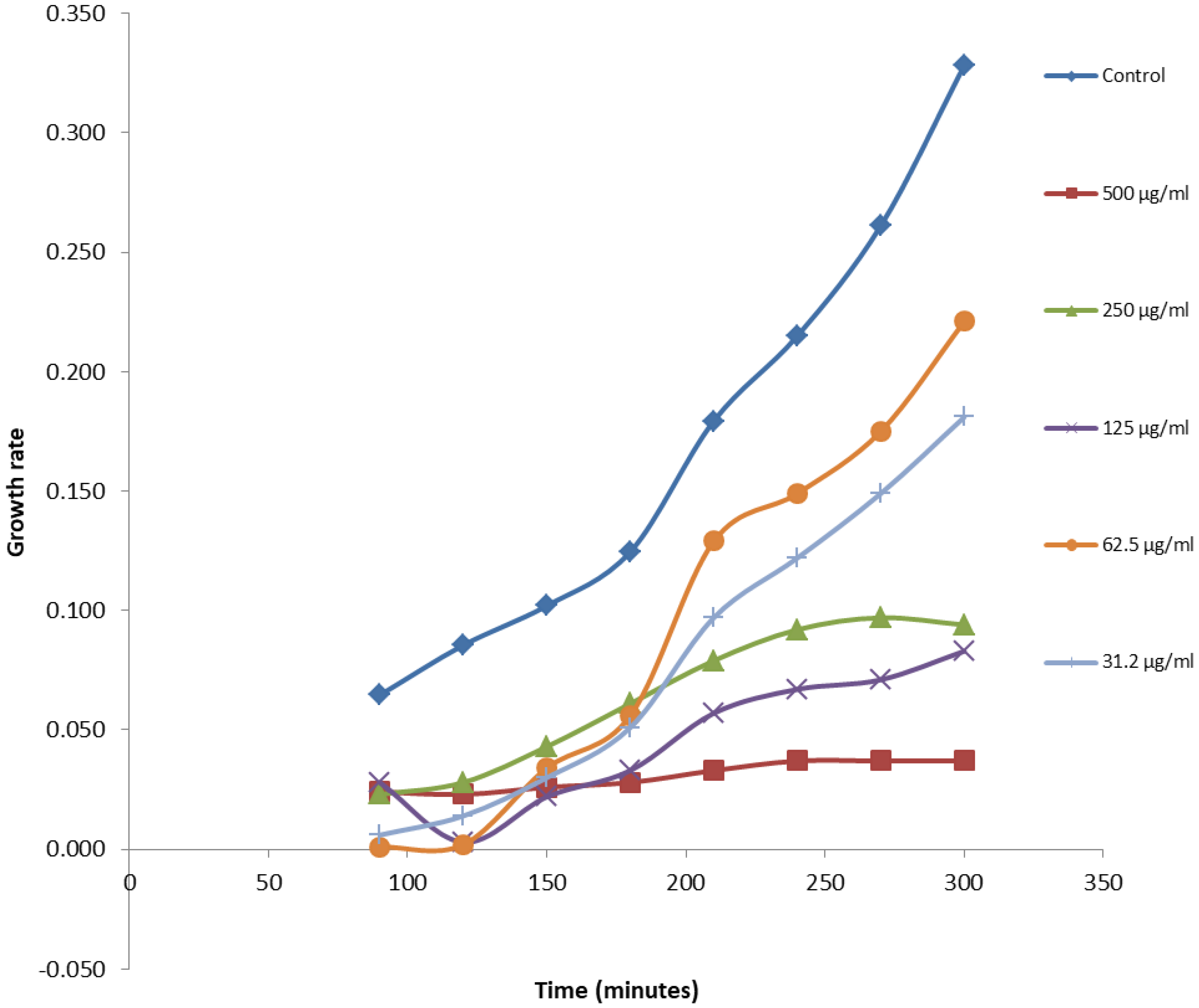
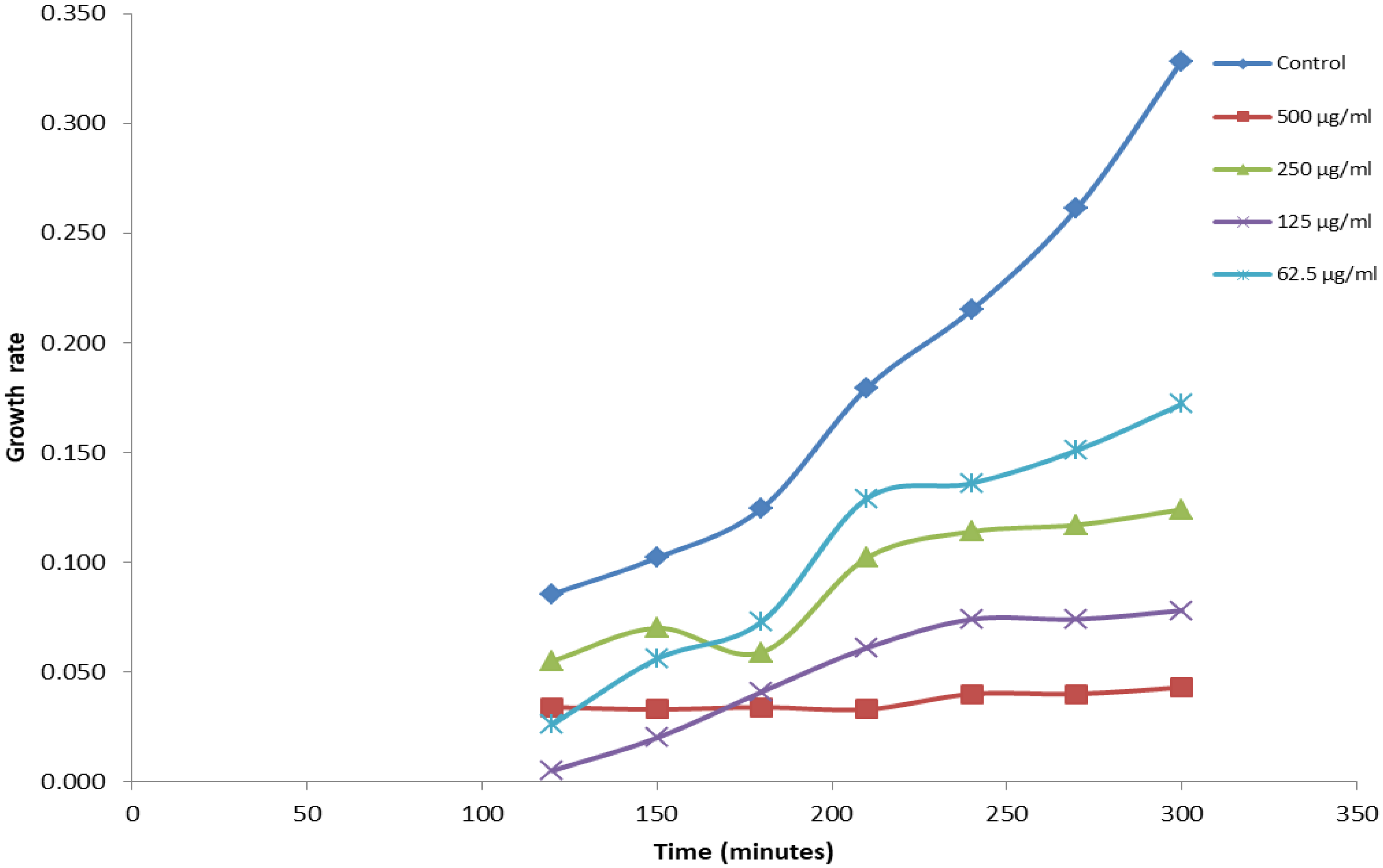
2.6.3. In Vitro Cytotoxicity Assay Using Saccharomyces cerevisiae

Further preliminary cytotoxicity study was likewise carried out on crude extract of CT using Saccharomyces cerevisiae cell culture [28]. However, S. cerevisiae cell cultures containing about 1.0 × 10⁷ cells per mL were realized via inoculation of Saccharomyces cerevisiae cells in 20 mL of YPD (yeast extract 1%, peptone 0.5%, and glucose 2%), followed by incubation at 30 °C for 16–20 h. Approximately 0.5 × 10⁶ cells were transferred into 4 mL disposable cuvettes containing YPD medium, and various concentrations (7.81–500 µg/mL) of CT and CTA were introduced to reach the volume of 2.2 mL. YPD medium without the organism was used as the negative control. The entire loaded disposable cuvettes were incubated in a Heidolph Inkubator 1000 with a shaker at 30 °C and 230 rpm agitation for 5 h. The initial absorbance was measured at 0 min and at intervals of 60 min for 300 min. The assay was performed in triplicates for each concentration. A covariance analysis was applied to the slopes of the growth curves to test the reproducibility of the method and, also, determine the percentage growth inhibition rate of S. cerevisiae in the presence of the sample [28,29,30].

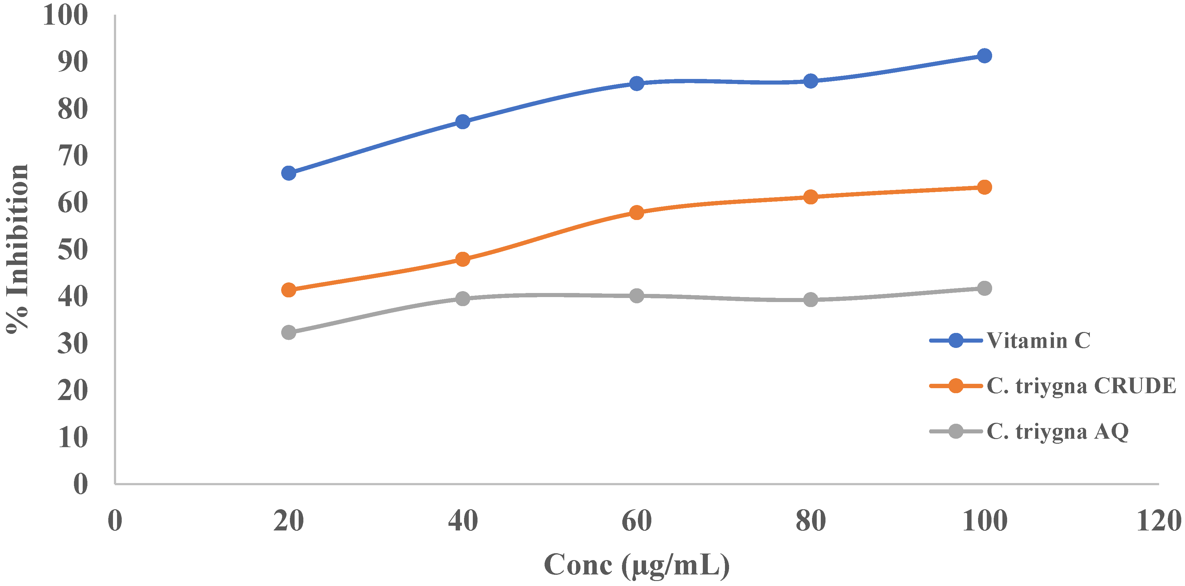
2.6.4. Determination of Antioxidant Activity

The radical scavenging activity of the CT and CTA was carried out against 2, 21-Diphenyl-1-picryl hydrazyl radical (DPPH, Sigma-Aldrich) using a method described by [31,32,33]. About 20–100 µg/mL of test samples and vitamin C were prepared in methanol (Analar grade). A volume of 1 mL of the sample was placed in a test tube, followed by 2 mL of 0.1 mM DPPH in methanol. A control solution was prepared without sample and absorbance was taken at 517 nm. The radical scavenging activity was calculated using the following formula:
% inhibition = {[Ab − Aa]/Ab} × 100
where Ab is the absorption of the blank sample and Aa is the absorption of the extract [31,34].

2.6.5. In Vitro Cytotoxicity against Human Cancer Cell Lines (Sulforhodamine-B (SRB) Assay)

Both samples of CT and CTA were subjected to in vitro cytotoxicity using human cancer cell lines involving semiautomatic procedure using Sulforhodamine-B (SRB) assay [1,34]. The procedure entails cultivating human cancer cell lines in a complete growth medium at a temperature of 37 °C, 5% CO2, and 90% relative humidity. Trypsin-EDTA was, in turn, used to treat the cells at the sub-confluent phase of growth. About 100 μL of cell suspension was introduced to each of the wells in the 96-well plates and incubated for 24 h. Thereafter, 100 μL of the samples at different concentrations were added to each well, incubated again for 24 h, and chilled. Subsequently, 50% TCA (50 μL) was introduced and incubated at 4 °C for 1 h, and washing was later carried out with distilled water 5–6 times. Thereafter, 100 μL of SRB dye (0.4% wt/vol in 1% acetic acid) was added and left at room temperature for 30 min. Thereafter, 1% acetic acid was used to wash the plates, air-dried, and Tris buffer (10 mM; pH 10.5; 100 μL) was, in turn, added to each well. The optical density (OD) of the solution in each well was recorded using a microplate reader at 540 nm.
% Growth inhibition = 100 − [OD (test sample) − OD (blank) OD (control) − OD (blank)] × 100

3. Results and Discussion

Prior to the adoption of medicinal plants by early humans for clothing, food, and shelter, the usage of such plants for medicine (treatment of a variety of ailments associated with humans and animals) has been employed. Natural products from minerals, plants, as well as animals aid the prevention, treatment, and management of an array of human diseases [1,5]. Undoubtedly, ecosystems have been an essential bedrock to plants, which, in turn, provide essential services for the survival of living and nonliving beings [35]. Complementary or alternative medicine in relation to cancer management can be described as therapies, products, and practices that are completely different from orthodox or conventional medicine in application and approach [3,4,21].
About 1.38 kg of Celosia trigyna was extracted with methanol (analytical grade) using Soxhlet extraction. In total, 62.28 g of extract was extracted, with a percentage yield of 4.51%. A total of 0.45 g (0.72%) and 57.40 g (92.16%) of chloroform (CTC) and aqueous fractions (CTA), respectively, were obtained upon solvent partitioning of the CT. Based on the low yield of organic fraction (chloroform fraction, CTC) obtained from solvent partitioning, all biological activities were based on CT and CTA alone, which later, in turn, showed promising results.
Different classes of secondary metabolites have been mentioned to have anticancer potential, as widely reported in the literature [2,5,35], some of which were likewise reported in CT and are, here, shown in Table 1. However, the presence of these compounds might be responsible for the cytotoxic activity observed against human breast, colon, and lung cancer cell lines, as observed hereinafter in the text [24].
In addition, certain bench-top assay methods have been used to ascertain if a drug possesses cytotoxic potential [26,29,31]. This has been imperative due to the high cost and unavailability associated with cancer cell lines and other advanced anticancer screening techniques. In this view, CT was subjected to preliminary bench-top screening using Raniceps ranninus and S. bicolor models. As shown in Figure 1, 100% mortality was recorded against R. ranninus at 200 and 400 µg/mL for C. trigyna crude extract (CT) and vincristine (positive control) at p < 0.05 relative to the negative control (3% DMSO).
Moreover, upon further subjection of CT to growth inhibitory assay using S. bicolor (SB), it was observed that CT was able to reduce the radicle length of SB in a concentration- and time-dependent manner. At 24–96 h, a decrease in S. bicolor radicle lengths was noted at all concentrations (1–30 mg/mL) in a concentration-dependent activity manner. Significant difference (p ≤ 0.05) was observed with the positive control (vincristine; 50 µg/mL) and all concentrations of CT (1–30 mg/mL) relative to the negative control (3% DMSO) for each day, as shown in Figure 2 below.
However, the observation of promising cytotoxic results in CT results in subsequent subjection of CT and CTA to another preliminary cytotoxic activity using the S. cerevisiae model. This assay is based on the similarity between human and S. cerevisiae cells (both cells are eukaryotic, with the former being multicellular, while the latter are unicellular eukaryotic organisms). However, the activity observed in the growth curve shown in Figure 3, Figure 4 and Figure 5 was mathematically extrapolated and reported in Table 2 below. Moreover, an average cytotoxic performance was observed in CT at a concentration nondependent manner, with the lowest and highest activities observed at 31.2 and 250 µg/mL, and at 26.40 ± 1.92% and 56.00 ± 2.12%, respectively. Improvement in activity was observed upon solvent partitioning of CT to give CTA, with a significant increase (p ≤ 0.05) noted at 125 and 250 µg/mL relative to negative control/vehicle (DMSO) with 31.2 ± 1.03% and 33.84 ± 1.03% at the same concentration. Significant activity was observed for nystatin (positive control) at all examined concentrations (7.81–250 µg/mL) at p ≤ 0.05 relative to DMSO (negative control/vehicle), as shown in Table 2.
Moreover, CT showed a concentration-dependent DPPH scavenging activity, with the lowest and highest inhibition observed at 20 and 100 µg/mL, and at 41.35 and 63.26%, respectively, while CTA showed a non-concentration-dependent scavenging activity, with 32.31 and 41.72% activity at the lowest (20 µg/mL) and highest (100 µg/mL) concentrations, respectively, as shown in Figure 6 below.
The amount of total phenolics in CT and CTA were observed to be 87.52 ± 2.45 and 81.35 ± 3.70 gallic acid equivalent, while their corresponding flavonoid content was shown to be 52.81 ± 1.97 and 39.11 ± 2.86 rutein equivalent, respectively, as shown in Table 3 below. Hence, this might be responsible for the pronounced cytotoxic and radical scavenging properties observed in the CT.
Due to the promising nature observed in all preliminary cytotoxic bioassays used, CT and CTA were finally subjected to human colon (HCT116), breast (MCF-7), and lung (H460) cancer cell lines using sulforhodamine B assay, as shown in Table 4 below. Improvement in cytotoxic activity was observed upon fractionation of CT to give CTA, further confirming the cytotoxic activity observed against S. cerevisiae. Moreover, improved cytotoxic activity was observed against human lung (H460), breast (MCF-7), and colon (HCT116) cancer cell lines with IC50 51.69 ± 5.13, 39.16 ± 9.21, and 38.52 ± 7.65, respectively, as shown in Table 4. Such evidence implies that solvent partitioning or aqueous extraction of the plant material enhances the cytotoxic activity of CT, which justifies the cooking (with water) or aqueous maceration of the plant material by locals in the management of cancer and cancer-related ailments.

Analysis of Data

All data collected from the entire study were analyzed using Microsoft Excel and GraphPad Prism 7. Relevant tables, charts, and descriptive statistics were used to present the pertinent points of the study. Data were expressed as the mean ± SEM of three replicates for the in vitro model. The data were subjected to statistical analysis using one-way analysis of variance (ANOVA) and complemented with Kruskal–Wallis test.

4. Conclusions

Based on the research findings from the performed experiments, CT and CTA were subjected to both preliminary and human breast, colon, and lung cancer cell lines using in vitro bioassay models. The resulting evidence allowed us to conclude that CT (leaves) exhibited significant anticancer activity, which increases upon solvent partitioning and utilization of the aqueous fraction CTA. This further experimentally justifies the ethnomedicinal use of CT in the management of cancer and cancer-related ailments in the southwestern part of Nigeria. At the same time, results corroborate the traditional cooking of CT (using water) for the preparation of local soups among the locals (which are characterized by a low incidence of cancer). Further research is ongoing to isolate, characterize, and elucidate the structure of the active principle(s) in CT using bioactivity-guided isolation techniques.

Author Contributions

Writing—original draft: A.O. Conceptualization: A.O., A.B. and P.R. Supervision: A.B. and P.R. Investigation: A.O., A.B., L.S. and P.R. Resources: A.O., A.B., E.N., S.P. and P.R. Data curation: A.O., A.B. and P.R. Writing—review and editing: A.B., S.P., E.N., L.S. and P.R. All authors have read and agreed to the published version of the manuscript.

Funding

This research was funded by Fundação para a Ciência e a Tecnologia (FCT) (Projects UIDB/04567/2020 and UIDP/04567/2020), UID/QUI/50006/2020 and supported by PADDIC 2019 (ALIES-COFAC) as part of the Ph.D. Program in Health Sciences from Universidad de Alcalá and Universidade Lusófona de Humanidades e Tecnologias.

Institutional Review Board Statement

Not applicable.

Informed Consent Statement

Not applicable.

Data Availability Statement

The data presented in this study are available on request from the corresponding author.

Acknowledgments

The authors appreciate CBIOS Research Center for Biosciences and Health Technologies, Lusófona University, Portugal for access to their laboratory and technical assistance during this research. We also appreciate the entire management of Igbinedion University, Okada, Nigeria, for the opportunity given to travel abroad to perform part of this research. Moreover, special appreciation to Odewo (Taxonomist from Forestry Research Institute of Nigeria), who suggested the name of the plant used for this research.

Conflicts of Interest

The authors declare no conflict of interest.

References

  1. Jamshidi-Kia, F.; Lorigooini, Z.; Amini-Khoei, H. Medicinal Plants: Past History and Future Perspective. J. HerbMed Pharmacol. 2018, 7, 1–7. [Google Scholar] [CrossRef]
  2. Shukla, S.; Mehta, A. Anticancer Potential of Medicinal Plants and Their Phytochemicals: A Review. Rev. Bras. Bot. 2015, 38, 199–210. [Google Scholar] [CrossRef]
  3. Salmerón-Manzano, E.; Garrido-Cardenas, J.A.; Manzano-Agugliaro, F. Worldwide Research Trends on Medicinal Plants. Int. J. Environ. Res. Public Health 2020, 17, 3376. [Google Scholar] [CrossRef] [PubMed]
  4. Ohiagu, F.O.; Chikezie, P.C.; Chikezie, C.M.; Enyoh, C.E. Anticancer Activity of Nigerian Medicinal Plants: A Review. Futur. J. Pharm. Sci. 2021, 7, 1–21. [Google Scholar] [CrossRef]
  5. Roy, A.; Attre, T.; Bharadvaja, N. Anticancer Agent from Medicinal Plants: A Review. New Apects Med. Plants Pharmacogn. 2017, 1, 54. [Google Scholar]
  6. Stewart, B.W.; Wild, C.P. World Cancer Report 2014; World Health Organization: Geneva, Switzerland; IARC Nonserial Publication: Lyon, France, 2014. [Google Scholar]
  7. Siegel, R.L.; Miller, K.D.; Fuchs, H.E.; Jemal, A. Cancer Statistics, 2021. CA Cancer J. Clin. 2021, 71, 359. [Google Scholar] [CrossRef]
  8. McGuire, S. World Cancer Report 2014. Geneva, Switzerland: World Health Organization, International Agency for Research on Cancer, WHO Press, 2015. Adv. Nutr. 2016, 7, 418–419. [Google Scholar] [CrossRef] [Green Version]
  9. Boyle, P.; Levin, B. World CanCer Report 2008. Cancer Control 2008, 199, 1828–1840. [Google Scholar] [CrossRef]
  10. Sung, H.; Ferlay, J.; Siegel, R.L.; Laversanne, M.; Soerjomataram, I.; Jemal, A.; Bray, F. Global Cancer Statistics 2020: GLOBOCAN Estimates of Incidence and Mortality Worldwide for 36 Cancers in 185 Countries. CA Cancer J. Clin. 2021, 71, 209–249. [Google Scholar] [CrossRef]
  11. Siegel, R.L.; Miller, K.D.; Jemal, A. Cancer Statistics, 2020. CA Cancer J. Clin. 2020, 70, 7–30. [Google Scholar] [CrossRef]
  12. Barker, K.; Eickmeyer, S. Therapeutic Exercise. Med. Clin. N. Am. 2020, 104, 189–198. [Google Scholar] [CrossRef] [PubMed]
  13. Wild, C.; Weiderpass, E.; Stewart, B. World Cancer Report: Cancer Research for Cancer Prevention; International Agency for Research on Cancer: Lyon, France, 2020; ISBN 978-92-832-0448-0. Available online: http://publications.iarc.fr/586 (accessed on 23 November 2022).
  14. Mazzola, R.; Corradini, S.; Eidemüeller, M.; Figlia, V.; Fiorentino, A.; Giaj-Levra, N.; Nicosia, L.; Ricchetti, F.; Rigo, M.; Musola, M.; et al. Modern Radiotherapy in Cancer Treatment during Pregnancy. Crit. Rev. Oncol. Hematol. 2019, 136, 13–19. [Google Scholar] [CrossRef] [PubMed]
  15. Chen, H.H.W.; Kuo, M.T. Improving Radiotherapy in Cancer Treatment: Promises and Challenges. Oncotarget 2017, 8, 62742–62758. [Google Scholar] [CrossRef] [Green Version]
  16. Luo, C.; Shuang, D.; Jing, Z.; Xi, C.; Ou, W.; Ran, F.; Yu, Q.; Wang, J.; Qing, H.; Sheng, H. Interim Guideline for Treatment Options on Cancer Patients during Pandemic of SARS-CoV-2. Open Access Online-First Publ. Res. Pap. COVID-19 2020, 47, 227–234. [Google Scholar]
  17. Xiao, Y.X.; Yang, W.X. KIFC1: A Promising Chemotherapy Target for Cancer Treatment? Oncotarget 2016, 7, 48656. [Google Scholar] [CrossRef] [Green Version]
  18. Lopes, C.M.; Dourado, A.; Oliveira, R. Phytotherapy and Nutritional Supplements on Breast Cancer. Biomed. Res. Int. 2017, 2017, 1–42. [Google Scholar] [CrossRef]
  19. Sowemimo, A.; van de Venter, M.; Baatjies, L.; Koekemoer, T. Cytotoxic Activity of Selected Nigerian Plants. Afr. J. Tradit. Complement. Altern. Med. 2009, 6, 526–528. [Google Scholar] [CrossRef] [Green Version]
  20. Ofusori, A.E.; Moodley, R.; Jonnalagadda, S.B. Elemental Distribution in the Edible Leaves of Celosia trigyna from the Western and Northern Regions of Nigeria. J. Environ. Sci. Health—Part B Pestic. Food Contam. Agric. Wastes 2019, 54, 61–69. [Google Scholar] [CrossRef]
  21. Nath, P.; Ohri, D.; Pal, M. Nuclear DNA Content in Celosia (Amaranthaceae ). Plant Syst. Evol. 1992, 182, 253–257. [Google Scholar] [CrossRef]
  22. El-Desouky, S.K.; Abdelgawad, A.A.; El-Hagrassi, A.M.; Hawas, U.W.; Kim, Y.K. Chemical Composition, Cytotoxic and Antioxidant Activities of Celosia trigyna L. Grown in Saudi Arabia. Acta Pol. Pharm.—Drug Res. 2019, 76, 691–699. [Google Scholar] [CrossRef]
  23. Onen, P.; Ocira, D.; Omara, T.; Nyeko, J.; Okwir, A. Preliminary Phytochemical Screening of Plumbago zeylanica L. Roots and Its Aphrodisiac Effect in Male Rats. Asian J. Appl. Chem. Res. 2021, 8, 24–31. [Google Scholar] [CrossRef]
  24. Adebayo, M.A.; Adedokun, O.A.; Akinpelu, L.A.; Okafor, P.O. Evaluation of Anti-Diarrheal Activity of Methanol Root Bark Extract of Milicia Excelsa (Welw) CC Berg (Moraceae) in Rats. Drug Res. 2019, 69, 439–444. [Google Scholar] [CrossRef] [PubMed]
  25. Ayinde, B.A.; Agbakwuru, U. Cytotoxic and Growth Inhibitory Effects of the Methanol Extract Struchium Sparganophora Ktze (Asteraceae) Leaves. Pharmacogn. Mag. 2010, 6, 293–297. [Google Scholar] [CrossRef] [PubMed] [Green Version]
  26. Ikpefan, E.O.; Ayinde, B.A.; Mudassar, B.A.; Farooq, A.D. Anticancer and Antioxidant Studies of the Methanol Extract and Fractions of Conyza sumatrensis (Retz.) E. H. Walker (Asteraceae). Niger. J. Nat. Prod. Med. 2020, 23, 77–82. [Google Scholar] [CrossRef]
  27. Buschini, A.; Poli, P.; Rossi, C. Saccharomyces Cerevisiae as an Eukaryotic Cell Model to Assess Cytotoxicity and Genotoxicity of Three Anticancer Anthraquinones. Mutagenesis 2003, 18, 25–36. [Google Scholar] [CrossRef] [Green Version]
  28. Roberto, A.; Caetano, P.P. A High-Throughput Screening Method for General Cytotoxicity Part I Chemical Toxicity Método Rápido Para Triagem Da Citotoxicidade Geral Parte I Toxicidade Química. Rev. Lusófona De Ciências E Tecnologias Da Saúde 2005, 2, 95–100. [Google Scholar]
  29. Oluwasegun, A.N.A. In vitro Free Radical Scavenging Activity and Total Phenolic Content of Kigelia Africana (LAM). Int. J. Sci. Res. 2014, 3, 368–370. [Google Scholar]
  30. Adedokun, O.; Gbolade, A.; Ayinde, B. 13, 14-Epoxyoleanan-3-Ol-Acetate: A Male Fertility Enhancing Constituent from Hexane Fraction of Momordica Charantia Linn (Curcubitaceae). Turk. J. Pharm. Sci. 2021, 19, 180–186. [Google Scholar] [CrossRef]
  31. Esther Ogunjinmi, O.; Oguntola Ogunjinmi, S.; Oluwagbemiga Alayande, S.; Omelebele Nwoke, E.; Adewale Adedosu, T. Evaluation of Antioxidant Activities of Celosia trigyna (Linn) Extracts African Extinction Vegetable. Sci. J. Chem. 2020, 8, 102. [Google Scholar] [CrossRef]
  32. Adedokun, O.; Ume, O.; Odunola, M.; Nnamani, D.; Jesumirhewe, C.; Ojo, T.; Aniebue, I. Evaluation of Toxicological Profile of Methanol Leaf Extract of Waltheria Indica (Sterculiaceae). GSC Biol. Pharm. Sci. 2021, 17, 34–43. [Google Scholar] [CrossRef]
  33. Vichai, V.; Kirtikara, K. Sulforhodamine B Colorimetric Assay for Cytotoxicity Screening. Nat. Protoc. 2006, 1, 1112–1116. [Google Scholar] [CrossRef] [PubMed]
  34. Thabit, M.G.; El Bialy, S.A.A.; Nasr, M.N.A. Synthesis and Biological Evaluation of New 3-(4-Substituted Phenyl)Aminoquinoxaline Derivatives as Anticancer Agents. Heterocycl. Commun. 2015, 21, 25–35. [Google Scholar] [CrossRef]
  35. Gunasekaran, J.; Sankaran, M. Anti Cancer; Anti Oxidant; Cytotoxicity; Lupeol Analogue; MCF-7 Cells Progress in Bioscience and Bioengineering Chemotherapeutic Potential of Lupeol and Its Analogue (Oxime) on Human Mammary Cancer Cell Line (MCF-7). Prog. Biosci. Bioeng. 2017, 1, 18–28. [Google Scholar] [CrossRef]
Figure 1. Cytotoxicity screening of CT using R. ranninus. n = 10. n ± SEM. Samples with superscript * indicate a significant difference at p < 0.05 relative to the negative control (3% DMSO) using one-way ANOVA (Kruskal–Wallis test).
Ijtm 02 00043 g001
Figure 2. Growth inhibitory screening (using S. bicolor) of C. trigyna (CT) at concentrations ranging from 1 to 30 mg/mL. n = 3. n ± SEM. Samples with superscript * indicate a significant difference at p < 0.05 relative to negative control (3% DMSO) using two-way ANOVA (Dunnett’s multiple comparisons test). NC—negative control (3%DMSO); PC—positive control (vincristine; 50 µg/mL).
Ijtm 02 00043 g002
Figure 3. Inhibitory effect of CT on the growth rate of S. cerevisiae over time (0–300 min).
Ijtm 02 00043 g003
Figure 4. Inhibitory effect of CTA on the growth rate of S. cerevisiae over time (0–300 min).
Ijtm 02 00043 g004
Figure 5. Inhibitory effect of nystatin (positive control) on the growth rate of S. cerevisiae over time (0–300 min).
Ijtm 02 00043 g005
Figure 6. Radical scavenging potential of vitamin C (positive control) and C. trigyna (crude and aqueous fraction) against DPPH. The values above are the mean of three replicates. n = 3. Mean ± SEM. The results of the extracts were compared to the corresponding percentage inhibition of vitamin C using one-way ANOVA (Kruskal–Wallis test). p < 0.05 was considered significant.
Ijtm 02 00043 g006
Table 1. Preliminary phytochemical screening of CT.
Class of PhytochemicalsCT
Saponins+
Flavonoids+
Steroids+
Reducing sugars+
Terpenoids+
Alkaloids
Phenolic compounds+
Key: + indicates present; − indicates negative.
Table 2. Comparative cytotoxicity study of CT and CTA using Saccharomyces cerevisiae bioassay model.
Concentration
(µg/mL)
% Inhibition
DMSONystatin (Positive Control)C. trigyna Crude (CT)C. trigyna Aq (CTA)
7.8112.67 ± 1.2197.25 ± 1.02 *44.00 ± 1.1328.00 ± 1.53
15.616.80 ± 1.0898.21 ± 0.98 *34.40 ± 2.1135.20 ± 2.10
31.217.60 ± 0.0198.78 ± 2.17 *26.40 ± 1.9250.40 ± 1.11
62.530.73 ± 1.1299.35 ± 2.92 *36.00 ± 1.1560.00 ± 1.89
12531.2 ± 1.0399.59 ± 1.87 *48.80 ± 2.1788.00 ± 2.08 *
25033.84 ± 1.0399.71 ± 1.34 *56.00 ± 2.1284.80 ± 1.62 *
The above reported values are the mean of three replicates. n = 3. Mean ± SEM. Values with superscript * indicate a significant difference at p < 0.05 when compared to the corresponding percentage inhibition of vehicle (DMSO) for each concentration using one-way ANOVA (Kruskal–Wallis test).
Table 3. Phenolic and flavonoid contents of CT and CTA.
SampleTotal Phenolics (Gallic Acid Equivalent)Total Flavonoids (Rutein Equivalent)
C. trigyna (Crude)87.52 ± 2.4552.81 ± 1.97
C. trigyna (Aqueous fraction)81.35 ± 3.7039.11 ± 2.86
The values above are the mean of three replicates, n = 3; mean ± SEM.
Table 4. Cytotoxic effect (IC50 (µg/mL)) of H. acida crude and aqueous fraction on different cancer cell lines using sulforhodamine B assay.
Cell LinesC. trigyna (CT)C. trigyna (Aq. Fraction; CTA)
H46058.15 ± 0.5151.69 ± 5.13
MCF-741.32 ± 2.6239.16 ± 9.21
HCT11651.82 ± 8.5138.52 ± 7.65
IC50 (µg/mL) of samples in H460, MCF-7, and HCT116 cell lines. The concentration that induced 50% of growth inhibition (IC50) was determined by sulforhodamine B assay after 48 h of treatment. Data = mean ± SEM of 4 (four) independent experiments. n = 4.
Publisher’s Note: MDPI stays neutral with regard to jurisdictional claims in published maps and institutional affiliations.

