Article

Isolation and Identification of a Strain of Aspergillus Tubingensis With Deoxynivalenol Biotransformation Capability

College of Veterinary Medicine, Nanjing Agricultural University, Nanjing 210095, P.R. China
*
Author to whom correspondence should be addressed.
Int. J. Mol. Sci. 2008, 9(12), 2366-2375; https://doi.org/10.3390/ijms9122366
Submission received: 14 June 2008 / Revised: 13 November 2008 / Accepted: 25 November 2008 / Published: 27 November 2008

Abstract

:
Deoxynivalenol (DON) is one of the most common contaminants of various foodstuffs. A biotransformation system was used in order to lessen the toxicity of DON. A strain of Aspergillus (NJA-1) was isolated from soil and cultured in an inorganic salt medium containing DON. Bt2a/Bt2b primers were used to amplify the β-tubulin gene of NJA-1. Sequence analysis the PCR product and morphology observation indicated that NJA-1 belonged to Aspergillus tubingensis (aerobic fungi). The DNA sequence information of the PCR product was deposited in GenBank (accession number DQ9025790). The DNA sequence had 99% similarity to the Aspergillus tubingensis accession number AY820009. An unknown compound in NJA-1 showed the ability to convert DON into another product. The molecular weight of the bioconversion product was 18.1 D (H2O) larger than that of DON. The analysis showed that DON could be hydrolyzed by NJA-1. The mean DON biotransformation rate was 94.4% after two weeks of cultivation. The finding presents a new method for DON biotransformation.

1. Introduction

Deoxynivalenol (DON), known as vomitoxin, a member of the trichothecenes which are secondary metabolites of fungi, mainly Fusarium, is one of the most common contaminants in various foodstuffs [15]. Epidemiological studies show an association of gastrointestinal diseases with DON, but critical data are still needed regarding its potential health damaging effects [6]. The toxic effects of DON in animals have been well documented and focused mainly on the immune system and the gastrointestinal tract [7]. In particular, the symptoms of acute toxicosis at high dosage include diarrhea, vomiting, leukocytosis, hemorrhage, circulatory shock and ultimately death [8]. Chronic toxicosis at low dosage leads to anorexia, reduced weight gain, nutrients malabsorption, neuroendocrine changes and immunosuppression [912]. Many methods are used for removal of mycotoxins in moldy feedstuff including chemical, physical and microbiological treatments [1322]. However, because of DON’s relatively high chemical stability and nonpolar structure, physical treatments have a very low efficacy to remove DON. Meanwhile, some adsorbents can also absorb nutrients from the foodstuff, such as minerals and vitamins. Chemical treatment has a high efficacy on DON detoxification, but it can greatly reduce nutritive value and palatability of foodstuffs. Microbiological and enzymatic treatments are the best choice to reduce the toxicity of DON.
Some microbes have been reported to produce enzymes during growth which could lessen the toxicity of DON. The process has been also called “bio-transformation”. In present paper, a strain of Aspergillus tubingensis (NJA-1) was isolated from soil samples. The article mainly reports the isolation and identification of NJA-1 and its efficacy on DON-transformation.

2. Results and Discussion

2.1. Isolation of NJA-1

Only one strain was isolated from fifteen soil samples and five rumen content samples and named NJA-1.

2.2. Identification of NJA-1

The purified colonies had radial villus and white mycelia at the edge of each colony in early culture stages (3~5 d). After seven days, the colony became pitchy at the head of mycelium and white in the reverse side in Czapek’s Agar plate, respectively. The vesicle of NJA-1 was round and radial. Diaphragms in hypha were visible with microscope. The value of metulae and conidial diameter were about 6 and 2.5 μm, respectively. Electron microscope scanning image showed that the spore was elliptical with introcession in head (Figure 1A). Sporangia of NJA-1 consisted of many spores (Figure 1B).
The β-tubulin gene of NJA-1 was amplified with primers Bt2a–Bt2b. The size of amplified fragment was 511bp. Example of PCR result was shown in Figure 2. The results of the DNA sequence analysis were submitted to GenBank (access number DQ902579). The DNA sequence had 99% similarity to the Aspergillus tubingensis accession number AY820009. The results of the DNA sequence analysis and the observation of morphology showed that the NJA-1 belonged to Aspergillus tubingensis.

2.3. The effect of transformation

The transformation results after 7d and 14d cultivation are shown in Table 1. The average transformation efficiency at 14 d was 94.4%. The detection of DON using HPLC is shown in Figure 3. The peak retention time of DON was 6.408 min, while that of the biotransformation product was 2.444 min. The difference between samples and control was significant (analyzed by SPASS 12.0 with One-Way ANOVA mode).

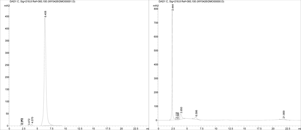
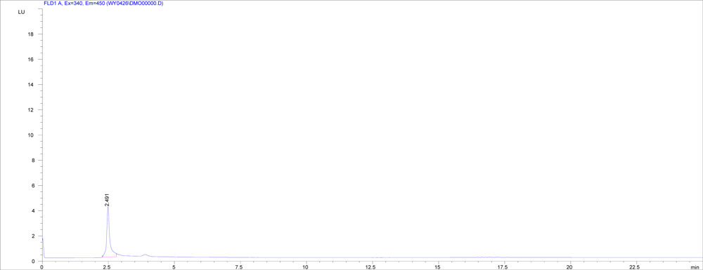
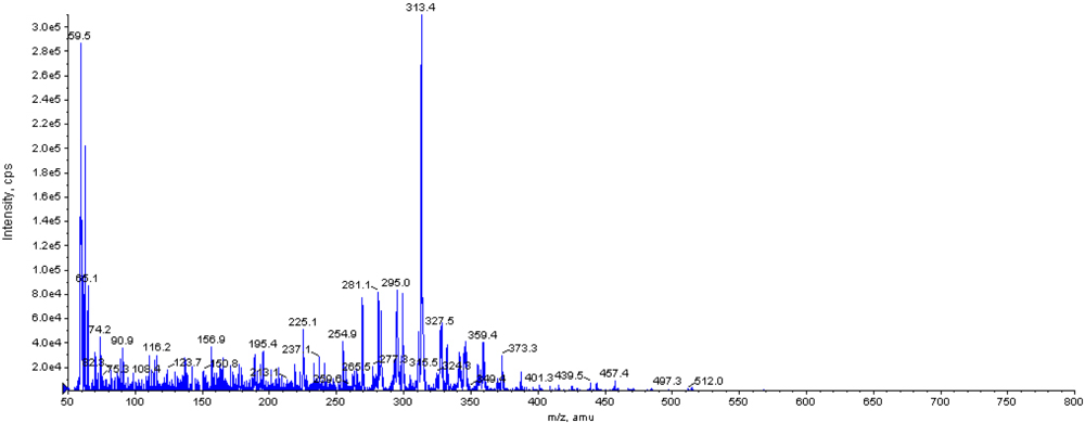
2.4. Screening for DON biotransformation product by NJA-1

The product of DON biotransformation by NJA-1 was analyzed by HPLC and HPLC-MS. The HPLC diagram showed that the biotransformation product could emit fluorescence (Ex=340, Em=450) (Figure 4). The result of HPLC-MS showed that the molecular weight of biotransformation product was 314.4 (Figure 5).
Biotransformation of DON into less toxic metabolites by microorganisms was the most promising approach to counteract the mycotoxin. A bacterial strain BBSH 797 was isolated by Fuchs et al. [23]. The strain could transform DON into a lower toxicity metabolite DOM-1 (molecular weight 280.3). Strain E3-39 isolated from a soil sample was reported to convert DON to 3-keto-DON (molecular formula of C15H18O6) [24]. A mixed culture, D107, was reported to biologically degradate DON under anaerobic conditions [25]. A UDP-glycosyltransferase was able to lessen the toxicity of DON by transferring a glucose from UDP-glucose to the hydroxyl group at carbon 3 of DON [20]. In this study the strain of Aspergillus tubingensis (NJA-1) was isolated from soil by inorganic salt culture media supplemented with DON. DON was the only carbon source during the isolation. Because the growth of fungi required the carbon source and nitrogen source, the NJA-1 could grow by using DON. This indicates that the NJA-1 had the ability to transform the DON into another product. The hypothesis was confirmed by the HPLC and HPLC-MS results. The product is different from that of previous reports. The molecular weight of bioconversion product detected by HPLC-MS was 18.1 D (H2O) higher than that of DON, which would show that the DON could be hydrolyzed by NJA-1. The previous researches mainly focused on anaerobic bacteria. NJA-1 was different from the most reported strains, because it is an aerobic fungi. We hope that the strain could have a wide application outlook for biotransformation of mycotoxin in mouldy feedstuffs, although further studies are needed to identify the biotransformation product of DON and compare the cytotoxicity of DON and its biotransformation product.

3. Experimental Section

3.1. Sampling

Fifteen soil samples (about 500 g per sample of clay soil was packed into sterile sampling bag and five rumen contents (about 10 mL per sample was extracted through stomach catheter and was placed in a 50 mL Erlenmeyer flask) were collected from Nanjing District and the dairy farm in Nanjing Agricultural University (NAU), respectively.

3.2. Culture media

Czapek’s Agar [26] contained (per liter): sucrose (30 g), NaNO3 (3 g), MgSO4·7H2O (0.5 g), KCl (0.5 g), FeSO4·7H2O (0.01 g), K2HPO4 (1 g), agar (13 g). The pH was adjusted to 7 with NaOH.
Inorganic salt culture media contained (per liter): (NH4)2SO4 (1.0 g), K2HPO4.3H2O (6.0 g), KH2PO4 (3.0 g), NaCl (0.5 g), MgSO4·7H2O (0.5 g), CaCl2 (0.05 g). The pH was adjusted to 7 with NaOH. Inorganic salt culture (50 mL) was added into a 250 mL conical flask. After autoclaving, DON (200 μg per conical flask, Sigma, purity: ≥97.0%) was added.
Liquid Czapek’s culture media was prepared similar to Czapek’s Agar but without the agar. Liquid Czapek’s culture media (50 mL) was added into a 250 mL conical flask. LB culture media contained (per liter): yeast extract (5 g), peptone (10 g), NaCl (10 g) and agar (15 g). The pH was adjusted to 7 with NaOH.

3.3. Purification and isolation

Soil (1 g) or rumen contents (1 mL) were added to sterile distilled water (50 mL), then mixed fully and centrifuged (2,000g × 10 min). Supernatant (1 mL) was added to inorganic salt culture media in a 250 mL conical flask in which DON was placed in advance. After cultivation at 30 °C for 7 d, a culture fluid sample (1 mL) was taken out and transferred into a 250 mL conical flask in which DON was added, and then was cultivated at 30 °C for another 7 d. Afterwards, a sample of the second culture fluid (1 mL) was spread on an inorganic salt culture media plate that contained DON. A single colony was cultured on Czapek’s Agar plate and LB culture media plate by streaking. A Strain of Aspergillus Tubingensis was isolated and named NJA-1.

3.4. Identification of NJA-1

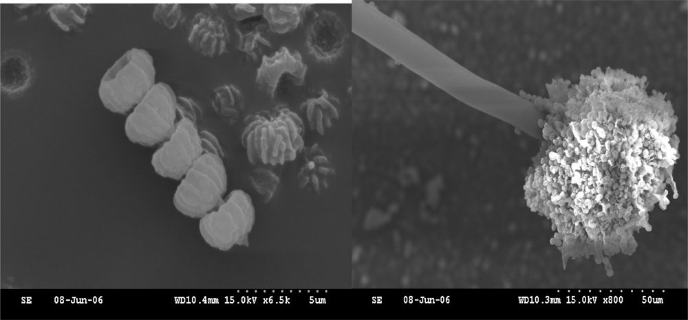
3.4.1. The observation of conventional morphology and scanning electron microscope

The taxonomic systems of Aspergillus were followed for observation and identification. The isolates were cultured on Czapek’s Agar at 30 °C. Macroscopical characters were assessed at were 3 and 7 days of age and microscopical characters were assessed after staining with lactophenol cotton blue. The colony color was observed by naked eye with the aid of color charts in the Methuen Handbook of Colour. The material on the slide mounts was examined under light microscope (OLYMPUS SZ-III Japan). The metulae, conidial diameter and conidial rough were determined under 1,000 × magnification. For scanning electron microscopic observations, the isolates were cultured on Czapek’s Agar at 30 °C for 7 days. The fungi were picked up on a 200-mesh copper screen and dried for viewing in the scanning electron microscope (HITACHI S-3000N Japan).

3.4.2. Identification of NJA-1 with PCR

The purified NJA-1 was inoculated into liquid Czapek’s culture media and cultivated at 30 °C for 7 d, then thallus of NJA-1were collected by filtration. Extraction of DNA from NJA-1 was performed following [27]. Almost the entire length of the β-tubulin gene of strain NJA-1 was amplified by PCR with the primers Bt2a (5′-GGTAACCAAATCGGTGCTGCTTTC) and Bt2b (5′-ACCCTCAGTGTAGTGACCCTTGGC) [28]. Genomic DNA of NJA-1 was added to PCR reactions containing forward and reverse primers. After 1 min incubation at 94 °C, thirty-three PCR cycles (94 °C, 1 min; 60 °C, 1 min and 72 °C, 1min) were performed, followed by one cycle of 10 min at 72 °C. The PCR product was separated by electrophoresis and then isolated and sequenced. A homology search was performed with GenBank database. The partial β-tubulin gene sequence was deposited into GeneBank Data Library with the accession number DQ902579.

3.5 The effect of DON transformation

The purified NJA-1 was inoculated into inorganic salt culture media with DON at concentrations of 40, 2, 1 and 0.4 mg/L, respectively. At the same time, negative and positive controls were performed. Only NJA-1 was added into the inorganic salt culture media as negative control and only DON was added as positive control. After cultivating at 30 °C for 14d, the concentration of DON was determined by high performance liquid chromatography (Hewlett Packard 1100 USA) equipped with fluorescence and UV detectors [29]. The HPLC conditions employed were as follows: Kromasil C18 column (250 × 4.6 mm, i.d., 5 μm); injection volume: 20 μL; column temperature: 29 °C; mobile phase: acetonitrile-water (15:85 v/v); flow rate: 0.8 mL/min; UV detection wavelength: 218 nm. To prevent trapping DON in cells and adsorption of DON to cell walls, cultures were split by ultrasonic wave before extraction. Three replicate experiments were run simultaneously. Data were analyzed by SPASS 12.0 with One-Way ANOVA mode.

3.6 Sscreening for DON biotransformation

An HPLC instrument (Hewlett Packard 1100 USA) equipped with UV-Vis diode array (G1315A) and fluorescence detectors (HP 1046A, USA) was used to screen the biotransformation product in culture media. The HPLC screening conditions were the same as the HPLC conditions for determination, except the UV-Vis diode array detector wavelength of 190–630 nm setting and fluorescence detector operated at Ex = 340, Em = 450.
An HPLC-MS instrument (Hewlett Packard 1100/API 2000) with atmospheric pressure chemical ionization (APCI) in negative ion mode were used to screen the DON product. The MS conditions employed were as follows: ion source temperature: 200 °C; APCI detection temperature: 450 °C; scanning range: m/z 50–800.

4. Conclusions

A strain of Aspergillus (NJA-1) was isolated from soil by inorganic salt culture media supplemented with DON. Sequence analysis of Bt2a–Bt2b gene and morphology observation indicated that NJA-1 belong to Aspergillus tubingensis (aerobic fungi). The DNA sequence of Bt2a–Bt2b PCR product was deposited in GenBank (accession number DQ9025790). The mean DON biotransformation rate was 94.4% after two weeks cultivation. The product of DON transformed by NJA-1 could emit fluorescence, and its molecular weight was 18.1 D (H2O) larger than that of DON analyzed by HPLC-MS. The analysis suggested that DON could be hydrolyzed by NJA-1. The finding could present a new method for DON biotransformation. To authors’ knowledge, it is the first report that Aspergillus tubingensis has the ability for DON biotransformation.

Acknowledgments

This study was supported by the National Natural Science Fundation of China (Grant No.30471277), the Doctoral Foundation of Ministry of Education (Grant No. 20040307043) and Youth Sci-Tech Innovation Fund, NAU (Grant No. KJ08017).

References

  1. Pieters, MN; Freijer, J; Baars, BJ; Fiolet, DC; van, KJ; Slob, W. Risk assessment of deoxynivalenol in food: concentration limits, exposure and effects. Adv. Exp. Med. Biol 2002, 504, 235–248. [Google Scholar]
  2. Trucksess, MW; Thomas, F; Young, K; Stack, ME; Fulgueras, WJ; Page, SW. Survey of deoxynivalenol in U.S. 1993 wheat and barley crops by enzyme-linked immunosorbent assay. J. AOAC Int 1995, 78, 631–636. [Google Scholar]
  3. Tanaka, T; Hasegawa, A; Matsuki, Y; Ueno, Y. A survey of the occurrence of nivalenol, deoxynivalenol and zearalenone in food stuffs and health foods in Japan. Food Addit. Contam 1985, 2, 259–265. [Google Scholar]
  4. Tanaka, T; Yamamoto, S; Hasegawa, A; Aoki, N; Besling, JR; Sugiura, Y; Ueno, Y. A survey of the natural occurrence of Fusarium mycotoxins, deoxynivalenol, nivalenol and zearalenone, in cereals harvested in the Netherlands. Mycopathologia 1990, 110, 19–22. [Google Scholar]
  5. Papadopoulou-Bouraoui, A; Vrabcheva, T; Valzacchi, S; Stroka, J; Anklam, E. Screening survey of deoxynivalenol in beer from the European market by an enzyme-linked immunosorbent assay. Food Addit. Contam 2004, 21, 607–617. [Google Scholar]
  6. Pestka, JJ; Smolinski, AT. Deoxynivalenol: toxicology and potential effects on humans. J. Toxicol. Environ. Health B Crit. Rev 2005, 8, 39–69. [Google Scholar]
  7. Overnes, G; Matre, T; Sivertsen, T; Larsen, HJ; Langseth, W; Reitan, LJ; Jansen, JH. Effects of diets with graded levels of naturally deoxynivalenol-contaminated oats on immune response in growing pigs. Zentralbl. Veterinarmed. A 1997, 44, 539–550. [Google Scholar]
  8. Rotter, BA; Prelusky, DB; Pestka, JJ. Toxicology of deoxynivalenol (vomitoxin). J. Toxicol. Environ. Health 1996, 48, 1–34. [Google Scholar]
  9. Drochner, W; Schollenberger, M; Piepho, HP; Gotz, S; Lauber, U; Tafaj, M; Klobasa, F; Weiler, U; Claus, R; Steffl, M. Serum IgA-promoting effects induced by feed loads containing isolated deoxynivalenol (DON) in growing piglets. J. Toxicol. Environ. Health A 2004, 67, 1051–1067. [Google Scholar]
  10. Johnson, PJ; Casteel, SW; Messer, NT. Effect of feeding deoxynivalenol (vomitoxin)-contaminated barley to horses. J. Vet. Diagn. Invest 1997, 9, 219–221. [Google Scholar]
  11. Prelusky, DB. The effect of low-level deoxynivalenol on neurotransmitter levels measured in pig cerebral spinal fluid. J. Environ. Sci. Health B 1993, 28, 731–761. [Google Scholar]
  12. Pieters, MN; Bakker, M; Slob, W. Reduced intake of deoxynivalenol in The Netherlands: A risk assessment update. Toxicol. Lett 2004, 153, 145–153. [Google Scholar]
  13. Bretz, M; Beyer, M; Cramer, B; Knecht, A; Humpf, HU. Thermal degradation of the Fusarium mycotoxin deoxynivalenol. J. Agric. Food Chem 2006, 54, 6445–6451. [Google Scholar]
  14. Lauren, DR; Smith, WA. Stability of the fusarium mycotoxins nivalenol, deoxynivalenol and zearalenone in ground maize under typical cooking environments. Food Addit. Contam 2001, 18, 1011–1016. [Google Scholar]
  15. O'Neill, K; Damoglou, AP; Patterson, MF. The stability of deoxynivalenol and 3-acetyl deoxynivalenol to gamma irradiation. Food Addit. Contam 1993, 10, 209–215. [Google Scholar]
  16. Yumbe-Guevara, BE; Imoto, T; Yoshizawa, T. Effects of heating procedures on deoxynivalenol, nivalenol and zearalenone levels in naturally contaminated barley and wheat. Food Addit. Contam 2003, 20, 1132–1140. [Google Scholar]
  17. Widestrand, J; Pettersson, H. Effect of time, temperature and solvent on the stability of T-2 toxin, HT-2 toxin, deoxynivalenol and nivalenol calibrants. Food Addit. Contam 2001, 18, 987–992. [Google Scholar]
  18. Doll, S; Danicke, S; Valenta, H; Flachowsky, G. In vitro studies on the evaluation of mycotoxin detoxifying agents for their efficacy on deoxynivalenol and zearalenone. Arch. Anim. Nutr 2004, 58, 311–324. [Google Scholar]
  19. Niderkorn, V; Morgavi, DP; Pujos, E; Tissandier, A; Boudra, H. Screening of fermentative bacteria for their ability to bind and biotransform deoxynivalenol, zearalenone and fumonisins in an in vitro simulated corn silage model. Food Addit. Contam 2007, 24, 406–415. [Google Scholar]
  20. Poppenberger, B; Berthiller, F; Lucyshyn, D; Sieberer, T; Schuhmacher, R; Krska, R; Kuchler, K; Glossl, J; Luschnig, C; Adam, G. Detoxification of the Fusarium mycotoxin deoxynivalenol by a UDP-glucosyltransferase from Arabidopsis thaliana. J. Biol. Chem 2003, 278, 47905–47914. [Google Scholar]
  21. Abolmaali, S; Mitterbauer, R; Spadiut, O; Peruci, M; Weindorfer, H; Lucyshyn, D; Ellersdorfer, G; Lemmens, M; Moll, WD; Adam, G. Engineered bakers yeast as a sensitive bioassay indicator organism for the trichothecene toxin deoxynivalenol. J. Microbiol. Method 2008, 72, 306–312. [Google Scholar]
  22. Avantaggiato, G; Havenaar, R; Visconti, A. Evaluation of the intestinal absorption of deoxynivalenol and nivalenol by an in vitro gastrointestinal model, and the binding efficacy of activated carbon and other adsorbent materials. Food Chem. Toxicol 2004, 42, 817–824. [Google Scholar]
  23. Fuchs, E; Binder, EM; Heidler, D; Krska, R. Structural characterization of metabolites after the microbial degradation of type A trichothecenes by the bacterial strain BBSH 797. Food Addit. Contam 2002, 19, 379–386. [Google Scholar]
  24. Shima, J; Takase, S; Takahashi, Y; Iwai, Y; Fujimoto, H; Yamazaki, M; Ochi, K. Novel detoxification of the trichothecene mycotoxin deoxynivalenol by a soil bacterium isolated by enrichment culture. Appl. Environ. Microbiol 1997, 63, 3825–3830. [Google Scholar]
  25. Volkl, A; Vogler, B; Schollenberger, M; Karlovsky, P. Microbial detoxification of mycotoxin deoxynivalenol. J. Basic Microbiol 2004, 44, 147–156. [Google Scholar]
  26. Fawole, MO; Oso, BA. Laboratory Manual in Microbiology; Spectrum Books Ltd: Ibadan, Nigeria, 1988; p. 14. [Google Scholar]
  27. Stirling, D. DNA extraction from fungi, yeast, and bacteria. Method. Mol. Biol 2003, 226, 53–54. [Google Scholar]
  28. Sheir-Neiss, G; Lai, MH; Morris, NR. Identification of a gene for beta-tubulin in Aspergillus nidulans. Cell 1978, 15, 639–647. [Google Scholar]
  29. Chang, HL; DeVries, JW; Larson, PA; Patel, HH. Rapid determination of deoxynivalenol (vomitoxin) by liquid chromatography using modified Romer column cleanup. J. Assoc. Anal. Chem 1984, 67, 52–54. [Google Scholar]
Figure 1. Electron microscope scanning photo of NJA-1. (A) spore; (B) sporangium.
Ijms 09 02366f1
Figure 2. PCR amplification of β-tubulin gene. 1, DNA marker (100 bp); 2, PCR product of β-tubulin gene; 3, Water as template.
Ijms 09 02366f2
Figure 3. The detection of DON using HPLC (A: The peak time of DON was 6.408 min; B: The peak time of product was 2.444 min). X axis = time (min), Y axis = Absorbance.
Ijms 09 02366f3
Figure 4. Fluorescence of the biotransformation product (Ex=340, Em=450) detected with HPLC equipped with fluorescence detector. The X axis = time (min), the Y axis = LU.
Ijms 09 02366f4
Figure 5. The detection of transformation product by HPLC-MS in negative ion mode (retention time 2.44 min, molecular weight 314.4). The X axis = z/m, the Y axis = intensity/cps.
Ijms 09 02366f5
Table 1. The concentration of DON in culture after cultivation for 7 d and 14 d.
No. of sampleOriginal concentration (mg/L)Concentration after 7d cultivation. (mg/L)Concentration after 14d cultivation. (mg/L)Transformation efficiency at 14d (%)
14020.32±0.263.28±0.0291.8%*
221.2±0.060.139±0.0293.1%*
310.56±0.160.035±0.0596.5%*
40.40.18±0.0560.015±0.0796.2%*
negative control0000
positive control0.50.498±0.0030.497±0.010.6%
Note: * indicates significant difference between samples and positive control (P <0.01).

