Article

Role of Mn-LIPA in Sex Hormone Regulation and Gonadal Development in the Oriental River Prawn, Macrobrachium nipponense

1
Wuxi Fisheries College, Nanjing Agricultural University, Wuxi 214081, China
2
Key Laboratory of Freshwater Fisheries and Germplasm Resources Utilization, Ministry of Agriculture and Rural Affairs, Freshwater Fisheries Research Center, Chinese Academy of Fishery Sciences, Wuxi 214081, China
*
Authors to whom correspondence should be addressed.
Int. J. Mol. Sci. 2024, 25(3), 1399; https://doi.org/10.3390/ijms25031399
Submission received: 15 December 2023 / Revised: 19 January 2024 / Accepted: 22 January 2024 / Published: 23 January 2024
(This article belongs to the Special Issue Molecular Advance on Reproduction and Fertility of Aquatic Animals)

Abstract

:
This study investigates the role of lysosomal acid lipase (LIPA) in sex hormone regulation and gonadal development in Macrobrachium nipponense. The full-length Mn-LIPA cDNA was cloned, and its expression patterns were analyzed using quantitative real-time PCR (qPCR) in various tissues and developmental stages. Higher expression levels were observed in the hepatopancreas, cerebral ganglion, and testes, indicating the potential involvement of Mn-LIPA in sex differentiation and gonadal development. In situ hybridization experiments revealed strong Mn-LIPA signaling in the spermatheca and hepatopancreas, suggesting their potential role in steroid synthesis (such as cholesterol, fatty acids, cholesteryl ester, and triglycerides) and sperm maturation. Increased expression levels of male-specific genes, such as insulin-like androgenic gland hormone (IAG), sperm gelatinase (SG), and mab-3-related transcription factor (Dmrt11E), were observed after dsMn-LIPA (double-stranded LIPA) injection, and significant inhibition of sperm development and maturation was observed histologically. Additionally, the relationship between Mn-LIPA and sex-related genes (IAG, SG, and Dmrt11E) and hormones (17β-estradiol and 17α-methyltestosterone) was explored by administering sex hormones to male prawns, indicating that Mn-LIPA does not directly control the production of sex hormones but rather utilizes the property of hydrolyzing triglycerides and cholesterol to provide energy while influencing the synthesis and secretion of self-sex hormones. These findings provide valuable insights into the function of Mn-LIPA in M. nipponense and its potential implications for understanding sex differentiation and gonadal development in crustaceans. It provides an important theoretical basis for the realization of a monosex culture of M. nipponense.

1. Introduction

The oriental river prawn (Macrobrachium nipponense) is a species of freshwater prawn that belongs to the phylum Arthropods and the subclass Crustacea [1,2]. This species has gained significant popularity in the aquaculture industry in China, Japan, Korea, Vietnam, and Myanmar due to its short breeding cycle, delicious taste, low susceptibility to diseases, adaptability to various environments, and stable market price [3]. However, females of M. nipponense undergo periodic and rapid ovarian maturation, particularly during the reproductive season when water temperatures exceed 22 °C [4]. This rapid maturation leads to a prolific multiplication of offspring within the pond, resulting in multigeneration reunion and causing adverse effects such as excessive density, oxygen depletion, and increased feed consumption [5]. This issue may be resolved by the development of reliable monosex cultivation techniques for M. nipponense [6]. By selectively producing males or females, population dynamics can be effectively controlled to minimize the negative effects of multigenerational recombination and increase production efficiency [7].
Lipase A (LIPA) catalyzes the hydrolysis of cholesterol esters or triglycerides that have been localized within lysosomes following the receptor-mediated endocytosis of low-density lipoprotein particles [8]. The LIPA gene is located on chromosome 10 of the human genome, is highly expressed in tissues throughout the body, and contains nine coding exons [9]. Human LIPA loss of function is often considered a major cause of recessive lysosomal diseases such as Wolman disease (WOD) and cholesteryl ester storage disease (CESD) [10]. Gene function studies have been conducted on LIPA in many other vertebrates such as rhesus monkeys [11], mice [12], rats [13], chickens [14], etc. It has been suggested that the LIP gene family (LIPA, LIPJ, LIPK, LIPM, LIPN, and LIPO) may have been conserved throughout vertebrate evolution and that they have been retained as a separate gene family for more than half a billion years to act in the primary role of acidic lysosomal lipases [15]. In a recent study in mice, the LIPA gene was found to be effective in regulating numerous cholesterol-induced problems, including hepatosplenomegaly, elevated serum transaminases, reduced LAL activity, and cholesterol and triglyceride accumulation in organs [16]. Common missense variants in the LIPA gene also caused higher levels of triglycerides and hepatic transaminase and lower levels of high-density lipoprotein cholesterol [17].
However, the function of the LIPA gene in crustaceans has not yet been studied. In evolutionary history, crustaceans diverged relatively early from other arthropods, and their lineage has distinctive features that differentiate them from vertebrates [18]. This has led to a variety of differences in their physiology and genetic makeup. Therefore, the LIPA gene may have different functions in crustaceans. Sex differentiation is the process by which undifferentiated gonads with bidirectional potential are programmed to develop into spermatophores or ovaries and develop secondary sexual traits through a series of events. Eyestalk ablation is commonly used to induce reproductive maturation in crustaceans [19], but it also affects growth and increases energy requirements [20]. This suggests that genes controlling growth are likely to be involved in sex differentiation and gonadal maturation. A previous study on the mechanism of sex determination in M. nipponense attempted to identify the “steroid biosynthesis” pathway by comparing the transcriptomes of normal female and unreversed male gonads [21]. A Kyoto Encyclopedia of Genes and Genomes (KEGG) enrichment analysis revealed that the genes in this pathway were significantly enriched in sex determination and gonadal differentiation, and LIPA was one of the differentially regulated genes [22].
In this study, the sequence characteristics and phylogenetic relationships of the Mn-LIPA gene were analyzed via bioinformatics. The expression pattern of the Mn-LIPA gene in different tissues and embryonic developmental stages of M. nipponense was investigated via qPCR. The localization of Mn-LIPA mRNA in the testes and hepatopancreas was detected via in situ hybridization (ISH) [23]. In addition, the role of the Mn-LIPA gene in the regulation of sex differentiation and gonadal development was investigated via RNA interference technology (RNAi). After the knockdown of the Mn-LIPA gene by RNAi, the levels of 17β-estradiol (E2) [24] and 17α-methyltestosterone (MT) [25] were measured via enzyme-linked immunosorbent assay, and the development of gonads was observed in combination with histological sections, confirming the role of the Mn-LIPA gene in gonadal development. Finally, we explored the regulatory role between the Mn-LIPA gene and sex-related genes. This study is the first functional exploration of the Mn-LIPA gene in crustaceans and provides a solid foundation for understanding the mechanism of sex differentiation and gonadal development in M. nipponense and for developing monosex cultivation techniques.

2. Results

2.1. Molecular Cloning and Structural Analysis of the LIPA Gene

As shown in Figure 1, the full length of the Mn-LIPA cDNA sequence was 849 base pairs (bp). The open reading frame was 630 bp and encoded 210 amino acids (GenBank accession number: OR602680.1). Upon analysis of the amino acid sequence of the Mn-LIPA gene shown in Figure 2, the estimated protein molecular weight was 23.66 kDa and the isoelectric point was 5.385. The amino acid sequence of the Mn-LIPA gene contained three conserved domains, namely, Sp1, Ap2, and CAT, which belong to PLN02872, the only member of the cl28691 superfamily.
BLAST (http://blast.ncbi.nim.nih.gov/Blast.cgi, accessed on 8 October 2023) and phylogenetic analysis of Mn-LIPA from M. nipponense confirmed that it is related to Insecta and crustaceans. In Figure 3, different colors are used to represent LIPA in different families, such as Pandalidae, Palaemonidae, Penaeidae, Nephropidae, Grapsidae, Crangonidae, Nitidulidae, Latreille, and so on. Mn-LIPA forms clusters with various crustaceans, including Penaeus vannamei, Eriocheir sinensis, Procambarus clarkia, and other crustaceans, and is then clustered with insects such as Aethina tumida.

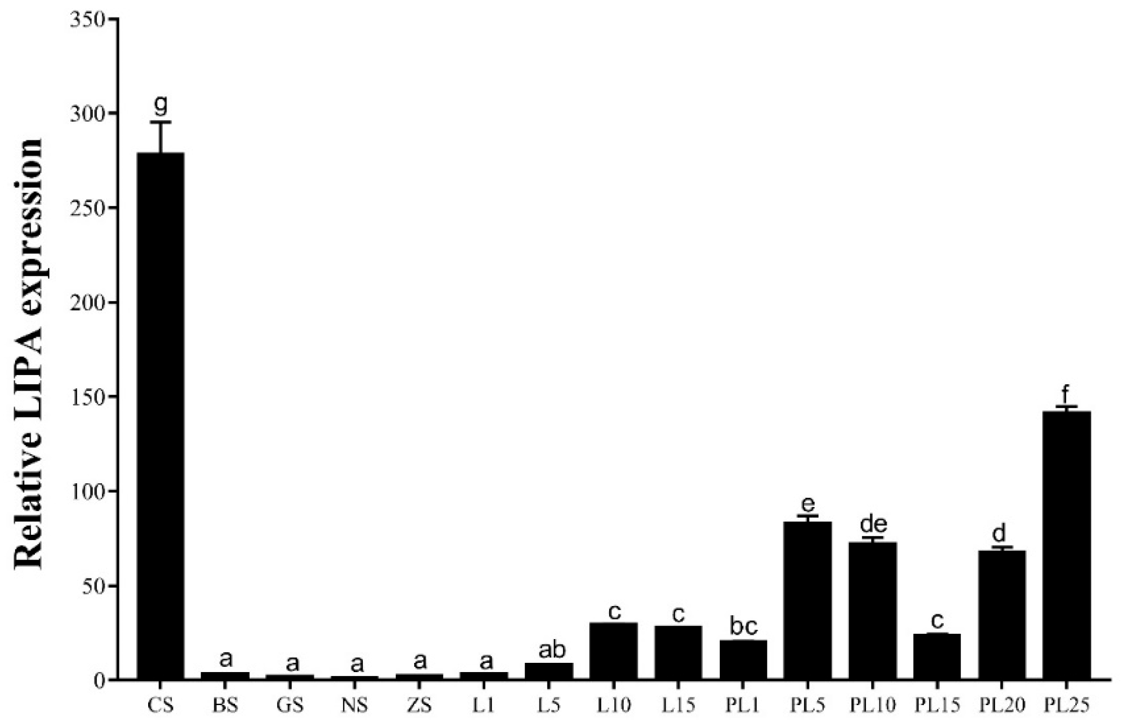
2.2. Expression of the Mn-LIPA Gene in Different Tissues in Males

Mn-LIPA is expressed in the eyestalk, cerebral ganglion, heart, hepatopancreas, testes, and the androgenic gland (Figure 4), but it is almost unexpressed in the gills and muscle. The highest expression level is observed in the hepatopancreas, significantly surpassing other tissues (p < 0.05). Following the hepatopancreas, the cerebral ganglion and testes demonstrate moderate expression, while the eyestalk, heart, and the androgenic gland exhibit lower levels of expression.

2.3. Expression of the Mn-LIPA Gene during Different Developmental Stages

Figure 5 shows the expression pattern of Mn-LIPA at different developmental stages. The expression level of Mn-LIPA is highest during the blastula stage, significantly exceeding subsequent stages (p < 0.05). Subsequently, it undergoes a rapid decline until the L1 stage. Among the later stages, the highest expression is observed at PL25, followed by PL5, PL10, and PL20.

2.4. Localization of the Mn-LIPA Gene in Testes and Hepatopancreas

ISH was used to examine the location of Mn-LIPA mRNA in the testes and hepatopancreas (Figure 6). Strong signals were observed in spermatogonium and spermatocyte cells. No signals were observed in sperm cells in Figure 6A. No signals were observed when the negative RNA probe was used. Figure 6B shows the same situation.

2.5. Effects of Mn-DCHR RNAi Knockdown on PL10-Stage M. nipponense

2.5.1. Sex Ratio

One hundred PL10-stage M. nipponense were injected using dsMn-LIPA at a concentration of 8 μg/g. The sex ratio was counted after the third injection (day 15). It is notable that in M. nipponense, the glands begin to develop at PL10 and sexual differentiation is complete by PL25, with distinctive secondary sexual characteristics enabling the identification of males and females. As shown in Figure 7, the number of prawns in the experimental and control groups decreased over time. However, the sex ratios of the experimental group for the three stages were 1.00, 0.99, and 1.01, which were not significantly different from the control group (p > 0.05).

2.5.2. Steroid Hormone Content

Figure 8 shows the sex hormone content of PL10 M. nipponense after five injections. The gonads of M. nipponense begin to develop at PL10, and sexual differentiation is completed at PL25 with the emergence of physiological males and females. Therefore, the results are only counted for 15, 22, and 30 days in Figure 8A,B. Figure 8A shows the level of MT in male prawns and Figure 8B shows the level of E2 in female prawns. As shown in Figure 8, the MT content in males decreased gradually over time and was consistently lower in the control group (p < 0.05). The E2 content in females was higher than the control at 15 days and lower than the control at 22 and 30 days (p < 0.05).

2.5.3. RNA Interference Efficiency

Figure 9 shows the relative expression of Mn-LIPA in the testes of male M. nipponense at different stages after the injection of dsRNA. The qPCR analysis showed that the expression of Mn-LIPA in the control group remained stable at days 1, 8, and 15 (p > 0.05) and displayed a decreasing trend at days 22 and 30. The Mn-LIPA expression in the experimental group remained consistently low and was 25.31%, 34.72%, 15.96%, 12.81%, and 10.30% of that in the control group on days 1, 8, 15, 22, and 30, respectively. Throughout the experiment, the expression level of Mn-LIPA in the RNAi group was significantly lower than that in the control group (p < 0.05).

2.6. Effects of Mn-DCHR RNAi Knockdown on Male M. nipponense

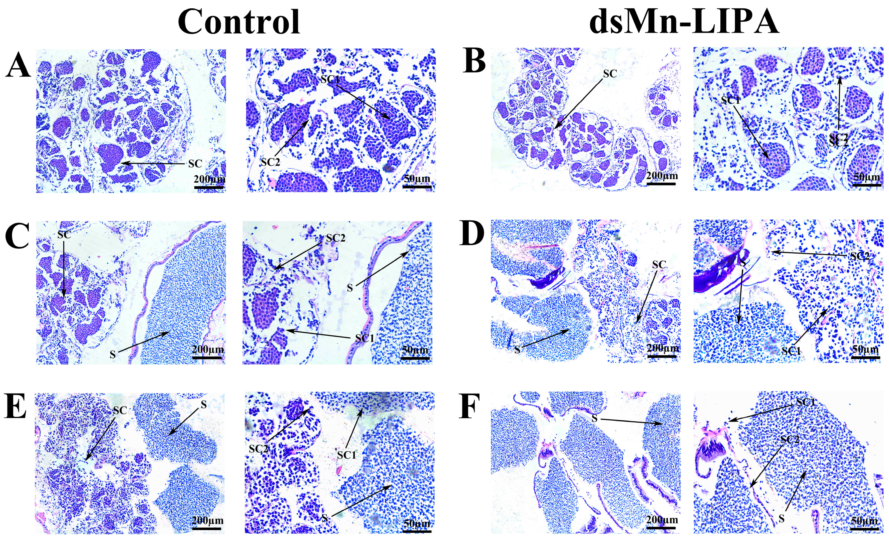
2.6.1. Histological Observations of Gonads

As shown in Figure 10A,B, 1 day after the injection, male prawns from both control and dsMn-LIPA groups were in the spermatid phase, with primary and secondary spermatocytes in the testes. Fifteen days after the injection, a large number of sperm appeared in the testes of both experimental and control groups (Figure 10C,D). Thirty days after the injection, large numbers of primary spermatocytes, secondary spermatocytes, and sperm were still observed in the control group (Figure 10E). However, as shown in Figure 10F, the testes of the dsMn-LIPA group were more mature and almost no spermatocytes were observed.

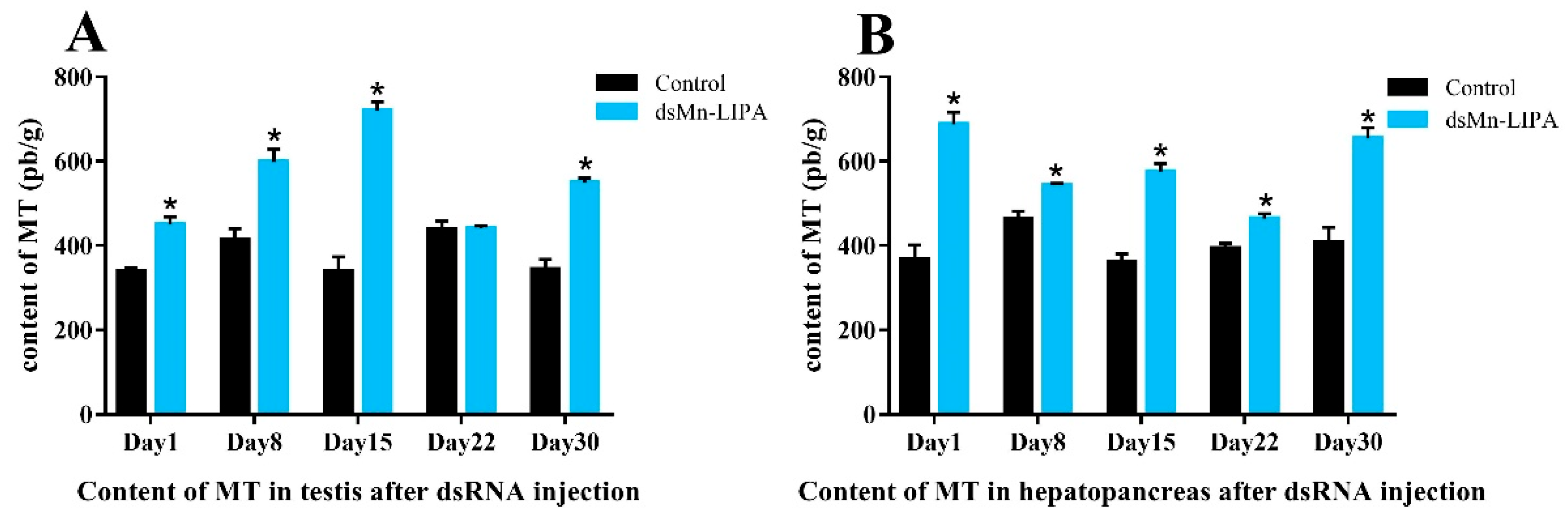
2.6.2. Steroid Hormone Content

Figure 11 shows the MT content in the testes and hepatopancreas of male M. nipponense after five injections. The MT content in the testes and hepatopancreas of male M. nipponense in the control group was relatively stable at 374.65 ± 22.82 pb/g and 397.87 ± 24.79 pb/g, respectively. As shown in Figure 11A, in the dsMn-LIPA groups, MT in the testes of male M. nipponense gradually increased and decreased over time, reaching the highest level of 720.09 pb/g on day 15 (p < 0.05). The levels of MT in the hepatopancreas decreased and then increased and were higher on day 1 and day 30 at 687.85 pb/g and 655.61 pb/g (p < 0.05), respectively (Figure 11B).

2.7. Effects of Steroid Hormones and RNAi on Male Sex-Related Genes in Male M. nipponense

Figure 12 shows the expression of male-related genes in male M. nipponense at different stages after 30 days of different treatments (RNAi, MT, and E2). After dsMn-LIPA injection, insulin-like androgenic gland hormone (IAG), sperm gelatinase (SG), and mab-3-related transcription factor (Dmrt11E) had similar trends of gradually increasing, whereas Mn-LIPA expression was significantly downregulated. Sex-related genes, including Mn-LIPA, were activated after feeding the prawns with sex hormones, and the expression was consistently higher than in the control group (p < 0.05). Negative feedback was shown between IAG and Mn-LIPA after feeding the prawns with MT, and the expression patterns of SG and Dmrt11E were similar. The expression of all three genes gradually decreased after feeding the prawns with E2, except for Dmrt11E, which showed a decrease followed by an increase (p < 0.05).

3. Discussion

Cholesterol ester lipase (LIPA), the only known intracellular lipase active at acidic pH levels, plays an essential role in cholesterol metabolism by hydrolyzing cholesteryl esters and triglycerides within lysosomes and catalyzing the formation of 25-hydroxycholesterol from cholesterol to repress cholesterol biosynthesis [26]. To date, Mn-LIPA has only been recognized for its cholesterol-regulating function in vertebrates, but whether it has other functions in crustacean species is not yet known. Crustaceans, like other arthropods, lack the capacity to synthesize the steroid nucleus de novo; therefore, they have to metabolize dietary sterols to form the cholesterol necessary for their growth and reproduction [27]. In this study, we analyzed the function of Mn-LIPA in M. nipponense, with a particular focus on the role of Mn-LIPA in the regulation of sex hormones, sex differentiation, and gonadal development. Our results may be useful in developing a technique to regulate the development of testes in this species.
We successfully cloned the full-length Mn-LIPA cDNA from M. nipponense. Multiple sequence alignments indicate that Mn-LIPA has three nuclear receptor DNA-binding domains (SP1, CAT, and AP2). From the point of view of biological habits, as the fitness of animals living in nutritionally variable environments depends on their ability to modify energy allocation to reproduction, growth, and maintenance in response to cues from the immediate environment, it is likely that the same genes have acted differently in vertebrates and crustaceans over the course of evolution. Phylogenetic analysis showed that crustaceans and insects were clearly delimited and clustered together, indicating that Mn-LIPA was differentiated in crustaceans and insects and was more conserved in the same class. To the best of our knowledge, the function of Mn-LIPA has not been well defined or analyzed in crustaceans and this is the first qPCR analysis of Mn-LIPA expression. We performed qPCR analysis of Mn-LIPA in various tissues and at various developmental stages of M. nipponense. Tissue-specific analysis revealed that transcripts were detected at much higher levels in the hepatopancreas than in other tissues, followed by the cerebral ganglion and testes, with less expression in the androgenic gland. The hepatopancreas is an important organ in M. nipponense. It is responsible for the absorption and metabolization of nutrients, the storage of energy reserves and minerals, the synthesis of lipoproteins for export to other organs, and other functions [28]. This finding suggests a potential role of Mn-LIPA in sex differentiation and gonadal development, possibly by controlling steroid synthesis (cholesterol, fatty acids, cholesteryl ester, and triglyceride) and thus influencing the endocrine axis of male reproductive activity, which connects the X-organ-sinus gland (XO-SG), AG, and testes in crustaceans [29].
Previous studies have reported that the sensitive period for sexual differentiation is from PL7 to PL19 [30]. The expression levels of Mn-LIPA were highest at the cleavage stage (CS) and PL25 (PL: post-larvae) during the larval and post-larval developmental stages, which are the beginning and end of sex differentiation, respectively, and gradually increased from the blastula stage (BS) to PL20, indicating that it is involved in the process of sexual differentiation in M. nipponense. ISH is used to locate a specific DNA or RNA sequence in a tissue or cell using labeled complementary DNA or RNA [31]. In this study, strong Mn-LIPA signals were detected in both the testes and hepatopancreas, suggesting that Mn-LIPA is involved in steroid synthesis in the hepatopancreas, which in turn promotes sperm maturation and testis development in M. nipponense. However, the mechanism of sexual differentiation in decapods is quite complex, and several sex-linked genes and non-coding RNAs have been successively identified, which together form a vast network of interactions that jointly determine sex [32].
RNAi, the most widely used tool in the functional research of crustacean genes, has enhanced our understanding of its target genes in many crustaceans [33,34,35]. In this study, we further confirmed the role of Mn-LIPA in sex differentiation and gonadal development of M. nipponense using RNAi technology. Oosperm were grown to PL10 and fully sexually differentiated male prawns were subjected to a 30-day RNAi interference experiment. There was no significant change in the sex ratio of PL10 prawns 30 days after a dsMn-LIPA injection. However, enzyme-linked immunosorbent assay (ELISA) indicated that levels of MT produced by males in the experimental group were increased whereas levels of E2 produced by females were decreased. Mn-LIPA expression in the RNAi group was significantly lower than that in the control group on the same day, indicating that dsRNA effectively inhibited Mn-LIPA expression. The expression of sex-determining genes is followed by a series of cascading reactions that gradually lead to differentiation in the sex dimension [36]. It has been shown that the sex-determining system is often controlled by multiple genes in lower aquatic animals [37,38]. It is now generally accepted that sexual manipulation in decapods only functions through the manipulation of AG [39], which controls behavior, spermatogenesis, and sexual differentiation through the secretion of IAG. Histological observations showed that dsMn-LIPA effectively inhibited the development and maturation of sperm in male M. nipponense. Moreover, the experimental group of male prawns had higher levels of MT in the testes and hepatopancreas than the control group. Therefore, we speculate that Mn-LIPA is likely to be involved in the formation and physiology of the AG, testes, and hepatopancreas. Mn-LIPA is not a master gene for controlling sex and gonadal development, but it can affect sex hormone synthesis. These results suggest that Mn-LIPA affects the development of the testes in male M. nipponense by regulating hormone levels. To the best of our knowledge, the reproductive function of Mn-LIPA has not been focused on in any species before this time.
To study the relationship of Mn-LIPA on the regulation of sex-related genes and sex hormones in male M. nipponense in more depth, we fed male prawns with 200 mg/kg MT and E2 and then used qPCR to characterize the expression of three male-specific genes, IAG, SG, and Dmrt11E. IAG is thought to be the active substance responsible for stimulating AG, which encodes and secretes several androgens [40,41]. SG is a key player in regulating various reproductive functions, including sperm motility (hyperactivation) [42], acrosome reaction [43], and sperm-egg fusion [44]. DMRT proteins share a distinctive zinc-finger DNA-binding motif termed the DM domain, the expression of which is upregulated in association with androgen-induced gonadal masculinization [45,46]. IAG and Dmrt11E have both been reported to be key genes for sex reversal in M. rosenbergii [47,48], and have been studied in M. nipponense in relation to male gonadal development [49]. The expression of IAG, SG, and Dmrt11E was increased at different times after the dsMn-LIPA injection, which is consistent with the histological observations.
The sex-related genes including Mn-LIPA were significantly elevated after feeding prawns with sex hormones, suggesting that Mn-LIPA plays a role similar to that of genes such as IAG. The expression of Mn-LIPA gradually increased after 30 days of MT feeding, in contrast to the expression of IAG. Previous studies have shown that exogenous androgens cause changes in gonadal development, but over-reliance on them can lead to a decrease in the self-synthesized secretion of androgens [50,51]. Combining the results of the histological observations and ELISA, we speculate that Mn-LIPA does not directly control the production of sex hormones but rather utilizes the property of hydrolyzing triglycerides and cholesterol to provide energy while influencing the synthesis and secretion of self-sex hormones [26,52]. The expression of IAG, SG, and Mn-LIPA in the experimental group gradually decreased with the feeding of E2 but was always higher than the control group. This suggests that Mn-LIPA is activated by sex hormones, similar to other sex genes. This may be one reason why feeding prawns with sex hormones can cause weight gain [53,54], and the expression of genes in the E2 group also supports this conjecture [55,56]. However, our study did not expose the reasons behind this, such as how Mn-LIPA affects the physiological activities of organs like AG, how Mn-LIPA regulates MT and E2 secretion, whether Mn-LIPA has similar functions in vertebrates, etc.

4. Materials and Methods

4.1. Experimental Prawns

Thirty healthy egg-holding female M. nipponense (body weight = 4.42 g ± 0.55 g) were obtained from Taihu Lake (Wuxi, China; 120°13′44′′ E, 31°28′22′′ N) and cultured until they hatched young prawns. Larvae were fed with Artemia until their body weight reached PL10 (0.0092 ± 0.0017 g) and PL30 (PL: post-larvae) male M. nipponense (1.4351 ± 0.1621 g), and then they were assigned randomly to the control and experimental groups. As shown in Figure 13, an injection was performed every 7 days; after each injection, samples of testes were collected for tissue observation, steroid hormone levels and sex ratios were measured, and qRT-PCR was performed.

4.2. Nucleotide Sequencing and Bioinformatics Analysis

The total RNA of M. nipponense was extracted using RNAiso Plus Reagent (TaKaRa, Dalian, China). The full-length Mn-LIPA cDNA sequence was obtained from the testis transcriptome library of female M. nipponense (accession number: OR602680) under Bioproject PRJNA830321. Three pairs of primers (1, 2, 3) were used to validate the ORF. The Mn-LIPA gene sequence was analyzed using GenBank BLASTX and BLASTN programs (http://www.ncbi.nlm.nih.gov/BLAST/, accessed on 12 October 2023). The open frame analysis of the full-length cDNA sequence of the obtained Mn-LIPA gene was performed using the online program ORF Finder (http://www.ncbi.nlm.nih.gov/gorf/gorf.html, accessed on 12 October 2023). The sequences were subjected to BLAST alignment to test for similarity and consistency. ExPASy ProtParam (http://web.expasy.org/compute_pi/, accessed on 12 October 2023) was used to predict the isoelectric points and mean relative molecular mass of the Mn-LIPA amino acid sequence. The phylogenetic tree analysis was performed using MEGA 5.0, and the calculation was repeated 1000 times using the bootstrap method. Multiple sequence alignment of Mn-LIPA amino acids was performed using DNAMAN 6.0 software.

4.3. Tissue Expression Analysis by Quantitative Real-Time PCR

The Bio-Rad iCycler iQ5 Real-Time PCR system (Bio-Rad, Hercules, CA, USA) was used to detect the expression of the Mn-LIPA gene in the above samples [57]. The specific primers are listed in Table 1. EIF was selected as the reference gene for qPCR [58]. The qPCR expression level of Mn-LIPA was calculated via the 2−ΔΔCT method [59].

4.4. In Situ Hybridization (ISH) and Histological Observations

The testes of mature male M. nipponense were collected and fixed in 4% paraformaldehyde in PBS (pH 7.4) at 4 °C overnight. Following the method of Satoet al. [60], we used the Zytofast PLUS CISH implementation kit and embedded the samples in paraffin. Primers were designed using Primer 5.0 software based on the Mn-LIPA cDNA sequence. The sequence 5′-CCTCATCATTTCCAAATAATCTCAGGACAAACTG-3′ was used as the ISH probe. The slides were examined under a light microscope.
Hematoxylin and eosin (HE) staining was used in order to study the histological changes in the testes and ovaries. On days 1, 15, and 30 after injection, testes samples from the control and Mn-LIPA groups were mounted on slides, which were stained via hematoxylin and eosin staining followed by cleaning with different concentrations of alcohol [61]. Observations took place using a stereo microscope (SZX16; Olympus Corporation, Tokyo, Japan). Comparative labeling was performed with various cell types based on cell morphology [21].

4.5. Sex Ratio Statistics

The prawns in the control and experimental groups were randomly assigned with more than 90 individuals in total to measure the sex ratio and feminization rate. Each group had at least three replicates.

4.6. RNA Interference of LIPA

The online software Snap Dragon (http://www.flyrnai.org/cgi-bin/RNAi_find_primers.pl, accessed on 20 October 2023) was used to design RNAi primers. The T7 promoter sequence was added to the 5′ end of the RNAi primer sequence. T7: TAATACGACTCACTATAGGG. Then, the Mn-LIPA fragment was amplified using RNAi primers. After detection, double-stranded RNA was synthesized using the Transcript AidTMT7 high yield transcription kit (Fermentas, Waltham, MA, USA). The dsRNA was verified using standard agarose gel electrophoresis and the concentration of dsRNA was measured using a BioPhotometer (Eppendorf, Hamburg, Germany). Finally, it was stored at −80 °C.

4.7. Enzyme Linked Immunosorbent Assay (ELISA)

The content of MT in the testes of male prawns and E2 in the ovaries of female prawns were detected by ELISA after RNAi. Tissues from experimental and control groups were collected on days 1, 8, 15, 22, and 30 post-injection, snap-frozen in liquid nitrogen, and stored at −80 °C until MT and E2 content were determined (n = 9). The sex hormone content in prawns was determined using double-antibody one-step sandwich ELISA following the instructions of the Shrimp EH ELISA Kit (Lot number: 20230724-YJ923014 and 20230724-YJ950014; Mlbio Shanghai, Shanghai, China).

4.8. Dietary Preparation

The diets used in this study came from the commercial prawn diet guidelines produced by the Freshwater Fisheries Research Center, Chinese Academy of Fisheries Sciences, China (Wuxi, China). The commercial diet is mainly composed of crude protein, fish meal, shrimp meal, squid meal, starch, soybean meal, ash, canola meal, soybean protein concentrate, crude lipid, etc. [62]. The MT (CAS number: 58-18-4, purity: 97.65%) and E2 (CAS number: 50-28-2, purity: 95.88%) were purchased from Beijing Solarbio Technology Co, Ltd. (Beijing, China). The method of dissolving hormones into the diets is described below [63]. Hormones were dissolved in 95% ethanol to prepare a stock solution at a concentration of 20 mg/mL, and then the solution was evenly sprayed on the feed (1 mL ethanol per 10 g diet) and stirred with a glass stick for at least three minutes. After that, it was placed under a ventilated laboratory hood and left in shade for 15 min. The treated feed was poured into 15 mL test tubes and placed in a refrigerator at 0 °C to evaporate the remaining alcohol naturally.

4.9. Data Analysis

The statistical analyses were all conducted using IBM SPSS Statistics for Windows, version 23.0. (IBM Corporation, Armonk, NY, USA). The significant differences between groups were determined via one-way ANOVA, followed by the least significant difference test and Tukey’s test [64]. Quantitative data are expressed as mean ± standard deviation (SD). Probability (p) values < 0.05 were considered statistically significant.

5. Conclusions

This study describes for the first time the role of Mn-LIPA in sex hormone regulation and gonadal development in M. nipponense. We found that Mn-LIPA is highly expressed in the hepatopancreas, cerebral ganglion, and testes, indicating its involvement in sex differentiation. ISH revealed Mn-LIPA signaling in the spermatheca and hepatopancreas, suggesting its role in steroid synthesis and sperm maturation. RNAi experiments confirmed the impact of Mn-LIPA on sperm development. Furthermore, this study demonstrated the relationship between Mn-LIPA and sex-related genes and hormones. Overall, these findings enhance our understanding of the function of Mn-LIPA in crustaceans and its effects on sex differentiation and gonadal development. This study provides an important theoretical basis for the realization of a monosex culture of M. nipponense.

Author Contributions

P.C. designed the experiment, injected and fed the prawns, and wrote the manuscript. H.F. supervised the experiment. S.J. (Sufei Jiang) and Y.X. provided the experimental prawns. S.J. (Shubo Jin), W.Z. and H.Q. performed the qPCR analysis. Z.G., H.Y. and Y.Z. performed the histological observation and made some further edits. All authors have read and agreed to the published version of the manuscript.

Funding

This research was supported by grants from the Jiangsu Agricultural Industry Technology System; the Central Public-Interest Scientific Institution Basal Research Fund, CAFS (2023TD39); the earmarked fund for CARS-48-07; the seed industry revitalization project of Jiangsu province (JBGS [2021] 118); the new cultivar breeding major project of Jiangsu province (PZCZ201745); and the Natural Science Foundation of Jiangsu Province (BK20221207).

Institutional Review Board Statement

The protocols of all experiments involving M. nipponense were approved in October 2019 (Authorization No. 20191015006) by the Institutional Animal Care and Use Ethics Committee of the Freshwater Fisheries Research Center, Chinese Academy of Fishery Sciences (Wuxi, China).

Informed Consent Statement

Not applicable.

Data Availability Statement

The data presented in this study are available on request from the corresponding author for scientific purposes.

Acknowledgments

Thanks to the Jiangsu Province Platform for the Conservation and Utilization of Agricultural Germplasm.

Conflicts of Interest

The authors declare no conflicts of interest.

References

  1. Cai, Y.; Ng, P. The freshwater palaemonid prawns (Crustacea: Decapoda: Caridea) of Myanmar. Hydrobiologia 2002, 487, 59–83. [Google Scholar] [CrossRef]
  2. Grave, S.; Ghane, A. The establishment of the Oriental River Prawn, Macrobrachium nipponense (de Haan, 1849) in Anzali Lagoon (Iran). Aquat. Invasions 2006, 1, 204–208. [Google Scholar] [CrossRef]
  3. Gul, I.; Abbas, M.; Kausar, S.; Luo, J.; Gao, X.; Mu, Y.; Fan, W.; Cui, H. Insight into crustacean cathepsins: Structure-evolutionary relationships and functional roles in physiological processes. Fish Shellfish Immunol. 2023, 139, 108852. [Google Scholar] [CrossRef] [PubMed]
  4. Li, Y.; Liu, Z.; Li, M.; Jiang, Q.; Wu, D.; Huang, Y.; Jiao, Y.; Zhang, M.; Zhao, Y. Effects of nanoplastics on antioxidant and immune enzyme activities and related gene expression in juvenile Macrobrachium nipponense. J. Hazard. Mater. 2020, 398, 122990. [Google Scholar] [CrossRef] [PubMed]
  5. Ekwudo, M.; Malek, M.; Anderson, C.; Yampolsky, L. The interplay between prior selection, mild intermittent exposure, and acute severe exposure in phenotypic and transcriptional response to hypoxia. Ecol. Evol. 2022, 12, e9319. [Google Scholar] [CrossRef] [PubMed]
  6. Jin, S.; Zhang, W.; Xiong, Y.; Fu, H. Recent progress of male sexual differentiation and development in the oriental river prawn (Macrobrachium nipponense): A review. Rev. Aquac. 2023, 15, 305–317. [Google Scholar] [CrossRef]
  7. Xiong, Y.; Wang, S.; Gui, J.; Mei, J. Artificially induced sex-reversal leads to transition from genetic to temperature-dependent sex determination in fish species. Sci. China Life Sci. 2020, 63, 157–159. [Google Scholar] [CrossRef]
  8. Morris, G.; Braund, P.; Moore, J.; Samani, N.; Codd, V.; Webb, T. Coronary artery disease–associated LIPA coding variant rs1051338 reduces lysosomal acid lipase levels and activity in lysosomes. Arterioscler. Thromb. Vasc. Biol. 2017, 37, 1050–1057. [Google Scholar] [CrossRef]
  9. Ameis, D.; Merkel, M.; Eckerskorn, C.; Greten, H. Purification, characterization and molecular cloning of human hepatic lysosomal acid lipase. Eur. J. Biochem. 1994, 219, 905–914. [Google Scholar] [CrossRef]
  10. Zhang, H. Lysosomal acid lipase and lipid metabolism: New mechanisms, new questions, and new therapies. Curr. Opin. Lipidol. 2018, 29, 218. [Google Scholar] [CrossRef]
  11. Sequencing, R.; Consortium, A. The Rhesus macaque genome sequence informs biomedical and evolutionary analyses. Science 2007, 316, 222–234. [Google Scholar] [CrossRef]
  12. Holmes, R.; Cox, L.; VandeBerg, J. Comparative studies of mammalian acid lipases: Evidence for a new gene family in mouse and rat (Lipo). Comp. Biochem. Physiol. Part D Genom. Proteom. 2010, 5, 217–226. [Google Scholar] [CrossRef] [PubMed]
  13. De, P.; Osoegawa, K.; Zhu, B.; Marra, M.; Schein, J.; Bosdet, I.; Fjell, C. Genome sequence of the Brown Norway rat yields insights into mammalian evolution. Nature 2004, 428, 493–521. [Google Scholar] [CrossRef]
  14. Hillier, W. International chicken genome sequencing consortium. Nature 2004, 432, 695–716. [Google Scholar] [CrossRef]
  15. Holmes, R.; VandeBerg, J.; Cox, L. Genomics and proteomics of vertebrate cholesterol ester lipase (LIPA) and cholesterol 25-hydroxylase (CH25H). 3 Biotech 2011, 1, 99–109. [Google Scholar] [CrossRef] [PubMed]
  16. Lam, P.; Ashbrook, A.; Zygmunt, D.; Yan, C.; Du, H.; Martin, P. Therapeutic efficacy of rscAAVrh74. miniCMV. LIPA gene therapy in a mouse model of lysosomal acid lipase deficiency. Mol. Ther. Methods Clin. Dev. 2022, 26, 413–426. [Google Scholar] [CrossRef] [PubMed]
  17. Pasta, A.; Borro, P.; Cremonini, A.; Formisano, E.; Tozzi, G.; Cecchi, S.; Fresa, R.; Labanca, S.; Djahandideh, A.; Sukkar, S. Effect of a common missense variant in LIPA gene on fatty liver disease and lipid phenotype: New perspectives from a single-center observational study. Pharmacol. Res. Perspect. 2021, 9, e00820. [Google Scholar] [CrossRef]
  18. Hou, Z.; Sket, B. A review of Gammaridae (Crustacea: Amphipoda): The family extent, its evolutionary history, and taxonomic redefinition of genera. Zool. J. Linn. Soc. 2016, 176, 323–348. [Google Scholar] [CrossRef]
  19. Uawisetwathana, U.; Leelatanawit, R.; Klanchui, A.; Prommoon, J.; Klinbunga, S.; Karoonuthaisiri, N. Insights into eyestalk ablation mechanism to induce ovarian maturation in the black tiger shrimp. PLoS ONE 2011, 6, e24427. [Google Scholar] [CrossRef]
  20. Liang, S.; Zeng, C.; Liang, J.; Luo, X.; Gao, X.; Ke, C.; Li, Y.; Chen, L.; Liu, H.; Guo, Y. Effects of eyestalk ablation on growth, feeding behavior, movement and circadian gene expression in Haliotis discus hannai. Aquac. Rep. 2023, 33, 101807. [Google Scholar] [CrossRef]
  21. Cai, P.; Yuan, H.; Gao, Z.; Qiao, H.; Zhang, W.; Jiang, S.; Xiong, Y.; Gong, Y.; Wu, Y.; Jin, S.; et al. 17β-Estradiol Induced Sex Reversal and Gonadal Transcriptome Analysis in the Oriental River Prawn (Macrobrachium nipponense): Mechanisms, Pathways, and Potential Harm. Int. J. Mol. Sci. 2023, 24, 8481. [Google Scholar] [CrossRef] [PubMed]
  22. Reed, K.; Cronan, J. Lipoic acid metabolism in Escherichia coli: Sequencing and functional characterization of the lipA and lipB genes. J. Bacteriol. 1993, 175, 1325–1336. [Google Scholar] [CrossRef]
  23. Jin, L.; Lloyd, R.V. In situ hybridization: Methods and applications. J. Clin. Lab. Anal. 1997, 11, 2–9. [Google Scholar] [CrossRef]
  24. Norbeck, L.; Sheridan, M. An in vitro model for evaluating peripheral regulation of growth in fish: Effects of 17β-estradiol and testosterone on the expression of growth hormone receptors, insulin-like growth factors, and insulin-like growth factor type 1 receptors in rainbow trout (Oncorhynchus mykiss). Gen. Comp. Endocrinol. 2011, 173, 270–280. [Google Scholar] [CrossRef] [PubMed]
  25. Abo-Al-Ela, H.G. Hormones and fish monosex farming: A spotlight on immunity. Fish Shellfish Immunol. 2018, 72, 23–30. [Google Scholar] [CrossRef]
  26. Li, F.; Zhang, H. Lysosomal acid lipase in lipid metabolism and beyond. Arterioscler. Thromb. Vasc. Biol. 2019, 39, 850–856. [Google Scholar] [CrossRef]
  27. Mondy, N.; Grossi, V.; Cathalan, E.; Delbecque, J.; Mermillod, F.; Douady, C. Sterols and steroids in a freshwater crustacean (Proasellus meridianus): Hormonal response to nutritional input. Invertebr. Biol. 2014, 133, 99–107. [Google Scholar] [CrossRef]
  28. Vogt, G. Functional cytology of the hepatopancreas of decapod crustaceans. J. Morphol. 2019, 280, 1405–1444. [Google Scholar] [CrossRef]
  29. Karplus, I.; Sagi, A.; Khalaila, I.; Barki, A. The Influence of Androgenic Gland Implantation on the Agonistic Behavior of Female Crayfish (Cherax quadricarinatus) in Interactions with Males. Behaviour 2003, 140, 649–663. [Google Scholar] [CrossRef]
  30. Chen, Y.; Zhu, Q.; Chen, H.; Zhu, X.; Cui, Z.; Qiu, G. The morphological and histological observation of embryonic development in the oriental river prawn Macrobrachium nipponense. J. Shanghai Ocean Univ. 2012, 21, 33–40. [Google Scholar]
  31. Tautz, D.; Pfeifle, C. A non-radioactive in situ hybridization method for the localization of specific RNAs in Drosophila embryos reveals translational control of the segmentation gene hunchback. Chromosoma 1989, 98, 81–85. [Google Scholar] [CrossRef]
  32. Farhadi, A.; Cui, W.; Zheng, H.; Li, S.; Zhang, Y.; Ikhwanuddin, M.; Ma, H. The regulatory mechanism of sexual development in decapod crustaceans. Front. Mar. Sci. 2021, 8, 679687. [Google Scholar] [CrossRef]
  33. Das, S.; Durica, D. Ecdysteroid receptor signaling disruption obstructs blastemal cell proliferation during limb regeneration in the fiddler crab, Uca pugilator. Mol. Cell. Endocrinol. 2013, 365, 249–259. [Google Scholar] [CrossRef] [PubMed]
  34. Xie, X.; Zhou, Y.; Liu, M.; Tao, T.; Jiang, Q.; Zhu, D. The nuclear receptor E75 from the swimming crab, Portunus trituberculatus: cDNA cloning, transcriptional analysis, and putative roles on expression of ecdysteroid-related genes. Comp. Biochem. Physiol. Part B Biochem. Mol. Biol. 2016, 200, 69–77. [Google Scholar] [CrossRef] [PubMed]
  35. Zhao, Y.; Zhu, X.; Li, Z.; Xu, W.; Dong, J.; Wei, H.; Li, Y.; Li, X. Genetic diversity and structure of Chinese grass shrimp, Palaemonetes sinensis, inferred from transcriptome-derived microsatellite markers. BMC Genet. 2019, 20, 75. [Google Scholar] [CrossRef] [PubMed]
  36. Vicoso, B. Molecular and evolutionary dynamics of animal sex-chromosome turnover. Nat. Ecol. Evol. 2019, 3, 1632–1641. [Google Scholar] [CrossRef] [PubMed]
  37. Toyota, K.; Miyakawa, H.; Hiruta, C.; Sato, T.; Katayama, H.; Ohira, T.; Iguchi, T. Sex determination and differentiation in decapod and cladoceran crustaceans: An overview of endocrine regulation. Genes 2021, 12, 305. [Google Scholar] [CrossRef]
  38. Zheng, S.; Tao, W.; Yang, H.; Kocher, T.D.; Wang, Z.; Peng, Z.; Jin, L.; Pu, D.; Zhang, Y.; Wang, D. Identification of sex chromosome and sex-determining gene of southern catfish (Silurus meridionalis) based on XX, XY and YY genome sequencing. Proc. R. Soc. B 2022, 289, 20212645. [Google Scholar] [CrossRef]
  39. Tan, K.; Jiang, H.; Jiang, D.; Wang, W. Sex reversal and the androgenic gland (AG) in Macrobrachium rosenbergii: A review. Aquac. Fish. 2020, 5, 283–288. [Google Scholar] [CrossRef]
  40. Amir, S.; Eviatar, S.; Isam, K. Sexual differentiation in decapod crustaceans: Role of the androgenic gland. Int. J. Invertebr. Reprod. 1997, 31, 55–61. [Google Scholar] [CrossRef]
  41. Ventura, T.; Sagi, A. The insulin-like androgenic gland hormone in crustaceans: From a single gene silencing to a wide array of sexual manipulation-based biotechnologies. Biotechnol. Adv. 2012, 30, 1543–1550. [Google Scholar] [CrossRef] [PubMed]
  42. Maya, B.; Debby, I.; Haim, B. Regulation of sperm motility by PIP2(4,5) and actin polymerization. Dev. Biol. 2013, 381, 62–72. [Google Scholar] [CrossRef]
  43. Breitbart, H.; Etkovitz, N. Role and regulation of EGFR in actin remodeling in sperm capacitation and the acrosome reaction. Asian J. Androl. 2011, 13, 106–110. [Google Scholar] [CrossRef] [PubMed]
  44. Inoue, N.; Hamada, D.; Kamikubo, H.; Hirata, K.; Kataoka, M.; Yamamoto, M.; Ikawa, M.; Okabe, M.; Hagihara, Y. Molecular dissection of IZUMO1, a sperm protein essential for sperm-egg fusion. Development 2013, 140, 3221–3229. [Google Scholar] [CrossRef] [PubMed]
  45. Zhang, T.; Zarkower, D. DMRT proteins and coordination of mammalian spermatogenesis. Stem Cell Res. 2017, 24, 195–202. [Google Scholar] [CrossRef] [PubMed]
  46. Abayed, F.; Manor, R.; Aflalo, E.; Sagi, A. Screening for Dmrt genes from embryo to mature Macrobrachium rosenbergii prawns. Gen. Comp. Endocrinol. 2019, 282, 113205. [Google Scholar] [CrossRef] [PubMed]
  47. Ventura, T.; Manor, R.; Aflalo, E.; Weil, S.; Raviv, S.; Glazer, L.; Sagi, A. Temporal silencing of an androgenic gland-specific insulin-like gene affecting phenotypical gender differences and spermatogenesis. Endocrinology 2009, 150, 1278–1286. [Google Scholar] [CrossRef]
  48. Xu, H.; Chen, Y.; Wang, Y.; Luo, J.; Li, J.; Shen, S.; Yang, J.; Ma, W. Full Functional Sex Reversal Achieved Through Silencing of MroDmrt11E Gene in Macrobrachium rosenbergii: Production of All-Male Monosex Freshwater Prawn. Front. Endocrinol. 2022, 12, 1867. [Google Scholar] [CrossRef]
  49. Wei, W.; Huang, J.; Yang, Q.; Zhou, F.; Jiang, S.; Li, Y.; Jiang, S.; Yang, L. Molecular characterization and functional analysis of DMRT11E in black tiger shrimp (Penaeus monodon). Aquac. Rep. 2022, 22, 100982. [Google Scholar] [CrossRef]
  50. Cai, P.; Yuan, H.; Gao, Z.; Daka, P.; Qiao, H.; Zhang, W.; Jiang, S.; Xiong, Y.; Gong, Y.; Wu, Y.; et al. Sex Reversal Induced by Dietary Supplementation with 17α-Methyltestosterone during the Critical Period of Sex Differentiation in Oriental River Prawn (Macrobrachium nipponense). Animals 2023, 13, 1369. [Google Scholar] [CrossRef]
  51. Zhu, Q.; Han, C.; Liu, S.; Ouyang, H.; Liu, D.; Zhang, Z.; Huang, J.; Han, L.; Li, S.; Li, G.; et al. Development and gene expression analysis of gonad during 17α-methyltestosterone-induced sex reversal in mandarin fish (Siniperca chuatsi). Aquac. Rep. 2022, 23, 101049. [Google Scholar] [CrossRef]
  52. Liang, F.; Sun, C.; Li, S.; Hou, T.; Li, C. Therapeutic effect and immune mechanism of chitosan-gentamicin conjugate on Pacific white shrimp (Litopenaeus vannamei) infected with Vibrio parahaemolyticus. Carbohydr. Polym. 2021, 269, 118334. [Google Scholar] [CrossRef] [PubMed]
  53. Greisy, Z.; Gamal, A. Monosex production of tilapia, Oreochromis niloticus using different doses of 17α-methyltestosterone with respect to the degree of sex stability after one year of treatment. Egypt. J. Aquat. Res. 2012, 38, 59–66. [Google Scholar] [CrossRef]
  54. Huang, J.; Liu, S.; Ouyang, H.; Han, C.; Li, M.; Han, L.; Li, S.; Li, G.; Lin, H.; Zhang, Y. Production of XX neo-male mandarin fish, Siniperca chuatsi, after sexual differentiation by oral administration of 17α-methyltestosterone. Water Biol. Secur. 2023, 2, 100095. [Google Scholar] [CrossRef]
  55. Voorhees, J.; Mamer, E.; Schill, D.; Adams, M.; Martinez, C.; Barnes, M. 17β-Estradiol can induce sex reversal in Brown Trout. Fishes 2023, 8, 103. [Google Scholar] [CrossRef]
  56. Black, D.; Minkin, M.; Graham, S.; Bernick, B.; Mirkin, S. Effects of combined 17β-estradiol and progesterone on weight and blood pressure in postmenopausal women of the REPLENISH trial. Menopause 2021, 28, 32. [Google Scholar] [CrossRef]
  57. Du, Y.; Ma, K.; Qiu, G. Discovery of the genes in putative GnRH signaling pathway with focus on characterization of GnRH-like receptor transcripts in the brain and ovary of the oriental river prawn Macrobrachium nipponense. Aquaculture 2015, 442, 1–11. [Google Scholar] [CrossRef]
  58. Liu, Z.; Xiao, J.; Xia, Y.; Wu, Q.; Zhao, C.; Li, D. Selection and validation of reference genes for RT-qPCR-based analyses of Anastatus japonicus Ashmead (Hymenoptera: Helicopteridae). Front. Physiol. 2022, 13, 1046204. [Google Scholar] [CrossRef]
  59. Andreani, J.; Le, M.; Duflot, I.; Jardot, P.; Rolland, C.; Boxberger, M.; Wurtz, N.; Rolain, J.; Colson, P.; La, B. In vitro testing of combined hydroxychloroquine and azithromycin on SARS-CoV-2 shows synergistic effect. Microb. Pathog. 2020, 145, 104228. [Google Scholar] [CrossRef]
  60. Sato, K.; Ohuchi, H. The Opsin 3/Teleost multiple tissue opsin system: mRNA localization in the retina and brain of medaka (Oryzias latipes). J. Comp. Neurol. 2021, 529, 2484–2516. [Google Scholar] [CrossRef]
  61. Faeed, M.; Kasra, R.; PourKazemi, M.; Darboee, M.; Haghighi, S. Study on effect feedings with probiotics in increasing resistance to Aeromonas hydrophila and Changes in gut bacterial communities Sander lucioperca. Biol. J. Microorg. 2018, 7, 1–12. [Google Scholar] [CrossRef]
  62. Hodar, A.; Vasava, R.; Mahavadiya, D.; Joshi, N. Fish meal and fish oil replacement for aqua feed formulation by using alternative sources: A review. J. Exp. Zool. India 2020, 23, 13–21. [Google Scholar] [CrossRef]
  63. Saleh, S.; Widodo, M.; Kilawati, Y. The Effect of 17α-Methyltestosterone Hormone Application on Masculinization and Growth Rate of Vaname Shrimp Larvae (Litopenaeus Vannamei, Boone 1931). Russ. J. Agric. Socio-Econ. Sci. 2018, 79, 352–356. [Google Scholar] [CrossRef]
  64. Rosales, M.; Castillo, S.; Pohlenz, C.; Gatlin, D. Evaluation of dried yeast and threonine fermentation biomass as partial fish meal replacements in the diet of red drum Sciaenops ocellatus. Anim. Feed Sci. Technol. 2017, 232, 190–197. [Google Scholar] [CrossRef]
Figure 1. The nucleotide and deduced amino acid sequences of Mn-LIPA. The numbers on the left of the sequence indicate the positions of nucleotides and amino acids. The start codon ATG and the stop codon TGA are indicated in red and underlined, and the TGA stop codon in the amino acid sequence is represented by an asterisk (*).
Ijms 25 01399 g001
Figure 2. Amino acid sequences from other species aligned with the sequence encoded by the Mn-LIPA gene. The black line indicates the PLN02872 superfamily. The red boxes mark the three conserved domain sites. GenBank accession number: Macrobrachium nipponense (OR602680.1), Crangon crangon (MH055770.1), Eriocheir sinensis (OU618541.1), Homarus americanus (XM_042384002.1), Penaeus chinensis (XM_047625243.1), Penaeus monodon (XM_037925897.1), Penaeus vannamei (XM_027357916.1), Portunus trituberculatus (XM_045258007.1), and Procambarus clarkia (XM_045760019.1).
Ijms 25 01399 g002
Figure 3. Phylogenetic tree of Mn-LIPA sequences. The diagram was generated via the neighbor-joining method using the MEGA 7.0 program, with 1000 bootstrapping replications. The red star indicates the location of the M. nipponense. The numbers indicate the reliability of branches.
Ijms 25 01399 g003
Figure 4. Expression pattern of the Mn-LIPA gene in different tissues. E: eyestalk, Cg: cerebral ganglion, H: heart, He: hepatopancreas, G: gill, M: muscle, T: testes, Ag: androgenic gland. Data are presented as the mean ± SD (n = 6). Different letters indicate significant differences (p < 0.05) which were analyzed via one-way ANOVA followed by least significant difference and Tukey’s test.
Ijms 25 01399 g004
Figure 5. The expression levels of Mn-LIPA during embryonic development stages. CS: cleavage stage; BS: blastula stage; GS: gastrula stage; NS: nauplius stage; ZS: zoea stage; L1: the first day after hatching; PL1: the first day post-larvae, and so on. Data are shown as mean ± SD (n = 6). Different letters indicate significant differences (p < 0.05) which were analyzed by one-way ANOVA followed by least significant difference and Tukey’s test.
Ijms 25 01399 g005
Figure 6. In situ hybridization analysis of M. nipponense. (A) Testes; (B) hepatopancreas. SG: spermatogonium; SC: spermatocyte; S: sperm; CT: collecting tissues, E: embryonic cell, R: resorption cells; F: fiber cells; B: blister cells, M: midget cells. Scale bars = 20 μm.
Ijms 25 01399 g006
Figure 7. The number of male and female prawns on days 15, 22, and 30 after the injection of PL2 M. nipponense. The number of males is above the black line and the number of females is below. Data are presented as the mean ± SD (n > 20).
Ijms 25 01399 g007
Figure 8. Sex hormone levels in males and females after PL10 M. nipponense received five injections of dsRNA. (A) Levels of 17α-methyltestosterone in males; (B) Levels of 17β-estradiol in females. Data are presented as the mean ± SD (n = 6); * indicates statistical significance at p < 0.05, which were analyzed by one-way ANOVA followed by least significant difference and Tukey’s test.
Ijms 25 01399 g008
Figure 9. Expression levels of Mn-LIPA in the testes after injection with dsMn-LIPA. Data are shown as mean ± SD (n = 6); * indicates statistical significance at p < 0.05, which were analyzed by one-way ANOVA followed by least significant difference and Tukey’s test.
Ijms 25 01399 g009
Figure 10. Histological sections of testes from control and dsMn-LIPA groups after five injections. (A,B) Histological sections of testes on day 1; (C,D) Histological sections of testes on day 15; (E,F). Histological sections of testes on day 30. SC: Spermatocyte; SC1: Primary spermatocyte; SC2: Second spermatocyte; S: Sperm. Scale bars: 200 µm and 50 µm.
Ijms 25 01399 g010
Figure 11. Levels of 17α-methyltestosterone in the testes and hepatopancreas of male M. nipponense after five injections of dsMn-LIPA. (A) Level of 17α-methyltestosterone in the testes; (B) Content of 17α-methyltestosterone in hepatopancreas. Data are presented as the mean ± SD (n = 6); * indicates statistical significance at p < 0.05, which were analyzed by one-way ANOVA followed by least significant difference and Tukey’s test.
Ijms 25 01399 g011
Figure 12. Expression levels of sex-related genes in testes after treatment with dsMn-LIPA, 17α-methyltestosterone, and 17β-estradiol. (A) Mn-IAG; (B) Mn-SG; (C) Mn-Dmrt11E; (D) Mn-LIPA. Data are shown as the mean ± SD (n = 6); different letters indicate significant differences (p < 0.05) which were analyzed by one-way ANOVA followed by least significant difference and Tukey’s test.
Ijms 25 01399 g012
Figure 13. Experimental design.
Ijms 25 01399 g013
Table 1. Primers for cDNA clone, qPCR analysis, and RNAi that were involved in this study.
Primer NameSequence (5′→3′)Usage
LIPA-F1ACTCATTTCTTTGTGGGACCCTTORF
LIPA-R1GAAATTCCTTGGGCAAAGTGGAAORF
LIPA-F2CGTAGCAACAGTTACAAGCATCAORF
LIPA-R2AAGGGTCCCACAAAGAAATGAGTORF
LIPA-F3ACTCAAACACTCTTCCACTTTGCORF
LIPA-R3ATAGACCAGAGGCTTTGCATCAGORF
Mn-LIPA-FACCGCCAGAATATGATGTTGCTAqPCR
Mn-LIPA-RAAAGGAAGTCCAGGTGGCTAAATqPCR
Mn-IAG-FCGCCTCCGTCTGCCTGAGATACqPCR
Mn-IAG-RCCTCCTCCTCCACCTTCAATGCqPCR
Mn-SG-FACCCTAGCCCCAGTACGTGTTqPCR
Mn-SG-RAGAGGTGGTGAAGCTGTCTCTCAqPCR
Mn-Dmrt11E-FACGACCTTAGTAGGATGGACAGTqPCR
Mn-Dmrt11E-RGAGTGGAGGCAATAGAATGGGTAqPCR
Mn-EIF-FCATGGATGTACCTGTGGTGAAACqPCR
Mn-EIF-RCTGTCAGCAGAAGGTCCTCATTAqPCR
dsMn-LIPA-FTAATACGACTCACTATAGGGACTGACCCATAACCCTGCTGdsRNA
dsMn-LIPA-RTAATACGACTCACTATAGGGTCAGTTGCCCAAAGGAAGTCdsRNA
Note: GenBank accession number: Mn-LIPA (OR602680.1), Mn-IAG (JX962354), Mn-SG (EF647641), Mn-Dmrt11E (MH636338), Mn-EIF (MH540106).
Disclaimer/Publisher’s Note: The statements, opinions and data contained in all publications are solely those of the individual author(s) and contributor(s) and not of MDPI and/or the editor(s). MDPI and/or the editor(s) disclaim responsibility for any injury to people or property resulting from any ideas, methods, instructions or products referred to in the content.

