Review

Cracking the Endothelial Calcium (Ca2+) Code: A Matter of Timing and Spacing

by
Francesco Moccia
1,*,
Valentina Brunetti
1,
Teresa Soda
2,
Roberto Berra-Romani
3 and
Giorgia Scarpellino
1
1
Laboratory of General Physiology, Department of Biology and Biotechnology “L. Spallanzani”, University of Pavia, 27100 Pavia, Italy
2
Department of Health Sciences, University of Magna Graecia, 88100 Catanzaro, Italy
3
Department of Biomedicine, School of Medicine, Benemérita Universidad Autónoma de Puebla, Puebla 72410, Mexico
*
Author to whom correspondence should be addressed.
Int. J. Mol. Sci. 2023, 24(23), 16765; https://doi.org/10.3390/ijms242316765
Submission received: 6 November 2023 / Revised: 16 November 2023 / Accepted: 24 November 2023 / Published: 26 November 2023
(This article belongs to the Special Issue Calcium Handling in Cell Physiology and Pathophysiology)

Abstract

:
A monolayer of endothelial cells lines the innermost surface of all blood vessels, thereby coming into close contact with every region of the body and perceiving signals deriving from both the bloodstream and parenchymal tissues. An increase in intracellular Ca2+ concentration ([Ca2+]i) is the main mechanism whereby vascular endothelial cells integrate the information conveyed by local and circulating cues. Herein, we describe the dynamics and spatial distribution of endothelial Ca2+ signals to understand how an array of spatially restricted (at both the subcellular and cellular levels) Ca2+ signals is exploited by the vascular intima to fulfill this complex task. We then illustrate how local endothelial Ca2+ signals affect the most appropriate vascular function and are integrated to transmit this information to more distant sites to maintain cardiovascular homeostasis. Vasorelaxation and sprouting angiogenesis were selected as an example of functions that are finely tuned by the variable spatio-temporal profile endothelial Ca2+ signals. We further highlighted how distinct Ca2+ signatures regulate the different phases of vasculogenesis, i.e., proliferation and migration, in circulating endothelial precursors.

1. Introduction

Once regarded as a mere physical barrier between the bloodstream and surrounding tissues, the vascular endothelium is now recognized as a distributed endocrine organ with a surface of 3000–7000 m2 and an estimated weight of 1 kg in humans [1,2,3]. Being located at the interface between flowing blood and parenchymal cells, vascular endothelial cells serve as a signal transduction platform that is simultaneously exposed to a myriad of chemical and physical inputs [4], which are integrated through an increase in intracellular Ca2+ concentration ([Ca2+]i) to generate the most appropriate output. Consequently, endothelial Ca2+ signals regulate a wide range of cardiovascular functions, including vascular resistance and permeability, coagulation, leukocyte transmigration, neurovascular coupling, angiogenesis and vasculogenesis [4,5,6,7,8,9,10,11]. Therefore, even a subtle dysregulation of endothelial Ca2+ signals can potentially lead to major human diseases [12,13,14,15,16,17,18,19,20,21,22,23,24], including hypertension, atherosclerosis, type 2 diabetes, obesity, deep vein thrombosis, sepsis, neurodegenerative disorders and cancer.
Vasodilation and sprouting angiogenesis represent the two most widely investigated functions regulated by endothelial [Ca2+]i [4,5,8]. The mechanisms whereby endothelial Ca2+ signals regulate vasodilation, i.e., an increase in lumen diameter to increase blood flow to downstream vessels, include the gasotransmitter nitric oxide (NO) and endothelium-dependent hyperpolarization (EDH) via small/intermediate conductance Ca2+-activated K+ channels (KCa), KCa2.3 (SKCa) and KCa3.1 (IKCa). However, the contribution of NO and EDH to endothelium-dependent vasodilation can vary along the vascular bed with NO being more effective in large conduit arteries and EDH in small resistance vessels [5,10]. Sprouting angiogenesis, i.e., the multistepped process that expands the existing vascular network through endothelial cell proliferation, migration and interaction with the surrounding environment, is also tuned by endothelial Ca2+ signals [8]. Vascular endothelial growth factor (VEGF), which is indispensable to initiate and direct the vascular sprout [8], may trigger a complex repertoire of Ca2+ signals to engage distinct Ca2+-sensitive pathways and regulate the different phases of the angiogenic response [8]. These examples embody the concept that endothelial Ca2+ signaling can recruit different Ca2+-dependent effectors to elicit specific vascular responses either depending on the vascular bed (e.g., vasorelaxation, which is primarily driven by NO in large vessels and EDH in small resistance vessels) or on the vascular function (e.g., vasorelaxation vs. angiogenesis) [4,25,26,27].
Herein, we provide an overview of how vascular endothelial cells regulate two representative examples of vascular functions, such as vasomotion and angiogenesis, by extracting the relevant information from the subcellular spatial profile and/or the temporal pattern of the underlying Ca2+ signal.

2. An Introduction to Endothelial Ca2+ Signaling: From Global to Local Ca2+ Signals

An increase in endothelial [Ca2+]i can be elicited by neurohumoral agonists, e.g., acetylcholine [28,29,30,31] and adenosine trisphosphate (ATP) [32,33], inflammatory mediators, e.g., histamine [34,35] and bradykinin [36], growth factors, e.g., VEGF [37,38,39] and angiopoietins [40], and reactive oxygen species (ROS) [41,42]. Interestingly, endothelial Ca2+ waves can also be evoked by synaptically released glutamate [43,44] and γ-aminobutyric acid (GABA) [45,46] at the blood–brain barrier (BBB) as well as by sympathetic stimulation of the adrenergic terminals innervating the mesenteric bed [47,48,49,50,51]. In addition, endothelial Ca2+ signals can be elicited by physical stimuli, such as laminar shear stress [52,53,54], changes in intravascular pressure [55], mechanical scraping [56,57] and heat [58].
A kaleidoscope of intracellular Ca2+ waveforms can be elicited by chemical and neurohumoral stimulation of vascular endothelial cells, as extensively reviewed in [4,6,8,26,27,59,60]. The synthesis of Ca2+-sensitive fluorescent indicators, followed by the development of genetic Ca2+ indicators (GECIs), the advent of high-resolution microscopic imaging and the generation of endothelial-specific Cre mouse models, contributed to unveiling how endothelial cells assemble their versatile Ca2+ handling machinery to generate an array of spatiotemporally diverse Ca2+ signals that are selectively coupled to distinct effectors to activate precise vascular responses [25]. Initial epifluorescence Ca2+ imaging primarily conducted on vascular endothelial cells loaded with a ratiometric Ca2+ indicator (i.e., Fura-2) showed that the endothelial Ca2+ responses to physiological autacoids and growth factors consist of transient, biphasic, monotonic or oscillatory elevations in [Ca2+]i that invade the whole cytoplasm and sometimes propagate to neighboring endothelial cells [25]. Nevertheless, high-speed Ca2+ imaging of endothelial cells loaded with non-ratiometric Ca2+ indicators (e.g., Fluo-3, Fluo-4 and Cal520) or expressing GECIs revealed that the physiological Ca2+ signals underlying vascular responses comprise a mosaic of spatially and temporally discrete Ca2+ events, which can remain highly localized to a specific subcellular site or propagate to generate intra- or intercellular endothelial Ca2+ waves [9]. These endothelial Ca2+ microdomains are mediated by the opening of Ca2+-permeable channels that are located both on the endoplasmic reticulum (ER) and on the plasma membrane (PM) and may be selectively coupled to distinct Ca2+-dependent effectors to modulate different functions.

2.1. General Mechanisms of Agonist-Induced Endothelial Ca2+ Signals

The endothelial Ca2+ response to chemical stimulation is primarily initiated by ER-dependent Ca2+ release and maintained by extracellular Ca2+ entry across the PM. ER Ca2+ mobilization requires agonist binding to Gq-Protein Coupled Receptors (GqPCRs) or tyrosine kinase receptors (TKRs) on the PM, which are coupled to distinct isoforms of phospholipase C (i.e., respectively, PLCβ and PLCγ) [8,9,27,61]. PLC cleaves the PM-associated phosphoinositide 4,5-bisphosphate (PIP2) into diacylglycerol (DAG) and InsP3, which mobilizes ER Ca2+ by gating InsP3 receptors (InsP3Rs). InsP3Rs represent the major family of ER Ca2+-releasing channels in the endothelial lineage, as all the known InsP3R isoforms (i.e., InsP3R1, InsP3R2 and InsP3R3) are expressed in vascular endothelial cells [27,61,62]. PLC-derived InsP3 primes InsP3Rs to be activated by local Ca2+ such that, in the presence of permissive InsP3 concentrations, a spatially restricted Ca2+ pulse can be regeneratively propagated via Ca2+-induced Ca2+ release (CICR) [63]. The Ca2+-dependent recruitment of InsP3Rs could be supported by lysosomal Ca2+ release through two-pore channels (TPCs) [64,65], which may present two distinct isoforms (i.e., TPC1 and TPC2) in vascular endothelial cells and are gated by nicotinic acid adenine dinucleotide phosphate (NAADP). In contrast, ryanodine receptors (RyRs), which are mainly responsible for Ca2+ release from the sarcoplasmic reticulum (SR) in vascular smooth muscle cells (VSMCs) [27], are unlikely to significatively shape endothelial Ca2+ dynamics [8,9,27].
The InsP3-induced increase in endothelial [Ca2+]i may rapidly return to the baseline due to the removal of cytosolic Ca2+ by Ca2+-clearing mechanisms, such as sarco-endoplasmic reticulum Ca2+-ATPase (SERCA), plasma membrane Ca2+-ATPase (PMCA), Na+/Ca2+ exchanger (NCX) and mitochondria. In most cases, however, the initial elevation in [Ca2+]i may be followed by repetitive cycles of InsP3-induced ER Ca2+ release that occur as prolonged Ca2+ oscillations or by a sustained plateau level. In both cases, extracellular Ca2+ entry is required to refill the ER and prolong the Ca2+ response [4,6,8,25,26,27]. Agonist-induced Ca2+ influx in vascular endothelial cells can occur either through store-operated channels (SOCs) [61,66], which couple ER Ca2+ load with the Ca2+ permeability of the PM, or through members of the transient receptor potential (TRP) superfamily of non-selective cation channels that are activated downstream of the PLC pathway [67,68], such as TRP Canonical 3 (TRPC3) [69,70,71] and TRP Vanilloid 4 (TRPV4) [72,73,74]. In addition, agonist-induced Ca2+ entry in endothelial cells could be supported by ionotropic receptors, such as P2X receptors [32] and N-methyl-D-aspartate receptors (NMDARs) [44].

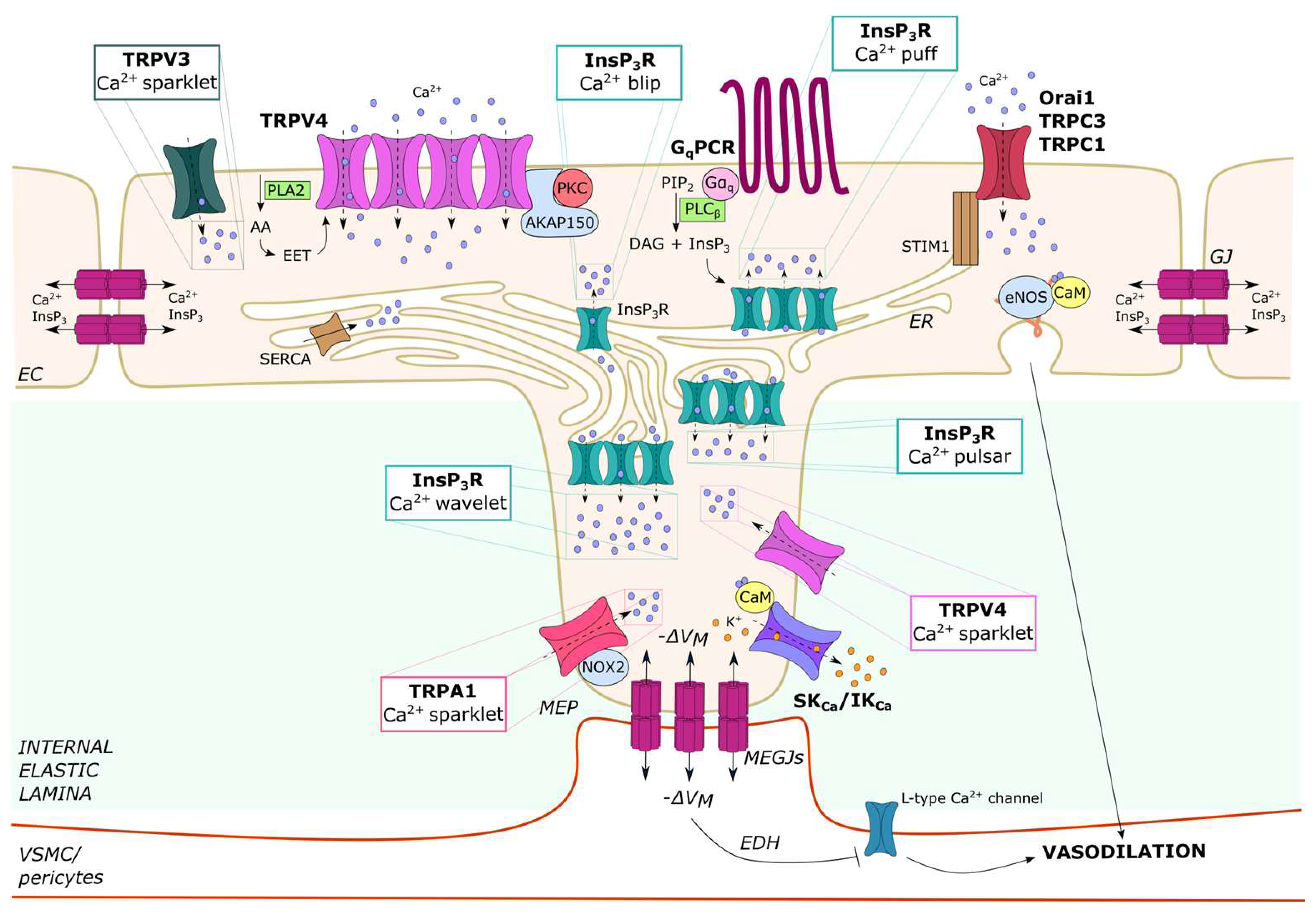
2.2. Endothelial Ca2+ Microdomains Generated by InsP3-Dependent ER Ca2+ Release

InsP3Rs are non-selective cation channels that present large single-channel conductance but only moderate Ca2+ selectivity over Na+ and K+ (PCa/PK ~7) [75]. The three InsP3R isoforms differ in their sensitivity to both InsP3- and Ca2+-dependent activation and, therefore, may encode distinct InsP3-mediated Ca2+ signals [76,77]: InsP3R2, which is the most sensitive to InsP3, sustains long-lasting oscillations in [Ca2+]i; InsP3R1 supports irregular Ca2+ oscillations; and InsP3R3, which is the less sensitive isoform to both InsP3 and Ca2+, serves as an anti-oscillatory unit that generates monophasic Ca2+ signals. To illustrate how this concept also applies to the vascular endothelium, acetylcholine and glutamate trigger intracellular Ca2+ oscillations in mouse cerebrovascular endothelial cells expressing InsP3R1 and InsP3R2 [43,78], while they induce a biphasic elevation in [Ca2+]i in their human counterparts expressing only InsP3R3 [31,79]. The global Ca2+ waves elicited by endothelial autacoids result from the summation of multiple local Ca2+ events that differ in spatial distribution and duration and are known as blips, puffs, pulsars and wavelets (Figure 1). Because of their spatial distribution, some of these discrete Ca2+ events (i.e., pulsars and wavelets) can be detected in native, but not cultured, endothelial cells. Local Ca2+ events sustained by rhythmic InsP3R activation may also spontaneously occur in the vascular intima. However, they are more likely to take place in resistance-sized vessels as compared to large conduit vessels [28,29,80,81].

2.2.1. Ca2+ Blips and Puffs in Cultured Endothelial Cells

Ca2+ puffs, which can also occur spontaneously [82,83], are the building blocks of the global Ca2+ signals elicited by physiological agonists (Figure 1). A preliminary characterization of the spatio-temporal organization of agonist-evoked changes in endothelial [Ca2+]i was conducted on calf pulmonary artery endothelial (CPAE) cells (Table 1). Confocal scanning microscopy revealed that the most elementary endothelial Ca2+ release event is represented by the Ca2+ blip, which reflects the opening of a single InsP3R (Figure 1) [84]. Endothelial Ca2+ blips present an average amplitude of 23 nM and a spatial spread of 1–3 µm and last <100 ms (Table 1) [84]. Superposition of discrete Ca2+ blips results in agonist-evoked Ca2+ puffs, which derive from the opening of a fixed cluster of adjacent InsP3Rs and present a higher amplitude (50–100 nM vs. 23 nM) and wider spatial spread (30 µm vs. 1–3 µm) than unitary blips (Table 1) [84]. Endothelial Ca2+ puffs precede by several hundred milliseconds the regenerative Ca2+ wave that propagates from the periphery to the central region of the cell at an average speed of 20–60 μm/s upon supra-physiological chemical stimulation [84]. Subsequent reports have shown that agonist-evoked cytosolic Ca2+ waves can propagate into the nucleus through CICR or by diffusion through nuclear pores in several types of cultured vascular endothelial cells [85,86,87].

2.2.2. Ca2+ Puffs, Pulsars and Wavelets in Native Endothelial Cells

Direct imaging of endothelial Ca2+ activity has confirmed that, in native macro- and microvascular endothelial cells, the regenerative Ca2+ wave elicited by physiological autacoids, such as acetylcholine [82,88,89,90] and adenosine [91,92], is initiated by the spatio-temporal recruitment of local Ca2+ puffs. InsP3-driven endothelial Ca2+, puffs in situ were first reported in rat ureteral arterioles [82] and then characterized in mouse small mesenteric precapillary arterioles [83] and rat tail artery [89], in which low concentrations of acetylcholine induced localized Ca2+ release events mainly originating at the distal ends of the cells. The spatio-temporal summation of InsP3-driven ER Ca2+ puffs leads to a global elevation of endothelial [Ca2+]i that could spread to neighboring endothelial cells to ultimately coordinate the vascular function, as outlined in Section 2.4.1.
However, the opening of a spatially restricted cluster of Ca2+-permeable channels may also generate local Ca2+ microdomains that are located around the inner mouth of the open channel, do not generate cell-wide Ca2+ waves and are tightly coupled to specific Ca2+-dependent effectors. These local Ca2+ signals are known as Ca2+ pulsars, Ca2+ wavelets and Ca2+ sparklets (Figure 1 and Table 1). Endothelial Ca2+ pulsars and Ca2+ wavelets are subcellular Ca2+ signals that are detectable only in native cells because they occur specifically at myo-endothelial projections (MEPs) (Figure 1) [10], whereas Ca2+ sparklets occur at the PM (Figure 1) and will be described in Section 2.3. Endothelial cells may send projections, known as MEPs, through holes in the internal elastic lamina (IEL) that separates the intimal and medial layers of the vascular wall (Figure 1) [5,10,80]. MEPs enable a single endothelial cell to make direct contact with multiple VSMCs; furthermore, MEPs may couple adjoining endothelial cells and VSMCs via myo-endothelial gap junctions (MEGJs), which allow the transmission of both electrical and chemical (e.g., Ca2+ and InsP3) signals [5,10,80]. The abundance of MEPs is inversely related to the vessel diameter such that they are more abundant in small resistance arteries and arterioles as compared to large vessels [5,93]. Studies carried out in mouse mesenteric third-order arterioles have shown that MEPs are enriched with the pro-oscillatory Ca2+ release units, InsP3R1 and InsP3R2 [94], which generate spontaneous, spatially fixed Ca2+ signals, known as Ca2+ pulsars [80]. These Ca2+ pulsars display a wider spatial spread as compared to Ca2+ puffs (~14 µm2) and occur repeatedly at the same sites with a frequency of 0.08 Hz (Table 1) [80]. Endothelial Ca2+ pulsars are driven by tonic PLC activity [95], but their frequency can be enhanced by the stimulation of GqPCRs, including muscarinic M3 receptors [80] and protease-activated receptor-2 (PAR-2) [96]. The mechanisms responsible for tonic PLC activation in pressurized resistance arteries are still unclear, but the spontaneous local Ca2+ activity recorded in the presence of physiological shear stress was due to flow-induced acetylcholine release and M3 receptor activation [29]. Nevertheless, an excessive increase in intraluminal pressure may suppress InsP3-induced Ca2+ release events by altering the endothelial cell shape and geometry, thereby limiting cytosolic Ca2+ diffusion that drives CICR through InsP3Rs [97]. The inhibitory effect of intraluminal pressure could decrease with aging, thereby leading to an increase in InsP3-induced ER Ca2+ release events [98]. Recruitment of Ca2+-calmodulin (CaM)-dependent protein kinase II (CaMKII) by the oscillatory Ca2+ signals may dampen the frequency of Ca2+ pulsars by inhibiting InsP3Rs, thereby acting as an additional feedback inhibition mechanism to fine-tune the spontaneous endothelial Ca2+ activity [94].
New endothelial Ca2+ pulsar sites can also be activated by sympathetic nerve stimulation of adjoining VSMCs via Ca2+ or InsP3 transfer through MEGJs in pressurized resistance arteries or arterioles [48,49]. The engagement of VSMC α1-adrenergic receptors may lead to InsP3 production within VSMCs overlying the endothelial monolayer; the newly formed InsP3 may then diffuse through MEGJs, thereby increasing the frequency of spontaneous Ca2+ pulsars and activating endothelium-dependent vasorelaxing pathways (i.e., EDH and NO) [49]. This mechanism was termed myo-endothelial feedback and could be mimicked by the pharmacological stimulation of arteriolar VSMCs with phenylephrine [99]. Subsequently, it was shown that VSMC depolarization was per se able to accelerate the frequency and increase the spatial distribution of endothelial Ca2+ pulsars in a voltage-dependent manner [48,81]: according to this model, the opening of voltage-gated L-type Ca2+ channels in arteriolar VSMCs leads to extracellular Ca2+ entry, which passes through MEGJs to stimulate endothelial InsP3Rs via CICR [48]. The endothelial Ca2+ signals that occur during myo-endothelial feedback are regulated by non-ER-based calreticulin, although the underlying mechanism is still unknown [100]. The myo-endothelial feedback could also be mediated by another type of local endothelial Ca2+ signals, known as Ca2+ wavelets, in hamster skeletal muscle arteries [50]. Endothelial Ca2+ wavelets can be activated at MEPs by rhythmic ER Ca2+ release through InsP3Rs upon stimulation of VSMC α1-adrenergic receptors with phenylephrine [50]. With respect to Ca2+ pulsars (Table 1), Ca2+ wavelets present a longer duration (~0.47 s vs. ~270 ms), a larger area (~41 µm2 vs. ~14 µm2) and a greater frequency (0.22 Hz vs. 0.08 Hz) [50].
Table 1. Local Ca2+ signals in vascular endothelial cells.
Ca2+ SignalSourceSpatio-Temporal Features and AmplitudeFunctionReference
Ca2+ blipsInsP3Rs in the ERSpatial spread: 1–3 µm; duration < 100 ms; amplitude: 23 nMBuilding block of Ca2+ puffs[84]
Ca2+ puffsInsP3Rs in the ERSpatial spread: 30 µm; duration: >>100 ms; amplitude: 50–100 nMBuilding blocks of the intracellular Ca2+ waves[84]
Ca2+ pulsarsInsP3Rs in the ERArea: ~14 µm2; duration: ~250 ms; frequency: ~0.08 HzRecruitment of SKCa/IKCa channels at MEPs and, to be demonstrated, of eNOS to induce endothelium-dependent vasorelaxation[48,49,80,94]
Ca2+ waveletsInsP3Rs in the ERArea: ~41 µm2; duration: ~470 ms; frequency: ~0.22 HzRecruitment of SKCa/IKCa channels at MEPs and, to be demonstrated, of eNOS to induce endothelium-dependent vasorelaxation[50]
TRPV4-mediated Ca2+ sparkletsTRPV4 in the PMArea: ~11 µm2; τ: ~37 ms; frequency: ~0.25 HzRecruitment of SKCa/IKCa channels at MEPs to induce endothelium-dependent vasodilation in systemic resistance arteries; recruitment of eNOS to induce vasodilation in pulmonary resistance vessels[47,53,55,101,102,103]
TRPV3-mediated Ca2+ sparkletsTRPV3 in the PMArea: ~1–2 µm2; duration: ~70 ms; frequency: ~0.6 HzSKCa/IKCa channels to induce endothelium-dependent vasodilation in cerebral parenchymal arterioles[104,105]
TRPA1-mediated Ca2+ sparkletsTRPA1 in the PMArea < 1 µm2; duration > 200 ms; frequency: ~0.3 HzSKCa/IKCa channels and TRPA1/Panx1/purinergic signaling to induce endothelium-dependent vasodilation in cerebral parenchymal arterioles[106,107,108,109]
NMDAR-mediated Ca2+ sparkletsNMDARs in the PMArea: 8 µm2; duration: 300 ms and 500 ms; frequency: ~0.15 HzSKCa/IKCa channels and eNOS to induce endothelium-dependent vasodilation in cerebral parenchymal arteries[14,110]
Abbreviations: eNOS: endothelial nitric oxide synthase; MEPs: myo-endothelial projections; PM: plasma membrane; τ: time constant.
Finally, an array of ER-dependent local Ca2+ events was also recorded in the swine coronary artery (SCA) endothelium [95], in which the basal production of InsP3 induces spatially restricted Ca2+ signals (~34 µm2) that show a lower frequency (0.016 Hz) and a greater propensity to develop into cell-wide Ca2+ waves as compared to Ca2+ pulsars described in mouse mesenteric arteries [80,95]. Furthermore, local Ca2+ signals in the SCA endothelium did not arise in proximity to MEPs but around the nucleus, which presented the highest density of InsP3Rs [95]. As observed for Ca2+ pulsars, however, the frequency of these local Ca2+ events may be increased by stimulating SCA endothelial cells with substance P [95]. Unlike pulsars, the local Ca2+ events arising in the SCA endothelium were more likely to develop into regenerative Ca2+ waves due to the orientation of InsP3Rs along the longitudinal axis of the cells that favors directional CICR [95]. Thus, the spatial location and distribution of the endothelial InsP3-driven local Ca2+ events may differ along the vascular tree (e.g., pulsars in mesenteric arteries vs. Ca2+ wavelets in hamster skeletal muscle arteries or the wider Ca2+ signals occurring in the SCA).
A compelling issue that warrants further investigation is the contribution of lysosomal Ca2+ release through TPCs to InsP3Rs-mediated local Ca2+ signals. A recent series of studies suggested that NAADP-induced lysosomal Ca2+ release through TPCs is required to prime InsP3Rs for InsP3-dependent activation, thereby triggering ER Ca2+ release in the endothelial lineage [31,43,111]. However, ultrastructural analysis failed to detect lysosomal vesicles at the MEPs [112,113]. This evidence suggests that the basal Ca2+ levels in vascular endothelial cells may be higher in the proximity of MEGJs such that locally synthesized InsP3 does not require NAADP-induced TPC activation to trigger Ca2+ release from the ER.

2.3. Endothelial Ca2+ Microdomains Generated by Extracellular Ca2+ Entry

Extracellular Ca2+ entry generates local Ca2+ microdomains that are spatially restricted in close proximity to the inner mouth of the protein channel, known as Ca2+ sparklets (Table 1) [6,9], and were originally described in rat ventricular myocytes [114]. Endothelial Ca2+ sparklets are mediated by TRPV4 [101], TRPV3 [105] or TRP Ankyrin 1 (TRPA1) [107] and by NMDARs in cerebrovascular endothelial cells [14]. Local endothelial Ca2+ signals can also be generated by SOCs [61].

2.3.1. Ca2+ Sparklets in Native Endothelial Cells: TRPV3, TRPV4 and TRPA1

Endothelial Ca2+ sparklets were first described by the Nelson group and found to be mediated by TRPV4 channels (Figure 1) [101]. TRPV4 is a polymodal non-selective cation channel that can integrate an array of chemical and physical cues [60,74]. These include, but are not limited to, phospholipase A2 (PLA2)-dependent generation of arachidonic acid (AA) and epoxyeicosatrienoic (EET) acids, PLC-dependent depletion of PIP2 or formation of InsP3 and DAG, shear stress, mechanical stretch and decrease in intravascular pressure [60,73,74,102,115,116]. Using high-resolution confocal microscopy to image local Ca2+ activity of an en face preparation of third-order mesenteric arteries explanted from a GCaMP2-expressing mouse, Sonkusare et al. recorded extracellular Ca2+ influx through single TRPV4 channels stimulated with the synthetic agonists GSK 1016790A (GSK) and 4α-Phorbol 12,13-didecanoate (4α-PDD) [101]. Additionally, submembrane Ca2+ microdomains were elicited by 11,12-EET, an endogenous ligand of TRPV4 channels. The endogenous TRPV4-mediated Ca2+ sparklets were not spike-like Ca2+ signals but presented a clear plateau phase, had a spatial distribution of ~11 µm2 and occurred repeatedly within the same subcellular sites, the majority of which were located within MEPs (Table 1) [101]. Quantal events of Ca2+ influx could be recorded, and, accordingly, the amplitude of TRPV4-mediated Ca2+ sparklets was sensitive to changes in the electrochemical gradient across the PM [101]. TRPV4 channels assembled into four-channel metastructures that were maintained by the kinase- and phosphatase-anchoring protein AKAP150 (A-kinase anchoring protein 150) (Figure 1), conferring them a cooperative gating behavior: local Ca2+ entry through an individual TRPV4 channel could be potentiated by the Ca2+-dependent activation of nearby channels [6,101,102]. Physiologically, TRPV4-mediated Ca2+ sparklets were triggered by acetylcholine via protein kinase C (PKC)-dependent phosphorylation, which required PKC anchoring to AKAP150 (Figure 1) [102]. The endothelial TRPV4-mediated Ca2+ sparklets could also be activated either by a drop in intravascular pressure below 50 mmHg [55] or by an increase in fluid shear stress below 4–8 dyne/cm2 [53]. In addition, TRPV4-mediated Ca2+ sparklets at MEPs could be indirectly activated upon stimulation of VSMC α1-adrenergic receptors with phenylephrine or thromboxane A2 receptors with U46619 [47]. Subsequently, InsP3 diffuses through MEGJs to the underlying endothelial cells, thereby activating TRPV4 in a Ca2+-independent manner [47].
Endothelial Ca2+ sparklets mediated by TRPV3 (Figure 1) and TRPA1 (Figure 1) were recorded in the rodent brain vasculature (Table 1). Ca2+ sparklets resulting from the opening of a single TRPV3 channel were activated by carvacrol, a typical ingredient of the Mediterranean diet (oregano), in rat pial and parenchymal endothelial cells [105]. However, this study did not evaluate whether TRPV3 channels are located at MEPs. Conversely, the endothelial TRPA1 was found to colocalize with NADPH oxidase 2 (NOX2) in the proximity of MEGJs in rat cerebral parenchymal arterioles (Figure 1) [107]. TRPA1-mediated Ca2+ sparklets occurred under basal conditions in pressurized arterioles, but their frequency was significantly increased by NOX2-generated reactive oxygens species (ROS) (Table 1), such as 4-hydroxy-nonenal (4-HNE) [107], a product of lipid peroxidation that can selectively gate TRPA1 [117,118], and Allyl Isothiocianate (AITC) [107], a pungent compound that is very abundant in cruciferous vegetables. The most common submembrane Ca2+ event was generated by the simultaneous opening of two TRPA1 channels [107]. TRPA1 channels have also been detected in human cerebrovascular endothelial cells, where they are gated by 4-HNE but not by AITC. However, TRPA1-mediated Ca2+ sparklets were not investigated in this study [119].

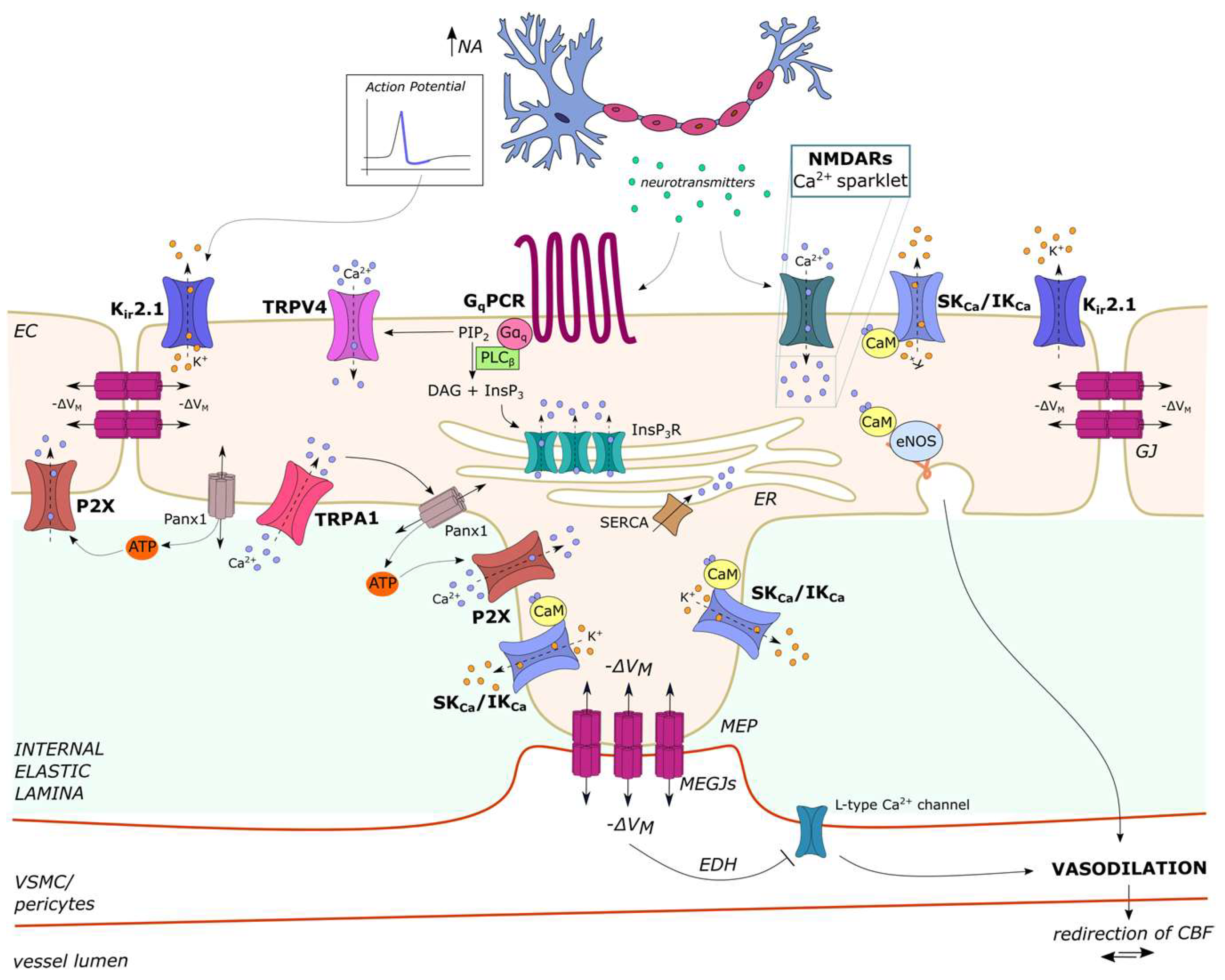
2.3.2. Ca2+ Sparklets in Native Cerebrovascular Endothelial Cells: NMDARs

Native and cultured cerebrovascular endothelial cells express ionotropic receptors classically belonging to the neuronal lineage, such as NMDARs [44,110,120] and GABA sub-type A receptors (GABAARs) [45,46]. Interestingly, both NMDARs and GABAARs contribute to shaping the endothelial Ca2+ response to their physiological agonists, the excitatory neurotransmitter glutamate [44,120] and the inhibitory neurotransmitter GABA [45,46], respectively. The Pires group has characterized the unitary Ca2+ entry events through NMDARs in mouse cerebral artery endothelial cells, showing that they consist of submembrane Ca2+ sparklets with a spatial spread of 8 µm2 (Table 1) [14]. NMDAR-mediated Ca2+ entry in the human cerebrovascular endothelial cell line, hCMEC/D3, is not detectable by epifluorescence imaging of Fura-2 loaded cells but leads to robust NO release [120]. This observation led the authors to suggest that NMDARs also mediate local Ca2+ sparklets in hCMEC/D3 cells.

2.3.3. Local Ca2+ Signals Activated by Store-Operated Ca2+ Entry (SOCE) in Vascular Endothelial Cells

Little information is available regarding the local Ca2+ signals induced by SOCE activation in the endothelial lineage [61]. SOCE is regarded as the major Ca2+ entry pathway that replenishes the ER Ca2+ store and sustains long-lasting oscillations in cultured endothelial cells [61,66] and in native macrovascular endothelial cells [20,121,122,123]. In accord, the distance between the ER and the PM in microvascular endothelial cells is significantly longer than in macrovascular endothelial cells, and the SOCE machinery is yet to be reported at MEPs [5]. Endothelial SOCE is activated upon InsP3-dependent reduction in ER Ca2+ concentration ([Ca2+]ER) and is mediated by the physical interaction between STIM1 (Figure 1), which serves as the sensor of [Ca2+]ER, and the Ca2+-permeable Orai1 channels (Figure 1) or the non-selective TRPC1/TRPC4 channels (Figure 1), which are located on the PM [61]. The store-operated current mediated by STIM1 and Orai1 channels has been termed Ca2+-release-activated Ca2+ current (ICRAC), whereas STIM1 and TRPC1/TRPC4 channels mediate a current known as ISOC. In addition, a heteromeric complex comprising STIM1, Orai1 and TRPC1/TRPC4 has been described in some endothelial cells. This complex mediates an ICRAC-like current with intermediate biophysical features between ICRAC and ISOC [61].
Local Ca2+ signals mediated by STIM1 activation at the leading edge of migration were recorded in cultured human umbilical vein endothelial cells (HUVECs) [124]. Furthermore, studies on CPAE cells have revealed that SOCE colocalizes with InsP3-induced ER Ca2+ release at sites of focal stimulation with sub-maximal doses of an agonist. However, SOCE can be activated at more remote locations by an increase in the strength of local stimulation because of intraluminal redistribution of ER Ca2+ [66,125]. Unfortunately, the measurement of local store-operated Ca2+ signals in native endothelial cells is still missing.

2.4. Intra- and Intercellular Endothelial Ca2+ Waves

The spatially restricted endothelial Ca2+ events that are generated by local Ca2+ puffs can be amplified by the recruitment of adjacent InsP3Rs via CICR and coalesce into a global elevation in [Ca2+]i, which can spread to neighboring endothelial cells, thereby originating both intra- (Table 2) and intercellular Ca2+ waves [4,9,10]. Extracellular Ca2+ entry through SOCs or TRP channels may sustain these regenerative fluctuations in endothelial [Ca2+]i, although the contribution of Ca2+ influx may be variable depending on the agonist or vascular bed (Table 2).

2.4.1. Intracellular Ca2+ Waves in Vascular Endothelial Cells

Ca2+ imaging analysis of either pressurized or en face vascular preparations by using high-speed confocal microscopy has revealed that physiological concentrations of neurohumoral mediators, such as acetylcholine, histamine or ATP, induce intracellular Ca2+ oscillations in native endothelial cells to regulate blood pressure or microvascular permeability. Growth factors, such as VEGF, have also been shown to elicit endothelial Ca2+ oscillations to stimulate sprouting angiogenesis in vivo [8] Table 2 lists some of the most representative examples of agonist-induced intracellular Ca2+ waves in vascular endothelial cells.
Further work is required to assess the mechanisms supporting endothelial Ca2+ oscillations, which could vary depending on the vascular bed and the species (Table 1). Do lysosomal TPCs support ER Ca2+ release through InsP3Rs (see also Section 2.2.2 and Section 6)? Does an SKF-sensitive SOCE sustain endothelial Ca2+ oscillations in large conduit arteries (Section 2.3.3 and Section 4.1), while TRPV4 channels fulfill this role in systemic resistance vessels (see Section 2.3.1 and Section 4.2.1)? May the Ca2+ entry pathway vary with the agonist?

2.4.2. Intracellular Ca2+ Waves Induced by Neuronal Activity in Cerebrovascular Endothelial Cells

A recent investigation revealed that synaptic activity can also trigger endothelial Ca2+ waves in the mouse brain microvasculature [131]. In vivo imaging in anesthetized transgenic mice selectively expressing GCaMP8 in endothelial cells revealed a hierarchy of InsP3-dependent Ca2+ signals, ranging from small, subsecond protoevents, caused by Ca2+ mobilization through a limited number of InsP3Rs, to high-magnitude, persistent (up to ~1 min) composite Ca2+ events sustained by large clusters of InsP3Rs [131]. These frequent InsP3-dependent local Ca2+ release events were sustained by TRPV4 activation upon PLCβ-dependent hydrolysis of PIP2 [131,132]. Somatosensory stimulation induced an increase in the amplitude and duration of these subcellular Ca2+ events and increased the prevalence of endothelial Ca2+ activity at the arteriole–capillary transitional zone [131]. The agonist responsible for GqPCR activation has not been identified [131,133], but it could be the synaptically released excitatory neurotransmitter glutamate [134]. In accord, glutamate can bind to metabotropic glutamate receptor 1 (mGluR1) and mGluR5 to induce intracellular Ca2+ oscillations in cultured cerebrovascular endothelial cells [43,79].

2.4.3. Intercellular Ca2+ Waves in Vascular Endothelial Cells

Intracellular Ca2+ waves that occur either spontaneously [91,127] or upon physiological stimulation can propagate along the vascular intima of resistance vessels as intercellular Ca2+ waves [27,135]. Local application of up to 1 µM acetylcholine induced intercellular Ca2+ waves that spread to adjacent endothelial cells in resistance vessels, such as mouse abdominal arterioles [90], the mouse cremaster muscle artery [136,137] and the rat mesenteric artery [29]. PAR-2 stimulation could also trigger endothelial Ca2+ waves that propagate across adjacent cells in the rat mesenteric artery [138]. Using transgenic mice selectively expressing GCaMP2 in vascular endothelial cells, Tallini et al. found that the local application of 1 µM acetylcholine induced intercellular Ca2+ waves that propagated for ~1 mm along the vascular intima at a speed of ~116 µm/s [137]. However, when acetylcholine was administered within the same concentration range (<3 µM), the intracellular Ca2+ waves did not spread beyond a limited cluster of endothelial cells, as observed in large conduit arteries [28].
The propagation of intercellular Ca2+ waves in the vascular intima can be supported either by the diffusion of cytosolic second messengers [128,139], e.g., Ca2+ and/or InsP3, through gap junctions or by the paracrine release of extracellular mediators [140,141], e.g., ATP. An alternative mechanism has been proposed for the hemodynamic response to prolonged (5 s) somatosensory stimulation in the whisker barrel cortex. Neuronal activity has been found to trigger an endothelial Ca2+ wave in brain capillaries that travels back to upstream feeding arterioles, thereby resulting in functional hyperemia [109]. It has been suggested that active neurons release ROS that can peroxidize membrane lipids and thereby generate metabolites, e.g., 4-HNE, in the proximity of brain capillaries [109,134]. ROS-derived metabolites, in turn, gate TRPA1 and induce the Ca2+-dependent opening of adjacent pannexin 1 (Panx1) channels, which release ATP into the extracellular milieu. The local elevation in extracellular ATP directly activates the Ca2+-permeable P2X receptors on neighboring endothelial cells, thus triggering a slow Ca2+ wave that spreads from the distant capillaries to the post-arteriole transitional segment [109]. A Panx1/purinergic signaling pathway also controls capillary-to-arteriolar communication in the hamster skeletal musculature [142], but it is still unclear whether it is triggered by TRPA1-mediated Ca2+ sparklets. Therefore, exploring the mechanisms driving the propagation of intracellular Ca2+ waves is mandatory to fully appreciate how they control vascular function (see below).

3. Vascular Endothelial Cells Use Ca2+ Signals to Interpret the Local Microenvironment and Transfer Information to Distant Sites

Vascular endothelial cells are immersed in a stream of information that must be independently processed to generate the most appropriate vascular output. These cues consist of modest changes in the chemical and physical composition of the local microenvironment that elicit an elevation in [Ca2+]i superimposed on the spontaneous Ca2+ activity, e.g., spontaneous Ca2+ puffs, also termed Ca2+ noise [4,26]. Therefore, endothelial cells need to reject the meaningless Ca2+ noise and extract the information that is encoded in the variety of Ca2+ signals induced by extracellular cues [4,26]. Recent studies have shown that vascular endothelial cells that are located at different positions of the vascular intima can detect distinct chemical signals due to the heterogeneity in the expression of their surface receptors. In addition, they have the potential to transmit local Ca2+ signals to more remote sites to coordinate specific vascular functions, such as vasodilation and local blood perfusion [4,26].
Immunofluorescence showed that purinergic P2Y2 receptors and muscarinic M3 receptors are segregated in spatially distinct clusters of endothelial cells in the rat carotid artery [143]. Functional analysis of Ca2+ activity in large populations of endothelial cells within native vessels confirmed that the endothelial Ca2+ response to extracellular stimuli is not homogeneous [4,26]. Acetylcholine evoked large Ca2+ signals at branching sites in the rat thoracic aorta rather than in neighboring non-branching areas, while the Ca2+ response to histamine displayed the opposite behavior [144]. Similarly, ATP evoked a robust increase in [Ca2+]i in ~90% of mouse aortic endothelial cells, while acetylcholine, substance P and bradykinin were significantly less effective [145]. The onset of the Ca2+ response to acetylcholine was constrained by the expression of M3 receptors on a limited fraction of endothelial cells [30]. Interestingly, the endothelial Ca2+ response to shear stress in the rat carotid artery was also not uniform and limited to a discrete number of cells [29].
To gain further insight into the heterogeneity of endothelial Ca2+ signals, Wilson and McCarron designed a miniature fluorescence endoscope that was developed around a gradient index (GRIN) lens to measure the Ca2+ activity in hundreds of endothelial cells in pressurized arteries [28]. Using this approach, they found that, in the rat carotid artery, low concentrations of acetylcholine (≤3 µM) elicited a Ca2+ signal in only a minority of endothelial cells. Increasing the concentration of acetylcholine up to the mid-to-high micromolar range increased both the number of activated cells (i.e., more endothelial cells were recruited) and the amplitude of the Ca2+ response within each single endothelial cell [28]. The concentration-dependent increase in the amplitude of the Ca2+ response of each endothelial cell spanned an order of magnitude of the acetylcholine concentration [28]. However, the Ca2+ sensitivity of the entire endothelial monolayer to acetylcholine spanned over three orders of magnitude of agonist concentration, with low-sensitive and high-sensitive endothelial cells clustered in spatially distinct domains of the vascular intima [28]. This arrangement renders the vascular endothelium highly sensitive to weak signals without undergoing any saturation, yet extraordinarily capable of responding even to increases in input strength [4,26]. Consistently, when individual endothelial cells in the clusters displayed spontaneous Ca2+ release events, these were not propagated to adjacent cells. Intracellular Ca2+ waves propagated along the vascular intima only when the agonist, i.e., acetylcholine, was applied and activated two or more neighboring endothelial cells within the cluster [28]. These findings demonstrated that clustered endothelial cells, with the same agonist sensitivity, serve as a local sensory platform that perceives extracellular cues only within a specific concentration range and thereby informs the more distant sites about agonist stimulation to regulate cardiovascular functions. Conversely, spontaneous Ca2+ activity cannot be propagated to the remaining endothelial monolayer and is rejected as background Ca2+ noise [4,26]. Thus, the vascular endothelium can be viewed as a complex mosaic of separate detection sites that are tuned to detect different agonists (e.g., acetylcholine and ATP) and different concentrations of the same agonist. A follow-up investigation has revealed that these detection sites convey the information encoded within the Ca2+ signal to a pre-determined ensemble of distant endothelial cells via “short-cut” connections that increase the speed and efficiency of signal transmission along the vascular intima [146]. Disruption of the organization of the endothelial network has been shown to impair Ca2+-dependent vascular functions, such as NO-dependent vasorelaxation, in a rat model of prediabetic obesity [17].
Subsequently, the McCarron group demonstrated that the endothelial clusters selectively expressing P2Y2 and M3 receptors are able to interact when the lumen of the pressurized rat carotid artery is stimulated with both ATP and acetylcholine [143]. They found that vascular endothelial cells are able to combine and extract information from multiple sources by generating a Ca2+ signal that did not derive from the simple summation or average of ATP- and acetylcholine-induced Ca2+ signals [143]. Consistently, while acetylcholine triggered intracellular Ca2+ oscillations and ATP induced transient Ca2+ signals, the combined application of both agonists induced Ca2+ signatures with intermediate features [143]. The ability of the vascular endothelium to generate new Ca2+ signals in response to different stimuli targeting distinct clusters of endothelial cells indicates that these small endothelial networks can communicate within the framework of a larger endothelial network, thereby increasing the computational capacity of the endothelial monolayer [26]. The computational screening of the incoming signals carried by endothelial cells is amplified by the interaction between the molecular pathways recruited downstream of distinct signaling systems. In this scenario, not only the spatial distribution but also the kinetics of the endothelial Ca2+ signal play a crucial role in the engagement of the most appropriate Ca2+-dependent decoder, as outlined in the next sections.

4. Local Endothelial Ca2+ Signals Regulate Mean Arterial Pressure and Local Blood Perfusion

Endothelial Ca2+ signals can induce vasorelaxation by recruiting a variety of Ca2+-dependent effectors [10,27,147,148], such as (1) endothelial nitric oxide synthase (eNOS) (Figure 1), which catalyzes the conversion of L-arginine to NO by requiring several cofactors, including CaM, tetrahydrobiopterin (BH4), flavin mononucleotide (FMN), flavin adenine dinucleotide (FAD) and iron protoporphyrin IX (Heme Fe); (2) phospholipase A2 (PLA2), which cleaves AA from the phospholipids that are located on the inner leaflet of the PM and ignites the signaling pathway leading to the conversion of AA to prostacyclin (or prostaglandin I2, PGI2) by cyclooxygenase; and (3) SKCa/IKCa channels (Figure 1), which are activated by an increase in submembrane Ca2+ concentration. NO is primarily responsible for the vasorelaxation of large conduit arteries, while SKCa/IKCa channels are critical for controlling the diameter of smaller resistance-sized arteries and resistance arterioles [5,10]. Nevertheless, NO also contributes to the regulation of vascular resistance, and NO release can be finely tuned by SKCa/IKCa-channel-mediated EDH [6,10,27]. Conversely, NO is central to the regulation of pulmonary resistance arteries, in which SKCa/IKCa channels only play a minor role [103,149].

4.1. The Selective Coupling of SOCE with eNOS: Indirect Evidence for Orai1-Mediated Ca2+ Sparklets in Vascular Endothelial Cells in Large Conduit Vessels

NO has been identified as the first endothelium-dependent vasorelaxant mediator in response to both neurohumoral stimuli and shear stress more than 30 years ago [150,151,152,153,154,155]. Endothelial NO is mainly synthesized by eNOS, also known as NOS3, which is located in Ω-shaped invaginations of the PM that are enriched in caveolin-1 and are termed caveolae [27,156]. Caveolin-1 exerts a tonic inhibitory effect on eNOS, thereby preventing NO production. An increase in submembrane Ca2+ levels stimulates CaM to bind to and displace caveolin-1 from eNOS, resulting in NO release. NO, in turn, diffuses to adjoining VSMCs, where it activates a soluble guanylate cyclase (sGC)/cyclic GMP (cGMP)/protein kinase G (PKG) signaling pathway to induce vasorelaxation and reduce blood pressure [6,27,147]. A seminal investigation by the Blatter group on CPAE cells has shown that the endothelial SOCE is selectively coupled to eNOS while being seemingly insensitive to InsP3-induced ER Ca2+ release [66,157]. In agreement with this finding, endothelial SOCs are also primarily located within caveolae, and this structural feature may have profound pathological implications [61,158,159]. Reducing membrane cholesterol can impair caveolar integrity and inhibit agonist-induced SOCE in pulmonary artery endothelial cells in a rat model of chronic hypoxia-induced pulmonary hypertension [160]. That SOCE is the major Ca2+ source for eNOS activation in large conduit vessels was also demonstrated by the reduction in NO-dependent vasorelaxation caused by the genetic deletion [161] or knockout [121,122] of STIM1 and the genetic deletion of TRPC4 [123] in several macrovascular beds (see [61]). The McCarron group showed that flow-induced vasodilation in the rat carotid artery is mediated by the autocrine action of endothelium-derived acetylcholine that triggers intracellular Ca2+ oscillations and NO release (Table 1) [29]. Although this investigation did not assess the molecular underpinnings of the Ca2+ entry pathway, acetylcholine is known to induce endothelial NO release by activating SOCE [162,163].
Measurement of local store-operated Ca2+ signals in native endothelial cells is still lacking. A combination of high-speed TIRF microscopy [164] and endothelial-cell-specific GCaMP2 or GCaMP5 mice [165] should enable the recording of Orai1 and TRPC1/TRPC4-mediated unitary Ca2+ entry events in en face vascular preparations. It is also still unclear whether Orai1 or TRPC1/TRPC4 are present at the MEPs that decorate resistance arteries and arterioles in the systemic vasculature and lungs [5]. Nevertheless, SOCE plays a major role in supporting eNOS activation in brain microcirculation [131,134], where NO release in response to various neurotransmitters and neuromodulators is severely hampered by Orai1 (in human) or Orai2 (mouse) inhibition [31,35,43,46,78,79,120]. Interestingly, acetylcholine triggered NO production only in the presence of a functional SOCE in mouse [78] and human [31] cerebrovascular endothelial cells, thereby suggesting a tight coupling between Orai1/Orai2 and eNOS.

4.2. Ca2+ Sparklets and Ca2+ Pulsars Recruit SKCa/IKCa Channels to Induce EDH-Dependent Vasorelaxation

Endothelial Ca2+ sparklets and Ca2+ pulsars at or near MEPs can relax overlying VSMCs, reduce vascular resistance and thereby increase local blood flow to downstream capillaries by activating juxtaposed SKCa/IKCa channels [5,6,9,10,27]. SKCa (KCa2.3) channels present a single-channel conductance of ~10 pS and are encoded by the KCNN3 gene, whereas IKCa (KCa3.1) channels show a unitary conductance of 20–30 pS and are encoded by the KCNN4 gene. The opening of SKCa/IKCa channels is regulated by CaM that is bound to their COOH-terminus: an increase in submembrane Ca2+ concentration stimulates CaM to gate both channels with a half-maximal activation (EC50) at 300–500 nM and a maximal activation at 1 µM [9,27]. Endothelial SKCa/IKCa channels are mainly concentrated at MEPs, where they can be activated by local Ca2+ sparklets and Ca2+ pulsars to mediate endothelial hyperpolarization. The negative shift in the membrane potential VM (up to ~−30 mV) is then propagated to the surrounding VSMCs via MEGJs, thereby reducing the open probability of voltage-gated L-type Ca2+ channels and inducing VSMC relaxation by a mechanism known as EDH [5,6,9,10,27]. Studies on rodents have shown that SKCa/IKCa channel currents were detectable in freshly isolated mesenteric artery endothelial cells [101] and in cerebral [166] and pulmonary [103] resistance arteries. Conversely, they are absent in mouse capillary endothelial cells [167], although preliminary evidence suggests that they are present at the human BBB [119].

4.2.1. Ca2+ Sparklets Induce EDH-Dependent Vasorelaxation in Systemic Resistance Vessels and in Brain Parenchymal Arterioles

EDH was first found to be activated by TRPV4-mediated Ca2+ sparklets (Table 1) [101,102]. The systemic activation of the TRPV4 channels with GSK induced a dose-dependent reduction in the mean arterial pressure (MAP) in mice, rats and dogs [168]. Subsequent work showed that acetylcholine-induced reduction in blood pressure and vasodilation in small mesenteric arteries were impaired in TRPV4-knocked-out mice [169]. Sonkusare et al. reported that acetylcholine induced EDH-dependent vasodilation of resistance arteries by eliciting TRPV4-mediated Ca2+ sparklets via the PLCβ-DAG-PKC-AKAP50 signaling pathway at MEPs [101,102]. Low levels of stimulation were primarily associated with IKCa activity, whereas three to eight individual TRPV4 channels were identified as responsible for the local Ca2+ sparklets driving the vasomotor response [101]. Interestingly, a significant increase in MAP was observed in transgenic mice lacking the endothelial AKAP50 or TRPV4 proteins [170]. Hypertension may disrupt TRPV4 channel coupling at MEPs by downregulating AKAP50 and reducing IKCa activation through a local elevation in peroxynitrite levels, thereby increasing the MAP [170]. Local Ca2+ entry through endothelial TRPV4 channels also stimulated SKCa/IKCa currents and induced vasodilation in mouse mesenteric arteries exposed to low fluid shear stress [53] and rat cremaster arteries experiencing a decrease in intraluminal pressure [55]. Additionally, InsP3-dependent TRPV4-mediated endothelial Ca2+ sparklets could be activated to generate vasorelaxant signals and attenuate vasoconstriction of mouse mesenteric arteries upon stimulation of VSMC α1-adrenergic receptors [47]. SKCa/IKCa currents may also be recruited by unitary Ca2+ entry signals through TRPV3 [105,171] or TRPA1 [105] and thereby induce vasodilation of rat cerebral parenchymal arterioles in response to, respectively, carvacrol and 4-HNE (Table 1). Finally, NMDAR-mediated Ca2+ sparklets induced dilation of mouse pial arteries by engaging SKCa/IKCa currents (Table 1), while the vasomotor response to NMDAR stimulation was impaired in a mouse model of Alzheimer’s disease, in 5x-FAD mice [14]. NMDAR-mediated Ca2+ sparklets could also recruit the eNOS to promote NO-dependent vasodilation, but this hypothesis remains to be probed [110,120].

4.2.2. InsP3-Driven Ca2+ Pulsars and Wavelets Induce EDH-Dependent Vasorelaxation in Systemic Resistance Vessels

EDH-dependent vasorelaxation can also be activated by InsP3-driven endothelial Ca2+ pulsars and wavelets (Table 1). Acetylcholine was found to recruit endothelial IKCa channels and induce vasorelaxation of mouse small mesenteric arteries by stimulating Ca2+ pulsars at MEPs [80]. This Ca2+ signaling pathway also contributes to establishing the myo-endothelial feedback [93]. Endothelial Ca2+ pulsars can activate SKCa and/or IKCa channels and limit VSMC depolarization and vasoconstriction to sympathetic nerve stimulation in rat cremasteric arteries [48], hamster cremaster arterioles [99] and mouse mesenteric arteries [49]. Endothelial Ca2+ wavelets at MEPs may also contribute to myo-endothelial feedback and moderate vasoconstriction by recruiting IKCa channels in hamster retractor muscle feed arteries [50].

4.2.3. Why Do Local Ca2+ Signals at MEPs in Systemic Resistance Vessels Fail to Stimulate Robust NO Release?

Acetylcholine-induced vasodilation of systemic mesenteric arteries also presents a modest NO-dependent component [172,173]. The presence of eNOS at MEPs has been reported [174], but NO diffusion to the overlying VSMC layer is prevented here by the alpha chain of hemoglobin (Hbα), which scavenges NO before it can induce vasorelaxation [175,176]. This signaling microdomain is absent in large conduit arteries and helps to understand why NO plays a minor role in the relaxation of smaller, resistance arteries [93,174]. TRPV4 is loosely coupled with eNOS in resistance arteries [103], which causes only a weak reduction in NO production in TRPV4-/- mice. Therefore, it has been proposed that eNOS stimulation in mesenteric resistance arteries is mainly driven by InsP3-induced local Ca2+ pulsars (Table 1) [80] or regenerative Ca2+ waves (Table 2) [101]. However, NO-dependent vasorelaxation in resistance vessels can also be supported by an STIM1-dependent Ca2+ source [121,122]. TRPC3 channels, which can be gated by STIM1 and have been detected at MEPs in rat third-order mesenteric arteries, could replenish the ER and thereby maintain acetylcholine-induced Ca2+ signaling and NO release (Figure 1) [5,177,178,179]. In line with this evidence, the MAP is also significantly reduced in transgenic mice lacking the eNOS [180,181], supporting the view that NO may play a role in the control of systemic resistance and MAP.

4.3. TRPV4-Mediated Ca2+ Sparklets Induce NO Release in Pulmonary Resistance Arteries

NO has long been regarded as the predominant vasorelaxant mediator in the pulmonary circulation [182]. Recent investigations have shown that, in contrast to the systemic vasculature, TRPV4 is preferentially coupled with eNOS to induce vasorelaxation in small, resistance-sized pulmonary arteries [149]. The endogenous agonist, ATP, was shown to elicit TRPV4-mediated Ca2+ sparklets that were not preferentially located to MEPs but were rather distributed along the endothelial cell membrane. Local Ca2+ entry through TRPV4 channels required caveolin-1 and selectively stimulated the eNOS to produce NO, which in turn reduced resting pulmonary arterial pressure (PAP) [103,183,184]. TRPV4-mediated caveolar Ca2+ sparklets were impaired in small pulmonary arteries isolated from a mouse model of pulmonary artery hypertension (PAH) and from PAH patients due to the local formation of peroxynitrite, which dismantles the caveolin-1-TRPV4 channel signaling network [184]. A follow-up investigation revealed that ATP is released through endothelial Panx 1 channels and subsequently activates purinergic P2Y2 receptors to stimulate PKCα and induce TRPV4 channel opening [185].

4.4. The Complex Regulation of Cerebral Blood Flow (CBF) by Endothelial Ca2+ Signaling

Neurovascular coupling, also known as functional hyperemia, is the mechanism whereby an increase in neuronal activity (NA) leads to the vasorelaxation of feeding arterioles, thereby redirecting local CBF to firing neurons [7,134,147]. Recent studies have unveiled that cerebrovascular endothelial cells can sense both neuronal [167] and synaptic [44,186] activity, thereby generating multiple vasorelaxant signals to increase CBF and maintain neuronal metabolism. Capillary endothelial cells, which are positioned in close contact with neurons in the brain, serve as a “sensory web” that generates an electrical signal in response to NA, thereby informing upstream arterioles to increase CBF locally at the site of neuronal firing [167]. Brain capillary endothelial cells express inwardly rectifying K+ (Kir2.1) channels that are activated by the modest elevation in extracellular K+ that occurs during repolarization of each action potential and translate it into a hyperpolarizing signal that propagates through homo-cellular gap junctions to upstream parenchymal arterioles to cause vasorelaxation (Figure 2) [135,167].
A follow-up study by Longden et al. showed that the complex Ca2+ signals induced by NA upon the activation of a GqPCR at the arteriole–capillary transitional zone (Figure 2) (see Section 2.4.2 were amplified by Kir2.1-mediated endothelial hyperpolarization, which enhanced extracellular Ca2+ entry through TRPV4 channels (Figure 2). Capillary Ca2+ activity led to the local production of NO (Figure 2), which relaxed overlying pericytes to increase the capillary diameter and redirect CBF to the actively signaling capillary bed [131]. It is still unknown why active brain capillary endothelial cells do not transmit their Ca2+ activity to adjacent cells via homo-cellular gap junctions [135], as observed throughout the vasculature (see Section 2.4.2). In addition, NA-induced intercellular Ca2+ waves in the distal capillary segment can reach the post-arteriole transitional segment via the TRPA1/Panx1/purinergic signaling pathway (Figure 2) [109], albeit at a lower speed as compared to Kir2.1-mediated hyperpolarization. Thakore et al. showed that the endothelial Ca2+ wave triggered at the site of NA is converted into a fast hyperpolarizing signal at the transitional segment, where endothelial cells express SKCa/IKCa channels (Figure 2) [109,118]. This fast electrical signal is locally amplified by Kir2.1 channels and is electrotonically transmitted via MEGJs to overlying VSMCs to deactivate voltage-gated L-type Ca2+ channels and induce vasodilation [109,118].
The hemodynamic Ca2+ response to somatosensory stimulation does not fully develop without the additional contribution of endothelial NMDARs, which are mainly located in parenchymal arterioles in the mouse brain and mediate local Ca2+ sparklets that recruit eNOS to produce NO and stimulate EDH via SKCa/IKCa channels (Figure 2) [14,44,120,186].
Recent investigations have shown that endothelial Ca2+ signaling and endothelium-dependent vasodilation are disrupted in neurodegenerative disorders, such as Alzheimer’s disease and chronic traumatic encephalopathy [14,187]. Rescuing endothelial ion signaling with synthetic or physiological agonists that stimulate endothelial Ca2+ sparklets, e.g., through TRPV3 or TRPA1 channels, may represent an alternative strategy to restore CBF in these and other brain pathologies [134,188].

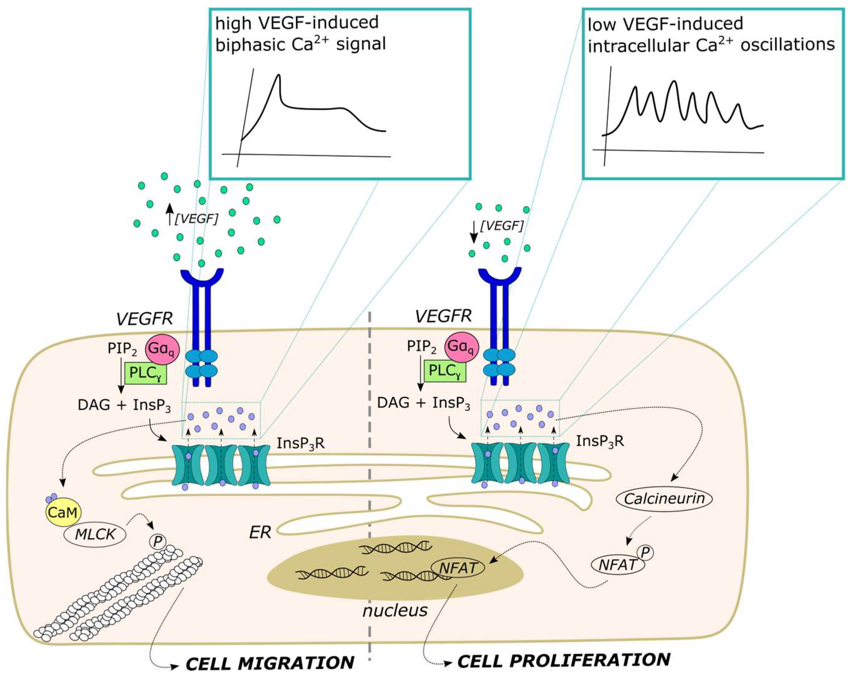
5. Distinct Spatio-Temporal Patterns of Endothelial Ca2+ Signals Regulate Angiogenesis

An increase in endothelial [Ca2+]i is the primary signal whereby VEGF, the master regulator of angiogenesis, regulates endothelial cell proliferation, motility, adhesion to the substrate, assembly into bidimensional tubular networks and neovessel formation [8]. VEGF stimulates endothelial Ca2+ signals by binding to VEGFR-2 (kdr/flk-1), thereby leading to ER Ca2+ release through InsP3Rs and lysosomal Ca2+ mobilization via TPC2, usually followed by SOCE activation [8,130,189,190]. During angiogenesis, VEGF activates a leading tip endothelial cell to become motile and spearhead a new sprout by migrating outward from the parental capillary toward the VEGF source. Trailing cells then begin to proliferate and migrate behind the tip cell as endothelial stalk cells to elongate the sprout and maintain the physical connection to the parental capillary vessel [8,191]. Recent studies have shown that VEGF can induce distinct Ca2+ signatures to induce proliferation or migration by recruiting different Ca2+-sensitive decoders. Additionally, neovessel formation during wound healing or tumor growth can be supported by de novo blood vessel formation through a process known as vasculogenesis. Endothelial colony-forming cells (ECFCs) are released into the bloodstream by vascular stem cell niches that are distributed along the vascular wall and home to the site of neovessel formation, where they deliver paracrine signals to support angiogenesis or physically engraft within neovessels [71,192]. Like sprouting angiogenesis, ECFC-mediated vasculogenesis is mediated by intracellular Ca2+ signals that adopt distinct temporal profiles in proliferating vs. migrating ECFCs [11].

5.1. VEGF-Induced Intracellular Ca2+ Oscillations Select Endothelial Tip and Stalk Cells

To explore how endothelial Ca2+ signaling finely tunes angiogenesis in vivo, Yokota et al. exploited high-speed, light-sheet microscopy in a transgenic zebrafish line expressing GCaMP7a in endothelial cells [38]. They found that VEGF triggered high-frequency Ca2+ oscillations in tip cells budding from the dorsal aorta, which were mediated by VEGFR-2. Trailing stalk cells that migrated behind the tip cells also showed VEGFR-2-dependent Ca2+ oscillations, although they were not synchronized with those occurring in the leading cells of the sprouts. Interestingly, VEGF-induced Ca2+ spikes also occurred in the adjacent endothelial cells before phenotype selection, when they became spatially restricted to the sprouting endothelial cells by Delta-like 4 (Dll4)/Notch signaling [38]. It was not clear whether the Ca2+ waves running along the dorsal aorta before tip/stalk cell selection were synchronized by homo-cellular gap junctions or were independently patterned [38]. These findings are consistent with the dynamic model of sprouting angiogenesis: leading tip cells exhibit higher levels of the Dll4 ligand that enhances their migration rate and induces Notch signaling in the follower stalk cells, which are less motile but exhibit a higher proliferation rate [193,194]. However, although Dll4/Notch-mediated lateral inhibition suppresses pro-angiogenic Ca2+ oscillations in the endothelial cells that remain within the parental vessel, it does not attenuate the Ca2+ spiking activity in stalk cells after they exit from the dorsal aorta [38]. Using a similar approach, Savage et al. showed that VEGF-induced intracellular Ca2+ oscillations in endothelial tip cells require the ER transmembrane protein 33 (TMEM33) and regulate phenotype selection by inducing extracellular-signal-regulated kinase (ERK) phosphorylation. The ERK signaling pathway, in turn, was crucial to induce the expression of several tip cell markers, including DLL4 and flt4, to promote filopodia formation and to stimulate endothelial cell migration [129]. Computational modeling confirmed that higher InsP3-dependent Ca2+ spiking is associated with tip cell phenotype selection and favors Dll4 expression [195]. Future studies will have to evaluate this mechanism in mammalian capillaries, which represent the main source of neovessels after an ischemic event or during tumor growth. It would also be helpful to assess whether the Ca2+ oscillations described by Yokota et al. [38] directly engage the Ca2+-dependent machinery driving proliferation (primarily in tip cells) and migration (primarily in stalk cells) or whether a distinct Ca2+ signaling activity must take place to stimulate these processes (see also Section 5.2). A fascinating hypothesis is that, during sprouting angiogenesis in vivo, VEGF elicits global Ca2+ signals to promote phenotype selection [38], whereas it triggers local elevations in [Ca2+]i to induce proliferation or migration [124].

5.2. VEGF Stimulates Endothelial Cell Proliferation and Migration through Distinct Ca2+ Signatures

A large number of in vitro studies, reviewed in detail in [8], have attempted to understand how VEGF-induced intracellular Ca2+ signals regulate endothelial cell proliferation and migration after phenotype selection. A recent investigation has revealed that VEGF stimulates endothelial cell proliferation through repetitive oscillations in [Ca2+]i, whereas biphasic Ca2+ signals were better suited to induce proliferation [37]. By studying porcine aortic endothelial cells (PAECs) and HUVECs, Noren et al. found that, when VEGF concentration is raised from the low to high nanomolar range, the percentage of endothelial cells showing repetitive Ca2+ oscillations decreases, while the percentage of endothelial cells displaying an initial Ca2+ peak followed by a persistent plateau phase increases (Figure 3) [37]. Interestingly, the higher concentrations of VEGF experienced by the leading tip cells induce migration by engaging myosin light chain kinase (MLCK) (Figure 3), whereas the following stalk cells are exposed to lower concentrations of VEGF and undergo proliferation upon the nuclear translocation of the Ca2+-sensitive transcription, nuclear factor of activated T-cells 2 (NFAT2) (Figure 3). In accord, only the prolonged increase in [Ca2+]i occurring during the plateau phase induced a sustained activation of MLCK [37], which phosphorylates the myosin light chain to promote stress fiber formation and contraction during cell movement [8]. Since the average [Ca2+]i achieved in proliferating and migrating endothelial cells was in the same range, the authors concluded that the Ca2+ waveform determines the endothelial cell behavior. By simultaneously measuring Ca2+ activity and MLCK activation, they showed that a single Ca2+ transient is not sufficient to recruit MLCK, whereas a persistent increase in [Ca2+]i above a precise threshold (5% of the baseline [Ca2+]i) stimulated MLCK. This low persistent Ca2+ signaling event did not occur in spiking cells because the [Ca2+]i returns to the baseline after each transient [37]. Conversely, the four members of the NFAT transcription factor family (NFAT1-NFAT4) are the most appropriate decoders of intracellular Ca2+ oscillations [196,197]. In the absence of extracellular stimulation, NFAT is retained in the cytosol by the extensive phosphorylation of its regulatory domain [198]. High-frequency intracellular Ca2+ oscillations engage calcineurin to fully dephosphorylate NFAT, thereby inducing its massive translocation into the nucleus [196,197]. In accord, Noren et al. showed that repetitive oscillations in [Ca2+]i, but not biphasic Ca2+ signals of the same average amplitude, supported the nuclear translocation of NFAT2 in vascular endothelial cells [37]. They clearly demonstrated discrete events of NFAT activation in response to each Ca2+ spike. Furthermore, by interrogating PAECs with a Bayesian decision model, they revealed that, when the cells are exposed to a gradient of VEGF, they can switch from a migratory to a proliferative phenotype depending on the underlying Ca2+ waveform, i.e., biphasic vs. oscillatory [37]. These findings demonstrate that the temporal profile of the Ca2+ signal is crucial to control the outcome of VEGF stimulation on endothelial cell behavior. In addition, it reinforces the view that vascular endothelial cells may display multiple Ca2+ signatures during sprouting angiogenesis (see Section 5.1). For instance, VEGF could first elicit intracellular Ca2+ oscillations that promote phenotype selection (tip vs. stalk cells) and then a novel pattern of repetitive Ca2+ spikes to promote proliferation in stalk cells or a different biphasic increase in [Ca2+]i to induce migration in the leading tip cells. Interestingly, stromal-derived factor-1α (SDF-1α), a chemokine that is released following a drop in O2 tension to stimulate endothelial cell migration, elicits biphasic Ca2+ signals, but not intracellular Ca2+ oscillations, in several types of vascular endothelial cells [190].
However, a number of issues warrant further investigation. In other cell types, Orai1 is physically coupled with calcineurin and NFAT1 through the scaffolding protein AKAP79 (A-kinase anchoring protein 79) and hence boosts NFAT1 activation by generating local Ca2+ nanodomains [199]. Orai1 stimulates the nuclear translocation of NFAT2 in vascular endothelial cells [8,61], such as HUVECs [19,200]. However, it is still unclear whether and how local Ca2+ entry events through endothelial Orai1 channels activate NFAT2. In addition, a previous investigation showed that NFAT2 regulates VEGF-induced HUVEC migration and bidimensional tube formation [201]. Additional experiments could be carried out to assess the waveform of the VEGF-induced Ca2+ response in the HUVEC cell line employed in [201].

5.3. Distinct Ca2+ Signatures Control ECFC Proliferation and Migration

The discovery that ECFCs can be mobilized into the circulation either to support vascular regeneration after an ischemic insult [192] or to promote neovessel formation in growing tumors [202] led to the investigation of the role of Ca2+ signaling in their angiogenic activity [11]. Dragoni et al. found that VEGF, at low nanomolar concentrations, elicits intracellular Ca2+ oscillations, the frequency of which is decreased as VEGF concentration is increased to the high nanomolar range [39], as reported in PAECs and HUVECs [37]. The spiking Ca2+ response to VEGF in circulating ECFCs was mediated by rhythmic ER Ca2+ release through InsP3Rs and supported by lysosomal Ca2+ release through TPC1 and by extracellular Ca2+ entry through SOCs [39,111]. Notably, the intracellular Ca2+ oscillations induced by 10 ng/mL VEGF in circulating ECFCs were in the same range as the frequency threshold required to recruit the Ca2+-dependent transcription factor nuclear factor kappa light chain enhancer of activated B cells (NF-κB) in artificially stimulated vascular endothelial cells (2.5 mHz vs. 5 mHz) [203,204]. In accord, 10 ng/mL VEGF stimulated ECFC proliferation and tube formation by inducing the nuclear translocation of the Ca2+-sensitive transcription factor, NF-κB [39]. Interestingly, SOCE in circulating ECFCs requires both Orai1 and TRPC1 [205,206], which is functionally coupled to NF-κB activation in the endothelial lineage [61,207,208]. Future work will have to assess whether TRPC1 only provides a molecular scaffold to couple NF-κB to local Ca2+ entry through Orai1, or whether it also contributes a pore-forming subunit to the SOCE machinery [61]. Moreover, previous studies have shown that the [Ca2+]i threshold that each Ca2+ spike must reach during an oscillatory train to induce the nuclear translocation of NF-κB in vascular endothelial cells is ~180 nM [209,210]. Whether this [Ca2+]i threshold must also be exceeded in circulating ECFCs remains to be determined. This signaling pathway could be exploited for therapeutic purposes [211]. In accord, the human amniotic fluid stem cell (hAFS) secretome, which promotes coronary neovascularization and partially rescues cardiac function in a mouse model of acute myocardial infarction [212,213,214], induced ECFC tubulogenesis by triggering intracellular Ca2+ oscillations that promoted the nuclear translocation of NF-κB [215]. Similarly, optical stimulation with the green light of the photosensitive conjugated polymer, regioregular Poly (3-hexyl-thiophene), rr-P3HT, induced repetitive Ca2+ spikes that promoted ECFC proliferation and tube formation by inducing the nuclear translocation of NF-κB [41,216]. This approach does not require viral manipulation of endothelial cells and may provide an alternative strategy to induce therapeutic angiogenesis of ischemic disorders by exploiting the high spatio-temporal resolution of light stimulation [217,218,219].
Recruitment of circulating ECFCs by growing neovessels is favored by chemokines, such as insulin-like growth factor-2 (IGF-2) [220] and SDF-1α [221]. As described above for migrating vascular endothelial cells, both IGF-2 [220] and SDF-1α [222] stimulate ECFC migration in vitro and neovessel formation in vivo through a biphasic increase in [Ca2+]i. Zuccolo et al. showed that SDF-1α stimulates the Gi-protein-coupled receptor, CXCR4 to recruit PLCβ2, thereby inducing an initial increase in [Ca2+]i driven by ER Ca2+ release through InsP3Rs, followed by a sustained plateau that was mediated by SOCE [222]. This biphasic Ca2+ signal stimulates ECFC homing in vivo by recruiting ERK 1/2 and PI3K/Akt [222], which have long been known to support SDF-1α-induced neovascularization of ischemic tissues [223].
These findings strongly support the view that the temporal profile of the Ca2+ signal is crucial to specifically control the different processes that take place during sprouting angiogenesis or vasculogenesis. However, it remains to be investigated whether NFAT may also be activated downstream of VEGF-induced Ca2+ oscillations and thereby contribute to triggering the transcriptional program responsible for ECFC proliferation. In addition, the spiking Ca2+ activity elicited by VEGF in umbilical cord blood-derived ECFCs (UCB-ECFCs) was remarkably higher as compared to their circulating counterparts [71,224,225]. Of note, the oscillatory Ca2+ response to VEGF in UCB-ECFCs was triggered by TRPC3-mediated extracellular Ca2+ entry [224], and UCB-ECFCs exhibited a higher proliferative capacity as compared to circulating ECFCs [226]. Future investigations could evaluate whether the higher proliferative potential of UCB-ECFCs depends on the higher frequency of VEGF-induced intracellular Ca2+ oscillations or on the selective recruitment of specific Ca2+-dependent effectors by TRPC3-mediated Ca2+ sparklets [225,227]. Finally, the role played by MLCK in ECFC-mediated neovessel formation has not been assessed. Nevertheless, MLCK was found to stimulate cytoskeletal rearrangement and migration in ECFCs [228] and could therefore be targeted by SDF-1α-induced Ca2+ signals.

6. Conclusions

The spatio-temporal profile of the underlying Ca2+ signals enables endothelial cells (as well as their immature precursors, ECFCs) to fine-tune specific vascular functions, thereby maintaining cardiovascular homeostasis. Elucidating the heterogeneity of the diverse mechanisms whereby local, oscillatory Ca2+ signals, such as Ca2+ pulsars, Ca2+ wavelets and Ca2+ sparklets, recruit SKCa/IKCa and eNOS in different vascular beds will also be essential for designing a more effective therapeutic strategy to target them in disease [134,188,211]. Furthermore, little attention has been paid to the spatio-temporal features of the Ca2+ signals that recruit other endothelium-dependent vasorelaxant pathways, including PLA2 and cystathionine ɣ-lyase enzyme, which, respectively, produce PGI2 and hydrogen sulfide [155,229]. It has long been known that TRPV4-mediated Ca2+ entry can stimulate PGI2 production [230], but whether PLA2 activation requires local Ca2+ sparklets at MEPs or a cell-wide increase in [Ca2+]i is still unclear. Similarly, it has long been known that VEGF only elicits biphasic Ca2+ signals in vascular endothelial cells, while it is now clear that it may also induce intracellular Ca2+ oscillations. Future work will have to scrutinize whether endothelial Ca2+ oscillations may also be triggered by other growth factors that are not known to elicit spiking Ca2+ activity, such as basic fibroblast growth factor, IGF-2 and angiopoietins [8,40,231]. Many other members of the TRP sub-family of Ca2+-permeable channels regulate crucial endothelium-dependent functions, including TRP Melastatin 2 (TRPM2), which can prevent endothelial dysfunction [232] and regulates both angiogenesis and vascular permeability [233,234]; TRPV1, which stimulates angiogenesis [191,218] and regulates NF-κB-dependent gene expression [216]; and finally, the mechano-sensitive TRP Polycystin 1 (TRPP1), which regulates blood pressure [235,236]. These channels are predicted to fine-tune endothelial functions by mediating Ca2+ sparklets at specific membrane sites where they are tightly coupled with their downstream Ca2+-dependent effectors. However, these discrete Ca2+ events are yet to be measured. Similarly, local events of lysosomal Ca2+ release through TPCs or TRP mucolipin (TRPML) channels, which could either recruit juxtaposed InsP3Rs on the ER or regulate vesicular trafficking and autophagy, also deserve to be visualized in the endothelial lineage [64,237]. The fascinating concept that spatially separated endothelial cell clusters are specialized to perceive distinct inputs and transmit them to more distant sites via pre-defined shortcuts, as well as the emergence of non-linear Ca2+ signals that are generated by the summation of multiple inputs, needs to be further explored [4,26,146].
The combination of endothelial-cell-specific knockout mouse models and high-speed imaging (e.g., 2P excitation microscopy and 3D light-sheet fluorescence imaging) will be instrumental in assessing how endothelial Ca2+ signals fine-tune vascular function in health and can be targeted in disease [6,238]. We also envision that the development of novel models of human organs, such as organ-on-a-chip, living myocardial slices and pluripotent stem-cell-derived 3D organoids and cell spheroids, will be the indispensable step for the therapeutic translation of the endothelial Ca2+ machinery [239,240,241,242,243]. Preliminary evidence showed that, in human peripheral arteries, native vascular endothelial cells present a wide variety of Ca2+ signals, ranging from discrete Ca2+ events to cell-wide Ca2+ waves, that can be severely attenuated by cardiovascular disorders [244].

Author Contributions

Conceptualization, F.M.; writing—original draft preparation, F.M.; writing—review and editing, V.B., T.S., R.B.-R. and G.S. All authors have read and agreed to the published version of the manuscript.

Funding

This research has been supported by #NEXTGENERATIONEU (NGEU) and funded by the Ministry of University and Research (MUR), National Recovery and Resilience Plan (NRRP), project MNESYS (PE0000006)—A Multiscale integrated approach to the study of the nervous system in health and disease (DN. 1553 11.10.2022) (F.M. and G.S.); the EU Horizon 2020 FETOPEN-2018-2020 Program under Grant Agreement N. 828984, LION-HEARTED (F.M.); and Fondo Ricerca Giovani from the University of Pavia (F.M.). R.B.R. was supported by the National Council of Science and Technology (CONACYT), Identification Number: CVU121216.

Institutional Review Board Statement

Not applicable.

Informed Consent Statement

Not applicable.

Data Availability Statement

Not applicable.

Conflicts of Interest

The authors declare no conflict of interest.

References

  1. Kruger-Genge, A.; Blocki, A.; Franke, R.P.; Jung, F. Vascular Endothelial Cell Biology: An Update. Int. J. Mol. Sci. 2019, 20, 4411. [Google Scholar] [CrossRef]
  2. Bkaily, G.; Jacques, D. Morphological and Functional Remodeling of Vascular Endothelium in Cardiovascular Diseases. Int. J. Mol. Sci. 2023, 24, 1998. [Google Scholar] [CrossRef] [PubMed]
  3. Marzoog, B.A. Tree of life: Endothelial cell in norm and disease, the good guy is a partner in crime! Anat. Cell Biol. 2023, 56, 166–178. [Google Scholar] [CrossRef] [PubMed]
  4. McCarron, J.G.; Lee, M.D.; Wilson, C. The Endothelium Solves Problems That Endothelial Cells Do Not Know Exist. Trends Pharmacol. Sci. 2017, 38, 322–338. [Google Scholar] [CrossRef]
  5. Murphy, T.V.; Sandow, S.L. Agonist-evoked endothelial Ca2+ signalling microdomains. Curr. Opin. Pharmacol. 2019, 45, 8–15. [Google Scholar] [CrossRef] [PubMed]
  6. Ottolini, M.; Hong, K.; Sonkusare, S.K. Calcium signals that determine vascular resistance. Wiley Interdiscip. Rev. Syst. Biol. Med. 2019, 11, e1448. [Google Scholar] [CrossRef] [PubMed]
  7. Negri, S.; Faris, P.; Soda, T.; Moccia, F. Endothelial signaling at the core of neurovascular coupling: The emerging role of endothelial inward-rectifier K(+) (Kir2.1) channels and N-methyl-d-aspartate receptors in the regulation of cerebral blood flow. Int. J. Biochem. Cell Biol. 2021, 135, 105983. [Google Scholar] [CrossRef]
  8. Moccia, F.; Negri, S.; Shekha, M.; Faris, P.; Guerra, G. Endothelial Ca2+ Signaling, Angiogenesis and Vasculogenesis: Just What It Takes to Make a Blood Vessel. Int. J. Mol. Sci. 2019, 20, 3962. [Google Scholar] [CrossRef]
  9. Jackson, W.F. Calcium-Dependent Ion Channels and the Regulation of Arteriolar Myogenic Tone. Front. Physiol. 2021, 12, 770450. [Google Scholar] [CrossRef]
  10. Lemmey, H.A.L.; Garland, C.J.; Dora, K.A. Intrinsic regulation of microvascular tone by myoendothelial feedback circuits. Curr. Top. Membr. 2020, 85, 327–355. [Google Scholar] [CrossRef]
  11. Moccia, F. Calcium Signaling in Endothelial Colony Forming Cells in Health and Disease. Adv. Exp. Med. Biol. 2020, 1131, 1013–1030. [Google Scholar] [CrossRef] [PubMed]
  12. Yuan, Q.; Yang, J.; Santulli, G.; Reiken, S.R.; Wronska, A.; Kim, M.M.; Osborne, B.W.; Lacampagne, A.; Yin, Y.; Marks, A.R. Maintenance of normal blood pressure is dependent on IP3R1-mediated regulation of eNOS. Proc. Natl. Acad. Sci. USA 2016, 113, 8532–8537. [Google Scholar] [CrossRef] [PubMed]
  13. Lin, Q.; Zhao, L.; Jing, R.; Trexler, C.; Wang, H.; Li, Y.; Tang, H.; Huang, F.; Zhang, F.; Fang, X.; et al. Inositol 1,4,5-Trisphosphate Receptors in Endothelial Cells Play an Essential Role in Vasodilation and Blood Pressure Regulation. J. Am. Heart Assoc. 2019, 8, e011704. [Google Scholar] [CrossRef] [PubMed]
  14. Peters, E.C.; Gee, M.T.; Pawlowski, L.N.; Kath, A.M.; Polk, F.D.; Vance, C.J.; Sacoman, J.L.; Pires, P.W. Amyloid-beta disrupts unitary calcium entry through endothelial NMDA receptors in mouse cerebral arteries. J. Cereb. Blood Flow. Metab. 2021, 42, 145–161. [Google Scholar] [CrossRef] [PubMed]
  15. Scarpellino, G.; Genova, T.; Avanzato, D.; Bernardini, M.; Bianco, S.; Petrillo, S.; Tolosano, E.; de Almeida Vieira, J.R.; Bussolati, B.; Fiorio Pla, A.; et al. Purinergic Calcium Signals in Tumor-Derived Endothelium. Cancers 2019, 11, 766. [Google Scholar] [CrossRef] [PubMed]
  16. Moccia, F. Endothelial Ca2+ Signaling and the Resistance to Anticancer Treatments: Partners in Crime. Int. J. Mol. Sci. 2018, 19, 217. [Google Scholar] [CrossRef]
  17. Wilson, C.; Zhang, X.; Lee, M.D.; MacDonald, M.; Heathcote, H.R.; Alorfi, N.M.N.; Buckley, C.; Dolan, S.; McCarron, J.G. Disrupted Endothelial Cell Heterogeneity and Network Organization Impair Vascular Function in Prediabetic Obesity. Metabolism 2020, 111, 154340. [Google Scholar] [CrossRef]
  18. Berra-Romani, R.; Guzman-Silva, A.; Vargaz-Guadarrama, A.; Flores-Alonso, J.C.; Alonso-Romero, J.; Trevino, S.; Sanchez-Gomez, J.; Coyotl-Santiago, N.; Garcia-Carrasco, M.; Moccia, F. Type 2 Diabetes Alters Intracellular Ca2+ Handling in Native Endothelium of Excised Rat Aorta. Int. J. Mol. Sci. 2019, 21, 250. [Google Scholar] [CrossRef]
  19. Noy, P.J.; Gavin, R.L.; Colombo, D.; Haining, E.J.; Reyat, J.S.; Payne, H.; Thielmann, I.; Lokman, A.B.; Neag, G.; Yang, J.; et al. Tspan18 is a novel regulator of the Ca2+ channel Orai1 and von Willebrand factor release in endothelial cells. Haematologica 2019, 104, 1892–1905. [Google Scholar] [CrossRef]
  20. Gandhirajan, R.K.; Meng, S.; Chandramoorthy, H.C.; Mallilankaraman, K.; Mancarella, S.; Gao, H.; Razmpour, R.; Yang, X.F.; Houser, S.R.; Chen, J.; et al. Blockade of NOX2 and STIM1 signaling limits lipopolysaccharide-induced vascular inflammation. J. Clin. Investig. 2013, 123, 887–902. [Google Scholar] [CrossRef]
  21. Hakim, M.A.; Chum, P.P.; Buchholz, J.N.; Behringer, E.J. Aging Alters Cerebrovascular Endothelial GPCR and K+ Channel Function: Divergent Role of Biological Sex. J. Gerontol. A Biol. Sci. Med. Sci. 2020, 75, 2064–2073. [Google Scholar] [CrossRef]
  22. Hakim, M.A.; Behringer, E.J. Development of Alzheimer’s Disease Progressively Alters Sex-Dependent KCa and Sex-Independent KIR Channel Function in Cerebrovascular Endothelium. J. Alzheimers Dis. 2020, 76, 1423–1442. [Google Scholar] [CrossRef] [PubMed]
  23. Perna, A.; Sellitto, C.; Komici, K.; Hay, E.; Rocca, A.; De Blasiis, P.; Lucariello, A.; Moccia, F.; Guerra, G. Transient Receptor Potential (TRP) Channels in Tumor Vascularization. Int. J. Mol. Sci. 2022, 23, 14253. [Google Scholar] [CrossRef] [PubMed]
  24. Behringer, E.J.; Segal, S.S. Impact of Aging on Calcium Signaling and Membrane Potential in Endothelium of Resistance Arteries: A Role for Mitochondria. J. Gerontol. A Biol. Sci. Med. Sci. 2017, 72, 1627–1637. [Google Scholar] [CrossRef] [PubMed]
  25. Taylor, M.S.; Francis, M. Decoding dynamic Ca2+ signaling in the vascular endothelium. Front. Physiol. 2014, 5, 447. [Google Scholar] [CrossRef] [PubMed]
  26. McCarron, J.G.; Wilson, C.; Heathcote, H.R.; Zhang, X.; Buckley, C.; Lee, M.D. Heterogeneity and emergent behaviour in the vascular endothelium. Curr. Opin. Pharmacol. 2019, 45, 23–32. [Google Scholar] [CrossRef] [PubMed]
  27. Ottolini, M.; Sonkusare, S.K. The Calcium Signaling Mechanisms in Arterial Smooth Muscle and Endothelial Cells. Compr. Physiol. 2021, 11, 1831–1869. [Google Scholar] [CrossRef] [PubMed]
  28. Wilson, C.; Saunter, C.D.; Girkin, J.M.; McCarron, J.G. Clusters of specialized detector cells provide sensitive and high fidelity receptor signaling in the intact endothelium. FASEB J. 2016, 30, 2000–2013. [Google Scholar] [CrossRef]
  29. Wilson, C.; Lee, M.D.; McCarron, J.G. Acetylcholine released by endothelial cells facilitates flow-mediated dilatation. J. Physiol. 2016, 594, 7267–7307. [Google Scholar] [CrossRef]
  30. Boittin, F.X.; Alonso, F.; Le Gal, L.; Allagnat, F.; Beny, J.L.; Haefliger, J.A. Connexins and M3 muscarinic receptors contribute to heterogeneous Ca2+ signaling in mouse aortic endothelium. Cell Physiol. Biochem. 2013, 31, 166–178. [Google Scholar] [CrossRef]
  31. Zuccolo, E.; Laforenza, U.; Negri, S.; Botta, L.; Berra-Romani, R.; Faris, P.; Scarpellino, G.; Forcaia, G.; Pellavio, G.; Sancini, G.; et al. Muscarinic M5 receptors trigger acetylcholine-induced Ca2+ signals and nitric oxide release in human brain microvascular endothelial cells. J. Cell Physiol. 2019, 234, 4540–4562. [Google Scholar] [CrossRef] [PubMed]
  32. Scarpellino, G.; Genova, T.; Quarta, E.; Distasi, C.; Dionisi, M.; Fiorio Pla, A.; Munaron, L. P2X Purinergic Receptors Are Multisensory Detectors for Micro-Environmental Stimuli That Control Migration of Tumoral Endothelium. Cancers 2022, 14, 2743. [Google Scholar] [CrossRef] [PubMed]
  33. Bintig, W.; Begandt, D.; Schlingmann, B.; Gerhard, L.; Pangalos, M.; Dreyer, L.; Hohnjec, N.; Couraud, P.O.; Romero, I.A.; Weksler, B.B.; et al. Purine receptors and Ca2+ signalling in the human blood-brain barrier endothelial cell line hCMEC/D3. Purinergic Signal 2012, 8, 71–80. [Google Scholar] [CrossRef] [PubMed]
  34. Bachkoenig, O.A.; Gottschalk, B.; Malli, R.; Graier, W.F. An unexpected effect of risperidone reveals a nonlinear relationship between cytosolic Ca2+ and mitochondrial Ca2+ uptake. Curr. Top. Membr. 2022, 90, 13–35. [Google Scholar] [CrossRef] [PubMed]
  35. Berra-Romani, R.; Faris, P.; Pellavio, G.; Orgiu, M.; Negri, S.; Forcaia, G.; Var-Gaz-Guadarrama, V.; Garcia-Carrasco, M.; Botta, L.; Sancini, G.; et al. Histamine induces intracellular Ca2+ oscillations and nitric oxide release in endothelial cells from brain microvascular circulation. J. Cell Physiol. 2020, 235, 1515–1530. [Google Scholar] [CrossRef] [PubMed]
  36. Dora, K.A.; Lin, J.; Borysova, L.; Beleznai, T.; Taggart, M.; Ascione, R.; Garland, C. Signaling and structures underpinning conducted vasodilation in human and porcine intramyocardial coronary arteries. Front. Cardiovasc. Med. 2022, 9, 980628. [Google Scholar] [CrossRef] [PubMed]
  37. Noren, D.P.; Chou, W.H.; Lee, S.H.; Qutub, A.A.; Warmflash, A.; Wagner, D.S.; Popel, A.S.; Levchenko, A. Endothelial cells decode VEGF-mediated Ca2+ signaling patterns to produce distinct functional responses. Sci. Signal 2016, 9, ra20. [Google Scholar] [CrossRef]
  38. Yokota, Y.; Nakajima, H.; Wakayama, Y.; Muto, A.; Kawakami, K.; Fukuhara, S.; Mochizuki, N. Endothelial Ca2+ oscillations reflect VEGFR signaling-regulated angiogenic capacity in vivo. Elife 2015, 4, e08817. [Google Scholar] [CrossRef]
  39. Dragoni, S.; Laforenza, U.; Bonetti, E.; Lodola, F.; Bottino, C.; Berra-Romani, R.; Carlo Bongio, G.; Cinelli, M.P.; Guerra, G.; Pedrazzoli, P.; et al. Vascular endothelial growth factor stimulates endothelial colony forming cells proliferation and tubulogenesis by inducing oscillations in intracellular Ca2+ concentration. Stem Cells 2011, 29, 1898–1907. [Google Scholar] [CrossRef]
  40. Pafumi, I.; Favia, A.; Gambara, G.; Papacci, F.; Ziparo, E.; Palombi, F.; Filippini, A. Regulation of Angiogenic Functions by Angiopoietins through Calcium-Dependent Signaling Pathways. Biomed. Res. Int. 2015, 2015, 965271. [Google Scholar] [CrossRef]
  41. Negri, S.; Faris, P.; Tullii, G.; Vismara, M.; Pellegata, A.F.; Lodola, F.; Guidetti, G.; Rosti, V.; Antognazza, M.R.; Moccia, F. Conjugated polymers mediate intracellular Ca2+ signals in circulating endothelial colony forming cells through the reactive oxygen species-dependent activation of Transient Receptor Potential Vanilloid 1 (TRPV1). Cell Calcium 2022, 101, 102502. [Google Scholar] [CrossRef]
  42. Hu, Q.; Corda, S.; Zweier, J.L.; Capogrossi, M.C.; Ziegelstein, R.C. Hydrogen peroxide induces intracellular calcium oscillations in human aortic endothelial cells. Circulation 1998, 97, 268–275. [Google Scholar] [CrossRef] [PubMed]
  43. Zuccolo, E.; Kheder, D.A.; Lim, D.; Perna, A.; Nezza, F.D.; Botta, L.; Scarpellino, G.; Negri, S.; Martinotti, S.; Soda, T.; et al. Glutamate triggers intracellular Ca2+ oscillations and nitric oxide release by inducing NAADP- and InsP3-dependent Ca2+ release in mouse brain endothelial cells. J. Cell Physiol. 2019, 234, 3538–3554. [Google Scholar] [CrossRef] [PubMed]
  44. Soda, T.; Brunetti, V.; Berra-Romani, R.; Moccia, F. The Emerging Role of N-Methyl-D-Aspartate (NMDA) Receptors in the Cardiovascular System: Physiological Implications, Pathological Consequences, and Therapeutic Perspectives. Int. J. Mol. Sci. 2023, 24, 3914. [Google Scholar] [CrossRef] [PubMed]
  45. Li, S.; Kumar, T.P.; Joshee, S.; Kirschstein, T.; Subburaju, S.; Khalili, J.S.; Kloepper, J.; Du, C.; Elkhal, A.; Szabo, G.; et al. Endothelial cell-derived GABA signaling modulates neuronal migration and postnatal behavior. Cell Res. 2018, 28, 221–248. [Google Scholar] [CrossRef]
  46. Negri, S.; Scolari, F.; Vismara, M.; Brunetti, V.; Faris, P.; Terribile, G.; Sancini, G.; Berra-Romani, R.; Moccia, F. GABA(A) and GABA(B) Receptors Mediate GABA-Induced Intracellular Ca2+ Signals in Human Brain Microvascular Endothelial Cells. Cells 2022, 11, 3860. [Google Scholar] [CrossRef]
  47. Hong, K.; Cope, E.L.; DeLalio, L.J.; Marziano, C.; Isakson, B.E.; Sonkusare, S.K. TRPV4 (Transient Receptor Potential Vanilloid 4) Channel-Dependent Negative Feedback Mechanism Regulates G(q) Protein-Coupled Receptor-Induced Vasoconstriction. Arterioscler. Thromb. Vasc. Biol. 2018, 38, 542–554. [Google Scholar] [CrossRef]
  48. Garland, C.J.; Bagher, P.; Powell, C.; Ye, X.; Lemmey, H.A.L.; Borysova, L.; Dora, K.A. Voltage-dependent Ca2+ entry into smooth muscle during contraction promotes endothelium-mediated feedback vasodilation in arterioles. Sci. Signal 2017, 10, eaal3806. [Google Scholar] [CrossRef]
  49. Nausch, L.W.; Bonev, A.D.; Heppner, T.J.; Tallini, Y.; Kotlikoff, M.I.; Nelson, M.T. Sympathetic nerve stimulation induces local endothelial Ca2+ signals to oppose vasoconstriction of mouse mesenteric arteries. Am. J. Physiol. Heart Circ. Physiol. 2012, 302, H594–H602. [Google Scholar] [CrossRef]
  50. Tran, C.H.; Taylor, M.S.; Plane, F.; Nagaraja, S.; Tsoukias, N.M.; Solodushko, V.; Vigmond, E.J.; Furstenhaupt, T.; Brigdan, M.; Welsh, D.G. Endothelial Ca2+ wavelets and the induction of myoendothelial feedback. Am. J. Physiol. Cell Physiol. 2012, 302, C1226–C1242. [Google Scholar] [CrossRef]
  51. Wier, W.G.; Mauban, J.R.H. Imaging sympathetic neurogenic Ca2+ signaling in blood vessels. Auton. Neurosci. 2017, 207, 59–66. [Google Scholar] [CrossRef]
  52. Swain, S.M.; Liddle, R.A. Piezo1 acts upstream of TRPV4 to induce pathological changes in endothelial cells due to shear stress. J. Biol. Chem. 2021, 296, 100171. [Google Scholar] [CrossRef] [PubMed]
  53. Geng, L.; Zhang, C.; He, C.; Zhang, K.; Kan, H.; Mao, A.; Ma, X. Physiological levels of fluid shear stress modulate vascular function through TRPV4 sparklets. Acta Biochim Biophys Sin 2022, 54, 1268–1277. [Google Scholar] [CrossRef] [PubMed]
  54. Li, J.; Hou, B.; Tumova, S.; Muraki, K.; Bruns, A.; Ludlow, M.J.; Sedo, A.; Hyman, A.J.; McKeown, L.; Young, R.S.; et al. Piezo1 integration of vascular architecture with physiological force. Nature 2014, 515, 279–282. [Google Scholar] [CrossRef] [PubMed]
  55. Bagher, P.; Beleznai, T.; Kansui, Y.; Mitchell, R.; Garland, C.J.; Dora, K.A. Low intravascular pressure activates endothelial cell TRPV4 channels, local Ca2+ events, and IKCa channels, reducing arteriolar tone. Proc. Natl. Acad. Sci. USA 2012, 109, 18174–18179. [Google Scholar] [CrossRef] [PubMed]
  56. Berra-Romani, R.; Raqeeb, A.; Avelino-Cruz, J.E.; Moccia, F.; Oldani, A.; Speroni, F.; Taglietti, V.; Tanzi, F. Ca2+ signaling in injured in situ endothelium of rat aorta. Cell Calcium 2008, 44, 298–309. [Google Scholar] [CrossRef]
  57. Zhao, Z.; Walczysko, P.; Zhao, M. Intracellular Ca2+ stores are essential for injury induced Ca2+ signaling and re-endothelialization. J. Cell Physiol. 2008, 214, 595–603. [Google Scholar] [CrossRef]
  58. Luo, H.; Rossi, E.; Saubamea, B.; Chasseigneaux, S.; Cochois, V.; Choublier, N.; Smirnova, M.; Glacial, F.; Perriere, N.; Bourdoulous, S.; et al. Cannabidiol Increases Proliferation, Migration, Tubulogenesis, and Integrity of Human Brain Endothelial Cells through TRPV2 Activation. Mol. Pharm. 2019, 16, 1312–1326. [Google Scholar] [CrossRef]
  59. Taylor, M.S.; Francis, M.; Qian, X.; Solodushko, V. Dynamic Ca2+ signal modalities in the vascular endothelium. Microcirculation 2012, 19, 423–429. [Google Scholar] [CrossRef]
  60. Thakore, P.; Earley, S. Transient Receptor Potential Channels and Endothelial Cell Calcium Signaling. Compr. Physiol. 2019, 9, 1249–1277. [Google Scholar] [CrossRef]
  61. Moccia, F.; Brunetti, V.; Perna, A.; Guerra, G.; Soda, T.; Berra-Romani, R. The Molecular Heterogeneity of Store-Operated Ca2+ Entry in Vascular Endothelial Cells: The Different roles of Orai1 and TRPC1/TRPC4 Channels in the Transition from Ca2+-Selective to Non-Selective Cation Currents. Int. J. Mol. Sci. 2023, 24, 3259. [Google Scholar] [CrossRef]
  62. Sun, M.Y.; Geyer, M.; Komarova, Y.A. IP3 receptor signaling and endothelial barrier function. Cell Mol. Life Sci. 2017, 74, 4189–4207. [Google Scholar] [CrossRef] [PubMed]
  63. Prole, D.L.; Taylor, C.W. Structure and Function of IP3 Receptors. Cold Spring Harb. Perspect. Biol. 2019, 11, a035063. [Google Scholar] [CrossRef] [PubMed]
  64. Moccia, F.; Negri, S.; Faris, P.; Perna, A.; De Luca, A.; Soda, T.; Romani, R.B.; Guerra, G. Targeting Endolysosomal Two-Pore Channels to Treat Cardiovascular Disorders in the Novel COronaVIrus Disease 2019. Front Physiol 2021, 12, 629119. [Google Scholar] [CrossRef]
  65. Galione, A.; Davis, L.C.; Martucci, L.L.; Morgan, A.J. NAADP-Mediated Ca2+ Signalling. Handb. Exp. Pharmacol. 2023, 278, 3–34. [Google Scholar] [CrossRef] [PubMed]
  66. Blatter, L.A. Tissue Specificity: SOCE: Implications for Ca2+ Handling in Endothelial Cells. Adv. Exp. Med. Biol. 2017, 993, 343–361. [Google Scholar] [CrossRef]
  67. Negri, S.; Faris, P.; Berra-Romani, R.; Guerra, G.; Moccia, F. Endothelial Transient Receptor Potential Channels and Vascular Remodeling: Extracellular Ca2+ Entry for Angiogenesis, Arteriogenesis and Vasculogenesis. Front. Physiol. 2019, 10, 1618. [Google Scholar] [CrossRef]
  68. Smani, T.; Gomez, L.J.; Regodon, S.; Woodard, G.E.; Siegfried, G.; Khatib, A.M.; Rosado, J.A. TRP Channels in Angiogenesis and Other Endothelial Functions. Front. Physiol. 2018, 9, 1731. [Google Scholar] [CrossRef]
  69. Muzorewa, T.T.; Buerk, D.G.; Jaron, D.; Barbee, K.A. TRPC channel-derived calcium fluxes differentially regulate ATP and flow-induced activation of eNOS. Nitric Oxide 2021, 111–112, 1–13. [Google Scholar] [CrossRef]
  70. Andrikopoulos, P.; Eccles, S.A.; Yaqoob, M.M. Coupling between the TRPC3 ion channel and the NCX1 transporter contributed to VEGF-induced ERK1/2 activation and angiogenesis in human primary endothelial cells. Cell Signal 2017, 37, 12–30. [Google Scholar] [CrossRef]
  71. Moccia, F.; Lucariello, A.; Guerra, G. TRPC3-mediated Ca2+ signals as a promising strategy to boost therapeutic angiogenesis in failing hearts: The role of autologous endothelial colony forming cells. J. Cell Physiol. 2018, 233, 3901–3917. [Google Scholar] [CrossRef]
  72. Adapala, R.K.; Talasila, P.K.; Bratz, I.N.; Zhang, D.X.; Suzuki, M.; Meszaros, J.G.; Thodeti, C.K. PKCalpha mediates acetylcholine-induced activation of TRPV4-dependent calcium influx in endothelial cells. Am. J. Physiol. Heart Circ. Physiol. 2011, 301, H757–H765. [Google Scholar] [CrossRef]
  73. Zuccolo, E.; Dragoni, S.; Poletto, V.; Catarsi, P.; Guido, D.; Rappa, A.; Reforgiato, M.; Lodola, F.; Lim, D.; Rosti, V.; et al. Arachidonic acid-evoked Ca2+ signals promote nitric oxide release and proliferation in human endothelial colony forming cells. Vascul Pharmacol. 2016, 87, 159–171. [Google Scholar] [CrossRef] [PubMed]
  74. Chen, Y.L.; Sonkusare, S.K. Endothelial TRPV4 channels and vasodilator reactivity. Curr. Top. Membr. 2020, 85, 89–117. [Google Scholar] [CrossRef]
  75. Foskett, J.K.; White, C.; Cheung, K.H.; Mak, D.O. Inositol trisphosphate receptor Ca2+ release channels. Physiol. Rev. 2007, 87, 593–658. [Google Scholar] [CrossRef]
  76. Miyakawa, T.; Maeda, A.; Yamazawa, T.; Hirose, K.; Kurosaki, T.; Iino, M. Encoding of Ca2+ signals by differential expression of IP3 receptor subtypes. EMBO J. 1999, 18, 1303–1308. [Google Scholar] [CrossRef]
  77. Zhang, S.; Fritz, N.; Ibarra, C.; Uhlen, P. Inositol 1,4,5-trisphosphate receptor subtype-specific regulation of calcium oscillations. Neurochem. Res. 2011, 36, 1175–1185. [Google Scholar] [CrossRef]
  78. Zuccolo, E.; Lim, D.; Kheder, D.A.; Perna, A.; Catarsi, P.; Botta, L.; Rosti, V.; Riboni, L.; Sancini, G.; Tanzi, F.; et al. Acetylcholine induces intracellular Ca2+ oscillations and nitric oxide release in mouse brain endothelial cells. Cell Calcium 2017, 66, 33–47. [Google Scholar] [CrossRef]
  79. Negri, S.; Faris, P.; Pellavio, G.; Botta, L.; Orgiu, M.; Forcaia, G.; Sancini, G.; Laforenza, U.; Moccia, F. Group 1 metabotropic glutamate receptors trigger glutamate-induced intracellular Ca2+ signals and nitric oxide release in human brain microvascular endothelial cells. Cell Mol. Life Sci. 2020, 77, 2235–2253. [Google Scholar] [CrossRef]
  80. Ledoux, J.; Taylor, M.S.; Bonev, A.D.; Hannah, R.M.; Solodushko, V.; Shui, B.; Tallini, Y.; Kotlikoff, M.I.; Nelson, M.T. Functional architecture of inositol 1,4,5-trisphosphate signaling in restricted spaces of myoendothelial projections. Proc. Natl. Acad. Sci. USA 2008, 105, 9627–9632. [Google Scholar] [CrossRef]
  81. Kansui, Y.; Garland, C.J.; Dora, K.A. Enhanced spontaneous Ca2+ events in endothelial cells reflect signalling through myoendothelial gap junctions in pressurized mesenteric arteries. Cell Calcium 2008, 44, 135–146. [Google Scholar] [CrossRef]
  82. Burdyga, T.; Shmygol, A.; Eisner, D.A.; Wray, S. A new technique for simultaneous and in situ measurements of Ca2+ signals in arteriolar smooth muscle and endothelial cells. Cell Calcium 2003, 34, 27–33. [Google Scholar] [CrossRef] [PubMed]
  83. Borisova, L.; Wray, S.; Eisner, D.A.; Burdyga, T. How structure, Ca signals, and cellular communications underlie function in precapillary arterioles. Circ. Res. 2009, 105, 803–810. [Google Scholar] [CrossRef] [PubMed]
  84. Huser, J.; Blatter, L.A. Elementary events of agonist-induced Ca2+ release in vascular endothelial cells. Am. J. Physiol. 1997, 273, C1775–C1782. [Google Scholar] [CrossRef]
  85. Moccia, F.; Berra-Romani, R.; Tritto, S.; Signorelli, S.; Taglietti, V.; Tanzi, F. Epidermal growth factor induces intracellular Ca2+ oscillations in microvascular endothelial cells. J. Cell Physiol. 2003, 194, 139–150. [Google Scholar] [CrossRef]
  86. Ikeda, M.; Ariyoshi, H.; Kambayashi, J.; Fujitani, K.; Shinoki, N.; Sakon, M.; Kawasaki, T.; Monden, M. Separate analysis of nuclear and cytosolic Ca2+ concentrations in human umbilical vein endothelial cells. J. Cell Biochem. 1996, 63, 23–36. [Google Scholar] [CrossRef]
  87. Garnier-Raveaud, S.; Usson, Y.; Cand, F.; Robert-Nicoud, M.; Verdetti, J.; Faury, G. Identification of membrane calcium channels essential for cytoplasmic and nuclear calcium elevations induced by vascular endothelial growth factor in human endothelial cells. Growth Factors 2001, 19, 35–48. [Google Scholar] [CrossRef]
  88. McSherry, I.N.; Spitaler, M.M.; Takano, H.; Dora, K.A. Endothelial cell Ca2+ increases are independent of membrane potential in pressurized rat mesenteric arteries. Cell Calcium 2005, 38, 23–33. [Google Scholar] [CrossRef]
  89. Mumtaz, S.; Burdyga, G.; Borisova, L.; Wray, S.; Burdyga, T. The mechanism of agonist induced Ca2+ signalling in intact endothelial cells studied confocally in in situ arteries. Cell Calcium 2011, 49, 66–77. [Google Scholar] [CrossRef]
  90. Socha, M.J.; Domeier, T.L.; Behringer, E.J.; Segal, S.S. Coordination of intercellular Ca2+ signaling in endothelial cell tubes of mouse resistance arteries. Microcirculation 2012, 19, 757–770. [Google Scholar] [CrossRef]
  91. Duza, T.; Sarelius, I.H. Localized transient increases in endothelial cell Ca2+ in arterioles in situ: Implications for coordination of vascular function. Am. J. Physiol. Heart Circ. Physiol. 2004, 286, H2322–H2331. [Google Scholar] [CrossRef] [PubMed]
  92. Duza, T.; Sarelius, I.H. Increase in endothelial cell Ca2+ in response to mouse cremaster muscle contraction. J. Physiol. 2004, 555, 459–469. [Google Scholar] [CrossRef] [PubMed]
  93. Garland, C.J.; Dora, K.A. Endothelium-Dependent Hyperpolarization: The Evolution of Myoendothelial Microdomains. J. Cardiovasc. Pharmacol. 2021, 78, S3–S12. [Google Scholar] [CrossRef]
  94. Toussaint, F.; Charbel, C.; Blanchette, A.; Ledoux, J. CaMKII regulates intracellular Ca2+ dynamics in native endothelial cells. Cell Calcium 2015, 58, 275–285. [Google Scholar] [CrossRef]
  95. Francis, M.; Waldrup, J.R.; Qian, X.; Solodushko, V.; Meriwether, J.; Taylor, M.S. Functional Tuning of Intrinsic Endothelial Ca2+ Dynamics in Swine Coronary Arteries. Circ. Res. 2016, 118, 1078–1090. [Google Scholar] [CrossRef]
  96. Hennessey, J.C.; Stuyvers, B.D.; McGuire, J.J. Small caliber arterial endothelial cells calcium signals elicited by PAR2 are preserved from endothelial dysfunction. Pharmacol. Res. Perspect. 2015, 3, e00112. [Google Scholar] [CrossRef]
  97. Wilson, C.; Saunter, C.D.; Girkin, J.M.; McCarron, J.G. Pressure-dependent regulation of Ca2+ signalling in the vascular endothelium. J. Physiol. 2015, 593, 5231–5253. [Google Scholar] [CrossRef]
  98. Wilson, C.; Saunter, C.D.; Girkin, J.M.; McCarron, J.G. Advancing Age Decreases Pressure-Sensitive Modulation of Calcium Signaling in the Endothelium of Intact and Pressurized Arteries. J. Vasc. Res. 2016, 53, 358–369. [Google Scholar] [CrossRef]
  99. Jackson, W.F.; Boerman, E.M.; Lange, E.J.; Lundback, S.S.; Cohen, K.D. Smooth muscle alpha1D-adrenoceptors mediate phenylephrine-induced vasoconstriction and increases in endothelial cell Ca2+ in hamster cremaster arterioles. Br. J. Pharmacol. 2008, 155, 514–524. [Google Scholar] [CrossRef]
  100. Biwer, L.A.; Good, M.E.; Hong, K.; Patel, R.K.; Agrawal, N.; Looft-Wilson, R.; Sonkusare, S.K.; Isakson, B.E. Non-Endoplasmic Reticulum-Based Calr (Calreticulin) Can Coordinate Heterocellular Calcium Signaling and Vascular Function. Arterioscler. Thromb. Vasc. Biol. 2018, 38, 120–130. [Google Scholar] [CrossRef]
  101. Sonkusare, S.K.; Bonev, A.D.; Ledoux, J.; Liedtke, W.; Kotlikoff, M.I.; Heppner, T.J.; Hill-Eubanks, D.C.; Nelson, M.T. Elementary Ca2+ signals through endothelial TRPV4 channels regulate vascular function. Science 2012, 336, 597–601. [Google Scholar] [CrossRef]
  102. Sonkusare, S.K.; Dalsgaard, T.; Bonev, A.D.; Hill-Eubanks, D.C.; Kotlikoff, M.I.; Scott, J.D.; Santana, L.F.; Nelson, M.T. AKAP150-dependent cooperative TRPV4 channel gating is central to endothelium-dependent vasodilation and is disrupted in hypertension. Sci. Signal 2014, 7, ra66. [Google Scholar] [CrossRef]
  103. Ottolini, M.; Daneva, Z.; Chen, Y.L.; Cope, E.L.; Kasetti, R.B.; Zode, G.S.; Sonkusare, S.K. Mechanisms underlying selective coupling of endothelial Ca2+ signals with eNOS vs. IK/SK channels in systemic and pulmonary arteries. J. Physiol. 2020, 598, 3577–3596. [Google Scholar] [CrossRef]
  104. Earley, S. Endothelium-dependent cerebral artery dilation mediated by transient receptor potential and Ca2+-activated K+ channels. J. Cardiovasc. Pharmacol. 2011, 57, 148–153. [Google Scholar] [CrossRef] [PubMed]
  105. Pires, P.W.; Sullivan, M.N.; Pritchard, H.A.; Robinson, J.J.; Earley, S. Unitary TRPV3 channel Ca2+ influx events elicit endothelium-dependent dilation of cerebral parenchymal arterioles. Am. J. Physiol. Heart Circ. Physiol. 2015, 309, H2031–H2041. [Google Scholar] [CrossRef]
  106. Qian, X.; Francis, M.; Solodushko, V.; Earley, S.; Taylor, M.S. Recruitment of dynamic endothelial Ca2+ signals by the TRPA1 channel activator AITC in rat cerebral arteries. Microcirculation 2013, 20, 138–148. [Google Scholar] [CrossRef] [PubMed]
  107. Sullivan, M.N.; Gonzales, A.L.; Pires, P.W.; Bruhl, A.; Leo, M.D.; Li, W.; Oulidi, A.; Boop, F.A.; Feng, Y.; Jaggar, J.H.; et al. Localized TRPA1 channel Ca2+ signals stimulated by reactive oxygen species promote cerebral artery dilation. Sci. Signal 2015, 8, ra2. [Google Scholar] [CrossRef]
  108. Pires, P.W.; Earley, S. Neuroprotective effects of TRPA1 channels in the cerebral endothelium following ischemic stroke. Elife 2018, 7, e35316. [Google Scholar] [CrossRef]
  109. Thakore, P.; Alvarado, M.G.; Ali, S.; Mughal, A.; Pires, P.W.; Yamasaki, E.; Pritchard, H.A.; Isakson, B.E.; Tran, C.H.T.; Earley, S. Brain endothelial cell TRPA1 channels initiate neurovascular coupling. Elife 2021, 10, e63040. [Google Scholar] [CrossRef] [PubMed]
  110. LeMaistre, J.L.; Sanders, S.A.; Stobart, M.J.; Lu, L.; Knox, J.D.; Anderson, H.D.; Anderson, C.M. Coactivation of NMDA receptors by glutamate and D-serine induces dilation of isolated middle cerebral arteries. J. Cereb. Blood Flow. Metab. 2012, 32, 537–547. [Google Scholar] [CrossRef]
  111. Moccia, F.; Zuccolo, E.; Di Nezza, F.; Pellavio, G.; Faris, P.S.; Negri, S.; De Luca, A.; Laforenza, U.; Ambrosone, L.; Rosti, V.; et al. Nicotinic acid adenine dinucleotide phosphate activates two-pore channel TPC1 to mediate lysosomal Ca2+ release in endothelial colony-forming cells. J. Cell Physiol. 2021, 236, 688–705. [Google Scholar] [CrossRef]
  112. Straub, A.C.; Billaud, M.; Johnstone, S.R.; Best, A.K.; Yemen, S.; Dwyer, S.T.; Looft-Wilson, R.; Lysiak, J.J.; Gaston, B.; Palmer, L.; et al. Compartmentalized connexin 43 s-nitrosylation/denitrosylation regulates heterocellular communication in the vessel wall. Arterioscler. Thromb. Vasc. Biol. 2011, 31, 399–407. [Google Scholar] [CrossRef]
  113. Straub, A.C.; Zeigler, A.C.; Isakson, B.E. The myoendothelial junction: Connections that deliver the message. Physiology (Bethesda) 2014, 29, 242–249. [Google Scholar] [CrossRef]
  114. Wang, S.Q.; Song, L.S.; Lakatta, E.G.; Cheng, H. Ca2+ signalling between single L-type Ca2+ channels and ryanodine receptors in heart cells. Nature 2001, 410, 592–596. [Google Scholar] [CrossRef]
  115. Harraz, O.F.; Longden, T.A.; Hill-Eubanks, D.; Nelson, M.T. PIP2 depletion promotes TRPV4 channel activity in mouse brain capillary endothelial cells. Elife 2018, 7, e38689. [Google Scholar] [CrossRef]
  116. Dragoni, S.; Guerra, G.; Fiorio Pla, A.; Bertoni, G.; Rappa, A.; Poletto, V.; Bottino, C.; Aronica, A.; Lodola, F.; Cinelli, M.P.; et al. A functional Transient Receptor Potential Vanilloid 4 (TRPV4) channel is expressed in human endothelial progenitor cells. J. Cell Physiol. 2015, 230, 95–104. [Google Scholar] [CrossRef] [PubMed]
  117. Thakore, P.; Ali, S.; Earley, S. Regulation of vascular tone by transient receptor potential ankyrin 1 channels. Curr. Top. Membr. 2020, 85, 119–150. [Google Scholar] [CrossRef]
  118. Alvarado, M.G.; Thakore, P.; Earley, S. Transient Receptor Potential Channel Ankyrin 1: A Unique Regulator of Vascular Function. Cells 2021, 10, 1167. [Google Scholar] [CrossRef] [PubMed]
  119. Berra-Romani, R.; Brunetti, V.; Pellavio, G.; Soda, T.; Laforenza, U.; Scarpellino, G.; Moccia, F. Allyl Isothiocianate Induces Ca2+ Signals and Nitric Oxide Release by Inducing Reactive Oxygen Species Production in the Human Cerebrovascular Endothelial Cell Line hCMEC/D3. Cells 2023, 12, 1732. [Google Scholar] [CrossRef] [PubMed]
  120. Negri, S.; Faris, P.; Maniezzi, C.; Pellavio, G.; Spaiardi, P.; Botta, L.; Laforenza, U.; Biella, G.; Moccia, D.F. NMDA receptors elicit flux-independent intracellular Ca2+ signals via metabotropic glutamate receptors and flux-dependent nitric oxide release in human brain microvascular endothelial cells. Cell Calcium 2021, 99, 102454. [Google Scholar] [CrossRef]
  121. Kassan, M.; Zhang, W.; Aissa, K.A.; Stolwijk, J.; Trebak, M.; Matrougui, K. Differential role for stromal interacting molecule 1 in the regulation of vascular function. Pflugers Arch. 2015, 467, 1195–1202. [Google Scholar] [CrossRef]
  122. Nishimoto, M.; Mizuno, R.; Fujita, T.; Isshiki, M. Stromal interaction molecule 1 modulates blood pressure via NO production in vascular endothelial cells. Hypertens. Res. 2018, 41, 506. [Google Scholar] [CrossRef]
  123. Freichel, M.; Suh, S.H.; Pfeifer, A.; Schweig, U.; Trost, C.; Weissgerber, P.; Biel, M.; Philipp, S.; Freise, D.; Droogmans, G.; et al. Lack of an endothelial store-operated Ca2+ current impairs agonist-dependent vasorelaxation in TRP4−/− mice. Nat. Cell Biol. 2001, 3, 121–127. [Google Scholar] [CrossRef] [PubMed]
  124. Tsai, F.C.; Seki, A.; Yang, H.W.; Hayer, A.; Carrasco, S.; Malmersjo, S.; Meyer, T. A polarized Ca2+, diacylglycerol and STIM1 signalling system regulates directed cell migration. Nat. Cell Biol. 2014, 16, 133–144. [Google Scholar] [CrossRef] [PubMed]
  125. Huser, J.; Holda, J.R.; Kockskamper, J.; Blatter, L.A. Focal agonist stimulation results in spatially restricted Ca2+ release and capacitative Ca2+ entry in bovine vascular endothelial cells. J. Physiol. 1999, 514 Pt 1, 101–109. [Google Scholar] [CrossRef] [PubMed]
  126. Taylor, M.S.; Choi, C.S.; Bayazid, L.; Glosemeyer, K.E.; Baker, C.C.P.; Weber, D.S. Changes in vascular reactivity and endothelial Ca2+ dynamics with chronic low flow. Microcirculation 2017, 24, e12354. [Google Scholar] [CrossRef] [PubMed]
  127. Ying, X.; Minamiya, Y.; Fu, C.; Bhattacharya, J. Ca2+ waves in lung capillary endothelium. Circ. Res. 1996, 79, 898–908. [Google Scholar] [CrossRef] [PubMed]
  128. Escue, R.; Kandasamy, K.; Parthasarathi, K. Thrombin Induces Inositol Trisphosphate-Mediated Spatially Extensive Responses in Lung Microvessels. Am. J. Pathol. 2017, 187, 921–935. [Google Scholar] [CrossRef] [PubMed]
  129. Savage, A.M.; Kurusamy, S.; Chen, Y.; Jiang, Z.; Chhabria, K.; MacDonald, R.B.; Kim, H.R.; Wilson, H.L.; van Eeden, F.J.M.; Armesilla, A.L.; et al. tmem33 is essential for VEGF-mediated endothelial calcium oscillations and angiogenesis. Nat. Commun. 2019, 10, 732. [Google Scholar] [CrossRef]
  130. Favia, A.; Desideri, M.; Gambara, G.; D’Alessio, A.; Ruas, M.; Esposito, B.; Del Bufalo, D.; Parrington, J.; Ziparo, E.; Palombi, F.; et al. VEGF-induced neoangiogenesis is mediated by NAADP and two-pore channel-2-dependent Ca2+ signaling. Proc. Natl. Acad. Sci. USA 2014, 111, E4706–E4715. [Google Scholar] [CrossRef]
  131. Longden, T.A.; Mughal, A.; Hennig, G.W.; Harraz, O.F.; Shui, B.; Lee, F.K.; Lee, J.C.; Reining, S.; Kotlikoff, M.I.; Konig, G.M.; et al. Local IP3 receptor-mediated Ca2+ signals compound to direct blood flow in brain capillaries. Sci. Adv. 2021, 7, eabh0101. [Google Scholar] [CrossRef] [PubMed]
  132. Harraz, O.F.; Longden, T.A.; Dabertrand, F.; Hill-Eubanks, D.; Nelson, M.T. Endothelial GqPCR activity controls capillary electrical signaling and brain blood flow through PIP2 depletion. Proc. Natl. Acad. Sci. USA 2018, 115, E3569–E3577. [Google Scholar] [CrossRef] [PubMed]
  133. Rosehart, A.C.; Longden, T.A.; Weir, N.; Fontaine, J.T.; Joutel, A.; Dabertrand, F. Prostaglandin E2 Dilates Intracerebral Arterioles When Applied to Capillaries: Implications for Small Vessel Diseases. Front. Aging Neurosci. 2021, 13, 695965. [Google Scholar] [CrossRef] [PubMed]
  134. Moccia, F.; Negri, S.; Faris, P.; Angelone, T. Targeting endothelial ion signalling to rescue cerebral blood flow in cerebral disorders. Vascul Pharmacol. 2022, 145, 106997. [Google Scholar] [CrossRef] [PubMed]
  135. Jackson, W.F. Endothelial Ion Channels and Cell-Cell Communication in the Microcirculation. Front. Physiol. 2022, 13, 805149. [Google Scholar] [CrossRef] [PubMed]
  136. Bagher, P.; Davis, M.J.; Segal, S.S. Visualizing calcium responses to acetylcholine convection along endothelium of arteriolar networks in Cx40BAC-GCaMP2 transgenic mice. Am. J. Physiol. Heart Circ. Physiol. 2011, 301, H794–H802. [Google Scholar] [CrossRef] [PubMed]
  137. Tallini, Y.N.; Brekke, J.F.; Shui, B.; Doran, R.; Hwang, S.M.; Nakai, J.; Salama, G.; Segal, S.S.; Kotlikoff, M.I. Propagated endothelial Ca2+ waves and arteriolar dilation in vivo: Measurements in Cx40BAC GCaMP2 transgenic mice. Circ. Res. 2007, 101, 1300–1309. [Google Scholar] [CrossRef]
  138. Zhang, X.; Lee, M.D.; Buckley, C.; Hollenberg, M.D.; Wilson, C.; McCarron, J.G. Endothelial PAR2 activation evokes resistance artery relaxation. J. Cell Physiol. 2023, 238, 776–789. [Google Scholar] [CrossRef]
  139. Domenighetti, A.A.; Beny, J.L.; Chabaud, F.; Frieden, M. An intercellular regenerative calcium wave in porcine coronary artery endothelial cells in primary culture. J. Physiol. 1998, 513 Pt 1, 103–116. [Google Scholar] [CrossRef]
  140. De Bock, M.; Culot, M.; Wang, N.; Bol, M.; Decrock, E.; De Vuyst, E.; da Costa, A.; Dauwe, I.; Vinken, M.; Simon, A.M.; et al. Connexin channels provide a target to manipulate brain endothelial calcium dynamics and blood-brain barrier permeability. J. Cereb. Blood Flow. Metab. 2011, 31, 1942–1957. [Google Scholar] [CrossRef]
  141. De Bock, M.; Culot, M.; Wang, N.; da Costa, A.; Decrock, E.; Bol, M.; Bultynck, G.; Cecchelli, R.; Leybaert, L. Low extracellular Ca2+ conditions induce an increase in brain endothelial permeability that involves intercellular Ca2+ waves. Brain Res. 2012, 1487, 78–87. [Google Scholar] [CrossRef]
  142. Lamb, I.R.; Novielli-Kuntz, N.M.; Murrant, C.L. Capillaries communicate with the arteriolar microvascular network by a pannexin/purinergic-dependent pathway in hamster skeletal muscle. Am. J. Physiol. Heart Circ. Physiol. 2021, 320, H1699–H1711. [Google Scholar] [CrossRef] [PubMed]
  143. Lee, M.D.; Wilson, C.; Saunter, C.D.; Kennedy, C.; Girkin, J.M.; McCarron, J.G. Spatially structured cell populations process multiple sensory signals in parallel in intact vascular endothelium. Sci. Signal 2018, 11, eaar4411. [Google Scholar] [CrossRef] [PubMed]
  144. Huang, T.Y.; Chu, T.F.; Chen, H.I.; Jen, C.J. Heterogeneity of [Ca2+](i) signaling in intact rat aortic endothelium. FASEB J. 2000, 14, 797–804. [Google Scholar] [CrossRef]
  145. Marie, I.; Beny, J.L. Calcium imaging of murine thoracic aorta endothelium by confocal microscopy reveals inhomogeneous distribution of endothelial cells responding to vasodilator agents. J. Vasc. Res. 2002, 39, 260–267. [Google Scholar] [CrossRef]
  146. Lee, M.D.; Buckley, C.; Zhang, X.; Louhivuori, L.; Uhlen, P.; Wilson, C.; McCarron, J.G. Small-world connectivity dictates collective endothelial cell signaling. Proc. Natl. Acad. Sci. USA 2022, 119, e2118927119. [Google Scholar] [CrossRef] [PubMed]
  147. Guerra, G.; Lucariello, A.; Perna, A.; Botta, L.; De Luca, A.; Moccia, F. The Role of Endothelial Ca2+ Signaling in Neurovascular Coupling: A View from the Lumen. Int. J. Mol. Sci. 2018, 19, 938. [Google Scholar] [CrossRef]
  148. Khaddaj Mallat, R.; Mathew John, C.; Kendrick, D.J.; Braun, A.P. The vascular endothelium: A regulator of arterial tone and interface for the immune system. Crit. Rev. Clin. Lab. Sci. 2017, 54, 458–470. [Google Scholar] [CrossRef]
  149. Sonkusare, S.K.; Laubach, V.E. Endothelial TRPV4 channels in lung edema and injury. Curr. Top. Membr. 2022, 89, 43–62. [Google Scholar] [CrossRef]
  150. Palmer, R.M.; Ferrige, A.G.; Moncada, S. Nitric oxide release accounts for the biological activity of endothelium-derived relaxing factor. Nature 1987, 327, 524–526. [Google Scholar] [CrossRef]
  151. Ignarro, L.J.; Buga, G.M.; Wood, K.S.; Byrns, R.E.; Chaudhuri, G. Endothelium-derived relaxing factor produced and released from artery and vein is nitric oxide. Proc. Natl. Acad. Sci. USA 1987, 84, 9265–9269. [Google Scholar] [CrossRef] [PubMed]
  152. Furchgott, R.F.; Zawadzki, J.V. The obligatory role of endothelial cells in the relaxation of arterial smooth muscle by acetylcholine. Nature 1980, 288, 373–376. [Google Scholar] [CrossRef] [PubMed]
  153. Joannides, R.; Haefeli, W.E.; Linder, L.; Richard, V.; Bakkali, E.H.; Thuillez, C.; Luscher, T.F. Nitric oxide is responsible for flow-dependent dilatation of human peripheral conduit arteries in vivo. Circulation 1995, 91, 1314–1319. [Google Scholar] [CrossRef] [PubMed]
  154. Hyre, C.E.; Unthank, J.L.; Dalsing, M.C. Direct in vivo measurement of flow-dependent nitric oxide production in mesenteric resistance arteries. J. Vasc. Surg. 1998, 27, 726–732. [Google Scholar] [CrossRef]
  155. Vanhoutte, P.M.; Zhao, Y.; Xu, A.; Leung, S.W. Thirty Years of Saying NO: Sources, Fate, Actions, and Misfortunes of the Endothelium-Derived Vasodilator Mediator. Circ. Res. 2016, 119, 375–396. [Google Scholar] [CrossRef] [PubMed]
  156. Chow, B.W.; Nunez, V.; Kaplan, L.; Granger, A.J.; Bistrong, K.; Zucker, H.L.; Kumar, P.; Sabatini, B.L.; Gu, C. Caveolae in CNS arterioles mediate neurovascular coupling. Nature 2020, 579, 106–110. [Google Scholar] [CrossRef] [PubMed]
  157. Dedkova, E.N.; Blatter, L.A. Nitric oxide inhibits capacitative Ca2+ entry and enhances endoplasmic reticulum Ca2+ uptake in bovine vascular endothelial cells. J. Physiol. 2002, 539, 77–91. [Google Scholar] [CrossRef]
  158. Murata, T.; Lin, M.I.; Stan, R.V.; Bauer, P.M.; Yu, J.; Sessa, W.C. Genetic evidence supporting caveolae microdomain regulation of calcium entry in endothelial cells. J. Biol. Chem. 2007, 282, 16631–16643. [Google Scholar] [CrossRef]
  159. Barbee, K.A.; Parikh, J.B.; Liu, Y.; Buerk, D.G.; Jaron, D. Effect of spatial heterogeneity and colocalization of eNOS and capacitative calcium entry channels on shear stress-induced NO production by endothelial cells: A modeling approach. Cell Mol. Bioeng. 2018, 11, 143–155. [Google Scholar] [CrossRef]
  160. Zhang, B.; Naik, J.S.; Jernigan, N.L.; Walker, B.R.; Resta, T.C. Reduced membrane cholesterol after chronic hypoxia limits Orai1-mediated pulmonary endothelial Ca2+ entry. Am. J. Physiol. Heart Circ. Physiol. 2018, 314, H359–H369. [Google Scholar] [CrossRef]
  161. Hirano, K.; Hirano, M.; Hanada, A. Involvement of STIM1 in the proteinase-activated receptor 1-mediated Ca2+ influx in vascular endothelial cells. J. Cell Biochem. 2009, 108, 499–507. [Google Scholar] [CrossRef] [PubMed]
  162. Erac, Y.; Selli, C.; Tosun, M. Alterations of store-operated calcium entry and cyclopiazonic acid-induced endothelium-derived relaxations in aging rat thoracic aorta. Physiol. Int. 2016, 103, 146–156. [Google Scholar] [CrossRef] [PubMed]
  163. Silva, J.; Ballejo, G. Pharmacological characterization of the calcium influx pathways involved in nitric oxide production by endothelial cells. Einstein 2019, 17, eAO4600. [Google Scholar] [CrossRef] [PubMed]
  164. Luik, R.M.; Wu, M.M.; Buchanan, J.; Lewis, R.S. The elementary unit of store-operated Ca2+ entry: Local activation of CRAC channels by STIM1 at ER-plasma membrane junctions. J. Cell Biol. 2006, 174, 815–825. [Google Scholar] [CrossRef] [PubMed]
  165. Chen, Y.L.; Baker, T.M.; Lee, F.; Shui, B.; Lee, J.C.; Tvrdik, P.; Kotlikoff, M.I.; Sonkusare, S.K. Calcium Signal Profiles in Vascular Endothelium from Cdh5-GCaMP8 and Cx40-GCaMP2 Mice. J. Vasc. Res. 2021, 58, 159–171. [Google Scholar] [CrossRef] [PubMed]
  166. Hannah, R.M.; Dunn, K.M.; Bonev, A.D.; Nelson, M.T. Endothelial SK(Ca) and IK(Ca) channels regulate brain parenchymal arteriolar diameter and cortical cerebral blood flow. J. Cereb. Blood Flow. Metab. 2011, 31, 1175–1186. [Google Scholar] [CrossRef] [PubMed]
  167. Longden, T.A.; Dabertrand, F.; Koide, M.; Gonzales, A.L.; Tykocki, N.R.; Brayden, J.E.; Hill-Eubanks, D.; Nelson, M.T. Capillary K(+)-sensing initiates retrograde hyperpolarization to increase local cerebral blood flow. Nat. Neurosci. 2017, 20, 717–726. [Google Scholar] [CrossRef]
  168. Willette, R.N.; Bao, W.; Nerurkar, S.; Yue, T.L.; Doe, C.P.; Stankus, G.; Turner, G.H.; Ju, H.; Thomas, H.; Fishman, C.E.; et al. Systemic activation of the transient receptor potential vanilloid subtype 4 channel causes endothelial failure and circulatory collapse: Part 2. J. Pharmacol. Exp. Ther. 2008, 326, 443–452. [Google Scholar] [CrossRef]
  169. Zhang, D.X.; Mendoza, S.A.; Bubolz, A.H.; Mizuno, A.; Ge, Z.D.; Li, R.; Warltier, D.C.; Suzuki, M.; Gutterman, D.D. Transient receptor potential vanilloid type 4-deficient mice exhibit impaired endothelium-dependent relaxation induced by acetylcholine in vitro and in vivo. Hypertension 2009, 53, 532–538. [Google Scholar] [CrossRef]
  170. Ottolini, M.; Hong, K.; Cope, E.L.; Daneva, Z.; DeLalio, L.J.; Sokolowski, J.D.; Marziano, C.; Nguyen, N.Y.; Altschmied, J.; Haendeler, J.; et al. Local Peroxynitrite Impairs Endothelial Transient Receptor Potential Vanilloid 4 Channels and Elevates Blood Pressure in Obesity. Circulation 2020, 141, 1318–1333. [Google Scholar] [CrossRef]
  171. Earley, S.; Gonzales, A.L.; Garcia, Z.I. A dietary agonist of transient receptor potential cation channel V3 elicits endothelium-dependent vasodilation. Mol. Pharmacol. 2010, 77, 612–620. [Google Scholar] [CrossRef] [PubMed]
  172. Wallis, L.; Donovan, L.; Johnston, A.; Phillips, L.C.; Lin, J.; Garland, C.J.; Dora, K.A. Tracking endothelium-dependent NO release in pressurized arteries. Front. Physiol. 2023, 14, 1108943. [Google Scholar] [CrossRef] [PubMed]
  173. MacKay, C.E.; Leo, M.D.; Fernandez-Pena, C.; Hasan, R.; Yin, W.; Mata-Daboin, A.; Bulley, S.; Gammons, J.; Mancarella, S.; Jaggar, J.H. Intravascular flow stimulates PKD2 (polycystin-2) channels in endothelial cells to reduce blood pressure. eLife 2020, 9, e56655. [Google Scholar] [CrossRef] [PubMed]
  174. Billaud, M.; Lohman, A.W.; Johnstone, S.R.; Biwer, L.A.; Mutchler, S.; Isakson, B.E. Regulation of cellular communication by signaling microdomains in the blood vessel wall. Pharmacol. Rev. 2014, 66, 513–569. [Google Scholar] [CrossRef] [PubMed]
  175. Straub, A.C.; Lohman, A.W.; Billaud, M.; Johnstone, S.R.; Dwyer, S.T.; Lee, M.Y.; Bortz, P.S.; Best, A.K.; Columbus, L.; Gaston, B.; et al. Endothelial cell expression of haemoglobin alpha regulates nitric oxide signalling. Nature 2012, 491, 473–477. [Google Scholar] [CrossRef] [PubMed]
  176. Shu, X.; Ruddiman, C.A.; Keller, T.C.S.t.; Keller, A.S.; Yang, Y.; Good, M.E.; Best, A.K.; Columbus, L.; Isakson, B.E. Heterocellular Contact Can Dictate Arterial Function. Circ. Res. 2019, 124, 1473–1481. [Google Scholar] [CrossRef] [PubMed]
  177. Senadheera, S.; Kim, Y.; Grayson, T.H.; Toemoe, S.; Kochukov, M.Y.; Abramowitz, J.; Housley, G.D.; Bertrand, R.L.; Chadha, P.S.; Bertrand, P.P.; et al. Transient receptor potential canonical type 3 channels facilitate endothelium-derived hyperpolarization-mediated resistance artery vasodilator activity. Cardiovasc. Res. 2012, 95, 439–447. [Google Scholar] [CrossRef]
  178. Kerr, P.M.; Wei, R.; Tam, R.; Sandow, S.L.; Murphy, T.V.; Ondrusova, K.; Lunn, S.E.; Tran, C.H.T.; Welsh, D.G.; Plane, F. Activation of endothelial IKCa channels underlies NO-dependent myoendothelial feedback. Vascul Pharmacol. 2015, 74, 130–138. [Google Scholar] [CrossRef]
  179. Yeon, S.I.; Kim, J.Y.; Yeon, D.S.; Abramowitz, J.; Birnbaumer, L.; Muallem, S.; Lee, Y.H. Transient receptor potential canonical type 3 channels control the vascular contractility of mouse mesenteric arteries. PLoS ONE 2014, 9, e110413. [Google Scholar] [CrossRef]
  180. Shesely, E.G.; Maeda, N.; Kim, H.S.; Desai, K.M.; Krege, J.H.; Laubach, V.E.; Sherman, P.A.; Sessa, W.C.; Smithies, O. Elevated blood pressures in mice lacking endothelial nitric oxide synthase. Proc. Natl. Acad. Sci. USA 1996, 93, 13176–13181. [Google Scholar] [CrossRef]
  181. Van Vliet, B.N.; Chafe, L.L.; Montani, J.P. Characteristics of 24 h telemetered blood pressure in eNOS-knockout and C57Bl/6J control mice. J. Physiol. 2003, 549, 313–325. [Google Scholar] [CrossRef]
  182. Monica, F.Z.; Bian, K.; Murad, F. The Endothelium-Dependent Nitric Oxide-cGMP Pathway. Adv. Pharmacol. 2016, 77, 1–27. [Google Scholar] [CrossRef]
  183. Marziano, C.; Hong, K.; Cope, E.L.; Kotlikoff, M.I.; Isakson, B.E.; Sonkusare, S.K. Nitric Oxide-Dependent Feedback Loop Regulates Transient Receptor Potential Vanilloid 4 (TRPV4) Channel Cooperativity and Endothelial Function in Small Pulmonary Arteries. J. Am. Heart Assoc. 2017, 6, e007157. [Google Scholar] [CrossRef] [PubMed]
  184. Daneva, Z.; Marziano, C.; Ottolini, M.; Chen, Y.L.; Baker, T.M.; Kuppusamy, M.; Zhang, A.; Ta, H.Q.; Reagan, C.E.; Mihalek, A.D.; et al. Caveolar peroxynitrite formation impairs endothelial TRPV4 channels and elevates pulmonary arterial pressure in pulmonary hypertension. Proc. Natl. Acad. Sci. USA 2021, 118, e2023130118. [Google Scholar] [CrossRef] [PubMed]
  185. Daneva, Z.; Ottolini, M.; Chen, Y.L.; Klimentova, E.; Kuppusamy, M.; Shah, S.A.; Minshall, R.D.; Seye, C.I.; Laubach, V.E.; Isakson, B.E.; et al. Endothelial pannexin 1-TRPV4 channel signaling lowers pulmonary arterial pressure in mice. Elife 2021, 10, e67777. [Google Scholar] [CrossRef] [PubMed]
  186. Hogan-Cann, A.D.; Lu, P.; Anderson, C.M. Endothelial NMDA receptors mediate activity-dependent brain hemodynamic responses in mice. Proc. Natl. Acad. Sci. USA 2019, 116, 10229–10231. [Google Scholar] [CrossRef] [PubMed]
  187. Freeman, K.; Sackheim, A.; Mughal, A.; Koide, M.; Bonson, G.; Ebner, G.; Hennig, G.; Lockette, W.; Nelson, M.T. Pathogenic soluble tau peptide disrupts endothelial calcium signaling and vasodilation in the brain microvasculature. bioRxiv 2023. [Google Scholar] [CrossRef]
  188. Moccia, F.; Negri, S.; Faris, P.; Berra-Romani, R. Targeting the endothelial Ca2+ toolkit to rescue endothelial dysfunction in obesity associated-hypertension. Curr. Med. Chem. 2019, 27, 240–257. [Google Scholar] [CrossRef] [PubMed]
  189. Li, J.; Cubbon, R.M.; Wilson, L.A.; Amer, M.S.; McKeown, L.; Hou, B.; Majeed, Y.; Tumova, S.; Seymour, V.A.L.; Taylor, H.; et al. Orai1 and CRAC channel dependence of VEGF-activated Ca2+ entry and endothelial tube formation. Circ. Res. 2011, 108, 1190–1198. [Google Scholar] [CrossRef]
  190. Moccia, F.; Bonetti, E.; Dragoni, S.; Fontana, J.; Lodola, F.; Romani, R.B.; Laforenza, U.; Rosti, V.; Tanzi, F. Hematopoietic progenitor and stem cells circulate by surfing on intracellular Ca2+ waves: A novel target for cell-based therapy and anti-cancer treatment? Curr. Signal Transd T 2012, 7, 161–176. [Google Scholar] [CrossRef]
  191. Negri, S.; Faris, P.; Rosti, V.; Antognazza, M.R.; Lodola, F.; Moccia, F. Endothelial TRPV1 as an Emerging Molecular Target to Promote Therapeutic Angiogenesis. Cells 2020, 9, 1341. [Google Scholar] [CrossRef] [PubMed]
  192. Faris, P.; Negri, S.; Perna, A.; Rosti, V.; Guerra, G.; Moccia, F. Therapeutic Potential of Endothelial Colony-Forming Cells in Ischemic Disease: Strategies to Improve their Regenerative Efficacy. Int. J. Mol. Sci. 2020, 21, 7406. [Google Scholar] [CrossRef] [PubMed]
  193. Eilken, H.M.; Adams, R.H. Dynamics of endothelial cell behavior in sprouting angiogenesis. Curr. Opin. Cell Biol. 2010, 22, 617–625. [Google Scholar] [CrossRef]
  194. Norton, K.A.; Popel, A.S. Effects of endothelial cell proliferation and migration rates in a computational model of sprouting angiogenesis. Sci. Rep. 2016, 6, 36992. [Google Scholar] [CrossRef] [PubMed]
  195. Debir, B.; Meaney, C.; Kohandel, M.; Unlu, M.B. The role of calcium oscillations in the phenotype selection in endothelial cells. Sci. Rep. 2021, 11, 23781. [Google Scholar] [CrossRef] [PubMed]
  196. Dolmetsch, R.E.; Xu, K.; Lewis, R.S. Calcium oscillations increase the efficiency and specificity of gene expression. Nature 1998, 392, 933–936. [Google Scholar] [CrossRef]
  197. Tomida, T.; Hirose, K.; Takizawa, A.; Shibasaki, F.; Iino, M. NFAT functions as a working memory of Ca2+ signals in decoding Ca2+ oscillation. EMBO J. 2003, 22, 3825–3832. [Google Scholar] [CrossRef]
  198. Lewis, R.S. Calcium oscillations in T-cells: Mechanisms and consequences for gene expression. Biochem. Soc. Trans. 2003, 31, 925–929. [Google Scholar] [CrossRef]
  199. Barak, P.; Parekh, A.B. Signaling through Ca2+ Microdomains from Store-Operated CRAC Channels. Cold Spring Harb. Perspect. Biol. 2020, 12, a035097. [Google Scholar] [CrossRef]
  200. Zhou, M.H.; Zheng, H.; Si, H.; Jin, Y.; Peng, J.M.; He, L.; Zhou, Y.; Munoz-Garay, C.; Zawieja, D.C.; Kuo, L.; et al. Stromal interaction molecule 1 (STIM1) and Orai1 mediate histamine-evoked calcium entry and nuclear factor of activated T-cells (NFAT) signaling in human umbilical vein endothelial cells. J. Biol. Chem. 2014, 289, 29446–29456. [Google Scholar] [CrossRef]
  201. Suehiro, J.; Kanki, Y.; Makihara, C.; Schadler, K.; Miura, M.; Manabe, Y.; Aburatani, H.; Kodama, T.; Minami, T. Genome-wide approaches reveal functional vascular endothelial growth factor (VEGF)-inducible nuclear factor of activated T cells (NFAT) c1 binding to angiogenesis-related genes in the endothelium. J. Biol. Chem. 2014, 289, 29044–29059. [Google Scholar] [CrossRef]
  202. Moccia, F.; Fotia, V.; Tancredi, R.; Della Porta, M.G.; Rosti, V.; Bonetti, E.; Poletto, V.; Marchini, S.; Beltrame, L.; Gallizzi, G.; et al. Breast and renal cancer-Derived endothelial colony forming cells share a common gene signature. Eur. J. Cancer 2017, 77, 155–164. [Google Scholar] [CrossRef]
  203. Zhu, L.P.; Luo, Y.G.; Chen, T.X.; Chen, F.R.; Wang, T.; Hu, Q. Ca2+ oscillation frequency regulates agonist-stimulated gene expression in vascular endothelial cells. J. Cell Sci. 2008, 121, 2511–2518. [Google Scholar] [CrossRef]
  204. Hu, Q.; Deshpande, S.; Irani, K.; Ziegelstein, R.C. [Ca2+](i) oscillation frequency regulates agonist-stimulated NF-kappaB transcriptional activity. J. Biol. Chem. 1999, 274, 33995–33998. [Google Scholar] [CrossRef]
  205. Lodola, F.; Laforenza, U.; Bonetti, E.; Lim, D.; Dragoni, S.; Bottino, C.; Ong, H.L.; Guerra, G.; Ganini, C.; Massa, M.; et al. Store-operated Ca2+ entry is remodelled and controls in vitro angiogenesis in endothelial progenitor cells isolated from tumoral patients. PLoS ONE 2012, 7, e42541. [Google Scholar] [CrossRef]
  206. Lodola, F.; Laforenza, U.; Cattaneo, F.; Ruffinatti, F.A.; Poletto, V.; Massa, M.; Tancredi, R.; Zuccolo, E.; Khdar, A.D.; Riccardi, A.; et al. VEGF-induced intracellular Ca2+ oscillations are down-regulated and do not stimulate angiogenesis in breast cancer-derived endothelial colony forming cells. Oncotarget 2017, 8, 95223–95246. [Google Scholar] [CrossRef] [PubMed]
  207. Bair, A.M.; Thippegowda, P.B.; Freichel, M.; Cheng, N.; Ye, R.D.; Vogel, S.M.; Yu, Y.; Flockerzi, V.; Malik, A.B.; Tiruppathi, C. Ca2+ entry via TRPC channels is necessary for thrombin-induced NF-kappaB activation in endothelial cells through AMP-activated protein kinase and protein kinase Cdelta. J. Biol. Chem. 2009, 284, 563–574. [Google Scholar] [CrossRef] [PubMed]
  208. Thippegowda, P.B.; Singh, V.; Sundivakkam, P.C.; Xue, J.; Malik, A.B.; Tiruppathi, C. Ca2+ influx via TRPC channels induces NF-kappaB-dependent A20 expression to prevent thrombin-induced apoptosis in endothelial cells. Am. J. Physiol. Cell Physiol. 2010, 298, C656–C664. [Google Scholar] [CrossRef] [PubMed]
  209. Song, S.; Li, J.; Zhu, L.; Cai, L.; Xu, Q.; Ling, C.; Su, Y.; Hu, Q. Irregular Ca2+ oscillations regulate transcription via cumulative spike duration and spike amplitude. J. Biol. Chem. 2012, 287, 40246–40255. [Google Scholar] [CrossRef] [PubMed]
  210. Zhu, L.; Song, S.; Pi, Y.; Yu, Y.; She, W.; Ye, H.; Su, Y.; Hu, Q. Cumulated Ca2+ spike duration underlies Ca2+ oscillation frequency-regulated NFkappaB transcriptional activity. J. Cell Sci. 2011, 124, 2591–2601. [Google Scholar] [CrossRef] [PubMed]
  211. Moccia, F.; Berra-Romani, R.; Rosti, V. Manipulating Intracellular Ca2+ Signals to Stimulate Therapeutic Angiogenesis in Cardiovascular Disorders. Curr. Pharm. Biotechnol. 2018, 19, 686–699. [Google Scholar] [CrossRef]
  212. Balbi, C.; Lodder, K.; Costa, A.; Moimas, S.; Moccia, F.; van Herwaarden, T.; Rosti, V.; Campagnoli, F.; Palmeri, A.; De Biasio, P.; et al. Reactivating endogenous mechanisms of cardiac regeneration via paracrine boosting using the human amniotic fluid stem cell secretome. Int. J. Cardiol. 2019, 287, 87–95. [Google Scholar] [CrossRef]
  213. Balbi, C.; Lodder, K.; Costa, A.; Moimas, S.; Moccia, F.; van Herwaarden, T.; Rosti, V.; Campagnoli, F.; Palmeri, A.; De Biasio, P.; et al. Supporting data on in vitro cardioprotective and proliferative paracrine effects by the human amniotic fluid stem cell secretome. Data Brief. 2019, 25, 104324. [Google Scholar] [CrossRef]
  214. Maghin, E.; Garbati, P.; Quarto, R.; Piccoli, M.; Bollini, S. Young at Heart: Combining Strategies to Rejuvenate Endogenous Mechanisms of Cardiac Repair. Front. Bioeng. Biotechnol. 2020, 8, 447. [Google Scholar] [CrossRef]
  215. Balducci, V.; Faris, P.; Balbi, C.; Costa, A.; Negri, S.; Rosti, V.; Bollini, S.; Moccia, F. The human amniotic fluid stem cell secretome triggers intracellular Ca2+ oscillations, NF-kappaB nuclear translocation and tube formation in human endothelial colony-forming cells. J. Cell Mol. Med. 2021, 25, 8074–8086. [Google Scholar] [CrossRef]
  216. Lodola, F.; Rosti, V.; Tullii, G.; Desii, A.; Tapella, L.; Catarsi, P.; Lim, D.; Moccia, F.; Antognazza, M.R. Conjugated polymers optically regulate the fate of endothelial colony-forming cells. Sci. Adv. 2019, 5, eaav4620. [Google Scholar] [CrossRef]
  217. Moccia, F.; Antognazza, M.R.; Lodola, F. Towards Novel Geneless Approaches for Therapeutic Angiogenesis. Front. Physiol. 2020, 11, 616189. [Google Scholar] [CrossRef] [PubMed]
  218. Moccia, F.; Negri, S.; Faris, P.; Ronchi, C.; Lodola, F. Optical excitation of organic semiconductors as a highly selective strategy to induce vascular regeneration and tissue repair. Vascul. Pharmacol. 2022, 144, 106998. [Google Scholar] [CrossRef] [PubMed]
  219. Di Maria, F.; Lodola, F.; Zucchetti, E.; Benfenati, F.; Lanzani, G. The evolution of artificial light actuators in living systems: From planar to nanostructured interfaces. Chem. Soc. Rev. 2018, 47, 4757–4780. [Google Scholar] [CrossRef] [PubMed]
  220. Maeng, Y.S.; Choi, H.J.; Kwon, J.Y.; Park, Y.W.; Choi, K.S.; Min, J.K.; Kim, Y.H.; Suh, P.G.; Kang, K.S.; Won, M.H.; et al. Endothelial progenitor cell homing: Prominent role of the IGF2-IGF2R-PLCbeta2 axis. Blood 2009, 113, 233–243. [Google Scholar] [CrossRef] [PubMed]
  221. Abbott, J.D.; Huang, Y.; Liu, D.; Hickey, R.; Krause, D.S.; Giordano, F.J. Stromal cell-derived factor-1alpha plays a critical role in stem cell recruitment to the heart after myocardial infarction but is not sufficient to induce homing in the absence of injury. Circulation 2004, 110, 3300–3305. [Google Scholar] [CrossRef] [PubMed]
  222. Zuccolo, E.; Di Buduo, C.; Lodola, F.; Orecchioni, S.; Scarpellino, G.; Kheder, D.A.; Poletto, V.; Guerra, G.; Bertolini, F.; Balduini, A.; et al. Stromal Cell-Derived Factor-1alpha Promotes Endothelial Colony-Forming Cell Migration Through the Ca2+-Dependent Activation of the Extracellular Signal-Regulated Kinase 1/2 and Phosphoinositide 3-Kinase/AKT Pathways. Stem Cells Dev. 2018, 27, 23–34. [Google Scholar] [CrossRef] [PubMed]
  223. Bromage, D.I.; Taferner, S.; He, Z.; Ziff, O.J.; Yellon, D.M.; Davidson, S.M. Stromal cell-derived factor-1alpha signals via the endothelium to protect the heart against ischaemia-reperfusion injury. J. Mol. Cell Cardiol. 2019, 128, 187–197. [Google Scholar] [CrossRef] [PubMed]
  224. Dragoni, S.; Laforenza, U.; Bonetti, E.; Lodola, F.; Bottino, C.; Guerra, G.; Borghesi, A.; Stronati, M.; Rosti, V.; Tanzi, F.; et al. Canonical transient receptor potential 3 channel triggers vascular endothelial growth factor-induced intracellular Ca2+ oscillations in endothelial progenitor cells isolated from umbilical cord blood. Stem Cells Dev. 2013, 22, 2561–2580. [Google Scholar] [CrossRef]
  225. Moccia, F.; Ruffinatti, F.A.; Zuccolo, E. Intracellular Ca2+ Signals to Reconstruct A Broken Heart: Still A Theoretical Approach? Curr. Drug Targets 2015, 16, 793–815. [Google Scholar] [CrossRef]
  226. Ingram, D.A.; Mead, L.E.; Tanaka, H.; Meade, V.; Fenoglio, A.; Mortell, K.; Pollok, K.; Ferkowicz, M.J.; Gilley, D.; Yoder, M.C. Identification of a novel hierarchy of endothelial progenitor cells using human peripheral and umbilical cord blood. Blood 2004, 104, 2752–2760. [Google Scholar] [CrossRef] [PubMed]
  227. Smedlund, K.; Bah, M.; Vazquez, G. On the role of endothelial TRPC3 channels in endothelial dysfunction and cardiovascular disease. Cardiovasc. Hematol. Agents Med. Chem. 2012, 10, 265–274. [Google Scholar] [CrossRef]
  228. Varberg, K.M.; Garretson, R.O.; Blue, E.K.; Chu, C.; Gohn, C.R.; Tu, W.; Haneline, L.S. Transgelin induces dysfunction of fetal endothelial colony-forming cells from gestational diabetic pregnancies. Am. J. Physiol. Cell Physiol. 2018, 315, C502–C515. [Google Scholar] [CrossRef]
  229. Wang, R. Roles of Hydrogen Sulfide in Hypertension Development and Its Complications: What, So What, Now What. Hypertension 2023, 80, 936–944. [Google Scholar] [CrossRef]
  230. Baylie, R.L.; Brayden, J.E. TRPV channels and vascular function. Acta Physiol 2011, 203, 99–116. [Google Scholar] [CrossRef]
  231. Filippini, A.; D’Amore, A.; D’Alessio, A. Calcium Mobilization in Endothelial Cell Functions. Int. J. Mol. Sci. 2019, 20, 4525. [Google Scholar] [CrossRef]
  232. Alves-Lopes, R.; Lacchini, S.; Neves, K.B.; Harvey, A.; Montezano, A.C.; Touyz, R.M. Vasoprotective effects of NOX4 are mediated via polymerase and transient receptor potential melastatin 2 cation channels in endothelial cells. J. Hypertens. 2023, 41, 1389–1400. [Google Scholar] [CrossRef]
  233. Zielinska, W.; Zabrzynski, J.; Gagat, M.; Grzanka, A. The Role of TRPM2 in Endothelial Function and Dysfunction. Int. J. Mol. Sci. 2021, 22, 7635. [Google Scholar] [CrossRef] [PubMed]
  234. Mittal, M.; Nepal, S.; Tsukasaki, Y.; Hecquet, C.M.; Soni, D.; Rehman, J.; Tiruppathi, C.; Malik, A.B. Neutrophil Activation of Endothelial Cell-Expressed TRPM2 Mediates Transendothelial Neutrophil Migration and Vascular Injury. Circ. Res. 2017, 121, 1081–1091. [Google Scholar] [CrossRef]
  235. Woudenberg-Vrenken, T.E.; Bindels, R.J.; Hoenderop, J.G. The role of transient receptor potential channels in kidney disease. Nat. Rev. Nephrol. 2009, 5, 441–449. [Google Scholar] [CrossRef] [PubMed]
  236. Hamzaoui, M.; Groussard, D.; Nezam, D.; Djerada, Z.; Lamy, G.; Tardif, V.; Dumesnil, A.; Renet, S.; Brunel, V.; Peters, D.J.M.; et al. Endothelium-Specific Deficiency of Polycystin-1 Promotes Hypertension and Cardiovascular Disorders. Hypertension 2022, 79, 2542–2551. [Google Scholar] [CrossRef]
  237. Negri, S.; Faris, P.; Moccia, F. Endolysosomal Ca2+ signaling in cardiovascular health and disease. Int. Rev. Cell Mol. Biol. 2021, 363, 203–269. [Google Scholar] [CrossRef]
  238. Moccia, F.; Fiorio Pla, A.; Lim, D.; Lodola, F.; Gerbino, A. Intracellular Ca2+ signalling: Unexpected new roles for the usual suspect. Front. Physiol. 2023, 14, 1210085. [Google Scholar] [CrossRef] [PubMed]
  239. King, O.; Cruz-Moreira, D.; Sayed, A.; Kermani, F.; Kit-Anan, W.; Sunyovszki, I.; Wang, B.X.; Downing, B.; Fourre, J.; Hachim, D.; et al. Functional microvascularization of human myocardium in vitro. Cell Rep. Methods 2022, 2, 100280. [Google Scholar] [CrossRef]
  240. Pitoulis, F.G.; Smith, J.J.; Pamias-Lopez, B.; de Tombe, P.P.; Hayman, D.; Terracciano, C.M. MyoLoop: Design, development and validation of a standalone bioreactor for pathophysiological electromechanical in vitro cardiac studies. Exp. Physiol. 2023. [Google Scholar] [CrossRef]
  241. Guan, N.N.; Sharma, N.; Hallen-Grufman, K.; Jager, E.W.H.; Svennersten, K. The role of ATP signalling in response to mechanical stimulation studied in T24 cells using new microphysiological tools. J. Cell Mol. Med. 2018, 22, 2319–2328. [Google Scholar] [CrossRef] [PubMed]
  242. Tao, T.; Wang, Y.; Chen, W.; Li, Z.; Su, W.; Guo, Y.; Deng, P.; Qin, J. Engineering human islet organoids from iPSCs using an organ-on-chip platform. Lab. Chip 2019, 19, 948–958. [Google Scholar] [CrossRef] [PubMed]
  243. Mussano, F.; Genova, T.; Laurenti, M.; Gaglioti, D.; Scarpellino, G.; Rivolo, P.; Faga, M.G.; Fiorio Pla, A.; Munaron, L.; Mandracci, P.; et al. Beta1-integrin and TRPV4 are involved in osteoblast adhesion to different titanium surface topographies. Appl. Surf. Sci. 2020, 507, 145112. [Google Scholar] [CrossRef]
  244. Taylor, M.S.; Lowery, J.; Choi, C.S.; Francis, M. Restricted Intimal Ca2+ Signaling Associated With Cardiovascular Disease. Front. Physiol. 2022, 13, 848681. [Google Scholar] [CrossRef] [PubMed]
Figure 1. Local Ca2+ signals in endothelial cells. Schematic representation of the wall of a microvessel presenting a myo−endothelial projection (MEP) that passes through the internal elastic lamina and establishes physical contact with the overlying vascular smooth muscle cells (VSMCs) in systemic resistance arterioles or pericytes at the arteriole-to-capillary transition zone. Local Ca2+ signals in endothelial cells (ECs) are produced by the opening of single InsP3R or a cluster of InsP3Rs in the ER, which, respectively, generate Ca2+ blips and Ca2+ puffs. The summation of spatially coupled Ca2+ puffs via the mechanism of Ca2+-induced Ca2+ release leads to global Ca2+ waves (not shown). Repetitive openings of smaller clusters of InsP3Rs in ER cisternae protruding within MEP originate the Ca2+ pulsars. The basal production of InsP3 is driven by tonic activation of PLCβ upon the activation of GqPCRs located in the PM, such as muscarinic M3 receptors. ER Ca2+ refilling during repetitive Ca2+ puffs and Ca2+ pulsars is likely to be sustained by store-operated Ca2+ entry (SOCE), which is mediated by the interaction of the ER-resident Ca2+ sensor, STIM1, and one or more Ca2+-permeable channels on the PM, such as Orai1, TRPC1 and TRPC3. Local Ca2+ signals can be produced also by the activation of Ca2+−permeable channels on the PM, known as Ca2+ sparklets, including TRPV4, TRPV3 and TRPA1. Ca2+ sparklets are mainly coupled with SKCa/IKCa channels, thereby promoting endothelium-dependent hyperpolarization (EDH), which spreads to overlying VSMCs or pericytes to deactivate voltage-gated L-type Ca2+ channels and reduce contractility. The Ca2+-sensitive eNOS can also be present in systemic resistance arteries and is widely expressed in brain microcirculation. The Ca2+ source responsible for eNOS activation could be provided by the global increase in [Ca2+]i, although the contribution of Ca2+ pulsars has also been postulated. In large conduit arteries and in brain microvascular endothelial cells, eNOS is mainly recruited by SOCE (not shown). Local and global InsP3Rs-mediated Ca2+ signals could propagate toward adjacent endothelial cells via homo-cellular gap junctions (GJ), thereby generating intercellular Ca2+ waves.
Ijms 24 16765 g001
Figure 2. Endothelial Ca2+ signaling controls CBF. Synaptic activity can stimulate endothelial GqPCRs that induce PIP2 hydrolysis, thereby promoting InsP3-induced ER Ca2+ release and TRPV4-mediated Ca2+ entry at capillary level. Here, synaptic activity also results in the local accumulation of extracellular K+, which results in Kir2.1-mediated endothelial hyperpolarization and amplifies extracellular Ca2+ entry. The overall increase in endothelial [Ca2+]i stimulates eNOS to produce NO and induce vasorelaxation at the arteriole-to-capillary transition zone. Furthermore, neuronal activity can result in the local accumulation of extracellular ROS that gate brain capillary endothelial TRPA1-channels. TRPA1-mediated Ca2+ entry stimulates ATP release via Panx1 channels, thereby triggering an intercellular Ca2+ wave that is sustained by the activation of P2X receptors on adjacent endothelial cells. When the intercellular Ca2+ wave reaches the arteriole-to-capillary transition zone, it activates SKCa/IKCa channels and promotes endothelial-hyperpolarization. The negative shift in the VM (−ΔVM; also known as endothelium-dependent hyperpolarization or EDH) spreads through hetero-cellular gap junctions to overlying pericytes, deactivates voltage-gated L-type Ca2+ channels and promotes the local increase in CBF. In parenchymal arterioles, synaptically released glutamate can also activate endothelial NMDARs, which engage eNOS and SKCa/IKCa channels to induce vasorelaxation.
Ijms 24 16765 g002
Figure 3. Distinct pro-angiogenic Ca2+ signature induced by VEGF in vascular endothelial cells. VEGF elicits an increase in endothelial [Ca2+]i by binding to VEGFR-2, a TKR that recruits PLCγ to induce ER Ca2+ release through InsP3Rs. InsP3-evoked ER Ca2+ mobilization may be supported by lysosomal Ca2+ efflux through TPCs (not shown) and is maintained over time by SOCE activation on the PM (not shown). High doses of VEGF elicit a biphasic increase in [Ca2+]i that consists of a rapid Ca2+ peak followed by a sustained plateau phase and stimulates endothelial cell migration by activating myosin light chain kinase (MLCK). Low doses of VEGF elicit intracellular Ca2+ oscillations that promote endothelial cell proliferation by recruiting calcineurin and inducing the nuclear translocation of NFAT.
Ijms 24 16765 g003
Table 2. Representative examples of agonist-evoked intracellular Ca2+ oscillations in native endothelial cells.
AgonistVascular BedFunctionMechanismReference
AcetylcholineRat carotid artery (pressurized)Flow-dependent vasodilationInsP3-induced ER Ca2+ release and an SKF-sensitive Ca2+ entry pathway[29]
AcetylcholineMouse carotid artery (en face)NO-mediated vasodilationInsP3-induced ER Ca2+ release, Ca2+ entry not assessed[126]
AcetylcholineMouse second-order mesenteric artery (pressurized)EDH-mediated vasodilationInsP3-induced ER Ca2+ release and Ca2+ entry[88]
AcetylcholineRat third-order mesenteric artery (pressurized)Flow-dependent vasodilationInsP3-induced ER Ca2+ release and an SKF-sensitive Ca2+ entry pathway[29]
AcetylcholineMouse superior epigastric arteries (en face)Not definedInsP3-induced ER Ca2+ release, no Ca2+ entry[90]
CarbacholRat tail artery (en face)Not definedInsP3-induced ER Ca2+ release and STIM1-mediated SOCE[89]
CarbacholRat ureteric precapillary arterioles (en face)NO- and EDH-mediated vasodilationInsP3-induced ER Ca2+ release, no Ca2+ entry[83]
ATPMouse carotid artery (pressurized)Not definedInsP3-induced ER Ca2+ release and an SKF-sensitive Ca2+ entry pathway[29]
AdenosineMouse cremaster muscle arterioles (pressurized)Not definedNot defined[91]
HistamineRat lung capillaries (pressurized)Not definedInsP3-induced ER Ca2+ release and Ca2+ entry, likely mediated by SOCE[35,127]
ThrombinRat lung capillaries and venules (pressurized)Lung microvascular permeabilityInsP3-induced ER Ca2+ release, Ca2+ entry not assessed[128]
LPSMouse lung microvesselsLung microvascular permeabilityER Ca2+ release through InsP3R2 and STIM1/Orai1-mediated SOCE[20]
VEGFZebrafish dorsal aorta and posterior cardinal veinAngiogenesisER Ca2+ release through InsP3R and STIM1/Orai1-mediated SOCE; plausible contribution of lysosomal Ca2+ release via TPC2[8,38,129,130]
Abbreviations: LPS lipopolysaccharide; SKF: SKF-96365; VEGF: vascular endothelial growth factor.
Disclaimer/Publisher’s Note: The statements, opinions and data contained in all publications are solely those of the individual author(s) and contributor(s) and not of MDPI and/or the editor(s). MDPI and/or the editor(s) disclaim responsibility for any injury to people or property resulting from any ideas, methods, instructions or products referred to in the content.

