Article

Molecular Characterization of Prunus lusitanica L. Fruit Extracts and Their Health-Promoting Potential in Inflammation, Diabetes, and Neurodegenerative Diseases

by
Ana Abraão
1,
Carlos Martins-Gomes
1,
Raúl Domínguez-Perles
2,†,
Ana Barros
1,† and
Amélia M. Silva
1,3,*
1
Centre for Research and Technology of Agro-Environmental and Biological Sciences (CITAB), University of Trás-os-Montes and Alto Douro (UTAD), 5001-801 Vila Real, Portugal
2
Phytochemistry and Healthy Foods Lab (LabFAS), Department of Food Science and Technology (CEBAS-CSIC), University Campus of Espinardo, Edif. 25, 30100 Murcia, Spain
3
Department of Biology and Environment (DeBA-ECVA), University of Trás-os-Montes and Alto Douro (UTAD), 5001-801 Vila Real, Portugal
*
Author to whom correspondence should be addressed.
These authors contributed equally to this work.
Int. J. Mol. Sci. 2023, 24(10), 8830; https://doi.org/10.3390/ijms24108830
Submission received: 1 April 2023 / Revised: 30 April 2023 / Accepted: 12 May 2023 / Published: 16 May 2023

Abstract

:
Prunus lusitanica L. is a shrub belonging to the genus Prunus L. (Rosaceae family) that produces small fruits with none known application. Thus, the aim of this study was to determine the phenolic profile and some health-promoting activities of hydroethanolic (HE) extracts obtained from P. lusitanica fruits, harvested from three different locations. Qualitative and quantitative analysis of extracts was performed using HPLC/DAD-ESI-MS and antioxidant activity was assessed by in vitro methods. Antiproliferative/cytotoxic activity was determined on Caco-2, HepG2, and RAW 264.7 cells, anti-inflammatory activity was assessed using lipopolysaccharide (LPS)-stimulated RAW 264.7 cells, and the antidiabetic, antiaging, and neurobiological action of extracts was determined in vitro by assessing their inhibitory effect against the activity of α-amylase, α-glucosidase, elastase, tyrosinase, and acetylcholinesterase (AChE). Results showed that P. lusitanica fruit HE extracts from the three different locations showed identical phytochemical profile and bioactivities, although small differences were observed regarding the quantities of some compounds. Extracts of P. lusitanica fruits contain high levels in total phenolic compounds, namely, hydroxycinnamic acids, as well as flavan-3-ols and anthocyanins, primarily cyanidin-3-(6-trans-p-coumaroyl)glucoside. P. lusitanica fruit extracts have a low cytotoxic/antiproliferative effect, with the lowest IC50 value obtained in HepG2 cells (352.6 ± 10.0 μg/mL, at 48 h exposure), but high anti-inflammatory activity (50–60% NO release inhibition, at 100 μg/mL extract) and neuroprotective potential (35–39% AChE inhibition, at 1 mg/mL), and moderate antiaging (9–15% tyrosinase inhibition, at 1 mg/mL) and antidiabetic (9–15% α-glucosidase inhibition, at 1 mg/mL) effects. The bioactive molecules present in the fruits of P. lusitanica deserve to be further explored for the development of new drugs of interest to the pharmaceutical and cosmetic industry.

1. Introduction

Due to the increase in life expectancy, as well as the boost in unhealthy lifestyles (e.g., sedentary habits and hypercaloric diets), the risk of metabolic diseases, such as type 2 diabetes mellitus, obesity, and hyperlipidemias, as well as of cancer and neurodegenerative diseases, is increasing [1]. In addition to the higher prevalence of metabolic diseases, the high cost of synthetic drugs used to treat their symptoms, as well as their adverse effects, influences the development of new synthetic pharmaceutical formulations and favors the discovery and screening of natural molecules [2,3], which usually present lower toxicity and cost. Other conditions are commonly associated with metabolic diseases, such as inflammatory processes and oxidative stress, resulting from imbalance in redox metabolism, which are also aggravated by the lifestyles mentioned above [4,5,6]. Thus, changes in lifestyle, such as ingestion of foods rich in antioxidants and in nutraceutical molecules, may prevent or treat a variety of these diseases or associated symptoms, as well as contribute to a healthier status. Natural products have long been studied as sources of molecules to target the inflammatory process, both chronical and acute, mediated by trauma or infection, through the modulation of signaling cascades and/or responses of immune system cells (e.g., macrophages and neutrophils) [7]. Regarding antioxidant activity, there is a growing effort to validate the results obtained by chemical assays in biological models, aiming to understand how to exploit the antioxidant potential of natural products to reduce oxidative stress in vivo. Furthermore, studies suggest that, as many metabolic diseases depend on several factors, the use of “polyherbal” formulations or combinations of drugs and herbs as a therapeutic approach may improve the treatment efficiency [2]. This may also apply to extracts, which contain various bioactive molecules, whose heterogeneity may be a key factor to address the multifactorial nature of metabolic diseases. Nevertheless, this requires intensive screening of molecular phytochemical compositions, as well as the study of their bioactivities.
Many plant species have been screened for their phytochemicals and respective bioactivities, resulting in the many natural products that are currently used as treatment drugs, or have served as inspiration to create more effective and less cytotoxic drugs [8]. Among these, galantamine (isolated from several members of the Amaryllidaceae family, such as Leucojum spp. and Galanthus spp.) used in Alzheimer’s disease treatment [9], taxol (originally isolated from Taxus brevifolia L.) used in chemotherapy [10], and artemisinin (isolated from Artemisia annua L.) used as an antimalarial [11] are some of the natural compounds in the list of drugs currently approved, a large portion of which consists of natural compounds or derivatives of natural compounds [8]. Another example is the antidiabetic drug metformin, whose development was based on the phytochemical galegine, present in Galega officinalis L., a plant historically used in traditional medicine to treat symptoms associated with diabetes [12,13]. Another prime example is acetylsalicylic acid, sold as aspirin, a nonsteroidal anti-inflammatory drug originally derived from salicin [14].
Natural products, such as plants and fruits, contain a diversity of biologically active molecules, which play a crucial role in regulating several cellular and extracellular processes, resulting in antioxidant, antiproliferative, anti-inflammatory, antidiabetic, and antiaging effects, among others [15,16,17]. Most of the biological effects result from direct interaction of phytochemicals with key enzymes involved in relevant metabolic processes. For example, compounds with ability to inhibit acetylcholinesterase (AChE) are promising molecules to develop new drug leads for neurodegenerative diseases treatment, such as Alzheimer’s and Parkinson’s diseases, as these pathologies are characterized by a decrease in acetylcholine release [9,18,19]. Another target enzyme is tyrosinase, whose involvement in Parkinson’s disease is still being unveiled; however, increased neuromelanin production in substantia nigra contributes to the degradation of dopaminergic neurons and is considered a hallmark of disease progression [20,21,22]. Furthermore, the search for anti-tyrosinase agents is also related to its role in skin melanin production, being a major target of cosmetic products, such as whitening agents [23]. Phytochemicals that inhibit α-amylase and α-glucosidase activity are sought as antidiabetic agents or hypoglycemic agents, as these enzymes are responsible for carbohydrate hydrolysis at the intestinal level; thus, their inhibition decreases or delays glucose absorption, thereby preventing postprandial hyperglycemia [24].
In this respect, screening new and unexplored plant species concerning their phytoconstituents and respective bioactivities is an ongoing process, aiming to identify and discover new potential drugs. Among the unexplored plants, Prunus lusitanica L., belonging to the Prunus L. genus of the Rosaceae family (Prunaceae or Amygdaloideae subfamily), is an example. This plant, considered a relict tree from the Tertiary, is distributed mainly in the Iberian Peninsula, Northern Morocco, and Macaronesia [25]; it is also known as laurel cherry, cherry bay, or Portuguese laurel [26]. P. lusitanica is an evergreen shrub with alternate and oval leaves, resembling those of bay laurel, whose small flowers (10–15 mm diameter) are produced on erect or spreading racemes in late spring, and whose fruits, consisting of small cherry-like drupes (8–13 mm in diameter), ripen in late summer or early autumn and are of dark purple or black color [27]. This species is mainly used as ornamental tree and easily adapts to its surrounding environment, contributing to a maintenance of ecological balance and ecosystem sustainability [26]. Despite its unique qualities and relative importance, only a few studies are available in the literature concerning P. lusitanica’s chemical composition. P. lusitanica leaf extracts are rich in triterpenoids, such as 2α,3α-dihydroxyurs-12-en-28-oic acid, ursolic acid, ursol aldehyde [28], and friedelin 1 [26]. Recently, our group reported the polyphenol composition of P. lusitanica fruit extracts, which consisted mainly of hydroxycinnamic acids, flavan-3-ols, anthocyanins, and flavonols [29]. The nutritional value of P. lusitanica fruit was recently reported, highlighting this fruit as rich in amino acids, both essential (e.g., arginine, leucine, and lysine) and non-essential, as well as minerals and fibers [22]. Considering the rich content of these fruits in phytonutrients, particularly phenolic compounds, and as this fruit is not commonly added to human diets, there is a need to further explore the potential health effects of this fruit, by performing an assessment of the bioactivities of P. lusitanica fruit extracts using in vitro models.
The aim of this study was to determine the phenolic profile and bioactivities of hydroethanolic extracts obtained from ripe P. lusitanica L. fruits, with different provenances. The antiproliferative activity of the extracts was determined in two human cancer cell lines, Caco-2 and HepG2, the anti-inflammatory activity was assessed in LPS-stimulated RAW 264.7 cells, and the potential antidiabetic, antiaging, and neurobiological actions of extracts were determined in vitro by assessing the extracts’ inhibitory effect against the activity of α-amylase, α-glucosidase, elastase, tyrosinase, and acetylcholinesterase (AChE). It should be emphasized that, to the best of our knowledge, this is the first report exploring the in vitro biological activity of P. lusitanica L. fruit extracts.

2. Results and Discussion

2.1. Extraction Yields, Phytochemical Characterization, and Antioxidant Activity

As shown in Table 1, the hydroethanolic extracts of P. lusitanica fruits, from the three different locations (see Section 3 for details), presented identical yields (54–59%, w/w). Although extracts from fruits harvested at PL2 showed, on average, higher extraction yield, this was not statistically different from those harvested at PL1. Yields in the same range have been obtained for hydroethanolic extracts of other fruits from the Prunus genus; for example, pulp extracts from Prunus domestica subsp. domestica and P. domestica subsp. syriaca yielded 63% and 46% [30] using the same ethanol/water ratio as this study (70:30).
Regarding the molecular composition of these extracts, HPLC-PAD–ESI-MS/MS (high-performance liquid chromatography with photodiode array detection coupled to electrospray ionization tandem mass spectrometry) analysis was used for the identification and quantification of the phenolic compounds present in P. lusitanica fruit extracts, and the results are shown in Table 2. According to a previous study [29], 28 phenolic compounds were identified and quantified in all P. lusitanica fruit extracts, regardless of the geographic location where the fruits were harvested (Table 2). These 28 compounds belonged to different classes, namely, hydroxycinnamic acids, flavonoids (e.g., flavan-3-ols, flavonols, anthocyanins), and secoiridoids, with hydroxycinnamic acids being the most represented both in number and in quantity. As observed, the phenolic profile of P. lusitanica fruit extracts from the different locations was identical with respect to the compounds present; however, some differences were observed when analyzing the quantities of the different compounds. Extracts of P. lusitanica fruits collected in Vila Real (PL1) had a higher amount of total phenolic compounds, which contributed to the higher amount of total hydroxycinnamic acids. However, this extract contained the lower amount of total flavan-3-ols and an intermediate level of total anthocyanins. On the other hand, samples from Mata da Margaraça (PL3) contained a higher amount of total flavan-3-ols (more than double that of the Vila Real sample) and a higher amount of total anthocyanins (more than double that of the Pardieiros PL2 sample). These results show that geographical location and, thus, local edaphoclimatic conditions affected the quantities of phenolic compounds produced by fruits. The biosynthesis of anthocyanins and anthocyanidins occurs most exclusively in plants, in a branch of the phenylpropanoid pathway, which is also involved in the biosynthesis of other flavonoids [31,32]. This biosynthetic pathway is one of the most studied regarding the production of plant secondary metabolites. The various enzymes involved in the different steps, as well as the factors that regulate their expression and activity, have also been well described in many publications (e.g., [31,32,33,34]). Several climatic factors have been reported to affect anthocyanin biosynthesis, e.g., light (intensity and type of radiation) and temperature [35]. Light intensity and ultraviolet radiation have been shown to stimulate anthocyanin production by inducing structural genes and regulatory factors involved in this biosynthetic pathway [35,36]. On the other hand, temperature seems to negatively influence the accumulation of anthocyanins, as cold weather favors their accumulation compared to warm weather, explained by the fact that higher temperatures inhibit structural genes [35]. Comparing the three locations, the average temperature was practically identical; however, nighttime temperatures in PL1 and PL3 could reach lower values than in PL2 [37] due to altitude and the microenvironment effect, partly explaining the lower values of anthocyanins in PL2 (Table 2). The PL1 and PL3 locations were also favored by sunlight exposure, which also contributed to the accumulation of anthocyanins. Hydroxycinnamic acids are also synthesized through the phenylpropanoid pathway [34]. Factors such as drought or senescence conditions were reported to increase hydroxycinnamic acid biosynthesis [38], due to the activation of structural genes. However, other factors, such as soil composition and microenvironment, might also influence the production of these secondary metabolites; these factors were not the focus of the current research.
It is interesting to note that, in extracts obtained from the fruits of other Prunus species, hydroxycinnamic acid and their derivatives are also the most abundant compounds, e.g., P. domestica subsp. syriaca [30] and Prunus spinosa (blackthorn) [39]. Several compounds were also identified as present in all samples, such as acetyl-p-coumaroylsucroses (peaks 3, 11, 12, 13–19, and 22–26, with peaks 11, 13, 19, and 26 present in higher amounts; Table 2), corroborating the pattern for this fruit [29]. Derivatives of acetyl-p-coumaroylsucrose were also reported to be present in fresh flowers of Prunus mume [40].
Concerning the antioxidant activity, three methods were performed, the ferric ion-reducing antioxidant power assay (FRAP) and the ABTS and DPPH free-radical-scavenging assays; the results are shown in Table 1. The same trend of antioxidant activity was observed in the different assays, with the antioxidant activity of samples ranked PL3 (Mata da Margaraça) > PL1 (Vila Real) > PL2 (Pardieiros). The antioxidant activity is related to the phytochemical profile, as well as the quantities of each class of compounds [17,29]. P. lusitanica fruit extracts collected in Mata da Margaraça showed the highest antioxidant activities, as assessed by the three different antioxidant assays, which can probably be attributed to the higher content of flavan-3-ols, e.g., B-type proanthocyanidin trimer (Table 2). The PL3 sample also contained a higher content of total anthocyanins (Table 2), which may have contributed to the high antioxidant activity (Table 1); this is corroborated by the fact that several extracts of the purple variety of plum, P. domestica subsp. domestica, showed higher antioxidant activity than the yellow variety, P. domestica subsp. syriaca, as the purple color is mainly due to the anthocyanin content [30].
The antioxidant potential is closely related to the molecular structure of polyphenols, which is influenced by the number and position of hydroxyl groups; thus, it is expected that different polyphenolic compositions produce different antioxidant activity, depending on the chemical structure of components, with flavan-3-ols and anthocyanins having higher antioxidant potential [17,41,42]. For the results presented herein, summing the total flavan-3-ols + total anthocyanins gave the same order (PL3 > PL1 > PL2) as that observed for the antioxidant activity, corroborating the structure-related antioxidant activity. Thus, in this case, antioxidant activity seemed to be directly correlated with the extracts’ content of total flavan-3-ols + total anthocyanins. Usually, phytochemical- and antioxidant-rich plant products have significant bioactive potential and are worth screening for new pharmaceutical and/or cosmeceutical applications.

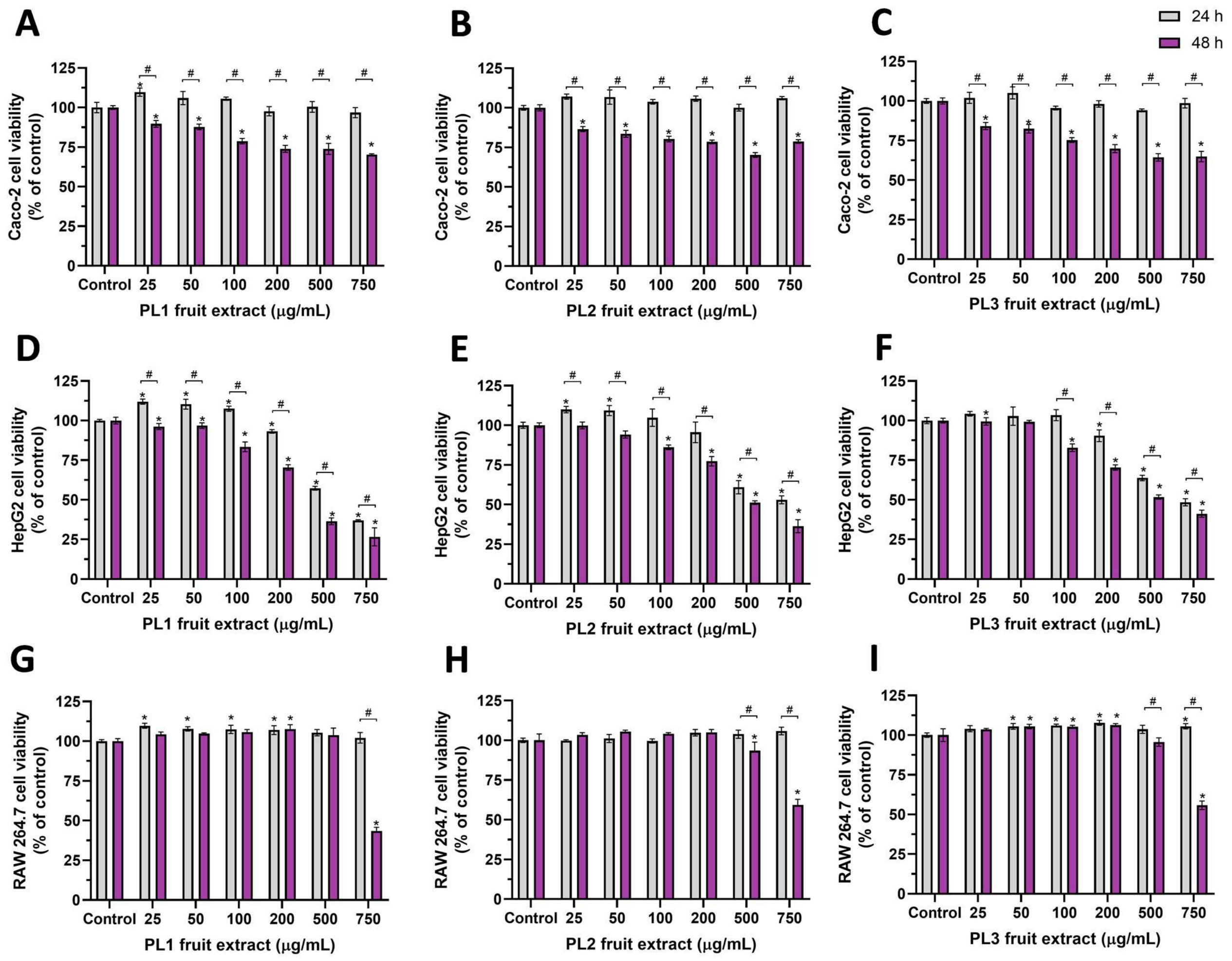
2.2. Antiproliferative/Cytotoxic Activity of P. lusitanica Fruit Extracts

The antiproliferative/cytotoxic activity of P. lusitanica fruit HE extracts was assessed using the Alamar Blue (AB) assay and three selected cell lines: the human cell lines Caco-2 and HepG2 as models of enterocyte and hepatocyte cells, respectively, and the murine macrophage cell line, RAW 264.7 cells. HE extracts of P. lusitanica fruits harvested in the three different locations (PL1, PL2, and PL3) were used. Cells were incubated, for 24 h or 48 h, with different concentrations of extracts (25, 50, 100, 200, 500, and 750 μg/mL) as depicted in Figure 1, and the results were compared with the respective positive control cells (nonexposed cells). As observed, 24 h exposure of Caco-2 cells with P. lusitanica fruit HE extracts did not produce a cytotoxic effect as cell viability was close to 100% of the control, at all tested concentrations (Figure 1A–C: gray bars), with no statistical difference (p > 0.05). However, exposure of Caco-2 cells to extracts for 48 h dose-dependently reduced cell viability, with the viability reduction profile being identical for the extracts obtained from the fruits harvested at the different locations (Figure 1A–C: purple bars). Despite the identical profile, cell viability after 48 h exposure to 750 μg/mL of LP1 or LP2 extract was about 70–78% of the control, reducing slightly (65% of the control) when exposed to 750 μg/mL of LP3 extract. The LP3 extract had higher amounts of flavan-3-ols and total anthocyanins (Table 2), which may have reduced the rate of proliferation of these cells, since a reduction in cell proliferation has been reported for several cell lines exposed to various flavan-3-ols [43,44] and anthocyanin-rich extracts [45].
As shown in Figure 1D–F, P. lusitanica fruit HE extracts dose-dependently reduced HepG2 cell viability with an identical profile at both exposure times but with slightly different potencies, with longer exposure being more cytotoxic. As observed, 24 h exposure to concentrations up to 100 μg/mL did not reduce HepG2 cell viability (cell viability: ~100% of the control or above), but 48 h exposure to the same concentrations reduced cell viability by about 15–20% (cell viability: 80–85% of the control). At the highest tested concentration of 750 μg/mL, cell viability was reduced to 50% of the control or even lower (Figure 1D–F). The IC50 values (Table 3) show that HepG2 cells were the most sensitive to these extracts; however, for the 24 h exposure, the IC50 values were higher than 500 μg/mL, denoting a very low cytotoxic effect. On the other hand, RAW 264.7 cells were the least affected (Figure 1G–I).
On the other hand, in Caco-2 and HepG2 cells, the obtained IC50 values were all higher than 350 μg/mL, which indicates that these extracts could be considered weakly active or inactive, with respect to antiproliferative/anticancer activity, according to the criteria of Geran et al. [46] (weakly active when IC50 0.20–0.50 mg/mL and inactive when IC50 > 0.50 mg/mL). Identical IC50 values were reported for the anthocyanin-rich fractions of blueberry (IC50 ~500 μg/mL) and blackcurrant (IC50 ~300 μg/mL) juices, against A2780, HeLa, and B10F10 cancer lines [47].
As observed, for RAW 264.7 cells (Figure 1G–I), only the higher tested concentrations, for the 48 h exposure, reduced cell viability to values close to 50% of the control (Figure 1H,I) or below (Figure 1G). The calculated IC50 values were close to 750 μg/mL (PL1 extract, 48 h exposure) or higher (Table 3, Figure 1G–I). According to Geran et al. [46], these extracts could be considered inactive against RAW 264.7 cells, since IC50 > 0.50 mg/mL (Table 3). The viability of Caco-2, HepG2 RAW 264.7 cells was not affected by concentrations up to 500 μg/mL of anthocyanin-rich elderberry (Sambucus nigra L.) extracts [48].

2.3. Anti-Inflammatory Activity of Prunus lusitanica Fruit Extracts

The anti-inflammatory activity of P. lusitanica fruit extracts (from the three locations) was evaluated on RAW 264.7 cells by their capacity to decrease the lipopolysaccharide (LPS)-induced nitric oxide (NO) release. The anti-inflammatory effect was evaluated at noncytotoxic concentrations, taking into account the results shown in Figure 1. RAW 264.7 cells were pre-exposed to P. lusitanica fruit extracts at concentrations up to 100 µg/mL, for 24 h, and then challenged with LPS (see Section 3 for details). Results concerning the anti-inflammatory potential of these extracts are shown in Figure 2. As observed, P. lusitanica fruit extracts from the different locations dose-dependently reduced NO release from LPS-stimulated RAW 264.7 cells, indicating that these extracts had the ability to target the TLR-4 (Toll-like receptor 4) pathway, as well as reduce iNOS (inducible nitric oxide synthase) expression and/or activity, resulting in lower amounts of released NO. PL1 and PL2 extracts at 25 µg/mL reduced NO release by about 40% (NO level: ~60% of the control), whereas PL3 reduced NO release to 70% of the control (Figure 2).
PL2 did not produce a significant dose-dependent inhibition of NO production at concentrations up to 100 µg/mL. On the other hand, PL1 and PL3 produced significant dose-dependent inhibition, inducing a significantly lower NO production at the highest tested concentration (100 µg/mL), with PL3 being the most effective extract, inducing ~64% inhibition (36% of the control’s NO production). PL3 presented a higher content of anthocyanins and flavan-3-ols (Table 2), which was likely correlated with the higher inhibition of NO release observed. Some of these compounds have been reported to produce anti-inflammatory activity.
Cyanidin-3-glucoside, for example, has been shown to reduce NO production in LPS-stimulated RAW 264.7 cells and in an intestinal cell model (HT-29), where it also induced a reduction in iNOS and cyclooxygenase 2 (COX-2) expression [49,50]. This activity was also observed for extracts obtained from other species rich in pro-anthocyanidin derivatives, which were present in a higher content in PL3 compared to PL1 and PL2. Some examples are extracts of cranberry [51], blueberry [52], elderberry [48], or grape [53]. Similarly, Musa acuminata extracts, rich in acetylated p-coumaroyl sucrose derivatives, such as various mumeoses and prunoses, also produced anti-inflammatory activity [54].
Specifically, in the Prunus genus, Prunus armeniaca leaf extracts were shown to inhibit COX-1 and COX-2 activity in in vitro assays [55]. Methanol and ethyl acetate extracts of Prunus yedoensis (50 and 100 µg/mL) reduced both NO and PGE2 (prostaglandin E2) production, as well as iNOS and COX-2 expression, in LPS-stimulated RAW 264.7 cells [56]. The anti-inflammatory effect of Prunus avium hydroethanolic extracts in RAW 264.7 cells was also evaluated, with leaf, stem, and flower extracts producing a dose-dependent inhibition of NO production, at concentrations ranging 50–400 µg/mL [57].

2.4. Assessment of the Inhibitory Effect of P. lusitanica Fruit Extracts on the Activity of Metabolically Relevant Enzymes

P. lusitanica fruit extracts were evaluated for their ability to inhibit key enzymes identified as therapeutic targets of common degenerative and metabolic pathologies, e.g., in the case of Alzheimer’s disease (acetylcholinesterase; AChE [58]), Parkinson’s disease (tyrosinase [59]), and diabetes mellitus (α-amylase and α-glucosidase [60,61]), as well as enzymes targeted for cosmetic purposes, such as skin-whitening (tyrosinase) and antiaging products (elastase) [23,62,63].
Two concentrations of Prunus lusitanica fruit HE extracts (from the three locations) were chosen: 0.5 and 1 mg/mL; the results are presented in Table 4.
Phytochemicals with the ability to inhibit AChE activity are promising molecules in the development of new drug leads for the treatment of neurodegenerative diseases, such as Alzheimer’s and Parkinson’s diseases, as these are characterized by decreased acetylcholine release [18]. As observed in Table 4, all samples dose-dependently inhibited AChE activity. The greatest inhibitory effect was observed for 1 mg/mL P. lusitanica extract, which inhibited AChE activity by 34% to 40% (Table 4). The PL1 extract produced the highest average inhibitory effect, at both 0.5 and 1 mg/mL, although the observed effect was not statistically significant at the latter concentration (p > 0.05) when compared with the other extracts. The highest inhibition induced by PL1 can likely be explained by the higher content of total phenolic compounds (Table 2), as this extract presented both the highest AChE inhibition and the greatest content of total phenolic compounds. PL3, the extract with a lower average AChE inhibition, presented the highest content of anthocyanins and flavan-3-ols, thus inferring that these components present a lower correlation with AChE inhibition. Among the main phytochemicals quantified (Table 2), various hydroxycinnamic acids and derivatives have been described as AChE inhibitors, e.g., p-coumaric, ferulic, and sinapic acids [64,65]. Regarding other Prunus spp., the hydroethanolic extracts of three cultivars of Prunus armeniaca L., containing various p-coumaroyl derivatives and flavanols, were shown to inhibit AChE activity, with IC50 values ranging between 5.53 and 10.33 mg/mL [55], concentrations ~5.5 and ~10 times higher than the highest concentration tested herein for P. lusitanica that presented ~40% inhibition (Table 4). Phenolic compounds present in sour cherry juice (Prunus cerasus), such as gallic or chlorogenic acids, showed inhibitory activity against AChE and monoamine oxidase A, with both enzymes being relevant in neuroprotective activity [66]. In another study, essential oils of P. armeniaca and Prunus domestica L. also inhibited AChE activity, with IC50 values ranging between 0.09 and 0.172 mg/mL [67]. These studies infer that Prunus spp. are sources of biologically active compounds with anti-AChE activity, which deserve further study.
Concerning the ability to inhibit tyrosinase activity, the same trend was observed as for AChE inhibition (Table 4), with PL1 extracts producing the greatest inhibitory effect (15% inhibition, at 1 mg/mL); the inhibition efficiency was in the order PL1 > PL2 > PL3, showing a dose-dependent effect (Table 4). This implies that these extracts could have some potential to counter neurodegeneration in Parkinson’s disease (by inhibiting neuromelanin production), as well as a cosmeceutical effect in skin-whitening agents [20,21,22]. Other Prunus species have been addressed for their potential as melanogenesis inhibitors. Prunus serrulata leaf aqueous and hydroethanolic extracts presented a higher potential as tyrosinase inhibitors [68] than P. lusitanica fruit extracts (Table 4), with 0.1 mg/mL aqueous and hydroethanolic extracts inhibiting tyrosinase activity by 63% and 39%, respectively [68]; however, the extract composition was different. A higher inhibitory effect against tyrosinase was also reported for hydroethanolic extracts of Prunus persica leaves that produced 14% inhibition at 0.5 mg/mL, when compared to Prunus persica fruit extracts that only produced 2% inhibition at the same concentration [69]. Thus, the tyrosinase-inhibitory effect of P. persica fruit extracts [69] is in line with the results here presented for P. lusitanica fruit extracts (Table 4). The ethanolic extracts from leaves of several varieties of P. domestica produced inhibition of tyrosinase activity, ranging from 0% to 11.60% at 3.43 mg/mL [70]. The data presented and discussed herein highlight the applicability of Prunus species to search for new cosmetic ingredients, as they combine antioxidant (Table 1) and tyrosinase-inhibitory activity, which might contribute to the regulation of melanin synthesis and attenuation of oxidative and age-related skin hyperpigmentation.
None of the extracts showed the ability to inhibit elastase activity, up to 1 mg/mL (Table 4). The absence of an elastase-inhibitory effect was also observed for methanolic extracts of elderberries (Sambucus nigra L.); these fruits were also rich in cinnamic acids and anthocyanins, such as cyanidin-3-O-glucoside [71], in line with the composition of P. lusitanica (Table 2), indicating that these compounds do not have ability to inhibit elastase. On the other hand, extracts obtained from leaves of several Prunus species (using organic solvents, such as hexane, ethyl acetate, and ethanol), were reported to produce anti-elastase activity depending on the solvent used (e.g., [72]). Stierlin et al. [72] obtained different extracts from leaves of various P. domestica varieties, showing that, although hexane and ethyl acetate extracts produced anti-elastase activity, the ethanolic extract produced no inhibition at 3.43 mg/mL. However, using a different extraction method, with ethanol as the solvent, Plainfossé et al. [70] reported significant anti-elastase activity for leaves of P. domestica. This indicates that both the solvent and the extraction method interfere with the extract’s bioactivities, which might be directly correlated with its phytochemical composition. Extracts from other sources have shown promising results concerning anti-elastase activity, such as extracts obtained from stems of different grape (Vitis vinifera L.) varieties, which inhibited 60% to 98% of elastase activity at 1 mg/mL [73]. Moreover, hydroethanolic extracts of orange thyme inhibited about 50% of elastase activity at 0.5 mg/mL [74]. Thus, although Prunus spp. extracts may present antiaging potential, the use of solvents with higher polarity is necessary to extract phytochemicals that contribute to this activity, which cannot be extracted using ethanol, as shown above (Table 4).
Concerning antidiabetic potential, unlike anti-AChE and anti-tyrosinase activity, no significant dose-dependent inhibition was observed for anti-α-amylase activity, with the exception of PL1 (Table 4), which also produced the highest average inhibition at 1 mg/mL. As observed, extracts produced low α-amylase-inhibitory activity. However, antidiabetic activity at the enzymatic level may also be exerted as α-glucosidase inhibition. Both PL1 and PL2 produced the highest α-glucosidase inhibition (~15%; Table 4); although, on average, the inhibitory effect increased with concentration, the differences were not statistically significant (p > 0.05). Despite the LP2 extract presenting a lower content of total phenolic compounds (Table 2), the unique phytochemical profile of each extract and the synergisms within its phytochemicals may have contributed to the variations observed. The LP3 extract produced a lower α-glucosidase-inhibitory effect; indeed, this extract contained lower amounts of di- and triacetylated p-coumaroyl derivatives, in the glycosylated form, which can promote competitive inhibition; however, further studies are needed to solidify this conclusion. Various individual components such as hydroxycinnamic acids [75], isoquercetin, rutin [76], and cyanidin-3-glucoside [77] have been shown to inhibit α-glucosidase; thus, these molecules were expected to contribute to the inhibition observed herein as they were identified in the extracts (Table 2). Regarding studies in other Prunus spp., Nan et al. [78] reported, in Prunus mume flower bud hydromethanolic extracts, the direct binding of phenolic compounds to the enzyme, contributing to the inhibition of α-glucosidase binding activity evaluated using an online methodology. Among the compounds studied by Nan et al. [78] in Prunus mume extracts (e.g., phenolic acids, flavonoids, and derivatives), some were also present in the P. lusitanica extracts reported herein (Table 2), likely responsible for the inhibition observed. Hydroethanolic extracts of P. armeniaca leaves produced 50% inhibition (IC50) of α-amylase at concentrations of 3.32–7.52 mg/mL [55], values well above the highest concentration tested in this study (1 mg/mL; Table 4). In addition, Wojdyło and Nowicka [55] reported, for the same extracts, lower IC50 values for α-glucosidase (0.71 to 3.35 mg/mL) inhibition, supporting a higher affinity of the components for α-glucosidase when compared to α-amylase, as observed herein for P. lusitanica extracts (Table 4). The low and moderate anti-α-amylase and anti-α-glucosidase activity, respectively, indicates the low antidiabetic effect of these extracts, with a low hypoglycemic potential.

3. Materials and Methods

3.1. Chemicals and Reagents

Sodium nitrate, aluminum chloride, and sodium hydroxide, all extra pure (>99%), and methanol were acquired from Merck (Merck, Darmstadt, Germany). Folin–Ciocâlteu’s reagent, 3,4,5-trihydroxybenzoic acid (gallic acid) and acetic acid, both extra pure (>99.0%), and sodium hydroxide were purchased from Panreac (Panreac Química S.L.U., Barcelona, Spain). Sodium molybdate (99.5%) was obtained from Chem-Lab (Chem-Lab N.V., Zedelgem, Belgium). The compounds 2,2-azino-bis (3-ethylbenzothiazoline-6-sulfonic acid) diammonium salt (ABTS), 2,2-diphenyl-1-picrylhidrazyl radical (DPPH), potassium phosphate, catechin, potassium persulfate, sodium acetate, 2,4,6-tripyridyl-s-triazine (TPTZ iron reagent), acetic acid, hydrochloric acid, and iron(III) chloride were obtained from Sigma-Aldrich/Merck (Algés, Portugal). Additionally, 6-hydroxy-2,5,7,8-tetramethylchroman-2-carboxylic acid (Trolox) was purchased from Fluka Chemika (Fluka Chemika, Neu-Ulm, Switzerland). The standards for chromatographic determinations were purchased from Sigma-Aldrich/Merck. Methanol, acetonitrile, and acetic acid (LC–MS-grade solvents) were provided by J.T. Baker (Philipsburg, NJ, USA). Milli-Q purified water (Millipore, Bedford, MA, USA) was used for all the extraction and chromatographic analyses. Enzymes and reagents for all enzymatic activities were purchased from Sigma-Aldrich/Merck.

3.2. Plant Material

To carry out this study, fruits of Prunus lusitanica L. were harvested, fully matured (characterized by a homogeneous purple color along the bunch) in early October, from three different locations: (1) at the University of Trás-os-Montes and Alto Douro (UTAD, Vila Real, Portugal; plants belonging to UTAD’s Botanical Garden collection), (2) in Pardieiros (Arganil, Portugal; voucher HVR 22669), which is currently included in the protected region of Serra do Açor, and (3) in Mata da Margaraça (Serra do Açor, Portugal; voucher HVR22670). These locations are denoted as PL1, PL2 and PL3, respectively. The specimens were properly identified by UTAD’s Botanical Garden office, where the vouchers were created and deposited.
After being collected, the samples were lyophilized (freeze-drier VirTis Benchtop Pro with Omnitronics TM), ground (using a coffee grinder), and stored in a hermetically sealed container protected from light until further analysis.

3.3. Preparation of Extracts from Prunus lusitanica L. Fruits

From each sample, three extracts were prepared by mixing 1.2 g of powder (lyophilized and grounded fruits) with 45 mL of hydroethanolic solution (70:30, % v/v). The mixture was homogenized and placed under stirring in an orbital shaker (GFL 3005, GEMINI, Apeldoorn, The Netherlands), at room temperature, for 30 min.
Afterward, the mixtures were centrifuged at 2291× g, 4 °C, for 15 min (Sigma 2-16KL Refrigerated Centrifuges, Sigma Laborzentrifugen, Berlin, Germany) and the supernatants were collected (this process was repeated four times). The resulting supernatants were combined and filtered through 0.2 μm regenerated polyamide membrane filters (NL 16 WhatmanTM). Ethanol was removed by repeated evaporation under vacuum at 35 °C, and the residue was freeze-dried. Solids were frozen for further analysis.

3.4. Determination of the Antioxidant Capacity

The antioxidant potential of fruit extracts was evaluated using three different spectrophotometric methods: ABTS and DPPH radical-scavenging methods, and FRAP (ferric ion-reducing antioxidant power). Procedures for DPPH and ABTS were carried out according to Lemos et al. (2020) [79], whereas the procedure for FRAP was carried out according to Beltrão et al. (2020) [80]. The methodologies were performed using 96-well microplate-adapted assays (PrimeSurface MS-9096MZ, Frilabo, Maia, Portugal) using a microplate reader (Multiskan GO Microplate Photometer, Thermo Fisher Scientific, Vantaa, Finland).
ABTS radicals were produced during a 12–16 h reaction period by mixing 5 mL of ABTS stock solution (7.0 mM in water) with 88 μL of potassium persulfate (148 mM), which was then diluted to a working solution, using sodium acetate buffer (20 mM, pH 4.5), at an absorbance of 0.70 at 734 nm. Then, 188 μL of ABTS working solution and 12 μL of each extract (distilled water served as blank) were combined and allowed to react under a light-protected environment, and the absorbance at 734 nm was measured after 30 min. Results were given in millimoles of Trolox equivalents per gram of dry extract (mmol TE/g dw). DPPH radical (8.87 mM in methanol) was diluted in a 70:30 (% v/v) methanol/water solution to achieve an absorbance of 1.00 at 520 nm before being used to create the DPPH working solution. Then, 190 μL of DPPH working solution and 10 μL of extract (using 70% hydromethanolic solution as blank) were mixed and incubated for 30 min (protected from light, at room temperature), after which the absorbance at 520 nm was measured to assess radical-scavenging activity. The scavenging capacity of samples was determined using interpolation of the Trolox calibration curve for DPPH and ABTS assays. Results were expressed as millimoles of Trolox equivalents per gram of dry extract (mmol TE/g dw). In order to measure the ferric ion-reducing antioxidant power (FRAP), extracts (20 μL) were added to the microplate, along with 280 μL of the FRAP working solution, consisting of a mixture at 10:1:1 (v:v:v) of acetate buffer (300 mM, pH 3.6), ferric chloride (20 mM in water), and TPTZ (10 mM in hydrochloric acid). This mixture was incubated for 30 min at 37 °C in the dark. Absorbance was then read at 593 nm. Once more, Trolox served as the reference, and the results were given in mmol TE/g dw.

3.5. HPLC-PAD–ESI-MS/MS Analysis of the Quantitative (Poly)phenolic Profile of Prunus lusitanica L. Fruit Extracts

The identification and quantification of phenolic compounds were performed according to Abellán et al. (2021) [81], with minor modifications. Briefly, chromatographic separations were carried out on a Kinetex Luna C18 column (250 × 46 mm, 5 µm particle size; Phenomenex, Macclesfield, UK) with a SecurityGuard C18-ODS cartridge system (Phenomenex). The chromatographic resolution of the phenolic profile was achieved using deionized water/formic acid (99:1, % v/v) (A) and acetonitrile (B) as the chromatographic solvents according to the following gradient (time in min, %B): (0, 5%), (30, 25%), (35, 50%), (37, 50%), and (38, 95%). The flow rate was 1 mL/min, and the injection volumes were 20 µL. The HPLC system was equipped with an Agilent 1100 Series diode array and a mass detector in series (Agilent Technologies, Waldbronn, Germany). It consisted of a G1312A binary pump, a G1313A auto-sampler, a G1322A degasser, and a G1315B photodiode array detector controlled by ChemStation software version 08.03 (Agilent Technologies, Waldbronn, Germany). Spectroscopic data from all peaks were accumulated in the range of 240–600 nm, and the spectral data were recorded at 280 nm, 320 nm, 330 nm, and 520 nm.
The mass detector was a G2445A Ion-Trap Mass Spectrometer equipped with an electrospray ionization (ESI) system and controlled by LCMSD software version 4.1 (Agilent, Waldbronn, Germany). Nitrogen was used as the nebulizing gas, at 60 psi with flow adjusted to 11 L/min. The heated capillary and voltage for ionization were maintained at 350 °C and 5 kV, respectively. Collision-induced fragmentation experiments were performed in the ion trap using helium as collision gas, with voltage ramping cycles from 0.3 up to 2 V. The full scan covered the range from m/z 100 to m/z 1600. Mass spectrometry data were acquired in negative and positive ionization modes. Total ion chromatograms were recorded as two alternating automatic scan events: full-scan mass spectra (MS) and MS/MS for fragmentation of the most abundant molecular ions. Identification of individual phenolic compounds was performed on the basis of retention time (min), parent ions, and fragmentation patterns, in comparison with authentic standards or, when not available, according to descriptions available in literature. Phenolic compounds were quantified by PDA chromatograms recorded at the above-indicated wavelengths, using, for each day of analysis, freshly prepared calibration curves.

3.6. In Vitro Cell-Based Assays

3.6.1. Cell Culture Maintenance and Handling

In this study, two human cell lines, Caco-2 (human colon adenocarcinoma; Cell Lines Service (CLS), Eppelheim, Germany) and HepG2 (human hepatocellular carcinoma; ATCC® Number: HB-8065TM), as well as a mouse cell line, RAW 264.7 (mouse macrophages, Abelson murine leukemia virus-induced tumor; CLS, Eppelheim, Germany), were used to evaluate the antiproliferative and anti-inflammatory activities of P. lusitanica fruit extracts. Cells were cultured in DMEM (Dulbecco’s modified Eagle medium) supplemented with 1 mM L-glutamine, 10% (v/v) fetal bovine serum (FBS), 100 U/mL penicillin, and 100 µg/mL streptomycin (all reagents from Gibco, Alfagene, Lisbon, Portugal), and then kept in an incubator (37 °C, 5% CO2/95% air, controlled humidity). Cells were treated and handled as reported by Silva et al. (2020) [82]. Briefly, near-confluence Caco-2 and HepG2 cells were treated with trypsin-EDTA, and the reaction was stopped using complete culture medium (1:1, trypsin/culture medium); in the case of RAW 264.7 cells, the cells were scratched off the flasks using a cell scratcher (Orange Scientific; Braine-L’Alleud, Belgium). Then, cells were counted, diluted in fresh culture medium at a final density of 5 × 10⁴ cells/mL, and seeded in 96-well culture microplates.

3.6.2. Cell Viability/Cytotoxicity Assay

In order to determine the effect of P. lusitanica fruit extracts on cell viability, Caco-2, HepG2, and RAW 264.7 cells seeded in 96-well microplates (see Section 3.6.1.) were allowed to adhere for 24 h before being subjected to extracts. After this period of time, the culture medium was removed, being immediately replaced with the test solutions replaced (extracts diluted in FBS-free culture medium at different concentrations: 25, 50, 100, 200, 500, and 750 µg/mL). Cells were then incubated for 24 or 48 h (in the incubator). Cell viability was assessed using the Alamar Blue (AB) metabolic indicator. After cell incubation with the extracts, the test solutions were removed, and the cells were exposed to 100 µL/well of AB solution (at 10% (v/v), in FBS-free culture medium). Next, 4–5 h after exposure to AB, the absorbance was read at 570 nm and 620 nm using a microplate reader (Multiskan EX; MTX Lab Systems, Inc., Bradenton, Fl, USA). Results were expressed as the cell viability (% of control; i.e., of positive control or nontreated cells), calculated as described by Andreani et al. (2014) [83].

3.6.3. Assessment of Anti-Inflammatory Activity

To assess the anti-inflammatory activity of P. lusitanica fruit extracts, the RAW 264.7 cell model stimulated by lipopolysaccharide (LPS) was used. RAW 264.7 cells were handled as described previously [82]. Briefly, RAW 264.7 cells seeded in 96-well plates (5 × 10⁴ cells/well) were allowed to adhere and stabilize for 48 h, before being exposed to noncytotoxic concentrations (25–100 µg/mL) of P. lusitanica fruit extracts for 24 h. Noncytotoxic concentrations were determined on the basis of the cell viability assay. Then, the extract solutions were removed, and the cells were gently rinsed with sterile phosphate buffer (PBS) to remove all traces of extracts (to avoid interference with the Griess assay, due to the chromophore effect), before being exposed to FBS-free culture medium (nonexposed cells, negative controls) or LPS (at 1 µg/mL in FBS-free culture medium) solution for 24 h (maintained in incubator). LPS induces the production of nitric oxide (NO) by RAW 264.7 cells, which is released into the incubation medium. After 24 h incubation, 50 µL of supernatant was removed from each well and transferred into a new 96-well plate. Then, 50 µL of Griess reagent [1% (w/v) sulfanilamide prepared in 5% (w/v) H3PO4 (v/v) and 0.1% (w/v) N-(1-naphthyl) ethylenediamine dihydrochloride in water] was added to each well. After 15 min incubation (room temperature, in the dark), the absorbance was read at 550 nm. The quantification of released NO was performed using a calibration curve obtained with sodium nitrite (NaNO2; range: 0 to 100 µM). Results were expressed as a percentage of the positive control (i.e., nitrite production by cells that were not exposed to extracts but stimulated with LPS) set to 100%, i.e., 0% anti-inflammatory effect.

3.7. In Vitro Bioassay for the Inhibition of Enzymes Involved in Several Relevant Metabolic Processes

The below-described enzyme inhibition assays were carried out in 96-well plates, and the absorbance of the respective products was read using a microplate reader.

3.7.1. Acetylcholinesterase (AChE) Inhibition Assay

The AChE inhibition assay was performed according to Elman’s method [84], with some modifications as described by Martins-Gomes et al. (2022) [85]. Briefly, to the extract (50 µL; at 0.5 or 1 mg/mL), DTNB (125 µL; 0.3 mM, in 50 mM Tris-HCl, pH 8) and acetylthiocholine iodide (ATCI; 25 µL; 1.5 mM). AChE (25 µL; 0.026 U/mL, in 20 mM Tris-HCl, pH 7.5) were sequentially added after 2 min of incubation. Then, the mixture was left to incubate for 10 min at 25 °C, after which the absorbance (Abs) was measured at 405 nm. Equation (1) was used to calculate the inhibition (%).
Inhibition (%) = [(Abs control − Abs sample)/(Abs control)] × 100.

3.7.2. Tyrosinase Inhibition Assay

The tyrosinase inhibition assay was performed on the basis of L-DOPA enzymatic oxidation, as described in [85]. Briefly, L-DOPA (40 µL; 2.5 mM) and phosphate buffer (80 µL; 50 mM, pH 6.8) were added to each extract (25 µL; at 0.5 or 1 mg/mL) and allowed to incubate for 2 min at 37 °C. Then, 40 µL of tyrosinase (40 U/mL, in phosphate buffer (50 mM, pH 6.5)) was added to start the reaction. Absorbance was measured at 492 nm after 10 min incubation at 37 °C. Kojic acid (1 mg/mL, in water) was used as the positive control, and the inhibition (%) was calculated using Equation (1).

3.7.3. Elastase Inhibition Assay

The anti-elastase activity of P. lusitanica fruit extracts was evaluated following a previously described procedure [21,62,86]. Briefly, to 50 µL of each extract (at 0.5 or 1 mg/mL), 160 µL of Tris-HCl buffer (0.2 M, pH 8) and 20 µL of N-(methoxysuccinyl)-ala-ala-pro-val-4-nitroanilide (0.8 mM, in buffer) were added. Then, this mixture was incubated for 10 min at room temperature. Next, elastase solution (20 µL; 0.4 U/mL in Tris-HCl buffer) was added to initiate the reaction. The absorbance was measured at 410 nm, after 20 min of incubation. Equation (1) was used to calculate the inhibition (%).

3.7.4. α-Amylase Inhibition Assay

For the α-amylase inhibition assay, the method described by Torres-Naranjo et al. (2016) [87] was modified as described in [21]. A mixture of each extract (5 µL; 0.5 or 1 mg/mL), PBS (35 µL), and starch solution (35 µL; 0.05%, pH 7) was preincubated for 2 min at 37 °C. Then, α-amylase solution (20 µL; 50 µg/mL; in phosphate buffer: 10 mM, pH 6.9) was added, and the mixture was allowed to react for 10 min at 37 °C. After incubation, the reaction was stopped by adding 50 µL of 0.1 M HCl solution. Then, 150 µL of Lugol solution (0.5 mM I2, 0.5 mM KI, in water) was added, and the mixture was incubated for 15 min. Absorbance was measured at 580 nm. Acarbose (1 mg/mL) was used as the positive control. Inhibition (%) was calculated according to Torres-Naranjo et al. (2016) [87].

3.7.5. α-Glucosidase Inhibition Assay

The α-Glucosidase inhibition was performed using rat intestinal acetone powder (Sigma-Aldrich/Merck) as a crude enzyme extract, as described previously [21]. Briefly, crude enzyme extract (250 mg) diluted in sodium phosphate buffer (10 mL; 0.1 M, pH 7.0) was centrifuged (5000× g, 20 min, 4 °C), and the supernatant was collected. To 50 µL of each extract (at 0.5 or 1 mg/mL), 100 µL of supernatant was added, and the mixture was incubated for 10 min at room temperature. Then, 50 µL of p-nitrophenyl-α-D-glucopyranoside (5 mM, in sodium phosphate buffer) were added, and the mixture was further incubated (30 min, 37 °C). After incubation, the absorbance was measured at 405 nm. Acarbose (1 mg/mL) was used as the positive control. Inhibition (%) was calculated according to Equation (1).

3.8. Data and Statistical Analysis

All assays were carried out in triplicate (n = 3), and the results were expressed as the mean ± standard deviation (SD). For the cytotoxicity assays, the IC50 (the concentration that reduces cell viability by 50%) values were calculated as described by Silva et al. (2019) [44]. The statistical differences were determined through analysis of variance (ANOVA) and a multiple range test (Tukey’s test) at p < 0.05. Statistical analyses and graphic design were performed using GraphPad Prism (Version 8; GraphPad Software Inc., San Diego, CA, USA).

4. Conclusions

In conclusion, the phytochemical profile of P. lusitanica fruit HE extracts was identical regarding the types of phytochemicals, independently of the location of harvest (PL1, PL2, and PL3); however, differences in the quantities of particular compounds were observed, potentially reflecting the edaphoclimatic differences across locations. Extracts from P. lusitanica fruits collected in Vila Real (PL1) had higher amounts of total phenolic compounds and total hydroxycinnamic acids, but contained a lower amount of total flavan-3-ols. Samples from Mata da Margaraça (PL3 extracts) contained higher amounts of total flavan-3-ols (more than twice that of PL1 sample) and total anthocyanins (more than twice that of Pardieiros sample, PL2). P. lusitanica fruit extracts generally exerted a low cytotoxic/antiproliferative effect; however, in HepG2 cells, a dose- and time-dependent decrease in cell viability was observed for concentrations higher than 100 μg/mL. The extracts showed high anti-inflammatory capacity, with all extracts reducing LPs-induced NO release by more than 50% at 100 μg/mL. The extracts also exhibited high neuroprotective potential and moderate antiaging and antidiabetic effects. Given that these fruits are not currently used for human consumption, it would be interesting in the future to assess whether they contain any antinutrients or potentially toxic compounds for humans, in order to promote these fruits as functional foods or as ingredients for the food industry. Additionally, the compounds present in these extracts are worth exploring for the development of new drugs of interest to the pharmaceutical and cosmetic industries.

Author Contributions

Conceptualization, A.M.S., R.D.-P. and A.B.; methodology, all authors; validation, A.M.S., R.D.-P. and A.B.; formal analysis, A.A., C.M.-G., R.D.-P., A.B. and A.M.S.; investigation, A.A., C.M.-G., R.D.-P., A.B. and A.M.S.; resources, A.M.S., R.D.-P. and A.B.; data curation, A.M.S., R.D.-P. and A.B.; writing—original draft preparation, A.A., C.M.-G. and A.M.S.; writing—review and editing, all authors; supervision, A.M.S., R.D.-P. and A.B.; funding acquisition, A.M.S., R.D.-P. and A.B. All authors have read and agreed to the published version of the manuscript.

Funding

This research was funded by the Portuguese Science and Technology Foundation, Ministry of Science and Education (FCT/MEC), through national and European funds, under the project UIDB/04033/2020 (CITAB), as well as PhD grants to A. Abraão (PD/BD/113614/2015) and C. Martins-Gomes (SFRH/BD/145855/2019).

Institutional Review Board Statement

Not applicable.

Informed Consent Statement

Not applicable.

Data Availability Statement

Not applicable.

Conflicts of Interest

The authors declare no conflict of interest.

References

  1. Vekic, J.; Zeljkovic, A.; Stefanovic, A.; Jelic-Ivanovic, Z.; Spasojevic-Kalimanovska, V. Obesity and dyslipidemia. Metabolism 2019, 92, 71–81. [Google Scholar] [CrossRef]
  2. Tabatabaei-Malazy, O.; Larijani, B.; Abdollahi, M. Targeting metabolic disorders by natural products. J. Diabetes Metab. Disord. 2015, 14, 57. [Google Scholar] [CrossRef]
  3. Martins-Gomes, C.; Souto, E.B.; Silva, A.M. Chapter 15—Nanophytosomes: A novel approach for the delivery of herbal drugs. In Systems of Nanovesicular Drug Delivery; Nayak, A.K., Hasnain, M.S., Aminabhavi, T.M., Torchilin, V.P., Eds.; Academic Press: Cambridge, MA, USA, 2022; pp. 239–257. [Google Scholar]
  4. Vona, R.; Gambardella, L.; Cittadini, C.; Straface, E.; Pietraforte, D. Biomarkers of Oxidative Stress in Metabolic Syndrome and Associated Diseases. Oxidative Med. Cell. Longev. 2019, 2019, 8267234. [Google Scholar] [CrossRef]
  5. Khutami, C.; Sumiwi, S.A.; Khairul Ikram, N.K.; Muchtaridi, M. The Effects of Antioxidants from Natural Products on Obesity, Dyslipidemia, Diabetes and Their Molecular Signaling Mechanism. Int. J. Mol. Sci. 2022, 23, 2056. [Google Scholar] [CrossRef]
  6. Baek, S.J.; Hammock, B.D.; Hwang, I.-K.; Li, Q.X.; Moustaid-Moussa, N.; Park, Y.; Safe, S.; Suh, N.; Yi, S.-S.; Zeldin, D.C.; et al. Natural Products in the Prevention of Metabolic Diseases: Lessons Learned from the 20th KAST Frontier Scientists Workshop. Nutrients 2021, 13, 1881. [Google Scholar] [CrossRef] [PubMed]
  7. Arulselvan, P.; Fard, M.T.; Tan, W.S.; Gothai, S.; Fakurazi, S.; Norhaizan, M.E.; Kumar, S.S. Role of Antioxidants and Natural Products in Inflammation. Oxidative Med. Cell. Longev. 2016, 2016, 5276130. [Google Scholar] [CrossRef]
  8. Atanasov, A.G.; Waltenberger, B.; Pferschy-Wenzig, E.-M.; Linder, T.; Wawrosch, C.; Uhrin, P.; Temml, V.; Wang, L.; Schwaiger, S.; Heiss, E.H.; et al. Discovery and resupply of pharmacologically active plant-derived natural products: A review. Biotechnol. Adv. 2015, 33, 1582–1614. [Google Scholar] [CrossRef] [PubMed]
  9. Halpin, C.M.; Reilly, C.; Walsh, J.J. Nature’s Anti-Alzheimer’s Drug: Isolation and Structure Elucidation of Galantamine from Leucojum aestivum. J. Chem. Educ. 2010, 87, 1242–1243. [Google Scholar] [CrossRef]
  10. Nicolaou, K.C.; Yang, Z.; Liu, J.J.; Ueno, H.; Nantermet, P.G.; Guy, R.K.; Claiborne, C.F.; Renaud, J.; Couladouros, E.A.; Paulvannan, K.; et al. Total synthesis of taxol. Nature 1994, 367, 630–634. [Google Scholar] [CrossRef] [PubMed]
  11. Elfawal, M.A.; Towler, M.J.; Reich, N.G.; Golenbock, D.; Weathers, P.J.; Rich, S.M. Dried whole plant Artemisia annua as an antimalarial therapy. PLoS ONE 2012, 7, e52746. [Google Scholar] [CrossRef]
  12. Ríos, J.L.; Francini, F.; Schinella, G.R. Natural Products for the Treatment of Type 2 Diabetes Mellitus. Planta Med. 2015, 81, 975–994. [Google Scholar] [CrossRef]
  13. Jugran, A.K.; Rawat, S.; Devkota, H.P.; Bhatt, I.D.; Rawal, R.S. Diabetes and plant-derived natural products: From ethnopharmacological approaches to their potential for modern drug discovery and development. Phytother. Res. 2021, 35, 223–245. [Google Scholar] [CrossRef]
  14. Vlachojannis, J.; Magora, F.; Chrubasik, S. Willow Species and Aspirin: Different Mechanism of Actions. Phytother. Res. 2011, 25, 1102–1104. [Google Scholar] [CrossRef] [PubMed]
  15. Martins-Gomes, C.; Nunes, F.M.; Silva, A.M. Modulation of Cell Death Pathways for Cellular Protection and Anti-Tumoral Activity: The Role of Thymus spp. Extracts and Their Bioactive Molecules. Int. J. Mol. Sci. 2023, 24, 1691. [Google Scholar] [CrossRef] [PubMed]
  16. Gouvinhas, I.; Machado, N.; Sobreira, C.; Domínguez-Perles, R.; Gomes, S.; Rosa, E.; Barros, A.I.R.N.A. Critical Review on the Significance of Olive Phytochemicals in Plant Physiology and Human Health. Molecules 2017, 22, 1986. [Google Scholar] [CrossRef] [PubMed]
  17. Souto, E.B.; Sampaio, A.C.; Campos, J.R.; Martins-Gomes, C.; Aires, A.; Silva, A.M. Chapter 2—Polyphenols for skin cancer: Chemical properties, structure-related mechanisms of action and new delivery systems. In Studies in Natural Products Chemistry; Atta ur, R., Ed.; Elsevier: Amsterdam, The Netherlands, 2019; Volume 63, pp. 21–42. [Google Scholar]
  18. Marucci, G.; Buccioni, M.; Ben, D.D.; Lambertucci, C.; Volpini, R.; Amenta, F. Efficacy of acetylcholinesterase inhibitors in Alzheimer’s disease. Neuropharmacology 2021, 190, 108352. [Google Scholar] [CrossRef]
  19. Sánchez-López, E.; Ettcheto, M.; Egea, M.A.; Espina, M.; Cano, A.; Calpena, A.C.; Camins, A.; Carmona, N.; Silva, A.M.; Souto, E.B.; et al. Memantine loaded PLGA PEGylated nanoparticles for Alzheimer’s disease: In Vitro and in vivo characterization. J. Nanobiotechnology 2018, 16, 32. [Google Scholar] [CrossRef]
  20. Carballo-Carbajal, I.; Laguna, A.; Romero-Giménez, J.; Cuadros, T.; Bové, J.; Martinez-Vicente, M.; Parent, A.; Gonzalez-Sepulveda, M.; Peñuelas, N.; Torra, A.; et al. Brain tyrosinase overexpression implicates age-dependent neuromelanin production in Parkinson’s disease pathogenesis. Nat. Commun. 2019, 10, 973. [Google Scholar] [CrossRef]
  21. Taghouti, M.; Martins-Gomes, C.; Schäfer, J.; Félix, L.M.; Santos, J.A.; Bunzel, M.; Nunes, F.M.; Silva, A.M. Thymus pulegioides L. as a rich source of antioxidant, anti-proliferative and neuroprotective phenolic compounds. Food Funct. 2018, 9, 3617–3629. [Google Scholar] [CrossRef] [PubMed]
  22. Abraão, A.; Yu, M.; Gouvinhas, I.; Ferreira, L.; Silva, A.M.; Domínguez-Perles, R.; Barros, A. Prunus lusitanica L. Fruits: A Promising Underexploited Source of Nutrients with Potential Economic Value. Foods 2023, 12, 973. [Google Scholar] [CrossRef]
  23. Pillaiyar, T.; Manickam, M.; Namasivayam, V. Skin whitening agents: Medicinal chemistry perspective of tyrosinase inhibitors. J. Enzym. Inhib. Med. Chem. 2017, 32, 403–425. [Google Scholar] [CrossRef]
  24. Tundis, R.; Loizzo, M.R.; Menichini, F. Natural products as alpha-amylase and alpha-glucosidase inhibitors and their hypoglycaemic potential in the treatment of diabetes: An update. Mini Rev. Med. Chem. 2010, 10, 315–331. [Google Scholar] [CrossRef]
  25. Pardo, A.; Cáceres, Y.; Pulido, F. Rangewide determinants of population performance in Prunus lusitanica: Lessons for the contemporary conservation of a Tertiary relict tree. Acta Oecologica 2018, 86, 42–48. [Google Scholar] [CrossRef]
  26. Costa, M.D.C.; Duarte, P.; Neng, N.R.; Nogueira, J.M.F.; Costa, F.; Rosado, C. Novel insights for permeant lead structures through in vitro skin diffusion assays of Prunus lusitanica L., the Portugal Laurel. J. Mol. Struct. 2015, 1079, 327–336. [Google Scholar] [CrossRef]
  27. Press, B. Green Guide to Trees of Britain and Europe; Bloomsbury Publishing: London, UK, 2016. [Google Scholar]
  28. Biessels, H.W.A.; van der Kerk-van Hoof, A.C.; Kettenes-van den Bosch, J.J.; Salemink, C.A. Triterpenes of Prunus serotina and P. lusitanica. Phytochemistry 1974, 13, 203–207. [Google Scholar] [CrossRef]
  29. Abraão, A.S.; Fernandes, N.; Silva, A.M.; Domínguez-Perles, R.; Barros, A. Prunus lusitanica L. Fruits as a Novel Source of Bioactive Compounds with Antioxidant Potential: Exploring the Unknown. Antioxidants 2022, 11, 1738. [Google Scholar] [CrossRef] [PubMed]
  30. Ullah, H.; Sommella, E.; Santarcangelo, C.; D’Avino, D.; Rossi, A.; Dacrema, M.; Minno, A.D.; Di Matteo, G.; Mannina, L.; Campiglia, P.; et al. Hydroethanolic Extract of Prunus domestica L.: Metabolite Profiling and In Vitro Modulation of Molecular Mechanisms Associated to Cardiometabolic Diseases. Nutrients 2022, 14, 340. [Google Scholar] [CrossRef]
  31. Holton, T.A.; Cornish, E.C. Genetics and Biochemistry of Anthocyanin Biosynthesis. Plant Cell 1995, 7, 1071–1083. [Google Scholar] [CrossRef]
  32. Mannino, G.; Gentile, C.; Ertani, A.; Serio, G.; Bertea, C.M. Anthocyanins: Biosynthesis, Distribution, Ecological Role, and Use of Biostimulants to Increase Their Content in Plant Foods—A Review. Agriculture 2021, 11, 212. [Google Scholar] [CrossRef]
  33. Geng, D.; Shen, X.; Xie, Y.; Yang, Y.; Bian, R.; Gao, Y.; Li, P.; Sun, L.; Feng, H.; Ma, F.; et al. Regulation of phenylpropanoid biosynthesis by MdMYB88 and MdMYB124 contributes to pathogen and drought resistance in apple. Hortic. Res. 2020, 7, 102. [Google Scholar] [CrossRef]
  34. Macoy, D.M.; Kim, W.-Y.; Lee, S.Y.; Kim, M.G. Biosynthesis, physiology, and functions of hydroxycinnamic acid amides in plants. Plant Biotechnol. Rep. 2015, 9, 269–278. [Google Scholar] [CrossRef]
  35. Sunil, L.; Shetty, N.P. Biosynthesis and regulation of anthocyanin pathway genes. Appl. Microbiol. Biotechnol. 2022, 106, 1783–1798. [Google Scholar] [CrossRef] [PubMed]
  36. Hernández, R.; Eguchi, T.; Deveci, M.; Kubota, C. Tomato seedling physiological responses under different percentages of blue and red photon flux ratios using LEDs and cool white fluorescent lamps. Sci. Hortic. 2016, 213, 270–280. [Google Scholar] [CrossRef]
  37. Costa, R.; Fraga, H.; Fernandes, P.M.; Santos, J.A. Implications of future bioclimatic shifts on Portuguese forests. Reg. Environ. Chang. 2017, 17, 117–127. [Google Scholar] [CrossRef]
  38. Torras-Claveria, L.; Jáuregui, O.; Codina, C.; Tiburcio, A.F.; Bastida, J.; Viladomat, F. Analysis of phenolic compounds by high-performance liquid chromatography coupled to electrospray ionization tandem mass spectrometry in senescent and water-stressed tobacco. Plant Sci. 2012, 182, 71–78. [Google Scholar] [CrossRef]
  39. Marčetić, M.; Samardžić, S.; Ilić, T.; Božić, D.D.; Vidović, B. Phenolic Composition, Antioxidant, Anti-Enzymatic, Antimicrobial and Prebiotic Properties of Prunus spinosa L. Fruits. Foods 2022, 11, 3289. [Google Scholar] [CrossRef]
  40. Yoshikawa, M.; Murakami, T.; Ishiwada, T.; Morikawa, T.; Kagawa, M.; Higashi, Y.; Matsuda, H. New Flavonol Oligoglycosides and Polyacylated Sucroses with Inhibitory Effects on Aldose Reductase and Platelet Aggregation from the Flowers of Prunus mume. J. Nat. Prod. 2002, 65, 1151–1155. [Google Scholar] [CrossRef]
  41. Rice-Evans, C.A.; Miller, N.J.; Paganga, G. Structure-antioxidant activity relationships of flavonoids and phenolic acids. Free Radic. Biol. Med. 1996, 20, 933–956. [Google Scholar] [CrossRef]
  42. Cherrak, S.A.; Mokhtari-Soulimane, N.; Berroukeche, F.; Bensenane, B.; Cherbonnel, A.; Merzouk, H.; Elhabiri, M. In Vitro Antioxidant versus Metal Ion Chelating Properties of Flavonoids: A Structure-Activity Investigation. PLoS ONE 2016, 11, e0165575. [Google Scholar] [CrossRef]
  43. Braicu, C.; Pilecki, V.; Balacescu, O.; Irimie, A.; Berindan Neagoe, I. The Relationships Between Biological Activities and Structure of Flavan-3-Ols. Int. J. Mol. Sci. 2011, 12, 9342–9353. [Google Scholar] [CrossRef]
  44. Silva, A.M.; Martins-Gomes, C.; Fangueiro, J.F.; Andreani, T.; Souto, E.B. Comparison of antiproliferative effect of epigallocatechin gallate when loaded into cationic solid lipid nanoparticles against different cell lines. Pharm. Dev. Technol. 2019, 24, 1243–1249. [Google Scholar] [CrossRef]
  45. Salamon, I.; Şimşek Sezer, E.N.; Kryvtsova, M.; Labun, P. Antiproliferative and Antimicrobial Activity of Anthocyanins from Berry Fruits after Their Isolation and Freeze-Drying. Appl. Sci. 2021, 11, 2096. [Google Scholar] [CrossRef]
  46. Geran, R.I.; Greenberg, N.H.; Donald, M.M.M. Protocols for screening chemical agents and natural products against animal tumors and other biological systems. Cancer Chemother. Rep. 1972, 3, 1–103. [Google Scholar]
  47. Diaconeasa, Z.; Leopold, L.; Rugină, D.; Ayvaz, H.; Socaciu, C. Antiproliferative and Antioxidant Properties of Anthocyanin Rich Extracts from Blueberry and Blackcurrant Juice. Int. J. Mol. Sci. 2015, 16, 2352–2365. [Google Scholar] [CrossRef]
  48. Ferreira, S.S.; Martins-Gomes, C.; Nunes, F.M.; Silva, A.M. Elderberry (Sambucus nigra L.) extracts promote anti-inflammatory and cellular antioxidant activity. Food Chem. X 2022, 15, 100437. [Google Scholar] [CrossRef]
  49. Jung, H.; Kwak, H.-K.; Hwang, K.T. Antioxidant and antiinflammatory activities of cyanidin-3-glucoside and cyanidin-3-rutinoside in hydrogen peroxide and lipopolysaccharide-treated RAW264.7 cells. Food Sci. Biotechnol. 2014, 23, 2053–2062. [Google Scholar] [CrossRef]
  50. Serra, D.; Paixão, J.; Nunes, C.; Dinis, T.C.P.; Almeida, L.M. Cyanidin-3-Glucoside Suppresses Cytokine-Induced Inflammatory Response in Human Intestinal Cells: Comparison with 5-Aminosalicylic Acid. PLoS ONE 2013, 8, e73001. [Google Scholar]
  51. Moore, K.; Howard, L.; Brownmiller, C.; Gu, I.; Lee, S.-O.; Mauromoustakos, A. Inhibitory effects of cranberry polyphenol and volatile extracts on nitric oxide production in LPS activated RAW 264.7 macrophages. Food Funct. 2019, 10, 7091–7102. [Google Scholar] [CrossRef]
  52. Grace, M.H.; Esposito, D.; Dunlap, K.L.; Lila, M.A. Comparative Analysis of Phenolic Content and Profile, Antioxidant Capacity, and Anti-inflammatory Bioactivity in Wild Alaskan and Commercial Vaccinium Berries. J. Agric. Food Chem. 2014, 62, 4007–4017. [Google Scholar] [CrossRef]
  53. Zhang, L.; Chen, J.; Liao, H.; Li, C.; Chen, M. Anti-inflammatory effect of lipophilic grape seed proanthocyanidin in RAW 264.7 cells and a zebrafish model. J. Funct. Foods 2020, 75, 104217. [Google Scholar] [CrossRef]
  54. Sandjo, L.P.; dos Santos Nascimento, M.V.P.; de H. Moraes, M.; Rodrigues, L.M.; Dalmarco, E.M.; Biavatti, M.W.; Steindel, M. NOx-, IL-1β-, TNF-α-, and IL-6-Inhibiting Effects and Trypanocidal Activity of Banana (Musa acuminata) Bracts and Flowers: UPLC-HRESI-MS Detection of Phenylpropanoid Sucrose Esters. Molecules 2019, 24, 4564. [Google Scholar] [CrossRef]
  55. Wojdyło, A.; Nowicka, P. Profile of Phenolic Compounds of Prunus armeniaca L. Leaf Extract Determined by LC-ESI-QTOF-MS/MS and Their Antioxidant, Anti-Diabetic, Anti-Cholinesterase, and Anti-Inflammatory Potency. Antioxidants 2021, 10, 1869. [Google Scholar] [CrossRef]
  56. Lee, J.; Yang, G.; Lee, K.; Lee, M.-H.; Eom, J.-W.; Ham, I.; Choi, H.-Y. Anti-inflammatory effect of Prunus yedoensis through inhibition of nuclear factor-κB in macrophages. BMC Complement. Altern. Med. 2013, 13, 92. [Google Scholar] [CrossRef]
  57. Nunes, A.R.; Flores-Félix, J.D.; Gonçalves, A.C.; Falcão, A.; Alves, G.; Silva, L.R. Anti-Inflammatory and Antimicrobial Activities of Portuguese Prunus avium L. (Sweet Cherry) By-Products Extracts. Nutrients 2022, 14, 4576. [Google Scholar] [CrossRef]
  58. Uddin, M.S.; Al Mamun, A.; Kabir, M.T.; Ashraf, G.M.; Bin-Jumah, M.N.; Abdel-Daim, M.M. Multi-Target Drug Candidates for Multifactorial Alzheimer’s Disease: AChE and NMDAR as Molecular Targets. Mol. Neurobiol. 2021, 58, 281–303. [Google Scholar] [CrossRef]
  59. Hasegawa, T. Tyrosinase-Expressing Neuronal Cell Line as in Vitro Model of Parkinson’s Disease. Int. J. Mol. Sci. 2010, 11, 1082–1089. [Google Scholar] [CrossRef]
  60. Li, X.; Bai, Y.; Jin, Z.; Svensson, B. Food-derived non-phenolic α-amylase and α-glucosidase inhibitors for controlling starch digestion rate and guiding diabetes-friendly recipes. LWT 2022, 153, 112455. [Google Scholar] [CrossRef]
  61. Ghosh, S.; More, P.; Derle, A.; Patil, A.B.; Markad, P.; Asok, A.; Kumbhar, N.; Shaikh, M.L.; Ramanamurthy, B.; Shinde, V.S.; et al. Diosgenin from Dioscorea bulbifera: Novel hit for treatment of type II diabetes mellitus with inhibitory activity against α-amylase and α-glucosidase. PLoS ONE 2014, 9, e106039. [Google Scholar]
  62. Frazão, D.F.; Martins-Gomes, C.; Steck, J.L.; Keller, J.; Delgado, F.; Gonçalves, J.C.; Bunzel, M.; Pintado, C.M.B.S.; Díaz, T.S.; Silva, A.M. Labdanum Resin from Cistus ladanifer L.: A Natural and Sustainable Ingredient for Skin Care Cosmetics with Relevant Cosmeceutical Bioactivities. Plants 2022, 11, 1477. [Google Scholar] [CrossRef]
  63. Azmi, N.; Hashim, P.; Hashim, D.M.; Halimoon, N.; Majid, N.M.N. Anti–elastase, anti–tyrosinase and matrix metalloproteinase–1 inhibitory activity of earthworm extracts as potential new anti–aging agent. Asian Pac. J. Trop. Biomed. 2014, 4, S348–S352. [Google Scholar] [CrossRef]
  64. Daroi, P.A.; Dhage, S.N.; Juvekar, A.R. p-Coumaric acid mitigates lipopolysaccharide induced brain damage via alleviating oxidative stress, inflammation and apoptosis. J. Pharm. Pharmacol. 2021, 74, 556–564. [Google Scholar] [CrossRef]
  65. Szwajgier, D.; Borowiec, K. Phenolic acids from malt are efficient acetylcholinesterase and butyrylcholinesterase inhibitors. J. Inst. Brew. 2012, 118, 40–48. [Google Scholar] [CrossRef]
  66. Cásedas, G.; Les, F.; Gómez-Serranillos, M.P.; Smith, C.; López, V. Bioactive and functional properties of sour cherry juice (Prunus cerasus). Food Funct. 2016, 7, 4675–4682. [Google Scholar] [CrossRef]
  67. Bonesi, M.; Tenuta, M.C.; Loizzo, M.R.; Sicari, V.; Tundis, R. Potential Application of Prunus armeniaca L. and P. domestica L. Leaf Essential Oils as Antioxidant and of Cholinesterases Inhibitors. Antioxidants 2018, 8, 2. [Google Scholar] [CrossRef]
  68. Park, J.W.; Yuk, H.G.; Lee, S.C. Antioxidant and tyrosinase inhibitory activities of different parts of oriental cherry (Prunus serrulata var. spontanea). Food Sci. Biotechnol. 2012, 21, 339–343. [Google Scholar] [CrossRef]
  69. Murata, K.; Suzuki, S.; Miyamoto, A.; Horimoto, M.; Nanko, S.; Mori, D.; Kanamaru, H.; Endo, Y. Tyrosinase Inhibitory Activity of Extracts from Prunus persica. Separations 2022, 9, 107. [Google Scholar] [CrossRef]
  70. Plainfossé, H.; Burger, P.; Verger-Dubois, G.; Azoulay, S.; Fernandez, X. Design Methodology for the Development of a New Cosmetic Active Based on Prunus domestica L. Leaves Extract. Cosmetics 2019, 6, 8. [Google Scholar] [CrossRef]
  71. Ferreira, S.S.; Silva, P.; Silva, A.M.; Nunes, F.M. Effect of harvesting year and elderberry cultivar on the chemical composition and potential bioactivity: A three-year study. Food Chem. 2020, 302, 125366. [Google Scholar] [CrossRef]
  72. Stierlin, E.; Azoulay, S.; Massi, L.; Fernandez, X.; Michel, T. Cosmetic potentials of Prunus domestica L. leaves. J. Sci. Food Agric. 2018, 98, 726–736. [Google Scholar] [CrossRef]
  73. Leal, C.; Gouvinhas, I.; Santos, R.A.; Rosa, E.; Silva, A.M.; Saavedra, M.J.; Barros, A.I.R.N.A. Potential application of grape (Vitis vinifera L.) stem extracts in the cosmetic and pharmaceutical industries: Valorization of a by-product. Ind. Crops Prod. 2020, 154, 112675. [Google Scholar] [CrossRef]
  74. Silva, A.M.; Félix, L.M.; Teixeira, I.; Martins-Gomes, C.; Schäfer, J.; Souto, E.B.; Santos, D.J.; Bunzel, M.; Nunes, F.M. Orange thyme: Phytochemical profiling, in vitro bioactivities of extracts and potential health benefits. Food Chem. X 2021, 12, 100171. [Google Scholar] [CrossRef] [PubMed]
  75. Aleixandre, A.; Gil, J.V.; Sineiro, J.; Rosell, C.M. Understanding phenolic acids inhibition of α-amylase and α-glucosidase and influence of reaction conditions. Food Chem. 2022, 372, 131231. [Google Scholar] [CrossRef] [PubMed]
  76. Li, Y.Q.; Zhou, F.C.; Gao, F.; Bian, J.S.; Shan, F. Comparative Evaluation of Quercetin, Isoquercetin and Rutin as Inhibitors of α-Glucosidase. J. Agric. Food Chem. 2009, 57, 11463–11468. [Google Scholar] [CrossRef] [PubMed]
  77. Akkarachiyasit, S.; Charoenlertkul, P.; Yibchok-anun, S.; Adisakwattana, S. Inhibitory Activities of Cyanidin and Its Glycosides and Synergistic Effect with Acarbose against Intestinal α-Glucosidase and Pancreatic α-Amylase. Int. J. Mol. Sci. 2010, 11, 3387–3396. [Google Scholar] [CrossRef]
  78. Nan, X.; Jia, W.; Zhang, Y.; Wang, H.; Lin, Z.; Chen, S. An on-line detection system for screening small molecule inhibitors of α-Amylase and α-Glucosidase in Prunus mume. J. Chromatogr. A 2022, 1663, 462754. [Google Scholar] [CrossRef]
  79. Mendes Lemos, A.; Machado, N.; Egea-Cortines, M.; Barros, A.I. Assessment of quality parameters and phytochemical content of thirty ‘Tempranillo’ grape clones for varietal improvement in two distinct sub-regions of Douro. Sci. Hortic. 2020, 262, 109096. [Google Scholar] [CrossRef]
  80. Beltrão Martins, R.; Gouvinhas, I.; Nunes, M.C.; Alcides Peres, J.; Raymundo, A.; Barros, A.I.R.N.A. Acorn Flour as a Source of Bioactive Compounds in Gluten-Free Bread. Molecules 2020, 25, 3568. [Google Scholar] [CrossRef]
  81. Abellán, Á.; Domínguez-Perles, R.; García-Viguera, C.; Moreno, D.A. In Vitro Evidence on Bioaccessibility of Flavonols and Cinnamoyl Derivatives of Cruciferous Sprouts. Nutrients 2021, 13, 4140. [Google Scholar] [CrossRef]
  82. Silva, A.M.; Martins-Gomes, C.; Souto, E.B.; Schäfer, J.; Santos, J.A.; Bunzel, M.; Nunes, F.M. Thymus zygis subsp. zygis an Endemic Portuguese Plant: Phytochemical Profiling, Antioxidant, Anti-Proliferative and Anti-Inflammatory Activities. Antioxidants 2020, 9, 482. [Google Scholar] [CrossRef]
  83. Andreani, T.; Kiill, C.P.; de Souza, A.L.; Fangueiro, J.F.; Fernandes, L.; Doktorovova, S.; Santos, D.L.; Garcia, M.L.; Gremiao, M.P.; Souto, E.B.; et al. Surface engineering of silica nanoparticles for oral insulin delivery: Characterization and cell toxicity studies. Colloids Surf B Biointerfaces 2014, 123, 916–923. [Google Scholar] [CrossRef]
  84. Ellman, G.L.; Courtney, K.D.; Andres, V.; Featherstone, R.M. A new and rapid colorimetric determination of acetylcholinesterase activity. Biochem. Pharmacol. 1961, 7, 88–95. [Google Scholar] [CrossRef] [PubMed]
  85. Martins-Gomes, C.; Coutinho, T.E.; Silva, T.L.; Andreani, T.; Silva, A.M. Neurotoxicity Assessment of Four Different Pesticides Using In Vitro Enzymatic Inhibition Assays. Toxics 2022, 10, 448. [Google Scholar] [CrossRef] [PubMed]
  86. Apraj, V.D.; Pandita, N.S. Evaluation of Skin Anti-aging Potential of Citrus reticulata Blanco Peel. Pharmacogn. Res. 2016, 8, 160–168. [Google Scholar] [CrossRef] [PubMed]
  87. Torres-Naranjo, M.; Suárez, A.; Gilardoni, G.; Cartuche, L.; Flores, P.; Morocho, V. Chemical Constituents of Muehlenbeckia tamnifolia (Kunth) Meisn (Polygonaceae) and Its In Vitro α-Amilase and α-Glucosidase Inhibitory Activities. Molecules 2016, 21, 1461. [Google Scholar] [CrossRef]
Figure 1. Antiproliferative/cytotoxic effect of P. lusitanica L. fruit extracts on Caco-2 (AC), HepG2 (DF), and Raw 264.7 (GI) cells, for two exposure times: 24 h (gray bars) and 48 h (dark-pink bars). Results are expressed as the mean ± SD (n = 4). * Statistically significant difference (p < 0.05) between the control and sample concentrations at respective incubation times; # statistically significant difference (p < 0.05) between exposure periods at the same concentration.
Ijms 24 08830 g001
Figure 2. Anti-inflammatory activity of P. lusitanica L. fruit extracts expressed as the inhibition of nitric oxide (NO) release by LPS-stimulated RAW 264.7 cells. Results are expressed as the mean ± SD (n = 4 independent assays). * Statistical differences (p < 0.05) between samples and the positive control (LPS-stimulated cells not exposed to extracts); # statistical differences (p < 0.05) between concentrations of the same sample.
Ijms 24 08830 g002
Table 1. Extraction yields (%, w/w) and in vitro antioxidant capacity of P. lusitanica L. fruit hydroethanolic extracts.
Extraction Yield (%, w/w)Antioxidant Activity (mmol TE/g dw)
FRAPDPPHABTS
PL155.28 ± 1.53 ab0.36 ± 0.01 a0.36 ± 0.01 b0.45 ± 0.01 a
PL259.70 ± 1.12 b0.33 ± 0.02 a0.32 ± 0.01 a0.42 ± 0.02 a
PL354.05 ± 3.12 a0.43 ± 0.01 b0.40 ± 0.01 c0.52 ± 0.02 b
Note: Results are presented as the mean ± SD (n = 3); mmol Trolox eq/g, mmol of Trolox equivalents per gram of dry extract. PL1, PL2, and PL3 refer to the three different locations where P. lusitanica fruits were harvested (see Section 3 for details). In the same column, different letters denote significant differences in extracts across locations according to Tukey’s test (p < 0.05).
Table 2. Quantitative (poly)phenolic profile (mg/g dw) of Prunus lusitanica L. fruit hydroethanolic extracts from three different locations.
R.T.
(min)
λ Max
(nm)
[M − H]/[M + H]+
(m/z)
MS n [M – H]/[M + H]+
(m/z)
IdentificationLocation
PL1PL2PL3
Hydroxycinnamic acids
114.4324353/-191,179/-3-O-Caffeoylquinic acid0.19 ± 0.01 b0.15 ± 0.01 a0.13 ± 0.00 a
215.2328503/-341,323,179,161,143,135/-Caffeoyl di-hexoside0.67 ± 0.03 c0.32 ± 0.02 b0.15 ± 0.01 a
317.3306487/-341,307,173,163,145/-p-Coumaroyl-3-O-sucrose1.55 ± 0.09 a1.74 ± 0.14 a2.26 ± 0.23 b
418.0310337/-191,163, 119/-3-p-Coumaroylquinic acid1.55 ± 0.14 c1.16 ± 0.04 b0.61 ± 0.07 a
518.9312487/-341,307,179,163,145/-Caffeic acid-O-(coumaroyl)hexoside4.82 ± 0.41 c3.35 ± 0.12 b1.64 ± 0.20 a
721.9326353/-191,179,173,155,135,127,111/-Caffeoyl-isocitrate1.70 ± 0.19 b2.03 ± 0.25 a2.36 ± 0.12 a
1026.4312337/-173,163,155,137,127/-4-p-Coumaroylquinic acid6.28 ± 0.47 c3.77 ± 0.38 b2.16 ± 0.24 a
1127.0312529/-487,349,307,173,162,145/-Mono-O-acetyl-3-O-p-coumaroylsucrose isomer17.97 ± 1.27 a19.96 ± 1.82 a27.24 ± 2.71 b
1227.4314529/-487,349,307,173,162,145/-Mono-O-acetyl-3-O-p-coumaroylsucrose isomer2.35 ± 0.30 b2.01 ± 0.14 b1.26 ± 0.04 a
1332.4314571/-529,511,487,469,393,383,341,307/-Di-O-acetyl-3-O-p-coumaroyl sucrose isomer20.15 ± 1.50 b17.85 ± 0.95 b13.93 ± 1.03 a
1432.7308571/-529,511,487,469,425,383,367,349,341,307,217,173,171,163/-Di-O-acetyl-3-O-p-coumaroyl sucrose isomer1.51 ± 0.20 b0.87 ± 0.06 a0.74 ± 0.01 a
1533.2314571/-529,511,487,469,425,383,367,341,307,289/-Di-O-acetyl-3-O-p-coumaroyl sucrose isomer1.52 ± 0.11 b1.41 ± 0.06 b1.05 ± 0.08 a
1633.8314613/-571,559,553,451,449,425,407,289,273/-Tri-O-acetyl-3-O-p-coumaroyl sucrose isomer1.05 ± 0.12 b0.76 ± 0.11 a0.59 ± 0.05 a
1734.9308613/-571,553,529,511,425,383,349,289,217,163/-Tri-O-acetyl-3-O-p-coumaroyl sucrose isomer2.54 ± 0.20 b1.26 ± 0.01 a1.37 ± 0.15 a
1835.9306571/-529,511,487,469,451,422,349,331,307,289,271,259,231,214,173,145/-Di-O-acetyl-3-O-p-coumaroyl sucrose isomer1.23 ± 0.11 a1.14 ± 0.11 a1.15 ± 0.16 a
1936.2310613/-571,553,529,511,487,469,467,425,407,383,349,307,277,228,219,201,163/-Tri-O-acetyl-3-O-p-coumaroyl sucrose isomer19.65 ± 1.25 b16.78 ± 0.66 a14.64 ± 1.25 a
2237.6316613/-571,553,511,493,469,425,365,349,331,307,289,271,269,245,214,187,163/-Tri-O-acetyl-3-O-p-coumaroyl sucrose isomer6.39 ± 0.63 b5.19 ± 0.01 a5.69 ± 0.30 ab
2338.1316613/-571,553,511,493,469,451,425,407,391,331,303,287,271,214,197/-Tri-O-acetyl-3-O-p-coumaroyl sucrose isomer3.39 ± 0.33 b2.39 ± 0.14 a3.27 ± 0.36 b
2438.2318613/-571,553,511,493,451,407,391,389,349,331,318,245,163/-Tri-O-acetyl-3-O-p-coumaroyl sucrose isomer5.78 ± 0.51 b4.38 ± 0.38 a4.86 ± 0.31 ab
2538.6316655/-613,595,553,535,493,391,349,330,313,287,270/-Tetra-O-acetyl-3-O-p-coumaroylsucrose isomer6.81 ± 0.41 b3.66 ± 0.07 a3.33 ± 0.28 a
2639.339398, sh334655/-613,595,553,535,511,493,393,331/-Tetra-O-acetyl-3-O-p-coumaroylsucrose isomer25.53 ± 0.49 b22.17 ± 0.17 a23.64 ± 1.19 a
Secoiridoids
621.6328581/-545,503,341,323,235,161/-6-O-β-D-Glucosyl swertiamarin (tentative)1.70 ± 0.19 b1.46 ± 0.13 ab1.25 ± 0.03 a
Flavan-3-ols
825.3278289/-245,205,203,179,165/-Catechin1.19 ± 0.15 a2.93 ± 0.19 c2.48 ± 0.09 b
925.9280865/-739,713,695,577,575,451,407,363,289,287,173,163/-B-type proanthocyanidin trimer4.73 ± 0.05a8.68 ± 0.58 b10.92 ± 0.81 c
Flavonols
2036.9340463/-301,271,255,229,213,193,179,151,121/-Quercetin-3-O-glucoside0.18 ± 0.01 b0.11 ± 0.01 a0.20 ± 0.02 b
2137.1356609/-301,271,255,229,211,193,179,151,121,107/-Quercetin-3-O-rutinoside2.40 ± 0.19 b1.19 ± 0.11 a2.33 ± 0.08 b
Anthocyanins
2728.6518-/449-/366,307,287,227,213,203,187,160Cyanidin-3-O-glucoside2.00 ± 0.01 b0.67 ± 0.01 a2.90 ± 0.06 c
2830.4520-/595-/467,329,287,269,259,252,219,127Cyanidin-3-(6-trans-p-coumaroyl)glucoside17.08 ± 0.28 b10.01 ± 0.27 a20.94 ± 2.21 c
Total phenolic compounds162.29 ± 4.55 b137.38 ±1.76 a153.11± 8.58 b
Total hydroxycinnamic acids133.02 ± 4.38 b112.34 ± 1.34 a112.07 ± 5.47 a
Total secoiridoids1.70 ± 0.19 b1.46 ± 0.13 ab1.25 ± 0.03 a
Total Flavan-3-ols5.92 ± 0.16 a11.61 ± 0.42 b13.41 ± 0.77 c
Total Flavonols2.58 ± 0.19 b1.30 ± 0.11 a2.53 ± 0.10 b
Total Anthocyanins19.08 ± 0.29 b10.68 ± 0.28 a23.85 ± 2.27 c
Note: Data are shown as the mean ± SD (n = 3). Different letters within the same row indicate significant differences in concentrations of extracts from the fruits retrieved in the different locations, according to the analysis of variance (ANOVA) and the multiple range Tukey test at p < 0.05. Fragments identified in bold correspond to the base peak. R.T.—retention time.
Table 3. Half-maximal inhibitory concentration (IC50; in µg/mL) of P. lusitanica extracts for the tested cell lines exposed for 24 and 48 h (see Section 3 for details).
IC50 (µg/mL)
Caco-2HepG2RAW 264.7
PL124 h-584.10 ± 25.51 c-
48 h-352.60 ± 10.00 a745.20 ± 36.01
PL224 h---
48 h-501.10 ± 13.45 b-
PL324 h-701.30 ± 26.42 d-
48 h-517.30 ± 22.66 b-
Note: Significant differences (p < 0.05) between samples within the same column are denoted by different letters.
Table 4. In vitro assessment of Prunus lusitanica L. fruits extracts’ inhibitory activity against acetylcholinesterase (AChE), tyrosinase, elastase, α-glucosidase, and α-amylase.
Enzymatic Inhibition (% of the Control)
ExtractConcentrationAChETyrosinaseElastaseα-Amylaseα-Glucosidase
PL10.5 mg/mL28.33 ± 1.04 c2.92 ± 0.79 an.d.1.81 ± 0.16 a14.59 ± 1.92 c
1 mg/mL39.41 ± 4.18 d15.33 ± 3.38 cn.d.2.58 ± 0.29 b15.10 ± 0.88 c
PL20.5 mg/mL23.15 ± 1.04 b2.00 ± 0.17 an.d.1.55 ± 0.92 ab12.67 ± 1.28 bc
1 mg/mL37.19 ± 0.85 d11.67 ± 3.6 bcn.d.2.03 ± 0.66 ab15.21 ± 0.96 c
PL30.5 mg/mL17.98 ± 1.04 a4.75 ± 1.80 an.d.1.31 ± 0.39 a1.98 ± 2.40 a
1 mg/mL34.24 ± 3.13 d9.48 ± 0.06 bn.d.2.14 ± 0.47 ab10.63 ± 0.80 b
Notes: Results are presented as the mean ± SD (n = 3); n.d.: not detected. In the same column, different letters denote significant differences across locations according to Tukey’s test (p < 0.05).
Disclaimer/Publisher’s Note: The statements, opinions and data contained in all publications are solely those of the individual author(s) and contributor(s) and not of MDPI and/or the editor(s). MDPI and/or the editor(s) disclaim responsibility for any injury to people or property resulting from any ideas, methods, instructions or products referred to in the content.

