Review

Potassium Channels, Glucose Metabolism and Glycosylation in Cancer Cells

by
Agata Wawrzkiewicz-Jałowiecka
1,*,
Anna Lalik
2,3,
Agnieszka Lukasiak
4,
Monika Richter-Laskowska
5,
Paulina Trybek
6,
Maciej Ejfler
7,
Maciej Opałka
7,
Sonia Wardejn
7 and
Domenico V. Delfino
8
1
Department of Physical Chemistry and Technology of Polymers, Silesian University of Technology, 44-100 Gliwice, Poland
2
Department of Systems Biology and Engineering, Silesian University of Technology, 44-100 Gliwice, Poland
3
Biotechnology Center, Silesian University of Technology, 44-100 Gliwice, Poland
4
Department of Physics and Biophysics, Institute of Biology, Warsaw University of Life Sciences, 02-776 Warsaw, Poland
5
The Centre for Biomedical Engineering, Łukasiewicz Research Network—Krakow Institute of Technology, 30-418 Krakow, Poland
6
Institute of Physics, University of Silesia in Katowice, 41-500 Chorzów, Poland
7
Faculty of Automatic Control, Electronics and Computer Science, Silesian University of Technology, 44-100 Gliwice, Poland
8
Section of Pharmacology, Department of Medicine and Surgery, University of Perugia, 06129 Perugia, Italy
*
Author to whom correspondence should be addressed.
Int. J. Mol. Sci. 2023, 24(9), 7942; https://doi.org/10.3390/ijms24097942
Submission received: 29 March 2023 / Revised: 24 April 2023 / Accepted: 25 April 2023 / Published: 27 April 2023

Abstract

:
Potassium channels emerge as one of the crucial groups of proteins that shape the biology of cancer cells. Their involvement in processes like cell growth, migration, or electric signaling, seems obvious. However, the relationship between the function of K + channels, glucose metabolism, and cancer glycome appears much more intriguing. Among the typical hallmarks of cancer, one can mention the switch to aerobic glycolysis as the most favorable mechanism for glucose metabolism and glycome alterations. This review outlines the interconnections between the expression and activity of potassium channels, carbohydrate metabolism, and altered glycosylation in cancer cells, which have not been broadly discussed in the literature hitherto. Moreover, we propose the potential mediators for the described relations (e.g., enzymes, microRNAs) and the novel promising directions (e.g., glycans-orinented drugs) for further research.

Graphical Abstract

1. Introduction

Throughout the years, advances in experimental techniques used in cellular and molecular have biology allowed the scientific community to make significant progress in our understanding of cancer pathogenesis and progression, which is gathered by the gradual improvement of therapeutic methods including, e.g., chemotherapy, radiotherapy, and immunotherapy. Still, however, the high incidence (over 19 million new cases per year), severity, and mortality of oncological diseases (ca. 10 million per year) remain one of the biggest global healthcare problems [1], which stimulates further search for new promising anti-cancer drug targets.
In that context, ion channels are gaining international attention due to their ability to regulate many aspects of cancer cells’ biology [2,3]. The ion channels enable the rapid and selective transport of ions through the biological membranes. Thus, its activity is critical for the fundamental tumor cell functions ranging from electrical excitation to cellular motility, which include cell volume regulation, migration, cell cycle progression, proliferation and apoptosis [2]. Thus, ion channels are one of the key proteins responsible for tumor cell survival and metastasis. It turns out that the altered expression of ion channels can be considered as one of the hallmarks of cancer, and several ion channel types have been linked to cancer cell chemoresistance [4]. From this perspective, ion channels emerge as a promising group of proteins that can be targeted in cancer treatment [5,6,7,8,9,10,11,12]. Not only the channels from the plasma membrane are involved in important physiological processes in cancer cells but also the ones present in its organelles, where mitochondrial channels are assumed to have a significant role as regulators of bioenergetics (ATP synthesis), intracellular C a 2 + homeostasis, production of reactive oxygen species, and apoptosis [3,13]. In this work, we will discuss the relatively rarely considered aspects of the ion channel activity in cancer cells—namely, their roles in carbohydrate metabolism and the consequences of altered glycosylation of channels for cancer biology. We restrict ourselves to the broadest ion channel family, i.e., potassium channels. Potassium channels are transmembrane proteins responsible for the fast and selective transport of K + ions through biological membranes down the electrochemical gradient. They are categorized into four main classes: calcium-activated potassium channels (K C a ), voltage-gated potassium channels (Kv), inward-rectifying potassium channels (Kir), and two-pore domain potassium channels (K 2 P ) [7].
Carbohydrates are the main, but not the only, source of energy for the human body (in their digested state, when they are converted into glucose). They can also be important components of the molecular structure of different constituents of the cells in the form of glycans, which are formed in a process called glycosylation. It turns out that cancer cells are characterized by both heterogeneous changes in carbohydrate metabolism and glycome alterations [14,15].
Among the most typical metabolic changes occurring in cancer cells, one should mention: enhanced glucose consumption, increased glycolysis associated with reduced pyruvate oxidation, and a higher rate of lactic acid production (glutaminolysis), which are called the ”Warburg effect”. The complete mechanism of metabolic reprogramming from the preferential energy source of oxidative phosphorylation (OxPhos) in the mitochondria to glycolysis and glutaminolysis (fermentation) is yet to be explained. The main consequences of this phenomenon are disturbances in mitochondrial function [16,17], including, among others, the broadly observed changes in the expression of crucial enzymes involved in metabolism regulation [18,19,20,21], which lead to extracellular acidification, and eventually accelerate malignant progression.
Among the plethora of intriguing aspects of cancer biology, the formation of phenotype-specific glycoconjugates should also be highlighted. It turns out that tumor cells exhibit some innate characteristics of the glycans formed by their proteins and lipids, which can drive metastatic properties, inhibit apoptosis, or yield resistance to chemotherapy [15,22,23].
The compelling question one can ask is: are there any interconnections between impaired glucose metabolism and glycosylation in cancer cells? It turns out that there are a plethora of examples where both processes can influence each other. First, the increase in glucose and glutamine metabolism in cancer cells provides nutrient transporters and specific enzymes required in downstream anabolic pathways, including the biosynthesis of proteins, lipids, nucleic acids, and glycoconjugates. In particular, fructose 6-phosphate, glutamine, and acetyl coenzyme A (acetyl-CoA) are critical metabolites and also substrates of the hexosamine biosynthesis pathway to UDP-N-acetylglucosamine (UDP-GlcNAc). In turn, the UDP-GlcNAc concentrations are essential for the glycosylation processes leading to the biosynthesis of glycosaminoglycan, O- and N-linked glycans, as well as O-linked-N-acetylglucosaminylation (O-GlcNAcylation) [24,25,26].
As can be observed, metabolism and glycosylation are mutually associated, and both exhibit aberrations in cancer cells, which results in shaping the characteristic microenvironment that facilitates proliferation, distinctive signaling, angiogenesis, and metastasis. This cancer microenvironment is, however, influenced by many factors, including dysregulation of K + homeostasis by abnormalities in the expression and activity of potassium channels. In turn, the intra- and extracellular K + concentrations are one of the regulating factors for glycolysis and OxPhos [13,27]. Considering the altered glycome in cancer, it affects—globally—the biophysical properties of membranes [28] (thus, it indirectly influences the potassium channels’ activity via protein–lipid and protein–protein interactions [29,30]) and—locally—the glycosylation of the structure of the channels, which predefines their functionality [31,32,33,34,35]. In this work, we focus on the second aspect, which more evidently affects the channel activity. As one can infer, there is a complex biochemical metabolic network that interconnects the K + channels functioning, preferred pathways of glucose metabolism, and carbohydrate-based modifications of the structural components of cancer cells at a molecular level, which inspired us to synthesize the current state of knowledge in this subject. This overview outlines the relations between potassium channels, metabolic switches, and the consequences of the changes in glycans’ composition for the biology of cancer cells. Particular attention is paid to the possible molecular mechanisms and mediating molecules (e.g., non-coding RNAs or enzymes). This strategy allows us to propose promising directions for further in-depth research in this area.

2. The Role of Potassium Channels in the Metabolism of Cancer Cells
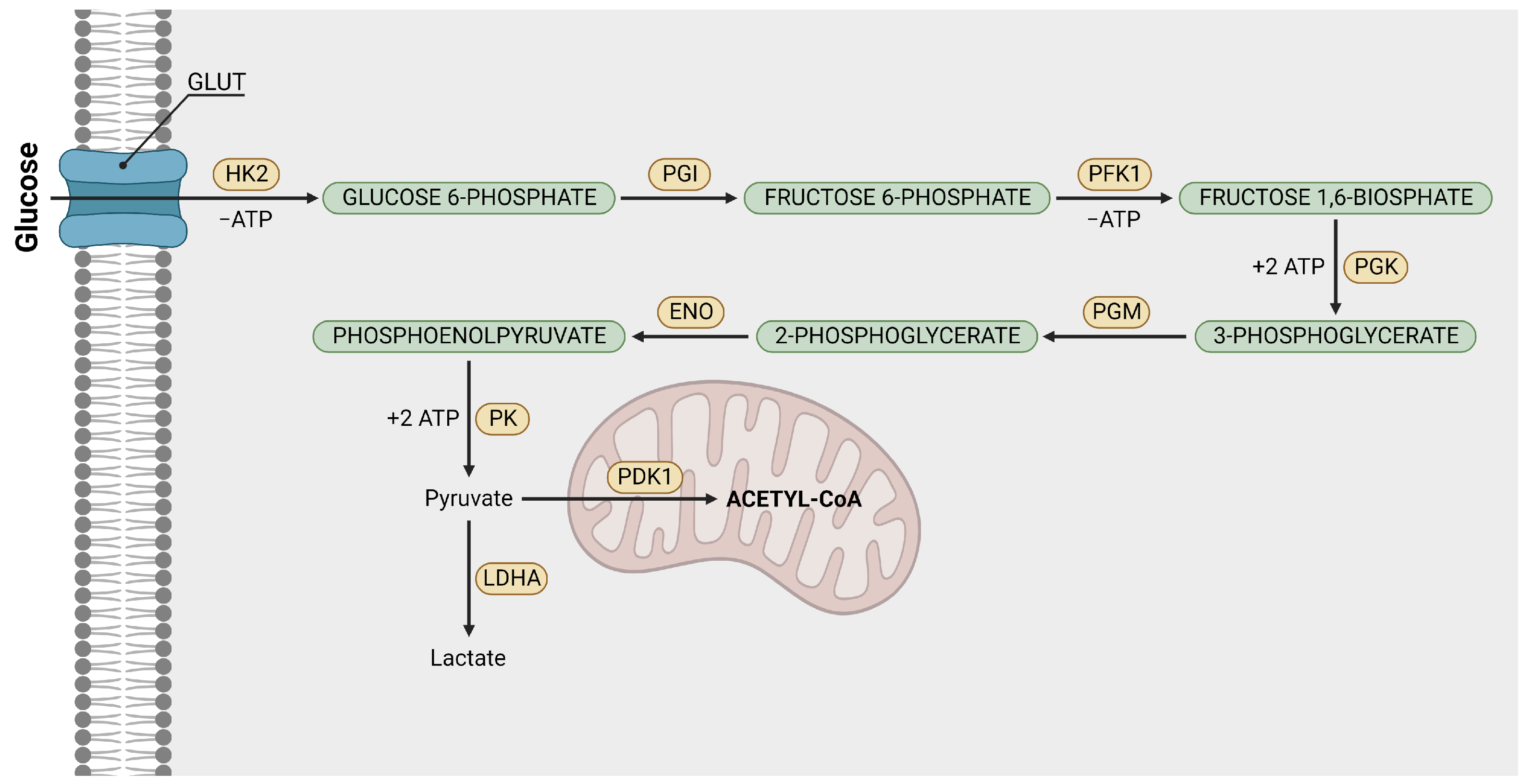
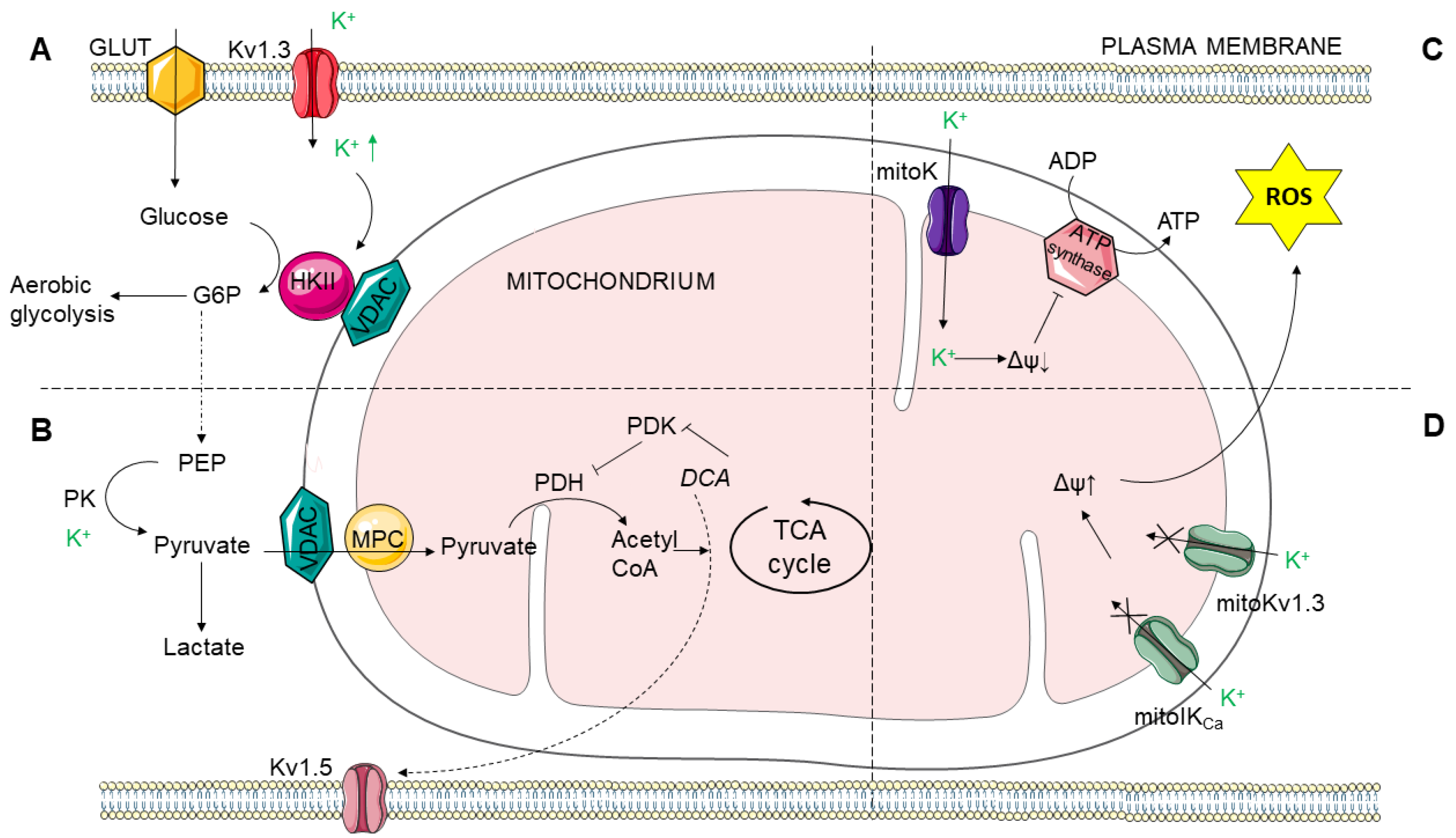
The metabolism of cancer cells is shaped by a variable proportion of oxidative phosphorylation, glycolysis, and glutaminolysis (Figure 1), which depends on the tumor type [36]. Among the typical metabolic features of cancer cells, one can list higher consumption of glucose compared with their non-transformed couplings, enhanced aerobic conversion of pyruvate to lactic acid (“Warburg effect”), and, consequently, excessive production of acidic metabolic products [37,38].
In the following Sections we describe the relationships between the potassium channels’ functioning and the crucial component processes of cancer metabolism. The particular interest is paid on the glycolysis as a preferred source of energy, enhanced glutaminolysis, hampered tricarboxylic acid cycles, and the effects of hypoxia in cancer cells.

2.1. Glycolysis

Glycolysis is considered the primary process of cellular respiration. Simply, it involves breaking glucose into two three-carbon molecules called pyruvate. In general, it is a multistage process which can be summarized as follows:
Glucose + 2 NAD + + 2 ADP + 2 P i 2 Pyruvate + 2 NADH + 2 ATP + 2 H 2 0 + 2 H + .
This process yields two ATP molecules containing free energy, two pyruvate molecules, two high-energy electron-carrying molecules of NADH, and two water molecules. In normal circumstances, generated pyruvate is used in the subsequent stages of cellular respiration to produce more ATPS. These processes (pyruvate oxidation, TCA cycle, oxidative phosphorylation) require, however, the presence of oxygen. Under hypoxic conditions, the pyruvate transforms into lactate, during which NADH drops its electrons off, turning into NAD + . It ensures a constant supply of NAD + needed for glycolysis to keep running.
In cancer cells, the process of glycolysis is similar to that occurring in normal cells under hypoxic conditions (as presented in Figure 2 (left panel)). A hypoxic cellular environment is present in some tumors. Nevertheless, glycolysis may also be predominant in cancer cells even under normoxic conditions (Figure 2 (right panel)). Simultaneously, the consumption of glucose is higher in comparison to normal cells. It results in a higher rate of glycolysis associated with reduced pyruvate oxidation and increased production of lactate. This “injured” form of respiration is called the Warburg effect [39,40] and is considered one of the hallmarks of cancer cells. This persistent aerobic glycolysis can be associated with the activation of oncogenes or the loss of tumor suppressors in certain neoplastic cells [41,42,43,44,45]. The Warburg effect alone does not explain why cancer cells abandon mitochondrial respiration in favor of much less efficient aerobic glycolysis. For comparison, from the mitochondrial oxidative phosphorylation we obtain 36 ATP vs. only 2 ATP generated from glycolysis. It seems, however, that glycolysis is faster and may be more suitable for tumor cells’ vital functions maintenance as long as constant glucose supplies are ensured [44,46]. It may also be advantageous in the process of adaptation to hypoxic conditions in the early phase of tumor development, where access to oxygen is limited. Moreover, glycolytic energy metabolism of tumor cells is beneficial for perpetual proliferation and meeting the high demand for non-essential amino acids, fatty acids, and nucleotides [47]. Alternatively, adjustment to an acidic microenvironment occurring in the tumor cells due to excessive lactate production may also promote the evolution of the glycolytic phenotype [48,49].
Warburg initially explained this impaired form of metabolism as mitochondrial dysfunctionality [50]. Recent findings indicate, however, that the increased rate of ATP production by glycolysis occurs in cells in which mitochondria are not damaged [16,42]. Nowadays, the Warburg effect is considered a consequence of mutations in mitochondrial enzymes, such as fumarate hydratase, succinate dehydrogenase, and isocitrate dehydrogenase, and by excessive production of mitochondrial reactive oxygen species (ROS) [14,51,52,53,54]. It is also regarded as the interplay between the normoxic/hypoxic activation of the transcription factor hypoxia-inducible factor-1 (HIF-1), oncogene activation, loss of function of tumor suppressors, altered signaling pathways and interaction with components of the tumor microenvironment (TME) [17].
In Figure 2, we present a very simplified scheme of aerobic glycolysis as a part of the metabolic pathway in neoplastic cells. In reality, the catabolism of glucose to pyruvate is a complicated process involving 9–10 biochemical steps. The most important ones are presented in Figure 3. The glucose is imported into cell with upregulated GLUT transporters [47]. Then, it is transformed into pyruvate through biochemical reactions catalyzed by enzymes: hexokinases, phosphoglucose isomerase (PGI), phosphofructokinase I (PFK1), phosphoglycerate kinase (PGK), phosphoglycerate mutase (PGM), enolase, pyruvate kinase (PK) and lactate dehydrogenase A (LDHA). Afterwards, pyruvate is transformed into lactate, which is subsequently ejected from the cell with pregulated monocarboxylate transporter 4 (MCT4). Then, it is accumulated in the extracellular space. Simultaneously, mitochondrial pyruvate dehydrogenase kinase 1 (PDK1) impedes the conversion of pyruvate to acetyl-CoA [17].
Having the above-described process in mind, we ask the following question: are we able to prevent tumor growth by forcing cells to produce energy with aerobic metabolism [55,56]? To answer this query, we need to better understand the glycolytic energy metabolism and action mechanism of most crucial enzymes, which become upregulated in most neoplastic cells [57]. One of the possible directions toward the development of novel anticancer strategies is to study the impact of potassium ion channels on aerobic glycolysis. In general, it has been reported that the presence or absence of these channels may affect the activity of enzymes taking part in glycolysis, oxidative phosphorylation, and the tricarboxylic acid (TCA) cycle [58,59]. It, in turn, is associated with an alternating cancer malignancy and influences the rate of cell division, migration, metastasis, migration, and resistance to anti-cancer therapies.
As for glycolysis, one of the most important enzymes catalyzing the process is pyruvate kinase (PK). Its main task consists of the conversion of phosphoenolpyruvate and ADP to pyruvate and ATP (Figure 3) in the presence of ions such as K + or M g 2 + . Therefore, it is likely that abnormal K + channel expression affects this reaction [60]. Another K + –dependent step in aerobic glycolysis is the transformation of glucose into glucose-6-phosphate which is catalyzed by hexokinase II. This enzyme is considered a crucial metabolic switch toward aerobic glycolysis. It has been demonstrated in refs. [27,61] that a decrease in intracellular K + ions impaired aerobic glycolysis and triggered energy stress pathways. The re-addition of the potassium ions could restore the ATPs generation via glycolysis. In ref. [62] it was underlined that the interaction of overexpressed hexokinase I (HKI) with VDAC (Voltage-Dependent Anion Channel) supports GLT-1 (glutamate transporter)-mediated transport activity. Another study confirmed the impact of the K 2 P 3.1 ion channel (considered a representative of the two-pore domain K + channels) on glucose metabolism in lung cancer cells [63]. Results demonstrated that overexpression of K 2 P 3.1 was responsible for diminished glucose uptake and, as a consequence, for a decreased rate of lactate production. Furthermore, it was shown that elevated expression of these potassium channels can lead to the downregulation of GLUT1 and LDHA.
It is also worth mentioning the review [64], which gathers knowledge about relationship between the Warburg effect and PAH (Pulmonary Artery Hypertension). It turns out that, similarly to cancer cells, the metabolism in pulmonary artery cells is altered; the ATPs are obtained through aerobic glycolysis rather than from mitochondrial oxidative phosphorylation. There are several therapeutic strategies that inhibit the development of this disease. One of them involves DCA Dichloroacetate which is an analog of acetic acid the inhibits the action of PDK1 (Figure 3) and enhances oxydative phosphorylation [65]. As previously emphasized, this enzyme impedes the conversion of pyruvate into acetyl-CoA. Nonetheless, DCA can stop this process [65,66,67] in the pulmonary artery as well as in cancer cells through the regulation of potassium channels. It has been demonstrated that, on contrary to normal cells, neoplastic cells are characterized by a high DJm (mitochondrial membrane potential) and low expression of the voltage–dependent potassium channel Kv1.5. Both these factors contribute to apoptosis resistance. Besides impeding the PDK1 (which results in metabolism alteration towards glucose oxidation) DCA decreases DJm, upregulates mitochondrial H 2 O 2 (relatively stable ROS reactive oxygen species), and stimulates the activity of potassium ion channels [13].
Yet in another study, the impact of small-conductance calcium-activated K + (SK) channels was investigated [68]. The authors revealed that CyPPA, an activator of SK channels, slightly reduced the level of mitochondrial respiration in favor of glycolysis and lactate production. In agreement with the studies, it was shown that DCA can reverse the process.
It has also been reported that astrocytes in vitro and in vivo maintain a cytosolic reservoir of lactate, which upregulates the plasma membrane K + ion channels. It results in the fast transport of K + ions to the extracellular space [69].
In ref. [70], it was pointed out that aerobic glycolysis in cancer cells generates an abundance of protons in a gradient across most solid tumors with an acidic core and an alkaline rim. The authors discovered that the rate of cell proliferation depends on extracellular pH. In the example of glioma cells, they demonstrated that changes in pH are detected by H + –sensitive K + ion channels, which translate the changes in pH into changes in membrane potential. These tonically active potassium channels can be blocked by quinine and ruthenium red. The downregulation of K + channels leads to glioma cells depolarization and, consequently, stops their proliferation.
The article [71] focused on the role of the K C a 3.1 channel in Liver Cancer Stem Cells (LCSC). The studies revealed enhanced expression of these potassium channels in LCSCs. The elevated K C a 3.1 led, in turn, to the upregulation of enzymes catalyzing the reactions of glycolysis, such as HK2 or PFK1.
In ref. [72], it was demonstrated that there exists a correlation between the expression of GLUT transporter and the expression of Kv11.1 (hERG) channels in colorectal cancer cells. The presence of hERG turned out to be associated with a lack of GLUT expression and resulted in a worse prognosis for patients suffering from this kind of neoplasm.
The authors of [73] provided direct evidence for interaction between Kir6.2 subunits in rat ventricles and pyruvate kinase. Since the expression of pyruvate kinase is increased in most tumor cells, this interaction may lead to their alternated metabolism.
In search of the potential modulators that shape the relations between ion channels and metabolic pathways in cancer, we paid particular attention to microRNAs. In recent work, Mirzaei and Hamblin cataloged microRNAs, which are associated with the cancer-related changes in glycolytic pathways according to experimental in vitro and/or in vivo models. To find a possible relation between the K + channels and cancer metabolism, we searched for the common microRNAs from the ones presented in [74] and the ones that are anticipated to regulate the expression of potassium channels according to at least two from three independent databases TargetScan [75], MirTarBase [76,77], and MirDB [78]. The results are presented in Table 1.
As one can see in Table 1, the brief investigations allow us to conclude that there are microRNAs, which can simultaneously affect the crucial biomolecules responsible for the metabolic characteristics of cancer (like, e.g., HK1, HK2, LDHA) and the expression of some types of potassium channels. In that way, they can further indirectly modulate the electrical microenvironment of cancer to enhance its progression. What is more, most of the indicated microRNAs can be found in exosomes. Therefore, they can affect cellular pathways that significantly regulate cancer biology (tumor growth, invasion, metastasis, angiogenesis, and its resistance to drugs) [107].

2.2. Mitochondrial Link to Cancer Cell Metabolism of Glucose and Potassium Channels

2.2.1. Mitochondrial Function

Mitochondria serve mainly as the powerhouse of the cell. However, mitochondrial function goes far beyond ATP production. They play prominent roles in the signaling pathways of many cellular processes. In cancer cells, mitochondria undergo many alterations, and their functioning is impaired, partly due to mutations encoding mitochondrial electron transport chain (ETC) complexes. Mitochondrial DNA mutations concerning complex I, III, and IV are well-described for various types of cancer [108]. Mitochondrial dysfunction in cancer cells also involves the impairment of key TCA cycle enzymes. Such enzymes as citrate synthase, aconitase, isocitrate dehydrogenase, succinate dehydrogenase, fumarate hydratase, and malic enzyme are upregulated. Others, such as oxoglutarate dehydrogenase, malate dehydrogenase, or pyruvate dehydrogenase, are downregulated [109]. Eventually, disruption of ETC as well as the TCA cycle leads to mitochondrial dysfunction. It can lead to aberrant mitochondrial ROS production and consequently regulate cancer cell metabolism. Increased ROS production is also correlated with the role of mitochondria in cell death regulation. This regulation involves apoptosis and cytochrome c release from mitochondria, as well as necroptosis and ROS-dependent MPTP activation [110].

2.2.2. Glutaminolysis and the TCA Cycle

The transition processes of glutamine and glucose in rapidly dividing cancer cells are highly combined. Under the influence of glutaminase (GLS), the glutamine is transformed into glutamate, which is further converted to α -ketoglutarate. The latter takes part in energy production through the tricarboxylic acid cycle (the TCA cycle). However, an alternate scenario is also possible, where α -ketoglutarate produced from glutamate in the GDH reaction is used for reductive (counter-clockwise) metabolism in the TCA cycle. The reductive TCA cycle pathway yields isocitrate, citrate, and acetyl-CoA, which serve as substrates for lipogenesis and the synthesis of other biomass in rapidly dividing cancer cells [111,112,113]. These, in turn, support cell growth and viability [114,115,116].
It is well-known that glutaminolysis (when metabolized in the TCA cycle in the oxidative, clockwise, direction) is a valuable energy source in cancer cells. The key enzymes in the process of glutamine metabolism are strongly involved in tumorigenesis [117]. It is also proven that the TCA cycle mediates the inhibition or promotion of tumor progression [118]. For these reasons, glutaminolysis is frequently considered a possible target in cancer therapy [119]. The glutamine additionally participates in the synthesis of glutathione (GSH), which is also localized in mitochondria and has great importance in counteracting reactive oxygen species (ROS). It protects the cellular organelles from the action of ROS [120] and has a proven impact on cancer metabolism [121]. A higher level of ROS is found in almost all cancers [122]. Despite the fact that there is no direct relation between the glutaminolysis and the potassium channels activity, the activation of potassium channels may impact the GSH synthesis and final production of ROS.

2.2.3. Hexokinase

Mitochondrial function is also strongly associated with the function of the crucial enzymes in glucose metabolism. The first critical enzyme in metabolism of glucose is hexokinase. Hexokinase is an enzyme that phosphorylates the glucose and forms glucose-6-phosphate. In humans, there are four hexokinase isoforms, where hexokinase I and II are related to mitochondria and are involved in cancer cell metabolism [123,124]. Hexokinase I and II are associated with the mitochondrial VDAC channel, and they are overexpressed in cancer cells. It was established that mitochondrial hexokinase is coupled with oxidative phosphorylation, and a high level of hexokinase correlates with high glycolytic activity in cancer cells [125]. Thus, hexokinase may serve as a switch between aerobic and anaerobic glycolysis. What is more, it was discovered that the activity of hexokinase II may be regulated by potassium ions and, thus, the channels in the plasma membrane (as presented in Figure 4A). Bischof and coworkers provided a link between Kv1.3 activity and cell metabolism. The increased expression and activation of the Kv1.3 channel leads to a decrease in intracellular K + concentration, which in turn lowers the glycolytic activity of the cell [27]. The mechanism involves hexokinase II, which is highly expressed in cancer cells, and the K + ions, which are essential to maintaining aerobic glycolysis activity.

2.2.4. Pyruvate Kinase

Glucose-6-phosphate is then used to form pyruvate. The pyruvate can later enter either the TCA cycle to fuel oxidative phosphorylation in mitochondria in high oxygen conditions or be used to synthesize lactic acid in the cytoplasm in low oxygen conditions. The enzyme that catalyzes the conversion of phosphoenolpyruvate to the final product of glycolysis- pyruvate, is pyruvate kinase (PK). There are four isoforms of pyruvate kinases. Among them, PKM2 is highly expressed in tumor cells. PKM2 can form either a more active tetramer or a less active dimer. In cancer cells, PKM2 usually forms a less active dimer and promotes the conversion of pyruvate to lactic acid. Therefore, pyruvate kinase can act as a metabolic switch in cancer cells [126]. There is a potential role for potassium channels in the process, as the activity of PK requires K + , along with other monovalent cations [59] (Figure 4B).

2.2.5. Pyruvate Dehydrogenase

Another enzyme that determines whether the metabolic pathway of glucose will lead to lactate production or to the oxidation pathway in mitochondria is pyruvate dehydrogenase (PDH). PDH is an enzyme that catalyzes the conversion of pyruvate to acetyl-CoA, which feeds the TCA cycle in mitochondria, producing electron donors such as NADH and FADH 2 . The activity of PDH is inhibited by PDH kinase (PDK), which phosphorylates PDH. What is interesting, the PDH and PDK activities are related to the functioning of the Kv1.5 potassium channels. It was discovered that inhibition of PDK with dichloroacetate (DCA) promotes glucose oxidation and activates Kv channels in cancer cells, but not in the normal ones [13]. Moreover, cancer cells show lower expression of the Kv1.5 channel, and DCA upregulates its expression in NFAT1-dependent way.

2.2.6. Mitochondrial Potassium Channels

Potassium channels are present not only in the plasma membrane but also in various intracellular compartments and organelles, such as mitochondria. The potassium channels that were discovered in the inner mitochondrial membrane are ATP-sensitive (mitoK A T P ), Ca 2 + -activated large-conductance (mitoBK C a ), Ca 2 + -activated intermediate-conductance (mitoIK C a ), Ca 2 + -activated small-conductance (mitoSK C a ), voltage-gated (mitoKv1.3 and mitoKv7.4), two-pore domain (mitoTASK-3), and sodium-activated (mitoSlo2) potassium channels [127]. According to the literature, mitoKv and mitoIK C a channels play substantial roles in cancer cells [9].
It is well-known that mitochondria play a pivotal role in cell death, while cancer cells escape this general route. Mitochondria-regulated cell death involves mitochondrial permeability transition pore (MPTP), which in turn is regulated by Ca 2 + overload or by ROS overproduction [128,129]. Mitochondrial potassium channels are well known regulators of ROS production (Figure 4C,D). It was found that Kv1.3 and mitoKv1.3 channels are overexpressed in many tumor cells [8]. It was also discovered that inhibition of the mitoKv1.3 channel, either by Bax protein or Kv1.3 channel inhibitors, caused apoptosis in lymphocytes. The mechanism of this phenomenon involved K + -related hyperpolarization of the inner mitochondrial membrane, consequent ROS generation, MPTP opening, and cytochrome c release [130]. The studies on pancreatic ductal adenocarcinoma lines [131] confirm the role of mitoKv1.3 channels in the regulation of cancer cells’ apoptosis via modulation of the ROS-related signalling. The data concerning cancer cells also indicates that inhibition of mitoKv1.3 induces cell death, however, in a Bax-independent way [132]. Similarly to Kv1.3, K C a 3.1 channel is overexpressed in many cancer cells, such as malignant glioma or lung cancer [133,134]. This overexpression is related to promalignant properties of the channel. Moreover, the studies [135,136] confirmed the localisation of the K C a channels in the inner mitochondrial membrane in human colon and lung cancer cells. Likewise the Kv1.3 channels, inhibition of K C a s (along with other anti-cancer drug administration) caused enhancement of apoptosis. The mechanism also involves changes in mitochondrial membrane potential and ROS overproduction [137]. The effect of potassium channels on mitochondria is associated not only with ROS production but also with the regulation of oxidative phosphorylation. In pancreatic ductal adenocarcinoma cells, it was discovered that K C a 3.1 channels regulate oxygen consumption, ATP production, and, consequently, cell proliferation. However, the studies do not clarify whether mitochondrial or plasma membrane channels are involved in this effect [138].
It is well known that mitochondrial potassium channels are involved in many cytoprotective effects. In the work of Malinska et al. [139], authors characterize the association between the mechanisms of the mitochondrial production of ROS and the potassium channels’ activation, which can result in cytoprotection. The activation of K + channels in the inner mitochondrial membrane entails a decrease in mitochondrial membrane potential and, consecutively, may lead to many different phenomena, including the deactivation of the mitochondrial intrinsic apoptosis pathway [140]. The specific mitoK A T P and mitoBK C a channels may play a primary role in cytoprotection [141]. In the context of the glutamine to glutamate transformation, it is worth mentioning the role of the K C a 2.2 (SK2) channels in glutamate toxicity. The activation of these channels prevents mitochondrial super-oxide formation and reduces the risk of cellular apoptosis [142]. The glutamate-induced toxicity can also be reduced by the inhibition of the mitoBK C a channels using a popular BK C a /mitoBK C a channel blocker, paxilline [143].

2.3. Effects of Hypoxia on the Potassium Channels Activity

Hypoxia is a common characteristic of tumors. It occurs when oxygen is not available in sufficient amounts to maintain proper homeostasis at the tissue level. Low availability of oxygen is a complex phenomenon triggered by uncontrolled cell proliferation, during which they quickly consume nutrients and oxygen from the vasculature [144], increased distance from blood vessels, and impaired blood flow caused by cell aggregates. Cancer cells, however, are able to adapt to the altered conditions through neo-angiogenesis, metabolic changes, and metastasis, which allows them to prolong proliferation. These adaptation paths, in turn, are associated with higher resistance of tumors to therapeutics applied and lead to higher aggressiveness of the tumor.
The main role in this process is played by hypoxia-inducible factor 1 (HIF-1). It is a dimeric protein complex regulating the cellular and homeostatic response to hypoxia [145]. It also drives aerobic glycolysis and prevents cells from hypoxia-stress damage [146]. Hypoxia leads to HIF-1 stabilization. Consequently, HIF-1 is translocated to the nucleus, where it takes part in the induction of transcription of multiple genes encoding transporters and enzymes necessary for cancer cells in the adaptation process. The HIF-1 level can be regulated by the potassium channels.
Hypoxia may alter the function of potassium channels either directly by affecting subunits of potassium channels or via various signaling molecules. Hypoxia induces depolarization of membrane potential by inhibiting the activity of several potassium channels. It was documented that hypoxia inhibits potassium channels such as voltage-gated (Kv1.2, Kv1.5, Kv2.1, Kv3.1, Kv3.3, Kv4.2, and Kv9.3) or two-pore domain potassium channel (TASK-1). In hypoxic conditions, these channels are closed, whereas in normoxic conditions, they remain open [147]. In cancer cells, it was shown that Kv3.1 and Kv3.4 are implicated in migration and invasion processes in a HIF-dependent way, involving the ERK (extra cellular signal-regulated kinase) pathway [148]. It is documented that the Kv10.1 channel is involved in the increased HIF-1 alpha expression and activity, which induces tumor angiogenesis via increased vascular endothelial growth factor (VEGF) [149]. Similarly, Kv11.1 is involved in tumor vascularization in colorectal cancer via VEGF secretion in a HIF-dependent way [150]. The action of HIF-1 has also been shown to correlate with the expression of Kv10.1 and Kv11.1 channels in the breast [151].
An intriguing relationship between potassium channels and hypoxia is observed not only for the effect of the channel proteins on second messengers in hypoxia but also for how hypoxia can affect the activity of the potassium channels. This complex interdependency was extensively reviewed recently by Girault et al. [152]. It has been found that hypoxia, either directly or via second messengers such as ROS, can alter the function of potassium channels. The effect on the BK C a channel varies depending on the localization of the channel protein. In plasma membrane, hypoxia inhibits BK C a channel. Whereas in mitochondria, it increases BK C a activity. The inhibitory effect of hypoxia on the potassium channels has also been documented for K 2 P 3.1 (also known as TASK-1). Furthermore, the K 2 P 3.1 channel is related to the changes in nutrient transport and the overexpression of Na + -coupled transporters for mio-inositol, biotin and glutamine, which is involved in metabolism changes [153]. In turn, the overexpressed two pore domain potassium channel KCNK9 gene (encoding K 2 P 9.1) present in human breast and small-cell lung tumors can contribute to the resistance to hypoxic conditions [154]. A possible signalling mechanism linking hypoxia to ion channel function is alternation in ROS production. In neuroblastoma cells it has been shown that hypoxia reduces hERG (Kv11.1) channel by increasing ROS production. Increased ROS level interferes in the association of hERG with Hsp90 (Heat Shock Protein 90) and causes retention of the channel in endoplasmic reticulum [155]. In medulloblastoma the mechanism of Kv2.1 channel inhibition involves HO-1 (heme oxygenase-1; also known as Hsp32). HO-1 upregulation induces CO production and consequent Kv2.1 channel inhibition [156]). Lower activity of Kv1.5 channel in Erwing sarcoma and neuroblastoma cells can be associated with lower Kv1.5 channel expression [157] Another study shows that hydrogen sulphide may be implicated in hypoxia-induced radio resistance. The mechanism involved KATP channel activation by H2S [158].

3. Glycosylation-Dependent Alterations in Properties of Potassium Channels in Cancer Cells

Glycosylation, one of the most common post-translational modifications of proteins, is the enzymatic process of attaching mono- or oligosaccharides to protein molecules. Glycosylation occurs in the endoplasmic reticulum and in the Golgi apparatus. Unlike the processes of transcription and translation, glycosylation is a non-templated process [22]. Therefore, the final structure of the glycans attached to a given protein molecule is a product of the presence of specific glycosyltransferases and glycosidases in the immediate vicinity of the peptide, the availability of substrates, and the overall state of the cell. The most common types of glycosylation are N- and O-glycosylation (where glycans are attached to the protein chain by an N- and O-glycosidic bond, respectively). The asparagine, present in the sequence Asn-X-Thr/Ser (X any amino acid except proline), undergoes N-glycosylation. O-glycans are attached to a serine or threonine residue within the protein molecule (there is no consensus sequence). However, N-glycans have also been shown to be attached within a non-consensus sequence and O-glycans to side chains of amino acids other than Ser or Thr [159]. Oligosaccharides attached to proteins affect their structure, stability, and activity [160]. Sugar structures on the cell surface form a complex layer called the glycocalyx [161]. Glycans attached to macromolecules play crucial roles in cellular processes and intercellular interactions [162]. The alteration in the glycosylation pattern is closely related to physiological and pathological changes that occur in the cell [15].

3.1. Glycosylation in Cancer Cells

The glycosylation pattern of cancer cells differs significantly from that of normal cells [163]. Oligosaccharide structures overrepresented in cancer cells include branched high-mannose N-glycans, truncated O-glycans with a large number of O-GLCNAc (O-linked N-Acetyl-D-glucosamine), glycans with abnormal core fucosylation, and oligosaccharides containing terminal sialic acid [164]. The reasons for the changes in the glycosylation pattern are not yet fully understood. Previous studies suggest that the changes in glycosylation that occur in cancer cells may be related to the expression levels of enzymes involved in glycan processing, the relocation of these enzymes within the endoplasmic reticulum and the Golgi apparatus, and changes in the pH of the Golgi apparatus [165]. Glycan structures found in cancer cells are directly related to cancer development, progression, and metastasis [22]. Sialic acid has a negative charge, which mediates intercellular interactions and signal transduction [166]. Overexpression of sugar antigens (e.g., sialyl-Lewis A antigen) is associated with poor survival in cancer patients [166]. Inhibition of fucosylation leads to inhibition of the proliferation, migration, and invasion of cancer cells [22]. Studies with tunicamycin, an antibiotic that inhibits N-glycosylation, have shown that N-glycans promote tumor growth and metastasis [167].

3.2. Functional Role of Glycans in Potassium Channels

Potassium ion channels are protein tetramers that undergo numerous post-translational modifications. Each channel subunit contains transmembrane segments linked by linkers [168]. Research on the effects of oligosaccharides attached to potassium channels has focused mainly on N-glycosylation, although the K + channels can also undergo O-glycosylation [169]. Most voltage-gated potassium Kv channels have one–two N-glycosylation sites located on the S1–S2 linker or two–three glycosylation sites in the S5-S6 linker [170,171]. Kv channels that lack an N-glycosylation site (e.g., Kv4.2, Kv4.3) are O-glycosylated [169]. Two-pore domain potassium channels (K 2 P ) have one–two N-glycosylation sites, while no O-glycan was found [172]. Inward rectifier potassium channels (Kir) have one, not always occupied, N-glycosylation site [173].
Glycosylation of potassium channels affects their activity, dynamics, stability, and localization [170]. Studies in hamster cells have shown that sialic acid attached by an O-glycosidic bond modulates the gating of Kv4.2 and Kv4.3 potassium channels, but it does not affect their voltage-dependent steady-state inactivation [169]. In contrast, studies of the Kv12.2 channel, which has three N-glycosylation sites within the S5-S6 loop, showed that N-glycans regulate voltage-dependent activation of this channel in a sialic acid-independent manner. The same study also found that N-glycosylation of at least one (any) of the glycosylation sites is required for the expression of the Kv12.2 channel on the cell surface [171]. Similarly, the attachment of a sugar chain within the S1-S2 linker promotes the surface expression and stability of ion channels and directly affects their gating and voltage sensitivity [174]. Studies conducted on wild-type and mutant (lacking N-glycosylation sites) Kv3.1 channels suggest that glycosylation is essential for proper folding and expression of this channel on the cell surface and also affects the opening of the voltage-dependent gate of the Kv3.1 channel [175].
The role of glycans depends on the channel type, and even within the same subfamily, channels may respond differently. For example, studies have shown that the Kv1.5 channel gate is modulated by sialylated N-glycans, while no effect of desialylation/deglycosylation was observed on the Kv1.4 channel gating [176]. The authors suggest that the distinct effects of sialylation on the gating of the Kv1.5 and Kv1.4 channels may be due to the difference in length and amino acid composition of the S1–S2 linker.
The effect of oligosaccharides on potassium channels also depends on the composition and structure of the glycans themselves. Studies on the Kv3.1 channel proteins showed that the localization of these channels within the plasma membrane and their activity vary depending on the type (hybrid or complex) of the attached N-glycan [177]. The effect of glycosylation on the function and stability of Kv channels has been extensively studied, but in other types of potassium channels, attached N-glycans also play notable physiological roles. For example, deglycosylation has been shown to cause a decrease in the number of K 2 P 3.1, K 2 P 9.1, K 2 P 2.1, and K 2 P 17.1 channels in the cell membrane [33,172,178]. Furthermore, changes in the glycosylation of the K 2 P 3.1, K 2 P 2.1, and K 2 P 17.1 channels have been linked to changes in membrane currents [33,172,178]. However, glycosylation does not affect the pH sensitivity of the K 2 P 2.1 channel [178]. Glycosylation in Kir channels affects gating, the magnitude of macroscopic currents, and the number of channels on the cell surface [173,179].

3.3. miRNAs as a Common Element in the Regulation of Potassium Channels and Sialotransferases

The tumor microenvironment plays a pivotal role in cancer progression. One element in the communication of tumor cells with the surrounding microenvironment and with other cells is miRNAs (short non-coding RNA molecules) [180]. MiRNAs are transported between cells by exosomes and can either promote or inhibit tumor growth [181,182]. The activity of potassium ion channels is modulated, among other things, by hypoxia (see Section 2.3) and sialylation (see Section 3.2). At the same time, the level of sialyltransferases, enzymes responsible for attaching sialic acid to glycans, depends on hypoxia [183], and an increase in ST6Gal-I sialyltransferase levels correlates with an increase in HIF-1 factor expression [184]. Additionally, studies suggest that hypoxia is directly related to miRNAs that regulate mitochondrial function [185]. Therefore, we decided to look for miRNAs that, such as hypoxia, can affect the expression of both potassium channels and sialyltransferases. To do this, we searched three algorithmically different databases (TargetScan [75], MirTarBase [76,77] and MirDB [78]). The results, miR indicated for a given sialyltransferase and individual potassium channels by at least two databases, are shown in Table 2. Almost all selected (except miR-135, miR-1297, miR-4319, miR-8485) can be directly linked to hypoxia (Table 2). Furthermore, all of these miRNAs (except miR-4319 and miR-6838) are exosomal miRNAs (Table 2), so these can be extracellular elements that regulate sialylation and potassium channel levels and can also affect cellular pathways associated with hypoxia.

4. Discussion

This review highlights the relations between the potassium channels, glucose metabolism, and glycosylation in cancer cells, which can be useful in deciphering rarely considered aspects of the molecular pathophysiology of oncological diseases. Ion channels emerge as a promising group of proteins that can be considered drug targets in cancer treatment. Let us provide a few examples.

4.1. Pharmacological Modulation of Potassium Channels in Cancer

Several studies have investigated the potential use of potassium channel inhibitors as a therapeutic strategy for cancer. In fact, potassium channels are highly expressed in both the plasma membrane and the inner mitochondrial membrane of many forms of cancer [8]. These channels include the intermediate-conductance calcium-dependent potassium channel (K C a 3.1) expressed in the plasma membrane and inner mitochondrial membrane (mitoK C a 3.1). In particular, blocking mitoK C a 3.1 but not K C a 3.1 with the inhibitor TRAM-34 results in the in vitro death of tumor cells and reduces their metastatic spread in vivo [215]. The role of potassium channels located in the mitochondrial membrane has also been highlighted by the discovery of a new inhibitor of the K 2 P 9.1 (TASK-3) channel. The mitochondriotropic version of this inhibitor (mitoIN-THPP) was seen to decrease the survival of breast cancer cells and kill melanoma cells, whereas IN-THPP was unable to do it, highlighting the importance of potassium channels located in the mitochondrial membrane as privileged pharmacological targets in the therapy of various forms of cancer [216]. Also in chronic lymphocytic leukemia (CLL) Kv1.3 and K C a 3.1 channels are highly expressed both in the plasma and mitochondrial membranes, and inhibiting mitoKv1.3 with PAPTP induces in vitro death of CLL cells, while inhibition of K C a 3.1 with TRAM-34 decreases their proliferation. The action of PAPTP was also exerted on CLL cells resistant to ibrutinib, and PAPTP also enhances the therapeutic action of Venetoclax by acting on mitoKv1.3. PAPTP also decreases CLL in vivo in animal models of CLL [217].
Other potassium channels, such as the voltage-gated potassium channels, are highly expressed in ductal pancreas adenocarcinoma (PDAC), and their blockade could be a useful therapeutic strategy to be added to conventional therapy [218]. Also in breast cancer, in particular the triple-negative type, it has been seen that the expression of potassium channels determines increased in vitro invasion, tumor growth in vivo, and metastases, so it is interesting to reposition already approved potassium channel-blocking drugs for the therapy of this type of particularly aggressive cancer [219,220]. In breast cancer, the expression of potassium channel subfamily K member 6 (K 2 P 6.1) is also increased, which enhances the proliferation, invasion, and migratory capacity of cancer cells. Therefore, K 2 P 6.1 can also be considered a target to be blocked for additional breast cancer therapies [221]. Another interesting finding is the cooperation between ion channels in promoting tumor growth. For example, the chloride intracellular channel 1 (CLIC1) cooperates with the potassium channel Kv10.2 (also called EAG2) in promoting the growth of medulloblastoma cells, and their simultaneous silencing synergistically suppresses tumor growth [222].

4.2. Is It Possible to Reprogram the Cancer Metabolism via Modulation of Potassium Channels?

Due to the characteristic switch in glucose metabolism from oxidative phosphorylation toward aerobic glycolysis, cancer can be considered a metabolic disease [37,38]. Moreover, the metabolic traits seem to depend on the stage of the oncological disease. Many metastases display metabolic differences in comparison with the primary tumors, as summarized in [223]. These changes enable the metastatic tumors to survive and grow in different environments. From this perspective, the idea of anti-glycolytic cancer therapy seems encouraging [224,225,226]. Nevertheless, the development of effective solutions of this kind is not an easy task due to the high energetic adaptability of cancer cells ensuring cell survival in terms of varying availability of energetic substrates, its metabolites, and enzymes for their conversions [16].
As described in this review, there are complex interconnections between potassium channels and metabolic reprogramming in cancer, which suggests that the K + channels are one of the emerging molecular targets in anti-cancer therapies. Among the promising metabolic modulators that affect the functioning of potassium channels, one should mention dicholoroacetate (DCA), which already has been mentioned in Section 2.1. The DCA upregulates mitochondrial Kv1.5 channels by an NFAT1-dependent mechanism in cancer cells, which results in restoring OxPhos and triggering apoptosis [13]. Other interesting substances are mitochondria-targeted derivatives of PAP-1, well-known Kv1.3 channel inhibitor. It was shown that these derivatives, namely PAPTP and PCARBTP, lead to ROS-dependent apoptosis of cancer cells via inhibition of the mitoKv1.3 channel. What’s interesting is that the tested compounds did not affect healthy cells [10].
Considering the perspectives of K + channel modulation in the aim to suppress cancer, one possible approach is to exploit the existing relationships between K + levels and the activity of metabolic enzymes and transcription factors. In this context, the hypoxia-inducible factor 1-alpha can be considered a therapeutic target. It increases glycolytic enzymes’ expression (e.g., LDHA, HKs), and at the same time, its signaling correlates with the expression and activity of Kv10.1 and Kv11.1 channels [149,150,151,155]. Considering enzymes, one should mention the possible down-regulation of hexokinase II via the increased expression and activation of the Kv1.3 channel [27] or KCa3.1 [71]. Lowering HK2 levels can be beneficial due to the decrease in glycolytic activity of cancer cells [27] and reducing their metastasis [225]. The increased production of lactate from pyruvate, which is typical for cancer cells, is gathered by the elevated LDH enzymes (LDHA, LDHB). In particular, LDHA is considered a viable target for drug design and discovery [227,228] since it has a higher affinity for pyruvate, preferentially converting pyruvate to lactate, and NADH to NAD + in anaerobic conditions. According to the studies [63], increased expression of the K 2 P 3.1 channels can lead to down-regulation of LDHA.
As we summarized in Section 2.2, the mitochondrial potassium channels play important roles in the regulation of cell death processes. The molecular mechanism of their involvement is mainly associated with regulation of ROS production (like Kv1.3 channels [8,130,131]), but also they can modulate the oxidative phosphorylation (like K C a 3.1 channels [138]), cytoprotection (like mitoK A T P and mitoBK C a channels [141,143]) and the cellular response to hypoxia (like Kv11.1 channels [149,150,151,155], K C a channels [229] including the BK C a ones [152], and the K 2 P channels: K 2 P 3.1 [153] and K 2 P 9.1 [154]). This may inspire forthcoming research where the K + channels in the mitochondrial membranes can be considered important targets for controlled induction of the pro-death processes in cancer cells.
As one can see, the metabolic changes in tumor cells are gathered with changes in K + channels’ expressions and their transport properties across the plasma- and mitochondrial membranes. Nevertheless, we are still far from a complete understanding of the mechanistic picture of this phenomenon. For example, enhanced investigations on the mechanisms and functions of microRNAs in the ion channel regulation, which can affect cancer metabolism, are needed. They could allow for the discovery of additional prognostic biomarkers as well as support the effective development of novel therapeutic strategies. Here, we presented some new microRNA-oriented directions for further research. In Table 1, we presented the channel types whose expression can be affected by the microRNAs involved in the regulation of glucose metabolism in cancer cells. The presented nine types of microRNAs are the ones that are most probable to interfere with the K + channels, but they are definitely not the only ones that can exhibit mutual relations. Moreover, most of the selected microRNAs from Table 1 have been identified in exosomes. Our approach has a limitation because the cell-type match with the exosomal release of particular microRNAs still needs to be experimentally verified. Nevertheless, the sole possibility of the exosomal delivery of the indicated metabolism- and K + channel-related microRNAs seems promising due to the anticipated profound effects on cell-cell communication that can facilitate tumor growth, invasion, metastasis, angiogenesis, and drug resistance.

4.3. The Therapeutic Potential of Controlling Potassium Channels Glycosylation

The activity and distribution of potassium channels in the cell affect proliferation, cell cycle, apoptosis, and tumor progression [6]. One of the factors that regulates the activity, distribution in the membrane, and stability of K + channels is glycosylation [177]. In particular, the negatively charged sialic acid present in glycans affects the electrical excitability of cells [230]. Altered sialylation occurs in most cancer cells, and the presence of sialic acid-containing tumor antigens correlates with poor survival [231]. Inhibition of sialization reduces metastasis and increases cell sensitivity to chemotherapy and radiotherapy [232]. It has also been shown that an increase in ST6Gal-I sialyltransferase levels correlates with an increase in hypoxia-inducible factor HIF-1 α and HIF-1 α -regulated genes, including glucose transporter genes GLUT1 and GLUT3 and the glycolytic enzyme gene PDHK1 [184]. Selective inhibition of sialotransferases and other enzymes involved in the synthesis of sugar structures overexpressed in cancer cells can stop cancer growth and restore potassium channel activity to normal. Thus, it seems that enzymes involved in glycosylation, particularly sialotransferases, may be great targets for molecular therapy.
Another promising idea is to exploit oligosaccharides attached to potassium channels to deliver potassium channel-specific inhibitors/modulators directly to the channel environment. Such an approach can enable the delivery of appropriate channel modulators and their release it in situ via glycan-mediated pH-dependent click chemistry methodology, similarly to the procedure described in [233].
The main regulatory elements of glycome in the cell are miRNAs [234]. The miRNAs are an interesting potential target for channel therapy in that there are miRNAs correlated with hypoxia that can simultaneously regulate the expression of enzymes involved in oligosaccharide synthesis (Table 2). Moreover, some of these miRNAs also regulate glycolysis (Table 1). Introduced into cells, miRNAs could therefore comprehensively affect both the expression of potassium channels and the metabolism of cancer cells. It is worth noting that most of the miRNAs we identified as miRNAs that could simultaneously regulate the expression of either of the sialotransferases and either of the potassium channels are exosomal miRNAs (Table 2). Exosomes are microvesicles that are part of intercellular communication present in physiological fluids [235]. There is a cell-type-specific mechanism for packaging miRNAs into exosomes [236]. Furthermore, the content of exosomes secreted by normal cells differs from the content of exosomes secreted by cancer cells,; hence, exosomal miRNAs may serve as a readily available biomarker of cancer [237].

4.4. Challenges and Perspectives

Finding new drugs that reprogram cancer metabolism and act on plasma membrane- and mitochondrial channels seems like a viable but challenging idea. Among the most problematic issues, one should mention that most of the relations between potassium channels and the biomolecules involved in the regulation of glucose metabolism are indirect. Thus, the response to channel modulation can be complex and needs an in-depth experimental inspection. Moreover, most types of cancer exhibit metabolic plasticity. Therefore, even if channel activation/blocking could be beneficial from the point of view of its interaction with a particular enzyme/transcription factor, or other mediating molecule (e.g., glucose intake and aerobic glycolysis) cancer cells are able to adapt to unfavorable environmental or cellular conditions by reprogramming their metabolism to survive and sustain proliferation.
Additional challenges in channel-oriented drug design refer to their specificity and possible undesired cytotoxicity outside the tumor. A possible problem with potassium channel blockers/activators is their poor selectivity due to structural similarities between the various channel types. For example, Kv10.1, highly expressed in 70% of human tumors but not in healthy tissue outside the brain, is very similar to Kv11.1 (also called hERG). It is therefore necessary to look for drugs that act with higher selectivity by studying the differences between these channels [238]. Nevertheless, different types of cancer may involve different mechanisms of K + channel activation depending on the origin of tumor. There are several very interesting tissues associated with ion channel specificity in the cancer context. First of all, the environment of cancer, including hypoxia, leads to the changeability of the activation of different channel types. Moreover, an extremely important and not sufficiently studied aspect is the impact of potassium channel mutations in various types of cancer [7]. The expression level of the specific potassium channels is inversely proportional to the level of tumor malignancy in glioma, lymphoma, or colorectal cancer [239,240,241,242].
In this review, we paid much attention to mitochondrial potassium channels. Their druggability is a complex issue [243]. Due to the structural and functional adaptations of the mitoK channels to operate in terms of relatively high membrane potential, high Ca 2 + concentrations, and alkaline pH in the mitochondrial matrix, they display unique regulatory properties. Nevertheless, the specificity of the openers and inhibitors to mitochondrial channel isoforms, the possibility of drug delivery to the mitochondrion and their accumulation within the matrix, as well as precluding drug interactions with the electron transport chain, are challenging problems for further investigations [243].
Another challenge for channel-oriented anti-cancer therapy is to recognize and control the possible non-channel off-targets. Sometimes, the channel modulators act in a pleiotropic manner. For example, the CGS7184, a mitoBK C a channel opener, induces cell death in neuronal cells. This effect is, however, related to the deregulation of calcium homeostasis by CGS7184 via, at least partly, activation of calpain proteases [244]. Analogously, the 3-chloro-4-methoxyphenyl derivative of bromotyramine purpurealidin E [245] being an effective Kv10.1 inhibitor shows dose-dependent cytotoxic and proapoptotic effects. Nevertheless, these effects are detectable both on Kv10.1 expressing- and non-expressing cell lines. Thus, they involve independent mediating biomolecules. Moreover, these cytotoxic and proapoptotic effects were also not restricted to cancer cell lines, which hinders the usage of this Kv10.1 inhibitor in cancer treatment.
It is also important to underline the role of pharmacological modulation of potassium channels to combat the undesirable effects of some drugs used in the treatment of cancer. For example, gemcitabine, commonly used in the treatment of pancreatic cancer and non-small cell lung cancer, may cause cardiotoxicity as it reduces the expression of Kv11.1 (human rapid delayed rectifier potassium channel, hERG). Therefore, by using hERG as a pharmacological target to increase its expression, it can be thought to reduce the cardiotoxicity of gemcitabine [246]. Another example is the drug oxaliplatin, which produces neurotoxicity as an adverse effect. In experimental models, riluzole prevents neurological deficits caused by oxaliplatin due to its action on the potassium channel K 2 P 2.1 (TREK-1) [247].
A possible solution to some of the aforementioned problems is drug repositioning [248]. The drug repurposing technique, which frequently uses artificial intelligence, has also been used to identify drugs blocking the Kv11.1 K + channels that have a role in the proliferation, survival, secretion of angiogenic factors, invasiveness, and metastasis of cells of various tumor types (epithelial, neuronal, leukemic and connective tissue). By using this technique, 26 already approved drugs have been identified, including, for example, bromocriptine, darglitazone, and troglitazone, that have a tumor inhibition effect, although further studies are needed to understand their mechanism [249]. Two other drugs, such as loperamide and amitriptyline, have been identified through a thallium influx-based assay and have been shown to block potassium channels Kv10.1 that induce the proliferation of different tumor cell types [250].
Another possible solution to the existing problems in channel-oriented anti-cancer drug design is to target only the cancer-specific glycosylated channel variants. In such an approach, the particular glycans (e.g., the sialylated N-glycans) can serve as anchoring points for channel modulator-directed chemistries [233]. To enable the utilization of this methodology, several issues should be explored. For example, the appropriate channels’ glycan attachment sites should be recognized to ensure the saccharide-specificity of the glycoproteomic method.
Considering glycosylated forms of ion channels, once again microRNAs deserve a particular scientific interest. According to our literature and database analysis (see Table 2, based on the analysis of TargetScan [75], MirTarBase [76,77], and MirDB [78]), some microRNAs are anticipated to affect both the expression of sialyltransferases responsible for forming sialylated protein glycans in terms of hypoxia and the potassium channels at the transcriptional level. This relation is particularly interesting in the case of the Kv4.3 channels. The glycolysation of their structure is important for cancer biology, as described in Section 3.2. In turn, the analysis of microRNA repositories allowed us to find the possible enzyme involved in binding saccharides to their structures (i.e., alpha-2,8-Sialyltransferase III, ST8SIA3), as well as the regulatory microRNAs (miR-148, miR-152), Table 2. Both miRs are related to hypoxia [194,195] and may be present in cancer-derived exosomes [189,196], which needs, however, experimental validation.

5. Conclusions

In this work, we discussed novel metabolism- and glycome-related aspects of tumor biology, with particular attention paid to the role of the expression and activity of potassium channels. The recognized complex interconnections between the channels’ functioning and metabolic alterations allow us to consider cancer a metabolic disease and a channelopathy. Therefore, the research of detailed mechanisms responsible for the involvement of plasma membrane- and mitochondrial channels in cancer energetics as well as the potential saccharide-mediated drug delivery to cancer-specific ion channel glycans are promising fields to explore in further biochemical and pharmacological investigations. Among the other rising directions for further research, we would like to particularly distinguish the microRNAs as viable cancer biomarkers and potential effective drug targets since they regulate both aspects of cancer biology—the metabolic switch and altered glycosylation, which have been discussed in this work.

Author Contributions

Conceptualization, A.W.-J. and A.L. (Anna Lalik); formal analysis, A.W.-J., A.L. (Anna Lalik) and A.L. (Agnieszka Lukasiak); investigation, A.W.-J., A.L. (Anna Lalik), A.L. (Agnieszka Lukasiak), M.R.-L., P.T. and D.V.D.; resources, A.W.-J., A.L. (Anna Lalik), A.L. (Agnieszka Lukasiak), M.R.-L., P.T., M.E., M.O., S.W. and D.V.D.; writing—original draft preparation, A.W.-J., A.L. (Anna Lalik), A.L. (Agnieszka Lukasiak), M.R.-L., P.T. and D.V.D.; writing—review and editing, A.W.-J., A.L. (Anna Lalik), A.L. (Agnieszka Lukasiak), S.W. and D.V.D.; visualization, A.W.-J., S.W., A.L. (Agnieszka Lukasiak), M.R.-L., A.L. (Anna Lalik) and M.O.; supervision, A.W.-J., A.L. (Anna Lalik), A.L. (Agnieszka Lukasiak) and D.V.D.; project administration, A.W.-J.; funding acquisition, A.W.-J., M.E., M.O. and S.W. All authors have read and agreed to the published version of the manuscript.

Funding

This research was funded by the Silesian University of Technology under the project 31/010/SDU20/0006-10 (to A.W.-J., M.E., M.O. and S.W.).

Institutional Review Board Statement

Not applicable.

Informed Consent Statement

Not applicable.

Data Availability Statement

Not applicable.

Conflicts of Interest

The authors declare no conflict of interest.

Abbreviations

The following abbreviations are used in this manuscript:
Acetyl-CoAacetyl coenzyme A
ADPadenosine diphosphate
ATPadenosine triphosphate
BK C a big conductance Ca 2 + -activated potassium channel
CLIC1chloride intracellular channel 1
DCAdichloroacetate
Enoenolase
ETCelectron transport chain
FADHreduced form of flavin adenine dinucleotide
G6Pglucose-6-phosphate
GDHglutamate dehydrogenase
GLSglutaminase
GLT-1glutamate transporter-1
GSHglutathione
hERGhuman rapid delayed rectifier potassium channel
HIF-1hypoxia-inducible-factor-1
HK1 (HKI)hexokinase-1
HK2 (HKII)hexokinase-2
HO-1heme oxygenase-1
Hsp90Heat Shock Protein 90
IKCaintermediate calcium activated potassium channel
K 2 P two-pore domain potassium channel
K I r inward rectifier potassium channel
K V voltage-gated potassium channel
LDHAlactate dehydrogenase A
LCSCliver cancer stem cells
MCT4monocarboxylate transporter-4
mitoK A T P mitochondrial ATP-sensitive potassium channel
mitoBKmitochondrial big potassium channel
mitoBK C a 2 + Ca 2 + -activated mitochondrial big potassium channel
mitoK A T P ATP-sensitive mitochondrial potassium channel
mitoSK C a Ca 2 + -activated mitochondrial small-conductance potassium channel
mitoSLO2mitochondrial sodium-activated potassium channel
mitoTASK-3mitochondrial TWIK-related acid-sensitive potassium channel
MPCmitochondrial pyruvate carrier
MPTPmitochondrial permeability transition pore
NADHreduced form of Nicotinamide adenine dinucleotide
NOX4NADPH oxidase 4
O-GLCNAcO-linked-N-Acetyl-D-glucosamine
O-GlcNAcylation O-linked-N-acetylglucosaminylation
OxPhosoxidative phosphorylation
PAHpulmonary artery hypertension
PDACductal pancreas adenocarcinoma
PDHpyruvate dehydrogenase
PDK1pyruvate dehydrogenase kinase-1
PDK4pyruvate dehydrogenase kinase-4
PEPphosphoenolopyruvate
PFK16-phosphofructokinase-1
PFKFB36-phosphofructo-2-kinase/fructose-2,6-biphosphatase 3
PGIphosphoglucose isomerase
PGKphoglycerate kinase
PGMphosphoglycerate mutase
PKpyruvate kinase
PKM2pyruvate kinase isoenzyme type M2
PRKAA1AMP-activated, alpha 1 catalytic subunit
ROSreactive oxygen species
TCAtricarboxylic acid (cycle)
SKsmall conductance calcium-activated potassium channel
Sp1transcription factor specificity protein 1
TMEtumor microenvironment
UDP-GlcNAcUDP-N-acetylglucosamine
VDACvoltage-dependent anion channel
VEGFvascular endothelial growth factor

References

  1. Sung, H.; Ferlay, J.; Siegel, R.L.; Laversanne, M.; Soerjomataram, I.; Jemal, A.; Bray, F. Global cancer statistics 2020: GLOBOCAN estimates of incidence and mortality worldwide for 36 cancers in 185 countries. CA Cancer J. Clin. 2021, 71, 209–249. [Google Scholar] [CrossRef] [PubMed]
  2. Prevarskaya, N.; Skryma, R.; Shuba, Y. Ion channels in cancer: Are cancer hallmarks oncochannelopathies? Physiol. Rev. 2018, 98, 559–621. [Google Scholar] [CrossRef] [PubMed]
  3. Leanza, L.; Biasutto, L.; Managò, A.; Gulbins, E.; Zoratti, M.; Szabò, I. Intracellular ion channels and cancer. Front. Physiol. 2013, 4, 227. [Google Scholar] [CrossRef] [PubMed]
  4. Kischel, P.; Girault, A.; Rodat-Despoix, L.; Chamlali, M.; Radoslavova, S.; Abou Daya, H.; Lefebvre, T.; Foulon, A.; Rybarczyk, P.; Hague, F.; et al. Ion channels: New actors playing in chemotherapeutic resistance. Cancers 2019, 11, 376. [Google Scholar] [CrossRef] [PubMed]
  5. Capatina, A.L.; Lagos, D.; Brackenbury, W.J. Targeting ion channels for cancer treatment: Current progress and future challenges. In Targets of Cancer Diagnosis and Treatment; Springer: Berlin/Heidelberg, Germany, 2020; pp. 1–43. [Google Scholar]
  6. Li, M.; Tian, P.; Zhao, Q.; Ma, X.; Zhang, Y. Potassium channels: Novel targets for tumor diagnosis and chemoresistance. Front. Oncol. 2022, 12, 1074469. [Google Scholar] [CrossRef]
  7. Huang, X.; Jan, L.Y. Targeting potassium channels in cancer. J. Cell Biol. 2014, 206, 151–162. [Google Scholar] [CrossRef]
  8. Prosdocimi, E.; Checchetto, V.; Leanza, L. Targeting the mitochondrial potassium channel KV1.3 to kill cancer cells: Drugs, strategies, and new perspectives. SLAS DISCOVERY: Adv. Life Sci. R&D 2019, 24, 882–892. [Google Scholar]
  9. Szabo, I.; Zoratti, M.; Biasutto, L. Targeting mitochondrial ion channels for cancer therapy. Redox Biol. 2021, 42, 101846. [Google Scholar] [CrossRef]
  10. Leanza, L.; Romio, M.; Becker, K.A.; Azzolini, M.; Trentin, L.; Managò, A.; Venturini, E.; Zaccagnino, A.; Mattarei, A.; Carraretto, L.; et al. Direct pharmacological targeting of a mitochondrial ion channel selectively kills tumor cells in vivo. Cancer Cell 2017, 31, 516–531. [Google Scholar] [CrossRef]
  11. Bugan, I.; Kucuk, S.; Karagoz, Z.; Fraser, S.P.; Kaya, H.; Dodson, A.; Foster, C.S.; Altun, S.; Djamgoz, M.B. Anti-metastatic effect of ranolazine in an in vivo rat model of prostate cancer, and expression of voltage-gated sodium channel protein in human prostate. Prostate Cancer Prostatic Dis. 2019, 22, 569–579. [Google Scholar] [CrossRef]
  12. Driffort, V.; Gillet, L.; Bon, E.; Marionneau-Lambot, S.; Oullier, T.; Joulin, V.; Collin, C.; Pagès, J.C.; Jourdan, M.L.; Chevalier, S.; et al. Ranolazine inhibits Na v 1.5-mediated breast cancer cell invasiveness and lung colonization. Mol. Cancer 2014, 13, 264. [Google Scholar] [CrossRef]
  13. Bonnet, S.; Archer, S.L.; Allalunis-Turner, J.; Haromy, A.; Beaulieu, C.; Thompson, R.; Lee, C.T.; Lopaschuk, G.D.; Puttagunta, L.; Bonnet, S.; et al. A mitochondria-K+ channel axis is suppressed in cancer and its normalization promotes apoptosis and inhibits cancer growth. Cancer Cell 2007, 11, 37–51. [Google Scholar] [CrossRef]
  14. Seyfried, T.N.; Arismendi-Morillo, G.; Mukherjee, P.; Chinopoulos, C. On the origin of ATP synthesis in cancer. Iscience 2020, 23, 101761. [Google Scholar] [CrossRef]
  15. Reily, C.; Stewart, T.J.; Renfrow, M.B.; Novak, J. Glycosylation in health and disease. Nat. Rev. Nephrol. 2019, 15, 346–366. [Google Scholar] [CrossRef]
  16. Shiratori, R.; Furuichi, K.; Yamaguchi, M.; Miyazaki, N.; Aoki, H.; Chibana, H.; Ito, K.; Aoki, S. Glycolytic suppression dramatically changes the intracellular metabolic profile of multiple cancer cell lines in a mitochondrial metabolism-dependent manner. Sci. Rep. 2019, 9, 18699. [Google Scholar] [CrossRef]
  17. Vaupel, P.; Multhoff, G. Revisiting the Warburg effect: Historical dogma versus current understanding. J. Physiol. 2021, 599, 1745–1757. [Google Scholar] [CrossRef]
  18. Pollard, P.; Wortham, N.; Tomlinson, I. The TCA cycle and tumorigenesis: The examples of fumarate hydratase and succinate dehydrogenase. Ann. Med. 2003, 35, 634–635. [Google Scholar] [CrossRef]
  19. Gottlieb, E.; Tomlinson, I.P. Mitochondrial tumour suppressors: A genetic and biochemical update. Nat. Rev. Cancer 2005, 5, 857–866. [Google Scholar] [CrossRef]
  20. Locasale, J.W.; Grassian, A.R.; Melman, T.; Lyssiotis, C.A.; Mattaini, K.R.; Bass, A.J.; Heffron, G.; Metallo, C.M.; Muranen, T.; Sharfi, H.; et al. Phosphoglycerate dehydrogenase diverts glycolytic flux and contributes to oncogenesis. Nat. Genet. 2011, 43, 869–874. [Google Scholar] [CrossRef]
  21. Dang, L.; White, D.W.; Gross, S.; Bennett, B.D.; Bittinger, M.A.; Driggers, E.M.; Fantin, V.R.; Jang, H.G.; Jin, S.; Keenan, M.C.; et al. Cancer-associated IDH1 mutations produce 2-hydroxyglutarate. Nature 2009, 462, 739–744. [Google Scholar] [CrossRef]
  22. Thomas, D.; Rathinavel, A.K.; Radhakrishnan, P. Altered glycosylation in cancer: A promising target for biomarkers and therapeutics. Biochim. Biophys. Acta (BBA)-Rev. Cancer 2021, 1875, 188464. [Google Scholar] [CrossRef] [PubMed]
  23. Berois, N.; Pittini, A.; Osinaga, E. Targeting tumor glycans for cancer therapy: Successes, limitations, and perspectives. Cancers 2022, 14, 645. [Google Scholar] [CrossRef] [PubMed]
  24. Abdel Rahman, A.M.; Ryczko, M.; Pawling, J.; Dennis, J.W. Probing the hexosamine biosynthetic pathway in human tumor cells by multitargeted tandem mass spectrometry. ACS Chem. Biol. 2013, 8, 2053–2062. [Google Scholar] [CrossRef]
  25. Nagel, A.K.; Ball, L.E. Intracellular protein O-GlcNAc modification integrates nutrient status with transcriptional and metabolic regulation. Adv. Cancer Res. 2015, 126, 137–166. [Google Scholar] [PubMed]
  26. Lee, J.B.; Pyo, K.H.; Kim, H.R. Role and function of O-GlcNAcylation in cancer. Cancers 2021, 13, 5365. [Google Scholar] [CrossRef]
  27. Bischof, H.; Burgstaller, S.; Springer, A.; Matt, L.; Rauter, T.; Bachkönig, O.A.; Schmidt, T.; Groschner, K.; Schindl, R.; Madl, T.; et al. Potassium ions promote hexokinase-II dependent glycolysis. Iscience 2021, 24, 102346. [Google Scholar] [CrossRef]
  28. Purushothaman, A.; Mohajeri, M.; Lele, T.P. The role of glycans in the mechanobiology of cancer. J. Biol. Chem. 2023, 299, 102935. [Google Scholar] [CrossRef]
  29. Dart, C. Symposium Review: Lipid microdomains and the regulation of ion channel function. J. Physiol. 2010, 588, 3169–3178. [Google Scholar] [CrossRef]
  30. Corradi, V.; Sejdiu, B.I.; Mesa-Galloso, H.; Abdizadeh, H.; Noskov, S.Y.; Marrink, S.J.; Tieleman, D.P. Emerging diversity in lipid–protein interactions. Chem. Rev. 2019, 119, 5775–5848. [Google Scholar] [CrossRef]
  31. Hall, M.K.; Cartwright, T.A.; Fleming, C.M.; Schwalbe, R.A. Importance of glycosylation on function of a potassium channel in neuroblastoma cells. PLoS ONE 2011, 6, e19317. [Google Scholar] [CrossRef]
  32. Sprovieri, P.; Martino, G. The role of the carbohydrates in plasmatic membrane. Physiol. Res. 2018, 67, 1–11. [Google Scholar] [CrossRef]
  33. Mant, A.; Williams, S.; Roncoroni, L.; Lowry, E.; Johnson, D.; O’Kelly, I. N-glycosylation-dependent control of functional expression of background potassium channels K2P3. 1 and K2P9. 1. J. Biol. Chem. 2013, 288, 3251–3264. [Google Scholar] [CrossRef]
  34. Zhu, J.; Yan, J.; Thornhill, W.B. N-glycosylation promotes the cell surface expression of Kv1. 3 potassium channels. FEBS J. 2012, 279, 2632–2644. [Google Scholar] [CrossRef]
  35. Baycin-Hizal, D.; Gottschalk, A.; Jacobson, E.; Mai, S.; Wolozny, D.; Zhang, H.; Krag, S.S.; Betenbaugh, M.J. Physiologic and pathophysiologic consequences of altered sialylation and glycosylation on ion channel function. Biochem. Biophys. Res. Commun. 2014, 453, 243–253. [Google Scholar] [CrossRef]
  36. Sonveaux, P.; Végran, F.; Schroeder, T.; Wergin, M.C.; Verrax, J.; Rabbani, Z.N.; De Saedeleer, C.J.; Kennedy, K.M.; Diepart, C.; Jordan, B.F.; et al. Targeting lactate-fueled respiration selectively kills hypoxic tumor cells in mice. J. Clin. Investig. 2008, 118, 3930–3942. [Google Scholar] [CrossRef]
  37. Gyamfi, J.; Kim, J.; Choi, J. Cancer as a metabolic disorder. Int. J. Mol. Sci. 2022, 23, 1155. [Google Scholar] [CrossRef]
  38. Seyfried, T.N.; Flores, R.E.; Poff, A.M.; D’Agostino, D.P. Cancer as a metabolic disease: Implications for novel therapeutics. Carcinogenesis 2014, 35, 515–527. [Google Scholar] [CrossRef]
  39. Warburg, O. On the origin of cancer cells. Science 1956, 123, 309–314. [Google Scholar] [CrossRef]
  40. Dang, C.V. Links between metabolism and cancer. Genes Dev. 2012, 26, 877–890. [Google Scholar] [CrossRef]
  41. Vander Heiden, M.G.; Cantley, L.C.; Thompson, C.B. Understanding the Warburg effect: The metabolic requirements of cell proliferation. Science 2009, 324, 1029–1033. [Google Scholar] [CrossRef]
  42. Koppenol, W.H.; Bounds, P.L.; Dang, C.V. Otto Warburg’s contributions to current concepts of cancer metabolism. Nat. Rev. Cancer 2011, 11, 325–337. [Google Scholar] [CrossRef] [PubMed]
  43. Levine, A.J.; Puzio-Kuter, A.M. The control of the metabolic switch in cancers by oncogenes and tumor suppressor genes. Science 2010, 330, 1340–1344. [Google Scholar] [CrossRef] [PubMed]
  44. Cairns, R.A.; Harris, I.S.; Mak, T.W. Regulation of cancer cell metabolism. Nat. Rev. Cancer 2011, 11, 85–95. [Google Scholar] [CrossRef] [PubMed]
  45. Hsu, P.P.; Sabatini, D.M. Cancer cell metabolism: Warburg and beyond. Cell 2008, 134, 703–707. [Google Scholar] [CrossRef]
  46. Semenza, G.L.; Artemov, D.; Bedi, A.; Bhujwalla, Z.; Chiles, K.; Feldser, D.; Laughner, E.; Ravi, R.; Simons, J.; Taghavi, P.; et al. ‘The metabolism of tumours’: 70 years later. In The Tumour Microenvironment: Causes and Consequences of Hypoxia and Acidity: Novartis Foundation Symposium 240; Wiley Online Library: Hoboken, NJ, USA, 2001; Volume 240, pp. 251–264. [Google Scholar]
  47. Li, X.b.; Gu, J.d.; Zhou, Q.h. Review of aerobic glycolysis and its key enzymes—New targets for lung cancer therapy. Thorac. Cancer 2015, 6, 17–24. [Google Scholar] [CrossRef]
  48. Gatenby, R.A.; Gillies, R.J. Why do cancers have high aerobic glycolysis? Nat. Rev. Cancer 2004, 4, 891–899. [Google Scholar] [CrossRef]
  49. Gillies, R.J.; Robey, I.; Gatenby, R.A. Causes and consequences of increased glucose metabolism of cancers. J. Nucl. Med. 2008, 49, 24S–42S. [Google Scholar] [CrossRef]
  50. Seyfried, T.N.; Shelton, L.M. Cancer as a metabolic disease. Nutr. Metab. 2010, 7, 7. [Google Scholar] [CrossRef]
  51. Ratcliffe, P.J. Fumarate hydratase deficiency and cancer: Activation of hypoxia signaling? Cancer Cell 2007, 11, 303–305. [Google Scholar] [CrossRef]
  52. Rasheed, M.R.H.A.; Tarjan, G. Succinate dehydrogenase complex: An updated review. Arch. Pathol. Lab. Med. 2018, 142, 1564–1570. [Google Scholar] [CrossRef]
  53. Smith, D.G.; Sturmey, R.G. Parallels between embryo and cancer cell metabolism. Biochem. Soc. Trans. 2013, 41, 664–669. [Google Scholar] [CrossRef]
  54. King, A.; Selak, M.; Gottlieb, E. Succinate dehydrogenase and fumarate hydratase: Linking mitochondrial dysfunction and cancer. Oncogene 2006, 25, 4675–4682. [Google Scholar] [CrossRef]
  55. Pelicano, H.; Martin, D.; Xu, R.; Huang, P. Glycolysis inhibition for anticancer treatment. Oncogene 2006, 25, 4633–4646. [Google Scholar] [CrossRef]
  56. Pan, J.G.; Mak, T.W. Metabolic targeting as an anticancer strategy: Dawn of a new era? Sci. STKE 2007, 2007, pe14. [Google Scholar] [CrossRef]
  57. Altenberg, B.; Greulich, K. Genes of glycolysis are ubiquitously overexpressed in 24 cancer classes. Genomics 2004, 84, 1014–1020. [Google Scholar] [CrossRef]
  58. Bygrave, F.L. The ionic environment and metabolic control. Nature 1967, 214, 667–671. [Google Scholar] [CrossRef]
  59. Gohara, D.W.; Di Cera, E. Molecular mechanisms of enzyme activation by monovalent cations. J. Biol. Chem. 2016, 291, 20840–20848. [Google Scholar] [CrossRef]
  60. Burgstaller, S.; Bischof, H.; Matt, L.; Lukowski, R. Assessing K+ ions and K+ channel functions in cancer cell metabolism using fluorescent biosensors. Free. Radic. Biol. Med. 2022, 181, 43–51. [Google Scholar] [CrossRef]
  61. Bischof, H.; Burgstaller, S.; Graier, W.F.; Lukowski, R.; Malli, R. Unveiling the K+-sensitivity of cell metabolism using genetically encoded, FRET-based K+, glucose, and ATP biosensors. STAR Protoc. 2021, 2, 100843. [Google Scholar] [CrossRef]
  62. Jackson, J.G.; O’Donnell, J.C.; Krizman, E.; Robinson, M.B. Displacing hexokinase from mitochondrial voltage-dependent anion channel impairs GLT-1-mediated glutamate uptake but does not disrupt interactions between GLT-1 and mitochondrial proteins. J. Neurosci. Res. 2015, 93, 999–1008. [Google Scholar] [CrossRef]
  63. Lin, G.; Lin, L.; Lin, H.; Chen, W.; Chen, L.; Chen, X.; Chen, S.; Lin, Q.; Xu, Y.; Zeng, Y. KCNK3 inhibits proliferation and glucose metabolism of lung adenocarcinoma via activation of AMPK-TXNIP pathway. Cell Death Discov. 2022, 8, 360. [Google Scholar] [CrossRef] [PubMed]
  64. Liu, N.; Parry, S.; Xiao, Y.; Zhou, S.; Liu, Q. Molecular targets of the Warburg effect and inflammatory cytokines in the pathogenesis of pulmonary artery hypertension. Clin. Chim. Acta 2017, 466, 98–104. [Google Scholar] [CrossRef] [PubMed]
  65. Bersin, R.M.; Stacpoole, P.W. Dichloroacetate as metabolic therapy for myocardial ischemia and failure. Am. Heart J. 1997, 134, 841–855. [Google Scholar] [CrossRef] [PubMed]
  66. Michelakis, E.; Sutendra, G.; Dromparis, P.; Webster, L.; Haromy, A.; Niven, E.; Maguire, C.; Gammer, T.L.; Mackey, J.; Fulton, D.; et al. Metabolic modulation of glioblastoma with dichloroacetate. Sci. Transl. Med. 2010, 2, 31ra34. [Google Scholar] [CrossRef] [PubMed]
  67. McMurtry, M.S.; Bonnet, S.; Wu, X.; Dyck, J.R.; Haromy, A.; Hashimoto, K.; Michelakis, E.D. Dichloroacetate prevents and reverses pulmonary hypertension by inducing pulmonary artery smooth muscle cell apoptosis. Circ. Res. 2004, 95, 830–840. [Google Scholar] [CrossRef]
  68. Krabbendam, I.E.; Honrath, B.; Dilberger, B.; Iannetti, E.F.; Branicky, R.S.; Meyer, T.; Evers, B.; Dekker, F.J.; Koopman, W.J.; Beyrath, J.; et al. SK channel-mediated metabolic escape to glycolysis inhibits ferroptosis and supports stress resistance in C. elegans. Cell Death Dis. 2020, 11, 263. [Google Scholar] [CrossRef]
  69. Sotelo-Hitschfeld, T.; Niemeyer, M.I.; Machler, P.; Ruminot, I.; Lerchundi, R.; Wyss, M.T.; Stobart, J.; Fernández-Moncada, I.; Valdebenito, R.; Garrido-Gerter, P.; et al. Channel-mediated lactate release by K+-stimulated astrocytes. J. Neurosci. 2015, 35, 4168–4178. [Google Scholar] [CrossRef]
  70. Honasoge, A.; Shelton, K.A.; Sontheimer, H. Autocrine regulation of glioma cell proliferation via pHe-sensitive K+ channels. Am. J. Physiol. Cell Physiol. 2014, 306, C493–C505. [Google Scholar] [CrossRef]
  71. Fan, J.; Tian, R.; Yang, X.; Wang, H.; Shi, Y.; Fan, X.; Zhang, J.; Chen, Y.; Zhang, K.; Chen, Z.; et al. KCNN4 promotes the stemness potentials of liver cancer stem cells by enhancing glucose metabolism. Int. J. Mol. Sci. 2022, 23, 6958. [Google Scholar] [CrossRef]
  72. Lastraioli, E.; Bencini, L.; Bianchini, E.; Romoli, M.R.; Crociani, O.; Giommoni, E.; Messerini, L.; Gasperoni, S.; Moretti, R.; Di Costanzo, F.; et al. hERG1 channels and Glut-1 as independent prognostic indicators of worse outcome in stage I and II colorectal cancer: A pilot study. Transl. Oncol. 2012, 5, 105–112. [Google Scholar] [CrossRef]
  73. Ying, H.; Kimmelman, A.C.; Lyssiotis, C.A.; Hua, S.; Chu, G.C.; Fletcher-Sananikone, E.; Locasale, J.W.; Son, J.; Zhang, H.; Coloff, J.L.; et al. Oncogenic Kras maintains pancreatic tumors through regulation of anabolic glucose metabolism. Cell 2012, 149, 656–670. [Google Scholar] [CrossRef]
  74. Mirzaei, H.; Hamblin, M.R. Regulation of glycolysis by non-coding RNAs in cancer: Switching on the Warburg effect. Mol. Ther.-Oncolytics 2020, 19, 218–239. [Google Scholar] [CrossRef]
  75. McGeary, S.E.; Lin, K.S.; Shi, C.Y.; Pham, T.M.; Bisaria, N.; Kelley, G.M.; Bartel, D.P. The biochemical basis of microRNA targeting efficacy. Science 2019, 366, eaav1741. [Google Scholar] [CrossRef]
  76. Huang, H.Y.; Lin, Y.C.D.; Li, J.; Huang, K.Y.; Shrestha, S.; Hong, H.C.; Tang, Y.; Chen, Y.G.; Jin, C.N.; Yu, Y.; et al. miRTarBase 2020: Updates to the experimentally validated microRNA–target interaction database. Nucleic Acids Res. 2020, 48, D148–D154. [Google Scholar] [CrossRef]
  77. Huang, H.Y.; Lin, Y.C.D.; Cui, S.; Huang, Y.; Tang, Y.; Xu, J.; Bao, J.; Li, Y.; Wen, J.; Zuo, H.; et al. mmiRTarBase update 2022: An informative resource for experimentally validated miRNA–target interactions. Nucleic Acids Res. 2022, 50, D222–D230. [Google Scholar] [CrossRef]
  78. Chen, Y.; Wang, X. miRDB: An online database for prediction of functional microRNA targets. Nucleic Acids Res. 2020, 48, D127–D131. [Google Scholar] [CrossRef]
  79. Snezhkina, A.; Krasnov, G.; Zhikrivetskaya, S.; Karpova, I.Y.; Fedorova, M.; Nyushko, K.; Belyakov, M.; Gnuchev, N.; Sidorov, D.; Alekseev, B.Y.; et al. Overexpression of microRNAs miR-9,-98, and-199 correlates with the downregulation of HK2 expression in colorectal cancer. Mol. Biol. 2018, 52, 190–199. [Google Scholar] [CrossRef]
  80. Song, W.; Chen, Y.; Zhu, G.; Xie, H.; Yang, Z.; Li, L. Exosome-mediated miR-9-5p promotes proliferation and migration of renal cancer cells both in vitro and in vivo by targeting SOCS4. Biochem. Biophys. Res. Commun. 2020, 529, 1216–1224. [Google Scholar] [CrossRef]
  81. Riancho, J.; Vázquez-Higuera, J.L.; Pozueta, A.; Lage, C.; Kazimierczak, M.; Bravo, M.; Calero, M.; Gonalezález, A.; Rodríguez, E.; Lleó, A.; et al. MicroRNA profile in patients with Alzheimer’s disease: Analysis of miR-9-5p and miR-598 in raw and exosome enriched cerebrospinal fluid samples. J. Alzheimer’S Dis. 2017, 57, 483–491. [Google Scholar] [CrossRef]
  82. Lu, J.; Wang, L.; Chen, W.; Wang, Y.; Zhen, S.; Chen, H.; Cheng, J.; Zhou, Y.; Li, X.; Zhao, L. miR-603 targeted hexokinase-2 to inhibit the malignancy of ovarian cancer cells. Arch. Biochem. Biophys. 2019, 661, 1–9. [Google Scholar] [CrossRef]
  83. Ling, Z.; Liu, D.; Zhang, G.; Liang, Q.; Xiang, P.; Xu, Y.; Han, C.; Tao, T. miR-361-5p modulates metabolism and autophagy via the Sp1-mediated regulation of PKM2 in prostate cancer. Oncol. Rep. 2017, 38, 1621–1628. [Google Scholar] [CrossRef]
  84. Tao, Y.; Zhou, J.; Wang, Z.; Tao, H.; Bai, J.; Ge, G.; Li, W.; Zhang, W.; Hao, Y.; Yang, X.; et al. Human bone mesenchymal stem cells-derived exosomal miRNA-361-5p alleviates osteoarthritis by downregulating DDX20 and inactivating the NF-κB signaling pathway. Bioorg. Chem. 2021, 113, 104978. [Google Scholar] [CrossRef] [PubMed]
  85. Huang, P.; Mao, L.f.; Zhang, Z.p.; Lv, W.w.; Feng, X.p.; Liao, H.j.; Dong, C.; Kaluba, B.; Tang, X.f.; Chang, S. Down-regulated miR-125a-5p promotes the reprogramming of glucose metabolism and cell malignancy by increasing levels of CD147 in thyroid cancer. Thyroid 2018, 28, 613–623. [Google Scholar] [CrossRef] [PubMed]
  86. Lv, A.; Tu, Z.; Huang, Y.; Lu, W.; Xie, B. Circulating exosomal miR-125a-5p as a novel biomarker for cervical cancer. Oncol. Lett. 2021, 21, 54. [Google Scholar] [CrossRef] [PubMed]
  87. Ding, M.; Pei, Y.; Zhang, C.; Qi, Y.; Xia, J.; Hao, C.; Yao, W. Exosomal miR-125a-5p regulates T lymphocyte subsets to promote silica-induced pulmonary fibrosis by targeting TRAF6. Ecotoxicol. Environ. Saf. 2023, 249, 114401. [Google Scholar] [CrossRef]
  88. Xueya, Z.; Yamei, L.; Sha, C.; Dan, C.; Hong, S.; Xingyu, Y.; Weiwei, C. Exosomal encapsulation of miR-125a-5p inhibited trophoblast cell migration and proliferation by regulating the expression of VEGFA in preeclampsia. Biochem. Biophys. Res. Commun. 2020, 525, 646–653. [Google Scholar] [CrossRef]
  89. Wang, D.; Hao, C.; Zhang, L.; Zhang, J.; Liu, S.; Li, Y.; Qu, Y.; Zhao, Y.; Huang, R.; Wei, J.; et al. Exosomal miR-125a-5p derived from silica-exposed macrophages induces fibroblast transdifferentiation. Ecotoxicol. Environ. Saf. 2020, 192, 110253. [Google Scholar] [CrossRef]
  90. Hui, L.; Zhang, J.; Guo, X. MiR-125b-5p suppressed the glycolysis of laryngeal squamous cell carcinoma by down-regulating hexokinase-2. Biomed. Pharmacother. 2018, 103, 1194–1201. [Google Scholar] [CrossRef]
  91. Cao, J.Y.; Wang, B.; Tang, T.T.; Wen, Y.; Li, Z.L.; Feng, S.T.; Wu, M.; Liu, D.; Yin, D.; Ma, K.L.; et al. Exosomal miR-125b-5p deriving from mesenchymal stem cells promotes tubular repair by suppression of p53 in ischemic acute kidney injury. Theranostics 2021, 11, 5248. [Google Scholar] [CrossRef]
  92. Shen, K.; Wang, X.; Wang, Y.; Jia, Y.; Zhang, Y.; Wang, K.; Luo, L.; Cai, W.; Li, J.; Li, S.; et al. miR-125b-5p in adipose derived stem cells exosome alleviates pulmonary microvascular endothelial cells ferroptosis via Keap1/Nrf2/GPX4 in sepsis lung injury. Redox Biol. 2023, 62, 102655. [Google Scholar] [CrossRef]
  93. Wu, Q.Q.; Zheng, B.; Weng, G.B.; Yang, H.M.; Ren, Y.; Weng, X.J.; Zhang, S.W.; Zhu, W.Z. Downregulated NOX4 underlies a novel inhibitory role of microRNA-137 in prostate cancer. J. Cell. Biochem. 2019, 120, 10215–10227. [Google Scholar] [CrossRef]
  94. Jiang, Y.; Liu, J.; Chen, L.; Jin, Y.; Zhang, G.; Lin, Z.; Du, S.; Fu, Z.; Chen, T.; Qin, Y.; et al. Serum secreted miR-137-containing exosomes affects oxidative stress of neurons by regulating OXR1 in Parkinson’s disease. Brain Res. 2019, 1722, 146331. [Google Scholar] [CrossRef]
  95. Zhang, D.; Cai, G.; Liu, K.; Zhuang, Z.; Jia, K.; Pei, S.; Wang, X.; Wang, H.; Xu, S.; Cui, C.; et al. Microglia exosomal miRNA-137 attenuates ischemic brain injury through targeting Notch1. Aging 2021, 13, 4079. [Google Scholar] [CrossRef]
  96. Lv, N.; Hao, S.; Luo, C.; Abukiwan, A.; Hao, Y.; Gai, F.; Huang, W.; Huang, L.; Xiao, X.; Eichmuller, S.B.; et al. miR-137 inhibits melanoma cell proliferation through downregulation of GLO1. Sci. China Life Sci. 2018, 61, 541–549. [Google Scholar] [CrossRef]
  97. Ferguson, S.W.; Wang, J.; Lee, C.J.; Liu, M.; Neelamegham, S.; Canty, J.M.; Nguyen, J. The microRNA regulatory landscape of MSC-derived exosomes: A systems view. Sci. Rep. 2018, 8, 1419. [Google Scholar] [CrossRef]
  98. Zhou, W.; Xu, M.; Wang, Z.; Yang, M. Engineered exosomes loaded with miR-449a selectively inhibit the growth of homologous non-small cell lung cancer. Cancer Cell Int. 2021, 21, 485. [Google Scholar] [CrossRef]
  99. Guo, C.J.; Cao, X.L.; Zhang, Y.F.; Yue, K.Y.; Han, J.; Yan, H.; Han, H.; Zheng, M.H. Exosome-mediated inhibition of microRNA-449a promotes the amplification of mouse retinal progenitor cells and enhances their transplantation in retinal degeneration mouse models. Mol. Ther.-Nucleic Acids 2023, 31, 763–778. [Google Scholar] [CrossRef]
  100. Hua, S.; Lei, L.; Deng, L.; Weng, X.; Liu, C.; Qi, X.; Wang, S.; Zhang, D.; Zou, X.; Cao, C.; et al. miR-139-5p inhibits aerobic glycolysis, cell proliferation, migration, and invasion in hepatocellular carcinoma via a reciprocal regulatory interaction with ETS1. Oncogene 2018, 37, 1624–1636. [Google Scholar] [CrossRef]
  101. Liu, H.; Wang, F.; Zhang, Y.; Xing, Y.; Wang, Q. Exosomal microRNA-139-5p from mesenchymal stem cells accelerates trophoblast cell invasion and migration by motivation of the ERK/MMP-2 pathway via downregulation of protein tyrosine phosphatase. J. Obstet. Gynaecol. Res. 2020, 46, 2561–2572. [Google Scholar] [CrossRef]
  102. Sun, H.; Dai, J.; Chen, M.; Chen, Q.; Xie, Q.; Zhang, W.; Li, G.; Yan, M. miR-139-5p Was Identified as Biomarker of Different Molecular Subtypes of Breast Carcinoma. Front. Oncol. 2022, 12, 857714. [Google Scholar] [CrossRef]
  103. Sun, K.; Hu, P.; Xu, F. LINC00152/miR-139-5p regulates gastric cancer cell aerobic glycolysis by targeting PRKAA1. Biomed. Pharmacother. 2018, 97, 1296–1302. [Google Scholar] [CrossRef] [PubMed]
  104. Weng, Y.; Shen, Y.; He, Y.; Pan, X.; Xu, J.; Jiang, Y.; Zhang, Q.; Wang, S.; Kong, F.; Zhao, S.; et al. The miR-15b-5p/PDK4 axis regulates osteosarcoma proliferation through modulation of the Warburg effect. Biochem. Biophys. Res. Commun. 2018, 503, 2749–2757. [Google Scholar] [CrossRef] [PubMed]
  105. Yadava, S.M.; Feng, A.; Parobchak, N.; Wang, B.; Rosen, T. miR-15b-5p promotes expression of proinflammatory cytokines in human placenta by inhibiting Apelin signaling pathway. Placenta 2021, 104, 8–15. [Google Scholar] [CrossRef] [PubMed]
  106. Ghafouri-Fard, S.; Khoshbakht, T.; Hussen, B.M.; Jamal, H.H.; Taheri, M.; Hajiesmaeili, M. A comprehensive review on function of miR-15b-5p in malignant and non-malignant disorders. Front. Oncol. 2022, 12, 1874. [Google Scholar] [CrossRef] [PubMed]
  107. Li, C.; Zhou, T.; Chen, J.; Li, R.; Chen, H.; Luo, S.; Chen, D.; Cai, C.; Li, W. The role of Exosomal miRNAs in cancer. J. Transl. Med. 2022, 20, 6. [Google Scholar] [CrossRef]
  108. Chatterjee, A.; Mambo, E.; Sidransky, D. Mitochondrial DNA mutations in human cancer. Oncogene 2006, 25, 4663–4674. [Google Scholar] [CrossRef]
  109. Gaude, E.; Frezza, C. Defects in mitochondrial metabolism and cancer. Cancer Metab. 2014, 2, 10. [Google Scholar] [CrossRef]
  110. Grasso, D.; Zampieri, L.X.; Capelôa, T.; Van de Velde, J.A.; Sonveaux, P. Mitochondria in cancer. Cell Stress 2020, 4, 114. [Google Scholar] [CrossRef]
  111. DeBerardinis, R.J.; Mancuso, A.; Daikhin, E.; Nissim, I.; Yudkoff, M.; Wehrli, S.; Thompson, C.B. Beyond aerobic glycolysis: Transformed cells can engage in glutamine metabolism that exceeds the requirement for protein and nucleotide synthesis. Proc. Natl. Acad. Sci. USA 2007, 104, 19345–19350. [Google Scholar] [CrossRef]
  112. Zhang, G.F.; Jensen, M.V.; Gray, S.M.; El, K.; Wang, Y.; Lu, D.; Becker, T.C.; Campbell, J.E.; Newgard, C.B. Reductive TCA cycle metabolism fuels glutamine-and glucose-stimulated insulin secretion. Cell Metab. 2021, 33, 804–817. [Google Scholar] [CrossRef]
  113. Metallo, C.M.; Gameiro, P.A.; Bell, E.L.; Mattaini, K.R.; Yang, J.; Hiller, K.; Jewell, C.M.; Johnson, Z.R.; Irvine, D.J.; Guarente, L.; et al. Reductive glutamine metabolism by IDH1 mediates lipogenesis under hypoxia. Nature 2012, 481, 380–384. [Google Scholar] [CrossRef]
  114. Jiang, L.; Shestov, A.A.; Swain, P.; Yang, C.; Parker, S.J.; Wang, Q.A.; Terada, L.S.; Adams, N.D.; McCabe, M.T.; Pietrak, B.; et al. Reductive carboxylation supports redox homeostasis during anchorage-independent growth. Nature 2016, 532, 255–258. [Google Scholar] [CrossRef]
  115. Wise, D.R.; Ward, P.S.; Shay, J.E.; Cross, J.R.; Gruber, J.J.; Sachdeva, U.M.; Platt, J.M.; DeMatteo, R.G.; Simon, M.C.; Thompson, C.B. Hypoxia promotes isocitrate dehydrogenase-dependent carboxylation of α-ketoglutarate to citrate to support cell growth and viability. Proc. Natl. Acad. Sci. USA 2011, 108, 19611–19616. [Google Scholar] [CrossRef]
  116. Mullen, A.R.; Wheaton, W.W.; Jin, E.S.; Chen, P.H.; Sullivan, L.B.; Cheng, T.; Yang, Y.; Linehan, W.M.; Chandel, N.S.; DeBerardinis, R.J. Reductive carboxylation supports growth in tumour cells with defective mitochondria. Nature 2012, 481, 385–388. [Google Scholar] [CrossRef]
  117. Masisi, B.K.; El Ansari, R.; Alfarsi, L.; Rakha, E.A.; Green, A.R.; Craze, M.L. The role of glutaminase in cancer. Histopathology 2020, 76, 498–508. [Google Scholar] [CrossRef]
  118. Eniafe, J.; Jiang, S. The functional roles of TCA cycle metabolites in cancer. Oncogene 2021, 40, 3351–3363. [Google Scholar] [CrossRef]
  119. Jin, L.; Alesi, G.; Kang, S. Glutaminolysis as a target for cancer therapy. Oncogene 2016, 35, 3619–3625. [Google Scholar] [CrossRef]
  120. Fernandez-Checa, J.C.; Kaplowitz, N.; Garcia-Ruiz, C.; Colell, A.; Miranda, M.; MARi, M.; Ardite, E.; Morales, A. GSH transport in mitochondria: Defense against TNF-induced oxidative stress and alcohol-induced defect. Am. J. Physiol.-Gastrointest. Liver Physiol. 1997, 273, G7–G17. [Google Scholar] [CrossRef]
  121. Estrela, J.M.; Ortega, A.; Obrador, E. Glutathione in cancer biology and therapy. Crit. Rev. Clin. Lab. Sci. 2006, 43, 143–181. [Google Scholar] [CrossRef]
  122. Liou, G.Y.; Storz, P. Reactive oxygen species in cancer. Free. Radic. Res. 2010, 44, 479–496. [Google Scholar] [CrossRef]
  123. Abu-Hamad, S.; Zaid, H.; Israelson, A.; Nahon, E.; Shoshan-Barmatz, V. Hexokinase-I protection against apoptotic cell death is mediated via interaction with the voltage-dependent anion channel-1: Mapping the site of binding. J. Biol. Chem. 2008, 283, 13482–13490. [Google Scholar] [CrossRef] [PubMed]
  124. Mathupala, S.; Ko, Y.; Pedersen, P. Hexokinase II: Cancer’s double-edged sword acting as both facilitator and gatekeeper of malignancy when bound to mitochondria. Oncogene 2006, 25, 4777–4786. [Google Scholar] [CrossRef] [PubMed]
  125. Bustamante, E.; Pedersen, P.L. High aerobic glycolysis of rat hepatoma cells in culture: Role of mitochondrial hexokinase. Proc. Natl. Acad. Sci. USA 1977, 74, 3735–3739. [Google Scholar] [CrossRef] [PubMed]
  126. Zahra, K.; Dey, T.; Mishra, S.P.; Pandey, U. Pyruvate kinase M2 and cancer: The role of PKM2 in promoting tumorigenesis. Front. Oncol. 2020, 10, 159. [Google Scholar] [CrossRef] [PubMed]
  127. Szewczyk, A.; Bednarczyk, P.; Jedraszko, J.; Kampa, R.P.; Koprowski, P.; Krajewska, M.; Kucman, S.; Kulawiak, B.; Laskowski, M.; Rotko, D.; et al. Mitochondrial potassium channels—An overview. Postep. Biochem. 2018, 64, 196–212. [Google Scholar] [CrossRef] [PubMed]
  128. Cui, Y.; Pan, M.; Ma, J.; Song, X.; Cao, W.; Zhang, P. Recent progress in the use of mitochondrial membrane permeability transition pore in mitochondrial dysfunction-related disease therapies. Mol. Cell. Biochem. 2021, 476, 493–506. [Google Scholar] [CrossRef] [PubMed]
  129. Bonora, M.; Patergnani, S.; Ramaccini, D.; Morciano, G.; Pedriali, G.; Kahsay, A.E.; Bouhamida, E.; Giorgi, C.; Wieckowski, M.R.; Pinton, P. Physiopathology of the permeability transition pore: Molecular mechanisms in human pathology. Biomolecules 2020, 10, 998. [Google Scholar] [CrossRef]
  130. Szabó, I.; Bock, J.; Grassmé, H.; Soddemann, M.; Wilker, B.; Lang, F.; Zoratti, M.; Gulbins, E. Mitochondrial potassium channel Kv1. 3 mediates Bax-induced apoptosis in lymphocytes. Proc. Natl. Acad. Sci. USA 2008, 105, 14861–14866. [Google Scholar] [CrossRef]
  131. Peruzzo, R.; Mattarei, A.; Romio, M.; Paradisi, C.; Zoratti, M.; Szabò, I.; Leanza, L. Regulation of proliferation by a mitochondrial potassium channel in pancreatic ductal adenocarcinoma cells. Front. Oncol. 2017, 7, 239. [Google Scholar] [CrossRef]
  132. Leanza, L.; Henry, B.; Sassi, N.; Zoratti, M.; Chandy, K.G.; Gulbins, E.; Szabò, I. Inhibitors of mitochondrial Kv1. 3 channels induce Bax/Bak-independent death of cancer cells. EMBO Mol. Med. 2012, 4, 577–593. [Google Scholar] [CrossRef]
  133. Turner, K.L.; Honasoge, A.; Robert, S.M.; McFerrin, M.M.; Sontheimer, H. A proinvasive role for the Ca2+-activated K+ channel KCa3. 1 in malignant glioma. Glia 2014, 62, 971–981. [Google Scholar] [CrossRef]
  134. Bulk, E.; Ay, A.S.; Hammadi, M.; Ouadid-Ahidouch, H.; Schelhaas, S.; Hascher, A.; Rohde, C.; Thoennissen, N.H.; Wiewrodt, R.; Schmidt, E.; et al. Epigenetic dysregulation of KCa3. 1 channels induces poor prognosis in lung cancer. Int. J. Cancer 2015, 137, 1306–1317. [Google Scholar] [CrossRef]
  135. De Marchi, U.; Sassi, N.; Fioretti, B.; Catacuzzeno, L.; Cereghetti, G.M.; Szabò, I.; Zoratti, M. Intermediate conductance Ca2+-activated potassium channel (KCa3. 1) in the inner mitochondrial membrane of human colon cancer cells. Cell Calcium 2009, 45, 509–516. [Google Scholar] [CrossRef]
  136. Bulk, E.; Todesca, L.M.; Bachmann, M.; Szabo, I.; Rieke, M.; Schwab, A. Functional expression of mitochondrial KCa3. 1 channels in non-small cell lung cancer cells. Pflugers Arch.-Eur. J. Physiol. 2022, 474, 1147–1157. [Google Scholar] [CrossRef]
  137. Bauer, D.; Werth, F.; Nguyen, H.A.; Kiecker, F.; Eberle, J. Critical role of reactive oxygen species (ROS) for synergistic enhancement of apoptosis by vemurafenib and the potassium channel inhibitor TRAM-34 in melanoma cells. Cell Death Dis. 2017, 8, e2594. [Google Scholar] [CrossRef]
  138. Kovalenko, I.; Glasauer, A.; Schockel, L.; Sauter, D.R.; Ehrmann, A.; Sohler, F.; Hagebarth, A.; Novak, I.; Christian, S. Identification of KCa3. 1 channel as a novel regulator of oxidative phosphorylation in a subset of pancreatic carcinoma cell lines. PLoS ONE 2016, 11, e0160658. [Google Scholar] [CrossRef]
  139. Malinska, D.; Mirandola, S.R.; Kunz, W.S. Mitochondrial potassium channels and reactive oxygen species. FEBS Lett. 2010, 584, 2043–2048. [Google Scholar] [CrossRef]
  140. Wallace, D.C. Mitochondria and cancer. Nat. Rev. Cancer 2012, 12, 685–698. [Google Scholar] [CrossRef]
  141. Laskowski, M.; Augustynek, B.; Kulawiak, B.; Koprowski, P.; Bednarczyk, P.; Jarmuszkiewicz, W.; Szewczyk, A. What do we not know about mitochondrial potassium channels? Biochim. Biophys. Acta (BBA)-Bioenerg. 2016, 1857, 1247–1257. [Google Scholar] [CrossRef]
  142. Garthwaite, G.; Williams, G.D.; Garthwaite, J. Glutamate toxicity: An experimental and theoretical analysis. Eur. J. Neurosci. 1992, 4, 353–360. [Google Scholar] [CrossRef]
  143. Kulawiak, B.; Szewczyk, A. Glutamate-induced cell death in HT22 mouse hippocampal cells is attenuated by paxilline, a BK channel inhibitor. Mitochondrion 2012, 12, 169–172. [Google Scholar] [CrossRef]
  144. Iorio, J.; Petroni, G.; Duranti, C.; Lastraioli, E. Potassium and sodium channels and the Warburg effect: Biophysical regulation of cancer metabolism. Bioelectricity 2019, 1, 188–200. [Google Scholar] [CrossRef] [PubMed]
  145. Ziello, J.E.; Jovin, I.S.; Huang, Y. Hypoxia-Inducible Factor (HIF)-1 regulatory pathway and its potential for therapeutic intervention in malignancy and ischemia. Yale J. Biol. Med. 2007, 80, 51. [Google Scholar] [PubMed]
  146. Eales, K.L.; Hollinshead, K.E.; Tennant, D.A. Hypoxia and metabolic adaptation of cancer cells. Oncogenesis 2016, 5, e190. [Google Scholar] [CrossRef] [PubMed]
  147. Patel, A.; Honore, E. Molecular physiology of oxygen-sensitive potassium channels. Eur. Respir. J. 2001, 18, 221–227. [Google Scholar] [CrossRef] [PubMed]
  148. Song, M.S.; Park, S.M.; Park, J.S.; Byun, J.H.; Jin, H.J.; Seo, S.H.; Ryu, P.D.; Lee, S.Y. Kv3.1 and Kv3.4, are involved in cancer cell migration and invasion. Int. J. Mol. Sci. 2018, 19, 1061. [Google Scholar] [CrossRef]
  149. Downie, B.R.; Sánchez, A.; Knötgen, H.; Contreras-Jurado, C.; Gymnopoulos, M.; Weber, C.; Stühmer, W.; Pardo, L.A. Eag1 expression interferes with hypoxia homeostasis and induces angiogenesis in tumors. J. Biol. Chem. 2008, 283, 36234–36240. [Google Scholar] [CrossRef]
  150. Crociani, O.; Zanieri, F.; Pillozzi, S.; Lastraioli, E.; Stefanini, M.; Fiore, A.; Fortunato, A.; D’Amico, M.; Masselli, M.; De Lorenzo, E.; et al. hERG1 channels modulate integrin signaling to trigger angiogenesis and tumor progression in colorectal cancer. Sci. Rep. 2013, 3, 3308. [Google Scholar] [CrossRef]
  151. Lai, Q.; Wang, T.; Guo, Q.; Zhang, Y.; Wang, Y.; Yuan, L.; Ling, R.; He, Y.; Wang, W. Positive correlation between the expression of hEag1 and HIF-1α in breast cancers: An observational study. BMJ Open 2014, 4, e005049. [Google Scholar] [CrossRef]
  152. Girault, A.; Ahidouch, A.; Ouadid-Ahidouch, H. Roles for Ca2+ and K+ channels in cancer cells exposed to the hypoxic tumour microenvironment. Biochim. Biophys. Acta (BBA)-Mol. Cell Res. 2020, 1867, 118644. [Google Scholar] [CrossRef]
  153. Leithner, K.; Hirschmugl, B.; Li, Y.; Tang, B.; Papp, R.; Nagaraj, C.; Stacher, E.; Stiegler, P.; Lindenmann, J.; Olschewski, A.; et al. TASK-1 regulates apoptosis and proliferation in a subset of non-small cell lung cancers. PLoS ONE 2016, 11, e0157453. [Google Scholar] [CrossRef]
  154. Mu, D.; Chen, L.; Zhang, X.; See, L.H.; Koch, C.M.; Yen, C.; Tong, J.J.; Spiegel, L.; Nguyen, K.C.; Servoss, A.; et al. Genomic amplification and oncogenic properties of the KCNK9 potassium channel gene. Cancer Cell 2003, 3, 297–302. [Google Scholar] [CrossRef]
  155. Vaddi, D.R.; Piao, L.; Khan, S.A.; Wang, N.; Prabhakar, N.R.; Nanduri, J. Hypoxia induced hERG trafficking defect linked to cell cycle arrest in SH-SY5Y cells. PLoS ONE 2019, 14, e0215905. [Google Scholar] [CrossRef]
  156. Al-Owais, M.M.; Scragg, J.L.; Dallas, M.L.; Boycott, H.E.; Warburton, P.; Chakrabarty, A.; Boyle, J.P.; Peers, C. Carbon monoxide mediates the anti-apoptotic effects of heme oxygenase-1 in medulloblastoma DAOY cells via K+ channel inhibition. J. Biol. Chem. 2012, 287, 24754–24764. [Google Scholar] [CrossRef]
  157. Ryland, K.E.; Svoboda, L.K.; Vesely, E.D.; McIntyre, J.C.; Zhang, L.; Martens, J.R.; Lawlor, E.R. Polycomb-dependent repression of the potassium channel-encoding gene KCNA5 promotes cancer cell survival under conditions of stress. Oncogene 2015, 34, 4591–4600. [Google Scholar] [CrossRef]
  158. Zhang, J.; Xie, Y.; Xu, Y.; Pan, Y.; Shao, C. Hydrogen sulfide contributes to hypoxia-induced radioresistance on hepatoma cells. J. Radiat. Res. 2011, 52, 622–628. [Google Scholar] [CrossRef]
  159. Dutta, D.; Mandal, C.; Mandal, C. Unusual glycosylation of proteins: Beyond the universal sequon and other amino acids. Biochim. Biophys. Acta (BBA)-Gen. Subj. 2017, 1861, 3096–3108. [Google Scholar] [CrossRef]
  160. Patel, C.; Saad, H.; Shenkman, M.; Lederkremer, G.Z. Oxidoreductases in glycoprotein glycosylation, folding, and ERAD. Cells 2020, 9, 2138. [Google Scholar] [CrossRef]
  161. Cohen, M.; Varki, A. Modulation of glycan recognition by clustered saccharide patches. Int. Rev. Cell Mol. Biol. 2014, 308, 75–125. [Google Scholar]
  162. Barchi Jr, J.J.; Strain, C.N. The effect of a methyl group on structure and function: Serine vs. threonine glycosylation and phosphorylation. Front. Mol. Biosci. 2023, 10, 1117850. [Google Scholar] [CrossRef]
  163. Mereiter, S.; Balmaña, M.; Campos, D.; Gomes, J.; Reis, C.A. Glycosylation in the era of cancer-targeted therapy: Where are we heading? Cancer Cell 2019, 36, 6–16. [Google Scholar] [CrossRef] [PubMed]
  164. Doud, E.H.; Yeh, E.S. Mass Spectrometry-Based Glycoproteomic Workflows for Cancer Biomarker Discovery. Technol. Cancer Res. Treat. 2023, 22, 15330338221148811. [Google Scholar] [CrossRef] [PubMed]
  165. Pearce, O.M. Cancer glycan epitopes: Biosynthesis, structure and function. Glycobiology 2018, 28, 670–696. [Google Scholar] [CrossRef] [PubMed]
  166. Guo, Y.; Jia, W.; Yang, J.; Zhan, X. Cancer glycomics offers potential biomarkers and therapeutic targets in the framework of 3P medicine. Biomol. Modif. -Endocr.-Relat. Cancers 2023, 16648714, 80. [Google Scholar] [CrossRef] [PubMed]
  167. Peric, L.; Vukadin, S.; Petrovic, A.; Kuna, L.; Puseljic, N.; Sikora, R.; Rozac, K.; Vcev, A.; Smolic, M. Glycosylation Alterations in Cancer Cells, Prognostic Value of Glycan Biomarkers and Their Potential as Novel Therapeutic Targets in Breast Cancer. Biomedicines 2022, 10, 3265. [Google Scholar] [CrossRef]
  168. Capera, J.; Serrano-Novillo, C.; Navarro-Pérez, M.; Cassinelli, S.; Felipe, A. The potassium channel odyssey: Mechanisms of traffic and membrane arrangement. Int. J. Mol. Sci. 2019, 20, 734. [Google Scholar] [CrossRef]
  169. Schwetz, T.A.; Norring, S.A.; Ednie, A.R.; Bennett, E.S. Sialic acids attached to O-glycans modulate voltage-gated potassium channel gating. J. Biol. Chem. 2011, 286, 4123–4132. [Google Scholar] [CrossRef]
  170. Lazniewska, J.; Weiss, N. The “Sweet” Side of Ion Channels. In Reviews of Physiology, Biochemistry and Pharmacology; Springer: Berlin/Heidelberg, Germany, 2014; Volume 167, pp. 67–114. [Google Scholar]
  171. Noma, K.; Kimura, K.; Minatohara, K.; Nakashima, H.; Nagao, Y.; Mizoguchi, A.; Fujiyoshi, Y. Triple N-glycosylation in the long S5-P loop regulates the activation and trafficking of the Kv12. 2 potassium channel. J. Biol. Chem. 2009, 284, 33139–33150. [Google Scholar] [CrossRef]
  172. Wiedmann, F.; Schlund, D.; Voigt, N.; Ratte, A.; Kraft, M.; Katus, H.A.; Schmidt, C. N-glycosylation–dependent regulation of hK2P17. 1 currents. Mol. Biol. Cell 2019, 30, 1425–1436. [Google Scholar] [CrossRef]
  173. Carrington, S.J.; Hernandez, C.C.; Swale, D.R.; Aluko, O.A.; Denton, J.S.; Cone, R.D. G protein–coupled receptors differentially regulate glycosylation and activity of the inwardly rectifying potassium channel Kir7. 1. J. Biol. Chem. 2018, 293, 17739–17753. [Google Scholar] [CrossRef]
  174. Lopez-Rodriguez, A.; Holmgren, M. Deglycosylation of Shaker KV channels affects voltage sensing and the open–closed transition. J. Gen. Physiol. 2018, 150, 1025–1034. [Google Scholar] [CrossRef]
  175. Brooks, N.L.; Corey, M.J.; Schwalbe, R.A. Characterization of N-glycosylation consensus sequences in the Kv3. 1 channel. FEBS J. 2006, 273, 3287–3300. [Google Scholar] [CrossRef]
  176. Schwetz, T.A.; Norring, S.A.; Bennett, E.S. N-glycans modulate Kv1. 5 gating but have no effect on Kv1. 4 gating. Biochim. Biophys. Acta (BBA)-Biomembr. 2010, 1798, 367–375. [Google Scholar] [CrossRef]
  177. Hall, M.K.; Weidner, D.A.; Dayal, S.; Pak, E.; Murashov, A.K.; Schwalbe, R.A. Membrane Distribution and Activity of a Neuronal Voltage-Gated K+ Channel is Modified by Replacement of Complex Type N-Glycans with Hybrid Type. J. Glycobiol. 2017, 6, 128. [Google Scholar]
  178. Wiedmann, F.; Schlund, D.; Faustino, F.; Kraft, M.; Ratte, A.; Thomas, D.; Katus, H.A.; Schmidt, C. N-glycosylation of TREK-1/hK2P2. 1 two-pore-domain potassium (K2P) channels. Int. J. Mol. Sci. 2019, 20, 5193. [Google Scholar] [CrossRef]
  179. Pabon, A.; Chan, K.W.; Sui, J.L.; Wu, X.; Logothetis, D.E.; Thornhill, W.B. Glycosylation of GIRK1 at Asn119 and ROMK1 at Asn117 has different consequences in potassium channel function. J. Biol. Chem. 2000, 275, 30677–30682. [Google Scholar] [CrossRef]
  180. Patil, N.; Allgayer, H.; Leupold, J.H. MicroRNAs in the tumor microenvironment. Tumor Microenviron. Mol. Play. Part B 2020, 1277, 1–31. [Google Scholar]
  181. Tang, Y.; Zong, S.; Zeng, H.; Ruan, X.; Yao, L.; Han, S.; Hou, F. MicroRNAs and angiogenesis: A new era for the management of colorectal cancer. Cancer Cell Int. 2021, 21, 221. [Google Scholar] [CrossRef]
  182. Zhou, X.; Liu, Q.; Wang, X.; Yao, X.; Zhang, B.; Wu, J.; Sun, C. Exosomal ncRNAs facilitate interactive ‘dialogue’between tumor cells and tumor-associated macrophages. Cancer Lett. 2022, 552, 215975. [Google Scholar] [CrossRef]
  183. Hassinen, A.; Khoder-Agha, F.; Khosrowabadi, E.; Mennerich, D.; Harrus, D.; Noel, M.; Dimova, E.Y.; Glumoff, T.; Harduin-Lepers, A.; Kietzmann, T.; et al. A Golgi-associated redox switch regulates catalytic activation and cooperative functioning of ST6Gal-I with B4GalT-I. Redox Biol. 2019, 24, 101182. [Google Scholar] [CrossRef]
  184. Jones, R.B.; Dorsett, K.A.; Hjelmeland, A.B.; Bellis, S.L. The ST6Gal-I sialyltransferase protects tumor cells against hypoxia by enhancing HIF-1α signaling. J. Biol. Chem. 2018, 293, 5659–5667. [Google Scholar] [CrossRef] [PubMed]
  185. Rencelj, A.; Gvozdenovic, N.; Cemazar, M. MitomiRs: Their roles in mitochondria and importance in cancer cell metabolism. Radiol. Oncol. 2021, 55, 379–392. [Google Scholar] [CrossRef] [PubMed]
  186. Xue, G.; Yan, H.; Zhang, Y.; Hao, L.; Zhu, X.; Mei, Q.; Sun, S. c-Myc-mediated repression of miR-15-16 in hypoxia is induced by increased HIF-2α and promotes tumor angiogenesis and metastasis by upregulating FGF2. Oncogene 2015, 34, 1393–1406. [Google Scholar] [CrossRef] [PubMed]
  187. Luo, F.; Guo, W.; Liu, W. Exosomes derived from bone marrow mesenchymal stem cells inhibit human aortic vascular smooth muscle cells calcification via the miR-15a/15b/16/NFATc3/OCN axis. Biochem. Biophys. Res. Commun. 2022, 635, 65–76. [Google Scholar] [CrossRef]
  188. Li, C.; Li, Y.; Lu, Y.; Niu, Z.; Zhao, H.; Peng, Y.; Li, M. miR-26 family and its target genes in tumorigenesis and development. Crit. Rev. Oncol. 2021, 157, 103124. [Google Scholar] [CrossRef]
  189. Chettimada, S.; Lorenz, D.R.; Misra, V.; Wolinsky, S.M.; Gabuzda, D. Small RNA sequencing of extracellular vesicles identifies circulating miRNAs related to inflammation and oxidative stress in HIV patients. BMC Immunol. 2020, 21, 57. [Google Scholar] [CrossRef]
  190. Xu, Y.; Ge, Y.; Chen, X.; Zhang, Y.; Chen, H.; Liu, D.; Lu, Y.; Liu, Y.; Tu, W. Hypoxic Cell-Derived Extracellular Vesicles Aggravate Rectal Injury Following Radiotherapy via MiR-122-5p. Front. Cell Dev. Biol. 2022, 10, 908. [Google Scholar] [CrossRef]
  191. Li, L.; Wang, Q.; Yuan, Z.; Chen, A.; Liu, Z.; Wang, Z.; Li, H. LncRNA-MALAT1 promotes CPC proliferation and migration in hypoxia by up-regulation of JMJD6 via sponging miR-125. Biochem. Biophys. Res. Commun. 2018, 499, 711–718. [Google Scholar] [CrossRef]
  192. Kot, A.; Kaczmarek, R. Exosomal miRNA Profiling in Vitreous Humor in Proliferative Diabetic Retinopathy. Cells 2023, 12, 123. [Google Scholar] [CrossRef]
  193. Parikh, M.; Pierce, G.N. A brief review on the biology and effects of cellular and circulating micrornas on cardiac remodeling after infarction. Int. J. Mol. Sci. 2021, 22, 4995. [Google Scholar] [CrossRef]
  194. Behera, J.; Govindan, S.; Ramasamy, M. Nitric oxide promotes cell-matrix adhesion of endothelial progenitor cells under hypoxia condition via ITGA5 CpG promoter demethylation. Biochem. Biophys. Res. Commun. 2023, 644, 162–170. [Google Scholar] [CrossRef]
  195. Zhao, Z.; Wu, C.; He, X.; Zhao, E.; Hu, S.; Han, Y.; Wang, T.; Chen, Y.; Liu, T.; Huang, S. miR-152-3p aggravates vascular endothelial cell dysfunction by targeting DEAD-box helicase 6 (DDX6) under hypoxia. Bioengineered 2021, 12, 4899–4910. [Google Scholar] [CrossRef]
  196. Li, Y.; Wu, A.; Dai, W.; Liu, R.; Jiang, B.; Zhou, R. Cerebrospinal fluid exosomal miR-152-3p predicts the occurrence of subarachnoid haemorrhage and regulates vascular smooth muscle cell dysfunction. Folia Neuropathol. 2022, 60, 185–194. [Google Scholar] [CrossRef]
  197. Blissenbach, B.; Nakas, C.T.; Kronke, M.; Geiser, T.; Merz, T.M.; Pichler Hefti, J. Hypoxia-induced changes in plasma micro-RNAs correlate with pulmonary artery pressure at high altitude. Am. J. Physiol.-Lung Cell. Mol. Physiol. 2018, 314, L157–L164. [Google Scholar] [CrossRef]
  198. Sotillo, J.; Robinson, M.W.; Kimber, M.J.; Cucher, M.; Ancarola, M.E.; Nejsum, P.; Marcilla, A.; Eichenberger, R.M.; Tritten, L. The protein and microRNA cargo of extracellular vesicles from parasitic helminths–current status and research priorities. Int. J. Parasitol. 2020, 50, 635–645. [Google Scholar] [CrossRef]
  199. Lin, J.; Maimaitiyiming, A.; Chen, S.; Xiao, M.; Xian, Z. Hypoxia-induced miR-27 and miR-195 regulate ATP consumption, viability, and metabolism of rat cardiomyocytes by targeting PPARγ and FASN expression. Aging 2021, 13, 10158. [Google Scholar] [CrossRef]
  200. Cheng, C.; Guo, F.; Yang, H.; Ma, J.; Li, H.; Yin, L.; Li, M.; Liu, S. Identification and analysis of the predictive urinary exosomal miR-195-5p in lupus nephritis based on renal miRNA-mRNA co-expression network. Lupus 2022, 31, 1786–1799. [Google Scholar] [CrossRef]
  201. Xu, Y.; Wu, D.; Hui, B.; Shu, L.; Tang, X.; Wang, C.; Xie, J.; Yin, Y.; Sagnelli, M.; Yang, N.; et al. A novel regulatory mechanism network mediated by lncRNA TUG1 that induces the impairment of spiral artery remodeling in preeclampsia. Mol. Ther. 2022, 30, 1692–1705. [Google Scholar] [CrossRef]
  202. Zhou, L.; Wang, Y.; Zhou, M.; Zhang, Y.; Wang, P.; Li, X.; Yang, J.; Wang, H.; Ding, Z. HOXA9 inhibits HIF-1α-mediated glycolysis through interacting with CRIP2 to repress cutaneous squamous cell carcinoma development. Nat. Commun. 2018, 9, 1480. [Google Scholar] [CrossRef]
  203. Coon, J.; Kingsley, K.; Howard, K.M. miR-365 (microRNA): Potential biomarker in oral squamous cell carcinoma exosomes and extracellular vesicles. Int. J. Mol. Sci. 2020, 21, 5317. [Google Scholar] [CrossRef]
  204. Cui, C.; Li, Y.; Liu, Y. Down-regulation of miR-377 suppresses high glucose and hypoxia-induced angiogenesis and inflammation in human retinal endothelial cells by direct up-regulation of target gene SIRT1. Human Cell 2019, 32, 260–274. [Google Scholar] [CrossRef] [PubMed]
  205. Wang, L.; Song, X.; Yu, M.; Niu, L.; Zhao, Y.; Tang, Y.; Zheng, B.; Song, X.; Xie, L. Serum exosomal miR-377-3p and miR-381-3p as diagnostic biomarkers in colorectal cancer. Future Oncol. 2022, 18, 793–805. [Google Scholar] [CrossRef] [PubMed]
  206. Tsai, S.H.; Huang, P.H.; Hsu, Y.J.; Chen, Y.W.; Wang, J.C.; Chen, Y.H.; Lin, S.J. Roles of the Hypoximir microRNA-424/322 on Acute Hypoxia and Hypoxia-Induced Pulmonary Vascular Leakage. EBioMedicine 2018. [Google Scholar] [CrossRef]
  207. Wang, Z.; Jiao, P.; Zhong, Y.; Ji, H.; Zhang, Y.; Song, H.; Du, H.; Ding, X.; Wu, H. The Endoplasmic Reticulum-Stressed Head and Neck Squamous Cell Carcinoma Cells Induced Exosomal miR-424-5p Inhibits Angiogenesis and Migration of Humanumbilical Vein Endothelial Cells Through LAMC1-Mediated Wnt/β-Catenin Signaling Pathway. Cell Transplant. 2022, 31, 09636897221083549. [Google Scholar] [CrossRef] [PubMed]
  208. Zhang, H.; Ye, W.; Li, Y. The effect of MiR-497 on the expression of target genes BCL-2 and LC3B on cardiomyocytes injured by hypoxia/reoxygenation. Cell. Mol. Biol. 2022, 68, 170–176. [Google Scholar] [CrossRef]
  209. Abdelrahman, A.; Negroni, C.; Sahm, F.; Adams, C.L.; Urbanic-Purkart, T.; Khalil, M.; Vergura, R.; Morelli, C.; Hanemann, C.O. miR-497 and 219 in blood aid meningioma classification. J. Neuro-Oncol. 2022, 160, 137–147. [Google Scholar] [CrossRef]
  210. Lin, X.; Shan, S.K.; Xu, F.; Zhong, J.Y.; Wu, F.; Duan, J.Y.; Guo, B.; Li, F.X.Z.; Wang, Y.; Zheng, M.H.; et al. The crosstalk between endothelial cells and vascular smooth muscle cells aggravates high phosphorus-induced arterial calcification. Cell Death Dis. 2022, 13, 650. [Google Scholar] [CrossRef]
  211. Luo, X.; Luo, S.Z.; Xu, Z.X.; Zhou, C.; Li, Z.H.; Zhou, X.Y.; Xu, M.Y. Lipotoxic hepatocyte-derived exosomal miR-1297 promotes hepatic stellate cell activation through the PTEN signaling pathway in metabolic-associated fatty liver disease. World J. Gastroenterol. 2021, 27, 1419. [Google Scholar] [CrossRef]
  212. Cao, W.; Zeng, Z.; He, Z.; Lei, S. Hypoxic pancreatic stellate cell-derived exosomal mirnas promote proliferation and invasion of pancreatic cancer through the PTEN/AKT pathway. Aging 2021, 13, 7120. [Google Scholar] [CrossRef]
  213. Zhang, G.; Ding, L.; Sun, G.; Liu, Z.; Ou, W.; Wang, B.; Sun, Y. LncRNA AZIN1-AS1 ameliorates myocardial ischemia–reperfusion injury by targeting miR-6838-5p/WNT3A axis to activate Wnt-β/catenin signaling pathway. In Vitr. Cell. Dev. Biol.-Anim. 2022, 58, 54–68. [Google Scholar] [CrossRef]
  214. Li, Z.; Wang, Y.; Xiang, S.; Zheng, Z.; Bian, Y.; Feng, B.; Weng, X. Chondrocytes-derived exosomal miR-8485 regulated the Wnt/β-catenin pathways to promote chondrogenic differentiation of BMSCs. Biochem. Biophys. Res. Commun. 2020, 523, 506–513. [Google Scholar] [CrossRef]
  215. Bachmann, M.; Rossa, A.; Varanita, T.; Fioretti, B.; Biasutto, L.; Milenkovic, S.; Checchetto, V.; Peruzzo, R.; Ahmad, S.A.; Patel, S.H.; et al. Pharmacological targeting of the mitochondrial calcium-dependent potassium channel KCa3. 1 triggers cell death and reduces tumor growth and metastasis in vivo. Cell Death Dis. 2022, 13, 1055. [Google Scholar] [CrossRef]
  216. Bachmann, M.; Rossa, A.; Antoniazzi, G.; Biasutto, L.; Carrer, A.; Campagnaro, M.; Leanza, L.; Gonczi, M.; Csernoch, L.; Paradisi, C.; et al. Synthesis and cellular effects of a mitochondria-targeted inhibitor of the two-pore potassium channel TASK-3. Pharmacol. Res. 2021, 164, 105326. [Google Scholar] [CrossRef]
  217. Severin, F.; Urbani, A.; Varanita, T.; Bachmann, M.; Azzolini, M.; Martini, V.; Pizzi, M.; Tos, A.P.D.; Frezzato, F.; Mattarei, A.; et al. Pharmacological modulation of Kv1. 3 potassium channel selectively triggers pathological B lymphocyte apoptosis in vivo in a genetic CLL model. J. Exp. Clin. Cancer Res. 2022, 41, 64. [Google Scholar] [CrossRef]
  218. Li, W.; Wilson, G.C.; Bachmann, M.; Wang, J.; Mattarei, A.; Paradisi, C.; Edwards, M.J.; Szabo, I.; Gulbins, E.; Ahmad, S.A.; et al. Inhibition of a mitochondrial potassium channel in combination with gemcitabine and abraxane drastically reduces pancreatic ductal adenocarcinoma in an immunocompetent orthotopic murine model. Cancers 2022, 14, 2618. [Google Scholar] [CrossRef]
  219. Payne, S.L.; Ram, P.; Srinivasan, D.H.; Le, T.T.; Levin, M.; Oudin, M.J. Potassium channel-driven bioelectric signalling regulates metastasis in triple-negative breast cancer. EBioMedicine 2022, 75, 103767. [Google Scholar] [CrossRef]
  220. Lastraioli, E. Focus on triple-negative breast cancer: Potassium channel expression and clinical correlates. Front. Pharmacol. 2020, 11, 725. [Google Scholar] [CrossRef]
  221. Hou, X.; Tang, L.; Li, X.; Xiong, F.; Mo, Y.; Jiang, X.; Deng, X.; Peng, M.; Wu, P.; Zhao, M.; et al. Potassium channel protein KCNK6 promotes breast cancer cell proliferation, invasion, and migration. Front. Cell Dev. Biol. 2021, 9, 616784. [Google Scholar] [CrossRef]
  222. Francisco, M.A.; Wanggou, S.; Fan, J.J.; Dong, W.; Chen, X.; Momin, A.; Abeysundara, N.; Min, H.K.; Chan, J.; McAdam, R.; et al. Chloride intracellular channel 1 cooperates with potassium channel EAG2 to promote medulloblastoma growth. J. Exp. Med. 2020, 217, e20190971. [Google Scholar] [CrossRef]
  223. Bergers, G.; Fendt, S.M. The metabolism of cancer cells during metastasis. Nat. Rev. Cancer 2021, 21, 162–180. [Google Scholar] [CrossRef]
  224. Abdel-Wahab, A.F.; Mahmoud, W.; Al-Harizy, R.M. Targeting glucose metabolism to suppress cancer progression: Prospective of anti-glycolytic cancer therapy. Pharmacol. Res. 2019, 150, 104511. [Google Scholar] [CrossRef] [PubMed]
  225. Ghanavat, M.; Shahrouzian, M.; Zayeri, Z.D.; Banihashemi, S.; Kazemi, S.M.; Saki, N. Digging deeper through glucose metabolism and its regulators in cancer and metastasis. Life Sci. 2021, 264, 118603. [Google Scholar] [CrossRef] [PubMed]
  226. Kim, S.H.; Baek, K.H. Regulation of cancer metabolism by deubiquitinating enzymes: The Warburg effect. Int. J. Mol. Sci. 2021, 22, 6173. [Google Scholar] [CrossRef] [PubMed]
  227. Urbańska, K.; Orzechowski, A. Unappreciated role of LDHA and LDHB to control apoptosis and autophagy in tumor cells. Int. J. Mol. Sci. 2019, 20, 2085. [Google Scholar] [CrossRef]
  228. Sharma, D.; Singh, M.; Rani, R. Role of LDH in tumor glycolysis: Regulation of LDHA by small molecules for cancer therapeutics. Semin. Cancer Biol. 2022, 87, 184–995. [Google Scholar] [CrossRef]
  229. Hui, A.S.; Bauer, A.L.; Striet, J.B.; Schnell, P.O.; Czyzyk-Krzeska, M.F. Calcium signaling stimulates translation of HIF-α during hypoxia. FASEB J. 2006, 20, 466–475. [Google Scholar] [CrossRef]
  230. Ednie, A.R.; Bennett, E.S. Reduced sialylation impacts ventricular repolarization by modulating specific K+ channel isoforms distinctly. J. Biol. Chem. 2015, 290, 2769–2783. [Google Scholar] [CrossRef]
  231. Nag, S.; Mandal, A.; Joshi, A.; Jain, N.; Srivastava, R.S.; Singh, S.; Khattri, A. Sialyltransferases and Neuraminidases: Potential Targets for Cancer Treatment. Diseases 2022, 10, 114. [Google Scholar] [CrossRef]
  232. Munkley, J. Aberrant Sialylation in Cancer: Therapeutic Opportunities. Cancers 2022, 14, 4248. [Google Scholar] [CrossRef]
  233. Stockmann, H.; Marin, V.L.; Nimmer, P.; Balut, C.M.; Davidson, D.J.; Richardson, P.L.; Vasudevan, A. Glycan-Mediated, Ligand-Controlled Click Chemistry for Drug-Target Identification. ChemBioChem 2016, 17, 150–154. [Google Scholar] [CrossRef]
  234. Thu, C.T.; Mahal, L.K. Sweet control: MicroRNA regulation of the glycome. Biochemistry 2019, 59, 3098–3110. [Google Scholar] [CrossRef]
  235. Falcone, G.; Felsani, A.; D’Agnano, I. Signaling by exosomal microRNAs in cancer. J. Exp. Clin. Cancer Res. 2015, 34, 32. [Google Scholar] [CrossRef]
  236. Garcia-Martin, R.; Wang, G.; Brandão, B.B.; Zanotto, T.M.; Shah, S.; Kumar Patel, S.; Schilling, B.; Kahn, C.R. MicroRNA sequence codes for small extracellular vesicle release and cellular retention. Nature 2022, 601, 446–451. [Google Scholar] [CrossRef]
  237. Barile, L.; Vassalli, G. Exosomes: Therapy delivery tools and biomarkers of diseases. Pharmacol. Ther. 2017, 174, 63–78. [Google Scholar] [CrossRef]
  238. Toplak, Ž.; Hendrickx, L.A.; Abdelaziz, R.; Shi, X.; Peigneur, S.; Tomašič, T.; Tytgat, J.; Peterlin-Mašič, L.; Pardo, L.A. Overcoming challenges of HERG potassium channel liability through rational design: Eag1 inhibitors for cancer treatment. Med. Res. Rev. 2022, 42, 183–226. [Google Scholar] [CrossRef]
  239. Preußat, K.; Beetz, C.; Schrey, M.; Kraft, R.; Wolfl, S.; Kalff, R.; Patt, S. Expression of voltage-gated potassium channels Kv1. 3 and Kv1. 5 in human gliomas. Neurosci. Lett. 2003, 346, 33–36. [Google Scholar] [CrossRef]
  240. Bielanska, J.; Hernandez-Losa, J.; Perez-Verdaguer, M.; Moline, T.; Somoza, R.; Cajal, S.; Condom, E.; Ferreres, J.; Felipe, A. Voltage-dependent potassium channels Kv1. 3 and Kv1. 5 in human cancer. Curr. Cancer Drug Targets 2009, 9, 904–914. [Google Scholar] [CrossRef]
  241. Than, B.L.; Goos, J.; Sarver, A.L.; O’Sullivan, M.G.; Rod, A.; Starr, T.K.; Fijneman, R.J.; Meijer, G.A.; Zhao, L.; Zhang, Y.; et al. The role of KCNQ1 in mouse and human gastrointestinal cancers. Oncogene 2014, 33, 3861–3868. [Google Scholar] [CrossRef]
  242. Rosa, P.; Sforna, L.; Carlomagno, S.; Mangino, G.; Miscusi, M.; Pessia, M.; Franciolini, F.; Calogero, A.; Catacuzzeno, L. Overexpression of large-conductance calcium-activated potassium channels in human glioblastoma stem-like cells and their role in cell migration. J. Cell. Physiol. 2017, 232, 2478–2488. [Google Scholar] [CrossRef]
  243. Wrzosek, A.; Augustynek, B.; Żochowska, M.; Szewczyk, A. Mitochondrial potassium channels as druggable targets. Biomolecules 2020, 10, 1200. [Google Scholar] [CrossRef]
  244. Augustynek, B.; Koprowski, P.; Rotko, D.; Kunz, W.S.; Szewczyk, A.; Kulawiak, B. Mitochondrial BK channel openers CGS7181 and CGS7184 exhibit cytotoxic properties. Int. J. Mol. Sci. 2018, 19, 353. [Google Scholar] [CrossRef] [PubMed]
  245. Moreels, L.; Bhat, C.; Voráčová, M.; Peigneur, S.; Goovaerts, H.; Maki-Lohiluoma, E.; Zahed, F.; Pardo, L.A.; Yli-Kauhaluoma, J.; Kiuru, P.; et al. Synthesis of novel purpurealidin analogs and evaluation of their effect on the cancer-relevant potassium channel KV10. 1. PLoS ONE 2017, 12, e0188811. [Google Scholar] [CrossRef] [PubMed]
  246. Wei, M.; Wang, P.; Zhu, X.; Morishima, M.; Liu, Y.; Zheng, M.; Liu, G.; Osanai, H.; Yoshimura, K.; Kume, S.; et al. Electrophysiological evaluation of an anticancer drug gemcitabine on cardiotoxicity revealing down-regulation and modification of the activation gating properties in the human rapid delayed rectifier potassium channel. PLoS ONE 2023, 18, e0280656. [Google Scholar] [CrossRef] [PubMed]
  247. Poupon, L.; Lamoine, S.; Pereira, V.; Barriere, D.A.; Lolignier, S.; Giraudet, F.; Aissouni, Y.; Meleine, M.; Prival, L.; Richard, D.; et al. Targeting the TREK-1 potassium channel via riluzole to eliminate the neuropathic and depressive-like effects of oxaliplatin. Neuropharmacology 2018, 140, 43–61. [Google Scholar] [CrossRef]
  248. Kale, V.P.; Amin, S.G.; Pandey, M.K. Targeting ion channels for cancer therapy by repurposing the approved drugs. Biochim. Biophys. Acta (BBA)-Biomembr. 2015, 1848, 2747–2755. [Google Scholar] [CrossRef]
  249. Patil, V.M.; Gaurav, A.; Garg, P.; Masand, N. Non-cancer to anti-cancer: Investigation of human ether-a-go-go-related gene potassium channel inhibitors as potential therapeutics. J. Egypt. Natl. Cancer Inst. 2021, 33, 33. [Google Scholar] [CrossRef]
  250. Loza-Huerta, A.; Milo, E.; Picones, A.; Hernández-Cruz, A.; Luis, E. Thallium-sensitive fluorescent assay reveals loperamide as a new inhibitor of the potassium channel Kv10.1. Pharmacol. Rep. 2021, 73, 1744–1753. [Google Scholar] [CrossRef]
Figure 1. A simplified picture of cancer metabolism. Glycolysis is a multistage process of glucose transformation into pyruvate in the presence of specific enzymes. The obtained pyruvate can be further converted to lactic acid (aerobic glycolysis) or access the tricarboxylic acid (TCA) cycle, which leads to its full metabolization to CO 2 . The NADH and FADH 2 released during the TCA cycle trigger the electron transport chain, which gives rise to oxidative phosphorylation (OxPhos). It turns out that aerobic glycolysis is a prime metabolic pathway to provide energy in cancer cells, while pyruvate oxidation is reduced, despite its higher energetic efficiency. Due to the high demand for NADH and the TCA cycle intermediates (needed to synthesize amino acids, nucleotides, and lipids), cancer cells render glutamine to sustain the tricarboxylic acid cycle in a process called glutaminolysis, where glutamine is converted to glutamate and further to α -ketoglutarate in the presence of glutaminase (GLS), GDH1, and/or other enzymes to, finally, enter the TCA cycle. Created with BioRender.com, (accessed on 27 September 2022).
Ijms 24 07942 g001
Figure 2. The simplified process of glycolysis occurring under hypoxic conditions in normal (left panel) cells and at normoxic conditions in cancer cells (right panel). In normal cells, the glucose is transformed into pyruvate which in the presence of oxygen participates in extremely energetic process called oxidative phosphorylation returning in total 32 ATPs. When the oxygen supplies are limited, the pyruvates turns into lactate resulting only in 2 ATPS. In tumour cells lactate is produced from pyruvate under hypoxic conditions. To counterbalance the deficiency in ATPs cancer cells have a 10- to 40-fold higher glucose uptake rate, and a lactate production which is 10–100 times faster than the complete oxidation of glucose in mitochondria (‘facilitated glycolytic flux’) in order to maintain energy homeostasis. Lactate is accumulated in the tumor extracellular space upon export from cancer cells (right panel). Created with BioRender.com, (accessed on 27 September 2022).
Ijms 24 07942 g002
Figure 3. Key biochemical steps of the aerobic glycolysis. Glucose is imported into cell through overexpressed GLUT transporters. It is then transformed into pyruvate through 9–10 chemical reactions accelerated by different enzymes placed in green boxes. They are hexokinase-II (HK2), phosphoglucose isomerase (PGI), phosphofructokinase (PFK), phosphoglycerate kinase (PGK), phosphoglycerate mutase (PGM), enolase (Eno), pyruvate kinase (PK) and lactate dehydrogenase A (LDHA). At the end of the process, pyruvate is converted into lactate which is transported outside the cell with upregulated monocarboxylate transporter 4 (MCT4). The pyruvate dehydrogenase kinase 1 (PDK1) inhibits the intra-mitochondrial conversion of pyruvate to acetyl-CoA. Note that the whole process produces only 2 ATPs in total (2 ATPs are consumed in order to return 4 ATPs). Created with BioRender.com, (accessed on 27 September 2022).
Ijms 24 07942 g003
Figure 4. The relationship between K + channels, metabolism of cancer cells, and mitochondria. (A) Glucose enters the cells via glucose transporters (GLUT). The glucose is transformed into glucose-6-phosphate (G6P) by hexokinase. Hexokinase II (HKII) is bound to the mitochondrial voltage-dependent anion channel (VDAC). HKII activity is regulated by the intracellular K + ions and the plasma membrane-Kv1.3 channel activity. (B) G6P is transformed into phosphoenolpyruvate (PEP). Pyruvate kinase (PK) transforms PEP into pyruvate. PK requires K + for its activity. Pyruvate can enter mitochondria via VDAC and mitochondrial pyruvate carrier (MPC) to fuel the TCA cycle. Pyruvate dehydrogenase (PDH) catalyzes the transformation of pyruvate to acetyl-CoA. PDH activity is controlled by K + -dependent pyruvate dehydrogenase kinase (PDK). Inhibition of PDK with dichloroacetate (DCA) is involved in the increased expression and activation of the Kv1.5 channel. (C) Mitochondrial potassium channels transport K + across the inner mitochondrial membrane into the mitochondrial matrix. The K + influx disrupts mitochondrial membrane potential ( Δ Ψ ) generated by the electron transport chain, which affects ATP-synthase activity and reactive oxygen species (ROS) production. (D) Inhibition of voltage-dependent and calcium-activated mitochondrial potassium channels (mitoKv1.3 or mitoIK C a ) increases Δ Ψ and subsequent ROS production in cancer cells. The Figure was partly generated using Servier Medical Art, provided by Servier, licensed under a Creative Commons Attribution 3.0 unported license.
Ijms 24 07942 g004
Table 1. The microRNAs anticipated as the common regulators of the expression of particular potassium channels and molecular targets responsible for reprogramming glucose metabolism in cancer. The involvement of microRNAs in glycolytic pathways in particular cancer models was confirmed experimentally. The arrows symbolize the observed changes in microRNA expression: ↓ downregulation, ↑ upregulation. The regulation of the expression of ion channels by a given microRNA was indicated according to the predictions from at least two of three independent repositories TargetScan [75], MirTarBase [76,77] and MirDB [78]. For each microRNA, we present also the references which determine whether a given microRNA could be present in exosomes ([miRNA–Exosomes]). Table is partly adapted from [74] with permission from Elsevier (2023).
Potassium ChannelsmicroRNATargetCancer (Model)ExpressionReferencesReferences [miRNA–Exosomes]
Kir2.1, K 2 P 4.1miR-9-5pHK2Colorectal cancer (human)[79][80,81]
Kir2.2miR-603HK2Ovarian cancer (in vitro, in vivo, human)[82]
Ovarian cancer (human)
Kir3.1miR-361-5pSp1/PKM2Bladder cancer (in vitro)[83][84]
Kv12.3, Kv9.3miR-125a-5pCD147Thyroid cancer (in vitro)[85][86,87,88,89]
Kv1.1, Kv12.3, Kv9.3miR-125b-5pHK2Laryngeal squamous (in vitro, human)[90][91,92]
Kv1.2miR-137NOX4Prostate cancer (in vitro)[93][94,95]
GLO1Melanoma (in vitro)[96]
Kv7.3miR-449aLDHALung cancer (in vitro)[97][98,99]
Kv7.5miR-139-5pHK1, PFKFB3Liver cancer (in vitro, in vivo)[100][101,102]
PRKAA1Gastric cancer (in vitro)[103]
KCa3.1miR-15b-5pPDK4Osteosarcoma (in vitro)[104][105,106]
Table 2. The microRNAs anticipated as the common regulators of the expression of particular sialyltransferases and potassium channels. The microRNAs were indicated by at least two of three independent repositories (TargetScan [75], MirTarBase [76,77] and MirDB [78]). For each microRNA we present the references which describe its association with hypoxia ([miRNA–Hypoxia]) and/or state that it is an exosomal microRNA ([miRNA–Hypoxia]).
microRNASialyltransferasePotassium ChannelReferences [miRNA–Hypoxia]References [miRNA–Exosomes]
miR-15ST8SIA3K C a 3.1Xue et al. (2015) [186]Luo et al. (2022) [187]
miR-16ST8SIA3K C a 3.1Xue et al. (2015) [186]Luo et al. (2022) [187]
miR-26ST6GAL2Kv11.3Li et al. (2021) [188]Chettimada et al. (2020) [189]
Kv7.4
K 2 P 1.1
miR-122ST6GALNAC4Kir7.1Xu et al. (2022) [190]Xu et al. (2022) [190]
miR-125ST6GAL1Kv1.1Li et al. (2018) [191]Kot et al. (2023) [192]
Kv9.3
Kv12.3
ST6GALNAC6Kv1.1
Kv12.3
Kv9.3
miR-135ST6GAL2Kv4.1Parikh et al. (2021) [193]
Kir3.2
ST8SIA3Kv4.1
Kir3.1
miR-148ST8SIA3Kv4.3Behara et al. (2023) [194]Chettimada et al (2020) [189]
Kir3.1
miR-152ST8SIA3Kv4.3Zhao et al. (2021) [195]Li et al. (2022) [196]
miR-190ST6GAL2Kv7.5Blissenbach et al. (2018) [197]Sotillo et al. (2020) [198]
miR-195ST8SIA3K C a 3.1Lin et al. (2021) [199]Cheng et al. (2022) [200]
miR-218ST8SIA5Kv4.2Xu et al. (2022) [201]Cheng et al. (2022) [200]
K 2 P 15.1
miR-365ST6GAL2Kir3.1Zhou et al. (2018) [202]Coon et al. (2020) [203]
Kv11.1
Kv7.1
miR-377ST6GALNAC5Kv1.4Cui et al. (2019) [204]Wang et al. (2022) [205]
miR-424ST8SIA3K C a 3.1Tsai et al. (2018) [206]Wang et al. (2022) [207]
miR-497ST8SIA3K C a 3.1Ye et al. (2022) [208]Abdelrahma et al. (2022) [209]
miR-670ST8SIA3Kv9.1Lin et al. (2022) [210]
miR-1297ST6GAL2Kv11.3Luo et al. (2021) [211]
Kv7.4
K 2 P 1.1
miR-4319ST6GAL1Kv9.3
Kv12.3
ST6GALNAC6Kv12.3
Kv9.3
miR-4465ST6GAL2Kv7.4Cao et al. (2021) [212]Cao et al. (2021) [212]
K 2 P 1.1
miR-6838ST8SIA3K C a 3.1Zhang et al. (2022) [213]
miR-8485ST8SIA4Kir2.2Li et al. (2020) [214]
Kir3.1
Kir4.1
Kv1.4
Kv2.1
Kv3.4
K 2 P 5.1
K 2 P 10.1
Disclaimer/Publisher’s Note: The statements, opinions and data contained in all publications are solely those of the individual author(s) and contributor(s) and not of MDPI and/or the editor(s). MDPI and/or the editor(s) disclaim responsibility for any injury to people or property resulting from any ideas, methods, instructions or products referred to in the content.

