Review

Interplay between Signaling Pathways and Tumor Microenvironment Components: A Paradoxical Role in Colorectal Cancer

by
Sonia Ben Hamouda
and
Khadija Essafi-Benkhadir
*
Laboratoire d’Epidémiologie Moléculaire et Pathologie Expérimentale, LR16IPT04, Institut Pasteur de Tunis, Université de Tunis El Manar, Tunis 1002, Tunisia
*
Author to whom correspondence should be addressed.
Int. J. Mol. Sci. 2023, 24(6), 5600; https://doi.org/10.3390/ijms24065600
Submission received: 17 February 2023 / Revised: 6 March 2023 / Accepted: 7 March 2023 / Published: 15 March 2023
(This article belongs to the Special Issue Signal Pathways in Cancer)

Abstract

:
The study of the tumor microenvironment (TME) has become an important part of colorectal cancer (CRC) research. Indeed, it is now accepted that the invasive character of a primary CRC is determined not only by the genotype of the tumor cells, but also by their interactions with the extracellular environment, which thereby orchestrates the development of the tumor. In fact, the TME cells are a double-edged sword as they play both pro- and anti-tumor roles. The interaction of the tumor-infiltrating cells (TIC) with the cancer cells induces the polarization of the TIC, exhibiting an antagonist phenotype. This polarization is controlled by a plethora of interconnected pro- and anti-oncogenic signaling pathways. The complexity of this interaction and the dual function of these different actors contribute to the failure of CRC control. Thus, a better understanding of such mechanisms is of great interest and provides new opportunities for the development of personalized and efficient therapies for CRC. In this review, we summarize the signaling pathways linked to CRC and their implication in the development or inhibition of the tumor initiation and progression. In the second part, we enlist the major components of the TME and discuss the complexity of their cells functions.

1. Introduction

Colorectal cancer (CRC) is one of the most common malignancies worldwide [1]. According to GLOBOCAN data, colorectal cancer ranks third in incidence, with more than 1.93 million new cases, and second in mortality, with 935,173 deaths in 2020 [2,3]. Approximately, 50% of patients exhibit a metastatic profile [4] and usually progress unfavorably due to their resistance to current therapies [5]. This worst prognosis is driven by several molecular mechanisms that play a critical role in the development, progression and chemoresistance of CRC [5,6].
Indeed, a malignant tumor is characterized by the acquisition of genetic and epigenetic alterations, which lead to uncontrolled pro-proliferative signaling, escape from tumor suppressors and immunosurveillance mechanisms, a resistance to cell death, deregulated energy metabolism, the induction of angiogenesis, invasion and metastatic dissemination. These properties contribute to the complexity of the neoplastic disease and are orchestrated by a network of cellular signaling pathways, such as Mitogen-activated protein kinase (MAPK), Phosphoinositide 3-kinase/Protein Kinase B (PI3K/AKT), Nuclear factor (NF)-κB, Janus kinase/Signal transducer and activator of transcription (JAK/STAT), Wingless-related integration site (Wnt)/β-catenin, Transforming growth factor-β (TGF)-β and Neurogenic locus notch homolog protein (Notch).
Furthermore, the development of solid tumors is not limited to these processes [7,8]. Indeed, numerous studies have highlighted the importance of interactions between precancerous and cancerous cells with their microenvironment during the different stages of their pathology [9]. The concept of the tumor microenvironment (TME) was first proposed by Virchow et al. [10], pointing out the relationship between inflammation and cancer [10]. Local inflammation at sites of solid malignancies results in the accumulation of a variety of cells that are closely associated with tumor growth promotion. Similar to most other solid tumors, colon cancer is infiltrated by various cells, such as CD4+ T cells, dendritic cells (DC), natural killer cells (NK), endothelial cells, endothelial progenitor cells (EPC), platelets, and mesenchymal stem cells (MSCs) [9]. This causes tumors have a complex multi-cellular ecosystem that facilitates the malignant potential of cancer development [11]. In addition, the metabolic changes that occur in TME can influence not only the biological activity of tumor cells, which become more aggressive and auto-sustained, but also the immune response against tumor cells, by either producing ineffective responses or polarizing the response toward pro-tumor activity [12]. Furthermore, immune cell composition within the TME can vary widely among patients with the same cancer type, suggesting that mapping the composition of immune infiltrates and their functional status within the TME is important for both diagnosis and therapeutic strategy development [13]. It has been reported that immune infiltrates cells can polarize into diverse cell types and exhibit a paradoxical role in the tumor tissue. Indeed, they can play both anti- and pro-tumor roles in TME orchestrating the regulation of cell polarity either by intrinsic and extrinsic factors [14].
Therefore, this review summarizes the cellular signaling pathways network linked to CRC, the paradoxical role of immune infiltrates cells within the TME and the cross talk between such different regulators.

2. The Role of the Signaling Pathways in the Carcinogenesis of CRC

A plethora of cellular signaling pathways that regulate multiple steps of tumor progression are involved in CRC development [15,16]. Among them, (Wnt)/β-catenin, MAPK, Notch, PI3K/AKT, NF-κB, TGF-β and JAK/STAT are key players of important roles in CRC malignancy [16,17].

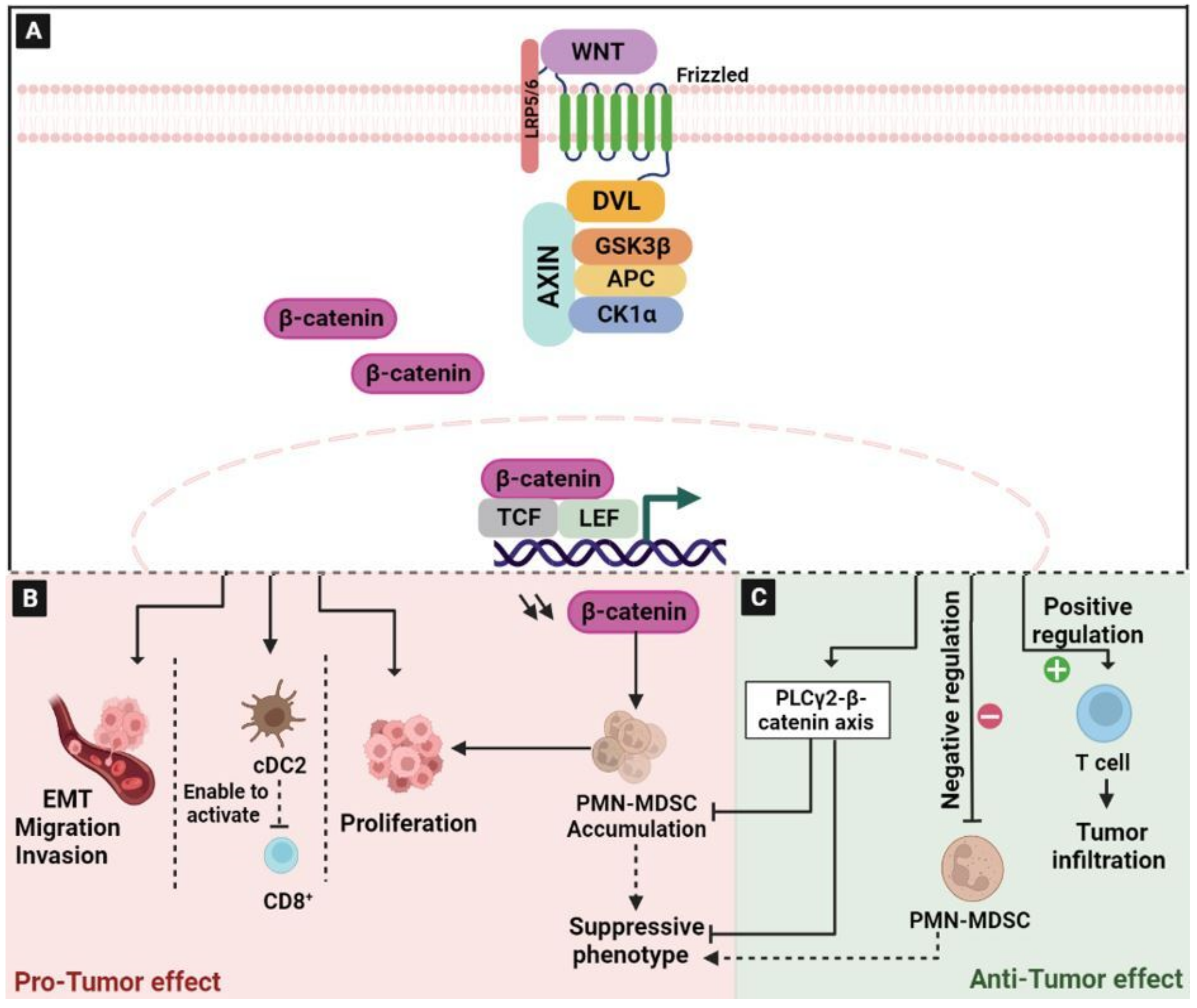
2.1. The Wingless/Integrated (Wnt)/β-Catenin Signaling Pathway

Wnt/β-catenin signaling is involved in cell differentiation both during embryogenesis and during adult tissue homeostasis, in cell–cell communication system and in the regulation of cell proliferation [18,19]. The aberrant activation of this pathway induces the accumulation of β-catenin in the nucleus, leading to the upregulation of many cell cycle-related proteins, such as cellular myelocytomatosis (c-Myc) and Cyclin D-1, which promote carcinogenesis [20]. Mechanically, without active stimuli, β-catenin would not accumulate in the cytoplasm based on the fact that the adenomatous polyposis coli/Axin/Glycogen synthase kinase-3 (APC/Axin/GSK-3) complex proceeds to its degradation via the ubiquitin–proteasome pathway. Indeed, in the presence of a Wnt ligand, casein kinase 1 (CK1) phosphorylates β-catenin at Ser45. This event primes β-catenin for subsequent phosphorylation by GSK-3β at Ser33, Ser37, and Thr41 residues [21]. The phosphorylation and the inhibition of GSK3β ensure an increase in the concentration of cytosolic β-catenin [22] (Figure 1A). However, unphosphorylated β-catenin accumulates in the cytoplasm and then migrates to the nucleus where it binds to the lymphoid enhanced factor-1/T-cell factor 4 (LEF-1/TCF4) and other co-regulators in order to promote the transcription of target genes such as Jun, c-Myc, and Cyclin D-1 in a tissue-specific manner [19,23].
Wnt/β-catenin plays an important role in the pathogenesis of CRC [24]. A large sequencing project conducted on 1134 CRC samples identified diverse alterations in core WNT regulators; this mainly included the loss-of-function mutations in APC, which are very frequent among CRC patients. In addition, it has been demonstrated that more than 96% of CRC patients showed WNT activation [25]. Wnt/β-catenin is considered to be the most vital pathway accelerating the process of epithelial–mesenchymal transition (EMT) and enhancing the metastatic properties of colon cancer [26]. The increase in the nuclear β-catenin levels is thought to be a hallmark of aggressive CRC, leading to the activation of Wnt-related targets, including c-myc, cyclin D1, matrix metallopeptidase (MMP) 2, and MMP 9, thereby increasing the proliferation, invasion, and migration potential of cells [27,28,29]. In addition, the analysis of 155 colorectal cancer tissues highlighted a correlation between the high β-catenin expression and the reduction in CD8+ T-cell infiltration. Mechanistically, β-catenin can regulate the c-c chemokine ligand (CCL) 4 expression to recruit CD103+ dendritic cells to enable CD8+ T cell activation [30]. The stabilization of β-catenin results in the tumorigenic phenotypes often observed in CRC. For this reason, the stabilized β-catenin remains the most promising target for curing the disease [31] (Figure 1B).
In contrast, there are cases in which WNT signaling exerts anticancer effects (Figure 1C). Indeed, β-catenin signaling plays an important role in the normal differentiation of myeloid progenitors [32]. It has been reported that the down-regulation of β-catenin is critical for the accumulation of myeloid-derived suppressor cells (MDSC), which suppresses T-cell responses and promotes tumor proliferation [33]. The dysregulation of this signaling pathway in myeloid cells has been implicated in promoting polymorphonuclear (PMN)-MDSC expansion in cancer [34]. Similarly, it has been demonstrated that the phospholipase Cγ2 (PLCγ2)-β-catenin axis inhibits the accumulation and suppressive phenotype of PMN-MDSCs in mouse models of lung carcinoma and melanoma [35]. In addition, it has been reported that the antagonist of the Wnt-β-catenin pathway supports tumor progression by creating an immune suppressive environment in which tumor cells can grow unabated [33]. Additionally, β-catenin signaling is important in maintaining CD8+ T cell stemness and central memory responses, as well as enabling T cell tumor infiltration [36]. Consequently, β-catenin signaling in the immune system may play an important role in multiple ways; on one hand, as a negative regulator of PMN-MDSCs, and on the other hand, as a positive regulator of T cell function [37].

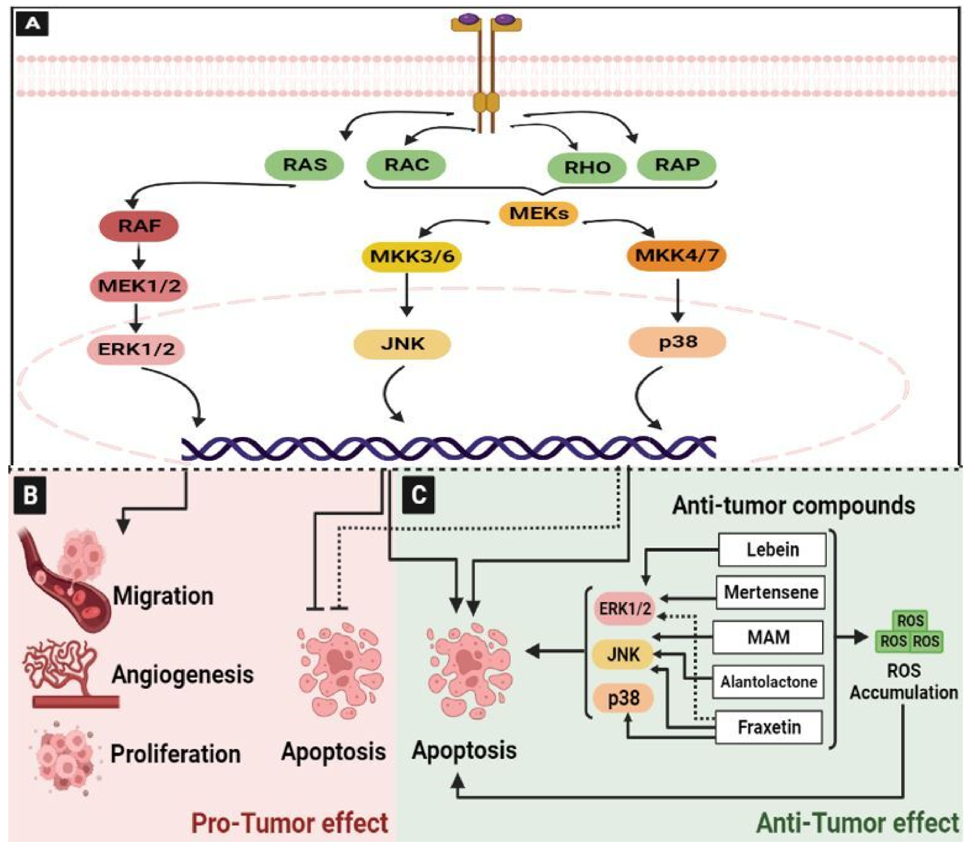
2.2. The Mitogen-Activated Protein Kinase (MAPK) Pathway

MAPK are ubiquitous signal transduction pathways that regulate all aspects of cell functions and are frequently altered in disease [38]. They are one of the most conserved signal transduction pathways, and have critical functions in cell proliferation, differentiation, death, and embryogenesis [39]. In the MAPK pathway, there are different phosphorylation cascades that modulate several series of vital processes. Each cascade is initiated by a specific extracellular stimulus and leads to the activation of particular MAPK kinase kinase (MAPKKK) and MAPK kinase (MAPKK). In a simplified model, the presence of mitogens and growth factors promotes the activation and dimerization of a canonical receptor tyrosine kinase subsequent to the activation of the small GTPases (Ras, Rac, RHO, or RAP). This induces the activation of MAPKKK (Raf or MEKs) and leads then to the stimulation of MAPKK (MEK1/2, MKK3/6, or MKK6/7). Finally, MAPKK activates the MAPK, including ERK1/2, p38, or JNK, by dual phosphorylation in order to interact and activate many transcription factors [39] (Figure 2A).
MAPK pathways play a critical role in different aspects of tumorigenesis, such as tumor growth, apoptosis, angiogenesis, invasion, metastasis, and drug resistance [39]. Dysregulated epidermal growth factor receptor (EGFR)/MAPK signaling pathway plays an oncogenic role in the initiation and development of CRC [40] (Figure 2B).
It has been reported that the long non-coding RNA H19 increases the migration and invasion of CRC cells by activating the RAS-MAPK signaling pathway, one of the most frequent carcinogenic events in human cancer [41]. Moreover, p38α MAPK signaling is a mediator of resistance in various agents in CRC patients, and it may also acquire an oncogenic role involving cancer related-processes, such as cell metabolism, invasion, inflammation, and angiogenesis [42]. In addition, the activation of the activator protein-1 (AP-1) by JNK and the nuclear factor-kappa B (NF-κB) by p38-MAPK promotes the invasion of human colon cancer cells [43]. For these reasons, MAPK pathways are mostly the target of cancer therapy.
The MAPK plays a crucial role in both cell proliferation and cell death [42]. The sustained activation of ERK1/2 promotes colon cancer cell death, and is induced by some anti-tumor compounds. In accordance, we previously reported that Lebein, a heterodimeric disintegrin isolated from Macrovipera lebetina snake venom, significantly inhibited the viability of LS174 colon cancer cells and induced their apoptosis by triggering the activation of the MAPK ERK1/2 pathway through the induction of reactive oxygen species (ROS) [44]. The halogenated monoterpene Mertensene from the red alga Pterocladiella capillacea induces G2/M cell cycle arrest and the caspase-dependent apoptosis of the human colon adenocarcinoma HT29 cell line through the modulation of intracellular ROS levels linked to the activation of the ERK1/2 in HT29 cells [45]. Fraxetin, a natural compound extracted from Fraxinus spp, induced apoptotic cell death in HT29 and HCT116 cells through mitochondria dysfunction associated with ROS induction, the modulation of ERK1/2, JNK, and P38 signaling pathways [46] (Figure 2C). JNKs have been also shown to play a role in apoptotic and non-apoptotic programmed cell death mechanisms. JNKs can either induce or inhibit cell death by stimulating the expression of specific genes and by modulating the activities of pro- and anti-apoptotic proteins through phosphorylation events [47]. It has been reported that treatment of HCT116 and HT29 colon cancer cells with a natural naphthoquinone 2-methoxy-6-acetyl-7-methyljuglone (MAM), isolated from Polygonum cuspidatu, validated the role of JNK in MAM-induced necroptosis, marked by mitochondrial depolarization, ATP depletion, and an increased production of mitochondrial ROS [48]. In addition, alantolactone, a plant-derived sesquiterpene lactone, showed anti-proliferative and pro-apoptotic effects in HCT116 colon cancer cells through activating the MAPK-JNK/c-Jun signaling pathway [49].
In addition, a synthetic alkaloid Lappaconitine hydrochloride exhibited antitumor activity in CRC HCT-116 cells by inducing their apoptosis through mitochondrial and MAPK signaling pathways [50].

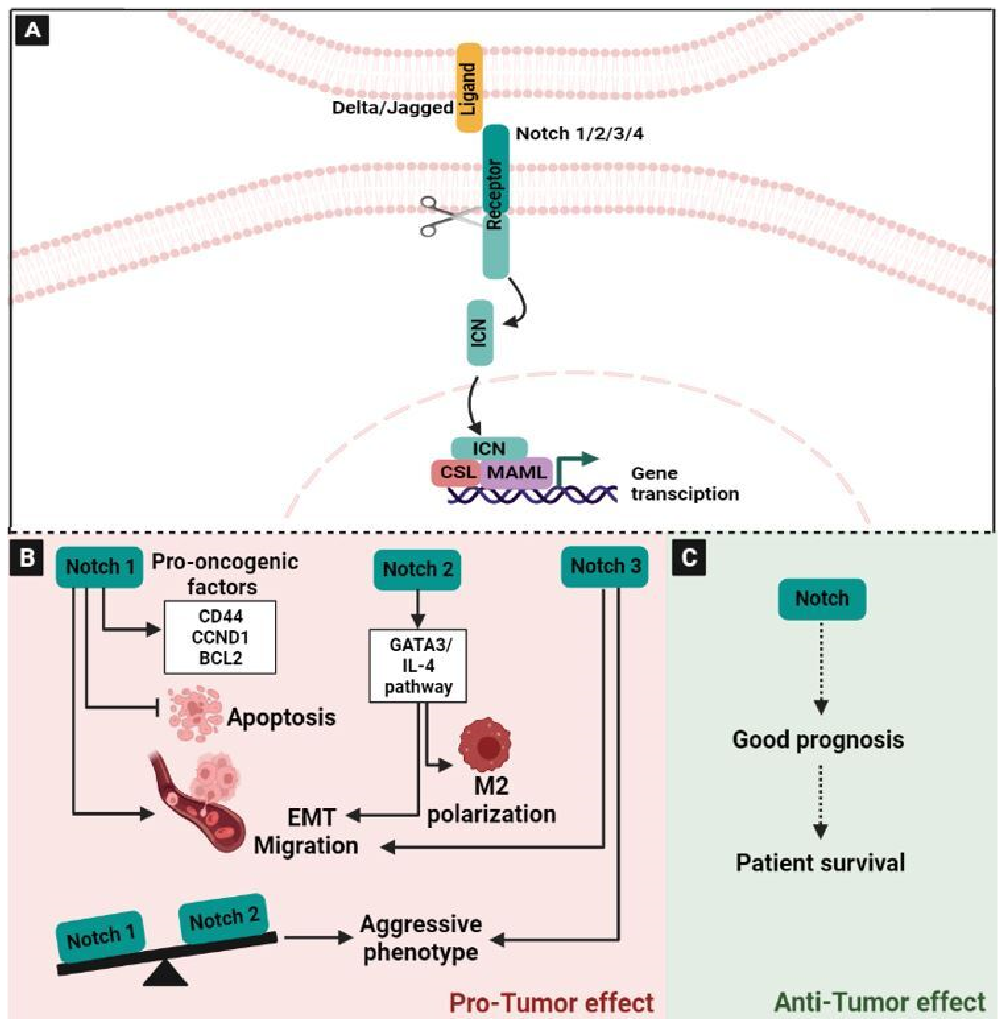
2.3. The Neurogenic Locus Notch Homolog Signaling (Notch) Pathway

The Notch signaling pathway modulated a series of fundamental cellular functions, including cell fate decision, the maintenance of stemness, proliferation, and apoptosis [51]. There are four receptors: Notch-1, Notch-2, Notch-3, and Notch-4 of the notch pathway, which are normally activated by interacting with ligands such as Delta-like and Jagged [52]. In total, there are three delta-like (Dl) ligands (Dll1, Dll3, and Dll4) and two Jagged (Jag) ligands (Jag1 and Jag2) [52,53]. In the canonical Notch pathway, ligand–receptor interaction results in a cascade of proteolytic cleavages, first mediated by metalloproteases and second by γ-secretase activity. These cleavage steps result in the release of a constitutively active intracytoplasmic nick (ICN) fragment, which is then translocated to the nucleus where it associates with CSL and MAML as part of a larger transcriptional complex [52] (Figure 3A). The precise signaling differences between the Notch receptors and Notch ligand pairs are unknown. While they all seem to go through the same pathway, there is evidence that different receptor–ligand parings yield distinct biological outcomes [54]. In addition to their role in cellular functions, the Notch signaling plays a crucial role in many aspects of cancer biology, either oncogenic or tumor-suppressive [55]. This pathway is aberrantly activated in many cancers, including CRC. Recently, the analysis of the Notch signaling pathway of 1116 CRC patients in East China highlighted that one locus at MINAR1 out of 133 genes is significantly associated with overall survival. This study proves that the Notch pathway plays a crucial role in the progression of CRC, likely affecting patient survival [56]. Moreover, in CRC patients, it has been reported that mutations in the Notch pathway components can activate anti-tumor immune responses, which are characterized by the up-regulation of checkpoint molecules [57]. Previously, it has been shown that the dysregulation of the Notch pathway is linked to the pathogenesis of CRC and plays an oncogenic role in CRC development and progression [58,59]. Indeed, the upregulation of Jagged-1, mediated by β-catenin, increases Notch-1 expression [60], which has been correlated with progression, tumor grade, and metastasis in CRC. This could be related to the inhibition of apoptosis promoted by Notch-1 [61] and induced invasiveness through the activation of several pro-oncogenic factors, including CD44, Cyclin D1 (CCND1), and BCL2 Apoptosis Regulator [62]. Notch-1 and Notch-2 have been associated with opposite clinical outcomes in CRC patients. Indeed, the increased expression of Notch-1 predicted a poor overall survival, while, on the contrary, the reduced expression pattern of Notch-2 was linked to a worse one [63]. In CRC pathogenesis, Notch2 overexpression may activate the GATA3/IL-4 pathway, which subsequently promotes the polarization of the tumor-associated macrophages toward an M2 phenotype and then enhances EMT [64]. Notch2 has also been shown to mediate stemness promotion and chemoresistance in CRC cells [65]. Notch-3 is also upregulated in metastatic CRC and may regulate CRC-related tumorigenesis [66]. The dysregulation of Notch-3, as well as Jagged-1 and Dll-4, is associated with a more aggressive phenotype in xenografts of CRC cells in vivo [66]. In addition, Dll-1 can increase Wnt and TGF-β by binding to Smad2/3 and Tcf-4, and overexpressing the CTGF gene, which regulates colon carcinogenesis independently of Notch signaling [58,67]. In addition, the dysregulation of the Notch pathway in the tumor skews the local cytokine composition, shaping the immunological landscape and affecting tumor growth, progression, and metastasis [68] (Figure 3B).
The research around the role of Notch signaling in tumorigenesis has focused on its role as an oncogene. Interestingly, Notch activity has been associated with both oncogenic and tumor suppressor functions, which depended on the cellular context and the nature of the induced response [55,69,70]. Indeed, various studies in different cancers have revealed a clear link between the loss of Notch activity and carcinogenesis, suggesting that Notch plays an important tumor suppressor function in certain tissues [55]. The role of Notch signaling as a tumor suppressor has also been highlighted in breast, prostate, liver, lung and skin cancers [71]. These studies suggested that Notch plays a role in solid tumors through the defective activation of signaling pathways. Furthermore, the cellular outcome of this aberrant Notch activity is highly dependent on contextual cues, such as interactions with the tumor microenvironment and crosstalk with other signaling pathways [71]. The anti-tumor role of the Notch signaling pathway in CRC has not yet been clearly illustrated. Previously, it has been reported that in a total of 146 colorectal cancer samples, Notch2 and JAG1 expression levels were associated with patients survival [72] (Figure 3C). In addition, it has been demonstrated that the higher levels of infiltration of CD4+ T cells, macrophages, neutrophils, and dendritic cells were positively correlated to the expression of Notch receptors in patients with gastric cancer [73].

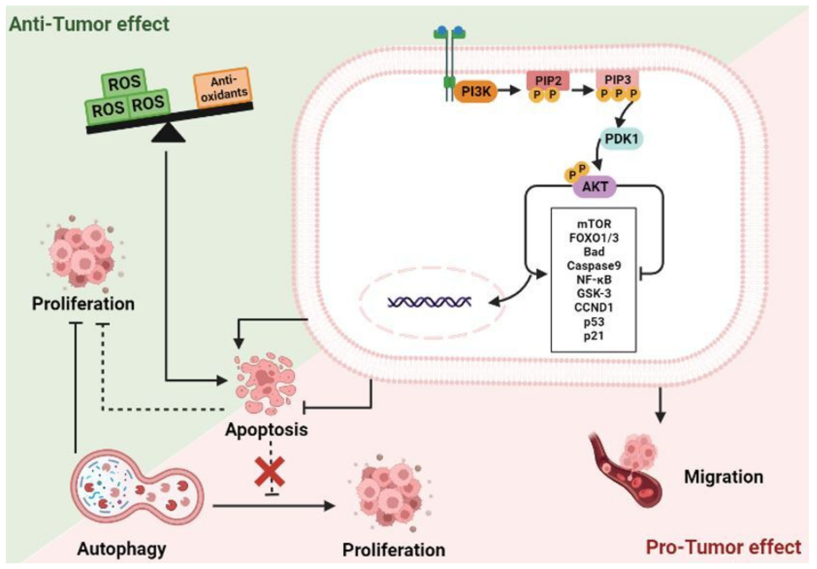
2.4. The Phosphoinositide 3-Kinase (PI3K)/Protein Kinase B (AKT) Signaling Pathway

PI3K/AKT signaling pathway plays a pivotal role in many biological and cellular processes, such as cell proliferation, growth, invasion, migration, and angiogenesis [74]. PI3K, a member of the lipid kinase family, is composed of a catalytic domain (p110) and a regulatory domain (p85). The activation of PI3K could catalyze the phosphorylation of phosphatidylinositol (PI) at the 3′-position of the inositol ring. The phosphorylated products have a critical influence on cellular functions, such as the enhancement of cell migration by PIP3, and the regulation of B cell activation and insulin sensitivity by the PI 3,4-bisphosphate. The PIP3 activates PDK1, which phosphorylates the serine/threonine kinase AKT at Thr308. AKT can also be phosphorylated and activated by PDK2 at Ser473. Activated AKT regulates cell proliferation, differentiation, migration, and apoptosis by activating or inhibiting downstream target proteins, such as Bad, Caspase 9, NF-κB, GSK-3, FOXO3, p21, p53, and FOXO1 (Figure 4). Additionally, AKT activates the mTOR pathway, thereby regulating cell growth through the modulation of the expression of cyclin D1 and p53. AKT boosts cell survival by inactivating the pro-apoptotic factor Bad and the transcription factor FKHR family [74]. The overexpression of PI3K/AKT/mTOR signaling, which is linked to the regulation of distinct oncogenic mechanisms, has been reported in various forms of cancers, especially in colorectal cancers (CRC). In addition, it plays a significant role in acquiring drug resistance, as well as in the metastatic initiation events of CRCs [75,76].
Due to the significant roles of this signaling pathway in the initiation and progression events of CRC, it is consistently recognized as a striking therapeutic target. PTEN, a tumor suppressor protein, possesses alkaline phosphatase and protein phosphatase activities, and can block PI3K/AKT signaling via the dephosphorylation of PIP3 to PIP2. In addition, Carboxyl-terminal modulator protein (CTMP) could block the activation of downstream signaling pathways by inhibiting AKT phosphorylation. The protein phosphatase 2A (PP2A) inhibits the activation of AKT through its dephosphorylation at Thr308 and Ser473 residues [74].
However, a study by Nogueira et al. [77], along with other research [78,79], reported upon a function of AKT and highlighted that it is not a single function kinase but, under certain conditions, can facilitate rather than inhibit cell death; this is via an increase in the reactive oxygen species and through suppressing antioxidant enzymes [77] (Figure 4). In accordance, our team previously reported that Mertensene, a halogenated monoterpene isolated from the red alga Pterocladiella capillacea, inhibited the viability of HT29 colon cancer cells and induced their apoptosis by triggering the activation of AKT [45]. The PI3K/AKT/mTOR pathway is also a critical regulator of cell autophagy. It has been reported that this process is involved in promoting cancer and participates in regulating the balance between the tumor and its microenvironment. Furthermore, the role of autophagy in cancer seems paradoxical due to its dual function as a survival or suppressor mechanism for tumor cells [80]. Indeed, it has been shown that targeting PI3K/AKT/mTOR-mediated autophagy is a double-edged sword in cancer. On the other hand, autophagy activation, by modulating the PI3K/AKT/mTOR pathway, increases the drug sensitivity of certain types of tumors and avoids drug resistance [81].

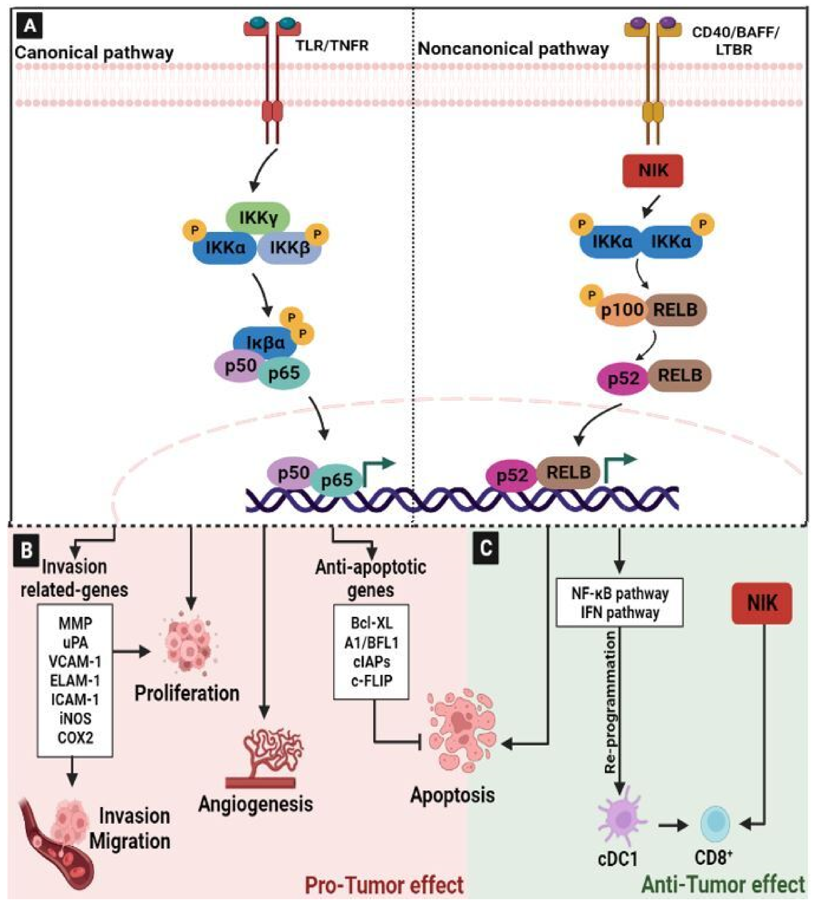
2.5. Nuclear Factor-kappaB (NF-κB) Signaling Pathway

NF-κB is one of the major signaling pathways involved in physiological and pathological conditions. It controls the expression of more than 400 genes, leading to their regulatory effects on different mechanisms, including immune response, inflammation, cell migration, apoptosis, and differentiation [82,83]. Indeed, extracellular stimuli, such as bacteria, viruses, cytokines, oncogenic molecules, chemo/radiotherapy, and cell surface receptors, including Toll-like receptors (TLRs), T/B cell receptors, and the interaction of tumor necrosis factor receptors (TNFR) with their specific ligands, cause the upregulation of the IκB kinase (IKK) complex [84]. The IKK complex phosphorylates p65/p50-bound IκB at serine residues -32 and -36. Phosphorylated IκB is degraded by the ubiquitin–proteasome pathway, thereby activating NFκB. The activated NF-κB is translocated to the nucleus, where it binds to the enhancer element of the immunoglobulin kappa light-chain of activated B cells (κB sites), thus triggering the expression of downstream genes that potentially leads to inflammation and the promotion of cancer development/progression [84,85,86,87] (Figure 5A). NF-κB exhibited an alternative pathway, initiated by ligands such as the cluster of differentiation (CD)-40, B-cell activating factor (BAFF), and lymphotoxin-β receptor (LTBR), and that includes RelB/p100 subunits of NF-κB, IKKα homo-dimer, and NF-κB-inducing kinase (NIK) [87].
The aberrant regulation of NF-κB is frequently reported in tumor cells and it is considered to be a poor prognostic marker in patients with CRC [88]. It has been reported that NF-κB suppression can induce apoptotic cell death in CRC cells [89]. In line with this, the activation of NF-κB signaling plays a significant role in the tumorigenesis process via the regulation of downstream NF-κB gene products linked to cell growth, inflammation, metastasis, angiogenesis and drug resistance in CRC cells [87] (Figure 5B). NF-κB inhibits apoptosis by up-regulating the expression of anti-apoptotic genes, including B-cell lymphoma-extra large (Bcl-xL), the Bcl-2-related gene (A1/BFL1), cellular inhibitors of apoptosis (cIAPs), and caspase-8/FAS-associated death domain-like IL-1beta-converting enzyme inhibitory protein (c-FLIP) [87,90]. It has been shown that NF-κB enhances the expression of various invasion-related genes, including MMPs, urokinase-type plasminogen activator (uPA), Vascular cell adhesion molecule 1 (VCAM-1), endothelial leukocyte adhesion molecule 1 (ELAM-1), Intercellular Adhesion Molecule 1 (ICAM-1), iNOS and COX2. In addition, it is involved in cancer-associated extracellular matrix (ECM) degradation [87,89]. Furthermore, the activation of NF-κB in response to chemotherapy reduces drug efficacy by decreasing the tumor cell chemosensitivity and cell death. Thus, the co-administration of chemo agents with NF-κB inhibitors can enhance chemosensitivity in CRC cells [91,92].
As described above, the NF-κB pathway plays a key role in supporting tumorigenesis, progression, and the chemoresistance of tumor cells. However, the role of the NF-κB signaling pathway in the induction of anti-tumor host immunity remains unclear. Recently, single-cell transcriptomics analyses have explored the molecular pathways that regulate the maturation of intratumoral conventional type 1 dendritic cells (cDC1s), which is critical for antitumor immunity, and have demonstrated the dynamic reprogramming of tumor-infiltrating cDC1s by NF-κB and IFN signaling pathways [93]. Moreover, the inactivation of NF-κB or IFN regulatory factor 1 (IRF1) in cDC1s, resulting in the impaired expression of IFN-γ-responsive genes, has been shown to lead to the ineffective recruitment and activation of antitumor CD8+ T cells [93] (Figure 5C). This study highlights the important role of NF-κB signaling in the polarization of conventional dendritic cells. Therefore, in cDC1, this pathway may represent an important focal point for the development of new diagnostic and therapeutic approaches in order to improve cancer immunotherapy.
Continuing in the discussion of the important role of NF-κB as an anti-tumor pathway, a recent study demonstrated that NF-κB-inducing kinase (NIK), which is known to be a mediator of noncanonical NF-κB activation, is a pivotal regulator of T cell metabolism and antitumor immunity [94]. Indeed, the transgenic expression of NIK, using an inducible system, strongly promoted the antitumor immunity associated with an increased glycolytic metabolism and the effector functions of CD8+ T cells (Figure 5C). This result was confirmed in a preclinical model of cancer therapy and proves that NIK expression rendered PD1+Tim3+ tumor-infiltrating CD8+ T cells competent for IFN-γ production [94]. In line with this, previously it has been reported that NIK plays a crucial role in mediating effector T cell function [95,96]. All these observations suggest that NIK regulates T cell function via both noncanonical NF-κB-dependent and independent mechanisms.

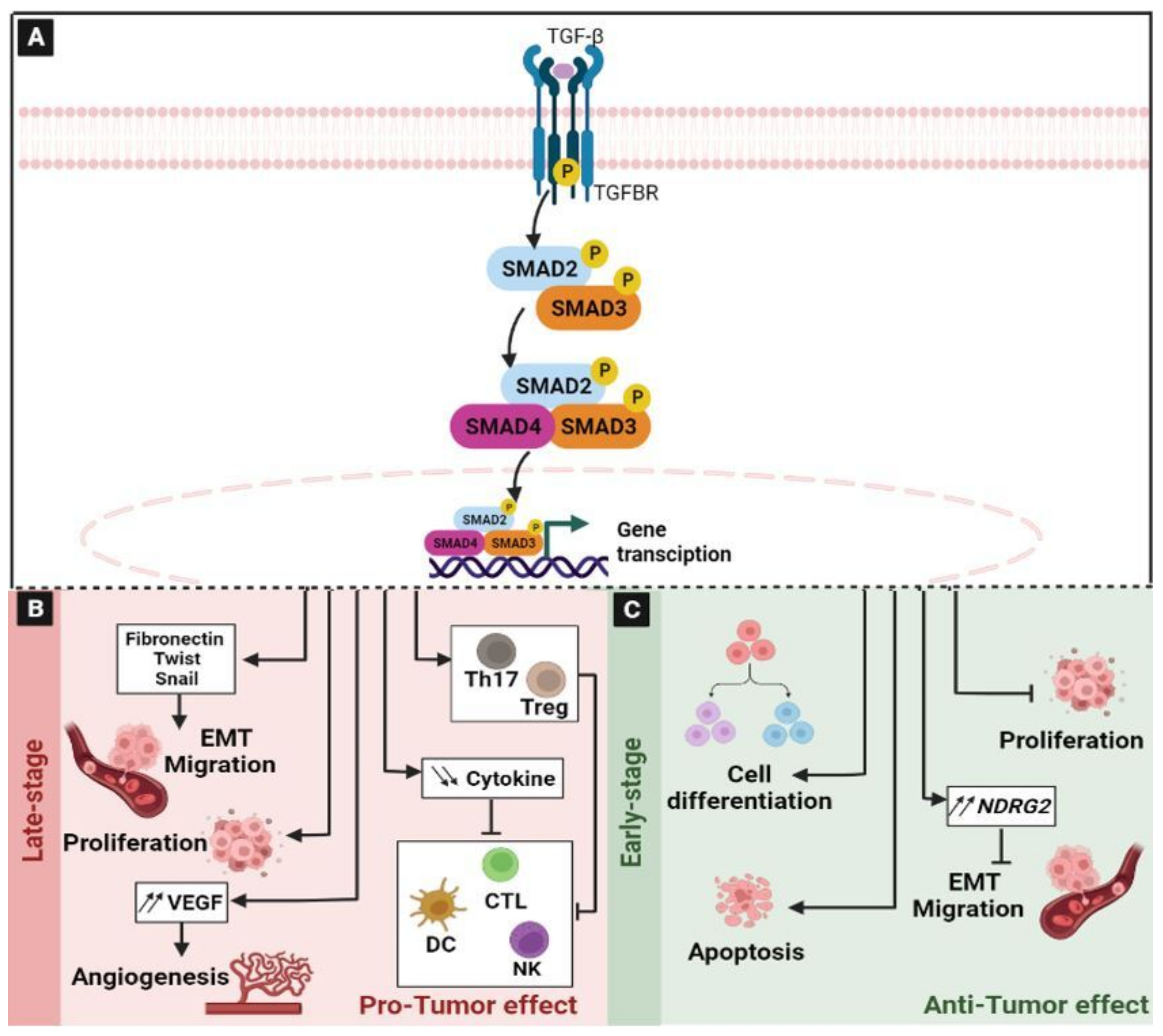
2.6. The Transforming Growth Factor-β (TGF-β) Signaling Pathway

The TGF-β signaling pathway is critical in many biological processes. It regulates cell growth, differentiation, apoptosis, cell motility, extracellular matrix production, epithelial–mesenchymal transition (EMT), angiogenesis, and cellular immunity [97,98,99]. There are three receptors in the TGF-β signaling pathway: Transforming growth factor-β receptor (TGFBR)-1, TGFBR2, and TGFBR3, and three ligand isoforms: TGF-β1, TGF-β2, and TGF-β3 [100,101]. The cascade initiates upon binding of the TGF-β ligand to the TGFBR2, inducing the formation of a heterotetrametric complex of TGFBR2 and TGFBR1 [100]. Subsequently, the TGFBR2 kinase domain activates TGFBR1 by phosphorylation, which, in turn, phosphorylates the suppressor of Mother Against Decapentaplegic (SMAD)2/3, which can be assembled into complexes with SMAD4 and then translocated to the nucleus where they can regulate the expression of target genes [102] (Figure 6A). The negative regulation of this pathway is assured by SMAD7, which can compete with SMAD2/3 for the catalytic site of phosphorylated or activated TGFBR1 and, thereby, inhibit the phosphorylation of SMAD2/3 [102]. The deregulation of this pathway is associated with many diseases, such as cancer [103]. Indeed, the aberrant activation of this signaling pathway promotes a variety of tumors, including hepatocellular carcinoma, pancreatic cancer, esophageal cancer, gastric cancer and CRC [99,104]. In tumor, the TGF-β signaling pathway exhibits a paradoxical role that is known as the “TGF-β paradox” [97] (Figure 6). On the one hand, TGF-β signaling could have a tumor suppressor function by inhibiting cell proliferation and stimulating cell differentiation in the early stages of cancer. On the other hand, in the late stages of cancer, it induces tumor progression and metastasis [97]. In CRC, its role also remains controversial. Indeed, it can promote or suppress the growth of colon cancer cells depending on the microenvironment [105]. Under normal conditions, TGF-β can induce the arrest of the cell cycle in G1 by increasing the expression of the cyclin-dependent kinase (CDK)4/6 inhibitor p15 [106] and by suppressing the multi-functional oncogene c-Myc [107]. In fact, during the early stages, TGF-β acts as a “tumor suppressor” and inhibits the proliferation of the tumor cells (Figure 6C). Previously, it has been reported that a bis-benzylisoquinoline alkaloid Tetrandrine inhibited the proliferation of colon cancer HCT116 cells and induced their apoptosis by increasing the mRNA and protein levels of TGF-β1. The upregulation of TGF-β1 decreases the phosphorylation of PTEN, thereby inactivating PI3K/Akt signaling [105]. In addition, it has been reported that TGF-β abrogated the epithelial–mesenchymal transition, and inhibited the invasion and migration of CRC cells, inducing the N-Myc downstream-regulated gene 2 (NDRG2) [108].
However, colon cancer cells can overcome the tumor-suppressing effects of the TGF-β pathway by inducing the loss of Smad proteins and deregulating the TGF-β type II receptor-mediated cell cycle [109]. Moreover, even if the TGF-β signaling contributes as a promoter of the tumor, its role in carcinogenesis is still complex. The ability of TGF-β to act either as a tumor suppressor or as an oncogenic agent is determined by the cell-to-cell communications and by the tumor stage [110]. Indeed, during late stages, TGF-β supports tumor cell proliferation, invasion, and metastasis (Figure 6B). The change in TGF-β expression and in the cellular responses tips the balance in favor of oncogenic activities by inducing the EMT, which is mediated by Fibronectin, Twist, and Snail, and accelerates tumor invasion and metastasis [108,111,112]. Previously, it has been reported that TGF-β1 increased the progression of colon cancer by upregulating the expression of Human Cripto-1 (CR-1) linked to tumorigenesis [113,114]. The overexpression of TGF-β has been highlighted in different tumors [97,115]. Moreover, this signaling pathway enhances angiogenesis via the up-regulation of the Vascular Endothelial Growth Factor (VEGF) [116] through Smad-dependent pathways [97,117,118]. Under normal conditions, TGF-β plays a key role in controlling immune responses. It promotes the differentiation of regulatory T cells and Th17 cells [119,120,121]. TGF-β suppresses the immune system by inhibiting NK-cell activity [122], decreasing cytokine production, inhibiting dendritic cell maturation [123], and altering T-cell cytotoxic properties [124]. All these properties suggest that TGF-β exhibited a facilitative and a direct role in tumor progression as it directly suppresses the immune system and allows tumor cells to acquire properties that help to evade the immune system [97].

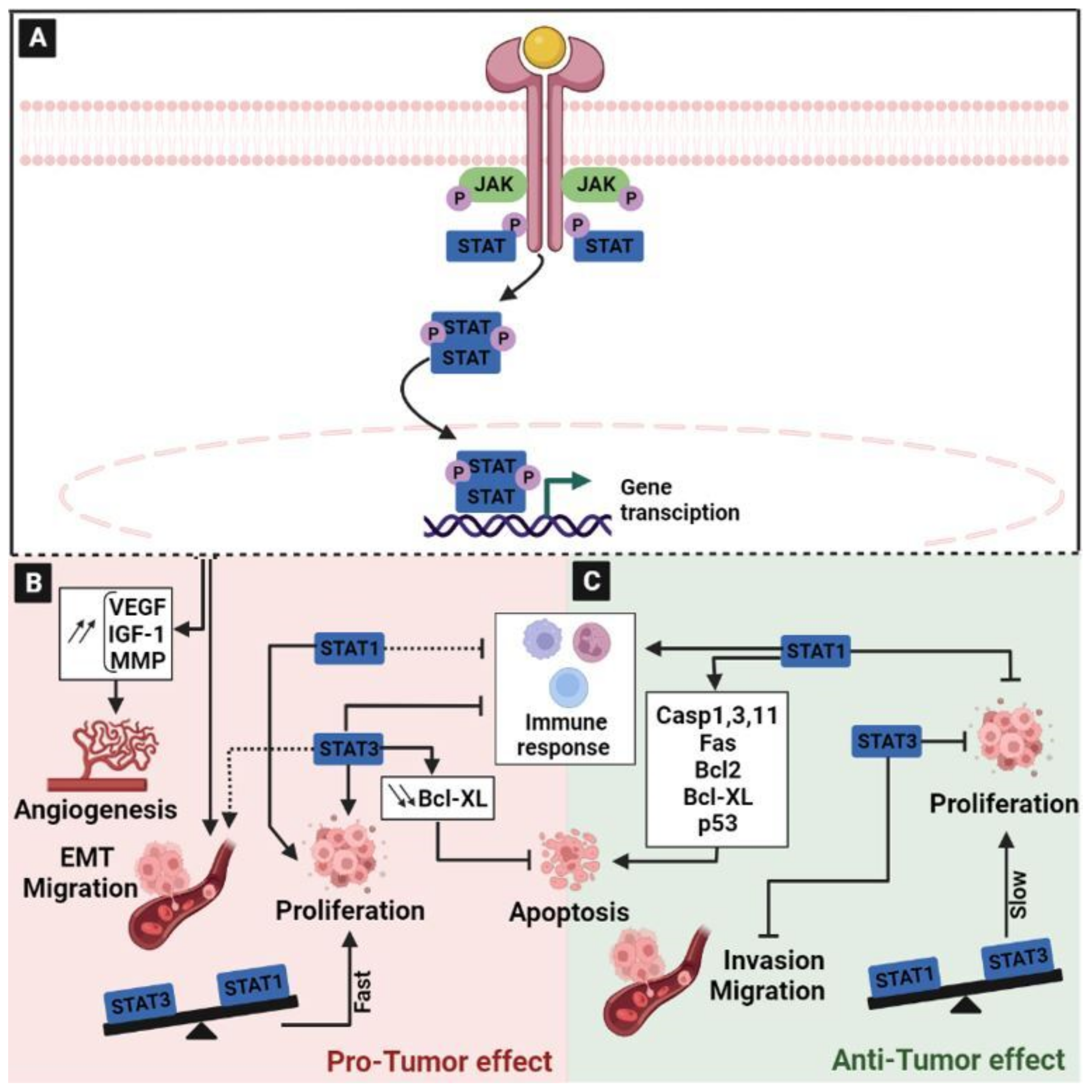
2.7. The Janus Kinase/Signal Transducer and Activator of Transcription (JAK/STAT) Signaling Pathway

The JAK/STAT signal transduction pathway is the common signaling pathway in which many growth factors and cytokines can transmit signals in cells [125,126]. The JAK family comprises four members: JAK1, JAK2, JAK3, and TYK2 [127], which will act through seven STAT family members, STAT1, STAT2, STAT3, STAT4, STAT5a, STAT5b, and STAT6 [126]. The cascade begins upon the binding of cytokines, inducing the receptor oligomerization and then leading to the recruitment of related JAKs. JAK activation induces the phosphorylation of tyrosine in the intracellular domain of the receptors. Once activated, JAKs serve as docking sites for STAT. Then, STATs dissociate from the receptor to form homodimers or heterodimers, and translocate to the nucleus to initiate the transcription of a repertoire of target genes [128] (Figure 7A). The JAK/STAT signal transduction pathway can promote the expression of several downstream genes that contribute to the many biological processes that are involved in immune function and cell growth [129]. In normal conditions, it plays an important role in the growth and development of the body. Under pathological conditions, the activation of the JAK/STAT pathway mediates the proliferation, differentiation, and migration of malignant tumor cells [130] (Figure 7B). During the early and late stages of CRC, JAK/STAT signaling is considered to be a clinical predictor and a prognosis marker for diagnosis, making this pathway as a target for therapeutic intervention [130,131,132]. Previously, it has been reported that the JAK/STAT pathway is activated in colon cells [133]. Case-control studies, using more than 1550 patients with colon cancer and 750 cases of rectal cancer, demonstrated that JAK2, SOCS2, STAT1, STAT3, STAT5A, STAT5B, and STAT6 were associated with colon cancer, and that STAT3, STAT4, STAT6, and TYK2 were linked to rectal cancer [129]. Thus, the rapid transduction of extracellular signals by the JAK/STAT pathway to the nucleus plays a pivotal role in the activation of oncogenes and the negative modulation of tumor suppressor genes in colon cancer [130]. Moreover, JAK/STAT can activate angiogenesis through the overexpression of many downstream growth factors, such as VEGF, insulin-like growth factor-1 (IGF-1), and MMP [134] (Figure 7B). JAK/STAT also plays regulatory roles in the inflammatory response, glycolysis, and epithelial–mesenchymal transition [130,135,136]. It has been reported that, depending on the cellular context, IFN/STAT signaling could mediate tumor cell growth, metastasis, and chemo- and radio-resistance to therapies [137,138]. Various studies have shown that the hyperactivation of STAT3 enhances the expression of its target genes, leading to an increase in tumor cell migration and proliferation, and thus contributing to colorectal carcinogenesis [139,140]. For this reason, the deletion of the hotspot mutation region in the DNA-binding domain of STAT3induced colon cancer cell growth and progression due to genome-wide changes in the transcription of STAT3-target genes [140]. In line with this, the analysis of the association of JAK-1 and STAT-3 protein levels with the clinicopathological parameters of patients with colon cancer has demonstrated that the expression of these two proteins is associated with the clinical stage of the pathology [130]. Indeed, STAT3 was shown to also inhibit cell apoptosis through down-regulating the apoptotic protein Bcl-xl [141]. In addition, based on its role in the regulation of the immune response, STAT3 has been reported to regulate the differentiation of Th17 cells [142]. In addition, it promoted the immunosuppression of tumor-associated macrophages and myeloid-derived suppressor cells [143,144] (Figure 7B).
Like several other signaling pathways, the JAK/STAT pathway could exhibit both a pro- or anti-tumor role. The complexity of these effects depends on several factors, such as the stage of the pathology and/or the cellular context. For example, IFN/STAT signaling has been known as an anti-tumorigenic pathway that regulates many effectors, such as caspases [145,146,147], cyclin-dependent kinase inhibitors [148], the anti-apoptotic protein BCL2 [149] and the IRF1/p53 pathway [150]. However, there is increasing evidence that IFN/STAT singling also contributes to tumor initiation and dissemination [137]. Furthermore, the role of STAT1 and STAT3 in CRC development and progression is controversial. It has been demonstrated in vivo that there are two opposite cell growth behaviors based on the STAT1/3 expression patterns (Figure 7). Indeed, a low STAT1/high STAT3 ratio highlighted a faster tumor growth in xenografts compared to high STAT1 and low STAT3 expressions, which were characterized in contrast slower ones. Interestingly, the simultaneous absence of nuclear STAT1 and STAT3 expression was associated with a reduction in the median survival by ≥33 months [132]. In CRC, the role of STAT3 is paradoxical. Indeed, as explained above, STAT3 activation can upregulate the expression of MMP, which, in turn, could favor cancer cell invasion and metastasis [151,152]. However, it has been shown that STAT3 activity may also exert limiting effects on colonic carcinoma development in murine models, as well as suppress tumor cell invasiveness [153,154]. In line with this, a study on CRC tissue microarrays showed that the expression and/or activation of STAT3 indicated a favorable clinical prognosis outcome [154] (Figure 7C). This double role may be explained by the presence of two splicing isoforms of STAT3, STAT3α and STAT3β, which have different functions in the regulation of tumors. STAT3α activation is believed to promote tumor initiation, while STAT3β is believed to inhibit the occurrence of cancer and is considered to be an effective tumor suppressor [128,155,156].
Another component of the JAK/STAT pathway that has a contradictory role is STAT1. On the one hand, the aberrant expression of STAT1 is found in tumor cells [157]. Moreover, STAT1 has a strong association with indoleamine-2,3-dioxygenase-1 expression in Paneth cells in the stem cells of intestinal crypts and tumors, and with subsequent immune escape in CRC [158] (Figure 7B). On the other hand, based on its anti-proliferative and pro-apoptotic effects, STAT1 is generally regarded to have tumor-suppressive functions and in CRC, its activity was shown to be associated with a favorable prognosis [154,159] (Figure 7C). In animal models, the loss of STAT1 expression promotes the development of CRC [131]. It has been demonstrated that STAT1 deficiency promotes rapid and extensive intestinal damage, leading to increased proliferation in the early stages of induced tumor formation and reduced apoptosis in advanced tumors [131]. This study supported the important role of STAT1 signaling during the development of cancer cells in vivo. Thus, STAT1 promoted cell apoptosis by activating the apoptotic caspases 1, 3, and 11 precursors, and by interacting with the p53 protein. Furthermore, STAT1 can also induce Fas, Bcl-2, and Bcl-X gene expression [128]. In addition, a recent study highlighted that increased STAT1 expression in tumor cells was strongly indicative of an immunogenic microenvironment, characterized by significantly high expression levels of MHC class I and PD-L1, both on tumor and non-tumor cells. Furthermore, tumor-infiltrating lymphocytes (TILs) were also increased in the positive-STAT1 group [160].
Thus, even if the activation of these signaling pathways and their link to CRC is well established, the interconnection between their different components, their dual functions and also the intercellular communications between tumor cells and the TME are still not fully understood; this contributes to the difficulty of achieving a successful therapy for CRC.
In accordance, in the next section, we will discuss how such crosstalk contributes to colorectal cancer development and progression, focusing on the double-edged role of some of the effectors regulating these processes. The comprehension of such mechanisms provides new opportunities for the development of efficient therapies for CRC.

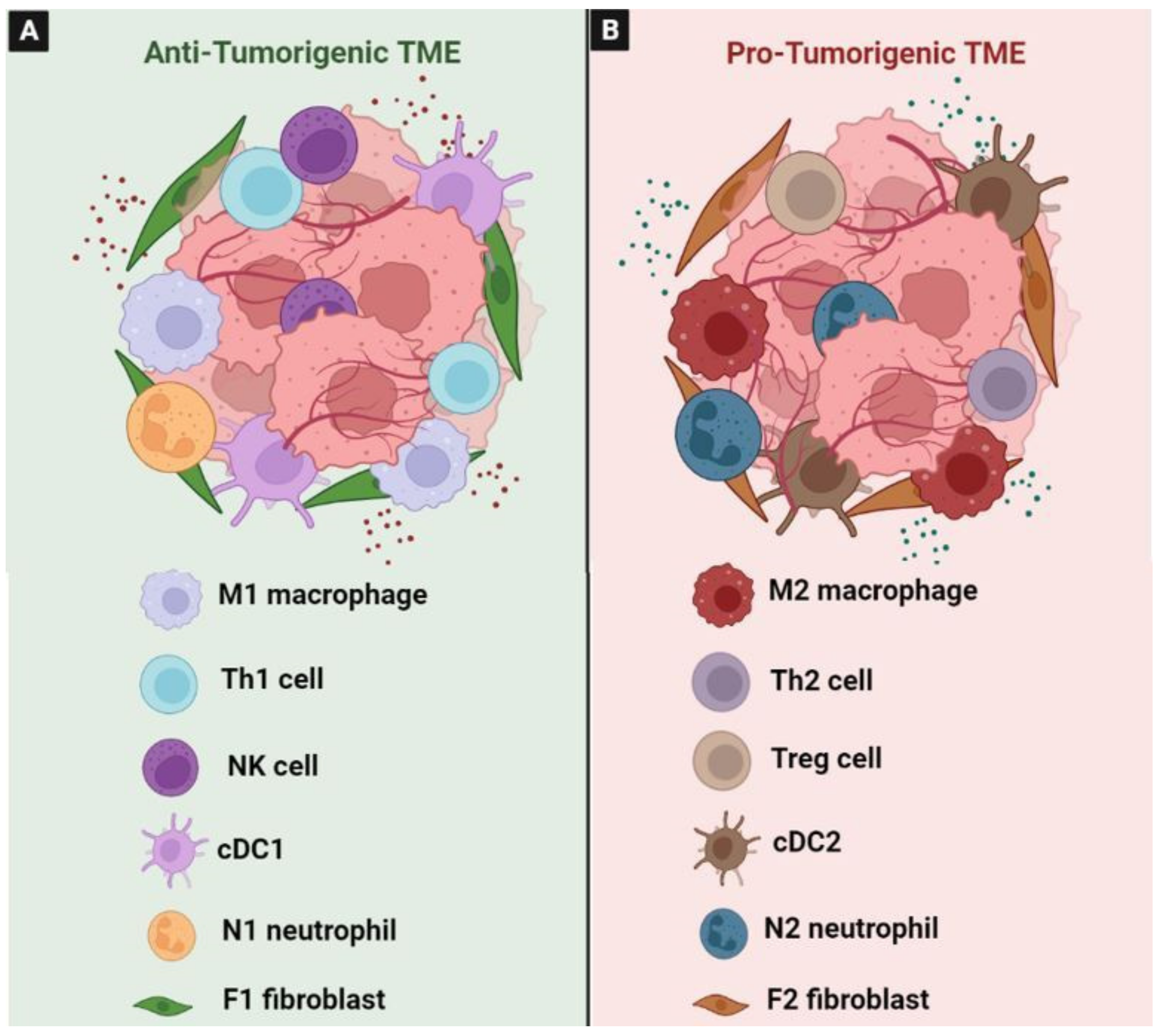
3. The Role of Tumor Microenvironment in the Carcinogenesis of CRC

The communication between tumor cells and the tumor microenvironment (TME) is established through paracrine factors that trigger the activation of numerous signaling pathways [161]. Indeed, carcinogenesis in colorectal cancer (CRC) is critically influenced by the TME, and guided by a plethora of different cells and effectors.

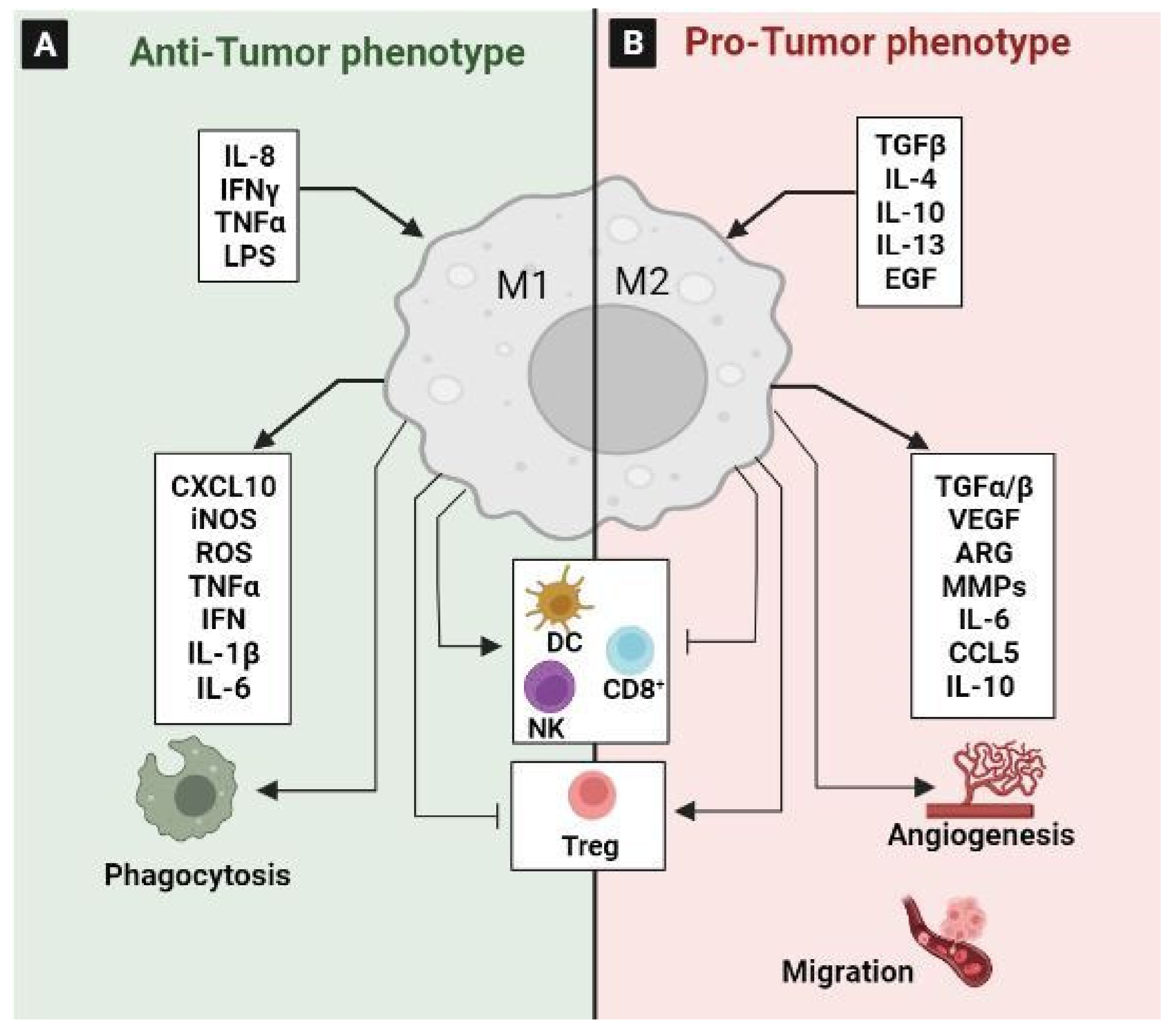
3.1. Tumor-Associated Macrophages (TAM)

TAMs are one of the most abundant cell types that play a pivotal role in the pathogenesis of CRC [162]. However, the cross talk between cancer cells and macrophages in TME is complicated and the underlying mechanisms are still poorly elucidated.
TAMs are divided into the anti-tumorigenic “M1” phenotype and the pro-tumorigenic “M2” phenotype (Figure 8). Typically, in response to inflammatory stimuli, monocytes differentiate into activated pro-inflammatory M1 cells [163]. Mechanistic studies showed that the expression of phospholipase D4 (PLD4) in TAMs promotes the activation of M1 macrophages, resulting in an antitumor effect on colon cancer cells [164]. It has been confirmed that PKCα acts as a tumor suppressor through the MKK3/6-p38 MAPK signaling pathway to promote IL12/GM-CSF-mediated M1 polarization, and inhibited the growth of mouse colon cancer [165]. M1 macrophages exert their pro-inflammatory effect by the secretion of different cytokines, such as TNFα, IL-1β, and IL-6. They also secreted factors such as TNFα and ROS to both exert antitumor effects and stimulate cytotoxic T cell recruitment into tumors [166,167]. Moreover, M1 macrophages can exhibit an anti-tumor effect by phagocytosing tumor cells [168] (Figure 8A).
In contrast, the promotion of the M2 polarization of TAM is due to the secretion of EGF by the colon cancer cells through the EGFR/PI3K/AKT/mTOR pathway [169]. Wnt5a could also induce the M2 polarization of TAMs by regulating CaKMII-ERK1/2-STAT3 pathway-mediated IL-10 secretion, ultimately promoting the tumor growth and metastasis of CRC [170]. Recently, it has been reported that exosomal miR-106b induces M2 macrophage polarization by directly suppressing programmed cell death 4 (PDCD4) to activate the PI3Kγ/AKT/mTOR signaling pathway. The activated M2 macrophages enhance the ability of CRC cells to migrate, invade, and induce metastasis in vitro and in vivo [171] (Figure 8B). They are also linked to chemoresistance and drug-induced apoptosis inhibition via the secretion of IL6, which regulates the STAT3-miR-204 axis in CRC cells [172]. High levels of growth differentiation factor 15 (GDF15), which is produced by TAMs, also impair the chemosensitivity of tumor cells via enhancing fatty acids β-oxidation [173]. Additionally, TAMs express the programmed cell death protein 1 (PD-1), which inhibits phagocytosis and antitumor immunity [174]. In addition, it has been reported that TAM-derived CCL5 facilitates the immune escape of CRC cells via the p65/STAT3-CSN5-PD-L1 pathway [175]. Similarly, a recent study characterized a new macrophage subpopulation with a high level of PD-L1. This population was induced by tumor cells during macrophage infiltration and was associated with a poor prognosis [162,176]. Mechanistically, CRC-derived multiple sEV-miRNAs synergistically induced TAM M2-like polarization to promote the inhibition of CD8+ T lymphocytes by expressing PD-L1 through the PTEN/AKT and SOCS1/STAT1 signaling pathways; they thus contribute to immune escape and CRC progression [162]. This finding proves that inhibiting the secretion of specific miRNAs from CRC and targeting PD-L1 in TAMs may serve as novel methods for CRC treatment. Moreover, to suppress the function of CD8+T cells and DCs, and to stimulate the amplification of Treg cells, TAMs secrete IL-10 and TGF-β [177]. The production of TGF-β1 by M2 macrophages through the VEGF/VEGFR2 signaling pathway highlights the fact that M2-TAMs suppressed the anti-tumor immune response through paracrine and autocrine VEGF signaling via VEGFR2 in the tumor microenvironment of CRC [178].

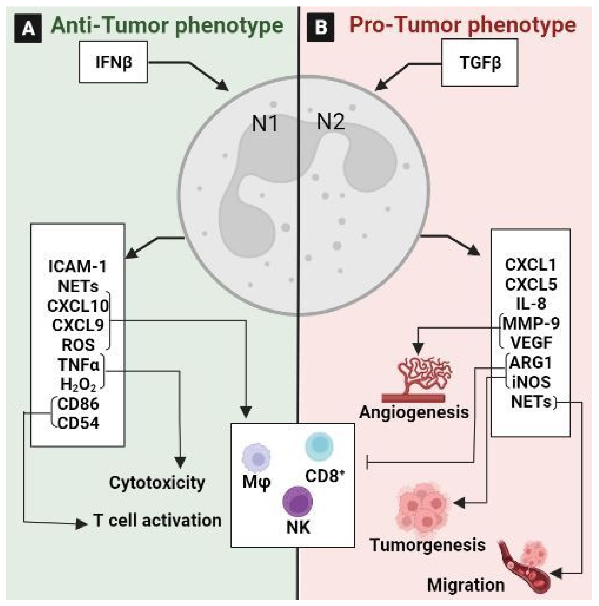
3.2. Tumor-Associated Neutrophils (TANs)

TANs constitute a significant part of the tumor microenvironment as they play a substantial role in linking inflammation and cancer, and are also involved in tumor progression and metastasis [179]. For this reason, neutrophils could be considered to be one of the emerging targets in multiple cancers [180]. TANs are divided into two populations: the anti-tumorigenic “N1” phenotype and the pro-tumorigenic “N2” phenotype, which means also that TANs exhibit considerable plasticity and are capable of polarization into either tumor-suppressive or -supportive cells [163] (Figure 9). Previously, it has been reported that N1 neutrophils increase cytotoxicity and reduce the immunosuppressive activity of immune cells by producing TNFα, the intercellular adhesion molecule (ICAM)-1, ROS, and Fas, and by decreasing arginase expression (Figure 9A). In contrast, N2 neutrophils support tumor expansion by expressing arginase, MMP-9, and VEGF, and by reducing ROS production and the intra-tumoral recruitment of cytotoxic T lymphocytes [181] (Figure 9B). The plasticity of TANs is regulated by TGF-β and IFN-β signaling [182]. Indeed, TGFβ signaling functions as a regulator between the N1 and N2 phenotypes, and its inhibition induces an anti-tumoral N1 phenotype. Hence, the anti-TGF-β treatment increases the cytotoxicity of TANs, decreases the metastasis, and significantly increases the apoptosis of CRC cells by suppressing the activation of the PI3K/AKT pathway in TANs and TGF-β/Smad signaling in tumor cells [183]. Furthermore, TANs contribute to tumor invasion and angiogenesis, and promote tumor cell dissemination by capturing circulating tumor cells using neutrophil extracellular traps (NET) and promoting their migration to distant sites. NETs can activate toll-like receptor 9 on CRC cells, resulting in cellular growth, migration, and invasion via the activation of MAPK signaling [182]. Recently, it has been reported that IL-8 upregulation induces neutrophil enrichment and NET formation in KRAS-mutant tissues, which promote the growth of CRC cells [184]. Moreover, the neutrophil-to-lymphocyte ratio is a well-defined predictive marker for CRC patients, and a high ratio is considered to be a poor prognostic factor for this cancer [182]. Similarly, a retrospective study using a cohort of 354 CRC patients with stage I–III cancer revealed a strong relationship between dynamic changes in the neutrophil-to-lymphocyte ratio and overall survival [185].
All these data support that TANs exhibit contradictory roles in CRC with both pro-tumoral and anti-tumoral properties depending on the immunological context.

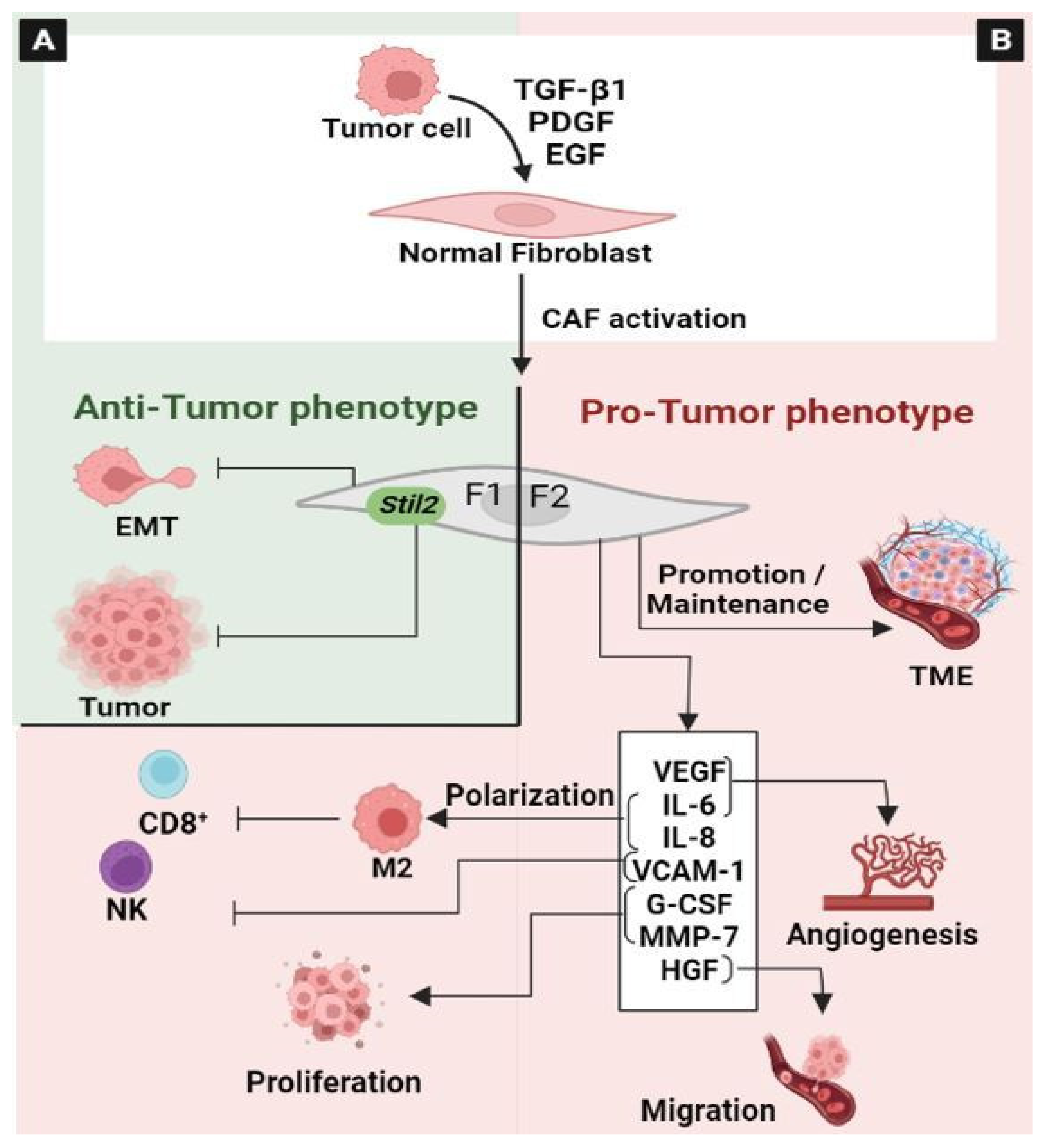
3.3. Cancer-Associated Fibroblasts (CAF)

Fibroblasts, commonly known as cancer-associated fibroblasts (CAFs), are the most abundant cells in the TME that influence tumor growth [186]. Nevertheless, the role of CAFs in colorectal cancer (CRC) development is not fully understood.
CAFs are a heterogeneous and plastic population. Different elements contribute to this heterogeneity, including the tissue type in which the tumor grows, the local paracrine environment, and the cell type of origin [187]. CAFs originate in their majority from the “activation” of local tissue fibroblasts via the action of tumor cell-secreted factors, such as TGFβ or platelet-derived growth factor (PDGF) [188]. Like other cells of the TME that display an enormous grade of plasticity, CAFs also have considerable plasticity and are capable of polarization [187]. The concept of “CAF polarization” highlights the functional heterogeneity of CAFs. They are divided into two functionally distinct subtypes: F1 and F2 polarized fibroblasts [187]. F1 represents CAF with antitumor effects, while F2 is CAF with tumor-promoting properties (Figure 10). However, the potential antitumor effects of CAFs are by far less studied compared to their tumor-promoting activity. Previously, it has been reported that primary fibroblasts, established from both normal and cancer tissues, can inhibit the proliferation of a panel of co-cultured cancer cells in vitro [189]. In breast cancer, the activation of Robo1 signaling by Slit2 from a stromal fibroblast prevents tumorigenesis, via the blocking of the PI3K/AKT/β-catenin pathway [190]. Similarly, it has been observed that fibroblast-derived Wnt3a could inhibit the growth of different patient-derived breast xenograft tumors [191]. A recent study demonstrated that Wnt induced a phenotypic switch in CAFs into non-aggressive cells and inhibited EMT in CRC [192]. Furthermore, the effects of CAFs on tumorigenesis appear to be less dependent on the instructive role of CAFs, but rather on the interacting compartments. For example, as described previously, Wnt/β-catenin signaling can be linked to the antitumoral effect of CAFs both in CRC and breast cancer [191,192]. In contrast, in CRC, it has been reported that WNT2 is selectively elevated in CAFs, leading to increased invasion and metastasis [193,194]. Similarly, as discussed above, one mechanism by which CAFs exert a tumor-suppressive effect is Slit2-induced Robo1 signaling, which is correlated positively with TGF-β [190]. However, TGF-β, for which CAFs are an important source, promotes tumor progression and metastasis [195] (Figure 10A).
It seems that CAFs can promote and suppress tumor formation, depending on their polarization profile in the local TME. The tumor-promoting activity of CAFs includes potent paracrine effects, which regulate the various cell types present in tumors [186,196] (Figure 10B). CAFs cooperate with tumor cells to promote the formation and maintenance of the tumor microenvironment by activating multiple signaling cascades, including the EGFR, JAK/STAT, TGF-β, and Wnt signaling pathways [197]. They promote an immunosuppressive microenvironment through the induction and accumulation of pro-tumoral macrophages [198]. However, the impact of the molecular crosstalk of tumor cells with CAFs and macrophages on monocyte recruitment and their phenotypic conversion is not fully depicted. It has been reported that colon fibroblasts and non-tumor cells recruit and dictate the fate of infiltrated monocytes towards a specific macrophage population, characterized by high CD163 expression and CCL2 production. Cytokine profiling revealed that CAFs produce M-CSF, IL6, IL8, and HGF. Moreover, macrophage/CAF/tumor cell co-cultures lead to the increased invasion of cancer cells [199]. CAFs can promote the polarization of macrophages toward the M2 phenotype, which contributes to the suppression of the functioning of natural killer cells through a synergistic mechanism. In addition, CAFs can also promote the adhesion of monocytes by up-regulating VCAM-1 expression in colorectal cancer cells. Thus, after VCAM-1 knocking-down in tumor cells or the depletion of macrophages, the pro-tumor effect of CAFs is partly abolished [200]. Recently, it has been demonstrated that IL1β, secreted by CRC cells, modifies surrounding normal fibroblasts in order to acquire protumorigenic characteristics and generate particularly chemoresistant cells [201]. Mechanistically, FGF-1/-3/FGFR4 signaling in cancer-associated fibroblasts promotes tumor progression in colon cancer through ERK and MMP-7 [202]. In addition, it has been reported that the constitutive activation of STAT3 in the CAFs of CRC promotes tumorigenesis [203]. Additionally, the concomitant activation of Wnt signaling and YAP/TAZ signaling coordinate to generate CAFs in CRC [204].

3.4. Tumor-Infiltrating Lymphocytes (TIL)

Immune responses play important and complex roles in immune surveillance and antitumor immunity during CRC progression [205]. TILs are located both within the tumor and in the peritumoral stroma. They are highly heterogeneous based on their cell-type compositions, gene expression profiles, and functional properties, which might contribute to the number of diverse responses to cancer immunotherapies [205]. TILs composed of NK cells, CD8+ cytotoxic T cells, and CD4+ helper T cells, are essential players in the defense against tumor cells [206]. Both CD8+ T cell and CD57+ NK cell infiltration, and Th1-type responses, are associated with a good prognosis in patients with colorectal cancer. However, the function of TILs can be controlled and inhibited by Treg cells [207]. Using the clustering analysis, Yang et al. identified eight T cell types from tumor tissues, including tumor Tregs, CD4+/CD8+ TRM T cells, CD4+/CD8+ effector memory T cells, Th17 cells, depleted CD8+ T cells, and CD8+ intraepithelial lymphocytes [208]. These T cell types likely represent the predominant tumor-infiltrating T cell subset in moderately differentiated CRC [208]. Treg cells in human colon tumor tissues express immunosuppressive molecules such as PD-1, cytotoxic T-lymphocyte-associated protein-4 (CTLA-4), T cell immunoglobulin and mucin domain-3 (TIM-3) and lymphocyte-activation gene 3 (LAG-3) [209]. In colon tumors, Treg cell depletion results in an increased accumulation of conventional T cells, including Th1-type T cells and IL-17A-producing T cells, suggesting that Treg cells not only regulate lymphocyte effector functions but also their recruitment to effector sites. Therefore, targeting Treg cells in antitumor immunotherapy can not only enhance the effector function of activated T cells, but also increase their numbers in tumors [210]. Of the TILs, helper T lymphocytes are considered to be the main players in tumor immunity that influence tumor progression. T helper type 1 (Th1), Th2, Th17, and regulatory T (Treg) cells have been identified as subtypes of helper T cells [210]. In TME, lymphoid cells exhibit immune-suppressive or stimulatory capacities. In the Th1/Th2 paradigm, cytotoxic T cell activity is supported by the Th1 lineage and M1 macrophages. In contrast, regulatory T lymphocytes, B-lymphocytes, and M2 macrophages are more closely related to tumor-promoting Th2 responses [211] (Figure 11). In healthy individuals, the Th1 and Th2 populations regulate each other. In patients with cancer, this balance shifts in favor of the Th2 cells. In CRC, it was reported that Th1 cell predominance is correlated with a good prognosis, whereas a high proportion of Th2 cells is associated with a worse one [207]. Indeed, M1 macrophages secrete cytokines such as IL-12, and can aid in the generation of Th1 adaptive immunity and impart a direct cytotoxic effect on tumor cells [212]. Recently, it has been reported that increased TGFβ in the tumor microenvironment represents a major mechanism for immune evasion, promoting T cell rejection and blocking the acquisition of a Th1 effector phenotype [213]. Interestingly, it has been demonstrated that exosomes derived from heat-stressed tumor cells possess a powerful capacity to convert immunosuppressive Tregs into Th17 cells via IL-6, which contributes to their potent antitumor effect [214]. In colon cancer, the immunoscore has extensively validated the prognostic significance of TILs. Based on this scoring system, the concept of “hot” (T-cell inflamed) and “cold” (non-T-cells inflamed) has emerged. Hot tumors are characterized by T cell infiltration and the molecular signatures of immune activation, whereas cold tumors show the striking features of T cell absence or exclusion [13]. Tumor intrinsic signaling pathways regulate T cell exclusion or their infiltration into tumors, which is crucial for developing novel therapeutic strategies against cancer. Local adaptive immunosuppression may be induced by involving the activation of various oncogenic pathways [215]. Recently, it has been reported that the downregulation of STAT1 induces immune escape in CRC, and that its upregulation implies a “hot” immunogenic microenvironment [160]. Furthermore, IFN-γ/JAK/STAT1 signaling stimulates PD-L1 expression in CRC cells [216]. The co-expression of the phosphorylated form of STAT1 and PD-L1 in CRC cells is strongly correlated with CD4- and CD8-positive TILs [217]. In addition, the intrinsic IFN type I (IFNα and IFNγ) signaling pathway is essential for the cytotoxic T lymphocyte (CTL) effector function in tumor suppression. Indeed, to evade host cancer immunosurveillance, human CRC may down-regulate the IFNAR1 on CTLs to impair the CTL effector function [218]. Another recent study has shown that TGF-β and PI3K signaling in CD4+ T cells (specifically Th17 cells) promotes the emergence of IL-22-producing Th17 cells and thereby tumorigenesis in mice [121]. An analysis of 155 colorectal cancer tissues highlighted that tumors with high β-catenin expression are characterized by a significant reduction in CD8+ T-cell infiltration. Mechanistically, β-catenin can regulate CCL4 expression to recruit CD103+ dendritic cells to enable CD8+ T cell activation [30]. Similarly, the immune escape in cancer is associated with the activation of various pathways, such as WNT–β-catenin, MAPK, JAK/STAT3, and NF- κB signaling [205]. The engagement of these pathways results in the production of cytokines and chemokines that ultimately mediate the exclusion of T cells from the TME, or the repression of factors that facilitate T cell recruitment.

3.5. Tumor-Associated Dendritic Cells (TADC)

TADCs play a key role in the orchestration of innate and adaptive antitumor immunity [219]. Therefore, dendritic cells (DCs) can strongly influence tumor progression and have a major impact on clinical outcomes in cancer patients [220]. They act as sentinels, detect tumor antigens, present them to CD8+ T-cells, and supply necessary signals for both the activation and suppression of CD8+ T-cells [221]. DCs play relevant roles in tumors by exerting both pro- and anti-tumorigenic functions depending on the local environment. The quantitative and functional impairment of DCs is widely observed in several cancer types, including CRC, and represents a tumor mechanism that is used by cancer cells to escape host immune surveillance [219]. Conventional DCs (cDCs) can be broadly divided into cDC1 and cDC2 populations (Figure 11), which arise through distinct pre-DC lineages [222]. Single-cell RNA sequencing, using CRC tissues, identifies three DC subsets: plasmacytoid DC (pDC), cDC2, and cDC1 cells, which are characterized by the high expression of HLA-DRs and the low expression of CD14, expressing specifically LILRA4/LILRB4, CD1C/FCER1A, and XCR1/ BATF3, respectively [223]. cDC1s are critical for generating anti-tumor T cell responses because cDC1s are capable of presenting tumor-associated antigens to CD8+ T cells and Th1 cells [221,224]. They produce type I and type III IFN and IL-12 at lower levels, and correlate with a favorable prognosis in cancer patients [224]. They are considered central to antitumor immunity and their presence in the tumor microenvironment is associated with improved outcomes in patients with cancer. Recently, it has been reported that the accumulation of CD8+ T cells indeed relies on BATF3-dependent cDC1s [225]. A direct correlation between Notch2 signaling, the infiltration of cDC1, and the association of the suppressed cDC1 signature with a poor prognosis in human CRC has also been shown. These findings reveal a critical role for Notch2-dependent cDC1s in preventing inflammation-associated transformation in the mouse model and tumor progression in human CRCs [226]. However, the role of cDC2s in tumors remains unclear. They exhibit substantial heterogeneity and they preferentially initiate CD4+ Tconv responses in a variety of immunological models [227]. Similarly, scRNA-seq and functional data in the TME demonstrate that two distinct populations of IRF4-dependent CD11b+ cDC2 are required for initiating the activation of antitumor CD4+ Tconv in vivo. cDC2 presented tumor-derived antigens to CD4+ Tconv, but failed to support antitumor CD4+ Tconv differentiation. However, Treg cell depletion enhanced their capacity to elicit strong CD4+ Tconv responses and ensure antitumor protection [228]. As reviewed, cDC2 expresses a large repertoire of pattern recognition receptors (PRRs) and pro- and anti-inflammatory cytokines, including IL-12. Mainly, they induce Th17 cell activation, but also Th1 cell, Th2 cell, Treg cell, and CD8+ T cell (cross-presentation) activation, which depends on the context and precise cDC2 subpopulation [224]. It has been reported that both cDC1 and cDC2 cells are programmed to differentiate into regulatory subsets upon their uptake of tumor antigens [229]. Thus, cDCs play an immunoregulatory role in the tumor microenvironment. The plasmacytoid dendritic cells (pDCs) can be stimulated to activate CD8+ T cells to generate powerful tumor antigen-specific CD8+ T cell responses [224,230]. The role that this DC subset plays in tolerogenic settings is poorly described, but correlates with a poor prognosis in cancer [224]. This is mainly due to the association that is made between the accumulation of pDC and the increase in Tregs cells, with the presence of TGF-β and/or IL10; these have an immunosuppressive phenotype, which results in a decreased overall survival rate in the patients [231]. Similarly, the analysis of a cohort of 64 gastric cancer patients without preoperative chemotherapy demonstrated an increased infiltration of BDCA2+ pDCs and Foxp3+ Tregs in tumors compared to normal ones. Indeed, BDCA2+ pDCs were positively associated with Foxp3+ Tregs [232], which were both considered to be immunosuppressive cells in the TME. Previously, it has been reported that pDCs can generate Tregs from naive CD4+ T cells, thereby contributing to tumor immune escape [233,234]. In addition, in ovarian cancers, they are essential for immunosuppression due to their expression of indoleamine 2,3-dioxygenase 1 (IDO1) and the inducible T cell costimulatory ligand (ICOSL) [235]. In contrast, it has been reported that higher densities of tumor-infiltrating pDCs are associated with better prognosis and the prolonged survival of patients with colon cancer. This effect is due to the co-localization of pDC and CD8+ T cells in tumor stroma, where activated pDCs may contribute to the stimulation of tumor-reactive CD8+ cytotoxic T cells [220]. These findings suggest that the TME plays a critical role in shaping the functional properties of infiltrating pDCs (tolerogenic vs activated). It has been shown that immunosuppressive DCs can be generated depending on the TME components, such as β-catenin, STAT3, TGF-β, IDO, endoplasmic reticulum (ER) stress, increased levels of lactate, VEGF, IL-10, TGF-β, prostaglandins, the accumulation of adenosine, and hypoxia [236,237].

4. Conclusions

Despite the progress made in the management of CRC, there are still many challenges to overcome in the development of antineoplastic treatments.
Through this review, we tried to actualize and compile what is known about the pivotal role of some signaling pathways in CRC development and progression, focusing on the paradoxical role of immune cells as beneficial or detrimental markers, and the crosstalk between tumor cells and the TME.
Hence, the double-edged sword function of the regulators that orchestrate tumor dissemination and the complex interactions that take place in the TME could explain the limited efficacy of therapeutic drugs and suggest that caution is applied before their administration to patients. Accordingly, an accurate staging of patients based on specific molecular groups, target identification and TME interactions could be a valuable tool to guide the optimal treatment.
Thus, modeling these complexities at the level of the individual patient and developing personalized anti-tumor approaches that block this crosstalk could rationalize the immunotherapy use and contribute not only to refining and to adapting treatment, but in improving the efficacy of therapies and bypassing anticancer treatment failure.

Author Contributions

K.E.-B. conceived of the review; S.B.H. conducted the literature review and drafted the original manuscript. K.E.-B. wrote, edited and revised the review. All authors have read and agreed to the published version of the manuscript.

Funding

This work was supported by the Tunisian Ministry of Higher Education and Research (LR16IPT04). The funders had no role in the decision to publish or in the preparation of the manuscript.

Institutional Review Board Statement

Not applicable.

Informed Consent Statement

Not applicable.

Data Availability Statement

Not applicable.

Conflicts of Interest

The authors declare no conflict of interest.

References

  1. Murphy, N.; Ward, H.A.; Jenab, M.; Rothwell, J.A.; Boutron-Ruault, M.-C.; Carbonnel, F.; Kvaskoff, M.; Kaaks, R.; Kühn, T.; Boeing, H.; et al. Heterogeneity of Colorectal Cancer Risk Factors by Anatomical Subsite in 10 European Countries: A Multinational Cohort Study. Clin. Gastroenterol. Hepatol. 2019, 17, 1323–1331.e6. [Google Scholar] [CrossRef] [PubMed] [Green Version]
  2. Global Cancer Observatory. Available online: https://gco.iarc.fr/ (accessed on 6 February 2023).
  3. Sung, H.; Ferlay, J.; Siegel, R.L.; Laversanne, M.; Soerjomataram, I.; Jemal, A.; Bray, F. Global Cancer Statistics 2020: GLOBOCAN Estimates of Incidence and Mortality Worldwide for 36 Cancers in 185 Countries. CA Cancer J. Clin. 2021, 71, 209–249. [Google Scholar] [CrossRef] [PubMed]
  4. Engstrand, J.; Nilsson, H.; Strömberg, C.; Jonas, E.; Freedman, J. Colorectal Cancer Liver Metastases—A Population-Based Study on Incidence, Management and Survival. BMC Cancer 2018, 18, 78. [Google Scholar] [CrossRef] [PubMed] [Green Version]
  5. Tieng, F.Y.F.; Baharudin, R.; Abu, N.; Mohd Yunos, R.-I.; Lee, L.-H.; Ab Mutalib, N.-S. Single Cell Transcriptome in Colorectal Cancer—Current Updates on Its Application in Metastasis, Chemoresistance and the Roles of Circulating Tumor Cells. Front. Pharm. 2020, 11, 135. [Google Scholar] [CrossRef] [PubMed] [Green Version]
  6. Riahi-Chebbi, I.; Souid, S.; Othman, H.; Haoues, M.; Karoui, H.; Morel, A.; Srairi-Abid, N.; Essafi, M.; Essafi-Benkhadir, K. The Phenolic Compound Kaempferol Overcomes 5-Fluorouracil Resistance in Human Resistant LS174 Colon Cancer Cells. Sci. Rep. 2019, 9, 195. [Google Scholar] [CrossRef] [Green Version]
  7. Luo, Z.; Wang, Q.; Lau, W.B.; Lau, B.; Xu, L.; Zhao, L.; Yang, H.; Feng, M.; Xuan, Y.; Yang, Y.; et al. Tumor Microenvironment: The Culprit for Ovarian Cancer Metastasis? Cancer Lett. 2016, 377, 174–182. [Google Scholar] [CrossRef]
  8. Ma, J.; Huang, L.; Hu, D.; Zeng, S.; Han, Y.; Shen, H. The Role of the Tumor Microbe Microenvironment in the Tumor Immune Microenvironment: Bystander, Activator, or Inhibitor? J. Exp. Clin. Cancer Res. 2021, 40, 327. [Google Scholar] [CrossRef] [PubMed]
  9. Peddareddigari, V.G.; Wang, D.; DuBois, R.N. The Tumor Microenvironment in Colorectal Carcinogenesis. Cancer Microenviron. 2010, 3, 149–166. [Google Scholar] [CrossRef] [Green Version]
  10. Virchow, R. Cellular Pathology. As Based upon Physiological and Pathological Histology. Lecture XVI—Atheromatous Affection of Arteries. 1858. Nutr. Rev. 1989, 47, 23–25. [Google Scholar] [CrossRef]
  11. Wang, H.; Yung, M.M.H.; Ngan, H.Y.S.; Chan, K.K.L.; Chan, D.W. The Impact of the Tumor Microenvironment on Macrophage Polarization in Cancer Metastatic Progression. Int. J. Mol. Sci. 2021, 22, 6560. [Google Scholar] [CrossRef]
  12. Corsale, A.M.; Di Simone, M.; Lo Presti, E.; Picone, C.; Dieli, F.; Meraviglia, S. Metabolic Changes in Tumor Microenvironment: How Could They Affect Γδ T Cells Functions? Cells 2021, 10, 2896. [Google Scholar] [CrossRef] [PubMed]
  13. Duan, Q.; Zhang, H.; Zheng, J.; Zhang, L. Turning Cold into Hot: Firing up the Tumor Microenvironment. Trends Cancer 2020, 6, 605–618. [Google Scholar] [CrossRef]
  14. Chen, C.-K.; Pan, C.-L. Cell Polarity Control by Wnt Morphogens. Dev. Biol. 2022, 487, 34–41. [Google Scholar] [CrossRef]
  15. Park, J.H.; Pyun, W.Y.; Park, H.W. Cancer Metabolism: Phenotype, Signaling and Therapeutic Targets. Cells 2020, 9, 2308. [Google Scholar] [CrossRef] [PubMed]
  16. Liu, X.; Ji, Q.; Fan, Z.; Li, Q. Cellular Signaling Pathways Implicated in Metastasis of Colorectal Cancer and the Associated Targeted Agents. Futur. Oncol. 2015, 11, 2911–2922. [Google Scholar] [CrossRef] [PubMed]
  17. Malki, A.; ElRuz, R.A.; Gupta, I.; Allouch, A.; Vranic, S.; Al Moustafa, A.-E. Molecular Mechanisms of Colon Cancer Progression and Metastasis: Recent Insights and Advancements. Int. J. Mol. Sci. 2020, 22, 130. [Google Scholar] [CrossRef] [PubMed]
  18. Nusse, R.; Clevers, H. Wnt/β-Catenin Signaling, Disease, and Emerging Therapeutic Modalities. Cell 2017, 169, 985–999. [Google Scholar] [CrossRef] [Green Version]
  19. Shang, S.; Hua, F.; Hu, Z.-W. The Regulation of β-Catenin Activity and Function in Cancer: Therapeutic Opportunities. Oncotarget 2017, 8, 33972–33989. [Google Scholar] [CrossRef] [Green Version]
  20. Vilchez, V.; Turcios, L.; Marti, F.; Gedaly, R. Targeting Wnt/β-Catenin Pathway in Hepatocellular Carcinoma Treatment. World J. Gastroenterol. 2016, 22, 823–832. [Google Scholar] [CrossRef]
  21. Polakis, P. Casein Kinase 1: A Wnt’er of Disconnect. Curr. Biol. 2002, 12, R499–R501. [Google Scholar] [CrossRef] [Green Version]
  22. Zhang, Y.; Wang, X. Targeting the Wnt/β-Catenin Signaling Pathway in Cancer. J. Hematol. Oncol. 2020, 13, 165. [Google Scholar] [CrossRef] [PubMed]
  23. Grumolato, L.; Liu, G.; Haremaki, T.; Mungamuri, S.K.; Mong, P.; Akiri, G.; Lopez-Bergami, P.; Arita, A.; Anouar, Y.; Mlodzik, M.; et al. β-Catenin-Independent Activation of TCF1/LEF1 in Human Hematopoietic Tumor Cells through Interaction with ATF2 Transcription Factors. PLOS Genet. 2013, 9, e1003603. [Google Scholar] [CrossRef] [Green Version]
  24. Bu, H.; Liu, D.; Cui, J.; Cai, K.; Shen, F. Wnt/β-Catenin Signaling Pathway Is Involved in Induction of Apoptosis by Oridonin in Colon Cancer COLO205 Cells. Transl. Cancer Res. 2019, 8, 1782–1794. [Google Scholar] [CrossRef]
  25. Yaeger, R.; Chatila, W.K.; Lipsyc, M.D.; Hechtman, J.F.; Cercek, A.; Sanchez-Vega, F.; Jayakumaran, G.; Middha, S.; Zehir, A.; Donoghue, M.T.A.; et al. Clinical Sequencing Defines the Genomic Landscape of Metastatic Colorectal Cancer. Cancer Cell 2018, 33, 125–136.e3. [Google Scholar] [CrossRef] [Green Version]
  26. Liu, C.-C.; Cai, D.-L.; Sun, F.; Wu, Z.-H.; Yue, B.; Zhao, S.-L.; Wu, X.-S.; Zhang, M.; Zhu, X.-W.; Peng, Z.-H.; et al. FERMT1 Mediates Epithelial-Mesenchymal Transition to Promote Colon Cancer Metastasis via Modulation of β-Catenin Transcriptional Activity. Oncogene 2017, 36, 1779–1792. [Google Scholar] [CrossRef] [PubMed]
  27. He, T.C.; Sparks, A.B.; Rago, C.; Hermeking, H.; Zawel, L.; da Costa, L.T.; Morin, P.J.; Vogelstein, B.; Kinzler, K.W. Identification of C-MYC as a Target of the APC Pathway. Science 1998, 281, 1509–1512. [Google Scholar] [CrossRef] [PubMed]
  28. Tetsu, O.; McCormick, F. Beta-Catenin Regulates Expression of Cyclin D1 in Colon Carcinoma Cells. Nature 1999, 398, 422–426. [Google Scholar] [CrossRef]
  29. Neth, P.; Ciccarella, M.; Egea, V.; Hoelters, J.; Jochum, M.; Ries, C. Wnt Signaling Regulates the Invasion Capacity of Human Mesenchymal Stem Cells. Stem Cells 2006, 24, 1892–1903. [Google Scholar] [CrossRef]
  30. Xue, J.; Yu, X.; Xue, L.; Ge, X.; Zhao, W.; Peng, W. Intrinsic β-Catenin Signaling Suppresses CD8+ T-Cell Infiltration in Colorectal Cancer. Biomed. Pharm. 2019, 115, 108921. [Google Scholar] [CrossRef]
  31. Zhong, Z.A.; Michalski, M.N.; Stevens, P.D.; Sall, E.A.; Williams, B.O. Regulation of Wnt Receptor Activity: Implications for Therapeutic Development in Colon Cancer. J. Biol. Chem. 2021, 296, 100782. [Google Scholar] [CrossRef]
  32. Famili, F.; Naber, B.A.E.; Vloemans, S.; de Haas, E.F.E.; Tiemessen, M.M.; Staal, F.J.T. Discrete Roles of Canonical and Non-Canonical Wnt Signaling in Hematopoiesis and Lymphopoiesis. Cell Death Dis. 2015, 6, e1981. [Google Scholar] [CrossRef] [PubMed] [Green Version]
  33. D’Amico, L.; Mahajan, S.; Capietto, A.-H.; Yang, Z.; Zamani, A.; Ricci, B.; Bumpass, D.B.; Meyer, M.; Su, X.; Wang-Gillam, A.; et al. Dickkopf-Related Protein 1 (Dkk1) Regulates the Accumulation and Function of Myeloid Derived Suppressor Cells in Cancer. J. Exp. Med. 2016, 213, 827–840. [Google Scholar] [CrossRef] [Green Version]
  34. Poh, T.W.; Bradley, J.M.; Mukherjee, P.; Gendler, S.J. Lack of Muc1-Regulated Beta-Catenin Stability Results in Aberrant Expansion of CD11b+Gr1+ Myeloid Derived Suppressor Cells from the Bone Marrow. Cancer Res. 2009, 69, 3554–3562. [Google Scholar] [CrossRef] [PubMed] [Green Version]
  35. Capietto, A.-H.; Kim, S.; Sanford, D.E.; Linehan, D.C.; Hikida, M.; Kumosaki, T.; Novack, D.V.; Faccio, R. Down-Regulation of PLCγ2–β-Catenin Pathway Promotes Activation and Expansion of Myeloid-Derived Suppressor Cells in Cancer. J. Exp. Med. 2013, 210, 2257–2271. [Google Scholar] [CrossRef] [PubMed] [Green Version]
  36. Pai, S.G.; Carneiro, B.A.; Mota, J.M.; Costa, R.; Leite, C.A.; Barroso-Sousa, R.; Kaplan, J.B.; Chae, Y.K.; Giles, F.J. Wnt/Beta-Catenin Pathway: Modulating Anticancer Immune Response. J. Hematol. Oncol. 2017, 10, 101. [Google Scholar] [CrossRef] [PubMed] [Green Version]
  37. Kramer, E.D.; Abrams, S.I. Granulocytic Myeloid-Derived Suppressor Cells as Negative Regulators of Anticancer Immunity. Front. Immunol. 2020, 11, 1963. [Google Scholar] [CrossRef]
  38. Lee, S.; Rauch, J.; Kolch, W. Targeting MAPK Signaling in Cancer: Mechanisms of Drug Resistance and Sensitivity. Int. J. Mol. Sci. 2020, 21, 1102. [Google Scholar] [CrossRef] [Green Version]
  39. Asl, E.R.; Amini, M.; Najafi, S.; Mansoori, B.; Mokhtarzadeh, A.; Mohammadi, A.; Lotfinejad, P.; Bagheri, M.; Shirjang, S.; Lotfi, Z.; et al. Interplay between MAPK/ERK Signaling Pathway and MicroRNAs: A Crucial Mechanism Regulating Cancer Cell Metabolism and Tumor Progression. Life Sci. 2021, 278, 119499. [Google Scholar] [CrossRef]
  40. Ros, J.; Baraibar, I.; Sardo, E.; Mulet, N.; Salvà, F.; Argilés, G.; Martini, G.; Ciardiello, D.; Cuadra, J.L.; Tabernero, J.; et al. BRAF, MEK and EGFR Inhibition as Treatment Strategies in BRAF V600E Metastatic Colorectal Cancer. Ther. Adv. Med. Oncol. 2021, 13, 1758835921992974. [Google Scholar] [CrossRef]
  41. Yang, W.; Redpath, R.E.; Zhang, C.; Ning, N. Long Non-Coding RNA H19 Promotes the Migration and Invasion of Colon Cancer Cells via MAPK Signaling Pathway. Oncol. Lett. 2018, 16, 3365–3372. [Google Scholar] [CrossRef]
  42. Grossi, V.; Peserico, A.; Tezil, T.; Simone, C. P38α MAPK Pathway: A Key Factor in Colorectal Cancer Therapy and Chemoresistance. World J. Gastroenterol. 2014, 20, 9744–9758. [Google Scholar] [CrossRef] [PubMed]
  43. Li, S.; Ung, T.T.; Nguyen, T.T.; Sah, D.K.; Park, S.Y.; Jung, Y.D. Cholic Acid Stimulates MMP-9 in Human Colon Cancer Cells via Activation of MAPK, AP-1, and NF-ΚB Activity. Int. J. Mol. Sci. 2020, 21, 3420. [Google Scholar] [CrossRef]
  44. Zakraoui, O.; Marcinkiewicz, C.; Aloui, Z.; Othman, H.; Grépin, R.; Haoues, M.; Essafi, M.; Srairi-Abid, N.; Gasmi, A.; Karoui, H.; et al. Lebein, a Snake Venom Disintegrin, Suppresses Human Colon Cancer Cells Proliferation and Tumor-Induced Angiogenesis through Cell Cycle Arrest, Apoptosis Induction and Inhibition of VEGF Expression: Mechanisms and Targets for Lebein in Colorectal Cancer. Mol. Carcinog. 2017, 56, 18–35. [Google Scholar] [CrossRef] [PubMed]
  45. Tarhouni-Jabberi, S.; Zakraoui, O.; Ioannou, E.; Riahi-Chebbi, I.; Haoues, M.; Roussis, V.; Kharrat, R.; Essafi-Benkhadir, K. Mertensene, a Halogenated Monoterpene, Induces G2/M Cell Cycle Arrest and Caspase Dependent Apoptosis of Human Colon Adenocarcinoma HT29 Cell Line through the Modulation of ERK-1/-2, AKT and NF-ΚB Signaling. Mar. Drugs 2017, 15, 221. [Google Scholar] [CrossRef] [PubMed] [Green Version]
  46. Lee, M.; Yang, C.; Park, S.; Song, G.; Lim, W. Fraxetin Induces Cell Death in Colon Cancer Cells via Mitochondria Dysfunction and Enhances Therapeutic Effects in 5-Fluorouracil Resistant Cells. J. Cell. Biochem. 2022, 123, 469–480. [Google Scholar] [CrossRef]
  47. Dhanasekaran, D.N.; Reddy, E.P. JNK-Signaling: A Multiplexing Hub in Programmed Cell Death. Genes Cancer 2017, 8, 682–694. [Google Scholar] [CrossRef] [Green Version]
  48. Sun, W.; Wu, X.; Gao, H.; Yu, J.; Zhao, W.; Lu, J.-J.; Wang, J.; Du, G.; Chen, X. Cytosolic Calcium Mediates RIP1/RIP3 Complex-Dependent Necroptosis through JNK Activation and Mitochondrial ROS Production in Human Colon Cancer Cells. Free Radic. Biol. Med. 2017, 108, 433–444. [Google Scholar] [CrossRef]
  49. Ren, Y.; Lv, C.; Zhang, J.; Zhang, B.; Yue, B.; Luo, X.; Yu, Z.; Wang, H.; Ren, J.; Wang, Z.; et al. Alantolactone Exhibits Antiproliferative and Apoptosis-Promoting Properties in Colon Cancer Model via Activation of the MAPK-JNK/c-Jun Signaling Pathway. Mol. Cell. Biochem. 2021, 476, 4387–4403. [Google Scholar] [CrossRef]
  50. Song, N.; Ma, J.; Hu, W.; Guo, Y.; Hui, L.; Aamer, M.; Ma, J. Lappaconitine Hydrochloride Inhibits Proliferation and Induces Apoptosis in Human Colon Cancer HCT-116 Cells via Mitochondrial and MAPK Pathway. Acta Histochem. 2021, 123, 151736. [Google Scholar] [CrossRef]
  51. Bray, S.J. Notch Signalling: A Simple Pathway Becomes Complex. Nat. Rev. Mol. Cell. Biol. 2006, 7, 678–689. [Google Scholar] [CrossRef]
  52. Mumm, J.S.; Kopan, R. Notch Signaling: From the outside in. Dev. Biol. 2000, 228, 151–165. [Google Scholar] [CrossRef] [PubMed] [Green Version]
  53. Ntziachristos, P.; Lim, J.S.; Sage, J.; Aifantis, I. From Fly Wings to Targeted Cancer Therapies: A Centennial for Notch Signaling. Cancer Cell 2014, 25, 318–334. [Google Scholar] [CrossRef] [PubMed] [Green Version]
  54. Andersson, E.R.; Sandberg, R.; Lendahl, U. Notch Signaling: Simplicity in Design, Versatility in Function. Development 2011, 138, 3593–3612. [Google Scholar] [CrossRef] [PubMed] [Green Version]
  55. Nowell, C.S.; Radtke, F. Notch as a Tumour Suppressor. Nat. Rev. Cancer 2017, 17, 145–159. [Google Scholar] [CrossRef] [PubMed]
  56. Qu, X.; Zhao, L.; Wang, M.; Zhang, R.; Cheng, L.; Qiu, L.; Tong, X.; Cai, S.; Wei, Q.; Li, Q. Novel Functional Variants in the Notch Pathway and Survival of Chinese Colorectal Cancer. Int. J. Cancer 2021, 149, 84–96. [Google Scholar] [CrossRef]
  57. Wang, F.; Long, J.; Li, L.; Zhao, Z.; Wei, F.; Yao, Y.; Qiu, W.; Wu, Z.; Luo, Q.; Liu, W.; et al. Mutations in the Notch Signalling Pathway Are Associated with Enhanced Anti-tumour Immunity in Colorectal Cancer. J. Cell. Mol. Med. 2020, 24, 12176–12187. [Google Scholar] [CrossRef]
  58. Vinson, K.E.; George, D.C.; Fender, A.W.; Bertrand, F.E.; Sigounas, G. The Notch Pathway in Colorectal Cancer. Int. J. Cancer 2016, 138, 1835–1842. [Google Scholar] [CrossRef]
  59. Sonoshita, M.; Aoki, M.; Fuwa, H.; Aoki, K.; Hosogi, H.; Sakai, Y.; Hashida, H.; Takabayashi, A.; Sasaki, M.; Robine, S.; et al. Suppression of Colon Cancer Metastasis by Aes through Inhibition of Notch Signaling. Cancer Cell 2011, 19, 125–137. [Google Scholar] [CrossRef] [Green Version]
  60. Rodilla, V.; Villanueva, A.; Obrador-Hevia, A.; Robert-Moreno, A.; Fernández-Majada, V.; Grilli, A.; López-Bigas, N.; Bellora, N.; Albà, M.M.; Torres, F.; et al. Jagged1 Is the Pathological Link between Wnt and Notch Pathways in Colorectal Cancer. Proc. Natl. Acad. Sci. USA 2009, 106, 6315–6320. [Google Scholar] [CrossRef] [Green Version]
  61. Zhang, Y.; Li, B.; Ji, Z.-Z.; Zheng, P.-S. Notch1 Regulates the Growth of Human Colon Cancers. Cancer 2010, 116, 5207–5218. [Google Scholar] [CrossRef]
  62. Tyagi, A.; Sharma, A.K.; Damodaran, C. A Review on Notch Signaling and Colorectal Cancer. Cells 2020, 9, 1549. [Google Scholar] [CrossRef] [PubMed]
  63. Chu, D.; Zhang, Z.; Zhou, Y.; Wang, W.; Li, Y.; Zhang, H.; Dong, G.; Zhao, Q.; Ji, G. Notch1 and Notch2 Have Opposite Prognostic Effects on Patients with Colorectal Cancer. Ann. Oncol. 2011, 22, 2440–2447. [Google Scholar] [CrossRef] [PubMed]
  64. Lin, X.; Wang, S.; Sun, M.; Zhang, C.; Wei, C.; Yang, C.; Dou, R.; Liu, Q.; Xiong, B. MiR-195-5p/NOTCH2-Mediated EMT Modulates IL-4 Secretion in Colorectal Cancer to Affect M2-like TAM Polarization. J. Hematol. Oncol. 2019, 12, 20. [Google Scholar] [CrossRef] [Green Version]
  65. Jin, Y.; Wang, M.; Hu, H.; Huang, Q.; Chen, Y.; Wang, G. Overcoming Stemness and Chemoresistance in Colorectal Cancer through MiR-195-5p-Modulated Inhibition of Notch Signaling. Int. J. Biol. Macromol. 2018, 117, 445–453. [Google Scholar] [CrossRef]
  66. Serafin, V.; Persano, L.; Moserle, L.; Esposito, G.; Ghisi, M.; Curtarello, M.; Bonanno, L.; Masiero, M.; Ribatti, D.; Stürzl, M.; et al. Notch3 Signalling Promotes Tumour Growth in Colorectal Cancer. J. Pathol. 2011, 224, 448–460. [Google Scholar] [CrossRef] [PubMed]
  67. Bordonaro, M.; Tewari, S.; Atamna, W.; Lazarova, D.L. The Notch Ligand Delta-like 1 Integrates Inputs from TGFbeta/Activin and Wnt Pathways. Exp. Cell Res. 2011, 317, 1368–1381. [Google Scholar] [CrossRef] [Green Version]
  68. Colombo, M.; Mirandola, L.; Chiriva-Internati, M.; Basile, A.; Locati, M.; Lesma, E.; Chiaramonte, R.; Platonova, N. Cancer Cells Exploit Notch Signaling to Redefine a Supportive Cytokine Milieu. Front. Immunol. 2018, 9, 1823. [Google Scholar] [CrossRef]
  69. Koch, U.; Radtke, F. Notch and Cancer: A Double-Edged Sword. Cell. Mol. Life Sci. 2007, 64, 2746–2762. [Google Scholar] [CrossRef] [Green Version]
  70. Majumder, S.; Crabtree, J.S.; Golde, T.E.; Minter, L.M.; Osborne, B.A.; Miele, L. Targeting Notch in Oncology: The Path Forward. Nat. Rev. Drug Discov. 2021, 20, 125–144. [Google Scholar] [CrossRef]
  71. Ranganathan, P.; Weaver, K.L.; Capobianco, A.J. Notch Signalling in Solid Tumours: A Little Bit of Everything but Not All the Time. Nat. Rev. Cancer 2011, 11, 338–351. [Google Scholar] [CrossRef]
  72. Jin, H.-Y.; Zhang, H.-Y.; Wang, X.; Xu, J.; Ding, Y. Expression and Clinical Significance of Notch Signaling Genes in Colorectal Cancer. Tumour Biol. 2012, 33, 817–824. [Google Scholar] [CrossRef] [PubMed]
  73. Hu, J.; Yu, J.; Gan, J.; Song, N.; Shi, L.; Liu, J.; Zhang, Z.; Du, J. Notch1/2/3/4 Are Prognostic Biomarker and Correlated with Immune Infiltrates in Gastric Cancer. Aging 2020, 12, 2595–2609. [Google Scholar] [CrossRef] [PubMed]
  74. Xue, C.; Li, G.; Lu, J.; Li, L. Crosstalk between CircRNAs and the PI3K/AKT Signaling Pathway in Cancer Progression. Signal Transduct. Target. Ther. 2021, 6, 400. [Google Scholar] [CrossRef]
  75. Narayanankutty, A. PI3K/ Akt/ MTOR Pathway as a Therapeutic Target for Colorectal Cancer: A Review of Preclinical and Clinical Evidence. Curr. Drug Targets 2019, 20, 1217–1226. [Google Scholar] [CrossRef] [PubMed]
  76. Prossomariti, A.; Piazzi, G.; Alquati, C.; Ricciardiello, L. Are Wnt/β-Catenin and PI3K/AKT/MTORC1 Distinct Pathways in Colorectal Cancer? Cell. Mol. Gastroenterol. Hepatol. 2020, 10, 491–506. [Google Scholar] [CrossRef]
  77. Nogueira, V.; Park, Y.; Chen, C.-C.; Xu, P.-Z.; Chen, M.-L.; Tonic, I.; Unterman, T.; Hay, N. Akt Determines Replicative Senescence and Oxidative or Oncogenic Premature Senescence and Sensitizes Cells to Oxidative Apoptosis. Cancer Cell 2008, 14, 458–470. [Google Scholar] [CrossRef] [Green Version]
  78. Dolado, I.; Nebreda, A.R. AKT and Oxidative Stress Team up to Kill Cancer Cells. Cancer Cell 2008, 14, 427–429. [Google Scholar] [CrossRef] [Green Version]
  79. Los, M.; Maddika, S.; Erb, B.; Schulze-Osthoff, K. Switching Akt: From Survival Signaling to Deadly Response. Bioessays 2009, 31, 492–495. [Google Scholar] [CrossRef] [Green Version]
  80. Rangel, M.; Kong, J.; Bhatt, V.; Khayati, K.; Guo, J.Y. Autophagy and Tumorigenesis. FEBS J. 2022, 289, 7177–7198. [Google Scholar] [CrossRef]
  81. Xu, Z.; Han, X.; Ou, D.; Liu, T.; Li, Z.; Jiang, G.; Liu, J.; Zhang, J. Targeting PI3K/AKT/MTOR-Mediated Autophagy for Tumor Therapy. Appl. Microbiol. Biotechnol. 2020, 104, 575–587. [Google Scholar] [CrossRef]
  82. Chen, T.; Yu, Q.; Xin, L.; Guo, L. Retracted: Circular RNA CircC3P1 Restrains Kidney Cancer Cell Activity by Regulating MiR-21/PTEN Axis and Inactivating PI3K/AKT and NF- k B Pathways. J. Cell. Physiol. 2020, 235, 4001–4010. [Google Scholar] [CrossRef] [PubMed]
  83. Karin, M.; Cao, Y.; Greten, F.R.; Li, Z.-W. NF-ΚB in Cancer: From Innocent Bystander to Major Culprit. Nat. Rev. Cancer 2002, 2, 301–310. [Google Scholar] [CrossRef] [PubMed]
  84. Bonizzi, G.; Karin, M. The Two NF-ΚB Activation Pathways and Their Role in Innate and Adaptive Immunity. Trends Immunol. 2004, 25, 280–288. [Google Scholar] [CrossRef]
  85. Sen, R.; Baltimore, D. Inducibility of κ Immunoglobulin Enhancer-Binding Protein NF-ΚB by a Posttranslational Mechanism. Cell 1986, 47, 921–928. [Google Scholar] [CrossRef]
  86. Hassanzadeh, P. Colorectal Cancer and NF-ΚB Signaling Pathway. Gastroenterol. Hepatol. Bed Bench 2011, 4, 127–132. [Google Scholar]
  87. Soleimani, A.; Rahmani, F.; Ferns, G.A.; Ryzhikov, M.; Avan, A.; Hassanian, S.M. Role of the NF-ΚB Signaling Pathway in the Pathogenesis of Colorectal Cancer. Gene 2020, 726, 144132. [Google Scholar] [CrossRef]
  88. Quinn, J.A.; Bennett, L.; Patel, M.; Frixou, M.; Park, J.H.; Roseweir, A.; Horgan, P.G.; McMillan, D.C.; Edwards, J. The Relationship between Members of the Canonical NF-KB Pathway, Tumour Microenvironment and Cancer Specific Survival in Colorectal Cancer Patients. Histol. Histopathol. 2020, 35, 569–578. [Google Scholar] [CrossRef] [PubMed]
  89. Park, M.; Hong, J. Roles of NF-ΚB in Cancer and Inflammatory Diseases and Their Therapeutic Approaches. Cells 2016, 5, 15. [Google Scholar] [CrossRef] [PubMed]
  90. Karin, M.; Lin, A. NF-KappaB at the Crossroads of Life and Death. Nat. Immunol. 2002, 3, 221–227. [Google Scholar] [CrossRef]
  91. Liu, T.; Liu, D.; Liu, J.; Song, J.-T.; Gao, S.-L.; Li, H.; Hu, L.-H.; Liu, B.-R. Effect of NF-ΚB Inhibitors on the Chemotherapy-Induced Apoptosis of the Colon Cancer Cell Line HT-29. Exp. Ther. Med. 2012, 4, 716–722. [Google Scholar] [CrossRef] [Green Version]
  92. Wu, T.; Wang, G.; Chen, W.; Zhu, Z.; Liu, Y.; Huang, Z.; Huang, Y.; Du, P.; Yang, Y.; Liu, C.-Y.; et al. Co-Inhibition of BET Proteins and NF-ΚB as a Potential Therapy for Colorectal Cancer through Synergistic Inhibiting MYC and FOXM1 Expressions. Cell Death Dis. 2018, 9, 315. [Google Scholar] [CrossRef] [PubMed] [Green Version]
  93. Ghislat, G.; Cheema, A.S.; Baudoin, E.; Verthuy, C.; Ballester, P.J.; Crozat, K.; Attaf, N.; Dong, C.; Milpied, P.; Malissen, B.; et al. NF-ΚB-Dependent IRF1 Activation Programs CDC1 Dendritic Cells to Drive Antitumor Immunity. Sci. Immunol. 2021, 6, eabg3570. [Google Scholar] [CrossRef] [PubMed]
  94. Gu, M.; Zhou, X.; Sohn, J.H.; Zhu, L.; Jie, Z.; Yang, J.-Y.; Zheng, X.; Xie, X.; Yang, J.; Shi, Y.; et al. NF-ΚB Inducing Kinase Maintains T Cell Metabolic Fitness in Antitumor Immunity. Nat. Immunol. 2021, 22, 193–204. [Google Scholar] [CrossRef] [PubMed]
  95. Sun, S.-C. The Non-Canonical NF-ΚB Pathway in Immunity and Inflammation. Nat. Rev. Immunol. 2017, 17, 545–558. [Google Scholar] [CrossRef]
  96. Li, Y.; Wang, H.; Zhou, X.; Xie, X.; Chen, X.; Jie, Z.; Zou, Q.; Hu, H.; Zhu, L.; Cheng, X.; et al. Cell Intrinsic Role of NF-ΚB-Inducing Kinase in Regulating T Cell-Mediated Immune and Autoimmune Responses. Sci. Rep. 2016, 6, 22115. [Google Scholar] [CrossRef] [Green Version]
  97. Haque, S.; Morris, J.C. Transforming Growth Factor-β: A Therapeutic Target for Cancer. Hum. Vaccin. Immunother. 2017, 13, 1741–1750. [Google Scholar] [CrossRef] [Green Version]
  98. Derynck, R.; Akhurst, R.J.; Balmain, A. TGF-Beta Signaling in Tumor Suppression and Cancer Progression. Nat. Genet. 2001, 29, 117–129. [Google Scholar] [CrossRef]
  99. Li, X.; Wu, Y.; Tian, T. TGF-β Signaling in Metastatic Colorectal Cancer (MCRC): From Underlying Mechanism to Potential Applications in Clinical Development. Int. J. Mol. Sci. 2022, 23, 14436. [Google Scholar] [CrossRef]
  100. Heldin, C.-H.; Moustakas, A. Signaling Receptors for TGF-β Family Members. Cold Spring Harb. Perspect. Biol. 2016, 8, a022053. [Google Scholar] [CrossRef] [Green Version]
  101. Gu, S.; Feng, X.-H. TGF-β Signaling in Cancer. Acta Biochim. Biophys. Sin. 2018, 50, 941–949. [Google Scholar] [CrossRef] [Green Version]
  102. Nakao, A.; Afrakhte, M.; Morén, A.; Nakayama, T.; Christian, J.L.; Heuchel, R.; Itoh, S.; Kawabata, M.; Heldin, N.E.; Heldin, C.H.; et al. Identification of Smad7, a TGFbeta-Inducible Antagonist of TGF-Beta Signalling. Nature 1997, 389, 631–635. [Google Scholar] [CrossRef]
  103. Akhurst, R.J.; Hata, A. Targeting the TGFβ Signalling Pathway in Disease. Nat. Rev. Drug Discov. 2012, 11, 790–811. [Google Scholar] [CrossRef] [PubMed] [Green Version]
  104. Gough, N.R.; Xiang, X.; Mishra, L. TGF-β Signaling in Liver, Pancreas, and Gastrointestinal Diseases and Cancer. Gastroenterology 2021, 161, 434–452.e15. [Google Scholar] [CrossRef] [PubMed]
  105. Chen, Q.-Z.; Li, Y.; Shao, Y.; Zeng, Y.-H.; Ren, W.-Y.; Liu, R.-X.; Zhou, L.-Y.; Hu, X.-L.; Huang, M.; He, F.; et al. TGF-Β1/PTEN/PI3K Signaling Plays a Critical Role in the Anti-Proliferation Effect of Tetrandrine in Human Colon Cancer Cells. Int. J. Oncol. 2017, 50, 1011–1021. [Google Scholar] [CrossRef] [PubMed] [Green Version]
  106. Iavarone, A.; Massagué, J. Repression of the CDK Activator Cdc25A and Cell-Cycle Arrest by Cytokine TGF-Beta in Cells Lacking the CDK Inhibitor P15. Nature 1997, 387, 417–422. [Google Scholar] [CrossRef]
  107. Lasorella, A.; Noseda, M.; Beyna, M.; Yokota, Y.; Iavarone, A. Id2 Is a Retinoblastoma Protein Target and Mediates Signalling by Myc Oncoproteins. Nature 2000, 407, 592–598. [Google Scholar] [CrossRef]
  108. Shen, L.; Qu, X.; Ma, Y.; Zheng, J.; Chu, D.; Liu, B.; Li, X.; Wang, M.; Xu, C.; Liu, N.; et al. Tumor Suppressor NDRG2 Tips the Balance of Oncogenic TGF-β via EMT Inhibition in Colorectal Cancer. Oncogenesis 2014, 3, e86. [Google Scholar] [CrossRef] [Green Version]
  109. Mishra, L.; Banker, T.; Murray, J.; Byers, S.; Thenappan, A.; He, A.R.; Shetty, K.; Johnson, L.; Reddy, E.P. Liver Stem Cells and Hepatocellular Carcinoma. Hepatology 2009, 49, 318–329. [Google Scholar] [CrossRef] [Green Version]
  110. Villalba, M.; Evans, S.R.; Vidal-Vanaclocha, F.; Calvo, A. Role of TGF-β in Metastatic Colon Cancer: It Is Finally Time for Targeted Therapy. Cell Tissue Res. 2017, 370, 29–39. [Google Scholar] [CrossRef] [PubMed]
  111. Elliott, R.L.; Blobe, G.C. Role of Transforming Growth Factor Beta in Human Cancer. J. Clin. Oncol. 2005, 23, 2078–2093. [Google Scholar] [CrossRef]
  112. De Caestecker, M.P.; Piek, E.; Roberts, A.B. Role of Transforming Growth Factor-Beta Signaling in Cancer. J. Natl. Cancer Inst. 2000, 92, 1388–1402. [Google Scholar] [CrossRef] [PubMed] [Green Version]
  113. Mancino, M.; Strizzi, L.; Wechselberger, C.; Watanabe, K.; Gonzales, M.; Hamada, S.; Normanno, N.; Salomon, D.S.; Bianco, C. Regulation of Human Cripto-1 Gene Expression by TGF-Beta1 and BMP-4 in Embryonal and Colon Cancer Cells. J. Cell. Physiol. 2008, 215, 192–203. [Google Scholar] [CrossRef]
  114. Bianco, C.; Strizzi, L.; Normanno, N.; Khan, N.; Salomon, D.S. Cripto-1: An Oncofetal Gene with Many Faces. Curr. Top. Dev. Biol. 2005, 67, 85–133. [Google Scholar] [CrossRef]
  115. Langenskiöld, M.; Holmdahl, L.; Falk, P.; Angenete, E.; Ivarsson, M.-L. Increased TGF-Beta 1 Protein Expression in Patients with Advanced Colorectal Cancer. J. Surg. Oncol. 2008, 97, 409–415. [Google Scholar] [CrossRef]
  116. Pertovaara, L.; Kaipainen, A.; Mustonen, T.; Orpana, A.; Ferrara, N.; Saksela, O.; Alitalo, K. Vascular Endothelial Growth Factor Is Induced in Response to Transforming Growth Factor-Beta in Fibroblastic and Epithelial Cells. J. Biol. Chem. 1994, 269, 6271–6274. [Google Scholar] [CrossRef] [PubMed]
  117. Schwarte-Waldhoff, I.; Volpert, O.V.; Bouck, N.P.; Sipos, B.; Hahn, S.A.; Klein-Scory, S.; Lüttges, J.; Klöppel, G.; Graeven, U.; Eilert-Micus, C.; et al. Smad4/DPC4-Mediated Tumor Suppression through Suppression of Angiogenesis. Proc. Natl. Acad. Sci. USA 2000, 97, 9624–9629. [Google Scholar] [CrossRef] [PubMed] [Green Version]
  118. Lu, S.; Lee, J.; Revelo, M.; Wang, X.; Lu, S.; Dong, Z. Smad3 Is Overexpressed in Advanced Human Prostate Cancer and Necessary for Progressive Growth of Prostate Cancer Cells in Nude Mice. Clin. Cancer Res. 2007, 13, 5692–5702. [Google Scholar] [CrossRef] [Green Version]
  119. Veldhoen, M.; Hocking, R.J.; Atkins, C.J.; Locksley, R.M.; Stockinger, B. TGFβ in the Context of an Inflammatory Cytokine Milieu Supports De Novo Differentiation of IL-17-Producing T Cells. Immunity 2006, 24, 179–189. [Google Scholar] [CrossRef] [Green Version]
  120. McGeachy, M.J.; Bak-Jensen, K.S.; Chen, Y.; Tato, C.M.; Blumenschein, W.; McClanahan, T.; Cua, D.J. TGF-Beta and IL-6 Drive the Production of IL-17 and IL-10 by T Cells and Restrain T(H)-17 Cell-Mediated Pathology. Nat. Immunol. 2007, 8, 1390–1397. [Google Scholar] [CrossRef]
  121. Perez, L.G.; Kempski, J.; McGee, M.H.; Pelzcar, P.; Agalioti, T.; Giannou, A.; Konczalla, L.; Brockmann, L.; Wahib, R.; Xu, H.; et al. TGF-β Signaling in Th17 Cells Promotes IL-22 Production and Colitis-Associated Colon Cancer. Nat. Commun. 2020, 11, 2608. [Google Scholar] [CrossRef]
  122. Ghiringhelli, F.; Ménard, C.; Terme, M.; Flament, C.; Taieb, J.; Chaput, N.; Puig, P.E.; Novault, S.; Escudier, B.; Vivier, E.; et al. CD4+CD25+ Regulatory T Cells Inhibit Natural Killer Cell Functions in a Transforming Growth Factor–β–Dependent Manner. J. Exp. Med. 2005, 202, 1075–1085. [Google Scholar] [CrossRef] [PubMed]
  123. Kao, J.Y.; Gong, Y.; Chen, C.-M.; Zheng, Q.-D.; Chen, J.-J. Tumor-Derived TGF-Beta Reduces the Efficacy of Dendritic Cell/Tumor Fusion Vaccine. J. Immunol. 2003, 170, 3806–3811. [Google Scholar] [CrossRef] [PubMed] [Green Version]
  124. Mulé, J.J.; Schwarz, S.L.; Roberts, A.B.; Sporn, M.B.; Rosenberg, S.A. Transforming Growth Factor-Beta Inhibits the in Vitro Generation of Lymphokine-Activated Killer Cells and Cytotoxic T Cells. Cancer Immunol. Immunother. 1988, 26, 95–100. [Google Scholar] [CrossRef]
  125. Watowich, S.S.; Wu, H.; Socolovsky, M.; Klingmuller, U.; Constantinescu, S.N.; Lodish, H.F. Cytokine Receptor Signal Transduction and the Control of Hematopoietic Cell Development. Annu. Rev. Cell Dev. Biol. 1996, 12, 91–128. [Google Scholar] [CrossRef]
  126. Darnell, J.E. STATs and Gene Regulation. Science 1997, 277, 1630–1635. [Google Scholar] [CrossRef] [PubMed]
  127. Verma, A.; Kambhampati, S.; Parmar, S.; Platanias, L.C. Jak Family of Kinases in Cancer. Cancer Metastasis Rev. 2003, 22, 423–434. [Google Scholar] [CrossRef] [PubMed]
  128. Hu, X.; Li, J.; Fu, M.; Zhao, X.; Wang, W. The JAK/STAT Signaling Pathway: From Bench to Clinic. Signal Transduct. Target. Ther. 2021, 6, 402. [Google Scholar] [CrossRef]
  129. Slattery, M.L.; Lundgreen, A.; Kadlubar, S.A.; Bondurant, K.L.; Wolff, R.K. JAK/STAT/SOCS-Signaling Pathway and Colon and Rectal Cancer. Mol. Carcinog. 2013, 52, 155–166. [Google Scholar] [CrossRef] [Green Version]
  130. Tang, S.; Yuan, X.; Song, J.; Chen, Y.; Tan, X.; Li, Q. Association Analyses of the JAK/STAT Signaling Pathway with the Progression and Prognosis of Colon Cancer. Oncol. Lett. 2019, 17, 159–164. [Google Scholar] [CrossRef] [Green Version]
  131. Leon-Cabrera, S.; Vázquez-Sandoval, A.; Molina-Guzman, E.; Delgado-Ramirez, Y.; Delgado-Buenrostro, N.L.; Callejas, B.E.; Chirino, Y.I.; Pérez-Plasencia, C.; Rodríguez-Sosa, M.; Olguín, J.E.; et al. Deficiency in STAT1 Signaling Predisposes Gut Inflammation and Prompts Colorectal Cancer Development. Cancers 2018, 10, 341. [Google Scholar] [CrossRef] [Green Version]
  132. Nivarthi, H.; Gordziel, C.; Themanns, M.; Kramer, N.; Eberl, M.; Rabe, B.; Schlederer, M.; Rose-John, S.; Knösel, T.; Kenner, L.; et al. The Ratio of STAT1 to STAT3 Expression Is a Determinant of Colorectal Cancer Growth. Oncotarget 2016, 7, 51096–51106. [Google Scholar] [CrossRef] [Green Version]
  133. Wang, Z.; Jin, H.; Xu, R.; Mei, Q.; Fan, D. Triptolide Downregulates Rac1 and the JAK/STAT3 Pathway and Inhibits Colitis-Related Colon Cancer Progression. Exp. Mol. Med. 2009, 41, 717. [Google Scholar] [CrossRef] [Green Version]
  134. Xue, C.; Xie, J.; Zhao, D.; Lin, S.; Zhou, T.; Shi, S.; Shao, X.; Lin, Y.; Zhu, B.; Cai, X. The JAK/STAT3 Signalling Pathway Regulated Angiogenesis in an Endothelial Cell/Adipose-derived Stromal Cell Co-culture, 3D Gel Model. Cell Prolif. 2016, 50, e12307. [Google Scholar] [CrossRef] [PubMed]
  135. Lei, K.; Tan, S.; Du, W.; Xu, Y.; Lin, S.; Zheng, Y.; Zou, F.; Xu, Y.; Liu, J. 3B, a Novel of Photosensitizer, Exhibited Anti-Tumor Effects via Mitochondrial Apoptosis Pathway in MCF-7 Human Breast Carcinoma Cells. Tumour Biol. 2015, 36, 5597–5606. [Google Scholar] [CrossRef] [PubMed]
  136. Park, K.B.; Kim, E.Y.; Chin, H.; Yoon, D.J.; Jun, K.-H. Leptin Stimulates Migration and Invasion and Maintains Cancer Stem-like Properties in Gastric Cancer Cells. Oncol. Rep. 2022, 48, 162. [Google Scholar] [CrossRef] [PubMed]
  137. Sakahara, M.; Okamoto, T.; Oyanagi, J.; Takano, H.; Natsume, Y.; Yamanaka, H.; Kusama, D.; Fusejima, M.; Tanaka, N.; Mori, S.; et al. IFN/STAT Signaling Controls Tumorigenesis and the Drug Response in Colorectal Cancer. Cancer Sci. 2019, 110, 1293–1305. [Google Scholar] [CrossRef] [PubMed] [Green Version]
  138. Khodarev, N.N.; Roach, P.; Pitroda, S.P.; Golden, D.W.; Bhayani, M.; Shao, M.Y.; Darga, T.E.; Beveridge, M.G.; Sood, R.F.; Sutton, H.G.; et al. STAT1 Pathway Mediates Amplification of Metastatic Potential and Resistance to Therapy. PLoS ONE 2009, 4, e5821. [Google Scholar] [CrossRef] [PubMed] [Green Version]
  139. Wang, S.-W.; Sun, Y.-M. The IL-6/JAK/STAT3 Pathway: Potential Therapeutic Strategies in Treating Colorectal Cancer (Review). Int. J. Oncol. 2014, 44, 1032–1040. [Google Scholar] [CrossRef] [Green Version]
  140. Xiong, Y.-J.; Liu, D.-Y.; Shen, R.-R.; Xiong, Y. A Short Deletion in the DNA-Binding Domain of STAT3 Suppresses Growth and Progression of Colon Cancer Cells. Aging 2021, 13, 5185–5196. [Google Scholar] [CrossRef] [PubMed]
  141. Catlett-Falcone, R.; Landowski, T.H.; Oshiro, M.M.; Turkson, J.; Levitzki, A.; Savino, R.; Ciliberto, G.; Moscinski, L.; Fernández-Luna, J.L.; Nuñez, G.; et al. Constitutive Activation of Stat3 Signaling Confers Resistance to Apoptosis in Human U266 Myeloma Cells. Immunity 1999, 10, 105–115. [Google Scholar] [CrossRef] [Green Version]
  142. Zhang, M.; Zhou, L.; Xu, Y.; Yang, M.; Xu, Y.; Komaniecki, G.P.; Kosciuk, T.; Chen, X.; Lu, X.; Zou, X.; et al. A STAT3 Palmitoylation Cycle Promotes TH17 Differentiation and Colitis. Nature 2020, 586, 434–439. [Google Scholar] [CrossRef] [PubMed]
  143. Ostrand-Rosenberg, S.; Sinha, P. Myeloid-Derived Suppressor Cells: Linking Inflammation and Cancer. J. Immunol. 2009, 182, 4499–4506. [Google Scholar] [CrossRef] [PubMed] [Green Version]
  144. Kortylewski, M.; Kujawski, M.; Wang, T.; Wei, S.; Zhang, S.; Pilon-Thomas, S.; Niu, G.; Kay, H.; Mulé, J.; Kerr, W.G.; et al. Inhibiting Stat3 Signaling in the Hematopoietic System Elicits Multicomponent Antitumor Immunity. Nat. Med. 2005, 11, 1314–1321. [Google Scholar] [CrossRef] [PubMed]
  145. Chin, Y.E.; Kitagawa, M.; Kuida, K.; Flavell, R.A.; Fu, X.Y. Activation of the STAT Signaling Pathway Can Cause Expression of Caspase 1 and Apoptosis. Mol. Cell. Biol. 1997, 17, 5328–5337. [Google Scholar] [CrossRef] [Green Version]
  146. Meister, N.; Shalaby, T.; von Bueren, A.O.; Rivera, P.; Patti, R.; Oehler, C.; Pruschy, M.; Grotzer, M.A. Interferon-Gamma Mediated up-Regulation of Caspase-8 Sensitizes Medulloblastoma Cells to Radio- and Chemotherapy. Eur. J. Cancer 2007, 43, 1833–1841. [Google Scholar] [CrossRef]
  147. Sironi, J.J.; Ouchi, T. STAT1-Induced Apoptosis Is Mediated by Caspases 2, 3, and 7. J. Biol. Chem. 2004, 279, 4066–4074. [Google Scholar] [CrossRef] [PubMed] [Green Version]
  148. Chin, Y.E.; Kitagawa, M.; Su, W.C.; You, Z.H.; Iwamoto, Y.; Fu, X.Y. Cell Growth Arrest and Induction of Cyclin-Dependent Kinase Inhibitor P21 WAF1/CIP1 Mediated by STAT1. Science 1996, 272, 719–722. [Google Scholar] [CrossRef]
  149. Stephanou, A.; Brar, B.K.; Knight, R.A.; Latchman, D.S. Opposing Actions of STAT-1 and STAT-3 on the Bcl-2 and Bcl-x Promoters. Cell Death Differ. 2000, 7, 329–330. [Google Scholar] [CrossRef] [Green Version]
  150. Townsend, P.A.; Scarabelli, T.M.; Davidson, S.M.; Knight, R.A.; Latchman, D.S.; Stephanou, A. STAT-1 Interacts with P53 to Enhance DNA Damage-Induced Apoptosis. J. Biol. Chem. 2004, 279, 5811–5820. [Google Scholar] [CrossRef] [Green Version]
  151. Tsareva, S.A.; Moriggl, R.; Corvinus, F.M.; Wiederanders, B.; Schütz, A.; Kovacic, B.; Friedrich, K. Signal Transducer and Activator of Transcription 3 Activation Promotes Invasive Growth of Colon Carcinomas through Matrix Metalloproteinase Induction. Neoplasia 2007, 9, 279–291. [Google Scholar] [CrossRef] [Green Version]
  152. Zugowski, C.; Lieder, F.; Müller, A.; Gasch, J.; Corvinus, F.M.; Moriggl, R.; Friedrich, K. STAT3 Controls Matrix Metalloproteinase-1 Expression in Colon Carcinoma Cells by Both Direct and AP-1-Mediated Interaction with the MMP-1 Promoter. Biol. Chem. 2011, 392, 449–459. [Google Scholar] [CrossRef] [PubMed]
  153. Musteanu, M.; Blaas, L.; Mair, M.; Schlederer, M.; Bilban, M.; Tauber, S.; Esterbauer, H.; Mueller, M.; Casanova, E.; Kenner, L.; et al. Stat3 Is a Negative Regulator of Intestinal Tumor Progression in Apc(Min) Mice. Gastroenterology 2010, 138, e1–e5. [Google Scholar] [CrossRef] [PubMed]
  154. Gordziel, C.; Bratsch, J.; Moriggl, R.; Knösel, T.; Friedrich, K. Both STAT1 and STAT3 Are Favourable Prognostic Determinants in Colorectal Carcinoma. Br. J. Cancer 2013, 109, 138–146. [Google Scholar] [CrossRef] [PubMed]
  155. Caldenhoven, E.; van Dijk, T.B.; Solari, R.; Armstrong, J.; Raaijmakers, J.A.; Lammers, J.W.; Koenderman, L.; de Groot, R.P. STAT3beta, a Splice Variant of Transcription Factor STAT3, Is a Dominant Negative Regulator of Transcription. J. Biol. Chem. 1996, 271, 13221–13227. [Google Scholar] [CrossRef] [Green Version]
  156. Avalle, L.; Camporeale, A.; Camperi, A.; Poli, V. STAT3 in Cancer: A Double Edged Sword. Cytokine 2017, 98, 42–50. [Google Scholar] [CrossRef] [PubMed]
  157. Kim, H.S.; Lee, M.-S. STAT1 as a Key Modulator of Cell Death. Cell. Signal. 2007, 19, 454–465. [Google Scholar] [CrossRef]
  158. Pflügler, S.; Svinka, J.; Scharf, I.; Crncec, I.; Filipits, M.; Charoentong, P.; Tschurtschenthaler, M.; Kenner, L.; Awad, M.; Stift, J.; et al. IDO1+ Paneth Cells Promote Immune Escape of Colorectal Cancer. Commun. Biol. 2020, 3, 252. [Google Scholar] [CrossRef]
  159. Simpson, J.A.D.; Al-Attar, A.; Watson, N.F.S.; Scholefield, J.H.; Ilyas, M.; Durrant, L.G. Intratumoral T Cell Infiltration, MHC Class I and STAT1 as Biomarkers of Good Prognosis in Colorectal Cancer. Gut 2010, 59, 926–933. [Google Scholar] [CrossRef]
  160. Liang, Y.-H.; Chen, K.-H.; Tsai, J.-H.; Cheng, Y.-M.; Lee, C.-C.; Kao, C.-H.; Chan, K.-Y.; Chen, Y.-T.; Hsu, W.-L.; Yeh, K.-H. Proteasome Inhibitors Restore the STAT1 Pathway and Enhance the Expression of MHC Class I on Human Colon Cancer Cells. J. Biomed. Sci. 2021, 28, 75. [Google Scholar] [CrossRef]
  161. AlMusawi, S.; Ahmed, M.; Nateri, A.S. Understanding Cell-Cell Communication and Signaling in the Colorectal Cancer Microenvironment. Clin. Transl. Med. 2021, 11, e308. [Google Scholar] [CrossRef]
  162. Yin, Y.; Liu, B.; Cao, Y.; Yao, S.; Liu, Y.; Jin, G.; Qin, Y.; Chen, Y.; Cui, K.; Zhou, L.; et al. Colorectal Cancer-Derived Small Extracellular Vesicles Promote Tumor Immune Evasion by Upregulating PD-L1 Expression in Tumor-Associated Macrophages. Adv. Sci. 2022, 9, 2102620. [Google Scholar] [CrossRef] [PubMed]
  163. Keeley, T.; Costanzo-Garvey, D.L.; Cook, L.M. Unmasking the Many Faces of Tumor-Associated Neutrophils and Macrophages: Considerations for Targeting Innate Immune Cells in Cancer. Trends Cancer 2019, 5, 789–798. [Google Scholar] [CrossRef]
  164. Gao, L.; Zhou, Y.; Zhou, S.-X.; Yu, X.-J.; Xu, J.-M.; Zuo, L.; Luo, Y.-H.; Li, X.-A. PLD4 Promotes M1 Macrophages to Perform Antitumor Effects in Colon Cancer Cells. Oncol. Rep. 2017, 37, 408–416. [Google Scholar] [CrossRef] [PubMed] [Green Version]
  165. Cheng, Y.; Zhu, Y.; Xu, W.; Xu, J.; Yang, M.; Chen, P.; Zhao, J.; Geng, L.; Gong, S. PKCα in Colon Cancer Cells Promotes M1 Macrophage Polarization via MKK3/6-P38 MAPK Pathway. Mol. Carcinog. 2018, 57, 1017–1029. [Google Scholar] [CrossRef] [PubMed]
  166. Mantovani, A.; Allavena, P. The Interaction of Anticancer Therapies with Tumor-Associated Macrophages. J. Exp. Med. 2015, 212, 435–445. [Google Scholar] [CrossRef] [PubMed] [Green Version]
  167. Klug, F.; Prakash, H.; Huber, P.E.; Seibel, T.; Bender, N.; Halama, N.; Pfirschke, C.; Voss, R.H.; Timke, C.; Umansky, L.; et al. Low-Dose Irradiation Programs Macrophage Differentiation to an INOS+/M1 Phenotype That Orchestrates Effective T Cell Immunotherapy. Cancer Cell 2013, 24, 589–602. [Google Scholar] [CrossRef] [Green Version]
  168. Munn, D.H.; Cheung, N.K. Phagocytosis of Tumor Cells by Human Monocytes Cultured in Recombinant Macrophage Colony-Stimulating Factor. J. Exp. Med. 1990, 172, 231–237. [Google Scholar] [CrossRef] [Green Version]
  169. Lian, G.; Chen, S.; Ouyang, M.; Li, F.; Chen, L.; Yang, J. Colon Cancer Cell Secretes EGF to Promote M2 Polarization of TAM Through EGFR/PI3K/AKT/MTOR Pathway. Technol. Cancer Res. Treat. 2019, 18, 1533033819849068. [Google Scholar] [CrossRef] [Green Version]
  170. Liu, Q.; Yang, C.; Wang, S.; Shi, D.; Wei, C.; Song, J.; Lin, X.; Dou, R.; Bai, J.; Xiang, Z.; et al. Wnt5a-Induced M2 Polarization of Tumor-Associated Macrophages via IL-10 Promotes Colorectal Cancer Progression. Cell Commun. Signal. 2020, 18, 51. [Google Scholar] [CrossRef] [Green Version]
  171. Yang, C.; Dou, R.; Wei, C.; Liu, K.; Shi, D.; Zhang, C.; Liu, Q.; Wang, S.; Xiong, B. Tumor-Derived Exosomal MicroRNA-106b-5p Activates EMT-Cancer Cell and M2-Subtype TAM Interaction to Facilitate CRC Metastasis. Mol. Ther. 2021, 29, 2088–2107. [Google Scholar] [CrossRef]
  172. Yuan, Y.; Surui, Y.; Yaling, H.; Yuyang, F.; Min, L.; Zehua, B.; Jiwei, Z.; Yan, Q.; Xiaowei, Q.; Leyuan, Z.; et al. The Immune-Microenvironment Confers Chemoresistance of Colorectal Cancer through Macrophage-Derived IL6. Clin. Cancer Res. Off. J. Am. Assoc. Cancer Res. 2017, 23, 7375–7387. [Google Scholar] [CrossRef] [PubMed] [Green Version]
  173. Zheng, H.; Yu, S.; Zhu, C.; Guo, T.; Liu, F.; Xu, Y. HIF1α Promotes Tumor Chemoresistance via Recruiting GDF15-Producing TAMs in Colorectal Cancer. Exp. Cell Res. 2021, 398, 112394. [Google Scholar] [CrossRef] [PubMed]
  174. Gordon, S.R.; Maute, R.L.; Dulken, B.W.; Hutter, G.; George, B.M.; McCracken, M.N.; Gupta, R.; Tsai, J.M.; Sinha, R.; Corey, D.; et al. PD-1 Expression by Tumor-Associated Macrophages Inhibits Phagocytosis and Tumor Immunity. Nature 2017, 545, 495–499. [Google Scholar] [CrossRef] [PubMed] [Green Version]
  175. Liu, C.; Yao, Z.; Wang, J.; Zhang, W.; Yang, Y.; Zhang, Y.; Qu, X.; Zhu, Y.; Zou, J.; Peng, S.; et al. Macrophage-Derived CCL5 Facilitates Immune Escape of Colorectal Cancer Cells via the P65/STAT3-CSN5-PD-L1 Pathway. Cell Death Differ. 2020, 27, 1765–1781. [Google Scholar] [CrossRef]
  176. Cao, L.; Li, T.; Ba, Y.; Chen, E.; Yang, J.; Zhang, H. Exploring Immune-Related Prognostic Signatures in the Tumor Microenvironment of Colon Cancer. Front. Genet. 2022, 13, 801484. [Google Scholar] [CrossRef]
  177. Batlle, E.; Massagué, J. Transforming Growth Factor-β Signaling in Immunity and Cancer. Immunity 2019, 50, 924–940. [Google Scholar] [CrossRef]
  178. Min, A.K.T.; Mimura, K.; Nakajima, S.; Okayama, H.; Saito, K.; Sakamoto, W.; Fujita, S.; Endo, H.; Saito, M.; Saze, Z.; et al. Therapeutic Potential of Anti-VEGF Receptor 2 Therapy Targeting for M2-Tumor-Associated Macrophages in Colorectal Cancer. Cancer Immunol. Immunother. 2021, 70, 289–298. [Google Scholar] [CrossRef]
  179. Wu, L.; Saxena, S.; Awaji, M.; Singh, R.K. Tumor-Associated Neutrophils in Cancer: Going Pro. Cancers 2019, 11, 564. [Google Scholar] [CrossRef] [Green Version]
  180. Berry, R.S.; Xiong, M.-J.; Greenbaum, A.; Mortaji, P.; Nofchissey, R.A.; Schultz, F.; Martinez, C.; Luo, L.; Morris, K.T.; Hanson, J.A. High Levels of Tumor-Associated Neutrophils Are Associated with Improved Overall Survival in Patients with Stage II Colorectal Cancer. PLoS ONE 2017, 12, e0188799. [Google Scholar] [CrossRef] [Green Version]
  181. Fridlender, Z.G.; Sun, J.; Kim, S.; Kapoor, V.; Cheng, G.; Ling, L.; Worthen, G.S.; Albelda, S.M. Polarization of Tumor-Associated Neutrophil Phenotype by TGF-Beta: “N1” versus “N2” TAN. Cancer Cell 2009, 16, 183–194. [Google Scholar] [CrossRef] [Green Version]
  182. Mizuno, R.; Kawada, K.; Itatani, Y.; Ogawa, R.; Kiyasu, Y.; Sakai, Y. The Role of Tumor-Associated Neutrophils in Colorectal Cancer. Int. J. Mol. Sci. 2019, 20, 529. [Google Scholar] [CrossRef] [PubMed] [Green Version]
  183. Qin, F.; Liu, X.; Chen, J.; Huang, S.; Wei, W.; Zou, Y.; Liu, X.; Deng, K.; Mo, S.; Chen, J.; et al. Anti-TGF-β Attenuates Tumor Growth via Polarization of Tumor Associated Neutrophils towards an Anti-Tumor Phenotype in Colorectal Cancer. J. Cancer 2020, 11, 2580–2592. [Google Scholar] [CrossRef] [PubMed]
  184. Shang, A.; Gu, C.; Zhou, C.; Yang, Y.; Chen, C.; Zeng, B.; Wu, J.; Lu, W.; Wang, W.; Sun, Z.; et al. Exosomal KRAS Mutation Promotes the Formation of Tumor-Associated Neutrophil Extracellular Traps and Causes Deterioration of Colorectal Cancer by Inducing IL-8 Expression. Cell Commun. Signal. 2020, 18, 52. [Google Scholar] [CrossRef] [PubMed] [Green Version]
  185. Li, Z.; Zhao, R.; Cui, Y.; Zhou, Y.; Wu, X. The Dynamic Change of Neutrophil to Lymphocyte Ratio Can Predict Clinical Outcome in Stage I-III Colon Cancer. Sci. Rep. 2018, 8, 9453. [Google Scholar] [CrossRef] [Green Version]
  186. Alkasalias, T.; Moyano-Galceran, L.; Arsenian-Henriksson, M.; Lehti, K. Fibroblasts in the Tumor Microenvironment: Shield or Spear? Int. J. Mol. Sci. 2018, 19, 1532. [Google Scholar] [CrossRef] [Green Version]
  187. Augsten, M. Cancer-Associated Fibroblasts as Another Polarized Cell Type of the Tumor Microenvironment. Front. Oncol. 2014, 4, 62. [Google Scholar] [CrossRef]
  188. Sahai, E.; Astsaturov, I.; Cukierman, E.; DeNardo, D.G.; Egeblad, M.; Evans, R.M.; Fearon, D.; Greten, F.R.; Hingorani, S.R.; Hunter, T.; et al. A Framework for Advancing Our Understanding of Cancer-Associated Fibroblasts. Nat. Rev. Cancer 2020, 20, 174–186. [Google Scholar] [CrossRef] [Green Version]
  189. Flaberg, E.; Markasz, L.; Petranyi, G.; Stuber, G.; Dicso, F.; Alchihabi, N.; Oláh, È.; Csízy, I.; Józsa, T.; Andrén, O.; et al. High-Throughput Live-Cell Imaging Reveals Differential Inhibition of Tumor Cell Prolif.eration by Human Fibroblasts. Int. J. Cancer 2011, 128, 2793–2802. [Google Scholar] [CrossRef]
  190. Chang, P.-H.; Hwang-Verslues, W.W.; Chang, Y.-C.; Chen, C.-C.; Hsiao, M.; Jeng, Y.-M.; Chang, K.-J.; Lee, E.Y.-H.P.; Shew, J.-Y.; Lee, W.-H. Activation of Robo1 Signaling of Breast Cancer Cells by Slit2 from Stromal Fibroblast Restrains Tumorigenesis via Blocking PI3K/Akt/β-Catenin Pathway. Cancer Res. 2012, 72, 4652–4661. [Google Scholar] [CrossRef] [Green Version]
  191. Green, J.L.; La, J.; Yum, K.W.; Desai, P.; Rodewald, L.-W.; Zhang, X.; Leblanc, M.; Nusse, R.; Lewis, M.T.; Wahl, G.M. Paracrine Wnt Signaling Both Promotes and Inhibits Human Breast Tumor Growth. Proc. Natl. Acad. Sci. USA 2013, 110, 6991–6996. [Google Scholar] [CrossRef] [Green Version]
  192. Mosa, M.H.; Michels, B.E.; Menche, C.; Nicolas, A.M.; Darvishi, T.; Greten, F.R.; Farin, H.F. A Wnt-Induced Phenotypic Switch in Cancer-Associated Fibroblasts Inhibits EMT in Colorectal Cancer. Cancer Res. 2020, 80, 5569–5582. [Google Scholar] [CrossRef] [PubMed]
  193. Unterleuthner, D.; Neuhold, P.; Schwarz, K.; Janker, L.; Neuditschko, B.; Nivarthi, H.; Crncec, I.; Kramer, N.; Unger, C.; Hengstschläger, M.; et al. Cancer-Associated Fibroblast-Derived WNT2 Increases Tumor Angiogenesis in Colon Cancer. Angiogenesis 2020, 23, 159–177. [Google Scholar] [CrossRef] [Green Version]
  194. Kramer, N.; Schmöllerl, J.; Unger, C.; Nivarthi, H.; Rudisch, A.; Unterleuthner, D.; Scherzer, M.; Riedl, A.; Artaker, M.; Crncec, I.; et al. Autocrine WNT2 Signaling in Fibroblasts Promotes Colorectal Cancer Progression. Oncogene 2017, 36, 5460–5472. [Google Scholar] [CrossRef] [PubMed]
  195. Stuelten, C.H.; Busch, J.I.; Tang, B.; Flanders, K.C.; Oshima, A.; Sutton, E.; Karpova, T.S.; Roberts, A.B.; Wakefield, L.M.; Niederhuber, J.E. Transient Tumor-Fibroblast Interactions Increase Tumor Cell Malignancy by a TGF-β Mediated Mechanism in a Mouse Xenograft Model of Breast Cancer. PLoS ONE 2010, 5, e9832. [Google Scholar] [CrossRef] [PubMed]
  196. Koliaraki, V.; Pallangyo, C.K.; Greten, F.R.; Kollias, G. Mesenchymal Cells in Colon Cancer. Gastroenterology 2017, 152, 964–979. [Google Scholar] [CrossRef] [Green Version]
  197. Yoshida, G.J. Regulation of Heterogeneous Cancer-Associated Fibroblasts: The Molecular Pathology of Activated Signaling Pathways. J. Exp. Clin. Cancer Res. 2020, 39, 112. [Google Scholar] [CrossRef]
  198. Takahashi, H.; Sakakura, K.; Kudo, T.; Toyoda, M.; Kaira, K.; Oyama, T.; Chikamatsu, K. Cancer-Associated Fibroblasts Promote an Immunosuppressive Microenvironment through the Induction and Accumulation of Protumoral Macrophages. Oncotarget 2016, 8, 8633–8647. [Google Scholar] [CrossRef] [Green Version]
  199. Stadler, M.; Pudelko, K.; Biermeier, A.; Walterskirchen, N.; Gaigneaux, A.; Weindorfer, C.; Harrer, N.; Klett, H.; Hengstschläger, M.; Schüler, J.; et al. Stromal Fibroblasts Shape the Myeloid Phenotype in Normal Colon and Colorectal Cancer and Induce CD163 and CCL2 Expression in Macrophages. Cancer Lett. 2021, 520, 184–200. [Google Scholar] [CrossRef]
  200. Zhang, R.; Qi, F.; Zhao, F.; Li, G.; Shao, S.; Zhang, X.; Yuan, L.; Feng, Y. Cancer-Associated Fibroblasts Enhance Tumor-Associated Macrophages Enrichment and Suppress NK Cells Function in Colorectal Cancer. Cell Death Dis. 2019, 10, 273. [Google Scholar] [CrossRef] [Green Version]
  201. Díaz-Maroto, N.G.; Garcia-Vicién, G.; Polcaro, G.; Bañuls, M.; Albert, N.; Villanueva, A.; Molleví, D.G. The Blockade of Tumoral IL1β-Mediated Signaling in Normal Colonic Fibroblasts Sensitizes Tumor Cells to Chemotherapy and Prevents Inflammatory CAF Activation. Int. J. Mol. Sci. 2021, 22, 4960. [Google Scholar] [CrossRef]
  202. Bai, Y.-P.; Shang, K.; Chen, H.; Ding, F.; Wang, Z.; Liang, C.; Xu, Y.; Sun, M.-H.; LI, Y.-Y. FGF-1/-3/FGFR4 Signaling in Cancer-Associated Fibroblasts Promotes Tumor Progression in Colon Cancer through Erk and MMP-7. Cancer Sci. 2015, 106, 1278–1287. [Google Scholar] [CrossRef] [PubMed] [Green Version]
  203. Heichler, C.; Scheibe, K.; Schmied, A.; Geppert, C.I.; Schmid, B.; Wirtz, S.; Thoma, O.-M.; Kramer, V.; Waldner, M.J.; Büttner, C.; et al. STAT3 Activation through IL-6/IL-11 in Cancer-Associated Fibroblasts Promotes Colorectal Tumour Development and Correlates with Poor Prognosis. Gut 2020, 69, 1269–1282. [Google Scholar] [CrossRef]
  204. Ferrari, N.; Ranftl, R.; Chicherova, I.; Slaven, N.D.; Moeendarbary, E.; Farrugia, A.J.; Lam, M.; Semiannikova, M.; Westergaard, M.C.W.; Tchou, J.; et al. Dickkopf-3 Links HSF1 and YAP/TAZ Signalling to Control Aggressive Behaviours in Cancer-Associated Fibroblasts. Nat. Commun. 2019, 10, 130. [Google Scholar] [CrossRef] [Green Version]
  205. Galon, J.; Bruni, D. Approaches to Treat Immune Hot, Altered and Cold Tumours with Combination Immunotherapies. Nat. Rev. Drug Discov. 2019, 18, 197–218. [Google Scholar] [CrossRef] [PubMed]
  206. Ohtani, H. Focus on TILs: Prognostic Significance of Tumor Infiltrating Lymphocytes in Human Colorectal Cancer. Cancer Immun. 2007, 7, 4. [Google Scholar] [PubMed]
  207. Tosolini, M.; Kirilovsky, A.; Mlecnik, B.; Fredriksen, T.; Mauger, S.; Bindea, G.; Berger, A.; Bruneval, P.; Fridman, W.-H.; Pagès, F.; et al. Clinical Impact of Different Classes of Infiltrating T Cytotoxic and Helper Cells (Th1, Th2, Treg, Th17) in Patients with Colorectal Cancer. Cancer Res. 2011, 71, 1263–1271. [Google Scholar] [CrossRef] [Green Version]
  208. Yang, X.; Qi, Q.; Pan, Y.; Zhou, Q.; Wu, Y.; Zhuang, J.; Xu, J.; Pan, M.; Han, S. Single-Cell Analysis Reveals Characterization of Infiltrating T Cells in Moderately Differentiated Colorectal Cancer. Front. Immunol. 2021, 11, 620196. [Google Scholar] [CrossRef]
  209. Toor, S.M.; Murshed, K.; Al-Dhaheri, M.; Khawar, M.; Abu Nada, M.; Elkord, E. Immune Checkpoints in Circulating and Tumor-Infiltrating CD4+ T Cell Subsets in Colorectal Cancer Patients. Front. Immunol. 2019, 10, 2936. [Google Scholar] [CrossRef]
  210. Akeus, P.; Langenes, V.; Kristensen, J.; von Mentzer, A.; Sparwasser, T.; Raghavan, S.; Quiding-Järbrink, M. Treg-Cell Depletion Promotes Chemokine Production and Accumulation of CXCR3(+) Conventional T Cells in Intestinal Tumors. Eur. J. Immunol. 2015, 45, 1654–1666. [Google Scholar] [CrossRef]
  211. Disis, M.L. Immune Regulation of Cancer. J. Clin. Oncol. 2010, 28, 4531–4538. [Google Scholar] [CrossRef]
  212. Sica, A.; Larghi, P.; Mancino, A.; Rubino, L.; Porta, C.; Totaro, M.G.; Rimoldi, M.; Biswas, S.K.; Allavena, P.; Mantovani, A. Macrophage Polarization in Tumour Progression. Semin. Cancer Biol. 2008, 18, 349–355. [Google Scholar] [CrossRef] [PubMed]
  213. Tauriello, D.V.F.; Palomo-Ponce, S.; Stork, D.; Berenguer-Llergo, A.; Badia-Ramentol, J.; Iglesias, M.; Sevillano, M.; Ibiza, S.; Cañellas, A.; Hernando-Momblona, X.; et al. TGFβ Drives Immune Evasion in Genetically Reconstituted Colon Cancer Metastasis. Nature 2018, 554, 538–543. [Google Scholar] [CrossRef] [PubMed] [Green Version]
  214. Guo, D.; Chen, Y.; Wang, S.; Yu, L.; Shen, Y.; Zhong, H.; Yang, Y. Exosomes from Heat-stressed Tumour Cells Inhibit Tumour Growth by Converting Regulatory T Cells to Th17 Cells via IL-6. Immunology 2018, 154, 132–143. [Google Scholar] [CrossRef] [Green Version]
  215. Yaguchi, T.; Kawakami, Y. Cancer-Induced Heterogeneous Immunosuppressive Tumor Microenvironments and Their Personalized Modulation. Int. Immunol. 2016, 28, 393–399. [Google Scholar] [CrossRef] [PubMed] [Green Version]
  216. Zhao, T.; Li, Y.; Zhang, J.; Zhang, B. PD-L1 Expression Increased by IFN-γ via JAK2-STAT1 Signaling and Predicts a Poor Survival in Colorectal Cancer. Oncol. Lett. 2020, 20, 1127–1134. [Google Scholar] [CrossRef]
  217. Kikuchi, T.; Mimura, K.; Okayama, H.; Nakayama, Y.; Saito, K.; Yamada, L.; Endo, E.; Sakamoto, W.; Fujita, S.; Endo, H.; et al. A Subset of Patients with MSS/MSI-Low-Colorectal.l Cancer Showed Increased CD8(+) TILs Together with up-Regulated IFN-γ. Oncol. Lett. 2019, 18, 5977–5985. [Google Scholar] [CrossRef] [PubMed] [Green Version]
  218. Lu, C.; Klement, J.D.; Ibrahim, M.L.; Xiao, W.; Redd, P.S.; Nayak-Kapoor, A.; Zhou, G.; Liu, K. Type I Interferon Suppresses Tumor Growth through Activating the STAT3-Granzyme B Pathway in Tumor-Infiltrating Cytotoxic T Lymphocytes. J. Immunother. Cancer 2019, 7, 157. [Google Scholar] [CrossRef] [PubMed] [Green Version]
  219. Legitimo, A.; Consolini, R.; Failli, A.; Orsini, G.; Spisni, R. Dendritic Cell Defects in the Colorectal Cancer. Hum. Vaccines Immunother. 2014, 10, 3224–3235. [Google Scholar] [CrossRef] [Green Version]
  220. Kießler, M.; Plesca, I.; Sommer, U.; Wehner, R.; Wilczkowski, F.; Müller, L.; Tunger, A.; Lai, X.; Rentsch, A.; Peuker, K.; et al. Tumor-Infiltrating Plasmacytoid Dendritic Cells Are Associated with Survival in Human Colon Cancer. J. Immunother. Cancer 2021, 9, e001813. [Google Scholar] [CrossRef]
  221. Böttcher, J.P.; Reis e Sousa, C. The Role of Type 1 Conventional Dendritic Cells in Cancer Immunity. Trends Cancer 2018, 4, 784–792. [Google Scholar] [CrossRef] [Green Version]
  222. Schlitzer, A.; Sivakamasundari, V.; Chen, J.; Sumatoh, H.R.B.; Schreuder, J.; Lum, J.; Malleret, B.; Zhang, S.; Larbi, A.; Zolezzi, F.; et al. Identification of CDC1- and CDC2-Committed DC Progenitors Reveals Early Lineage Priming at the Common DC Progenitor Stage in the Bone Marrow. Nat. Immunol. 2015, 16, 718–728. [Google Scholar] [CrossRef] [PubMed]
  223. Zhang, L.; Li, Z.; Skrzypczynska, K.M.; Fang, Q.; Zhang, W.; O’Brien, S.A.; He, Y.; Wang, L.; Zhang, Q.; Kim, A.; et al. Single-Cell Analyses Inform Mechanisms of Myeloid-Targeted Therapies in Colon Cancer. Cell 2020, 181, 442–459.e29. [Google Scholar] [CrossRef] [PubMed]
  224. Wculek, S.K.; Cueto, F.J.; Mujal, A.M.; Melero, I.; Krummel, M.F.; Sancho, D. Dendritic Cells in Cancer Immunology and Immunotherapy. Nat. Rev. Immunol. 2020, 20, 7–24. [Google Scholar] [CrossRef] [PubMed]
  225. Cueto, F.J.; del Fresno, C.; Brandi, P.; Combes, A.J.; Hernández-García, E.; Sánchez-Paulete, A.R.; Enamorado, M.; Bromley, C.P.; Gomez, M.J.; Conde-Garrosa, R.; et al. DNGR-1 Limits Flt3L-Mediated Antitumor Immunity by Restraining Tumor-Infiltrating Type I Conventional Dendritic Cells. J. Immunother. Cancer 2021, 9, e002054. [Google Scholar] [CrossRef] [PubMed]
  226. Wang, L.; Yu, S.; Chan, E.R.; Chen, K.-Y.; Liu, C.; Che, D.; Awadallah, A.; Myers, J.; Askew, D.; Huang, A.Y.; et al. Notch-Regulated Dendritic Cells Restrain Inflammation-Associated Colorectal Carcinogenesis. Cancer Immunol. Res. 2021, 9, 348–361. [Google Scholar] [CrossRef]
  227. Krishnaswamy, J.K.; Gowthaman, U.; Zhang, B.; Mattsson, J.; Szeponik, L.; Liu, D.; Wu, R.; White, T.; Calabro, S.; Xu, L.; et al. Migratory CD11b+ Conventional Dendritic Cells Induce T Follicular Helper Cell–Dependent Antibody Responses. Sci. Immunol. 2017, 2, eaam9169. [Google Scholar] [CrossRef] [PubMed]
  228. Binnewies, M.; Mujal, A.M.; Pollack, J.L.; Combes, A.J.; Hardison, E.A.; Barry, K.C.; Ruhland, M.K.; Kersten, K.; Tsui, J.; Abushawish, M.A.; et al. Unleashing Type-2 Dendritic Cells to Drive Protective Antitumor CD4+ T Cell Immunity. Cell 2019, 177, 556–571.e16. [Google Scholar] [CrossRef]
  229. Kim, C.W.; Kim, K.-D.; Lee, H.K. The Role of Dendritic Cells in Tumor Microenvironments and Their Uses as Therapeutic Targets. BMB Rep. 2021, 54, 31–43. [Google Scholar] [CrossRef] [PubMed]
  230. Poropatich, K.; Dominguez, D.; Chan, W.-C.; Andrade, J.; Zha, Y.; Wray, B.; Miska, J.; Qin, L.; Cole, L.; Coates, S.; et al. OX40+ Plasmacytoid Dendritic Cells in the Tumor Microenvironment Promote Antitumor Immunity. J. Clin. Investig. 2020, 130, 3528–3542. [Google Scholar] [CrossRef]
  231. Sisirak, V.; Vey, N.; Goutagny, N.; Renaudineau, S.; Malfroy, M.; Thys, S.; Treilleux, I.; Labidi-Galy, S.I.; Bachelot, T.; Dezutter-Dambuyant, C.; et al. Breast Cancer-Derived Transforming Growth Factor-β and Tumor Necrosis Factor-α Compromise Interferon-α Production by Tumor-Associated Plasmacytoid Dendritic Cells. Int. J. Cancer 2013, 133, 771–778. [Google Scholar] [CrossRef] [Green Version]
  232. Ling, Z.; Shao, L.; Liu, X.; Cheng, Y.; Yan, C.; Mei, Y.; Ji, F.; Liu, X. Regulatory T Cells and Plasmacytoid Dendritic Cells Within the Tumor Microenvironment in Gastric Cancer Are Correlated with Gastric Microbiota Dysbiosis: A Preliminary Study. Front. Immunol. 2019, 10, 533. [Google Scholar] [CrossRef] [PubMed] [Green Version]
  233. Ito, T.; Yang, M.; Wang, Y.-H.; Lande, R.; Gregorio, J.; Perng, O.A.; Qin, X.-F.; Liu, Y.-J.; Gilliet, M. Plasmacytoid Dendritic Cells Prime IL-10–Producing T Regulatory Cells by Inducible Costimulator Ligand. J. Exp. Med. 2007, 204, 105–115. [Google Scholar] [CrossRef] [PubMed]
  234. Huang, X.-M.; Liu, X.-S.; Lin, X.-K.; Yu, H.; Sun, J.-Y.; Liu, X.-K.; Chen, C.; Jin, H.-L.; Zhang, G.-E.; Shi, X.-X.; et al. Role of Plasmacytoid Dendritic Cells and Inducible Costimulator-Positive Regulatory T Cells in the Immunosuppression Microenvironment of Gastric Cancer. Cancer Sci. 2014, 105, 150–158. [Google Scholar] [CrossRef] [PubMed]
  235. Conrad, C.; Gregorio, J.; Wang, Y.-H.; Ito, T.; Meller, S.; Hanabuchi, S.; Anderson, S.; Atkinson, N.; Ramirez, P.T.; Liu, Y.-J.; et al. Plasmacytoid Dendritic Cells Promote Immunosuppression in Ovarian Cancer via ICOS Costimulation of Foxp3+ T-Regulatory Cells. Cancer Res. 2012, 72, 5240–5249. [Google Scholar] [CrossRef] [PubMed] [Green Version]
  236. Fucikova, J.; Palova-Jelinkova, L.; Bartunkova, J.; Spisek, R. Induction of Tolerance and Immunity by Dendritic Cells: Mechanisms and Clinical Applications. Front. Immunol. 2019, 10, 2393. [Google Scholar] [CrossRef] [Green Version]
  237. Liu, S.; Ren, J.; ten Dijke, P. Targeting TGFβ Signal Transduction for Cancer Therapy. Signal Transduct. Target. Ther. 2021, 6, 8. [Google Scholar] [CrossRef]
Figure 1. The dual function of Wnt/β-catenin signaling pathway in colorectal cancer. (A) Schematic representation of activated Wnt/β-catenin pathway: The ligation of WNT to their receptor Frizzled/LRP5/6-activated DVL and acted as a suppressor of GSK3β. This inhibition induces the accumulation of β-catenin in the cytoplasm. Subsequently, β-catenin is translocated into the nucleus and binds to TCF/LEF transcription factors, leading to the target genes’ transcription. (B) The wnt/β-catenin pathway increases the proliferative rate of tumor cells, accelerates the process of the EMT, and enhances the metastatic properties of cancer cells. In addition, β-catenin can regulate the CCL 4 expression to recruit cDC2 to enable CD8+ T cell activation. The down-regulation of β-catenin is critical for MDSC accumulation, which leads to the suppression of T-cell responses and promotes tumor proliferation. (C) β-catenin is a negative regulator of PMN-MDSCs and a positive regulator of T cell function. In addition, PLCγ2-β-catenin axis inhibits the accumulation and suppressive phenotype of PMN-MDSCs. Abbreviations: Wnt: Wingless/integrated, LRP5/6: Lipoprotein Receptor-related Protein 5/6, DVL: Dishevelled, GSK3β: Glycogen synthase kinase-3 beta, APC: Adenomatous Polyposis Coli, CK1α: Casein kinase-1 alpha, TCF: T-Cell Factor, LEF: Lymphoid Enhanced Factor, PMN-MDSC: Polymorphonuclear Myeloid-derived suppressor cells, EMT: Epithelial–Mesenchymal Transition, cDC2: Conventional Dendritic Cell 2, PLCγ2: Phospholipase Cγ2.
Ijms 24 05600 g001
Figure 2. The MAPK signaling pathway in colorectal cancer. (A) Schematic representation of activated MAPK pathway: The interaction of mitogens and growth factors with the receptor tyrosine kinase promotes its activation and dimerization. Then, it activates the small GTPases (Ras, Rac, RHO, or RAP) subsequent to the activation of MAPKKK (Raf or MEKs) and MAPKK (MEK1/2, MKK3/6, or MKK6/7). Finally, MAPKK activates ERK1/2, p38, or JNK, and they are translocated to the nucleus to interact and activate many transcription factors, leading to the target genes’ transcription. (B) MAPK pathway is involved in many oncogenic processes such as cell migration, angiogenesis, and tumor proliferation. In addition, it can inhibit apoptosis. (C) MAPK also exhibited anti-proliferative and pro-apoptotic activities in CRC. Many natural anti-tumor compounds induce tumor cell apoptosis through ERK1/2, JNK, or/and p38. Indeed, Lebein and Mertensene activate ERK1/2, MAM and Alantolactone activate JNK, and Fraxetin induces ERK1/2, JNK and p38 phosphorylations. These compounds increase the production of ROS and induce their accumulation, thereby promoting apoptosis. Abbreviations: RAS: Rat Sarcoma, RAF: Rapidly Accelerated Fibrosarcoma, MEK and MKK: Mitogen-activated protein kinase kinase, ERK: Extracellular signal-regulated protein kinase, JNK: Jun N-terminal kinase, ROS: Reactive oxygen species, MAM: 2-methoxy-6-acetyl-7-methyljuglone.
Ijms 24 05600 g002
Figure 3. The NOTCH signaling pathway in colorectal cancer. (A) Schematic representation of activated NOTCH pathway: After the interaction of the ligand and the receptor, a cascade of proteolytic cleavage is started, resulting in the release of a constitutively active ICN fragment. Then, the ICN fragment is translocated to the nucleus, where it associates with CSL and MAML as part of a larger transcriptional complex. (B) Notch1 inhibits apoptosis and induces invasiveness and metastasis in CRC by activating several pro-oncogenic factors, such as CD44, CCND1, and the BCL2. Notch 2 activates the GATA3/IL-4 pathway, thereby promoting macrophage M2 polarization and enhancing EMT. Moreover, Notch1/2 had an opposite clinical outcome, where the increased expression of Notch1 and a reduced Notch2 were associated with poor overall survival. Notch 3 is linked to metastatic CRC and it is associated with an aggressive phenotype. (C) Notch2 was considered to be a good prognosis marker for CRC patients. Abbreviations: NOTCH: Neurogenic locus notch homolog, ICN: Intracytoplasmic nick, CSL: “CBF-1, Suppressor of Hairless, Lag-2”, MAML: Mastermind-like, EMT: Epithelial–Mesenchymal Transition, M2: Macrophage 2, CD: Cluster of differentiation, CCND1: Cyclin D1, BCL2: B-cell lymphoma 2, GATA3: GATA Binding Protein 3, IL: interleukin.
Ijms 24 05600 g003
Figure 4. The PI3K/AKT signaling pathway in colorectal cancer. PI3K/AKT plays a double role in cancer as a survival and deadly signaling. Activated AKT regulates cell proliferation, differentiation, migration, and apoptosis in CRC by activating or inhibiting downstream target proteins, such as Bad, Caspase 9, NF-κB, GSK-3, FOXO1/3, p21, and p53. It can suppress tumor proliferation also by decreasing the expression of antioxidant enzymes and increasing the production of ROS, thereby inducing cell apoptosis. In contrast, PI3K/AKT could modulate the expression of pro-apoptotic factors to inhibit apoptosis. Autophagy is another process controlled by PI3K/AKT/mTOR signaling. It is a double-edged sword in cancer. It can inhibit or induce cell proliferation. Abbreviations: PIP2: Phosphatidylinositol 4,5-bisphosphate, PIP3: Phosphatidylinositol (3,4,5)-trisphosphate, PDK1: phosphoinositide-dependent protein kinase-1, mTOR: Mammalian target of rapamycin, Foxo: Forkhead box O, Bad: Bcl-2-associated death, NF-κB: Nuclear factor-kappaB, GSK-3: Glycogen synthase kinase 3, CCND1: Cyclin D1, ROS: Reactive oxygen species.
Ijms 24 05600 g004
Figure 5. The NF-κB signaling pathway in colorectal cancer. (A) Schematic representation of activated NF-κB pathway: The canonical pathway is induced by TLR and TNFR. Activation of this cascade leads to the degradation of inhibitory protein IκB through its phosphorylation by a trimeric complex composed of IKKα and IKKβ, and IKKγ. The IKK complex phosphorylates p65/p50-bound IκB then translocates into the nucleus. The non-canonical pathway is dependent on the activation of the p100/ RelB complex by BAFFR, CD40, and LTBR. This cascade induces the phosphorylation of NIK, which subsequently phosphorylates IKKα. Then, p52-RelB heterodimer is activated and translocates to the nucleus. (B) NF-κB signaling plays a significant role in the tumorigenesis process via the regulation of downstream NF-κB gene products linked to cell growth, inflammation, metastasis, angiogenesis, and the inhibition of apoptosis. It up-regulates the expression of invasion related-genes (MMP, uPA, VCAM-1, ELAM-1, ICAM-1, iNOS, and COX2) and anti-apoptotic genes (Bcl-KL, A1/BFL1, cIAPs, and cFLIP). (C) NF-κB and IFN pathways also play an important role in the re-programmation of conventional dendritic cells into cDC1, leading to effective recruitment and the activation of CD8+ T cells. The non-canonical pathway of NF-κB through NIK plays an anti-tumor role by increasing glycolytic metabolism and the effector functions of CD8+ T cells. Abbreviations: TLR: Toll-like receptors, TNFR: Tumor necrosis factor receptor, IKK: IkappaB kinase, Iκβα: Nuclear factor-kappa B inhibitor alpha, CD40: Cluster of differentiation 40, BAFF: B-cell activating factor, LTBR: Lymphotoxin beta receptor, NIK: NF-κB-inducing kinase, MMP: Matrix metalloproteinasec, uPA: Urokinase-type plasminogen activator, VCAM-1: Vascular cell adhesion molecule 1, ELAM-1: Endothelial leukocyte adhesion molecule 1, ICAM-1: Intercellular adhesion molecule 1, iNOS: Inducible nitric oxide synthase, COX2: Cyclooxygenase-2, Bcl-XL: B-cell lymphoma extra-large, A1/BFL1: Bcl-2-related protein A1, cIAPs: Cellular inhibitors of apoptosis, c-FLIP: Caspase-8/FAS-associated death domain-like IL-1beta-converting enzyme inhibitory protein, IFN: interferon, cDC1: Conventional dendritic cell 1.
Ijms 24 05600 g005
Figure 6. The paradoxical role of TGF-β signaling pathway in colorectal cancer. (A) Schematic representation of activated TGF-β pathway: The ligation of TGF-β to the receptor induces the formation of a heterotetrameric complex of TGFBRs. The activation of TGFBR1 by phosphorylation leads to phosphorylating the SMAD2/3, which can be assembled into complexes with SMAD4. Then, the complex is translocated to the nucleus where it can regulate the expression of target genes. (B) During the late stage, TGF-β acts as an oncogene and increases tumor proliferation. It induces the EMT, accelerating tumor invasion and metastasis through Fibronectin, Twist, and Snail. In addition, TGF-β enhances the expression of VEGF, inducing angiogenesis. TGF-β decreases cytokine production, thereby suppressing the immune system and inducing cell differentiation into Treg and Th17. (C) At the early stage of cancer, TGF-β acts as a tumor suppressor by inhibiting cell proliferation and EMT. It also induces apoptosis and stimulates cell differentiation. Abbreviations: TGFBR: Transforming growth factor-beta receptor, SMAD: Suppressor of Mother Against Decapentaplegic, VEGF: Vascular Endothelial Growth Factor, DC: Dendritic cell, CTL: Cytotoxic T Lymphocyte, NK: Natural Killer, NDRG2: N-Myc downstream-regulated gene 2, EMT: Epithelial–Mesenchymal Transition, Th: T helper, Treg: T regulator.
Ijms 24 05600 g006
Figure 7. The anti-tumorigenic and the pro-tumorigenic effects of the JAK/STAT signaling pathway. (A) Schematic representation of activated JAK/STAT pathway: The interaction of growth factor or cytokines with the receptor induces its oligomerization, leading to the recruitment of related JAKs. Once activated, JAKs serve as docking sites for STAT. Then, STATs dissociate from the receptor to form homodimers or heterodimers, and translocate to the nucleus to initiate the transcription of a repertoire of target genes. (B) JAK/STAT acts as an oncogene and enhances cell migration and angiogenesis. STAT1 induces cell proliferation and inhibits the immune system. STAT3 inhibits anti-tumor response and apoptosis by down-regulating the apoptotic protein Bcl-xl. In addition, a low STAT1/high STAT3 ratio highlighted faster tumor growth. (C) JAK/STAT acts as a tumor suppressor through the inhibition of cell proliferation and invasion. It also induces cell death and enhances the immune response. Indeed, STAT1 induces cell apoptosis by activating the apoptotic caspases 1/3/11, p53, Fas, Bcl-2, and Bcl-X. It also inhibits tumor proliferation and the anti-tumor immune response. STAT3 exhibits an anti-tumor effect by affecting cell viability and cell migration. Furthermore, a low STAT3/high STAT1 ratio highlighted slower tumor growth. Abbreviations: VEGF: Vascular Endothelial Growth Factor, EMT: Epithelial–Mesenchymal Transition, MMP: Matrix metalloproteinase, IGF: Insulin-like growth factor, Bcl-XL: B-cell lymphoma extra-large, Casp: Caspase, Bcl2: B-cell lymphoma 2.
Ijms 24 05600 g007
Figure 8. The role of tumor-associated macrophages (TAM) in the tumor microenvironment. Macrophages polarize to anti-tumoral M1 and pro-tumoral M2 phenotypes: Depending on the microenvironmental stimuli, TAMs exhibit anti-tumorigenic “M1” and the pro-tumorigenic “M2” phenotypes. (A) M1 macrophages are stimulated by IL-8, IFNγ, TNFα, or LPS. The stimulation of M1 macrophages triggers many downstream factors, such as CXCL10, iNOS, ROS, TNFα, IFN, IL-1β, and IL-6. These factors activate a pro-inflammatory response, stimulate tumor cell recruitment into tumors and inhibit Treg recruitment. In addition, M1 macrophages can exhibit an anti-tumor effect by phagocytosing tumor cells. (B) M2 macrophages are stimulated by TGFβ, IL-4, IL-10, IL-13, or EGF. This activation leads to the induction of downstream factors such asTGFα/β, VEGF, ARG, MMPs, IL-6, CCL5, and IL-10, which allow the M2 macrophage to play the role of a pro-tumor. Indeed, M2 macrophages induce angiogenesis and cell migration. They exert the function of an immunosuppressor by enhancing the Treg recruitment into the tumor. Abbreviations: TAM: Tumor associated macrophage, IL: interleukin, IFN: interferon, TNF: tumor necrosis factor, LPS: Lipopolysaccharide, TGF: Tumor growth factor, CXCL: Chemokine CXC ligand, iNOS: Inducible nitric oxide synthase, ROS: Reactive oxygen species, ARG: Arginase, VEGF: Vascular Endothelial Growth Factor, MMP: Matrix metalloproteinase, NK: Natural killer, DC: Dendritic cell, Treg: Regulatory T cell, EGF: Epidermal growth factor, CCL: CC chemokine ligand.
Ijms 24 05600 g008
Figure 9. The role of tumor-associated neutrophils (TANs) in the tumor microenvironment. Neutrophils polarize to anti-tumoral N1 and pro-tumoral N2 phenotypes: Depending on microenvironmental stimuli, TAN can be polarized into two sub-populations. (A) Upon binding to the receptor, IFNβ stimulated N1 and activated several transcription factors involved in the anti-tumoral effect of the N1 by increasing cytotoxicity through the production of TNFα and H2O2. Many downstream factors are activated by N1 neutrophils (such as ICAMs, NETs, CXCL9/10, ROS, CD86, and CD54) to induce T-cell activation and enhance the effective immune response. (B) Upon the activation of N2 by TGFβ, various effectors are secreted to induce pathological angiogenesis (MMPs and VEGF) and cell migration (NETs), contributing thus to the inhibition of the anti-tumor immune response (ARG1 and iNOS). Abbreviations: TAN: Tumor-associated neutrophil, IL: interleukin, IFN: interferon, ICAM: Intercellular adhesion molecule, NETs: Neutrophil extracellular traps, CXCL: chemokine CXC ligand, ROS: reactive oxygen species, TNF: tumor necrosis factor, CD: Cluster of differentiation, Mϕ: Macrophage, NK: Natural killer, CCXL: C-X-C motif chemokine ligand, TGF: Tumor growth factor, HMGB: High mobility group box protein, MMP: matrix metalloproteinase, VEGF: Vascular endothelial growth factor, Arg1: Arginase 1, iNOS: Inducible nitric oxide synthase.
Ijms 24 05600 g009
Figure 10. The role of cancer-associated fibroblasts (CAF) in the tumor microenvironment. Fibroblasts are stimulated by tumor cells that secrete TGF-β1, PDGF, or EGF. This activation promotes the polarization of CAF into antitumoral F1 and pro-tumoral F2. (A) F1 inhibits tumor proliferation and EMT through Stil2. (B) The polarization into F2 induces the expression of several factors, thereby exacerbating tumor growth (GSF and MMP-7), angiogenesis (VEGF and IL-6), cell migration (HGF), and promoting and maintaining the TME. In addition, it inhibits the immune response by producing VCAM-1 and inducing M2 polarization through IL-6 and IL-8. Abbreviations: IL: Interleukin, TGF: Tumor growth factor, PDGF: Platelet-derived growth factor, EGF: Epidermal growth factor, EMT: Epithelial–mesenchymal transition, TME: Tumor microenvironment, VEGF: Vascular endothelial growth factor, VCAM: Vascular cell adhesion molecule, HGF: Hepatocyte growth factor, G-CSF: Granulocyte-colony stimulating factor, MMP: matrix metalloproteinase, NK: Natural killer, M2: Macrophage 2.
Ijms 24 05600 g010
Figure 11. The anti-tumorigenic and the pro-tumorigenic effects of the TME components: (A) The anti-tumorigenic TME is characterized by the presence of cells that possess anti-tumor functions: M1 macrophages, Th1 cells, NK cells, cDC1, N1 neutrophils and F1 fibroblasts. (B) On the other hand, the pro-tumorigenic TME is composed mainly of cells, which have a pro-tumor effect: M2 macrophages, Th2 cells, Treg cells, cDC2, N2 neutrophils and F2 fibroblasts. Abbreviations: TME: Tumor Microenvironment, Th: T helper, NK: natural killer, cDC: Conventional Dendritic cell, Treg: T regulator.
Ijms 24 05600 g011
Disclaimer/Publisher’s Note: The statements, opinions and data contained in all publications are solely those of the individual author(s) and contributor(s) and not of MDPI and/or the editor(s). MDPI and/or the editor(s) disclaim responsibility for any injury to people or property resulting from any ideas, methods, instructions or products referred to in the content.

