
Does Photobiomodulation Affects CK10 and CK14 in Oral Mucositis Radioinduced Repair?

 and
and
Abstract
:1. Introduction
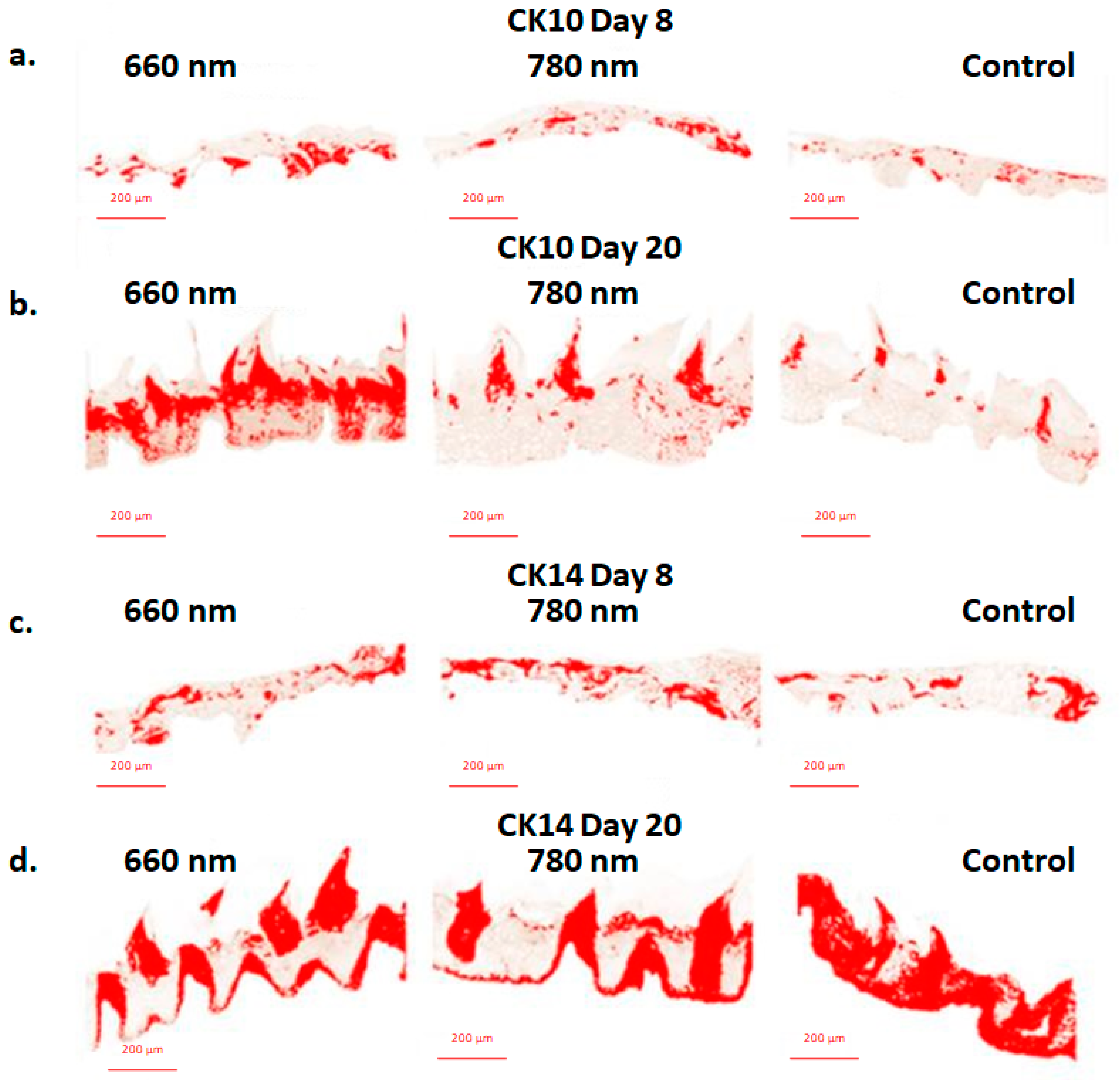
2. Results
2.1. Statistical Analysis
2.2. Percentages of CK10 Expression by IHQ
2.3. Percentages of CK14 Expression by IHQ
3. Discussion
4. Materials and Methods
4.1. OM Induced by Ionizing Radiation
4.2. PBMT Experimental Design
4.3. IHQ Analysis
5. Conclusions
Author Contributions
Funding
Institutional Review Board Statement
Data Availability Statement
Conflicts of Interest
References
- Sonis, S.T.; Elting, L.S.; Keefe, D.; Peterson, D.E.; Schubert, M.; Hauer-Jensen, M.; Bekele, B.N.; Raber-Durlacher, J.; Donnelly, J.P.; Rubenstein, E.B. Perspectives on Cancer Therapy-Induced Mucosal Injury: Pathogenesis, Measurement, Epidemiology, and Consequences for Patients. Cancer 2004, 100, 1995–2025. [Google Scholar] [CrossRef] [PubMed]
- Antunes, H.S.; Schluckebier, L.F.; Herchenhorn, D.; Small, I.A.; Araújo, C.M.M.; Viégas, C.M.P.; Rampini, M.P.; Ferreira, E.M.S.; Dias, F.L.; Teich, V.; et al. Cost-Effectiveness of Low-Level Laser Therapy (LLLT) in Head and Neck Cancer Patients Receiving Concurrent Chemoradiation. Oral Oncol. 2016, 52, 85–90. [Google Scholar] [CrossRef] [PubMed]
- Epstein, J.B.; Thariat, J.; Bensadoun, R.-J.; Barasch, A.; Murphy, B.A.; Kolnick, L.; Popplewell, L.; Maghami, E. Oral Complications of Cancer and Cancer Therapy. CA Cancer J. Clin. 2012, 62, 400–422. [Google Scholar] [CrossRef] [PubMed]
- Logan, R.M.; Gibson, R.J.; Sonis, S.T.; Keefe, D.M.K. Nuclear Factor-ΚB (NF-ΚB) and Cyclooxygenase-2 (COX-2) Expression in the Oral Mucosa Following Cancer Chemotherapy. Oral Oncol. 2007, 43, 395–401. [Google Scholar] [CrossRef]
- Sonis, S.T. New Thoughts on the Initiation of Mucositis. Oral Dis. 2010, 16, 597–600. [Google Scholar] [CrossRef]
- Chaveli-López, B.; Bagán-Sebastián, J.V. Treatment of Oral Mucositis Due to Chemotherapy. J. Clin. Exp. Dent. 2016, 8, e201–e209. [Google Scholar] [CrossRef]
- Hong, C.H.L.; Gueiros, L.A.; Fulton, J.S.; Cheng, K.K.F.; Kandwal, A.; Galiti, D.; Fall-Dickson, J.M.; Johansen, J.; Ameringer, S.; Kataoka, T.; et al. Systematic Review of Basic Oral Care for the Management of Oral Mucositis in Cancer Patients and Clinical Practice Guidelines. Support. Care Cancer 2019, 27, 3949–3967. [Google Scholar] [CrossRef] [Green Version]
- Elad, S.; Cheng, K.K.F.; Lalla, R.V.; Yarom, N.; Hong, C.; Logan, R.M.; Bowen, J.; Gibson, R.; Saunders, D.P.; Zadik, Y.; et al. MASCC/ISOO Clinical Practice Guidelines for the Management of Mucositis Secondary to Cancer Therapy. Cancer 2020, 126, 4423–4431. [Google Scholar] [CrossRef] [PubMed]
- Eduardo, F.D.P.; Bezinelli, L.M.; De Carvalho, D.L.C.; Lopes, R.M.D.G.; Fernandes, J.F.; Brumatti, M.; Vince, C.S.C.; De Azambuja, A.M.P.; Vogel, C.; Hamerschlak, N.; et al. Oral Mucositis in Pediatric Patients Undergoing Hematopoietic Stem Cell Transplantation: Clinical Outcomes in a Context of Specialized Oral Care Using Low-Level Laser Therapy. Pediatr. Transplant. 2015, 19, 316–325. [Google Scholar] [CrossRef] [PubMed]
- Peng, H.; Chen, B.B.; Chen, L.; Chen, Y.P.; Liu, X.; Tang, L.L.; Mao, Y.P.; Li, W.F.; Zhang, Y.; Lin, A.H.; et al. A Network Meta-Analysis in Comparing Prophylactic Treatments of Radiotherapy-Induced Oral Mucositis for Patients with Head and Neck Cancers Receiving Radiotherapy. Oral Oncol. 2017, 75, 89–94. [Google Scholar] [CrossRef]
- Soares, R.G.; Farias, L.C.; da Silva Menezes, A.S.; de Oliveira e Silva, C.S.; Tabosa, A.T.L.; Chagas, P.V.F.; Santiago, L.; Santos, S.H.S.; de Paula, A.M.B.; Guimarães, A.L.S. Treatment of Mucositis with Combined 660- and 808-Nm-Wavelength Low-Level Laser Therapy Reduced Mucositis Grade, Pain, and Use of Analgesics: A Parallel, Single-Blind, Two-Arm Controlled Study. Lasers Med. Sci. 2018, 33, 1813–1819. [Google Scholar] [CrossRef] [PubMed]
- Elad, S.; Arany, P.; Bensadoun, R.J.; Epstein, J.B.; Barasch, A.; Raber-Durlacher, J. Photobiomodulation Therapy in the Management of Oral Mucositis: Search for the Optimal Clinical Treatment Parameters. Support. Care Cancer 2018, 26, 3319–3321. [Google Scholar] [CrossRef] [PubMed]
- El Bousaadani, A.; Eljahd, L.; Abada, R.; Rouadi, S.; Roubal, M.; Mahtar, M. Actualités de la Prévention et du Traitement des Mucites Orales Chez les Enfants Cancéreux: Recommandations Pratiques. Cancer/Radiotherapie 2016, 20, 226–230. [Google Scholar] [CrossRef] [PubMed]
- Lalla, R.V.; Brennan, M.T.; Gordon, S.M.; Sonis, S.T.; Rosenthal, D.I.; Keefe, D.M. Oral Mucositis Due to High-Dose Chemotherapy and/or Head and Neck Radiation Therapy. J. Natl. Cancer Inst. Monogr. 2019, 2019, 17–24. [Google Scholar] [CrossRef]
- Zadik, Y.; Arany, P.R.; Fregnani, E.R.; Bossi, P.; Antunes, H.S.; Bensadoun, R.-J.; Gueiros, L.A.; Majorana, A.; Nair, R.G.; Ranna, V.; et al. Systematic Review of Photobiomodulation for the Management of Oral Mucositis in Cancer Patients and Clinical Practice Guidelines. Support. Care Cancer 2019, 27, 3969–3983. [Google Scholar] [CrossRef] [Green Version]
- Wardill, H.R.; Tissing, W.J.E.; Kissow, H.; Stringer, A.M. Animal Models of Mucositis: Critical Tools for Advancing Pathobiological Understanding and Identifying Therapeutic Targets. Curr. Opin. Support. Palliat. Care 2019, 13, 119–133. [Google Scholar] [CrossRef]
- Moll, R.; Franke, W.W.; Schiller, D.L.; Geiger, B.; Krepler, R. The Catalog of Human Cytokeratins: Patterns of Expression in Normal Epithelia, Tumors and Cultured Cells. Cell 1982, 31, 11–24. [Google Scholar] [CrossRef]
- Coulombe, P.A.; Omary, M.B. “Hard” and “Soft” Principles Defining the Structure, Function and Regulation of Keratin Intermediate Filaments. Curr. Opin. Cell Biol. 2002, 14, 110–122. [Google Scholar] [CrossRef]
- Dale, B.A.; Holbrook, K.A.; Kimball, J.R.; Hoff, M.; Sun, T.T. Expression of Epidermal Keratins and Filaggrin during Human Fetal Skin Development. J. Cell Biol. 1985, 101, 1257–1269. [Google Scholar] [CrossRef] [Green Version]
- Werner, S.; Krieg, T.; Smola, H. Keratinocyte-Fibroblast Interactions in Wound Healing. J. Investig. Dermatol. 2007, 127, 998–1008. [Google Scholar] [CrossRef]
- Bonan, P.R.F.; Kaminagakura, E.; Pires, F.R.; Vargas, P.A.; De Almeida, O.P. Cytokeratin Expression in Initial Oral Mucositis of Head and Neck Irradiated Patients. Oral Surg. Oral Med. Oral Pathol. Oral Radiol. Endodontology 2006, 101, 205–211. [Google Scholar] [CrossRef] [PubMed]
- Sawaf, M.H.; Ouhayoun, J.P.; Forest, N. Cytokeratin Profiles in Oral Epithelial: A Review and a New Classification. J. Biol. Buccale 1991, 19, 187–198. [Google Scholar]
- Liu, A.Y.; Destoumieux, D.; Wong, A.V.; Park, C.H.; Valore, E.V.; Liu, L.; Ganz, T. Human β-Defensin-2 Production in Keratinocytes Is Regulated by Interleukin-1, Bacteria, and the State of Differentiation. J. Investig. Dermatol. 2002, 118, 275–281. [Google Scholar] [CrossRef] [PubMed] [Green Version]
- Piipponen, M.; Li, D.; Landén, N.X. The Immune Functions of Keratinocytes in Skin Wound Healing. Int. J. Mol. Sci. 2020, 21, 8790. [Google Scholar] [CrossRef] [PubMed]
- Aragón-Sánchez, J.; Quintana-Marrero, Y.; Aragón-Hernández, C.; Hernández-Herero, M.J. ImageJ: A Free, Easy, and Reliable Method to Measure Leg Ulcers Using Digital Pictures. Int. J. Low. Extrem. Wounds 2017, 16, 269–273. [Google Scholar] [CrossRef] [PubMed]
- Landini, G.; Martinelli, G.; Piccinini, F. Colour Deconvolution: Stain Unmixing in Histological Imaging. Bioinformatics 2021, 37, 1485–1487. [Google Scholar] [CrossRef]
- Maria, O.M.; Syme, A.; Eliopoulos, N.; Muanza, T. Single-Dose Radiation-Induced Oral Mucositis Mouse Model. Front. Oncol. 2016, 6, 154. [Google Scholar] [CrossRef] [Green Version]
- Kim, J.H.; Jung, M.H.; Kim, J.P.; Kim, H.-J.; Jung, J.H.; Hahm, J.R.; Kang, K.M.; Jeong, B.-K.; Woo, S.H. Alpha Lipoic Acid Attenuates Radiation-Induced Oral Mucositis in Rats. Oncotarget 2017, 8, 72739–72747. [Google Scholar] [CrossRef]
- Cini, N.; Gruber, S.; Arican Alicikus, Z.; Dörr, W. Modulation of Radiation-Induced Oral Mucositis (Mouse) by Dermatan Sulfate: Effects on Differentiation Processes. Strahlenther. Onkol. 2020, 196, 85–94. [Google Scholar] [CrossRef] [Green Version]
- Sonis, S.T. Mucositis: The Impact, Biology and Therapeutic Opportunities of Oral Mucositis. Oral Oncol. 2009, 45, 1015–1020. [Google Scholar] [CrossRef]
- de Castro, J.R.; da Silva Pereira, F.; Chen, L.; Arana-Chavez, V.E.; Ballester, R.Y.; DiPietro, L.A.; Simões, A. Improvement of Full-Thickness Rat Skin Wounds by Photobiomodulation Therapy (PBMT): A Dosimetric Study. J. Photochem. Photobiol. B Biol. 2020, 206, 111850. [Google Scholar] [CrossRef] [PubMed]
- Sperandio, F.F.; Simões, A.; Corrêa, L.; Aranha, A.C.; Giudice, F.S.; Hamblin, M.R.; Sousa, S.C. Low-Level Laser Irradiation Promotes the Proliferation and Maturation of Keratinocytes during Epithelial Wound Repair. J. Biophotonics 2015, 8, 795–803. [Google Scholar] [CrossRef] [Green Version]
- Antunes, H.S.; Wajnberg, G.; Pinho, M.B.; Jorge, N.A.N.; de Moraes, J.L.M.; Stefanoff, C.G.; Herchenhorn, D.; Araújo, C.M.M.; Viégas, C.M.P.; Rampini, M.P.; et al. CDNA Microarray Analysis of Human Keratinocytes Cells of Patients Submitted to Chemoradiotherapy and Oral Photobiomodulation Therapy: Pilot Study. Lasers Med. Sci. 2018, 33, 11–18. [Google Scholar] [CrossRef] [PubMed]
- Bashkatov, A.N.; Genina, E.A.; Kochubey, V.I.; Tuchin, V.V. Optical Properties of Human Skin, Subcutaneous and Mucous Tissues in the Wavelength Range from 400 to 2000 Nm. J. Phys. D Appl. Phys. 2005, 38, 2543–2555. [Google Scholar] [CrossRef]
- Rusu, D.; Calenic, B.; Greabu, M.; Kralev, A.; Boariu, M.; Bojin, F.; Anghel, S.; Paunescu, V.; Vela, O.; Calniceanu, H.; et al. Evaluation of Oral Keratinocyte Progenitor and T-Lymphocite Cells Response during Early Healing after Augmentation of Keratinized Gingiva with a 3D Collagen Matrix—A Pilot Study. BMC Oral Health 2016, 17, 9. [Google Scholar] [CrossRef] [PubMed] [Green Version]
- de Freitas Cuba, L.; Braga Filho, A.; Cherubini, K.; Salum, F.G.; de Figueiredo, M.A.Z. Topical Application of Aloe Vera and Vitamin E on Induced Ulcers on the Tongue of Rats Subjected to Radiation: Clinical and Histological Evaluation. Support. Care Cancer 2016, 24, 2557–2564. [Google Scholar] [CrossRef]
- Wellington, D.; Mikaelian, I.; Singer, L. Comparison of Ketamine-Xylazine and Ketamine-Dexmedetomidine Anesthesia and Intraperitoneal Tolerance in Rats. J. Am. Assoc. Lab. Anim. Sci. 2013, 52, 481–487. [Google Scholar]


| Group | Mean ± SEM CK10 | Mean ± SEM CK14 |
|---|---|---|
| 660 nm day 8 | 8.69 ± 1.271 | 23.82 ± 2.22 |
| 660 nm day 20 | 38.01 ± 1.228 1 | 32.11 ± 6.485 |
| 780 nm day 8 | 7.32 ± 1.099 | 19.8 ± 1.551 |
| 780 nm day 20 | 10.35 ± 1.822 | 28.88 ± 4.237 |
| Control day 8 | 4.315 ± 1.269 | 26.82 ± 6.064 |
| Control day 20 | 5.87 ± 0.836 | 39.12 ± 5.972 |
| Groups | Day 8 | Day 20 | Total Rats in Each Group |
|---|---|---|---|
| λ = 780 nm, 30 mW, 7,5 J/cm2, 10 s, Spot size = 0.04 mm; Irradiation = 48/48 h | 07 | 07 | 14 |
| λ = 660 nm, 30 mW, 7,5 J/cm2, 10 s, Spot size = 0.04 mm; Irradiation = 48/48 h | 07 | 05 | 12 |
| Control | 04 | 04 | 8 |
Publisher’s Note: MDPI stays neutral with regard to jurisdictional claims in published maps and institutional affiliations. |
© 2022 by the authors. Licensee MDPI, Basel, Switzerland. This article is an open access article distributed under the terms and conditions of the Creative Commons Attribution (CC BY) license (https://creativecommons.org/licenses/by/4.0/).
Share and Cite
Sardo, A.V.N.; Andrade, M.F.; Figueiredo, A.; Rosin, F.C.P.; Corrêa, L.; Zezell, D.M. Does Photobiomodulation Affects CK10 and CK14 in Oral Mucositis Radioinduced Repair? Int. J. Mol. Sci. 2022, 23, 15611. https://doi.org/10.3390/ijms232415611
Sardo AVN, Andrade MF, Figueiredo A, Rosin FCP, Corrêa L, Zezell DM. Does Photobiomodulation Affects CK10 and CK14 in Oral Mucositis Radioinduced Repair? International Journal of Molecular Sciences. 2022; 23(24):15611. https://doi.org/10.3390/ijms232415611
Chicago/Turabian StyleSardo, Ariane Venzon Naia, Maíra Franco Andrade, Anaeliza Figueiredo, Flávia Cristina Perillo Rosin, Luciana Corrêa, and Denise Maria Zezell. 2022. "Does Photobiomodulation Affects CK10 and CK14 in Oral Mucositis Radioinduced Repair?" International Journal of Molecular Sciences 23, no. 24: 15611. https://doi.org/10.3390/ijms232415611