Article

β-Galactosylceramidase Deficiency Causes Upregulation of Long Pentraxin-3 in the Central Nervous System of Krabbe Patients and Twitcher Mice

1
Department of Molecular and Translational Medicine, School of Medicine, University of Brescia, 25123 Brescia, Italy
2
Department of Medical Biotechnology and Translational Medicine, University of Milan, 20133 Milan, Italy
3
Department of Pediatrics, University of Pittsburgh School of Medicine, Pittsburgh, PA 15224-1334, USA
*
Author to whom correspondence should be addressed.
Int. J. Mol. Sci. 2022, 23(16), 9436; https://doi.org/10.3390/ijms23169436
Submission received: 27 July 2022 / Revised: 17 August 2022 / Accepted: 18 August 2022 / Published: 21 August 2022
(This article belongs to the Special Issue Sphingolipid Metabolism and Signaling in Diseases 2.0)

Abstract

:
Globoid cell leukodystrophy (GLD), or Krabbe disease, is a neurodegenerative sphingolipidosis caused by genetic deficiency of lysosomal β-galactosylceramidase (GALC), characterized by neuroinflammation and demyelination of the central (CNS) and peripheral nervous system. The acute phase protein long pentraxin-3 (PTX3) is a soluble pattern recognition receptor and a regulator of innate immunity. Growing evidence points to the involvement of PTX3 in neurodegeneration. However, the expression and role of PTX3 in the neurodegenerative/neuroinflammatory processes that characterize GLD remain unexplored. Here, immunohistochemical analysis of brain samples from Krabbe patients showed that macrophages and globoid cells are intensely immunoreactive for PTX3. Accordingly, Ptx3 expression increases throughout the course of the disease in the cerebrum, cerebellum, and spinal cord of GALC-deficient twitcher (Galctwi/twi) mice, an authentic animal model of GLD. This was paralleled by the upregulation of proinflammatory genes and M1-polarized macrophage/microglia markers and of the levels of PTX3 protein in CNS and plasma of twitcher animals. Crossing of Galctwi/twi mice with transgenic PTX3 overexpressing animals (hPTX3 mice) demonstrated that constitutive PTX3 overexpression reduced the severity of clinical signs and the upregulation of proinflammatory genes in the spinal cord of P35 hPTX3/Galctwi/twi mice when compared to Galctwi/twi littermates, leading to a limited increase of their life span. However, this occurred in the absence of a significant impact on the histopathological findings and on the accumulation of the neurotoxic metabolite psychosine when evaluated at this late time point of the disease. In conclusion, our results provide the first evidence that PTX3 is produced in the CNS of GALC-deficient Krabbe patients and twitcher mice. PTX3 may exert a protective role by reducing the neuroinflammatory response that occurs in the spinal cord of GALC-deficient animals.

Graphical Abstract

1. Introduction

Globoid cell leukodystrophy (GLD), or Krabbe disease, is an autosomal recessive neurodegenerative sphingolipidosis caused by genetic deficiency of the lysosomal hydrolase β-galactosylceramidase (GALC) [1]. GALC degrades galactosylceramide (a major component of myelin) and other terminal β-galactose-containing sphingolipids, including β-galactosylsphingosine (psychosine). The pathogenesis of GLD has been proposed to arise from the accumulation of the neurotoxic metabolite psychosine present at high levels in the CNS of Krabbe patients [2,3,4,5]. The disease is characterized by neuroinflammation and loss of oligodendroglia and Schwann cells, leading to demyelination of the brain, spinal cord, nerve roots and peripheral nerves [6]. A pathognomonic feature of GLD is the presence in the white matter of globoid cells [7,8], giant multinucleated cells likely originated from resident microglia [9]. Clinically, GLD may manifest in early infancy with fatal neurological dysfunctions [6,10,11]. The standard of care for this disease is hematopoietic stem cell transplantation [12,13].
The acute phase protein long pentraxin-3 (PTX3) is an important regulator of peripheral innate immunity and a key mediator of inflammation during cardiovascular and cerebrovascular diseases [14]. Growing evidence points to the involvement of PTX3 in neurodegenerative disorders [15]. PTX3 expression is upregulated in the central nervous system (CNS) following pro-inflammatory cytokine stimulation [16], seizure-induced neurodegeneration [17], and ischemia [18]. Accordingly, peripheral PTX3 levels are increased after experimental stroke in mice [19], and plasma PTX3 levels correlate with mortality after ischemic stroke in humans [20]. In addition, serum levels of PTX3 have been proposed as a novel biochemical marker in Parkinson’s disease [21]. Despite this evidence, no data are available about the expression and role of PTX3 in sphingolipid disorders, including GLD.
Here, PTX3 production was investigated in the brain of Krabbe patients and in the CNS of GALC-deficient twitcher (Galctwi/twi) mice, an authentic murine model of GLD [22,23]. Analysis of brain samples from Krabbe patients demonstrated that macrophages and globoid cells are intensely immunoreactive for PTX3. Accordingly, twitcher mice are characterized by the progressive upregulation of Ptx3 expression along the CNS caudal-rostral axis. This was paralleled by the upregulation of various proinflammatory genes and M1-polarized macrophage/microglia markers, with no changes in the peripheral organs. Crossing of Galctwi/twi mice with transgenic PTX3 overexpressing animals (hPTX3 mice) caused an amelioration of clinical features and attenuated spinal cord inflammation in hPTX3/Galctwi/twi offspring when compared to Galctwi/twi littermates at postnatal day P35, when animals show clear neurologic defects. However, PTX3 overexpression did ameliorate the histopathological findings and did not affect psychosine accumulation in the CNS of GALC-deficient animals when evaluated at this late stage of the disease.
Our results demonstrate that PTX3 is produced in the CNS of GALC-deficient Krabbe patients and twitcher mice and point to a possible protective role of this immune modulator by reducing, at least in part, the neuroinflammatory response that characterizes GLD.

2. Results

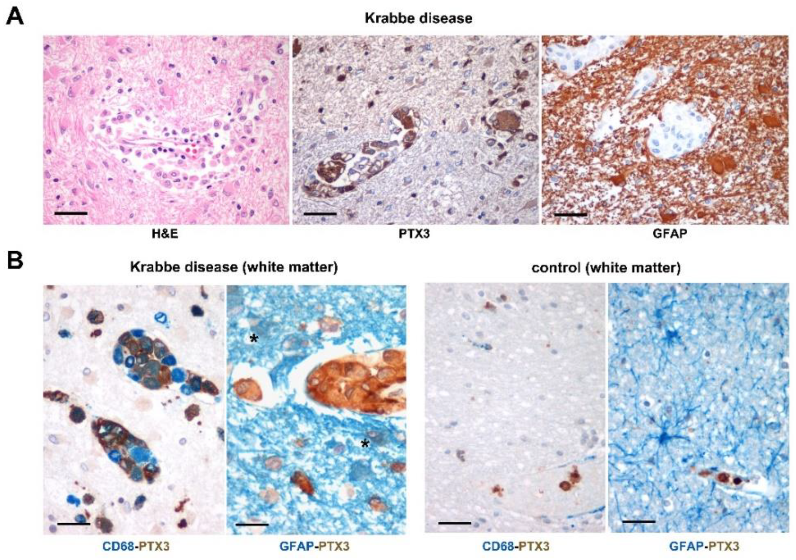
2.1. PTX3 Immunoreactivity in the Brain of Krabbe Patients

To assess whether GALC deficiency may cause PTX3 upregulation in human CNS, we analyzed autopsy brain cortex samples from nine Krabbe patients (Table S1) and two matched controls. Most of the Krabbe samples showed the classical histopathological hallmarks of the disease, such as diffuse demyelination along with marked loss of oligodendrocytes, intense reactive gliosis evidenced by the diffuse immunoreactivity of the astrocytic marker GFAP, and infiltration of globoid cells (Figure 1A). Previous observations had shown that activated astrocytes and microglia represent a major source of PTX3 in the brain [16,24]. Accordingly, single immunostains indicated that GLD macrophages and globoid cells are diffusely and intensely immunoreactive for PTX3. Double immunostains confirmed that PTX3 is mainly expressed within macrophages and globoid cells in Krabbe samples, its positivity being related to the extent of pathology (i.e., presence of perivascular globoid cells) and of GFAP-positive astrocytic gliosis (Table 1), while GFAP/PTX3 double positive astrocytes were barely detected. Control brain samples did not show double positive PTX3 immunoreactive cells (Figure 1B). Overall, these findings demonstrate that PTX3 is upregulated in the brain of GLD patients, its immunoreactivity being mainly expressed in monocyte-derived cells and related to the extent of the pathology.

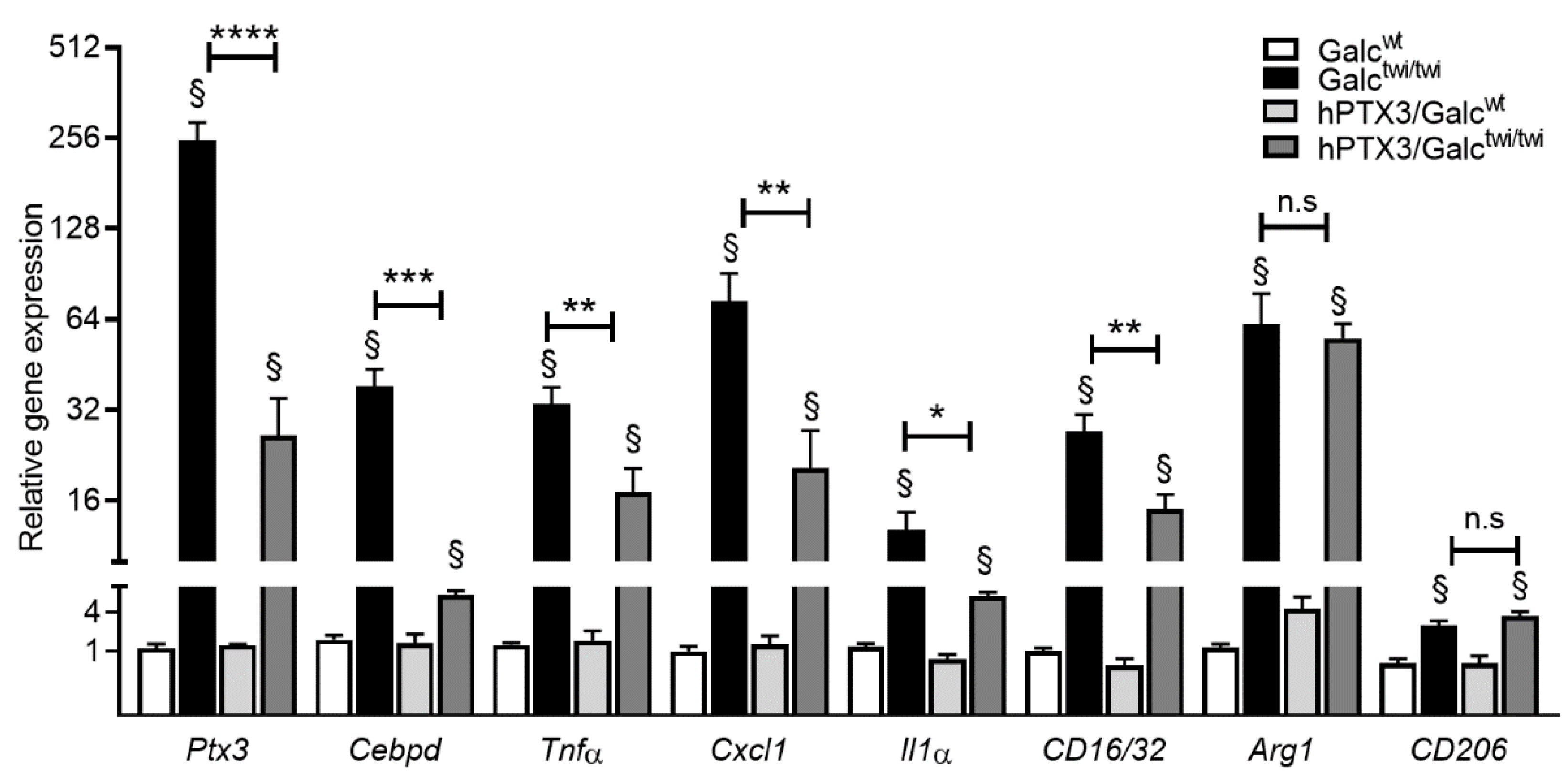
2.2. Ptx3 Upregulation in Twitcher CNS

Based on the data obtained in Krabbe patients, the expression of Ptx3 was investigated in the CNS of GALC-deficient twitcher (Galctwi/twi) mice, an authentic murine model of GLD [22,23]. In a first set of experiments, Ptx3 expression was assessed by qPCR analysis in the cerebrum of littermate Galcwt and homozygous Galctwi/twi mice. Measurements were performed at postnatal day P15 before the onset of evident neurologic signs, at P24 when pathologic alterations were occasionally detectable, and at P35 when Galctwi/twi mice showed clear neurologic defects, including tremor and hind limb paralysis [23,25,26]. As shown in Figure 2A, the brain levels of Ptx3 transcripts were already increased at P15, to further increase at P24 and P35. This was paralleled by the upregulation of the inflammatory cytokines Tnfα, Cxcl1 and Il1α and of the CCAAT/enhancer binding protein delta (Cebpd) gene, which encodes for a transcription factor mediating PTX3 expression in astrocytes during neuroinflammation [27]. Accordingly, a progressive increase in the levels of the astrocytic marker of gliosis Gfap and the microglial marker ionized calcium-binding adaptor molecule-1 (Iba1) [28] occurred in Galctwi/twi cerebrum when compared to controls.
In keeping with the neuroinflammatory response that occurs in twitcher brain, we observed an increase in the levels of expression of the classical M1 macrophage polarization/microglia markers Fc receptor, IgG, low affinity III and II (Fcgr3/CD16 and Fcgr2/CD32) [29] with no changes in the expression of the alternative M2 macrophage polarization markers arginase-1 (Arg1) and mannose receptor C-type1 (Mrc1/CD206) [30]. A similar transcriptional profile was observed in the cerebellum (Figure S1) and spinal cord (Figure 2B) of Galctwi/twi mice that also showed an increase in the M2 polarization markers Arg1 and CD206. Notably, Ptx3 mRNA levels in the spinal cord of Galctwi/twi mice were significantly higher than those measured in the cerebrum and cerebellum counterparts at all time points investigated.
Ptx3 upregulation in the CNS of twitcher mice resulted in high levels of PTX3 protein in the cerebrum, cerebellum, and spinal cord of P35 twitcher mice when compared to control littermates as assessed by immunohistochemistry and Western blot analysis (Figure 3A–C). In addition, the levels of PTX3 in the blood of P35 twitcher mice were significantly increased when compared to Galcwt animals (Figure 3D).
On this basis, immunohistochemical analysis was performed to identify PTX3-positive cells in the CNS of twitcher animals. As shown in Figure 4, double immunostains indicated that PTX3 immunoreactivity was prevalently detectable in IBA1-positive globoid cells in the white matter of the brain cortex and spinal cord of Galctwi/twi animals, no significant immunoreactivity being observed in control samples. In addition, PTX3 immunoreactivity was also present in GFAP-positive astrocytes of the gray matter of the CNS of twitcher mice, but not of Galcwt animals that lacked signs of gliosis (Figure 4). Similar results were obtained in twitcher cerebellum samples (Figure S2).
At variance with the results obtained by the analysis of the twitcher CNS, no Ptx3 upregulation occur in the peripheral tissues of Galctwi/twi mice, including kidney, liver, and lungs, in which only limited psychosine accumulation and inflammatory responses occur because of GALC deficiency [23,25,31,32,33] (Figure S3). Nevertheless, in keeping with the observation that the serum levels of PTX3 are increased after stroke [19,20] and in Parkinson’s disease [21], the levels of PTX3 in the blood of P35 twitcher mice were significantly increased when compared to Galcwt animals (Figure 3D).

2.3. PTX3 Overexpression Reduces Clinical Symptoms and Spinal Cord Inflammation in Twitcher Mice

TgN(Tie2-hPTX3) mice (hPTX3 mice) ubiquitously express human PTX3 under the control of the Tie2 promoter, leading to a significant accumulation of the PTX3 protein in serum and tissues that occurs from birth throughout the whole life of the transgenic mice [34]. The constitutive hPTX3 expression does not result in apparent defects in embryonic development; adult animals are normal and fertile, and no macroscopic or microscopic morphological abnormalities were observed in organs and tissues of hPTX3 mice [34].
On this basis, to assess a possible impact of the constitutive PTX3 overexpression in twitcher mice, syngeneic PTX3-overexpressing hPTX3 male mice were crossed with female Galctwi/+ animals to obtain hPTX3/Galctwi/+ breeders that were then crossed to generate hPTX3/Galcwt and hPTX3/Galctwi/twi animals. Next, hPTX3/Galcwt and hPTX3/Galctwi/twi mice were compared to the corresponding Galcwt and Galctwi/twi littermates. qPCR analysis using oligonucleotide primers designed to recognize simultaneously both human and murine PTX3 transcripts (Table S2) indicated that the levels of hPTX3 mRNA in the cerebrum of hPTX3/Galcwt mice were like the levels of the murine Ptx3 transcripts measured in Galctwi/twi mice at P35. In addition, hPTX3 overexpression together with the upregulation of its murine counterpart resulted in a cumulative increase in their mRNA levels in the brain of P35 hPTX3/Galctwi/twi mice when compared to Galctwi/twi littermates (Figure S4A).
As shown in Figure S4C,D, constitutive hPTX3 overexpression did not affect the body weight gain of both hPTX3/Galcwt and hPTX3/Galctwi/twi mice when compared to the corresponding control Galcwt and Galctwi/twi animals, with Galc deficiency leading to a similar decrease in body weight starting from approximately day P20 and P28 both in the absence or in the presence of constitutive hPTX3 overexpression in male and female animals, respectively. Nevertheless, hPTX3 overexpression resulted in statistically significant, albeit limited 4-day increases in the overall survival of twitcher animals, extending their life span from 42 to 46 days (p < 0.001) (Figure 5A), thus suggesting that PTX3 may exert an impact on the disease course. Indeed, when compared to Galctwi/twi animals, hPTX3/Galctwi/twi mice showed a reduced frequency and severity of twitching, which appeared around day P22 in both groups of animals (Figure 5B,C). In addition, hPTX3/Galctwi/twi mice displayed a less severe atypical tail suspension reflex (hind limbs clenching) (Figure 5D). No animal death and appearance of clinical symptoms were instead observed in hPTX3/Galcwt and Galcwt mice during the same investigation period.
On this basis, we assessed the levels of expression of the microglial marker Iba1, of the astrocytic marker of gliosis Gfap, and of the myelin basic protein-encoding gene mbp in the cerebrum, cerebellum, and spinal cord of the different experimental groups at P35. As shown in Figure 6A, GALC deficiency caused a similar upregulation of Iba1 and Gfap genes and downregulation of mbp in the CNS of hPTX3/Galctwi/twi and Galctwi/twi mice at this late stage of the disease when compared to Galcwt and hPTX3/Galcwt animals. Accordingly, immunohistochemical analysis demonstrated that the IBA1-positive microglia infiltrate, GFAP-positive gliosis, and myelin degradation of MBP-positive fibers were present to a similar extent in the CNS of P35 hPTX3/Galctwi/twi and Galctwi/twi mice (Figure 6B).
In keeping with the lack of a significant impact of constitutive hPTX3 overexpression on the histopathological features of the CNS of GALC-deficient animals, the levels of the neurotoxic GALC substrate psychosine were increased to a similar extent in the CNS of Galctwi/twi and hPTX3/Galctwi/twi animals (Table 2).
Finally, qPCR analysis of cerebrum, cerebellum, and spinal cord of P35 animals was performed to assess the impact of constitutive hPTX3 overexpression on the inflammatory response of the CNS in GALC-deficient mice. As shown in Figure 7, the expression of the inflammatory mediators Tnfα, Il1α, and Cxcl1 and of the M1 polarization macrophage/microglia markers CD16/CD32, albeit still higher than that observed in control wild type animals, was significantly reduced in the spinal cord of hPTX3/Galctwi/twi mice when compared to the Galctwi/twi counterpart, whereas Arg1 and CD206 expression remained unchanged.
At variance, no differences in gene expression were detected in the cerebrum and cerebellum of the two groups of animals, the only exceptions being represented by a slight increase in the expression of TNFα in the cerebrum of hPTX3/Galctwi/twi mice when compared to Galctwi/twi animals and a decreased cerebellar expression of the M2 polarization markers Arg1 and CD206 that occurred also in hPTX3/Galcwt mice, possibly due to the PTX3 upregulation present in both groups (Figure S5). In keeping with these observations, the levels of transcription factor Cebpd were significantly reduced in the spinal cord of hPTX3/Galctwi/twi mice when compared to Galctwi/twi animals (Figure 7), no differences being instead observed between the two groups in the cerebrum and cerebellum (Figure S5). Accordingly, the levels of murine Ptx3 transcript were reduced only in the spinal cord of hPTX3/Galctwi/twi animals (Figure 7). Thus, at variance with what was observed in the cerebrum (see above), the cumulative levels of the human plus murine PTX3 transcripts were reduced in the spinal cord of hPTX3/Galctwi/twi animals relative to the levels of murine Ptx3 mRNA in Galctwi/twi mice (see Figure S4B).

3. Discussion

In humans, genetic deficiency of the sphingolipid-metabolizing enzyme GALC leads to Krabbe disease, a neuroinflammatory degenerative disorder. Here, we demonstrated that the soluble pattern recognition receptor PTX3 was expressed by monocyte-derived cells in brain specimens from Krabbe patients, its immunoreactivity being related to the extent of the pathology and gliosis. In keeping with these observations, twitcher mice, an authentic model of Krabbe disease [22,23], were characterized by the progressive upregulation of PTX3 along the CNS caudal-rostral axis, with no changes in the peripheral organs. The upregulation of the Ptx3 transcript levels in cerebrum, cerebellum, and spinal cord of twitcher mice resulted in an increase in PTX3 protein levels in the affected tissues and plasma.
Ptx3 expression can be induced in the CNS following stimulation by LPS or pro-inflammatory cytokines [16]. Accordingly, Ptx3 upregulation is observed in mice under various experimental neuroinflammatory conditions, including neurotrauma [35], ischemia [18], limbic seizure [17] and autoimmune encephalomyelitis [36]. In addition, serum levels of PTX3 are increased in patients affected by neurodegenerative disorders, including Parkinson’s disease [21], ischemic stroke [20], and multiple sclerosis [36].
Our data extend these observations to Krabbe patients and demonstrate for the first time that Ptx3 upregulation occurs in the CNS of twitcher mice in parallel with an increased expression of neuroinflammation-related genes, including various cytokines/chemokines, the marker of gliosis Gfap, the microglial marker Iba1, and the M1 macrophage polarization/microglia markers CD16/CD32. These changes were detectable at postnatal day P15, before the onset of evident neurologic signs, and were increased at P24, when pathologic alterations were occasionally noticeable, and at P35, when clear neurologic defects occurred, including tremor and hind limb paralysis [23,25,26]. These findings indicate that the inflammatory environment progressively established after birth in the CNS of GALC-deficient mice drives the expression of Ptx3, as confirmed by the observed upregulation of Cebpd, a transcription factor known to mediate PTX3 expression in astrocytes during neuroinflammation [27].
Previous findings had shown that PTX3 can be expressed by neurons, astrocytes, and/or microglia following cytokine stimulation or under neuroinflammatory conditions, depending on the specific disorder and acute versus chronic phase of the disease (see [15,35,36] and references therein). Here, immunohistochemical analysis demonstrated that autopsy brain specimens from Krabbe infants were characterized by an intense gliosis and PTX3 immunoreactivity that was detectable in macrophages and globoid cells, a hallmark of Krabbe disease [9]. These observations were confirmed by the analysis of Galctwi/twi mice, in which PTX3 immunoreactivity was prevalently detectable in IBA1-positive globoid cells of the white matter of the brain cortex, cerebellum, and spinal cord of these animals, as well as in GFAP-positive astrocytes of the gray matter.
PTX3 is an important mediator of innate immune responses, produced locally at the site of inflammation [37]. In CNS, PTX3 modulates the activity of microglia by inhibiting phagocytosis of apoptotic cells and favoring the uptake of pathogens [24]. PTX3 binding helps the rescue of neurons from phagocytic clearance by macrophages [27] and protects them from ischemic damage [19,38], trauma [39], and in Parkinson’s disease [40]. Accordingly, neurogenesis and angiogenesis were inhibited after cerebral ischemia in Ptx3 null mice, and neuronal damage was increased after limbic seizure when Ptx3 deficient animals were compared to controls [17,18]. Together, these data point to a protective role of PTX3 during neuroinflammation.
Our data demonstrate that the constitutive overexpression of hPTX3 that occurs from birth throughout the whole life of hPTX3/Galctwi/twi mice attenuates the severity of clinical signs, such as twitching and hind limbs clenching, and causes a significant, albeit limited, increase in their life span. However, when assessed at the late P35 stage of the disease, hPTX3 overexpression did not exert any significant effect on the CNS of GALC-deficient mice in terms of IBA1-positive microglia infiltrate, GFAP-positive gliosis, and myelin degradation of MBP-positive fibers. In addition, a similar accumulation of the neurotoxic GALC substrate psychosine was observed in the cerebrum and spinal cord of Galctwi/twi and hPTX3/Galctwi/twi mice at this time point. These data appear to be in keeping with previous observations indicating that PTX3 is unable to exert a significant impact on the histopathological damage that occurs in mice after experimental neurotrauma or to affect the course of experimental autoimmune encephalomyelitis [35,36]. Nevertheless, we found that hPTX3 overexpression reduced the upregulation of proinflammatory genes in the spinal cord of P35 hPTX3/Galctwi/twi mice when compared to Galctwi/twi animals, including endogenous Ptx3 and its transcription factor Cebpd, with no effect on the neuroinflammatory response observed in the cerebrum and cerebellum of these animals. Relevant to this point, neurohistopathological and neurochemical alterations caused by GALC deficiency in the CNS of twitcher mice arise in a temporal and region-dependent fashion, the first alterations being observed in the spinal cord to progress along the caudal-rostral axis in the cerebellum and cerebrum (see [41] and references therein). In addition, spinal cord alterations were already observed in 18–21-week-old GLD fetuses [42,43]. Thus, the spinal cord represents the earliest tissue affected by GALC deficiency in murine and human CNS and appears to be more prone to a protective effect exerted by PTX3 on the neuroinflammatory response that occurs in twitcher mice.
Different hypotheses can be raised to attempt to explain why the spinal cord is more responsive to the partial protecting role exerted by genetic, constitutive hPTX3 upregulation in GALC-deficient mice. The ubiquitous production of PTX3 in hPTX3 animals is under the control of the Tie2 promoter [34] that drives the vascular expression of the transgene throughout embryogenesis and adulthood [44]. Thus, the early and long-lasting PTX3 overexpression that occurs in hPTX3/Galctwi/twi animals may prevent, at least in part, the first lesions that arise in the spinal cord of GALC-deficient mice, being instead less effective against the later CNS lesions (further experiments performed on hPTX3/Galctwi/twi mice at earlier stages of the disease will be required to elucidate this point). In addition, the metabolic alterations that occur in parallel with the progression of gliosis, neurodegeneration, microglial activation, and apoptosis along the rostral-caudal axis in a regional and age-dependent fashion [41] may exert a different impact on the protective activity of PTX3 in hPTX3/Galctwi/twi mice. A further hypothesis can be based on the fact that PTX3 may also exert detrimental effects following CNS damage (see [15] and references therein), indicating that the activity of PTX3 in neuroinflammation may represent the result of a fine tuning in different areas of the CNS between pro- and anti-neurodegenerative mechanisms of action that remain poorly defined. Finally, other members of the long pentraxin family, represented by the neuronal pentraxins NP1, NP2 and NPR, may exert an impact on neurodegeneration and interfere with PTX3 activity [35]. Experiments performed in a Ptx3 null background would be useful to assess these hypotheses. However, due to the female infertility of Ptx3 null mice [45] and the early lethality of Galctwi/twi animals, the generation of double Ptx3−/−/Galctwi/twi mice would require a breeding program with a very large number of animals, incompatible with the rules of the local and national ethical committees.
The neurotoxic GALC substrate psychosine has been used as a biomarker to identify patients with infantile GLD, and possibly late onset GLD patients, and to monitor the timing and efficacy of hematopoietic stem cell transplantation in these patients [46]. Our data demonstrate that serum levels of PTX3 increase in twitcher mice. Further studies will be required to assess whether longitudinal blood measurement of PTX3 levels may represent a novel biomarker more sensitive than psychosine to predict onset after newborn screening and to follow disease progression in transplanted and/or late-onset GLD patients.
In conclusion, our results provide the first evidence that PTX3 is produced in the CNS of Krabbe patients and GALC-deficient twitcher mice and may exert a partial protective role by reducing the neuroinflammatory response that occurs in the spinal cord of Galctwi/twi animals, with possible implications for GLD management and therapy.

4. Materials and Methods

4.1. Histopathology of Human GLD Biopsies

Autopsy brain specimens of GLD patients (Table S1) were obtained from the Program for Neurodevelopmental Function in Rare Disorders Brain and Tissue Bank, University of Pittsburgh School of Medicine. Use of this material was approved by the Committee for Oversight of Research and Clinical Training Involving Decedents (CORID) protocol No 583. Two matched control brains from 4-year-old and 11-year-old patients who died from unrelated non-neurological complications were obtained from the Archive of Pathological Department of Spedali Civili of Brescia. Their use was approved by the Ethics Board of Spedali Civili di Brescia (patient consent was not needed for retrospective and exclusively observational study on archival material obtained for diagnostic purposes (Delibera del Garante n. 52 del 24/7/2008 and DL 193/2003).
Formalin-fixed, paraffin-embedded tissue sections were submitted to H&E and single or double immunohistochemical staining. Briefly, sections were de-waxed, rehydrated, and endogenous peroxidase activity blocked with 0.3% H2O2 in methanol for 20 min. Antigen retrieval was performed using a microwave oven or thermostatic bath in 1.0 mM EDTA buffer (pH 8.0). Sections were then washed in TBS (pH 7.4) and incubated for 1 h with the specific primary antibody diluted in TBS 1% bovine serum albumin. Signal was revealed using the DAKO Envision+System-HRP Labelled Polymer Anti-Mouse or Anti-Rabbit (Dako Cytomation, Santa Clara, CA, USA), followed by Diaminobenzydine (DAB) as chromogen and hematoxylin as counterstain.
For double immunostains, after completing the first immune reaction, the second primary antibody was applied and labelled using MACH 4TM Universal AP Polymer Kit (Biocare Medical, Pacheco, CA, USA); chromogen reaction was developed with Ferangi BlueTM Chromogen System (Biocare Medical), and nuclei were faintly counterstained with Methyl Green. Images were then acquired with an Olympus DP70 camera mounted on an Olympus Bx60 microscope using AnalySIS imaging software (Soft Imaging System GmbH, Münster, Germany). The following primary antibodies were used: rabbit anti-PTX3 polyclonal antibody (1:500, kindly provided by B. Bottazzi Humanitas Clinical Institute, Rozzano, Italy), monoclonal mouse anti human CD68, clone PG-M1 (1:200, Dako Cytomation), and monoclonal mouse anti human GFAP, clone 6F2 (1:100, Dako Cytomation).

4.2. Animals

Breeder twitcher heterozygous mice (C57BL/6J, Galctwi/+; Jackson Laboratories, Bar Harbor, ME, USA) were maintained under standard housing conditions. Experiments were performed according to the Italian laws (D.L. 116/92 and following additions) that enforce the EU 86/109 Directive and were approved by the local animal ethics committee (OPBA, Università degli Studi di Brescia, Italy). Twitcher mutation was determined by polymerase chain reaction (PCR) on DNA extracted from clipped tails [47]. In all of the experiments, littermate wild type (Galcwt) and homozygous (Galctwi/twi) animals were used. To generate PTX3-overexpressing twitcher mice, syngeneic TgN(Tie2-hPTX3) male mice (hPTX3 mice) that ubiquitously express human PTX3 under the control of the Tie2 promoter [34] were bred with female Galctwi/+ mice to obtain hPTX3/Galctwi/+ animals. Next, hPTX3/Galcwt and hPTX3/Galctwi/twi mice were generated by crossing hPTX3/Galctwi/+ breeder mice that were genotyped for Galc status by PCR and for hPTX3 overexpression by RT-PCR. For the survival studies, the end point was established for each mouse by the animal house veterinarian unaware of the animal genotype, according to the rules of the local ethics committee.

4.3. Quantitative RT-PCR Analysis

Cerebrum, cerebellum, and spinal cord specimens were analyzed for gene expression by quantitative RT-PCR (qPCR) at P15, P24 and/or P35, and data were normalized for Gapdh expression [48]. For this purpose, total RNA was extracted from frozen samples using TRIzol Reagent according to the manufacturer’s instructions (Invitrogen, Carlsbad, CA, USA), and contaminating DNA was digested using DNAse (Promega, Madison, WI, USA). Total RNA (2 μg) was retrotranscribed with MMLV reverse transcriptase (Invitrogen) using random hexaprimers in a final 20 μL volume. Quantitative PCR was performed with a ViiATM 7 Real-Time PCR Detection System (Applied Biosystems, Waltham, MA, USA) using an iQTM SYBR Green Supermix (Biorad, Hercules, CA, USA) according to the manufacturer’s instructions. Ptx3 expression levels were analyzed by qPCR also in kidneys, liver, and lungs from Galcwt and Galctwi/twi mice at P35. In each experiment, an arbitrary value equal to 1.0 was assigned to the levels of expression of the gene(s) measured in one sample that was used as reference. The specific primers are shown in Table S2.

4.4. Immunohistochemical Analysis

Formalin-fixed, paraffin-embedded tissue sections (7 µm) of the brain cortex, cerebellum, and spinal cord from P35 Galcwt, Galctwi/twi, hPTX3/Galcwt and hPTX3/Galctwi/twi mice were submitted to single or double immunohistochemical staining. For single staining, sections were incubated overnight at 4°C with the anti-PTX3 polyclonal antibody, the anti-Iba1 mouse monoclonal antibody (1:100; Genetex, Irvine, CA, USA), the anti-GFAP mouse monoclonal antibody, clone 6F2 (1:100; Dako), or the anti-Myelin Basic Protein MAB386 antibody (1:50, Millipore). Next, sections were incubated for 1 h with anti-Rabbit or anti-Mouse Envision+System-HRP Labelled Polymer (Dako) or with Rat-on Mouse HRP-Polymer (Biocare Medical) for myelin staining, followed by Diaminobenzydine (DAB) as chromogen and hematoxylin as counterstain.
For double immunostains, incubation with the anti-PTX3 polyclonal antibody was followed by 1 h incubation with biotin anti-rabbit antibody and by 1 h incubation with streptavidin Alexa Fluor 594. Sections were then incubated for 2 h with anti-GFAP or anti-Iba1 monoclonal antibody, followed by 1 h incubation with Alexa Fluor 488 anti-mouse antibody. All tissue sections were incubated for 30 min with DAPI for nuclear staining and mounted in Dako fluorescent mounting medium.
Images were taken with an Axiovert 200 M microscope equipped with ApoTome optical sectioning device (Carl Zeiss, Oberkochen, Germany) using the same settings for comparison of all samples.

4.5. Western Blotting

Cerebrum, cerebellum, and spinal cord specimens from Galcwt and Galctwi/twi mice at P35 were used for Western blotting analysis. The tissues were homogenized in lysis buffer (1.0% NP-40, 20 mM Tris-HCl pH 8.0, 137 mM NaCl, 10% glycerol, 2.0 mM EDTA, 1.0 mM sodium orthovanadate, 10 mg/mL aprotinin, 10 mg/mL leupeptin). For PTX3 protein level analysis, tissue extracts (40 μg of protein) were probed with an anti-PTX3 polyclonal antibody. Monoclonal anti-vinculin antibody (Sigma-Aldrich, St Louis, MO, USA) was used as the loading control.

4.6. Serum PTX3

The blood levels of PTX3 were evaluated by ELISA (R&D Systems, Minneapolis, MN, USA) using 7.0 μL serum from Galcwt and Galctwi/twi mice at P28–34.

4.7. Assessment of Animal Clinical Features

Body weight, life span, twitching and hind limbs clenching were monitored daily from day P8 until mice reached a moribund condition. Each mouse was observed for at least 1 min by two trained observers. The extent of frequency and severity of twitching was scored as: 1, fine; 2, mild; 3, mild-moderate; 4, moderate; 5, severe. Hind limbs clenching frequency was scored: 1, rare; 2, mild; 3, intermittent; 4, moderate; 5, severe [49].

4.8. Psychosine Extraction and MS Analysis

Psychosine extraction was performed as previously described [50]. Briefly, 20 mg of lyophilized brain or spinal cord were extracted with 15 mL of acetone using a Polytron homogenizer to selectively extract psychosine. The extract was filtered through a Büchner funnel and evaporated to dryness. The dried residue was dissolved in 2.0 mL chloroform/methanol (2/1 v/v), and the psychosine-enriched fraction was evaporated to dryness, dissolved in methanol, and further analyzed by mass spectrometry (MS).
All MS analyses were performed at the Unitech OMICs platform (University of Milano, Italy) using an ExionLC™ AD system connected to TripleTOF™ 6600 System equipped with Turbo V™ Ion Source with ESI Probe (SCIEX, Framingham, MA, USA). Samples were separated on a Kinetex® EVO C18 100 (Length) × 2.1 mm (ID) × 1.7 µm (particle size). The temperature was set at 40 °C. The analytes were eluted with the following gradient: from 60% buffer A (0.01% formic acid in water) to 99% buffer B (0.01% formic acid in methanol) in 7.5 min. Constant flow rate: 450 µL/min. Total run: 12 min. MS spectra were collected, in positive polarity, in full-mass scan from 250 to 800 Da (100 ms accumulation time) and in IDA® mode (information-dependent acquisition) from 100 to 800 Da (40 ms accumulation time, top 18 spectra per cycle 0.87 s). Nitrogen was used as a nebulizing gas (GS1, 55 psi), turbo spray gas (GS2, 65 psi), and curtain gas (CUR, 35 psi). Spray voltage was fixed at 5.0 kV, de-clustering potential (DP) was 50 eV, the collision energy was 30 eV with a collision energy spread (CES) of 15 eV, and source temperature was 300 °C.
The data were analyzed using SCIEX OS 1.4 software (SCIEX™), together with LibraryView™ (version 1.0) containing the hexosylsphingosine mass spectra.

4.9. Statistical Analysis

Comparisons among multiple groups were performed using a one-way ANOVA model, followed by post hoc comparisons with adjustment for multiple comparisons (Sidak procedure). Comparison between two groups was performed using the Student’s unpaired t-test.

Supplementary Materials

The following supporting information can be downloaded at: https://www.mdpi.com/article/10.3390/ijms23169436/s1.

Author Contributions

Investigation, D.C., A.M.K.C., M.B., P.L.P., M.C. (Manuela Cominelli), F.P., M.C. (Miriam Capra) and S.P.; data curation, D.C., L.M. and A.P.; resources, P.L.P., J.K.K. and M.L.E.; statistical analysis, S.C.; funding acquisition, P.L.P. and M.P; supervision, writing, original draft preparation, reviewing and editing, M.P. All authors have read and agreed to the published version of the manuscript.

Funding

This research was funded by Associazione Italiana per la Ricerca sul Cancro (AIRC, IG 23116) to M.P., The Legacy of Angels Foundation, Partners for Krabbe Research to M.L.E., and Associazione “dedicato A te” to P.L.P.

Institutional Review Board Statement

The study was conducted in accordance with the Declaration of Helsinki and approved by the Committee for Oversight of Research and Clinical Training Involving Decedents (CORID) of the University of Pittsburgh School of Medicine (protocol No 583) and by the Ethics Board of Spedali Civili di Brescia (protocol n. 52 24/7/2008 and DL 193/2003). The animal study was performed according to the Italian laws (D.L. 116/92 and following additions) that enforce the EU 86/109 Directive and was approved by the local animal ethics committee (OPBA, Università degli Studi di Brescia, Italy).

Informed Consent Statement

Not applicable.

Data Availability Statement

Not applicable.

Conflicts of Interest

The authors declare no conflict of interest.

References

  1. Suzuki, K.; Suzuki, Y. Globoid cell leucodystrophy (Krabbe’s disease): Deficiency of galactocerebroside beta-galactosidase. Proc. Natl. Acad. Sci. USA 1970, 66, 302–309. [Google Scholar] [CrossRef] [PubMed] [Green Version]
  2. Ballabio, A.; Gieselmann, V. Lysosomal disorders: From storage to cellular damage. Biochim. Biophys. Acta 2009, 1793, 684–696. [Google Scholar] [CrossRef] [PubMed] [Green Version]
  3. Suzuki, K. Twenty five years of the “psychosine hypothesis”: A personal perspective of its history and present status. Neurochem. Res. 1998, 23, 251–259. [Google Scholar] [CrossRef] [PubMed]
  4. Igisu, H.; Suzuki, K. Progressive accumulation of toxic metabolite in a genetic leukodystrophy. Science 1984, 224, 753–755. [Google Scholar] [CrossRef]
  5. Li, Y.; Xu, Y.; Benitez, B.A.; Nagree, M.S.; Dearborn, J.T.; Jiang, X.; Guzman, M.A.; Woloszynek, J.C.; Giaramita, A.; Yip, B.K.; et al. Genetic ablation of acid ceramidase in Krabbe disease confirms the psychosine hypothesis and identifies a new therapeutic target. Proc. Natl. Acad. Sci. USA 2019, 116, 20097–20103. [Google Scholar] [CrossRef] [PubMed] [Green Version]
  6. Wenger, D.A.; Rafi, M.A.; Luzi, P.; Datto, J.; Costantino-Ceccarini, E. Krabbe disease: Genetic aspects and progress toward therapy. Mol. Genet. Metab. 2000, 70, 1–9. [Google Scholar] [CrossRef] [PubMed]
  7. Kanazawa, T.; Nakamura, S.; Momoi, M.; Yamaji, T.; Takematsu, H.; Yano, H.; Sabe, H.; Yamamoto, A.; Kawasaki, T.; Kozutsumi, Y. Inhibition of cytokinesis by a lipid metabolite, psychosine. J. Cell Biol. 2000, 149, 943–950. [Google Scholar] [CrossRef] [Green Version]
  8. Kozutsumi, Y.; Kanazawa, T.; Sun, Y.; Yamaji, T.; Yamamoto, H.; Takematsu, H. Sphingolipids involved in the induction of multinuclear cell formation. Biochim. Biophys. Acta 2002, 1582, 138–143. [Google Scholar] [CrossRef]
  9. Nicaise, A.M.; Bongarzone, E.R.; Crocker, S.J. A microglial hypothesis of globoid cell leukodystrophy pathology. J. Neurosci. Res. 2016, 94, 1049–1061. [Google Scholar] [CrossRef] [Green Version]
  10. Suzuki, K. Globoid cell leukodystrophy (Krabbe’s disease): Update. J. Child Neurol. 2003, 18, 595–603. [Google Scholar] [CrossRef]
  11. Loonen, M.C.; Van Diggelen, O.P.; Janse, H.C.; Kleijer, W.J.; Arts, W.F. Late-onset globoid cell leucodystrophy (Krabbe’s disease). Clinical and genetic delineation of two forms and their relation to the early-infantile form. Neuropediatrics 1985, 16, 137–142. [Google Scholar] [CrossRef] [PubMed]
  12. Hill, C.H.; Graham, S.C.; Read, R.J.; Deane, J.E. Structural snapshots illustrate the catalytic cycle of beta-galactocerebrosidase, the defective enzyme in Krabbe disease. Proc. Natl. Acad. Sci. USA 2013, 110, 20479–20484. [Google Scholar] [CrossRef] [PubMed] [Green Version]
  13. Escolar, M.L.; Poe, M.D.; Provenzale, J.M.; Richards, K.C.; Allison, J.; Wood, S.; Wenger, D.A.; Pietryga, D.; Wall, D.; Champagne, M.; et al. Transplantation of umbilical-cord blood in babies with infantile Krabbe’s disease. N. Engl. J. Med. 2005, 352, 2069–2081. [Google Scholar] [CrossRef] [PubMed] [Green Version]
  14. Garlanda, C.; Bottazzi, B.; Bastone, A.; Mantovani, A. Pentraxins at the crossroads between innate immunity, inflammation, matrix deposition, and female fertility. Annu. Rev. Immunol. 2005, 23, 337–366. [Google Scholar] [CrossRef]
  15. Fornai, F.; Carrizzo, A.; Ferrucci, M.; Damato, A.; Biagioni, F.; Gaglione, A.; Puca, A.A.; Vecchione, C. Brain diseases and tumorigenesis: The good and bad cops of pentraxin3. Int. J. Biochem. Cell Biol. 2015, 69, 70–74. [Google Scholar] [CrossRef] [PubMed]
  16. Polentarutti, N.; Bottazzi, B.; Di Santo, E.; Blasi, E.; Agnello, D.; Ghezzi, P.; Introna, M.; Bartfai, T.; Richards, G.; Mantovani, A. Inducible expression of the long pentraxin PTX3 in the central nervous system. J. Neuroimmunol. 2000, 106, 87–94. [Google Scholar] [CrossRef]
  17. Ravizza, T.; Moneta, D.; Bottazzi, B.; Peri, G.; Garlanda, C.; Hirsch, E.; Richards, G.J.; Mantovani, A.; Vezzani, A. Dynamic induction of the long pentraxin PTX3 in the CNS after limbic seizures: Evidence for a protective role in seizure-induced neurodegeneration. Neuroscience 2001, 105, 43–53. [Google Scholar] [CrossRef]
  18. Rodriguez-Grande, B.; Varghese, L.; Molina-Holgado, F.; Rajkovic, O.; Garlanda, C.; Denes, A.; Pinteaux, E. Pentraxin 3 mediates neurogenesis and angiogenesis after cerebral ischaemia. J. Neuroinflammation 2015, 12, 15. [Google Scholar] [CrossRef] [Green Version]
  19. Rodriguez-Grande, B.; Swana, M.; Nguyen, L.; Englezou, P.; Maysami, S.; Allan, S.M.; Rothwell, N.J.; Garlanda, C.; Denes, A.; Pinteaux, E. The acute-phase protein PTX3 is an essential mediator of glial scar formation and resolution of brain edema after ischemic injury. J. Cereb. Blood Flow Metab. 2014, 34, 480–488. [Google Scholar] [CrossRef]
  20. Ryu, W.S.; Kim, C.K.; Kim, B.J.; Kim, C.; Lee, S.H.; Yoon, B.W. Pentraxin 3: A novel and independent prognostic marker in ischemic stroke. Atherosclerosis 2012, 220, 581–586. [Google Scholar] [CrossRef]
  21. Lee, H.W.; Choi, J.; Suk, K. Increases of pentraxin 3 plasma levels in patients with Parkinson’s disease. Mov. Disord. 2011, 26, 2364–2370. [Google Scholar] [CrossRef] [PubMed]
  22. Kobayashi, T.; Yamanaka, T.; Jacobs, J.M.; Teixeira, F.; Suzuki, K. The Twitcher mouse: An enzymatically authentic model of human globoid cell leukodystrophy (Krabbe disease). Brain Res. 1980, 202, 479–483. [Google Scholar] [CrossRef]
  23. Suzuki, K. The twitcher mouse: A model for Krabbe disease and for experimental therapies. Brain Pathol. 1995, 5, 249–258. [Google Scholar] [CrossRef] [PubMed]
  24. Jeon, H.; Lee, S.; Lee, W.H.; Suk, K. Analysis of glial secretome: The long pentraxin PTX3 modulates phagocytic activity of microglia. J. Neuroimmunol. 2010, 229, 63–72. [Google Scholar] [CrossRef]
  25. Suzuki, K. The twitcher mouse. A model of human globoid cell leukodystrophy (krabbe’s disease). Am. J. Pathol. 1983, 111, 394–397. [Google Scholar]
  26. Belleri, M.; Ronca, R.; Coltrini, D.; Nico, B.; Ribatti, D.; Poliani, P.L.; Giacomini, A.; Alessi, P.; Marchesini, S.; Santos, M.B.; et al. Inhibition of angiogenesis by beta-galactosylceramidase deficiency in globoid cell leukodystrophy. Brain 2013, 136, 2859–2875. [Google Scholar] [CrossRef] [Green Version]
  27. Ko, C.Y.; Chang, L.H.; Lee, Y.C.; Sterneck, E.; Cheng, C.P.; Chen, S.H.; Huang, A.M.; Tseng, J.T.; Wang, J.M. CCAAT/enhancer binding protein delta (CEBPD) elevating PTX3 expression inhibits macrophage-mediated phagocytosis of dying neuron cells. Neurobiol. Aging 2012, 33, 422.e411–422.e425. [Google Scholar] [CrossRef]
  28. Ito, D.; Imai, Y.; Ohsawa, K.; Nakajima, K.; Fukuuchi, Y.; Kohsaka, S. Microglia-specific localisation of a novel calcium binding protein, Iba1. Mol. Brain Res. 1998, 57, 1–9. [Google Scholar] [CrossRef]
  29. Claycomb, K.I.; Winokur, P.N.; Johnson, K.M.; Nicaise, A.M.; Giampetruzzi, A.W.; Sacino, A.V.; Snyder, E.Y.; Barbarese, E.; Bongarzone, E.R.; Crocker, S.J. Aberrant production of tenascin-C in globoid cell leukodystrophy alters psychosine-induced microglial functions. J. Neuropathol. Exp. Neurol. 2014, 73, 964–974. [Google Scholar] [CrossRef] [Green Version]
  30. Yao, K.; Zu, H.B. Microglial polarization: Novel therapeutic mechanism against Alzheimer’s disease. Inflammopharmacology 2020, 28, 95–110. [Google Scholar] [CrossRef]
  31. Hill, C.H.; Cook, G.M.; Spratley, S.J.; Fawke, S.; Graham, S.C.; Deane, J.E. The mechanism of glycosphingolipid degradation revealed by a GALC-SapA complex structure. Nat. Commun. 2018, 9, 151. [Google Scholar] [CrossRef] [PubMed] [Green Version]
  32. Kobayashi, T.; Shinoda, H.; Goto, I.; Yamanaka, T.; Suzuki, Y. Globoid cell leukodystrophy is a generalized galactosylsphingosine (psychosine) storage disease. Biochem. Biophys. Res. Commun. 1987, 144, 41–46. [Google Scholar] [CrossRef]
  33. Whitfield, P.D.; Sharp, P.C.; Taylor, R.; Meikle, P. Quantification of galactosylsphingosine in the twitcher mouse using electrospray ionization-tandem mass spectrometry. J. Lipid Res. 2001, 42, 2092–2095. [Google Scholar] [CrossRef]
  34. Ronca, R.; Giacomini, A.; Di Salle, E.; Coltrini, D.; Pagano, K.; Ragona, L.; Matarazzo, S.; Rezzola, S.; Maiolo, D.; Torrella, R.; et al. Long-Pentraxin 3 Derivative as a Small-Molecule FGF Trap for Cancer Therapy. Cancer Cell 2015, 28, 225–239. [Google Scholar] [CrossRef] [Green Version]
  35. Oggioni, M.; Mercurio, D.; Minuta, D.; Fumagalli, S.; Popiolek-Barczyk, K.; Sironi, M.; Ciechanowska, A.; Ippati, S.; De Blasio, D.; Perego, C.; et al. Long pentraxin PTX3 is upregulated systemically and centrally after experimental neurotrauma, but its depletion leaves unaltered sensorimotor deficits or histopathology. Sci. Rep. 2021, 11, 9616. [Google Scholar] [CrossRef]
  36. Ummenthum, K.; Peferoen, L.A.; Finardi, A.; Baker, D.; Pryce, G.; Mantovani, A.; Bsibsi, M.; Bottazzi, B.; Peferoen-Baert, R.; van der Valk, P.; et al. Pentraxin-3 is upregulated in the central nervous system during MS and EAE, but does not modulate experimental neurological disease. Eur. J. Immunol. 2016, 46, 701–711. [Google Scholar] [CrossRef] [Green Version]
  37. Daigo, K.; Inforzato, A.; Barajon, I.; Garlanda, C.; Bottazzi, B.; Meri, S.; Mantovani, A. Pentraxins in the activation and regulation of innate immunity. Immunol. Rev. 2016, 274, 202–217. [Google Scholar] [CrossRef] [Green Version]
  38. Park, H.W.; Moon, H.E.; Kim, H.S.; Paek, S.L.; Kim, Y.; Chang, J.W.; Yang, Y.S.; Kim, K.; Oh, W.; Hwang, J.H.; et al. Human umbilical cord blood-derived mesenchymal stem cells improve functional recovery through thrombospondin1, pantraxin3, and vascular endothelial growth factor in the ischemic rat brain. J. Neurosci. Res. 2015, 93, 1814–1825. [Google Scholar] [CrossRef]
  39. Zhou, C.; Chen, H.; Zheng, J.F.; Guo, Z.D.; Huang, Z.J.; Wu, Y.; Zhong, J.J.; Sun, X.C.; Cheng, C.J. Pentraxin 3 contributes to neurogenesis after traumatic brain injury in mice. Neural Regen. Res. 2020, 15, 2318–2326. [Google Scholar]
  40. Lian, C.; Huang, Q.; Zhong, X.; He, Z.; Liu, B.; Zeng, H.; Xu, N.; Yang, Z.; Liao, C.; Fu, Z.; et al. Pentraxin 3 secreted by human adipose-derived stem cells promotes dopaminergic neuron repair in Parkinson’s disease via the inhibition of apoptosis. FASEB J. 2021, 35, e21748. [Google Scholar] [CrossRef]
  41. Meisingset, T.W.; Ricca, A.; Neri, M.; Sonnewald, U.; Gritti, A. Region- and age-dependent alterations of glial-neuronal metabolic interactions correlate with CNS pathology in a mouse model of globoid cell leukodystrophy. J. Cereb. Blood Flow Metab. 2013, 33, 1127–1137. [Google Scholar] [CrossRef] [PubMed] [Green Version]
  42. Ida, H.; Rennert, O.M.; Watabe, K.; Eto, Y.; Maekawa, K. Pathological and biochemical studies of fetal Krabbe disease. Brain Dev. 1994, 16, 480–484. [Google Scholar] [CrossRef]
  43. Martin, J.J.; Leroy, J.G.; Ceuterick, C.; Libert, J.; Dodinval, P.; Martin, L. Fetal Krabbe leukodystrophy. A morphologic study of two cases. Acta Neuropathol. 1981, 53, 87–91. [Google Scholar] [CrossRef]
  44. Schlaeger, T.M.; Bartunkova, S.; Lawitts, J.A.; Teichmann, G.; Risau, W.; Deutsch, U.; Sato, T.N. Uniform vascular-endothelial-cell-specific gene expression in both embryonic and adult transgenic mice. Proc. Natl. Acad. Sci. USA 1997, 94, 3058–3063. [Google Scholar] [CrossRef] [PubMed] [Green Version]
  45. Salustri, A.; Garlanda, C.; Hirsch, E.; De Acetis, M.; Maccagno, A.; Bottazzi, B.; Doni, A.; Bastone, A.; Mantovani, G.; Beck Peccoz, P.; et al. PTX3 plays a key role in the organization of the cumulus oophorus extracellular matrix and in in vivo fertilization. Development 2004, 131, 1577–1586. [Google Scholar] [CrossRef] [PubMed] [Green Version]
  46. Guenzel, A.J.; Turgeon, C.T.; Nickander, K.K.; White, A.L.; Peck, D.S.; Pino, G.B.; Studinski, A.L.; Prasad, V.K.; Kurtzberg, J.; Escolar, M.L.; et al. The critical role of psychosine in screening, diagnosis, and monitoring of Krabbe disease. Genet. Med. 2020, 22, 1108–1118. [Google Scholar] [CrossRef]
  47. Sakai, N.; Inui, K.; Tatsumi, N.; Fukushima, H.; Nishigaki, T.; Taniike, M.; Nishimoto, J.; Tsukamoto, H.; Yanagihara, I.; Ozono, K.; et al. Molecular cloning and expression of cDNA for murine galactocerebrosidase and mutation analysis of the twitcher mouse, a model of Krabbe’s disease. J. Neurochem. 1996, 66, 1118–1124. [Google Scholar] [CrossRef]
  48. Marmiroli, P.; Rodriguez-Menendez, V.; Rigamonti, L.; Tonoli, E.; Rigolio, R.; Cavaletti, G.; Tredici, G.; Vercelli, A. Neuropathological changes in the peripheral nervous system and spinal cord in a transgenic mouse model of Niemann-Pick disease type A. Clin. Neuropathol. 2009, 28, 263–274. [Google Scholar]
  49. Shen, J.S.; Watabe, K.; Ohashi, T.; Eto, Y. Intraventricular administration of recombinant adenovirus to neonatal twitcher mouse leads to clinicopathological improvements. Gene Ther. 2001, 8, 1081–1087. [Google Scholar] [CrossRef] [Green Version]
  50. Li, Y.T.; Li, S.C.; Buck, W.R.; Haskins, M.E.; Wu, S.W.; Khoo, K.H.; Sidransky, E.; Bunnell, B.A. Selective extraction and effective separation of galactosylsphingosine (psychosine) and glucosylsphingosine from other glycosphingolipids in pathological tissue samples. Neurochem. Res. 2011, 36, 1612–1622. [Google Scholar] [CrossRef] [Green Version]
Figure 1. PTX3 immunolocalization in the brain of Krabbe patients. (A) Representative images from Krabbe brain cortex specimens showing the classical histopathological hallmarks of the disease in the white matter, with diffuse infiltration of macrophages (globoid cells) mainly distributed around the blood vessels (left image, H&E). Immunostains for PTX3 (brown) and GFAP (brown, middle and right images, respectively) show intense and diffuse immunoreactivity of PTX3 and gliosis. (B) Double immunostainings from Krabbe and control brain cortex samples. The large majority of CD68+ monocyte-derived cells (blue) are intensely immunoreactive for PTX3 (brown) within the white matter of Krabbe patients (left image), while reactive GFAP+ astrocytes (blue) expressing PTX3 (brown) are barely detected (middle left image; asterisk). Conversely, no PTX3+ or CD68+ cells were detected in these samples, except for rare circulating CD68-PTX3+ monocytes within the vessels (middle right image), and PTX3 was not detected in resident GFAP+ astrocytes from control matched brains (right image). Original magnification: 40× (A); 60× (B). Scale bars: 50 μm (A); 30 μm (B).
Ijms 23 09436 g001
Figure 2. Ptx3 and proinflammatory gene expression in the cerebrum and spinal cord of twitcher mice. Steady state mRNA levels of the indicated genes were evaluated by qPCR in the cerebrum (A) and spinal cord (B) of Galcwt and Galctwi/twi mice harvested at P15, P24 and P35. Data were normalized to Gapdh expression and are the mean ± S.D. of 3–6 animals per group, * p < 0.05; # p < 0.01; § p < 0.001, Galcwt versus Galctwi/twi mice, Student’s t-test.
Ijms 23 09436 g002
Figure 3. PTX3 protein levels in twitcher mice. (A) Immunohistochemical analysis shows an increased PTX3 immunoreactivity (brown) in the CNS of P35 Galctwi/twi mice when compared to control littermates; 20× magnification; scale bar: 100 μm. (B) Image analysis of PTX3 positive area. Data are the mean ± S.D. of 3 samples per experimental group (5 fields/sample). (C) Western blot analysis of PTX3 immunoreactivity in the CNS of Galcwt and Galctwi/twi mice at P35. Vinculin was used as a loading control. (D) Blood samples were collected from Galcwt (n = 22) and Galctwi/twi mice (n = 27) at P28–34. Then, serum levels of PTX3 were assessed by ELISA. Each point represents one animal. Data are shown as mean ± S.D. ** p < 0.01; *** p < 0.001, Student’s t test.
Ijms 23 09436 g003
Figure 4. PTX3 immunolocalization in the CNS of twitcher mice. Paraffin-embedded sections of the white and gray matter of the brain cortex (A) and spinal cord (B) of P35 Galctwi/twi mice were double-immunostained with anti-PTX3/GFAP or anti-PTX3/IBA1 antibodies. Inserts show enlarged areas marked by dashed squares. Scale bar, 50 μm; scale bar in inserts, 5 μm.
Ijms 23 09436 g004
Figure 5. Impact of hPTX3 overexpression on the clinical features of twitcher mice. (A) The life span of hPTX3/Galctwi/twi mice (n = 15) was compared to that of Galctwi/twi mice (n = 11). hPTX3 overexpressing animals had a significant extension of life span compared to control twitcher mice. p < 0.001, Gehan–Breslow–Wilcoxon test. (BD) The extent of frequency (B) and severity (C) of twitching and hind limbs clenching frequency (D) were scored in hPTX3/Galctwi/twi mice (n = 15) and Galctwi/twi mice (n = 11). No neurological signs were observed before day P20 in both groups of animals. No animal death and clinical symptoms were observed in hPTX3/Galcwt and Galcwt mice throughout the whole experimental period. * p < 0.05; # p < 0.01; § p < 0.001, Student’s t-test.
Ijms 23 09436 g005
Figure 6. Impact of hPTX3 overexpression on histopathological features of twitcher mice. (A) mRNA levels of the indicated genes were evaluated by qPCR in the CNS of P35 hPTX3/Galcwt and hPTX3/Galctwi/twi mice and compared to the corresponding control Galcwt and Galctwi/twi animals. Data were normalized to Gapdh expression and are the mean ± SEM of 7–10 animals per group. n.s., not significant for Galctwi/twi versus hPTX3/Galctwi/twi mice; ° p < 0.05, # p < 0.01, § p < 0.001 for Galcwt versus Galctwi/twi mice or for hPTX3/Galcwt versus hPTX3/Galctwi/twi mice. One-way ANOVA with post hoc comparisons with adjustment for multiple comparisons (Sidak). (B) Immunohistochemical analysis shows similar IBA1, GFAP and MBP immunoreactivity (brown) in the CNS of P35 hPTX3/Galctwi/twi mice when compared to Galctwi/twi animals; 20× magnification; scale bar: 100 μm.
Ijms 23 09436 g006
Figure 7. qPCR analysis of the spinal cord of hPTX3/Galcwt and hPTX3/Galctwi/twi mice. Steady state mRNA levels of the indicated genes were evaluated by qPCR in the spinal cord of hPTX3/Galcwt and hPTX3/Galctwi/twi mice and compared to those measured in the corresponding control Galcwt and Galctwi/twi mice harvested at P35. Data were normalized to Gapdh expression and are the mean ± SEM of 7–10 animals per group. n.s., not significant; * p < 0.1; ** p < 0.05; *** p < 0.01, **** p < 0.001 for Galctwi/twi versus hPTX3/Galctwi/twi mice; §, p < 0.001 for Galcwt versus Galctwi/twi mice or for hPTX3/Galcwt versus hPTX3/Galctwi/twi animals. One-way ANOVA with post hoc comparisons with adjustment for multiple comparisons (Sidak).
Ijms 23 09436 g007
Table 1. GFAP/PTX3 immunostaining in Krabbe patients.
Deidentified Case ID* Pathology§ GFAP+§ PTX3+
CW16 0643 (severe)2 (gliosis)2 (mainly in perivascular globoid cells)
CW17 0603 (severe)2 (gliosis)2 (mainly in perivascular globoid cells)
CW18 0603 (severe)2 (gliosis)2 (mainly in perivascular globoid cells)
CW16 0663 (severe)2 (gliosis)2 (mainly in perivascular globoid cells)
CW16 0612 (moderate)2 (gliosis)2 (mainly in perivascular globoid cells)
CW16 0651 (mild to moderate)2 (gliosis)1 (mainly in perivascular globoid cells)
CW18 0641 (modest)2 (gliosis)1 (only few inflammatory cells)
CW16 0620 (no evidence)1 (moderate gliosis)1 (only few inflammatory cells)
CW15 1030 (no evidence)1 (mild gliosis)0 (no inflammatory cells)
* Perivascular globoid cells: 0, no cells; 1, rare cells; 2, moderate number of cells; 3, high number of cells. § GFAP and PTX3 semiquantitative immunoreactivity score: 0, no immunoreactivity; 1, mild immunoreactivity; 2, moderate immunoreactivity.
Table 2. Psychosine levels in the CNS of GALC-deficient mice.
Cerebrum *Spinal Cord *
hPTX3 mice.5.7 ± 2.81.5 ± 0.8
Galctwi/twi mice514.4 ± 288.1281.6 ± 38.1
hPTX3/Galctwi/twi mice552.0 ± 90.9393.2 ± 170.1
* Psychosine levels were assessed at P35 and normalized to the levels measured in Galcwt mice. Data are the mean ± S.E.M. of 3 animals per group.
Publisher’s Note: MDPI stays neutral with regard to jurisdictional claims in published maps and institutional affiliations.

