Review

Innate Immunity Crosstalk with Helicobacter pylori: Pattern Recognition Receptors and Cellular Responses

1
Department of Medical Microbiology, Faculty of Medicine, University of Malaya, Kuala Lumpur 50603, Malaysia
2
Department of Medicine, Faculty of Medicine, University of Malaya, Kuala Lumpur 50603, Malaysia
3
School of Biosciences, Faculty of Health & Medical Sciences, Taylor’s University, Subang Jaya 47500, Malaysia
*
Author to whom correspondence should be addressed.
Present address: Institute of Systems, Molecular and Integrative Biology, University of Liverpool, Liverpool L7 8TX, UK.
Present address: School of Health and Biomedical Sciences, Royal Melbourne Institute of Technology (RMIT) University, Bundoora, VIC 3083, Australia.
Int. J. Mol. Sci. 2022, 23(14), 7561; https://doi.org/10.3390/ijms23147561
Submission received: 23 May 2022 / Revised: 2 July 2022 / Accepted: 4 July 2022 / Published: 8 July 2022
(This article belongs to the Special Issue Helicobacter: Infection, Diagnosis and Treatment 2.0)

Abstract

:
Helicobacter pylori is one of the most successful gastric pathogens that has co-existed with human for centuries. H. pylori is recognized by the host immune system through human pattern recognition receptors (PRRs), such as toll-like receptors (TLRs), C-type lectin like receptors (CLRs), NOD-like receptors (NLRs), and RIG-I-like receptors (RLRs), which activate downstream signaling pathways. Following bacterial recognition, the first responders of the innate immune system, including neutrophils, macrophages, and dendritic cells, eradicate the bacteria through phagocytic and inflammatory reaction. This review provides current understanding of the interaction between the innate arm of host immunity and H. pylori, by summarizing H. pylori recognition by PRRs, and the subsequent signaling pathway activation in host innate immune cells.

1. Introduction

Helicobacter pylori is a microaerophilic, Gram-negative bacterium that colonizes the gastric epithelium of roughly half of the world’s population [1,2]. Although its infection remains asymptomatic in the majority of infected individuals, some patients develop gastroduodenal pathologies that progress into gastritis, peptic ulcer, gastric adenocarcinoma, and mucosa-associated lymphoid tissue lymphoma [3,4]. The unique characteristics of H. pylori contribute to its efficient infection in human hosts. For instance, the helical shape of the bacteria, its ability to produce urease, and its flagella-dependent corkscrew movement pattern allow bacterial invasion and colonization at the unconducive gastric mucosal layer. The release of virulence proteins, such as vacuolating cytotoxin A (VacA) or cytotoxin-associated gene A (CagA) through type IV secretion system (T4SS), causes damage to the gastric epithelial cells and initiates the inflammatory reaction that precedes gastritis and gastric carcinoma.
The host innate immune system is the first layer of defense against foreign pathogen invasion. H. pylori infection triggers activation of neutrophils, macrophages, and dendritic cells through the recognition of the bacteria motifs via innate immune receptors [5]. Interestingly, evidence has increasingly revealed the exploitation of immune cells and pattern recognition receptors (PRRs) to circumvent immune activation during H. pylori infection [6,7]. This warrants further understanding on the interaction between host immune response and H. pylori.
This current review discusses the interaction of H. pylori with the host innate immune system, from the molecular recognition by PRRs to the response of innate immune cells toward H. pylori. This helps in the comprehension of H. pylori–host interactions based on the latest literature, hence providing possibilities for developing effective treatments and prophylactic strategies in the future.

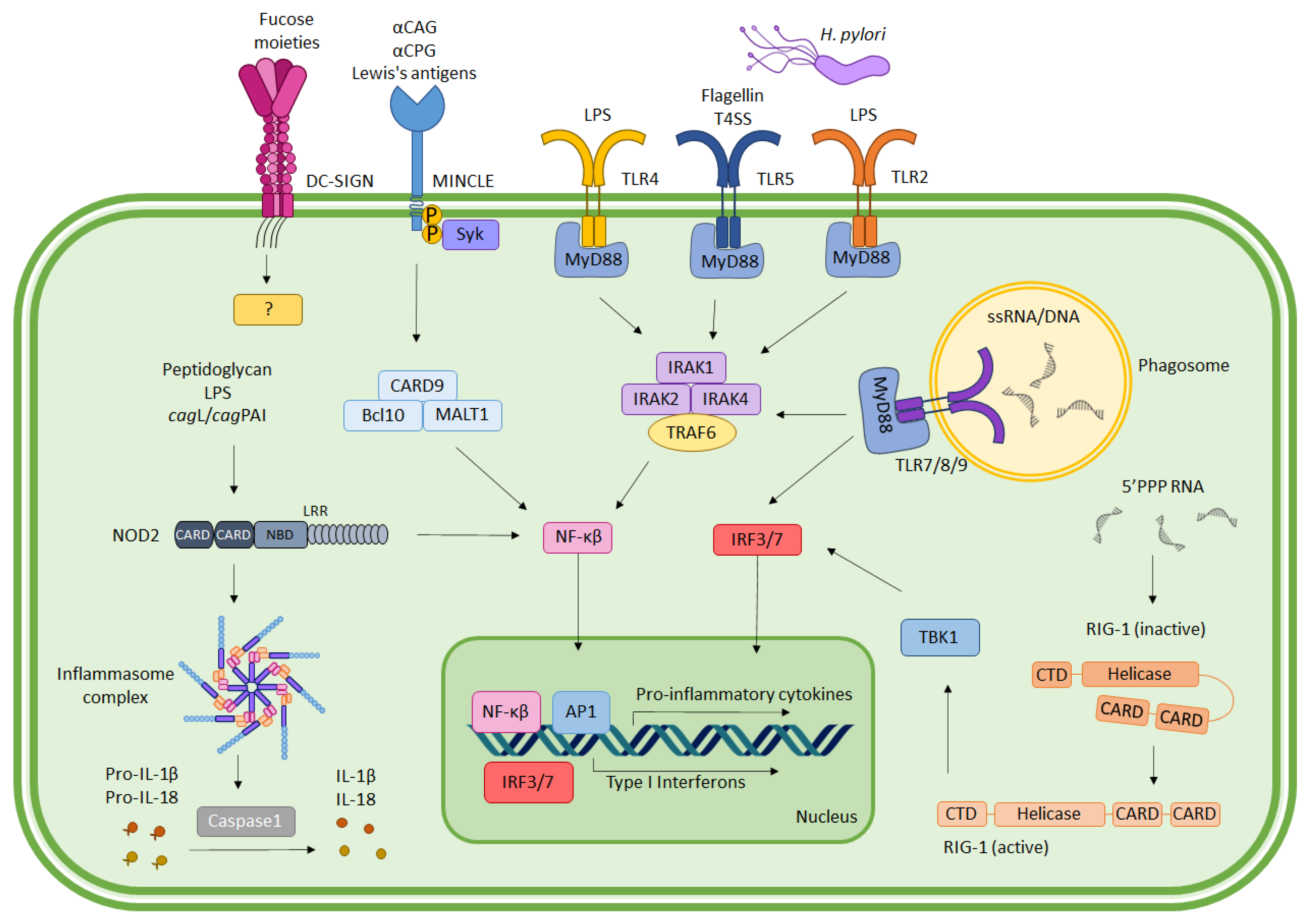
2. Immune Recognition of H. pylori

Immune activation elicited by H. pylori begins with the recognition of the bacterial-pathogen-associated molecular patterns (PAMPs) by PRRs that are expressed by the innate immune cells or gastric epithelial cells. The major classifications of PRRs, including toll-like receptors (TLRs), NOD-like receptors (NLRs), C-type lectin receptors (CLRs), and retinoic acid-inducible gene (RIG)-I-like receptors (RLRs), are all involved in H. pylori recognition and innate immune activation [8,9,10]. Figure 1 depicts the interaction of different PRRs with H. pylori ligands and their downstream signaling pathway activation.

2.1. Toll-Like Receptors (TLRs)

TLRs are a family of type I transmembrane proteins. Each receptor harbors a leucine-rich repeat ectodomain, a transmembrane region involved in PAMP recognition, and an intracellular Toll-IL-1 receptor (TIR) domain to mediate signal transduction. Upon binding to PAMPs, TLRs form complexes and activate either myeloid differentiation primary response 88 (MyD88)-dependent or -independent cascade to switch on nuclear factor kappa B (NF-κB) or interferon regulatory factors (IRFs) transcription factors, thus initiating an immune reaction to counter the pathogen invasion [11]. There are a total of 10 TLRs in humans, some of which (TLR1, TLR2, TLR4, TLR5, TLR6, and TLR10) are present on cellular plasma membranes to recognize extracellular microbial components such as lipopolysaccharide (LPS), lipoproteins, and peptidoglycans; others (TLR3, TLR7, TLR8, and TLR9) are attached within the lipid bilayer of the intracellular vesicles to recognize microbial nucleic acids.
TLR4 is the most intensively studied member of the TLR family. It recognizes H. pylori LPS [11] and activates the NF-ĸB transcription factor, which triggers production of pro-inflammatory cytokines, including interleukin-1β (IL-1β), IL-2, IL-6, IL-8, and IL-12 [12]. However, some studies denounce the role of TLR4 in immunity to H. pylori [13,14,15,16]. Instead, TLR2 was suggested as the receptor for H. pylori LPS despite its common role for detecting bacterial lipoproteins [17]. Together with TLR10, the TLR2/TLR10 heterodimer elicits NF-κB activation following exposure to heat-inactivated H. pylori or LPS [7,18]. H. pylori LPS-bound TLR2 also interacts with TLR4 to promote gastric metaplasia, dysplasia, and adenocarcinoma [19,20].
TLR5 is well-known for its recognition of bacterial flagellin, and is constitutively expressed on the surface of epithelial cells as well as some innate immune cells [21]. Previous research showed that live H. pylori or its purified flagellin activates the NF-κB pathway through binding to TLR5 [13]. Significant upregulation of TLR5 is also detected in THP-1 cells following H. pylori infection, causing cytokine secretion of IL-8 and TNF-α, which initiate inflammation [22]. Recent studies have reported that components of T4SS, such as CagL and CagY, in pathogenic H. pylori strains can serve as TLR5 agonists in driving the innate immune activation and T helper 1 (TH1) cells’ recruitment [23,24].
Inside the intracellular vesicles, TLR7 and TLR8 recognize H. pylori RNA [13], whereas TLR9 recognizes unmethylated CpG DNAs. TLR8 was shown to be induced in THP-1 monocytic cells following the phagocytosis of H. pylori [25], whereby an absence of MyD88 abrogated the production of cytokines such as IL-6 and IL-12 following H. pylori RNA stimulation [26]. H. pylori or its purified DNA induces secretion of IL-8 in neutrophils. Depletion of TLR9, by neutralizing antibodies or inhibitory oligonucleotide, abrogates IL-8 production, suggesting its vital involvement in the bacterial recognition and cell activation [27]. Intriguingly, another study indicated an anti-inflammatory role of TLR9, possibly through the secretion of type I interferon (IFN) in the early stage of H. pylori infection-mediated gastritis [28]. Mice deficient in TLR9 display an intensively augmented inflammation and IL-17 production when infected with H. pylori strains with intact T4SS, an important channel for transferring bacteria DNA into host cells [29]. It was reported that TLR9-mediated transcriptional activity can differ depending on cellular polarity, in which basolateral TLR9 signals IkappaB (IκB) degradation and activation of the NF-κB pathway, whereas apical TLR9 inhibits NF-κB activation. Hence, we anticipate that the anti-inflammatory response invoked by TLR9 signaling following H. pylori infection may be attributable to bacterial-T4SS-mediated immune suppression, which diverts TLR9 to the apical surface to promote a tolerogenic response.

2.2. C-Type Lectin Receptors (CLRs)

CLRs belong to an important class of PRRs that bind to carbohydrate moieties using a conserved carbohydrate-recognition domain (CRD). In general, CLRs induce downstream signaling via two pathways: one involves immunoreceptor tyrosine-based activation motif (ITAM)-containing adaptor molecules such as Fc receptor γ-chain (FcRγ); the other involves phosphatases or kinases that either directly or indirectly interact with cytoplasmic domain of the receptor [30,31].
Among CLR family members, dendritic-cell-specific intercellular nonintegrin (DC-SIGN) plays a pivotal role in H. pylori recognition and pathogenesis. DC-SIGN interacts with mannose and fucose moieties, and is capable of releasing signals to modulate TLR signaling. A study by Gringhuis, et al. [32] demonstrated that H. pylori, a fucose-expressing bacteria, inhibits TH1 polarization by enhancing IL-10 and decreasing IL-12 production. Manipulation of DC-SIGN by H. pylori also extends to their ability to spontaneously switch the expression of Lewis antigens on LPS on or off, whereby the presence of Lewis antigenic expression triggers DC-SIGN on dendritic cells to suppress TH1 polarization and vice versa [33].
In addition to DC-SIGN, the expression of macrophage-inducible C-type lectin (MINCLE) is also upregulated in THP-1 human monocytic cell line during direct or indirect infection with H. pylori. Lipid cholesteryl acyl α-glucoside (αCAG) and cholesteryl phosphatidyl α-glucoside (αCPG) are components of H. pylori that bind to MINCLE to induce secretion of proinflammatory cytokines such as tumor necrosis factor (TNF) and macrophage inflammatory protein (MIP) in dendritic cells, hence exacerbating gastritis in the host [34]. The interactions between MINCLE with the Lewis antigens of H. pylori LPS, on the contrary, induces an anti-inflammatory response in macrophages [35]. Taken together, H. pylori exploits both DC-SIGN and MINCLE to evade the host immune response while inflicting chronic inflammation.

2.3. NOD-Like Receptors (NLRs)

The human nucleotide-binding and oligomerization domain (NOD)-like receptor (NLR) family consists of 22 members that share common features: an N-terminal effector domain to mediate signal transduction, a centrally located nucleotide-binding domain (NBD/NATCH) for oligomerization, and a C-terminal leucine-rich repeat (LRR) domain for ligand sensing. NLRs are involved in the formation of inflammasome, which recruits adaptor protein apoptosis-associated speck-like protein containing a CARD (ASC) to cleave effector molecule pro-caspase-1 into active caspase-1 and mediate secretion of pro-inflammatory cytokines IL-1 and IL-18. Some NLRs, such as NOD1 and NOD2, signal mitogen-activated protein kinase (MAPK) and NF-κB in an inflammasome-independent manner [36,37].
Gene expression analysis showed elevated NLRs levels, including NLRC4, NLRC5, NLRP9, NLRP12, and NLRX1, in the H. pylori challenged human monocytic THP1 cells [38]. Kim, et al. [39] discovered that NOD2, but not NOD1, is required for the activation of NLRP3 inflammasome and pro-IL-1β production in H. pylori challenged bone-marrow-derived dendritic cells.
The production of mature IL-1β in dendritic cells can be triggered by H. pylori virulence factor cytotoxin-associated gene pathogenicity island and CagL [39]. However, NLRP3 activation following acute H. pylori infection results in low levels of mature IL-1β secretion, despite producing abundant premature pro-IL-1β, suggesting H. pylori interference with NLRP3 inflammasome activity to avoid host immune reactions [40]. A deficiency of caspase-1 and IL-1β, but not NLRP3, causes impairment of bacteria clearance from the stomach in H. pylori infected mice [39], which may be due to the involvement of another pathway in the cleavage of caspase-1, which is necessary for IL-1 maturation [41].
Interestingly, genetic polymorphisms involving different NLR members or pathway molecules (NLRP3, NLRP12, NLRX1, and CARD8) are associated with the incidence of gastric cancer. For instance, NLRP12 rs2866112 polymorphism increases the risk of H. pylori infection [38]. More studies are required to determine the involvement and function of these different members of NLRs family in H. pylori recognition. Together, these studies indicate the participation of NLRs in H. pylori recognition and immune cell activation through the release of IL-1.

2.4. RIG-I-Like Receptors (RLRs)

RLRs consist of three members known as retinoic acid-inducible gene I (RIG-I), melanoma differentiation association gene 5 (MDA5), and laboratory of genetics and physiology 2 (LGP2), which detect intracellular RNAs to trigger MyD88-independent secretion of type 1 interferon (IFN) [42].
It was reported that H. pylori 5′-triphosphorylated RNA can be recognized by the RIG-I receptor [26]. HEK293 cells transfected with RIG-I expressing plasmid activate the IFNβ promoter in response to H. pylori RNA stimulation. RIG-1 recognition of H. pylori RNA occurs in a phosphate-dependent manner, as a prior RNA dephosphorylation or usage of RIG-I K858E mutant, which cannot interact with 5’-triphosphate RNA, abrogates cell activation [26].
Furthermore, a clinical study suggested that expression of RIG-1 significantly correlates with poor gastric cancer prognosis [43]. Knockdown of RIG-1 using RNA interference technology in human gastric cancer cell lines results in augmented cell migration, enhanced division in G2/M phase, and higher cell viability [43], suggesting RIG-1 as a crucial component for the prevention of cancer cell growth and invasion. A study using human gastric biopsies revealed a significant upregulation of MDA5 expression in H. pylori infected samples, whereby the level of expression correlated with clinical parameters of gastric atrophy and intestinal metaplasia [44].

3. Innate Immune Cell Activation and Suppression by H. pylori

The immune response against H. pylori is initiated across gastric epithelial cells (GECs), either through the signaling or recruitment by activated GEC or through the direct recognition of various bacterial components or virulence factors [45]. This causes a cascade of innate immune cell activation by neutrophils, macrophages, and dendritic cells. Nonetheless, the host innate immune response can be a double-edged sword in the context of H. pylori infection, where it controls both the bacterial load in the gastric mucosa, as well as the proinflammatory responses that lead to gastritis. Hence, polymorphonuclear cells infiltration has been long used in histological analysis as an indicator of gastritis severity [46]. Figure 2 summarizes the mechanism utilized by H. pylori to promote inflammation and enhance survival, as discussed in the following subsections.

3.1. Neutrophils

The extensive neutrophil recruitment and activation during H. pylori infection lead to an excessive degree of inflammation and mucosal damage [47]. The peripheral blood neutrophil to lymphocyte ratio was shown to be lower in H. pylori positive patients compared with the uninfected individuals [48]. Neutrophils are highly sensitive to the presence of H. pylori, mainly due to the secretion of the H. pylori virulence factor neutrophil activating protein (HP-NAP), which serves as a chemotactic factor [49,50]. Neutrophils rapidly secrete cytokines IL-8, IL-1β, and TNF-α following H. pylori infection [51]. Additionally, H. pylori infection induces hepatoma-derived growth factor (HDGF) expression to enhance neutrophilic infiltration, which is pivotal in gastric carcinogenesis [52]. HDGF ablation in a mouse model blocked neutrophil recruitment following H. pylori infection, and demonstrated an alleviation of gastric lesions [52]. The presence of a flagellin A (FlaA)-positive strain, and a subunit of the T4SS, CagL, induce the secretion of the proinflammatory cytokine IL-1β from neutrophils [53].
The activation of neutrophils can be dampened by H. pylori to promote bacterial persistence. Cathepsin C expression was implicated in playing such a role, whereby cagA-positive H. pylori strain is capable of inhibiting cathepsin C in gastric epithelial cells to prevent neutrophils infiltration and bacterial clearance [54]. In addition, H. pylori is able to activate specific cellular carcinoembryonic antigen-related cell adhesion molecule (CEACAM) in order to promote bacterial uptake by neutrophils, while supporting their intracellular survival through CEACAM–H. pylori outer membrane adhesin HopQ [55].

3.2. Monocytes/Macrophages

Monocytes and macrophages serve as phagocytes and antigen-presenting cells upon H. pylori infection. They are responsible for the polarization of TH1 via secretions of IL-12 and IL-23 [56]. The importance of macrophages in H. pylori infection is highlighted by their marked increases in inflammatory markers, cell motility [57], and infiltration into the peritoneum as early as 2 days post-infection [58]. Furthermore, transient removal of CD11b+ macrophage population was identified to reduce gastric pathology in H. pylori infected mice without altering the bacterial load, implying its critical role in gastric pathology [59]. The activation of macrophages also relies on bacterial factors, whereby the lipopolysaccharide (LPS) inner -core heptose metabolites (ADP-heptose) reportedly induce NF-κB activation and early maturation of the cells [60].
Recently studies highlighted the involvement of the Notch signaling pathway in H. pylori challenged macrophages [61]. The upregulation of Notch ligand Jagged1 was detected in macrophages co-cultured with H. pylori, and clinical specimens from H. pylori infected patients displayed higher Jagged1+ macrophages [61]. The overexpression of Jagged1 in macrophages promotes bactericidal activity through facilitating pro-inflammatory mediator release. Conversely, siRNA-mediated knockdown or anti-Jagged1 neutralizing antibodies attenuate these activities. Additionally, γ-secretase, an inhibitor of Notch signaling was shown to suppress the function of macrophages against H. pylori infection through nitric oxide (NO) production, proinflammatory cytokine secretion, phagocytosis, as well as bactericidal activities [61].
Polarization of macrophages remains an important topic during H. pylori infection. The classically activated macrophages (M1) display a vigorous proinflammatory response and phagocytic function, leading to the reduction in bacterial load and enhanced pathology; whereas alternatively activated macrophages (M2) exhibit anti-inflammatory and immune-modulating functions during tissue remodeling and healing. It is evident that M1 polarization is prominent in H. pylori infected C57BL/6 mice, and can be further enhanced with vaccination using the sublingual administration of H. pylori lysate, although a mixed M1/M2 phenotype is commonly reported in human infection settings [62,63].
Factors influencing the polarization and subsequent outcome of infection have been identified in various reports, including the expression of the cation channel transient receptor potential melastatin 2 (TRPM2). The absence of TRPM2 promotes M1 polarization in response to H. pylori infection. Mice deficient in TRPM2 demonstrate higher gastric inflammation and lower bacterial colonization compared with control mice, owing to overloading calcium levels in macrophages. High intracellular calcium levels elicit an enhanced MAPK signaling pathway and increased production of iNOS and IL-6, which perpetuate inflammation for the resolution of bacterial infection [64].
Another study highlighted the importance of matrix metalloproteinase 7 (MMP7) during polarization. MMP7 null mice displayed increased M1 cells that synthesized more IL-1β than the wildtype counterpart during both in vivo and ex vivo infection with H. pylori [65]. Increased incidences of gastric hyperplasia and dysplasia were detected when MMP was depleted in hypergastrinemic mice due to increased M1 macrophage polarization and mucosal inflammation [65].
Apart from host factors, bacterial virulence, such as CagA, was also shown to influence the M1/M2 balance. The phosphorylation of CagA increases gene expression encoding heme oxygenase-1, causing macrophage polarization toward the M2 phenotype, which functions poorly in bacterial elimination [66]. Taken together, the interplay between various host and bacterial factors is crucial in controlling the outcome of infection, targeting at achieving optimal activation for bacterial clearance (M1) and sufficient healing to prevent pathology (M2).
To achieve long-term survival in the host, H. pylori develops strategies to impede macrophage functions. One example is the bacterial ability to inhibit macrophage phagocytosis [67,68] and to induce macrophage arginase II (ARG2) to inhibit nitric oxide (NO) production [69,70]. Additionally, H. pylori disrupts proliferation [71] and causes the apoptotic program by promoting hydrogen peroxide release and mitochondrial membrane depolarization [72,73]. Thus, the crosstalk between bacteria and macrophages is important to determine if infection can be circumvented in the host.

3.3. Dendritic Cells

H. pylori infection induces dendritic cells’ activation and secretion of cytokines IL-10, IL-12, IL-6, and IL-8 in a dose-dependent manner [74]. It was identified that human HLA-DR+ gastric dendritic cells and monocytes-derived dendritic cells engulf the bacteria for antigen presentation to CD4+ T cells, which preferentially skews T cells into TH1 cells, which release interferon-γ (IFN-γ) [75]. The induction of T helper 17 cells (TH17) is also present alongside TH1 cells, as shown in other reports [76,77]. H. pylori cagPAI influences the maintenance and development of TH1 and TH17 cells through augmenting the secretion of IL-12 and IL-23 from dendritic cells [76,77].
In H. pylori infected pediatric patients, an increased expression of the high-affinity IgE receptor (FcεRI) was observed on peripheral myeloid and plasmacytoid dendritic cells [78]. Monocyte-derived dendritic cells exhibit high FcεRI and IL-10 expressions, suggesting H. pylori drives regulatory dendritic cell differentiation to diminish the hostile immune environment [78]. The immunomodulatory role of dendritic cells was supported by Russler-Germain, et al. [79], whose demonstrated that migratory classical dendritic cells promote peripheral regulatory T cell differentiation to exert immune tolerance. Programmed cell death ligand 1 (PD-L1)-expressing dendritic cells interact with PD-1-expressing T cells in gastritis lesions to impede Helicobacter induced inflammation, thus warranting persistent Helicobacter colonization in mice [80]. Additionally, H. pylori infection can impair the antigen-presentation function of dendritic cells to T cells, thereby blocking TH1 differentiation [81].

4. Conclusions

H. pylori–mediated immune activation begins with the recognition of bacterial PAMPs by gastric epithelial or immune cell PRRs. Upon recognition, a cascade of immune activation is made possible for bacterial clearance as well as chronic gut inflammation. Although multiple studies have helped to deepen our understanding of host–H. pylori interactions, more remains to be investigated to explain the different outcomes of H. pylori infection. Recent studies have focused on identification of various PRRs-associated risk alleles [82], which would provide a better understanding on the risk of infection and degree of severity.

Author Contributions

Conceptualization, W.F.W. and C.Y.L.; writing—original draft preparation, Y.Y.C.; writing—review and editing, G.M.Y.T. and C.Y.Q.L.; funding acquisition, S.A. and W.F.W. All authors have read and agreed to the published version of the manuscript.

Funding

This research was funded by Malaysia Ministry of Higher Education Fundamental Research Grant Scheme FRGS/1/2019/SKK06/UM/02/4 (FP133-2019A) and the Institut Mérieux Young Investigator Fund (IF039-2017). Y.Y. Cheok was funded by a Doctoral Scholarship from the Malaysia Public Service Department (JPA).

Conflicts of Interest

The authors declare no conflict of interest. The funders had no role in the design of the study; in the collection, analyses, or interpretation of data; in the writing of the manuscript; or in the decision to publish the results.

References

  1. WHO. Schistosomes, liver flukes and Helicobacter pylori. IARC Working Group on the Evaluation of Carcinogenic Risks to Humans. Lyon, 7–14 June 1994. In IARC Monographs on the Evaluation of Carcinogenic Risks to Humans; World Health Organization: Lyon, France, 1994; Volume 61, pp. 1–241. [Google Scholar]
  2. Peleteiro, B.; Bastos, A.; Ferro, A.; Lunet, N. Prevalence of Helicobacter pylori infection worldwide: A systematic review of studies with national coverage. Dig. Dis. Sci. 2014, 59, 1698–1709. [Google Scholar] [CrossRef] [PubMed]
  3. Falkeis-Veits, C.; Vieth, M. Non-malignant Helicobacter pylori-Associated Diseases. Adv. Exp. Med. Biol. 2019, 1149, 121–134. [Google Scholar] [CrossRef] [PubMed]
  4. Hatakeyama, M. Malignant Helicobacter pylori-Associated Diseases: Gastric Cancer and MALT Lymphoma. Adv. Exp. Med. Biol. 2019, 1149, 135–149. [Google Scholar] [CrossRef] [PubMed]
  5. Reyes, V.E.; Peniche, A.G. Helicobacter pylori Deregulates T and B Cell Signaling to Trigger Immune Evasion. Curr. Top. Microbiol. Immunol. 2019, 421, 229–265. [Google Scholar] [CrossRef] [PubMed]
  6. Zhang, X.; Arnold, I.C.; Müller, A. Mechanisms of persistence, innate immune activation and immunomodulation by the gastric pathogen Helicobacter pylori. Curr. Opin. Microbiol. 2020, 54, 1–10. [Google Scholar] [CrossRef]
  7. Nagashima, H.; Yamaoka, Y. Importance of Toll-like Receptors in Pro-inflammatory and Anti-inflammatory Responses by Helicobacter pylori Infection. Curr. Top. Microbiol. Immunol. 2019, 421, 139–158. [Google Scholar] [CrossRef]
  8. Davis, B.K.; Wen, H.; Ting, J.P.Y. The Inflammasome NLRs in Immunity, Inflammation, and Associated Diseases. Annu. Rev. Immunol. 2011, 29, 707–735. [Google Scholar] [CrossRef] [Green Version]
  9. Unterholzner, L.; Keating, S.E.; Baran, M.; Horan, K.A.; Jensen, S.B.; Sharma, S.; Sirois, C.M.; Jin, T.; Latz, E.; Xiao, T.S.; et al. IFI16 is an innate immune sensor for intracellular DNA. Nat. Immunol. 2010, 11, 997–1004. [Google Scholar] [CrossRef] [Green Version]
  10. Castaño-Rodríguez, N.; Kaakoush, N.O.; Mitchell, H.M. Pattern-Recognition Receptors and Gastric Cancer. Front. Immunol. 2014, 5, 336. [Google Scholar] [CrossRef]
  11. Smith, S.M. Role of Toll-like receptors in Helicobacter pylori infection and immunity. World J. Gastrointest. Pathophysiol. 2014, 5, 133–146. [Google Scholar] [CrossRef]
  12. Takeuchi, O.; Akira, S. Toll-like receptors; their physiological role and signal transduction system. Int. Immunopharmacol. 2001, 1, 625–635. [Google Scholar] [CrossRef]
  13. Smith, M.F.; Mitchell, A.; Li, G.; Ding, S.; Fitzmaurice, A.M.; Ryan, K.; Crowe, S.; Goldberg, J.B. Toll-like Receptor (TLR) 2 and TLR5, but Not TLR4, Are Required for Helicobacter pylori-induced NF-κB Activation and Chemokine Expression by Epithelial Cells. J. Biol. Chem. 2003, 278, 32552–32560. [Google Scholar] [CrossRef] [PubMed] [Green Version]
  14. Bäckhed, F.; Rokbi, B.; Torstensson, E.; Zhao, Y.; Nilsson, C.; Seguin, D.; Normark, S.; Buchan, A.M.J.; Richter-Dahlfors, A. Gastric Mucosal Recognition of Helicobacter pylori Is Independent of Toll-Like Receptor 4. J. Infect. Dis. 2003, 187, 829–836. [Google Scholar] [CrossRef] [PubMed] [Green Version]
  15. Yokota, S.-I.; Ohnishi, T.; Muroi, M.; Tanamoto, K.-I.; Fujii, N.; Amano, K.-I. Highly-purified Helicobacter pylori LPS preparations induce weak inflammatory reactions and utilize Toll-like receptor 2 complex but not Toll-like receptor 4 complex. FEMS Immunol. Med. Microbiol. 2007, 51, 140–148. [Google Scholar] [CrossRef] [Green Version]
  16. Tongtawee, T.; Simawaranon, T.; Wattanawongdon, W.; Dechsukhum, C.; Leeanansaksiri, W. Toll-like receptor 2 and 4 polymorphisms associated with Helicobacter pylori susceptibility and gastric cancer. Turk. J. Gastroenterol. 2019, 30, 15–20. [Google Scholar] [CrossRef]
  17. Hashimoto, M.; Tawaratsumida, K.; Kariya, H.; Aoyama, K.; Tamura, T.; Suda, Y. Lipoprotein is a predominant Toll-like receptor 2 ligand in Staphylococcus aureus cell wall components. Int. Immunol. 2006, 18, 355–362. [Google Scholar] [CrossRef]
  18. Nagashima, H.; Iwatani, S.; Cruz, M.; Jiménez Abreu, J.A.; Uchida, T.; Mahachai, V.; Vilaichone, R.-k.; Graham, D.Y.; Yamaoka, Y. Toll-like Receptor 10 in Helicobacter pylori Infection. J. Infect. Dis. 2015, 212, 1666–1676. [Google Scholar] [CrossRef] [Green Version]
  19. Yokota, S.-I.; Okabayashi, T.; Rehli, M.; Fujii, N.; Amano, K.-I. Helicobacter pylori Lipopolysaccharides Upregulate Toll-Like Receptor 4 Expression and Proliferation of Gastric Epithelial Cells via the MEK1/2-ERK1/2 Mitogen-Activated Protein Kinase Pathway. Infect. Immun. 2010, 78, 468–476. [Google Scholar] [CrossRef] [Green Version]
  20. Chochi, K.; Ichikura, T.; Kinoshita, M.; Majima, T.; Shinomiya, N.; Tsujimoto, H.; Kawabata, T.; Sugasawa, H.; Ono, S.; Seki, S.; et al. Helicobacter pylori Augments Growth of Gastric Cancers via the Lipopolysaccharide-Toll-like Receptor 4 Pathway whereas Its Lipopolysaccharide Attenuates Antitumor Activities of Human Mononuclear Cells. Clin. Cancer Res. 2008, 14, 2909–2917. [Google Scholar] [CrossRef] [Green Version]
  21. Wen, S.; Wu, Z.; Zhong, S.; Li, M.; Shu, Y. Factors influencing the immunogenicity of influenza vaccines. Hum. Vaccines Immunother. 2021, 17, 2706–2718. [Google Scholar] [CrossRef]
  22. Pachathundikandi, S.K.; Brandt, S.; Madassery, J.; Backert, S. Induction of TLR-2 and TLR-5 Expression by Helicobacter pylori Switches cagPAI-Dependent Signalling Leading to the Secretion of IL-8 and TNF-α. PLoS ONE 2011, 6, e19614. [Google Scholar] [CrossRef] [PubMed]
  23. Tegtmeyer, N.; Neddermann, M.; Lind, J.; Pachathundikandi, S.K.; Sharafutdinov, I.; Gutiérrez-Escobar, A.J.; Brönstrup, M.; Tegge, W.; Hong, M.; Rohde, M.; et al. Toll-like Receptor 5 Activation by the CagY Repeat Domains of Helicobacter pylori. Cell Rep. 2020, 32, 108159. [Google Scholar] [CrossRef] [PubMed]
  24. Pachathundikandi, S.K.; Tegtmeyer, N.; Arnold, I.C.; Lind, J.; Neddermann, M.; Falkeis-Veits, C.; Chattopadhyay, S.; Brönstrup, M.; Tegge, W.; Hong, M.; et al. T4SS-dependent TLR5 activation by Helicobacter pylori infection. Nat. Commun. 2019, 10, 5717. [Google Scholar] [CrossRef] [PubMed] [Green Version]
  25. Gantier, M.P.; Irving, A.T.; Kaparakis-Liaskos, M.; Xu, D.; Evans, V.A.; Cameron, P.U.; Bourne, J.A.; Ferrero, R.L.; John, M.; Behlke, M.A.; et al. Genetic Modulation of TLR8 Response following Bacterial Phagocytosis. Hum. Mutat. 2010, 31, 1069–1079. [Google Scholar] [CrossRef]
  26. Rad, R.; Ballhorn, W.; Voland, P.; Eisenacher, K.; Mages, J.; Rad, L.; Ferstl, R.; Lang, R.; Wagner, H.; Schmid, R.M.; et al. Extracellular and intracellular pattern recognition receptors cooperate in the recognition of Helicobacter pylori. Gastroenterology 2009, 136, 2247–2257. [Google Scholar] [CrossRef]
  27. Alvarez-Arellano, L.; Cortés-Reynosa, P.; Sánchez-Zauco, N.; Salazar, E.; Torres, J.; Maldonado-Bernal, C. TLR9 and NF-κB are partially involved in activation of human neutrophils by Helicobacter pylori and its purified DNA. PLoS ONE 2014, 9, e101342. [Google Scholar] [CrossRef] [Green Version]
  28. Otani, K.; Tanigawa, T.; Watanabe, T.; Nadatani, Y.; Sogawa, M.; Yamagami, H.; Shiba, M.; Watanabe, K.; Tominaga, K.; Fujiwara, Y.; et al. Toll-like receptor 9 signaling has anti-inflammatory effects on the early phase of Helicobacter pylori-induced gastritis. Biochem. Biophys. Res. Commun. 2012, 426, 342–349. [Google Scholar] [CrossRef]
  29. Varga, M.G.; Piazuelo, M.B.; Romero-Gallo, J.; Delgado, A.G.; Suarez, G.; Whitaker, M.E.; Krishna, U.S.; Patel, R.V.; Skaar, E.P.; Wilson, K.T.; et al. TLR9 activation suppresses inflammation in response to Helicobacter pylori infection. Am. J. Physiol. Gastrointest. Liver Physiol. 2016, 311, G852–G858. [Google Scholar] [CrossRef] [Green Version]
  30. Tone, K.; Stappers, M.H.T.; Willment, J.A.; Brown, G.D. C-type lectin receptors of the Dectin-1 cluster: Physiological roles and involvement in disease. Eur. J. Immunol. 2019, 49, 2127–2133. [Google Scholar] [CrossRef] [Green Version]
  31. Geijtenbeek, T.B.H.; Gringhuis, S.I. Signalling through C-type lectin receptors: Shaping immune responses. Nat. Rev. Immunol. 2009, 9, 465–479. [Google Scholar] [CrossRef]
  32. Gringhuis, S.I.; den Dunnen, J.; Litjens, M.; van der Vlist, M.; Geijtenbeek, T.B.H. Carbohydrate-specific signaling through the DC-SIGN signalosome tailors immunity to Mycobacterium tuberculosis, HIV-1 and Helicobacter pylori. Nat. Immunol. 2009, 10, 1081–1088. [Google Scholar] [CrossRef] [PubMed]
  33. Bergman, M.P.; Engering, A.; Smits, H.H.; van Vliet, S.J.; van Bodegraven, A.A.; Wirth, H.-P.; Kapsenberg, M.L.; Vandenbroucke-Grauls, C.M.J.E.; van Kooyk, Y.; Appelmelk, B.J. Helicobacter pylori Modulates the T Helper Cell 1/T Helper Cell 2 Balance through Phase-variable Interaction between Lipopolysaccharide and DC-SIGN. J. Exp. Med. 2004, 200, 979–990. [Google Scholar] [CrossRef] [PubMed]
  34. Nagata, M.; Toyonaga, K.; Ishikawa, E.; Haji, S.; Okahashi, N.; Takahashi, M.; Izumi, Y.; Imamura, A.; Takato, K.; Ishida, H.; et al. Helicobacter pylori metabolites exacerbate gastritis through C-type lectin receptors. J. Exp. Med. 2020, 218, e20200815. [Google Scholar] [CrossRef]
  35. Devi, S.; Rajakumara, E.; Ahmed, N. Induction of Mincle by Helicobacter pylori and consequent anti-inflammatory signaling denote a bacterial survival strategy. Sci. Rep. 2015, 5, 15049. [Google Scholar] [CrossRef] [PubMed]
  36. Platnich, J.M.; Muruve, D.A. NOD-like receptors and inflammasomes: A review of their canonical and non-canonical signaling pathways. Arch. Biochem. Biophys. 2019, 670, 4–14. [Google Scholar] [CrossRef]
  37. Velloso, F.J.; Trombetta-Lima, M.; Anschau, V.; Sogayar, M.C.; Correa, R.G. NOD-like receptors: Major players (and targets) in the interface between innate immunity and cancer. Biosci. Rep. 2019, 39, BSR20181709. [Google Scholar] [CrossRef] [Green Version]
  38. Castaño-Rodríguez, N.; Kaakoush, N.O.; Goh, K.-L.; Fock, K.M.; Mitchell, H.M. The NOD-Like Receptor Signalling Pathway in Helicobacter pylori Infection and Related Gastric Cancer: A Case-Control Study and Gene Expression Analyses. PLoS ONE 2014, 9, e98899. [Google Scholar] [CrossRef]
  39. Kim, D.-J.; Park, J.-H.; Franchi, L.; Backert, S.; Núñez, G. The Cag pathogenicity island and interaction between TLR2/NOD2 and NLRP3 regulate IL-1β production in Helicobacter pylori infected dendritic cells. Eur. J. Immunol. 2013, 43, 2650–2658. [Google Scholar] [CrossRef] [Green Version]
  40. Pachathundikandi, S.K.; Blaser, N.; Bruns, H.; Backert, S. Helicobacter pylori Avoids the Critical Activation of NLRP3 Inflammasome-Mediated Production of Oncogenic Mature IL-1β in Human Immune Cells. Cancers 2020, 12, 803. [Google Scholar] [CrossRef] [Green Version]
  41. Basak, C.; Pathak, S.K.; Bhattacharyya, A.; Mandal, D.; Pathak, S.; Kundu, M. NF-κB- and C/EBPβ-driven Interleukin-1β Gene Expression and PAK1-mediated Caspase-1 Activation Play Essential Roles in Interleukin-1β Release from Helicobacter pylori Lipopolysaccharide-stimulated Macrophages. J. Biol. Chem. 2005, 280, 4279–4288. [Google Scholar] [CrossRef] [Green Version]
  42. Rehwinkel, J.; Gack, M.U. RIG-I-like receptors: Their regulation and roles in RNA sensing. Nat. Rev. Immunol. 2020, 20, 537–551. [Google Scholar] [CrossRef]
  43. Chen, L.; Feng, J.; Wu, S.; Xu, B.; Zhou, Y.; Wu, C.; Jiang, J. Decreased RIG-I expression is associated with poor prognosis and promotes cell invasion in human gastric cancer. Cancer Cell Int. 2018, 18, 144. [Google Scholar] [CrossRef]
  44. Tatsuta, T.; Imaizumi, T.; Shimoyama, T.; Sawaya, M.; Kunikazu, T.; Matsumiya, T.; Yoshida, H.; Satoh, K.; Fukuda, S. Expression of melanoma differentiation associated gene 5 is increased in human gastric mucosa infected with Helicobacter pylori. J. Clin. Pathol. 2012, 65, 839–843. [Google Scholar] [CrossRef] [PubMed]
  45. Cheok, Y.Y.; Lee, C.Y.Q.; Cheong, H.C.; Vadivelu, J.; Looi, C.Y.; Abdullah, S.; Wong, W.F. An Overview of Helicobacter pylori Survival Tactics in the Hostile Human Stomach Environment. Microorganisms 2021, 9, 2502. [Google Scholar] [CrossRef]
  46. Siriviriyakul, P.; Werawatganon, D.; Phetnoo, N.; Somanawat, K.; Chatsuwan, T.; Klaikeaw, N.; Chayanupatkul, M. Genistein attenuated gastric inflammation and apoptosis in Helicobacter pylori-induced gastropathy in rats. BMC Gastroenterol. 2020, 20, 410. [Google Scholar] [CrossRef] [PubMed]
  47. Toyoshima, O.; Nishizawa, T.; Arita, M.; Kataoka, Y.; Sakitani, K.; Yoshida, S.; Yamashita, H.; Hata, K.; Watanabe, H.; Suzuki, H. Helicobacter pylori infection in subjects negative for high titer serum antibody. World J. Gastroenterol. 2018, 24, 1419–1428. [Google Scholar] [CrossRef] [PubMed]
  48. Guclu, M.; Faruq Agan, A. Association of Severity of Helicobacter pylori Infection with Peripheral Blood Neutrophil to Lymphocyte Ratio and Mean Platelet Volume. Euroasian J. Hepatogastroenterol. 2017, 7, 11–16. [Google Scholar] [CrossRef] [PubMed] [Green Version]
  49. Evans, D.J.; Evans, D.G.; Takemura, T.; Nakano, H.; Lampert, H.C.; Graham, D.Y.; Granger, D.N.; Kvietys, P.R. Characterization of a Helicobacter pylori neutrophil-activating protein. Infect. Immun. 1995, 63, 2213–2220. [Google Scholar] [CrossRef] [PubMed] [Green Version]
  50. Wen, S.-H.; Hong, Z.-W.; Chen, C.-C.; Chang, H.-W.; Fu, H.-W. Helicobacter pylori Neutrophil-Activating Protein Directly Interacts with and Activates Toll-like Receptor 2 to Induce the Secretion of Interleukin-8 from Neutrophils and ATRA-Induced Differentiated HL-60 Cells. Int. J. Mol. Sci. 2021, 22, 1560. [Google Scholar] [CrossRef]
  51. Alvarez-Arellano, L.; Camorlinga-Ponce, M.; Maldonado-Bernal, C.; Torres, J. Activation of human neutrophils with Helicobacter pylori and the role of Toll-like receptors 2 and 4 in the response. FEMS Immunol. Med. Microbiol. 2007, 51, 473–479. [Google Scholar] [CrossRef] [Green Version]
  52. Chu, T.-H.; Huang, S.-T.; Yang, S.-F.; Li, C.-J.; Lin, H.-W.; Weng, B.-C.; Yang, S.-M.; Huang, S.-C.; Wu, J.-C.; Chang, Y.-C.; et al. Hepatoma-derived growth factor participates in Helicobacter Pylori-induced neutrophils recruitment, gastritis and gastric carcinogenesis. Oncogene 2019, 38, 6461–6477. [Google Scholar] [CrossRef]
  53. Jang, A.-R.; Kang, M.-J.; Shin, J.-I.; Kwon, S.-W.; Park, J.-Y.; Ahn, J.-H.; Lee, T.-S.; Kim, D.-Y.; Choi, B.-G.; Seo, M.-W.; et al. Unveiling the Crucial Role of Type IV Secretion System and Motility of Helicobacter pylori in IL-1β Production via NLRP3 Inflammasome Activation in Neutrophils. Front. Immunol. 2020, 11, 1121. [Google Scholar] [CrossRef] [PubMed]
  54. Gang Liu, Y.; Teng, Y.S.; Cheng, P.; Kong, H.; Lv, P.Y.; Mao, F.Y.; Wu, X.L.; Hao, C.J.; Chen, W.; Yang, S.M.; et al. Abrogation of cathepsin C by Helicobacter pylori impairs neutrophil activation to promote gastric infection. FASEB J. 2019, 33, 5018–5033. [Google Scholar] [CrossRef] [PubMed]
  55. Behrens, I.-K.; Busch, B.; Ishikawa-Ankerhold, H.; Palamides, P.; Shively John, E.; Stanners, C.; Chan, C.; Leung, N.; Gray-Owen, S.; Haas, R.; et al. The HopQ-CEACAM Interaction Controls CagA Translocation, Phosphorylation, and Phagocytosis of Helicobacter pylori in Neutrophils. mBio 2020, 11, e03256-19. [Google Scholar] [CrossRef] [PubMed] [Green Version]
  56. Peek, R.M., Jr.; Fiske, C.; Wilson, K.T. Role of innate immunity in Helicobacter pylori-induced gastric malignancy. Physiol. Rev. 2010, 90, 831–858. [Google Scholar] [CrossRef] [Green Version]
  57. Cheok, Y.Y.; Tan, G.M.Y.; Fernandez, K.C.; Chan, Y.T.; Lee, C.Y.Q.; Cheong, H.C.; Looi, C.Y.; Vadivelu, J.; Abdullah, S.; Wong, W.F. Podoplanin Drives Motility of Active Macrophage via Regulating Filamin C During Helicobacter pylori Infection. Front. Immunol. 2021, 12, 702156. [Google Scholar] [CrossRef]
  58. Algood, H.M.; Gallo-Romero, J.; Wilson, K.T.; Peek, R.M., Jr.; Cover, T.L. Host response to Helicobacter pylori infection before initiation of the adaptive immune response. FEMS Immunol. Med. Microbiol. 2007, 51, 577–586. [Google Scholar] [CrossRef] [Green Version]
  59. Kaparakis, M.; Walduck, A.K.; Price, J.D.; Pedersen, J.S.; van Rooijen, N.; Pearse, M.J.; Wijburg, O.L.C.; Strugnell, R.A. Macrophages Are Mediators of Gastritis in Acute Helicobacter pylori Infection in C57BL/6 Mice. Infect. Immun. 2008, 76, 2235–2239. [Google Scholar] [CrossRef] [Green Version]
  60. Faass, L.; Stein, S.C.; Hauke, M.; Gapp, M.; Albanese, M.; Josenhans, C. Contribution of Heptose Metabolites and the cag Pathogenicity Island to the Activation of Monocytes/Macrophages by Helicobacter pylori. Front. Immunol. 2021, 12, 632154. [Google Scholar] [CrossRef]
  61. Wen, J.; Chen, C.; Luo, M.; Liu, X.; Guo, J.; Wei, T.; Gu, X.; Gu, S.; Ning, Y.; Li, Y. Notch Signaling Ligand Jagged1 Enhances Macrophage-Mediated Response to Helicobacter pylori. Front. Microbiol. 2021, 12, 1741. [Google Scholar] [CrossRef]
  62. Quiding-Järbrink, M.; Raghavan, S.; Sundquist, M. Enhanced M1 macrophage polarization in human helicobacter pylori-associated atrophic gastritis and in vaccinated mice. PLoS ONE 2010, 5, e15018. [Google Scholar] [CrossRef] [PubMed]
  63. Fehlings, M.; Drobbe, L.; Moos, V.; Renner Viveros, P.; Hagen, J.; Beigier-Bompadre, M.; Pang, E.; Belogolova, E.; Churin, Y.; Schneider, T.; et al. Comparative Analysis of the Interaction of Helicobacter pylori with Human Dendritic Cells, Macrophages, and Monocytes. Infect. Immun. 2012, 80, 2724–2734. [Google Scholar] [CrossRef] [PubMed] [Green Version]
  64. Beceiro, S.; Radin, J.N.; Chatuvedi, R.; Piazuelo, M.B.; Horvarth, D.J.; Cortado, H.; Gu, Y.; Dixon, B.; Gu, C.; Lange, I.; et al. TRPM2 ion channels regulate macrophage polarization and gastric inflammation during Helicobacter pylori infection. Mucosal Immunol. 2017, 10, 493–507. [Google Scholar] [CrossRef] [Green Version]
  65. Krakowiak, M.S.; Noto, J.M.; Piazuelo, M.B.; Hardbower, D.M.; Romero-Gallo, J.; Delgado, A.; Chaturvedi, R.; Correa, P.; Wilson, K.T.; Peek, R.M. Matrix metalloproteinase 7 restrains Helicobacter pylori-induced gastric inflammation and premalignant lesions in the stomach by altering macrophage polarization. Oncogene 2015, 34, 1865–1871. [Google Scholar] [CrossRef] [PubMed] [Green Version]
  66. Gobert, A.P.; Verriere, T.; Asim, M.; Barry, D.P.; Piazuelo, M.B.; de Sablet, T.; Delgado, A.G.; Bravo, L.E.; Correa, P.; Peek, R.M.; et al. Heme oxygenase-1 dysregulates macrophage polarization and the immune response to Helicobacter pylori. J. Immunol. 2014, 193, 3013–3022. [Google Scholar] [CrossRef] [Green Version]
  67. Ramarao, N.; Gray-Owen, S.D.; Backert, S.; Meyer, T.F. Helicobacter pylori inhibits phagocytosis by professional phagocytes involving type IV secretion components. Mol. Microbiol. 2000, 37, 1389–1404. [Google Scholar] [CrossRef] [PubMed]
  68. Allen, L.-A.H.; Schlesinger, L.S.; Kang, B. Virulent strains of Helicobacter pylori demonstrate delayed phagocytosis and stimulate homotypic phagosome fusion in macrophages. J. Exp. Med. 2000, 191, 115–128. [Google Scholar] [CrossRef] [PubMed]
  69. Lewis, N.D.; Asim, M.; Barry, D.P.; de Sablet, T.; Singh, K.; Piazuelo, M.B.; Gobert, A.P.; Chaturvedi, R.; Wilson, K.T. Immune evasion by Helicobacter pylori is mediated by induction of macrophage arginase II. J. Immunol. 2011, 186, 3632–3641. [Google Scholar] [CrossRef] [Green Version]
  70. Gobert, A.P.; Cheng, Y.; Wang, J.-Y.; Boucher, J.-L.; Iyer, R.K.; Cederbaum, S.D.; Casero, R.A.; Newton, J.C.; Wilson, K.T. Helicobacter pylori induces macrophage apoptosis by activation of arginase II. J. Immunol. 2002, 168, 4692–4700. [Google Scholar] [CrossRef] [Green Version]
  71. Tan, G.M.Y.; Looi, C.Y.; Fernandez, K.C.; Vadivelu, J.; Loke, M.F.; Wong, W.F. Suppression of cell division-associated genes by Helicobacter pylori attenuates proliferation of RAW264.7 monocytic macrophage cells. Sci. Rep. 2015, 5, 11046. [Google Scholar] [CrossRef] [Green Version]
  72. Asim, M.; Chaturvedi, R.; Hoge, S.; Lewis, N.D.; Singh, K.; Barry, D.P.; Algood, H.S.; de Sablet, T.; Gobert, A.P.; Wilson, K.T. Helicobacter pylori induces ERK-dependent formation of a phospho-c-Fos c-Jun activator protein-1 complex that causes apoptosis in macrophages. J. Biol. Chem. 2010, 285, 20343–20357. [Google Scholar] [CrossRef] [PubMed] [Green Version]
  73. Chaturvedi, R.; Cheng, Y.; Asim, M.; Bussière, F.I.; Xu, H.; Gobert, A.P.; Hacker, A.; Casero, R.A., Jr.; Wilson, K.T. Induction of polyamine oxidase 1 by Helicobacter pylori causes macrophage apoptosis by hydrogen peroxide release and mitochondrial membrane depolarization. J. Biol. Chem. 2004, 279, 40161–40173. [Google Scholar] [CrossRef] [PubMed] [Green Version]
  74. Kranzer, K.; Eckhardt, A.; Aigner, M.; Knoll, G.; Deml, L.; Speth, C.; Lehn, N.; Rehli, M.; Schneider-Brachert, W. Induction of Maturation and Cytokine Release of Human Dendritic Cells by Helicobacter pylori. Infect. Immun. 2004, 72, 4416–4423. [Google Scholar] [CrossRef] [PubMed] [Green Version]
  75. Bimczok, D.; Clements, R.H.; Waites, K.B.; Novak, L.; Eckhoff, D.E.; Mannon, P.J.; Smith, P.D.; Smythies, L.E. Human primary gastric dendritic cells induce a Th1 response to H. pylori. Mucosal Immunol. 2010, 3, 260–269. [Google Scholar] [CrossRef] [PubMed] [Green Version]
  76. Khamri, W.; Walker Marjorie, M.; Clark, P.; Atherton John, C.; Thursz Mark, R.; Bamford Kathleen, B.; Lechler Robert, I.; Lombardi, G. Helicobacter pylori Stimulates Dendritic Cells To Induce Interleukin-17 Expression from CD4+ T Lymphocytes. Infect. Immun. 2010, 78, 845–853. [Google Scholar] [CrossRef] [Green Version]
  77. Guiney Donald, G.; Hasegawa, P.; Cole Sheri, P. Helicobacter pylori Preferentially Induces Interleukin 12 (IL-12) Rather than IL-6 or IL-10 in Human Dendritic Cells. Infect. Immun. 2003, 71, 4163–4166. [Google Scholar] [CrossRef] [Green Version]
  78. León, M.A.; Palma, C.; Hernández, C.; Sandoval, M.; Cofre, C.; Perez-Mateluna, G.; Borzutzky, A.; Harris, P.R.; Serrano, C.A. Helicobacter pylori pediatric infection changes FcεRI expression in dendritic cells and Treg profile in vivo and in vitro. Microbes Infect. 2019, 21, 449–455. [Google Scholar] [CrossRef]
  79. Russler-Germain, E.V.; Yi, J.; Young, S.; Nutsch, K.; Wong, H.S.; Ai, T.L.; Chai, J.N.; Durai, V.; Kaplan, D.H.; Germain, R.N.; et al. Gut Helicobacter presentation by multiple dendritic cell subsets enables context-specific regulatory T cell generation. Elife 2021, 10, e54792. [Google Scholar] [CrossRef]
  80. Go, D.-M.; Lee, S.H.; Lee, S.-H.; Woo, S.-H.; Kim, K.; Kim, K.; Park, K.S.; Park, J.-H.; Ha, S.-J.; Kim, W.H.; et al. Programmed Death Ligand 1-Expressing Classical Dendritic Cells Mitigate Helicobacter-Induced Gastritis. Cell. Mol. Gastroenterol. Hepatol. 2021, 12, 715–739. [Google Scholar] [CrossRef]
  81. Mitchell, P.; Germain, C.; Fiori, P.L.; Khamri, W.; Foster, G.R.; Ghosh, S.; Lechler, R.I.; Bamford, K.B.; Lombardi, G. Chronic exposure to Helicobacter pylori impairs dendritic cell function and inhibits Th1 development. Infect. Immun. 2007, 75, 810–819. [Google Scholar] [CrossRef] [Green Version]
  82. Ma, X.; Lu, L.; Tang, Y.; Luo, W.; Li, J.; Tang, M. Association between Toll-like receptor gene polymorphisms and risk of Helicobacter pylori infection: A protocol for systematic review and meta-analysis. Medicine 2021, 100, e25729. [Google Scholar] [CrossRef] [PubMed]
Figure 1. Activation of pattern recognition receptors (PRRs) by H. pylori. Toll-like receptors (TLRs) 2, 4, and 5 recognize different extracellular bacterial factors to mediate MyD88-dependent or -independent activation of proinflammatory cytokine secretion. TLR7/8/9 recognize ingested bacterial DNA or single-stranded RNA (ssRNA) in phagosomes for activation of type I interferons production. Transcription factors NF-ĸB, activator protein 1 (AP1), and IRF3/7 play prominent roles in transactivation of inflammatory cytokines and type I interferons. C-type lectin-like receptors (CLRs) including MINCLE also induce NF-ĸB through interaction with CARD9/BCL10/MALT1. DC-SIGN transmits signals through an unknown pathway during H. pylori infection. NOD-like receptors (NLRs) such as NOD2 activate the inflammasome complex to induce production and maturation of IL-1β and IL-18. RIG-I-like receptors (RLRs) such as retinoic acid-inducible gene I (RIG-I) are activated by 5′triphosphate (PPP) single-stranded RNA, leading to conformation change, which subsequently activates Tank-binding kinase 1 (TBK1). This, in turn, causes production of type I interferons through IRF3/7 signaling.
Ijms 23 07561 g001
Figure 2. Innate immune cells interaction with H. pylori across gastric epithelial cells (GECs). Neutrophils are directly activated by bacterial antigens, including H. pylori neutrophil activating protein (HP-NAP), type IV secretion system (T4SS), and flagellin A (FlaA), or indirectly through gastric epithelial cells secretion of hepatoma-derived growth factor (HDGF). Activation may result in proinflammatory cytokines secretion (IL-8 and IL-1β) and the production of reactive oxygen species (ROS) for the clearance of bacterial load. H. pylori can inhibit cathepsin C production to dampen neutrophil activation. It can also lead to cellular carcinoembryonic antigen-related cell adhesion molecules (CEACAMs) expression, which enhances neutrophils phagocytic activity without bacterial killing. Macrophages can be activated by bacterial lipopolysaccharide (LPS) or by the presence of the virulence gene cag pathogenicity island (cagPAI), resulting in mixed polarization of proinflammatory (M1) with high expressions of IL-1β, IL-6, and ROS, and the anti-inflammatory (M2) macrophages. Dendritic cells process bacterial antigens that are presented to T cells for differentiation. These cells upregulate programmed death ligand 1 (PD-L1) on regulatory T cell (Treg) to increase IL-10 secretion, leading to bacterial persistence. It can also upregulate proinflammatory cytokines IL-12 and IL-23 to stimulate T helper 1 (TH1) and 17 (TH17) polarization, which releases high amounts of interferon-γ (IFN-γ) and IL-17.
Ijms 23 07561 g002
Publisher’s Note: MDPI stays neutral with regard to jurisdictional claims in published maps and institutional affiliations.

