Review

Glycan-Lectin Interactions as Novel Immunosuppression Drivers in Glioblastoma

1
Laboratory of Tumor Immunology and Cell Therapy, Department of Experimental Medicine, “Sapienza” University of Rome, Viale Regina Elena 324, 00161 Rome, Italy
2
Department of Neurology and Psychiatry, Neurosurgery, “Sapienza” University of Rome, Viale dell’ Università 30, 00185 Rome, Italy
*
Author to whom correspondence should be addressed.
Int. J. Mol. Sci. 2022, 23(11), 6312; https://doi.org/10.3390/ijms23116312
Submission received: 14 April 2022 / Revised: 23 May 2022 / Accepted: 3 June 2022 / Published: 5 June 2022

Abstract

:
Despite diagnostic and therapeutic improvements, glioblastoma (GB) remains one of the most threatening brain tumor in adults, underlining the urgent need of new therapeutic targets. Lectins are glycan-binding proteins that regulate several biological processes through the recognition of specific sugar motifs. Lectins and their ligands are found on immune cells, endothelial cells and, also, tumor cells, pointing out a strong correlation among immunity, tumor microenvironment and vascularization. In GB, altered glycans and lectins contribute to tumor progression and immune evasion, shaping the tumor-immune landscape promoting immunosuppressive cell subsets, such as myeloid-derived suppressor cells (MDSCs) and M2-macrophages, and affecting immunoeffector populations, such as CD8+ T cells and dendritic cells (DCs). Here, we discuss the latest knowledge on the immune cells, immune related lectin receptors (C-type lectins, Siglecs, galectins) and changes in glycosylation that are involved in immunosuppressive mechanisms in GB, highlighting their interest as possible novel therapeutical targets.

1. Introduction

Gliomas are malignant brain tumors arising from transformed primitive neural stem cells, glial progenitor cells or dedifferentiated mature cell type [1]. Recently, the World Health Organization (WHO) released the newest 2021 classification of brain tumors, based on not only canonical criteria, such as histological and molecular features, but also novel diagnostic technologies, such as DNA methylome profiling. According to that, glioblastoma (GB) is characterized by the IDH-wildtype phenotype, while his mutant counterpart is identified as Astrocytoma IDH-mutant (grades 2–4) [2].
GB is the most common malignant brain tumor in adults, mostly in males compared to females. Nowadays, the first line of therapy requires maximal safe resection followed by radiotherapy (RT) and a high-dose of chemotherapy with temozolomide (TMZ). Despite the heavy treatment, most of the patients experience a recurrence, and the median overall survival remains below 18 months [3]. At recurrence, the standard of care is less well defined. Indeed, therapeutic options include afresh surgery and reirradiation in combination with systemic therapies, such as chemotherapy (TMZ, lomustine, carmustine) or anti-VEGF monoclonal antibody Bevacizumab [3,4,5]. Emerging therapies, such as immunotherapy and tumor-treating fields (TTFields), are available. TTFields and their ability to disrupt specifically mitotic process in cancer cells had shown promising results in prolonging the survival time, and several clinical trials are ongoing to study its safety and tolerability for further use [3,6]. On the other hand, immunotherapeutic approaches as immune-checkpoint inhibitors (ICIs) or personalized vaccination did not show in GB patients the same efficacy and benefits in terms of overall survival and improvement of prognosis as for other neoplasms [7,8,9,10]. Resistance to common therapies and failure of immunotherapeutic approaches are strongly associated to GB aggressiveness and microenvironment. Tumor cells within stromal cells cooperate in the establishment of a tumor-promoting microenvironment through the induction of immunosuppressive cytokines (i.e., TNF-α, interleukin (IL)-1α/β, IL-6), metalloproteases, several molecular pathways involved in angiogenesis, hypoxia and cell proliferation [11,12,13]. Moreover, several immunosuppressive cell subsets, such as tumor associated macrophages (TAMs) and myeloid-derived suppressor cells (MDSCs), are involved in supporting tumor evasion and resistance [14].
Glycosylation is a post-translational modification of proteins and lipids mostly expressed on the cellular membrane, ruling several physiological mechanisms such as cell metabolism, cell adhesion, cell–matrix interactions, cell–cell communication and immune homeostasis [15]. In tumors, aberrant glycosylation processes occur, leading to the generation of tumor-associated glycans as truncated and more intensely glycosylated structures for O-glycosylation or increased branching of N-glycans [16,17,18]. Lectins are a large family of proteins involved in the recognition of physiological and tumor associated-carbohydrates and they can be expressed by immune cells. Several studies have shown the role of glycan–lectins interactions in immunosuppressive mechanisms that occur in tumor immune escape [19,20,21,22,23].
In this review, we will discuss the state-of-the-art regarding immunosuppressive cell subsets, glycosylation changes and lectins as possible factors involved in immunosuppressive mechanisms and potential target for GB treatment.

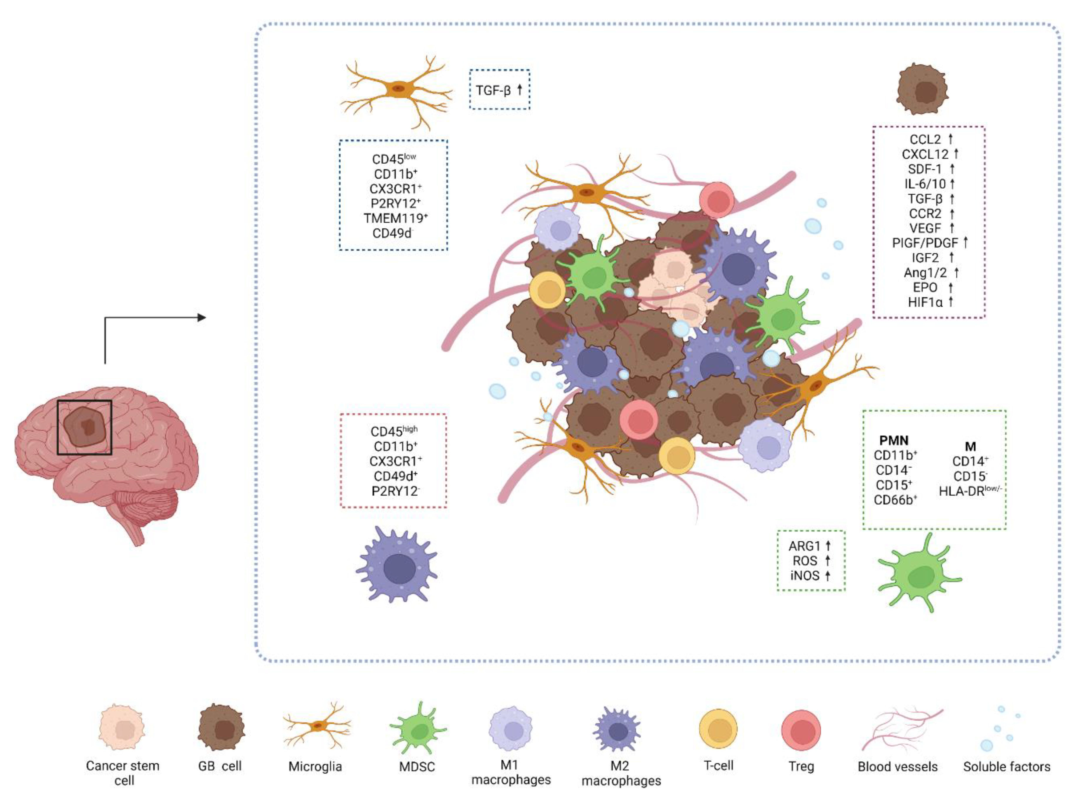
2. Glioblastoma and Unique Immunosuppressive Networks

2.1. The GB Microenvironment

Tumor development is related not only to cancer cells, but also to the interactions with surrounding cells and the microenvironment. Indeed, tumor microenvironment (TME) actively sustains tumors, and at the same time, is directly or indirectly modulated by the tumor itself. It contributes to the stabilization of immune and drug resistance mechanisms [24,25]. Although GB is highly vascularized, the circulation is inefficient, triggering the generation of a hypoxic microenvironment [26]. Hypoxic regions are disseminated in the entire tumor and different oxygen levels can be associated to distinct cell types [27]. It has been shown that hypoxia-induced factor 1 (HIF-1α) promotes the expansion of the CD133+ glioma stem cells (GSC), which cooperate in the tumor resistance to therapies [28]. Moreover, HIF-1α regulates the transcription of several genes involved in anaerobic and aerobic glycolysis, lipid metabolism, autophagy and angiogenesis [29]. Hypoxia promotes the adaptation of cancer cells through a metabolic reprogramming of aerobic glycolysis and uptake of glucose. This results in the protection of tumor cells from cell damage, in the abnormal proliferation and in the development of resistance, facilitating malignant progression [29]. In addition, HIF-1α sustains cell migration facilitating epithelial-mesenchymal transition (EMT) through Snail proteins and ECM remodeling and increasing tumor invasive potential by dysregulating glycosylation processes and altering the blood vessel permeability [29,30]. Hypoxic environment is firmly linked with angiogenesis, where HIF-1α stimulate the expression of pro-angiogenic genes, such as vascular endothelial growth factor (VEGF), placenta-like growth factor (PlGF), platelet-derived growth factor (PDGF), angiopoietin (Ang)-1 and -2; erythropoietin (EPO) and insulin-like growth factor 2 (IGF2) [27,29,31]. VEGF acts as a key regulator of angiogenesis modulating blood vessel formation and permeability and providing mitogenic and survival stimuli for endothelial cells [32]. VEGF also contributes to the stabilization of immunosuppressive networks that are a typical feature of GB immune microenvironments [30,33]. Lastly, hypoxia has multiple direct effects on the immune system, triggering several mechanisms besides VEGF production, such as immune suppressive myeloid cell shift and recruitment, impairing T cell response, thus promoting a complex immunosuppressive network, fostering tumor progression and poor prognosis for patients [29,31,33].

2.2. Immune Cells in GB Microenvironment

Notoriously, GB has been considered a “cold” tumor due to its privileged location and the presence of few immunoregulatory cells, mostly immunosuppressive actors [34]. Actually, the “privileged term” has been reconsidered after the discovery of lymphatic vessels outlining the dural sinuses in mice and potentially analogous structures can exist in the human brain, underlining how it is possible for the immune system to enter and interact with the central nervous system (CNS) [35]. Moreover, the blood–brain-barrier (BBB), which naturally separates the brain from the systemic circulation, is an obstacle for the delivery of therapeutic agents. In a tumor context, BBB can lose its integrity due to microvascular sprouting, new vasculature branching and dysfunction of tight junctions, allowing the infiltration of inflammatory cells [36]. Several studies have shown the presence of different immune subsets with distinct localization and function in GB microenvironments [37,38]. Within the TME composition, GB cells release a variety of chemoattractive factors such as CCL2, CXCL12, and SDF-1 that actively promote the recruitment of immune cells [39].
Myeloid cells are the most represented cell type in GB microenvironment (Figure 1) [14,40].
Among them, TAMs and MDSCs are the most diffused myeloid cells that cooperate in immunosuppressive mechanisms and tumor resistance. TAM population includes infiltrating bone marrow-derived macrophages (BMDMs) and microglia (MG), that make up to 30–50% of the total tumor mass [39,41]. MG are brain resident and resting macrophages that are involved in CNS development and homeostasis and immune surveillance [42]. MG and BMDMs are distinguished by the expression of surface markers, as CD49d and CD45: MG is usually identified as CD45low CD11b+ CX3CR1+ P2RY12+ CD49d while macrophages are CD45high CD11b+ CX3CR1+ CD49d+ P2RY12 [43]. Recently, the transmembrane protein 119 (TMEM119) has been reported as novel unique marker of resident MG both in humans and mice [44]. Moreover, MG and BMDMs differs for their localization underlining their distinct contribution to immunosuppression. MG cells appears stationary, large and branched and infiltrate the white matter, outlining the edge of the tumor, while BMDMs are usually localized near blood vessels in central and necrotic area of the tumor [14,43]. MG takes part in the tumorigenesis process through TGF-β release, enhancing the immunosuppression in the TME by blocking T-cells and NK cells activity and promoting T regulatory cells (Tregs) [38,45]. In response to chemokines and cytokines released by GB cells, BMDMs are recruited from the periphery to the TME. By interacting with tumor cells and TME, BMDMs change their expression profiles and mostly acquire a M2 pro-tumor phenotype through a modulation of immunosuppressive and phagocytic mechanisms and oxidative and iron metabolisms [14,46]. A recent study on patients-derived GB sections revealed the high infiltration of CD163+ cells (M2-like TAMs) in both the tumor core (TC) and peritumoral area (PTA). Interestingly the high frequency of these cells in the TC correlate with a significant prevalence of immunosuppressive markers, such as IDO and PD-L1, underlining a pro-tumoral immune system within GB TME [47]. Moreover, in a recent study the selective targeting of TAMs by inhibiting CSF-1R resulted in the alteration of macrophage polarization and block of glioma progression, proposing TAMs as potential therapeutic targets [48].
MDSCs play a fundamental role in promoting tumor progression impairing T-cell functions and inducing Treg cells [49]. MDSCs consist of two large group of cells, i.e., monocytic MDSCs (M-MDSCs) and polymorphonuclear MDSCs (PMN-MDSCs), which are differentiated by molecular and phenotypical features [14].
In the tumor site, MDSCs functions are strongly controlled by several soluble factors, such as IL-6, IL-10, TGF-β, CCR2, VEGF secreted by GB cells and immune cells, leading to the modulation of molecular pathways mediated by Janus tyrosine kinase (JAK) and signal transducer and activator of transcription 1, 3 and 6 (STAT1; STAT3; STAT6). This results in the promotion of MDSCs proliferation and survival and induction of immunosuppressive molecules, such as arginase 1 (ARG1), reactive oxygen species (ROS) and inducible nitric oxide synthase (iNOS). Thus, high levels of circulating ARG1 in GB patients’ blood have been considered as markers of MDSCs activation [50].
Several studies have reported the contribution of both MDSCs subpopulations to the progression of gliomas and high levels of circulating MDSCs are associated with the worst prognosis for GB patients [50,51,52,53,54]. Indeed, PMN-MDSCs have been identified as the main circulating MDSC subpopulation in GB patients’ blood and their proportion is higher than M-MDSC in GB tumors [55,56,57]. Moreover, Gielen et al., showed that MDSC levels of GB patients’ blood were significantly higher than healthy controls and found that PMN-MDSCs were representative of MDSC infiltrating tumor mass [58]. The discovery of novel markers, such as LOX1, helps to further distinguish MDSC subpopulations and elucidate their role and mechanisms in tumor progression [59]. Indeed, a recent study showed that LOX1+ identifies a specific PMN-MDSC subpopulation that is increased in GB patients’ blood and tumor tissue and is characterized by a high expression of ARG1 and iNOS and strong immunosuppressive ability to inhibit CD3+ T cells [52].

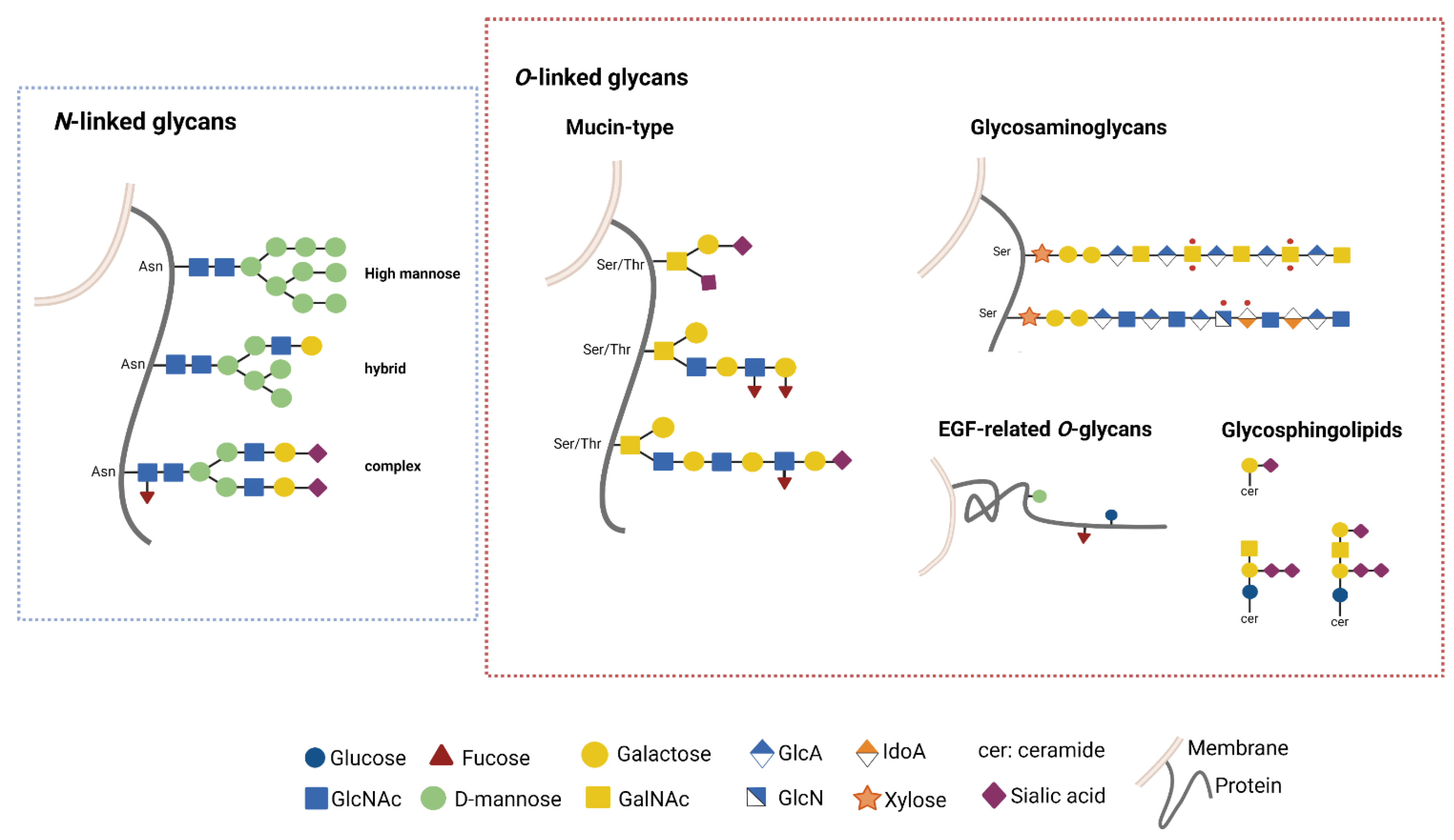
3. GB and Glycosylation Pathways

3.1. Glycosylation Pathways

Glycosylation is the main post-translational modification of proteins and lipids, that requires a highly monitored enzymatic complex network from nucleus to Golgi apparatus [60,61]. Indeed, specific enzymes participate to the finely tuned phases of the glycosylation process, i.e., core extension, elongation and branching, and capping of the carbohydrate chains. This generates an astonishing diversity in the glycome that corresponds to highly specific and selective biological functions [60,61].
Most of the glycoconjugates and glycans are exposed on the cellular membrane and play a crucial role in tissue lubrification and protection, cell–matrix interactions and cell signaling [62]. Glycans are also involved in immunomodulation mechanisms through the interaction with glycan-binding proteins of lectins family, regulating immune homeostasis and host–pathogen interactions [63].
N- and O-linked glycans are the most common post-translational modification (PTMs) in extracellular components (Figure 2).
O-linked glycosylation is characterized by the addition of monosaccharides, as such as N-acetyl-D-galactosamine (GalNAc), D-glucose (Glc) or D-mannose (Man), to serin or threonin residues (Ser, Thr). O-linked Glc, Fucose (Fuc) or Man are found on the EGF-related O-glycans. O-linked GalNAc is the substrate for the extension of long polylactosamine side chains in the so called “Mucin-type glycosylation”. In this pathway, the synthesis of GalNAc α1-O-Ser/Thr (the Tn antigen) is required for the next synthesis of the T antigen (Galβ1-3GalNAcα1-O-Ser/Thr) and further extension of long polylactosamine side chains that decor the protein backbone of mucins. These molecular structures function as protection and lubrification of epithelia and become truncated in carcinoma. Additionally, O-linked xylose may trigger the elongation of glycosaminoglycans (GAGs) on the backbone of proteoglycans. In response to nutrient and stress response, addition of O-linked N-Acetyl-D-glucosamine (O-GlcNAcylation) can occur on many intracellular proteins [64].
N-linked glycosylation consists in the covalent binding of the oligosaccharide N-Acetyl-D-glucosamine (GlcNAc) to the asparagine (Asn) residue of polypeptide chains mediated by the oligosaccharyltransferase (OST) complex. Elongation processes give rise to a common pentasaccharide core region composed by GlcNAc and Man residues. This N-core structure is the substrate for the branching of the side chains that are further diversified in carbohydrate composition. On the basis of that, N-glycoconjugates are classified in high mannose, hybrid or complex [65,66]. High mannose structures are composed only by Man, while complex structures are made also by other monosaccarides (GalNAc, Fuc, Galactose, Sialic Acid (Sia)). Hybrid structures are characterized by the presence of both high mannose and complex structures on distinct branches [65,66].
Lipid glycosylation is also crucial for the biology of cells. In particular, glycosphingolipids (GLS), which are the major component of the outer cell plasmamembrane, are composed by a hydrophobic ceramide backbone linked to a first glycan moiety (Gal or Glc) through a glycosid linkage [67]. A complex enzyme network located among the intracellular compartments mediates the subsequent addition of other carbohydrate residues as such as GlcNAc, GalNAc, Fuc and Sia generating structurally different molecules, which potentially correspond to a distinct function. Gangliosides (GG) are acidic GLS containing sialic acid residues, enriched in cell membrane microdomains, and are more abundantly found in the nervous system [68,69].
Despite the diversity of glycosylation pathways, the carbohydrate chains can undergo to non-specific modifications thus generating molecular structures shared by distinct glycoconjugates. Sulfation, the addition of SO3 group to the carbohydrate backbone, is the most abundant glycan modification generating a large pattern of sulfated structures that play wide essential biological role. Moreover, the terminal addition of Sia (Sialylation) or Fuc (Fucosylation) are crucial events in the capping of the glycan chains, thus blocking the elongation process and generating glycan structures specifically recognized by glycan-binding proteins [60,70].
The variety and complexity of the glycome is shaped by the cellular stress and becomes a strategy for the cell to modulate its function and interactions with the microenvironment. Indeed, glycosylation alterations may be regarded as key features of diseases and a key factor for immune recognition [71].

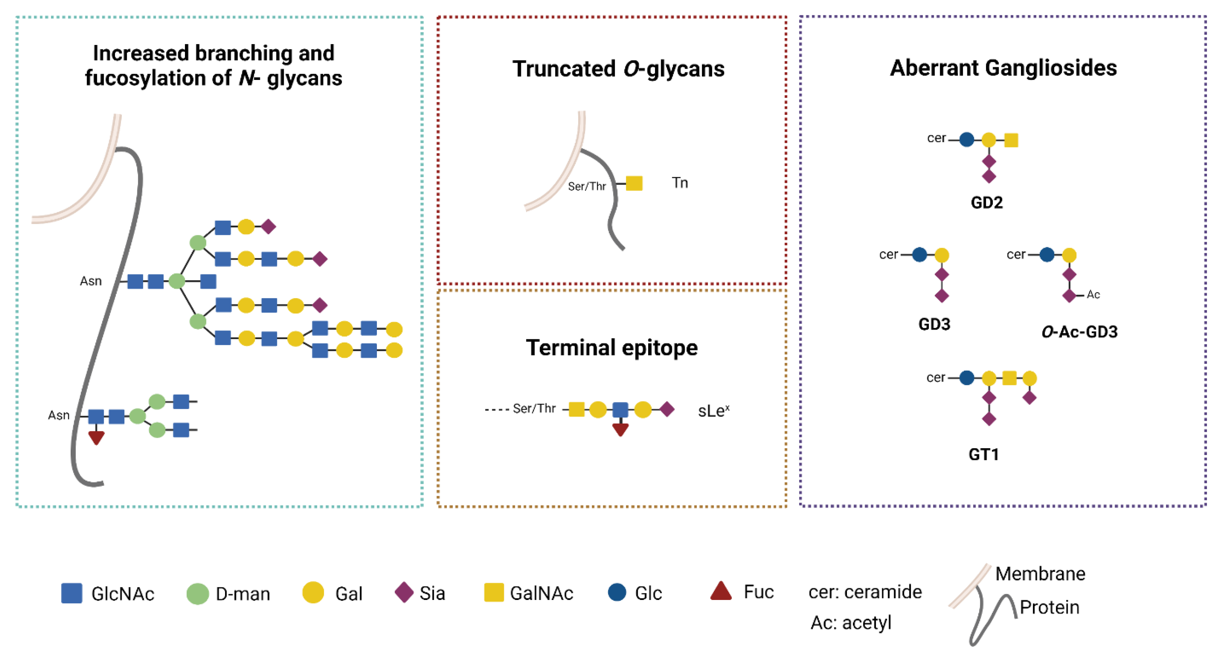
3.2. Aberrant Glycosylation Processes in GB

Aberrant changes in the glycosylation machinery early occur during tumor transformation, i.e., increased N-glycan branching, truncated O-glycans and augmented O-glycan density on the protein backbone, and synthesis of neo-glycans carrying numerous terminal Sia or Fuc [19,72]. These aberrant glycosylation processes generate tumor associated glycans that play a key-role in tumorigenesis, metastasis and immune modulation [72,73].
Thus far, an accumulating body of evidences indicate that glycosylation changes occur in GB and involve a large variety of cellular substrates (proteins and lipids) and distinct metabolic pathways, thus contributing to the complex biology and impaired immunogenicity of GB (Figure 3) [18,74].
In GB, changes in N-glycosylation are characterized by altered branching, increased fucosylation and sialylation. Several receptors may be aberrantly N-glycosylated and this impacts on cell functions. N-acetylgalactosyltransferases (GnTs) are key enzymes for the N-glycan synthesis. MGAT1 (GnT1) and MGAT5 (GnT5) are up-regulated in glioma cells and in GB tissues [75,76]. MGAT1 plays a crucial role in the conversion of the carbohydrate core from High mannose to complex/hybrid structures.
This change increases the stability of the GLUT1 transporter, which is N-glycosylated, promoting cell proliferation [75]. MGAT5 catalyzes the addition of terminal β-1,6-GlcNAc to the N-glycan and its overexpression in cancers supports N-glycan branching and correlates with tumor invasion [76,77]. Increased N-branching augments terminal sialylation and fucosylation processes that contributes to tumor spreading [78].
The tumor associated Sialyl-Lewis Acid (S-Lewis X) glycan generated by these changes is recognized by immune lectins [79]. Dysregulated fucosylation is a feature associated to GB aggressiveness. Fucosylation of the first GlcNAc residue of the N-core structure (Figure 3) is catalyzed by the fucosyltrasferases 8 (FUT8). In human GB FUT8, overexpression is associated with high tumor grade and aggressive disease. Interestingly, MET and EGFR molecules are substrate for FUT8 and the altered glycosylation increases their function with an overall gain of motility and proliferation for the tumor cells [80].
Indeed, inhibition of N-linked glycosylation has been proposed as an appealing pharmacological strategy to dampen receptor kinase activity and enhance radiosensitivity of GB cells [81].
The increased sialylation levels observed in glioma cells are mainly due to the overexpression of the α-2,3-sialyl transferase (ST3GAL1). Its up-regulation correlates with poor prognosis in GB patients and is associated to enhanced invasive potential [82]. On the contrary, α-2,6-sialylation is downregulated in the tumor cells, but it is upregulated in the GB vasculature, suggesting its crucial role for endothelial survival [83,84,85]. Therefore, two distinct sialylation pathways underlie distinct biological mechanisms for GB progression [74]. Little is known about changes in the O-linked mucin type glycosylation. In carcinoma, alterations of this enzymatic pathway generate truncated O- glycans, i.e., Tn and T and their sialylated version STn (NeuAcα2-6GalNAcα1-O-Ser/Thr) and ST (NeuAcα2-3Galβ1-3GalNAcα1-O-Ser/Thr) [86]. In GB, alteration of the enzymatic O-glycosylation pathway was found and Tn glycan expression was revealed by the binding of anti-Tn lectins in mouse model as well as in human tumor cells [87,88]. In GB mouse model, the overexpression of Tn has been associated to strong immunosuppression (as described in Section 4.1) [88].
Additionally, glycolipid components are altered in GB. Early work had shown changes in GLS components in human gliomas, in particular ganglioside GD3 was shown to be increased in gliomas and correlated with malignancy [89,90]. Thus far, increase of GD2 and GD3 in GB has been associated with resistance to therapy and increase on invasiveness [91,92]. The aberrant GG pattern alters the organization of membrane microdomains with huge impact on the activity of tyrosin-kinase receptors and downstream cell signaling [93]. Indeed, GD3 plays a crucial role in the stemness and tumorigenicity of GB by activating c-MET signaling [94]. At the same time, GD3 has been shown to modulate innate immune response by specifically interacting with Siglec7 (see Section 4.2). More recently, increase of O-Ac-GD3 (GD3 carrying acetylated Sia) and the ganglioside GT1 were found in human GB samples, while GM1 was found in peritumoral brain tissue [95,96]. These differential patterns may be relevant to both tumor biology and immunomodulation mediated by GGs [97].
Another crucial source of glycan diversity is the matrisome. In GB, the ECM is increased due to the up-regulated secretion of PGs and the associated O-linked GAGs [98,99,100]. These ECM components can undergo to aberrant sulfation processes that appear to be relevant for the glycan alteration in GB ECM. Modulation of sulfation among distinct PGs was found and several core matrisome PGs (as such as decorin, agrin, glypcan-1), and laminin, tenascin, fibronectin among the others, were differentially regulated in tumor vs. control tissues. Interestingly, changes in PG composition were also found within the GB samples in accordance to the IDH mutant status. From this analysis, O-glycosylated peptides from PGs were found only in the GB tissue and not in the control, again indicating that dysregulated glycosylation process generates specific glyco-profile [101].
Indeed, a glycoproteomic fingerprinting would be of great interest to in depth characterize the ECM components to unveil biological mechanisms aiding cell growth and metastatic spreading that could be useful as potential therapeutic targets [74,102].

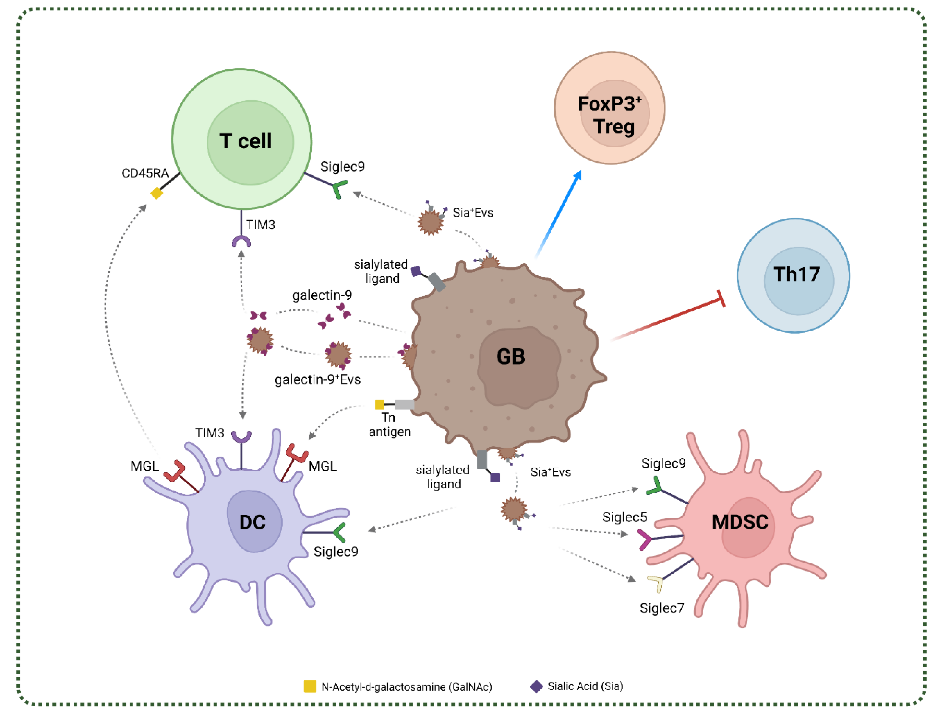
4. Glycan-Lectin Interactions and Immunosuppressive Networks in GB

The de novo exposed tumor glycan array triggers novel immune interactions mediated by lectins that are able to skew the immune system function with high impact on the overall tumor progression [103]. Lectins constitute a large protein family that share a common carbohydrate recognition domain (CRD) for the binding of specific glycosylated structures [19].
Human lectins are classified by considering their subcellular location and their structures. Lectins can be incorporate in the cellular membrane or have a soluble form and based on their form they can be divided into groups as C-type lectins, I-type lectins (including Siglecs), S-type lectins (also known as galectins), P-type lectins (known as Selectins) and pentraxins [104]. In tumor, they have been proposed to decipher the tumor glycocode that modulate immune tolerance/suppression [103].
In GB, the expression of lectins from the C-type, Siglec and galectin family has been described and their possible role in the stabilization of immune networks has been proposed [105,106,107]. The main glycan–lectin interactions found in GB are represented in Figure 4.

4.1. C-Type Lectins in GB

C-type lectins (CTLs) are a large family of proteins that share one or more conserved C-type lectin-like domain (CTLD) able to recognize a broad range of carbohydrate motifs. CTLs are mainly expressed on myeloid cells, and their expression is carefully regulated or induced under specific conditions, as inflammation. CTLs are mainly known for their role in antimicrobial immunity and tissue homeostasis, but they can both promote or suppress the immune response in disease context, such as cancer [23,108,109].
For example, alteration of MHC class I molecules due to oncogenic transformation or infection can be sensed by natural killer (NK) cells and can activate the C-type lectin NKG2D-mediated cytotoxicity. However, several C-type lectins, such as DC-SIGN, CLEC14A, macrophage galactose C-type lectin (MGL) contribute to tumor progression inducing immunosuppressive responses by sensing abnormal or altered tumor-associated carbohydrates [110]. Thus far, the few data on the expression and role of CTLs in brain diseases suggest they may play an immunosuppressive role in the immunological networks. CTLs expression associated to microglia has been observed in neuroinflammatory degenerative diseases as Alzheimer disease (AD) and Multiple Sclerosis [111,112].
In these pathological settings, expression of CTL by microglia has been observed to mitigate the inflammatory reaction suggesting a possible protective role in inflammatory mediated neurodegenerative diseases. Expression of MGL by macrophages and microglia in experimental autoimmune encephalomyelitis (EAE) mouse model, induced apoptosis of autoreactive T cells and release of immunosuppressive IL-10, thus proposing a role of MGL as negative regulator of autoimmune-driven neuroinflammation [112]. MGL is a Type II C-type lectin, with specificity for the truncated Tn O-glycan (GalNAc-α-Ser/Thr) (Table 1) [113].
MGL engagement by its ligand triggers phenotypic and metabolic changes in the antigen presenting cells [114] that are finely tuned by the structure of the Tn carrying ligand [113]. In physiological context, MGL expressing APCs appears to act as negative regulator for naïve T cell homeostasis and Treg cells through the interaction with the Tn-glycosylated CD45RA [115]. Expression of MGL and its Tn O-glycan ligand has been described in GB human tissues, preferentially associated to tumor infiltrating CD163+ cells [88]. Results from a GB syngenic mouse model highly expressing the Tn O-glycan showed that distinct myeloid infiltrating cell subsets heterogeneously expressed MGL. Interestingly, in the MGL+ tissue areas, a strong infiltration of PD-L1+ TAMs was found [88]. These results support the hypothesis that the MGL-Tn axis may contribute to the immunosuppressive network in GB. It is interesting to recall that the Tn-MGL axis has been validated as a therapeutic target in an ovarian cancer mouse model by means of glycomimetic peptides [116,117].
Table 1. Classification, expression, binding preference, glycosylated ligand, known molecular mechanisms and role of C-type lectins and Siglecs in GB. Abbreviations: GalNAc: N-Acetyl-d-galactosamine; MUC1: mucin-1; MUC5: mucin-5; MUC16: mucin-16; MUC24: mucin-24; ERP44: Endoplasmatic Reticulum Resident Protein 44; LAMP1: Lysosome-associated membrane glycoprotein 1; LAMP2: Lysosome-associated membrane glycoprotein 2; QSOX1: Sulphydryl oxidase 1; SEL1L: Protein Sel-1 homolog 1; LRR8CD: Leucin-rich repeated-containing protein 8D; AGRN: Agrin; APP: Amyloid beta A4 protein; DAG1: Dystroglycan; FN1: Fibronectin; NID-2: Nidogen-2; PODXL: Podocalyxin; SDC3: syndecan-3; VCAN: Versican core protein; GBS β-protein: group B Streptococcus (GBS) β-protein; GD3: ganglioside.
LectinExpressionRecognized Carbohydrate MotifGlycosylated LigandMolecular MechanismRole in GBRef.
MGLDC and cDC2
Macrophages
CD163+ cells Activated MG
GalNAc-α-Ser/ThrCD45RA
Matrix/cell adhesion (VCAN, SDC3, PODXL NID-2, FN1, DAG1, APP, AGRN)
Cell metabolism (ERP44,LAMP1/2, QSOX1, SEL1L, LRR8CD)
Mucins (MUC1/16/24)
Promotes ERK phosphorylation and NfkB
Enhanced secretion of IL10
Activation TLR signaling
Promotes DC
TAM/CD163+ cells mediated immunosuppression[111,112,113,118,119]
Siglec5Monocytes
DC
Neutrophils
Macrophages
α-(2-3)-Sialic acid
α-(2-6)-Sialic acid
α-(2-8)-Sialic acid
GBS β-proteinECM remodellingMDSCs mediated immunosuppression[63,73,104,107,120,121]
Siglec7NK cells
Monocytes
Macrophages
Mast cells
DC
α-(2-3)-Sialic acid
α-(2-6)-Sialic acid
α-(2-8)-Sialic acid
disialogangliosides
CD43
GD3
ECM remodellingMDSCs mediated immunosuppression[63,73,104,107,120,121]
Siglec9NK cells
DC
T cells
Neutrophils
Macrophages
Monocytes
α-(2-3)-Sialic acid
α-(2-6)-Sialic acid
α-(2-8)-Sialic acid
Glycophorin
Hyaluronic acid
MUC1/5
Modulation of MAPK/ERK
Neutrophils inhibition/death
M2 polarization
ECM remodelling
Inhibits macrophages phagocytosis
MDSCs mediated immunosuppression[63,73,104,107,120,121,122,123]

4.2. Siglecs in GB

Siglecs belong to the I-type lectins immunoglobulin superfamily that mainly recognize sialic acids (Sia) and other carbohydrate residues. They are classified based on their structure in CD33-related Siglecs (Siglec3 or CD33; Siglecs 5-11; Siglecs14 and 16) and other Siglecs (Siglec1 or CD169; Siglec2 or CD22; Siglec4a or MAG and Siglec15).
Siglecs function as immunomodulatory receptors that mainly mediate immunosuppressive responses through the phosphorylation of the immunoreceptor tyrosine-based inhibition motif (ITIM) depending on the cell which they are expressed by and on the ligand they interact with. All Siglecs are expressed on immune cells, except for the myelin-associated glycoprotein (MAG) [104]. Siglec1 and 12 expression is restricted on macrophages, CD22 on B lymphocytes, Siglec16 on MG while other Siglecs, such as CD33 and Siglec7, can be found on several immune cell subsets [121,122,123,124].
MAG is selectively expressed in the nervous system and is known to have an important role in the maintenance of myelinated axons, the physiology of oligodendrocytes and the suppression of axonal regeneration after injury [125]. MAG exerts its function through the interaction with Nogo receptor and gangliosides containing 2,3-linked sialic acid [125]. Interestingly, in U87MG glioma cells, it has been shown that MAG is able to reduce the migratory capacity of these cells in culture [126].
It has been shown that in tumor models, sialoglycan–Siglecs interactions are able to impair and suppress the effector immune cells, such as NK and CD8+ T cells. Moreover, Siglec7 and 9 glycosylated-ligands have been found in several cancers, including GB [127]. In a recent work, the authors characterized the expression of Siglec5, 7, 9 on MDSC both from peripheral blood and glioma infiltrating cells and found the respective ligands both on GB cell lines and patient-derived glioma tissue samples (Table 1). These evidences underline the possible interaction between glioma cells and MDSC via the sialic acid–siglec axis, modulating their function and shaping the immunosuppressive TME [107].
Interestingly, Dusoswa et al. showed a high presence of N-linked and O-linked α-2,3 sialic acids and N-linked α-2,6 sialic acids on GB-derived extracellular vesicles (EVs). GB-derived EVs express Sia-glycoconjugates which are ligands for Siglec9, whereas no ligand for the other Siglecs was detected [128]. Siglec9 is mainly found on immune cells, as NK, T and DC and can mediate immunosuppressive functions through the activation of the two ITIM sequences comprise in the intracellular domain. In this work, the authors showed that the modification of EVs-glycosylation profile through enzymatic desialylation enhanced EV’s internalization by DCs, thus suggesting that glycosylation degree may indirectly modulate antigen processing, as shown for other glycan carrying EV systems [129]. Although, these results sustain the relevance of that Siglecs as sensors of sialylated carbohydrates in GB, further studies are required to elucidate their functional contribution to the immunosuppressive networks.

4.3. Galectins in GB

S-type lectins, or galectins, are defined by the presence of at least one conserved carbohydrate recognition domain (CRD) with high affinity for lactose or N-acetyllactosamine (LacNAc; Galβ1-4GlcNAc)-containing ligands. Currently, galectins are structurally differentiated in three groups, as they can exist as dimeric form (galectin-1, -2, -5, -7, -10, -11, -13, -14, -15), tandem-repeat form (galectin-4, -6, -8, -9) and the monomer or multivalent chimera type (galectin-3) [130].
Galectins can be retained in the cytosol or secreted both by transformed and normal cells, such as fibroblasts, endothelial and immune cells [131,132]. Galectins are involved in a wide range of basic cellular functions, as apoptosis, proliferation, cell adhesion and signaling, but also in immune system modulation and cancer development [130]. Interestingly, galectins exert their function both intracellularly and extracellularly [133].
Galectins have been shown to play a key role in tumor progression, promoting angiogenesis and ECM remodeling through the binding with highly glycosylated ligands expressed by several cell types. Tumoral galectins, interacting with glycosylated immune-related ligands, induce immunosuppression promoting tolerogenic DCs, CD4+ and CD8+ T cell apoptosis and exhaustion, FoxP3+ Treg cells expansion and NK impairment [21]. Upon interaction with galectins, glycoproteins and glycolipids undergo structural rearrangement, thus modulating their function and the downstream signaling pathways [133]. Galectin-1 shapes the immune compartment, promoting the proliferation of tolerogenic DCs and M2-macrophages, the apoptosis of Th1 and Th17 T cells and the expansion of Treg cells. Moreover, galectin-3 exerts its immunomodulatory functions affecting effector T cells activity by altering the immunological synapses organization and distancing TCR from CD8. These molecular events induce anergy, exhaustion and suppression of T cells. At the same time, galectin-3 promotes immunosuppressive cells, as Tregs and MDSCs, thus dampening the anti-tumor immune response [133,134,135].
The abundant galectins levels both in TME and peripheral blood have been proposed as prognostic factors and associated to a negative prognosis for oncologic patients [130,136]. Several galectins have been found in both neurons and glial cells and physiologically contribute to CNS development and functions. Alterations in galectins levels are observed in neuroinflammatory and neurodegenerative disease, and also in CNS tumors [105,137]. Indeed galectin-1, -3, -8 and -9 are expressed in GB and the interaction with their respective glycan-carrying molecules blocks the anti-tumor response, thus promoting immunosuppression (Table 2) [138].
Galectin-1 and -9 are the two galectins studied for their association with immunosuppressive mechanisms [21]. In vitro and mice model studies have shown that galectin-1 promotes tumor cells’ migratory capability and invasiveness through modification of cytoskeleton and small GTPases [138,139,140,141,142]. The selective silencing of glioma-derived galectin-1 delays tumor progression and prolongs the survival of glioma-bearing mice impairing angiogenesis and the recruitment of myeloid cells as MDSCs and macrophages [143]. Gou et al., showed that the new putative oncogene FAM92A1-289 is involved in DNA methylation in GB cell line and interacts with galectin-1 contributing to tumor progression [144]. Recently, LGALS1 gene, coding for the galectin-1, has been included among the genes that define a glioma microenvironmental gene signature and has been identified as a key immunosuppressive gene in GB [145].
Galectin-1 levels directly correlated with the grade of GB samples and its selective knockdown impaired immunosuppressive mechanisms by down regulating M2 macrophages and MDSC cells and by inhibiting immunosuppressive cytokines [145]. Expression of galectin-1 was also associated with a clinical response: GB patients treated with radiotherapy with a low expression of galectin-1 showed a better median survival than patients with high levels [146]. The silencing or knockdown of galectin-1 in vivo via intranasal administration or through siRNA-loaded chitosan lipid nanocapsule showed promising results by altering the GB TME and immune compartment and reducing TMZ resistance, unraveling galectin-1 importance in immunosuppression and tumor promotion [147,148,149].
Galectin-9 is known as eosinophil chemoattractant and, more recently, as a negative modulator of adaptative immune response. The major mechanism through which galectin-9 exerts its immunosuppressive mechanisms involves the binding to TIM3, that is widely expressed on immune cells, especially on T-cells [21]. Moreover, other immune checkpoints are recognized by galectin-9, therefore galectin-9 is extremely relevant for immunosuppressive mechanisms (see Table 2) [21]. Through a comprehensive analysis of 1027 glioma patients, Yuan et al., found a strong common upregulation of galectin-9 in GB compared to normal brain tissue. In addition, high galectin-9 expression levels correlate with a shorter overall survival in lower-grade glioma patients. In GB tissue samples, galectin-9 expression is also associated to immunosuppressive M2-macrophages and its levels positively correlate with immune checkpoint molecules [150,151]. Notably, galectin-9 also exerts its functions when release in EVs. A fascinating study underlined that GB derived-exosomes (GB-Exos), enriched in galectin-9, anontribute to tumor progression by impairing DCs and CD8+ T cells function by TIM3 binding. Indeed, exosomal galectin-9 activity on DCs is TIM3 dependent and knockout of TIM3 in DCs, restore DCs function and activation [152]. Taken together, these studies suggest that galectin-9/TIM3 can be one of the key mechanisms that sustains immunosuppression in GB TME and it has been proposed as therapeutical intervention for targeting cell–cell interaction and exosomes communication [153,154,155].
Galectin-3 and -8 role in GB progression is less well defined. Little is known about their immune-related functions, while a better knowledge on their interplay with ECM is available (see Table 2) [130,137]. Depending on its expression, galectin-3 is able to reinforce or weak cell adhesion and cell–matrix interactions promoting the activation of pathway involved in ECM remodeling and EMT [156,157]. Moreover, galectin-3 upregulation under hypoxic and nutrient deprivation has been correlated to tumor aggressiveness and poor prognosis in several cancer type [21,158]. Galectin-3 has been also proposed as biomarker for differential grading and diagnosis of gliomas [159], resistance to standard therapies and high levels are associated with a poor survival [160,161,162].
Lastly, galectin-8 contribution to GB is very poorly understood. It is interesting to note that galectin-8 is the only one of this family that shows an high affinity for α-2-3-sialylated glycans, of which GB is rich [85,163]. In a recent study was showed that galectin-8 promotes proliferation and prevents apoptosis of GB cell lines [164]. Indeed, galectin-8 is strongly implicated in cell adhesion, growth and cytoskeletal organization, thus underlining a possible favorable function in tumor development and progression [165]. Moreover, high levels of galectin-8 had been found in oncologic patients and had been often considered as a poor prognostic factor in several malignancy, as multiple myeloma and prostate cancer [166,167,168]. Interestingly the selective knockdown of galectin-8 in a mouse model showed a notable reduction of the tumor size and a minor metastatic process, highlighting its importance as potential therapeutical target. However, high levels of galectin-8 have been also associated to a better survival for gastric cancer patients, pointing out the need to better clarify galectins role in carcinogenesis [169].
Table 2. Classification, expression, binding preference, glycosylated ligand, known molecular mechanisms and role of galectins in GB. Abbreviations: N-acetyllactosamine: LacNAc; Galβ1-4GlcNAc; Forssman pentasaccharide: GalNAc-α-(1,3)-GalNAc-β-(1,3)-Gal-α-(1,4)-Gal-β-(1,4)-Glc; MCAM: Melanoma Cell Adhesion Molecule; LFA-1: Lymphocyte function-associated antigen 1; TLR-4: Toll Like Receptor 4; LAG-3: Lymphocyte activation gene 3 protein; VEGF-R2: Vascular endothelial growth factor receptor 2; Glut-2: Glucose transporter 2; tDCs: tolerogenic dendritic cells; c-Maf/aryl: transcription factor Maf/aryl hydrocarbon receptor; STAT/JAK1-2: Janus kinases 1-2 and activator of transcription proteins; GSK-3β: Glycogen synthase kinase 3 beta; NRP-1: neuropilin-1; IL-2: Interleuchin 2 receptor; TGF-β1: Transforming growth factor 1; TGF-βR: Transforming growht factor beta receptor; Lck: non-receptor tyrosine-protein kinase; Bat3: Large proline-rich protein BAT3.
LectinExpressionRecognized Carbohydrate MotifGlycosylated LigandMolecular MechanismRole in GBRef.
Galectin-1Endothelial cells
Astrocytes
APCs
Treg
Lactose
poly-N-acetyllactosamine
Immune markers (CD3, CD4, CD7, CD43, CD45, CD69)Activation of Fas-induced death, mitochondria apoptotic pathway, VEGF-R2/NRP-1; STAT/JAK1-2; c-Jun/AP-1; Lck/ZAP-70
Differentiation of IL-27/IL-10-producing tDCs
Increased expression of IL-10 and IL-21
Modulation of the c-Maf/aryl receptor pathway
Loss of mitochondrial membrane potential
Release of cytocrome c
Tumor progression
Angiogenesis
Macrophage differentiation
MDSC recruitment
[21,132,135,138,139,140,141,142,143,144,145,146]
Galectin-3Endothelial cells
Activated microglia
Activated astrocytes
Myeloid cells
Fibroblasts
Lactose
N-acetyllactosamine
Cell adhesion/matrix (Laminin, Vitronectin, Collagen I/IV, MCAM)
Immune markers (TCR complex, CD7, CD29, CD45, CD71, LFA-1, TLR-4, LAG-3, CTLA-4)
VEGF-R2
Inhibition of NKp30 signaling pathway
Interferes with MICA-NKG2D affinity
Increases/impairs cell-matrix adhesion (integrins)
Activation of GSK-3β, RAS/PI3K/AKT, MEK/ERK
Modulation of β-catenin and RAS/Bcl-2/Myc
Increases Akt activity
Stabilization of TGF- βR and signaling
Supports IL-6 production
Proliferation
Motility
Resistance to radiotherapy
[21,130,133,134,135,137,156,157,158,160,161,162]
Galectin-8Endothelial cellsα-(2-3)-Sialic acid
Lactose
N-acetyllactosamine
Cell adhesion/ matrix (Laminin, Fibronectin, Vitronectin, Collagen IV, CD166, Integrins α1β1/α3β1/ α5β1/α6β1)
Immune markers (IL-
2R, TGF-βr, CD44)
Activation of VEGF-R2/NRP-1 and integrin-mediated signaling pathway
Modulation of TGF- βR and IL-2R signaling pathway
Promotes Treg differentiation and proliferation through STAT5 and Smad3 phosphorylation
Proliferation
Prevent apoptosis
Migration
[21,85,133,134,135,164,165,166,167,168,169]
Galectin-9Activated astrocytes
Microglia
Endothelial cells
Lactose
N-acetyllactosamine
Forssman pentasaccharide
Immune markers
(TIM3, DECTIN-1, CD44, VISTA, CD274, PD-L1, IDO1, LAG3)
β3-interini
Glut-2
TGF-β1- induced Treg differentiation
Activation of VEGF-R2/NRP-1 pathway
Promotes Smad3 phosphorylation
Promote expansion of CD11b+Ly-6G+ MDSCs
Upregulation of caspase-1, Granzyme B and Bid
Downregulation of Lck and Bat3 signaling
Block T helper 17
Expansion of FoxP3+ Treg
Apoptosis
T cell exhaustion
M2 polarization
[21,133,134,135,152,153,154,155]

5. Conclusions

Despite medical innovation, GB remains one of the most fatal malignant tumors [1].
Novel therapeutic strategies targeting GB cell self-renewal and growth as well as immunotherapies potentiating/eliciting anti-tumor immune response are currently investigated. However, unsatisfactory results have been reached with little impact on the availability of novel therapies [170,171].
Thus far, it has become clear that most therapeutic interventions aimed to dampen tumor growth, promote anti-tumor response as a bystander effect by reducing immune suppression and/or enhancing antigen presentation and eliciting T cell responses [172,173,174]. This indirect immune modulation contributes to the overall clinical benefit induced by the therapy [175].
It is also clear that the immunosuppressive, tumor-promoting microenvironment halts the drug-induced immunological benefits or the efficacy of immunotherapeutic interventions as such as Immune checkpoint blockade, now standard of therapy for several tumors, or more experimental immunotherapies as vaccination, adoptive T cell therapy, CAR-T [176,177,178].
In this scientific and biological framework, the tumor associated glycans that are generated by aberrant tumor glycosylation acquire a crucial importance for their immune-coding significance, and not just as markers of tumor phenotype and invasiveness [79,103].
Indeed, the lectin immune receptors that selectively sense and recognize these tumor associated structures have emerged as markers and possible therapeutic targets to dismantle the immunosuppressive networks [179].
The GB TME is characterized by a peculiar array of diverse myeloid cell types, whose molecular profile and functions are a challenging research field [180].
The diverse expression profiles of lectins displayed by these cell subsets is strongly suggestive of their role in the triggering and fueling of immunosuppression. Indeed, several compounds have been developed to block galectin- and Siglec-mediated interactions for therapeutic purposes in tumor and other pathological settings [181,182,183]. Glycomimetic peptides have been successfully employed for such purpose [182,184]; indeed the MGL-Tn glycan axis can be targeted with therapeutic benefit in ovarian cancer settings [116]. The availability of novel glycoproteomic approaches may allow in depth glycosylation profiling and mapping to identify the glycoconjugated ligands that in vivo participate to the lectin mediated immune networks in GB. Therefore, further investigations are required in order to better understand and clarify the contribution of these interactions in GB development and progression.
Glycan–lectin interactions mediate also immune cell migration [185]. Indeed, the matrisome has emerged as a key factor in the motility of tumor as well as immune cells and its modulation has been regarded as potential therapeutic option [101,186]. From an immunological point of view, the knowledge of the mechanisms that regulate immune cells infiltration is essential to switch the immune tumor contexture and improve the efficacy of treatments [187,188].
The characterization of the changes in glycan composition and the unveiling of the mechanisms of glycan-lectin interactions that occur in GB can critically contribute to identify novel therapeutic strategies and ameliorate already available immunotherapeutic treatment in GB.

Author Contributions

Conceptualization, A.P. and A.R.; writing—original draft preparation, A.P., A.R. and F.S.; Drawings, F.S. and H.R.; writing—review and editing, M.N., A.P., F.S., H.R., C.N., I.G.Z., L.D., A.S. and A.R.; funding acquisition and supervision, A.R. and M.N. All authors have read and agreed to the published version of the manuscript.

Funding

This research and APC were funded by MUR/”Sapienza” University of Rome, grants number RM1201772B803DB14, RM12117A7B767D0D and RM12117A85361029.

Acknowledgments

We are most grateful to M. Cristiani and G.J.F. for their collaboration throughout the preparation of the manuscript. We thank A. Di Filippo for helpful discussion and suggestions. A.P. and F.S. are fellows of the PhD course “Network Oncology and Precision Medicine”; Dpt. Experimental Medicine, “Sapienza” University of Rome.

Conflicts of Interest

The authors declare no conflict of interest. The funders had no role in the design of the study; in the collection, analyses, or interpretation of data; in the writing of the manuscript, or in the decision to publish the results.

References

  1. Ferris, S.P.; Hofmann, J.W.; Solomon, D.A.; Perry, A. Characterization of Gliomas: From Morphology to Molecules. Virchows Arch. 2017, 471, 257–269. [Google Scholar] [CrossRef] [PubMed]
  2. Louis, D.N.; Perry, A.; Wesseling, P.; Brat, D.J.; Cree, I.A.; Figarella-Branger, D.; Hawkins, C.; Ng, H.K.; Pfister, S.M.; Reifenberger, G.; et al. The 2021 WHO Classification of Tumors of the Central Nervous System: A Summary. Neuro Oncol. 2021, 23, 1231–1251. [Google Scholar] [CrossRef] [PubMed]
  3. Tan, A.C.; Ashley, D.M.; López, G.Y.; Malinzak, M.; Friedman, H.S.; Khasraw, M. Management of Glioblastoma: State of the Art and Future Directions. CA Cancer J. Clin. 2020, 70, 299–312. [Google Scholar] [CrossRef] [PubMed]
  4. Friedman, H.S.; Prados, M.D.; Wen, P.Y.; Mikkelsen, T.; Schiff, D.; Abrey, L.E.; Yung, W.K.A.; Paleologos, N.; Nicholas, M.K.; Jensen, R.; et al. Bevacizumab Alone and in Combination with Irinotecan in Recurrent Glioblastoma. J. Clin. Oncol. 2009, 27, 4733–4740. [Google Scholar] [CrossRef] [Green Version]
  5. Yang, K.; Wu, Z.; Zhang, H.; Zhang, N.; Wu, W.; Wang, Z.; Dai, Z.; Zhang, X.; Zhang, L.; Peng, Y.; et al. Glioma Targeted Therapy: Insight into Future of Molecular Approaches. Mol. Cancer 2022, 21, 39. [Google Scholar] [CrossRef]
  6. Liu, S.; Shi, W.; Zhao, Q.; Zheng, Z.; Liu, Z.; Meng, L.; Dong, L.; Jiang, X. Progress and Prospect in Tumor Treating Fields Treatment of Glioblastoma. Biomed. Pharmacother. 2021, 141, 111810. [Google Scholar] [CrossRef]
  7. Sampson, J.H.; Gunn, M.; Fecci, P.E.; Ashley, D.M. Brain Immunology and Immunotherapy in Brain Tumours John. Nat. Rev. Cancer 2020, 20, 12–25. [Google Scholar] [CrossRef]
  8. Tomaszewski, W.; Al, E. Brain Tumor Micro-Environment and Host State—Implications for Immunotherapy. Clin. Cancer Res. 2019, 25, 4202–4210. [Google Scholar] [CrossRef] [Green Version]
  9. Keskin, D.B.; Anandappa, A.J.; Sun, J.; Tirosh, I.; Mathewson, N.D.; Li, S.; Oliveira, G.; Giobbie-Hurder, A.; Felt, K.; Gjini, E.; et al. Neoantigen Vaccine Generates Intratumoral T Cell Responses in Phase Ib Glioblastoma Trial. Nature 2019, 565, 234–239. [Google Scholar] [CrossRef]
  10. Jackson, C.M.; Choi, J.; Lim, M. Mechanisms of Immunotherapy Resistance: Lessons from Glioblastoma. Nat. Immunol. 2019, 20, 1100–1109. [Google Scholar] [CrossRef]
  11. Basheer, A.S.; Abas, F.; Othman, I.; Naidu, R. Role of Inflammatory Mediators, Macrophages, and Neutrophils in Glioma Maintenance and Progression: Mechanistic Understanding and Potential Therapeutic Applications. Cancers 2021, 13, 4226. [Google Scholar] [CrossRef] [PubMed]
  12. Yeo, E.C.F.; Brown, M.P.; Gargett, T.; Ebert, L.M. The Role of Cytokines and Chemokines in Shaping the Immune Microenvironment of Glioblastoma: Implications for Immunotherapy. Cells 2021, 10, 607. [Google Scholar] [CrossRef] [PubMed]
  13. Brown, N.F.; Carter, T.J.; Ottaviani, D.; Mulholland, P. Harnessing the Immune System in Glioblastoma. Br. J. Cancer 2018, 119, 1171–1181. [Google Scholar] [CrossRef] [PubMed] [Green Version]
  14. De Leo, A.; Ugolini, A.; Veglia, F. Myeloid Cells in Glioblastoma Microenvironment. Cells 2020, 10, 18. [Google Scholar] [CrossRef] [PubMed]
  15. Rodrigues, J.G.; Balmaña, M.; Macedo, J.A.; Al, E. Glycosylation in Cancer: Selected Roles in Tumour Progression, Immune Modulation and Metastasis. Cell. Immunol. 2018, 333, 46–57. [Google Scholar] [CrossRef] [PubMed] [Green Version]
  16. Ho, W.L.; Hsu, W.M.; Huang, M.C.; Kadomatsu, K.; Nakagawara, A. Protein Glycosylation in Cancers and Its Potential Therapeutic Applications in Neuroblastoma. J. Hematol. Oncol. 2016, 9, 1–15. [Google Scholar] [CrossRef] [Green Version]
  17. Fu, C.; Zhao, H.; Wang, Y.; Cai, H.; Xiao, Y.; Zeng, Y.; Chen, H. Tumor-Associated Antigens: Tn Antigen, STn Antigen, and T Antigen. Hla 2016, 88, 275–286. [Google Scholar] [CrossRef]
  18. Veillon, L.; Fakih, C.; Abou-El-Hassan, H.; Kobeissy, F.; Mechref, Y. Glycosylation Changes in Brain Cancer. ASC Chem. Neurosci. 2018, 9, 51–52. [Google Scholar] [CrossRef]
  19. Kremsreiter, S.M.; Kroell, A.H.; Weinberger, K.; Boehm, H. Glycan—Lectin Interactions in Cancer and Viral Infections and How to Disrupt Them. Int. J. Mol. Sci. 2021, 22, 10577. [Google Scholar] [CrossRef]
  20. Pillai, S.; Netravali, I.A.; Cariappa, A.; Mattoo, H. Siglecs and Immune Regulation. Annu. Rev. Immunol. 2012, 30, 357–392. [Google Scholar] [CrossRef] [Green Version]
  21. Cedeno-Laurent, F. Galectins and Their Ligands: Negative Regulators of Anti-Tumor Immunity. Glycoconj J. 2012, 29, 619–625. [Google Scholar] [CrossRef] [PubMed] [Green Version]
  22. Drickamer, K.; Taylor, M.E. Recent Insights into Structures and Functions of C-Type Lectins in the Immune System. Curr. Opin. Struct. Biol. 2015, 34, 26–34. [Google Scholar] [CrossRef] [PubMed] [Green Version]
  23. Mayer, S.; Raulf, M.K.; Lepenies, B. C-Type Lectins: Their Network and Roles in Pathogen Recognition and Immunity. Histochem. Cell Biol. 2017, 147, 223–237. [Google Scholar] [CrossRef] [PubMed]
  24. Spill, F.; Reynolds, D.S.; Kamm, R.D.; Zaman, M.H. Impact of the Physical Microenvironment on Tumor Progression and Metastasis. Curr. Opin. Biotechnol. 2016, 40, 41–48. [Google Scholar] [CrossRef] [Green Version]
  25. Khalaf, K.; Hana, D.; Chou, J.T.-T.; Singh, C.; Mackiewicz, A.; Kaczmarek, M. Aspects of the Tumor Microenvironment Involved in Immune Resistance and Drug Resistance. Front. Immunol. 2021, 12, 656364. [Google Scholar] [CrossRef]
  26. Di Cintio, F.; Dal Bo, M.; Baboci, L.; De Mattia, E.; Polano, M.; Toffoli, G. The Molecular and Microenvironmental Landscape of Glioblastomas: Implications for the Novel Treatment Choices. Front. Neurosci. 2020, 14, 603647. [Google Scholar] [CrossRef]
  27. Colwell, N.; Larion, M.; Giles, A.J.; Seldomridge, A.N.; Sizdahkhani, S.; Gilbert, M.R.; Park, D.M. Hypoxia in the Glioblastoma Microenvironment: Shaping the Phenotype of Cancer Stem-like Cells. Neuro Oncol. 2017, 19, 887–896. [Google Scholar] [CrossRef]
  28. Soeda, A.; Park, M.; Lee, D.; Mintz, A.; Androutsellis-Theotokis, A.; McKay, R.D.; Engh, J.; Iwama, T.; Kunisada, T.; Kassam, A.B.; et al. Hypoxia Promotes Expansion of the CD133-Positive Glioma Stem Cells through Activation of HIF-1α. Oncogene 2009, 28, 3949–3959. [Google Scholar] [CrossRef] [Green Version]
  29. Dom, M.; Hern, A.; Plaja, A.; Mart, E. Hypoxia: The Cornerstone of Glioblastoma. Int. J. Mol. Sci. 2021, 22, 12608. [Google Scholar]
  30. Silva-Filho, A.F.; Sena, W.L.B.; Lima, L.R.A.; Carvalho, L.V.N.; Pereira, M.C.; Santos, L.G.S.; Santos, R.V.C.; Tavares, L.B.; Pitta, M.G.R.; Rêgo, M.J.B.M. Glycobiology Modifications in Intratumoral Hypoxia: The Breathless Side of Glycans Interaction. Cell. Physiol. Biochem. 2017, 41, 1801–1829. [Google Scholar] [CrossRef]
  31. Kaur, B.; Khwaja, F.W.; Severson, E.A.; Matheny, S.L.; Brat, D.J.; Van Meir, E.G. Hypoxia and the Hypoxia-Inducible-Factor Pathway in Glioma Growth and Angiogenesis. Neuro Oncol. 2005, 7, 134–153. [Google Scholar] [CrossRef] [PubMed] [Green Version]
  32. Carmeliet, P. VEGF as a Key Mediator of Angiogenesis in Cancer. Oncology 2005, 69, 4–10. [Google Scholar] [CrossRef] [PubMed]
  33. Duru, G.; van Egmond, M.; Heemskerk, N. A Window of Opportunity: Targeting Cancer Endothelium to Enhance Immunotherapy. Front. Immunol. 2020, 11, 584723. [Google Scholar] [CrossRef] [PubMed]
  34. Yang, F.; He, Z.; Duan, H.; Zhang, D.; Li, J.; Yang, H.; Dorsey, J.F.; Zou, W.; Ali Nabavizadeh, S.; Bagley, S.J.; et al. Synergistic Immunotherapy of Glioblastoma by Dual Targeting of IL-6 and CD40. Nat. Commun. 2021, 12, 1–15. [Google Scholar] [CrossRef] [PubMed]
  35. Louveau, A.; Smirnov, I.; Keyes, T.J.; Eccles, J.D.; Sherin, J.; Peske, J.D.; Derecki, N.C.; Castle, D.; Mandell, J.W.; Kevin, L.; et al. Structural and Functional Features of Central Nervous System Lymphatics. Nature 2015, 523, 337–341. [Google Scholar] [CrossRef]
  36. Eder, K.; Kalman, B. The Dynamics of Interactions Among Immune and Glioblastoma Cells. Neuro Mol. Med. 2015, 17, 335–352. [Google Scholar] [CrossRef]
  37. Zhang, B.; Shen, R.; Cheng, S.; Feng, L. Immune Microenvironments Differ in Immune Characteristics and Outcome of Glioblastoma Multiforme. Cancer Med. 2019, 8, 2897–2907. [Google Scholar] [CrossRef]
  38. DeCordova, S.; Shastri, A.; Tsolaki, A.G.; Yasmin, H.; Klein, L.; Singh, S.K.; Kishore, U. Molecular Heterogeneity and Immunosuppressive Microenvironment in Glioblastoma. Front. Immunol. 2020, 11, 1402. [Google Scholar] [CrossRef]
  39. Chen, Z.; Feng, X.; Herting, C.J.; Garcia, V.A.; Nie, K.; Pong, W.W.; Rasmussen, R.; Dwivedi, B.; Seby, S.; Wolf, S.A.; et al. Cellular and Molecular Identity of Tumor-Associated Macrophages in Glioblastoma. Cancer Res. 2017, 77, 2266–2278. [Google Scholar] [CrossRef] [Green Version]
  40. Pinton, L.; Masetto, E.; Vettore, M.; Solito, S.; Magri, S.; D’Andolfi, M.; Del Bianco, P.; Lollo, G.; Benoit, J.P.; Okada, H.; et al. The Immune Suppressive Microenvironment of Human Gliomas Depends on the Accumulation of Bone Marrow-Derived Macrophages in the Center of the Lesion. J. Immunother. Cancer 2019, 7, 58. [Google Scholar] [CrossRef]
  41. Brandenburg, S.; Blank, A.; Bungert, A.D.; Vajkoczy, P. Distinction of Microglia and Macrophages in Glioblastoma: Close Relatives, Different Tasks? Int. J. Mol. Sci. 2021, 22, 194. [Google Scholar] [CrossRef] [PubMed]
  42. Geribaldi-Doldán, N.; Fernández-Ponce, C.; Quiroz, R.N.; Sánchez-Gomar, I.; Escorcia, L.G.; Velásquez, E.P.; Quiroz, E.N. The Role of Microglia in Glioblastoma. Front. Oncol. 2021, 10, 603495. [Google Scholar] [CrossRef] [PubMed]
  43. Daubon, T.; Hemadou, A.; Romero Garmendia, I.; Saleh, M. Glioblastoma Immune Landscape and the Potential of New Immunotherapies. Front. Immunol. 2020, 11, 585616. [Google Scholar] [CrossRef] [PubMed]
  44. Satoh, J.; Kino, Y.; Asahina, N.; Takitani, M.; Miyoshi, J.; Ishida, T.; Saito, Y. TMEM119 Marks a Subset of Microglia in the Human Brain. Neuropathology 2016, 36, 39–49. [Google Scholar] [CrossRef] [PubMed]
  45. Crane, C.A.; Ahn, B.J.; Han, S.J.; Parsa, A.T. Soluble Factors Secreted by Glioblastoma Cell Lines Facilitate Recruitment, Survival, and Expansion of Regulatory T Cells: Implications for Immunotherapy. Neuro. Oncol. 2012, 14, 584–595. [Google Scholar] [CrossRef] [PubMed]
  46. Buonfiglioli, A.; Hambardzumyan, D. Macrophages and Microglia: The Cerberus of Glioblastoma. Acta Neuropathol. Commun. 2021, 9, 1–21. [Google Scholar] [CrossRef] [PubMed]
  47. Koshkaki, H.R.; Minasi, S.; Ugolini, A.; Trevisi, G.; Napoletano, C.; Zizzari, I.G.; Gessi, M.; Giangaspero, F.; Mangiola, A.; Nuti, M.; et al. Immunohistochemical Characterization of Immune Infiltrate in Tumor Microenvironment of Glioblastoma. J. Pers. Med. 2020, 10, 112. [Google Scholar] [CrossRef]
  48. Pyonteck, S.M.; Akkari, L.; Schuhmacher, A.J.; Bowman, R.L.; Sevenich, L.; Quail, D.F.; Olson, O.C.; Quick, M.L.; Huse, J.T.; Teijeiro, V.; et al. CSF-1R Inhibition Alters Macrophage Polarization and Blocks Glioma Progression. Nat. Med. 2013, 19, 1264–1272. [Google Scholar] [CrossRef] [Green Version]
  49. Lindau, D.; Gielen, P.; Kroesen, M.; Wesseling, P.; Adema, G.J. The Immunosuppressive Tumour Network: Myeloid-Derived Suppressor Cells, Regulatory T Cells and Natural Killer T Cells. Immunology 2013, 138, 105–115. [Google Scholar] [CrossRef]
  50. Lakshmanachetty, S.; Cruz-Cruz, J.; Hoffmeyer, E.; Cole, A.P.; Mitra, S.S. New Insights into the Multifaceted Role of Myeloid-Derived Suppressor Cells (MDSCs) in High-Grade Gliomas: From Metabolic Reprograming, Immunosuppression, and Therapeutic Resistance to Current Strategies for Targeting MDSCs. Cells 2021, 10, 893. [Google Scholar] [CrossRef]
  51. Bayik, D.; Zhou, Y.; Park, C.; Hong, C.; Vail, D.; Silver, D.J.; Lauko, A.; Roversi, G.; Watson, D.C.; Lo, A.; et al. Myeloid-Derived Suppressor Cell Subsets Drive Glioblastoma Growth in a Sex.-Specific Manner. Cancer Discov. 2020, 10, 1210–1225. [Google Scholar] [CrossRef] [PubMed] [Green Version]
  52. Chai, E.; Zhang, L.; Li, C. LOX-1+ PMN-MDSC Enhances Immune Suppression Which Promotes Glioblastoma Multiforme Progression. Cancer Manag. Res. 2019, 11, 7307–7315. [Google Scholar] [CrossRef] [PubMed] [Green Version]
  53. Dubinski, D.; Wölfer, J.; Hasselblatt, M.; Schneider-Hohendorf, T.; Bogdahn, U.; Stummer, W.; Wiendl, H.; Grauer, O.M. CD4+ T Effector Memory Cell Dysfunction Is Associated with the Accumulation of Granulocytic Myeloid-Derived Suppressor Cells in Glioblastoma Patients. Neuro Oncol. 2016, 18, 807–818. [Google Scholar] [CrossRef] [PubMed] [Green Version]
  54. Parizi, M.S.; Parizi, F.S.; Abdolhosseini, S.; Vanaei, S.; Manzouri, A.; Ebrahimzadeh, F. Myeloid-Derived Suppressor Cells (MDSCs) in Brain Cancer: Challenges and Therapeutic Strategies. Inflammopharmacology 2021, 29, 1613–1624. [Google Scholar] [CrossRef]
  55. Raychaudhuri, B.; Rayman, P.; Huang, P.; Grabowski, M.; Hambardzumyan, D.; Finke, J.H.; Vogelbaum, M.A. Myeloid Derived Suppressor Cell Infiltration of Murine and Human Gliomas Is Associated with Reduction of Tumor Infiltrating Lymphocytes. J. Neurooncol. 2015, 122, 293–301. [Google Scholar] [CrossRef]
  56. Raychaudhuri, B.; Ireland, P.R.J.; Ko, J.; Rini, B.; Borden, E.C.; Garcia, J.; Vogelbaum, M.A.; Finke, J. Myeloid-Derived Suppressor Cell Accumulation and Function in Patients with Newly Diagnosed Glioblastoma. Neuro Oncol. 2011, 13, 591–599. [Google Scholar] [CrossRef] [Green Version]
  57. Del Bianco, P.; Pinton, L.; Magri, S.; Canè, S.; Masetto, E.; Basso, D.; Padovan, M.; Volpin, F.; d’Avella, D.; Lombardi, G.; et al. Myeloid Diagnostic and Prognostic Markers of Immune Suppression in the Blood of Glioma Patients. Front. Immunol. 2022, 12, 809826. [Google Scholar] [CrossRef]
  58. Gielen, P.R.; Schulte, B.M.; Kers-Rebel, E.D.; Verrijp, K.; Petersen-Baltussen, H.M.J.M.; Ter Laan, M.; Wesseling, P.; Adema, G.J. Increase in Both CD14-Positive and CD15-Positive Myeloid-Derived Suppressor Cell Subpopulations in the Blood of Patients with Glioma but Predominance of CD15-Positive Myeloid-Derived Suppressor Cells in Glioma Tissue. J. Neuropathol. Exp. Neurol. 2015, 74, 390–400. [Google Scholar] [CrossRef] [Green Version]
  59. Condamine, T.; Gabrilovich, D.I.; Dominguez, G.A.; Youn, J.-I.; Kossenkov, A.V.; Mony, S.; Alicea-Torres, K.T.; Tcyganov, E.; Hashimoto, A.; Nefedova, Y.; et al. Lectin-Type Oxidized LDL Receptor-1 Distinguishes Population of Human Polymorphonuclear Myeloid-Derived Suppressor Cells in Cancer Patients. Sci. Immunol. 2016, 1, aaf8943. [Google Scholar] [CrossRef] [Green Version]
  60. Schjoldager, K.T.; Narimatsu, Y.; Joshi, H.J.; Clausen, H. Global View of Human Protein Glycosylation Pathways and Functions. Nat. Rev. Mol. Cell Biol. 2020, 21, 729–749. [Google Scholar] [CrossRef]
  61. Zhang, X.; Wang, Y. Glycosylation Quality Control by the Golgi Structure. J. Mol. Biol. 2016, 428, 3183–3193. [Google Scholar] [CrossRef] [PubMed] [Green Version]
  62. Varki, A. Biological Roles of Glycans. Glycobiology 2017, 27, 3–49. [Google Scholar] [CrossRef] [PubMed] [Green Version]
  63. Johannssen, T.; Lepenies, B. Glycan-Based Cell Targeting to Modulate Immune Responses. Trends Biotechnol. 2017, 35, 334–346. [Google Scholar] [CrossRef] [PubMed] [Green Version]
  64. Yang, X.; Qian, K. Protein O-GlcNAcylation: Emerging Mechanisms and Functions. Nat. Rev. Mol. Cell Biol. 2017, 18, 452–465. [Google Scholar] [CrossRef]
  65. Schwarz, F.; Aebi, M. Mechanisms and Principles of N-Linked Protein Glycosylation. Curr. Opin. Struct. Biol. 2011, 21, 576–582. [Google Scholar] [CrossRef]
  66. Cherepanova, N.; Al, E. N-Linked Glycosylation and Homeostasis of the Endoplasmic Reticulum. Curr. Opin. Cell Biol. 2016, 41, 57–65. [Google Scholar] [CrossRef] [Green Version]
  67. Angelo, G.D.; Capasso, S.; Sticco, L.; Russo, D. Glycosphingolipids: Synthesis and Functions. FEBS J. 2013, 280, 6338–6353. [Google Scholar] [CrossRef]
  68. Yu, R.K.; Tsai, Y.T.; Ariga, T.; Yanagisawa, M. Structures, Biosynthesis, and Functions of Gangliosides-an Overview. J. Oleo Sci. 2011, 60, 537–544. [Google Scholar] [CrossRef] [Green Version]
  69. Sipione, S.; Monyror, J.; Galleguillos, D.; Steinberg, N.; Kadam, V. Gangliosides in the Brain: Physiology, Pathophysiology and Therapeutic Applications. Front. Neurosci. 2020, 14, 572965. [Google Scholar] [CrossRef]
  70. Varki, A. Glycan-Based Interactions Involving Vertebrate Sialic-Acid-Recognizing Proteins. Nature 2007, 446, 1023–1029. [Google Scholar] [CrossRef]
  71. Alves, I.; Fernandes, Â.; Santos-Pereira, B.; Azevedo, C.M.; Pinho, S.S. Glycans as a Key Factor in Self and Non-self Discrimination: Impact on the Breach of Immune Tolerance. FEBS Lett. 2022, 1–18. [Google Scholar] [CrossRef]
  72. Reily, C.; Stewart, T.J.; Renfrow, M.B.; Novak, J. Glycosylation in Health and Disease. Nat. Rev. Nephrol 2019, 15, 346–366. [Google Scholar] [CrossRef] [PubMed]
  73. Mantuano, N.R.; Natoli, M.; Zippelius, A.; Läubli, H. Tumor-Associated Carbohydrates and Immunomodulatory Lectins as Targets for Cancer Immunotherapy. J. Immunother. Cancer 2020, 8, e001222. [Google Scholar] [CrossRef] [PubMed]
  74. Tondepu, C.; Karumbaiah, L. Glycomaterials to Investigate the Functional Role of Aberrant Glycosylation in Glioblastoma. Adv. Healthc. Mater. 2022, 11, 2101956. [Google Scholar] [CrossRef]
  75. Li, Y.; Liu, Y.; Zhu, H.; Chen, X.; Tian, M.; Wei, Y.; Gong, Y.; Jiang, J. N-Acetylglucosaminyltransferase I Promotes Glioma Cell Proliferation and Migration through Increasing the Stability of the Glucose Transporter GLUT1. FEBS Lett. 2020, 594, 358–366. [Google Scholar] [CrossRef]
  76. Marhuenda, E.; Fabre, C.; Zhang, C.; Martin-Fernandez, M.; Iskratsch, T.; Saleh, A.; Bauchet, L.; Cambedouzou, J.; Hugnot, J.P.; Duffau, H.; et al. Glioma Stem Cells Invasive Phenotype at Optimal Stiffness Is Driven by MGAT5 Dependent Mechanosensing. J. Exp. Clin. Cancer Res. 2021, 40, 1–14. [Google Scholar] [CrossRef]
  77. Cuello, H.A.; Ferreira, G.M.; Gulino, C.A.; Toledo, A.G.; Segatori, V.I.; Gabri, M.R. Terminally Sialylated and Fucosylated Complex N-Glycans Are Involved in the Malignant Behavior of High-Grade Glioma. Oncotarget 2021, 11, 4822–4835. [Google Scholar] [CrossRef]
  78. Magalhães, A.; Duarte, H.O.; Reis, C.A. Aberrant Glycosylation in Cancer: A Novel Molecular Mechanism Controlling Metastasis. Cancer Cell 2017, 31, 733–735. [Google Scholar] [CrossRef] [Green Version]
  79. Olio, F.D.; Pucci, M.; Malagolini, N. The Cancer-Associated Antigens Sialyl Lewis a/x and Sd a: Two Opposite Faces of Terminal Glycosylation. Cancers 2021, 13, 5273. [Google Scholar]
  80. Wei, K.-C.; Lin, Y.-C.; Chen, C.-H.; Chu, Y.-H.; Huang, C.-Y.; Liao, W.-C.; Liu, C.-H. Fucosyltransferase 8 Modulates Receptor Tyrosine Kinase Activation and Temozolomide Resistance in Glioblastoma Cells. Am. J. Cancer Res. 2021, 11, 5472–5484. [Google Scholar]
  81. Granta, B.D.; Smithb, C.A.; Castlec, P.E.; Al, E. Oligosaccharyltransferase Inhibition Reduces Receptor Tyrosine Kinase Activation and Enhances Glioma Radiosensitivity. Clin. Cancer Red. 2019, 25, 784–795. [Google Scholar] [CrossRef]
  82. Chong, Y.K.; Sandanaraj, E.; Koh, L.W.H.; Thangaveloo, M.; Tan, M.S.Y.; Koh, G.R.H.; Toh, T.B.; Lim, G.G.Y.; Holbrook, J.D.; Kon, O.L.; et al. ST3GAL1-Associated Transcriptomic Program in Glioblastoma Tumor Growth, Invasion, and Prognosis. J. Natl. Cancer Inst. 2016, 108, djv326. [Google Scholar] [CrossRef] [PubMed] [Green Version]
  83. Imamaki, R.; Ogawa, K.; Kizuka, Y.; Komi, Y.; Kojima, S.; Kotani, N.; Honke, K.; Honda, T.; Taniguchi, N.; Kitazume, S. Glycosylation Controls Cooperative PECAM-VEGFR2-Β3 Integrin Functions at the Endothelial Surface for Tumor Angiogenesis. Oncogene 2018, 37, 4287–4299. [Google Scholar] [CrossRef] [PubMed] [Green Version]
  84. Kroes, R.A.; He, H.; Emmett, M.R.; Nilsson, C.L.; Leach, F.E.; Amster, I.J.; Marshall, A.G.; Moskal, J.R. Overexpression of ST6GalNAcV, a Ganglioside-Specific A2,6- Sialyltransferase, Inhibits Glioma Growth in Vivo. Proc. Natl. Acad. Sci. USA 2010, 107, 12646–12651. [Google Scholar] [CrossRef] [Green Version]
  85. Yamamoto, H.; Oviedo, A.; Sweeley, C.; Saito, T.; Moskal, J.R. A2, 6-Sialylation of Cell-Surface N-Glycans Inhibits Glioma Formation in Vivo. Cancer Res. 2001, 61, 6822–6829. [Google Scholar]
  86. Burchell, J.M.; Beatson, R.; Graham, R.; Taylor-Papadimitriou, J.; Tajadura-Ortega, V. O-Linked Mucin-Type Glycosylation in Breast Cancer. Biochem. Soc. Trans. 2018, 46, 779–788. [Google Scholar] [CrossRef]
  87. Lou, Y.W.; Wang, P.Y.; Yeh, S.C.; Chuang, P.K.; Li, S.T.; Wu, C.Y.; Khoo, K.H.; Hsiao, M.; Hsu, T.L.; Wong, C.H. Stage-Specific Embryonic Antigen-4 as a Potential Therapeutic Target in Glioblastoma Multiforme and Other Cancers. Proc. Natl. Acad. Sci. USA 2014, 111, 2482–2487. [Google Scholar] [CrossRef] [Green Version]
  88. Dusoswa, S.A.; Verhoeff, J.; Abels, E.; Méndez-Huergo, S.P.; Croci, D.O.; Kuijper, L.H.; de Miguel, E.; Wouters, V.M.C.J.; Best, M.G.; Rodriguez, E.; et al. Glioblastomas Exploit Truncated O-Linked Glycans for Local and Distant Immune Modulation via the Macrophage Galactose-Type Lectin. Proc. Natl. Acad. Sci. USA 2020, 117, 3693–3703. [Google Scholar] [CrossRef]
  89. Mennel, H.D.; Wiegandt, H.; Bauer, B.L.; Jennemann, R.; Rodden, A.F.; Schachenmayr, W. Tissue Architecture and Glycosphingolipid Content in Human Gliomas II–IV. Pathol. Res. Pract. 1991, 187, 157–165. [Google Scholar] [CrossRef]
  90. Nakamura, O.; Iwamori, M.; Matsutani, M.; Takakura, K. Ganglioside GD3 Shedding by Human Gliomas. Acta Neurochir. 1991, 109, 34–36. [Google Scholar] [CrossRef]
  91. Iwasawa, T.; Zhang, P.; Ohkawa, Y.; Momota, H.; Wakabayashi, T.; Ohmi, Y.; Bhuiyan, R.H.; Furukawa, K.; Furukawa, K. Enhancement of Malignant Properties of Human Glioma Cells by Ganglioside GD3/GD2. Int. J. Oncol. 2018, 52, 1255–1266. [Google Scholar] [CrossRef] [PubMed] [Green Version]
  92. Spaeth, E.L.; Dembinski, J.L.; Sasser, A.K.; Watson, K.; Klopp, A.; Hall, B.; Andreeff, M.; Marini, F. Mesenchymal Stem Cell Transition to Tumor-Associated Fibroblasts Contributes to Fibrovascular Network Expansion and Tumor Progression. PLoS ONE 2009, 4, e4992. [Google Scholar] [CrossRef] [PubMed] [Green Version]
  93. Head, B.P.; Al, E. Interaction of Membrane/Lipid Rafts with the Cytoskeleton: Impact on Signaling and Function: Membrane/Lipid Rafts, Mediators of Cytoskeletal Arrangement and Cell Signaling. Biochim. Biophys. Acta 2014, 1838. [Google Scholar] [CrossRef]
  94. Yeh, S.C.; Wang, P.Y.; Lou, Y.W.; Khoo, K.H.; Hsiao, M.; Hsu, T.L.; Wong, C.H. Glycolipid GD3 and GD3 Synthase Are Key Drivers for Glioblastoma Stem Cells and Tumorigenicity. Proc. Natl. Acad. Sci. USA 2016, 113, 5592–5597. [Google Scholar] [CrossRef] [PubMed] [Green Version]
  95. Fabris, D.; Rožman, M.; Sajko, T.; Vukelić, Ž. Aberrant Ganglioside Composition in Glioblastoma Multiforme and Peritumoral Tissue: A Mass Spectrometry Characterization. Biochimie 2017, 137, 56–68. [Google Scholar] [CrossRef] [PubMed]
  96. Sarbu, M.; Petrica, L.; Clemmer, D.E.; Vukelić, Ž.; Zamfir, A.D. Gangliosides of Human Glioblastoma Multiforme: A Comprehensive Mapping and Structural Analysis by Ion Mobility Tandem Mass Spectrometry. J. Am. Soc. Mass Spectrom. 2021, 32, 1249–1257. [Google Scholar] [CrossRef] [PubMed]
  97. Groux-Degroote, S.; Delannoy, P. Cancer-Associated Glycosphingolipids as Tumor Markers and Targets for Cancer Immunotherapy. Int. J. Mol. Sci. 2021, 22, 6145. [Google Scholar] [CrossRef]
  98. Belousov, A.; Titov, S.; Shved, N.; Garbuz, M.; Malykin, G.; Gulaia, V.; Kagansky, A.; Kumeiko, V. The Extracellular Matrix and Biocompatible Materials in Glioblastoma Treatment. Front. Bioeng. Biotechnol. 2019, 7, 341. [Google Scholar] [CrossRef] [Green Version]
  99. Barnes, J.M.; Kaushik, S.; Bainer, R.O.; Sa, J.K.; Woods, E.C.; Kai, F.B.; Przybyla, L.; Lee, M.; Lee, H.W.; Tung, J.C.; et al. A Tension-Mediated Glycocalyx–Integrin Feedback Loop Promotes Mesenchymal-like Glioblastoma. Nat. Cell Biol. 2018, 20, 1203–1214. [Google Scholar] [CrossRef]
  100. Ma, D.; Liu, S.; Lal, B.; Al, E. Extracellular Matrix Protein Tenascin C Increases Phagocytosis Mediated by CD47 Loss of Function in Glioblastoma. Cancer Res. 2019, 79, 2697–2708. [Google Scholar] [CrossRef] [Green Version]
  101. Sethi, M.K.; Downs, M.; Shao, C.; Hackett, W.E.; Phillips, J.J.; Zaia, J. In-Depth Matrisome and Glycoproteomic Analysis of Human Brain Glioblastoma Versus Control Tissue. Mol. Cell. Proteom. 2022, 21, 100216. [Google Scholar] [CrossRef] [PubMed]
  102. Berois, N.; Pittini, A.; Osinaga, E. Targeting Tumor Glycans for Cancer Therapy: Successes, Limitations, and Perspectives. Cancers 2022, 14, 645. [Google Scholar] [CrossRef] [PubMed]
  103. Rodriguez, E.; Schetters, S.T.T.; van Kooyk, Y. The Tumour Glyco-Code as a Novel Immune Checkpoint for Immunotherapy. Nat. Rev. Immunol. 2018, 18, 204–211. [Google Scholar] [CrossRef] [PubMed]
  104. Raposo, C.D.; Canelas, A.B.; Barros, M.T. Human Lectins, Their Carbohydrate Affinities and Where to Find Them. Biomolecules 2021, 11, 188. [Google Scholar] [CrossRef] [PubMed]
  105. Le Mercier, M.; Fortin, S.; Mathieu, V.; Kiss, R.; Lefranc, F. Galectins and Gliomas. Brain Pathol. 2010, 20, 17–27. [Google Scholar] [CrossRef]
  106. Fan, H.W.; Ni, Q.; Fan, Y.N.; Ma, Z.X.; Li, Y. Bin C-Type Lectin Domain Family 5, Member A (CLEC5A, MDL-1) Promotes Brain Glioblastoma Tumorigenesis by Regulating PI3K/Akt Signalling. Cell Prolif. 2019, 52, e12584. [Google Scholar] [CrossRef]
  107. Santegoets, K.C.M.; Gielen, P.R.; Büll, C.; Schulte, B.M.; Kers-Rebel, E.D.; Küsters, B.; Bossman, S.A.J.F.H.; ter Laan, M.; Wesseling, P.; Adema, G.J. Expression Profiling of Immune Inhibitory Siglecs and Their Ligands in Patients with Glioma. Cancer Immunol. Immunother. 2019, 68, 937–949. [Google Scholar] [CrossRef] [Green Version]
  108. Dambuza, I.M.; Brown, G.D. C-Type Lectins in Immunity: Recent Developments. Curr. Opin. Immunol. 2015, 32, 21–27. [Google Scholar] [CrossRef] [Green Version]
  109. Sancho, D.; Sousa, C.R. Signaling by Myeloid C-Type Lectin Receptors in Immunity and Homeostasis. Annu. Rev. Immunol. 2012, 30, 491–529. [Google Scholar] [CrossRef] [Green Version]
  110. Brown, G.D.; Willment, J.A.; Whitehead, L. C-Type Lectins in Immunity and Homeostasis. Nat. Rev. Immunol. 2018, 18, 374–389. [Google Scholar] [CrossRef]
  111. Wu, X.; Saito, T.; Saido, T.C.; Barron, A.M.; Ruedl, C. Microglia and CD206+ Border-Associated Mouse Macrophages Maintain Their Embryonic Origin during Alzheimer’s Disease. eLife 2021, 10, e71879. [Google Scholar] [CrossRef] [PubMed]
  112. Ilarregui, J.M.; Kooij, G.; Rodríguez, E.; Van Der Pol, S.M.A.; Koning, N.; Kalay, H.; Van Der Horst, J.C.; Van Vliet, S.J.; García-Vallejo, J.J.; De Vries, H.E.; et al. Macrophage Galactose-Type Lectin (MGL) Is Induced on M2 Microglia and Participates in the Resolution Phase of Autoimmune Neuroinflammation. J. Neuroinflammation 2019, 16, 1–14. [Google Scholar] [CrossRef] [PubMed]
  113. Zizzari, I.G.; Napoletano, C.; Battisti, F.; Rahimi, H.; Caponnetto, S.; Pierelli, L.; Nuti, M.; Rughetti, A. MGL Receptor and Immunity: When the Ligand Can Make the Difference. J. Immunol. Res. 2015, 2015, 450695. [Google Scholar] [CrossRef] [PubMed] [Green Version]
  114. Napoletano, C.; Rughetti, A.; Agervig Tarp, M.P.; Coleman, J.; Bennett, E.P.; Picco, G.; Sale, P.; Denda-Nagai, K.; Irimura, T.; Mandel, U.; et al. Tumor-Associated Tn-MUC1 Glycoform Is Internalized through the Macrophage Galactose-Type C-Type Lectin and Delivered to the HLA Class I and II Compartments in Dendritic Cells. Cancer Res. 2007, 67, 8358–8367. [Google Scholar] [CrossRef] [PubMed] [Green Version]
  115. Zizzari, I.G.; Martufi, P.; Battisti, F.; Rahimi, H.; Caponnetto, S.; Bellati, F.; Nuti, M.; Rughetti, A.; Napoletano, C. The Macrophage Galactose-Type C-Type Lectin (MGL) Modulates Regulatory T Cell Functions. PLoS ONE 2015, 10, e0132617. [Google Scholar] [CrossRef] [Green Version]
  116. Eggink, L.L.; Roby, K.F.; Cote, R.; Kenneth Hoober, J. An Innovative Immunotherapeutic Strategy for Ovarian Cancer: CLEC10A and Glycomimetic Peptides. J. Immunother. Cancer 2018, 6, 28. [Google Scholar] [CrossRef] [Green Version]
  117. Napoletano, C.; Steentoff, C.; Battisti, F.; Ye, Z.; Rahimi, H.; Zizzari, I.G.; Dionisi, M.; Cerbelli, B.; Tomao, F.; French, D.; et al. Investigating Patterns of Immune Interaction in Ovarian Cancer: Probing the O-glycoproteome by the Macrophage Galactose-like C-type Lectin (Mgl). Cancers 2020, 12, 2841. [Google Scholar] [CrossRef]
  118. Heger, L.; Balk, S.; Lühr, J.J.; Heidkamp, G.F.; Lehmann, C.H.K.; Hatscher, L.; Purbojo, A.; Hartmann, A.; Garcia-Martin, F.; Nishimura, S.; et al. CLEC10A Is a specific marker for human CD1c+ dendritic cells and enhances their toll-like receptor 7/8-Induced cytokine secretion. Front. Immunol. 2018, 9, 744. [Google Scholar] [CrossRef]
  119. Van Vliet, S.J.; Bay, S.; Vuist, I.M.; Kalay, H.; García-Vallejo, J.J.; Leclerc, C.; van Kooyk, Y. MGL signaling augments TLR2-mediated responses for enhanced IL-10 and TNF-α secretion. J. Leukoc. Biol. 2013, 94, 315–323. [Google Scholar] [CrossRef]
  120. Pearce, O.M.T.; Läubli, H. Sialic acids in cancer biology and immunity. Glycobiology 2015, 26, 111–128. [Google Scholar] [CrossRef] [Green Version]
  121. Gianchecchi, E.; Arena, A.; Fierabracci, A. Sialic acid-siglec axis in human immune regulation, involvement in autoimmunity and cancer and potential therapeutic treatments. Int. J. Mol. Sci. 2021, 22, 5774. [Google Scholar] [CrossRef] [PubMed]
  122. Wielgat, P.; Rogowski, K.; Niemirowicz-Laskowska, K. Sialic Acid-Siglec Axis as Molecular Checkpoints Targeting of Immune System: Smart Players in Pathology and Conventional Therapy. Int. J. Mol. Sci. 2020, 21, 4361. [Google Scholar] [CrossRef] [PubMed]
  123. Gonzalez-Gil, A.; Schnaar, R.L. Siglec Ligands. Cells 2021, 10, 1260. [Google Scholar] [CrossRef]
  124. Bochner, B.S.; Zimmermann, N. Role of Siglecs and Related Glycan-Binding Proteins in Immune Responses and Immunoregulation. J. Allergy Clin. Immunol. 2015, 135, 598–608. [Google Scholar] [CrossRef] [Green Version]
  125. Quarles, R.H. Myelin-Associated Glycoprotein (MAG): Past, Present and Beyond. J. Neurochem. 2007, 100, 1431–1448. [Google Scholar] [CrossRef]
  126. Liao, H.; Duka, T.; Teng, F.Y.H.; Sun, L.; Bu, W.Y.; Ahmed, S.; Tang, B.L.; Xiao, Z.C. Nogo-66 and Myelin-Associated Glycoprotein (MAG) Inhibit the Adhesion and Migration of Nogo-66 Receptor Expressing Human Glioma Cells. J. Neurochem. 2004, 90, 1156–1162. [Google Scholar] [CrossRef] [PubMed]
  127. Van de Wall, S.; Santegoets, K.C.M.; van Houtum, E.J.H.; Büll, C.; Adema, G.J. Sialoglycans and Siglecs Can Shape the Tumor Immune Microenvironment. Trends Immunol. 2020, 41, 274–285. [Google Scholar] [CrossRef] [PubMed]
  128. Dusoswa, S.A.; Horrevorts, S.K.; Ambrosini, M.; Kalay, H.; Paauw, N.J.; Nieuwland, R.; Pegtel, M.D.; Würdinger, T.; Van Kooyk, Y.; Garcia-Vallejo, J.J. Glycan Modification of Glioblastoma-Derived Extracellular Vesicles Enhances Receptor-Mediated Targeting of Dendritic Cells. J. Extracell. Vesicles 2019, 8, 1648995. [Google Scholar] [CrossRef] [Green Version]
  129. Battisti, F.; Napoletano, C.; Koshkaki, H.R.; Belleudi, F.; Zizzari, I.G.; Ruscito, I.; Palchetti, S.; Bellati, F.; Panici, P.B.; Torrisi, M.R.; et al. Tumor-Derived Microvesicles Modulate Antigen Cross-Processing via Reactive Oxygen Species-Mediated Alkalinization of Phagosomal Compartment in Dendritic Cells. Front. Immunol. 2017, 8, 1179. [Google Scholar] [CrossRef]
  130. Li, C.-H.; Chang, Y.-C.; Chan, M.-H.; Yang, Y.-F.; Liang, S.-M.; Hsiao, M. Galectins in Cancer and the Microenvironment: Functional Roles, Therapeutic Developments, and Perspectives. Biomedicines 2021, 9, 1159. [Google Scholar] [CrossRef]
  131. Thiemann, S.; Baum, L.G. Galectins and Immune Responses-Just How Do They Do Those Things They Do? Annu. Rev. Immunol. 2016, 34, 243–264. [Google Scholar] [CrossRef] [PubMed]
  132. Elola, M.T.; Ferragut, F.; Méndez-Huergo, S.P.; Croci, D.O.; Bracalente, C.; Rabinovich, G.A. Galectins: Multitask Signaling Molecules Linking Fibroblast, Endothelial and Immune Cell Programs in the Tumor Microenvironment. Cell. Immunol. 2018, 333, 34–45. [Google Scholar] [CrossRef] [PubMed] [Green Version]
  133. Johannes, L.; Jacob, R.; Leffler, H. Galectins at a Glance. J. Cell Sci. 2018, 131, jcs208884. [Google Scholar] [CrossRef] [PubMed] [Green Version]
  134. Gilson, R.C.; Gunasinghe, S.D.; Johannes, L.; Gaus, K. Galectin-3 Modulation of T-Cell Activation: Mechanisms of Membrane Remodelling. Prog. Lipid Res. 2019, 76, 101010. [Google Scholar] [CrossRef] [PubMed]
  135. Méndez-Huergo, S.P.; Blidner, A.G.; Rabinovich, G.A. Galectins: Emerging Regulatory Checkpoints Linking Tumor Immunity and Angiogenesis. Curr. Opin. Immunol. 2017, 45, 8–15. [Google Scholar] [CrossRef] [PubMed]
  136. Laderach, D.J.; Compagno, D. Unraveling How Tumor-Derived Galectins Contribute to Anti-Cancer Immunity Failure. Cancers 2021, 13, 4529. [Google Scholar] [CrossRef] [PubMed]
  137. Nio-Kobayashi, J.; Itabashi, T. Galectins and Their Ligand Glycoconjugates in the Central Nervous System Under Physiological and Pathological Conditions. Front. Neuroanat. 2021, 15, 767330. [Google Scholar] [CrossRef]
  138. Huang, Y.; Wang, H.C.; Zhao, J.; Wu, M.H.; Shih, T.C. Immunosuppressive Roles of Galectin-1 in the Tumor Microenvironment. Biomolecules 2021, 11, 1398. [Google Scholar] [CrossRef]
  139. Jung, T.Y.; Jung, S.; Ryu, H.H.; Jeong, Y.; Jin, Y.H.; Jin, S.G.; Kim, I.Y.; Kang, S.S.; Kim, H.S. Role of Galectin-1 in Migration and Invasion of Human Glioblastoma Multiforme Cell Lines: Laboratory Investigation. J. Neurosurg. 2008, 109, 273–284. [Google Scholar] [CrossRef] [Green Version]
  140. Fortin, S.; Le Mercier, M.; Camby, I.; Spiegl-Kreinecker, S.; Berger, W.; Lefranc, F.; Kiss, R. Galectin-1 Is Implicated in the Protein Kinase c ε/Vimentin-Controlled Trafficking of Integrin-Β1 in Glioblastoma Cells. Brain Pathol. 2010, 20, 39–49. [Google Scholar] [CrossRef] [Green Version]
  141. Camby, I.; Belot, N.; Lefranc, F.; Sadeghi, N.; De Launoit, Y.; Kaltner, H.; Musette, S.; Darro, F.; Danguy, A.; Salmon, I.; et al. Galectin-1 Modulates Human Glioblastoma Cell Migration into the Brain through Modifications to the Actin Cytoskeleton and Levels of Expression of Small GTPases. J. Neuropathol. Exp. Neurol. 2002, 61, 585–596. [Google Scholar] [CrossRef] [PubMed] [Green Version]
  142. Toussaint, L.G.; Nilson, A.E.; Goble, J.M.; Ballman, K.V.; James, C.D.; Lefranc, F.; Kiss, R.; Uhm, J.H. Galectin-1, a Gene Preferentially Expressed at the Tumor Margin, Promotes Glioblastoma Cell Invasion. Mol. Cancer 2012, 11, 32. [Google Scholar] [CrossRef] [PubMed] [Green Version]
  143. Verschuere, T.; Toelen, J.; Maes, W.; Poirier, F.; Boon, L.; Tousseyn, T.; Mathivet, T.; Gerhardt, H.; Mathieu, V.; Kiss, R.; et al. Glioma-Derived Galectin-1 Regulates Innate and Adaptive Antitumor Immunity. Int. J. Cancer 2014, 134, 873–884. [Google Scholar] [CrossRef] [PubMed]
  144. Guo, X.R.; Wu, M.Y.; Dai, L.J.; Huang, Y.; Shan, M.Y.; Ma, S.N.; Wang, J.; Peng, H.; Ding, Y.; Zhang, Q.F.; et al. Nuclear FAM289-Galectin-1 Interaction Controls FAM289-Mediated Tumor Promotion in Malignant Glioma. J. Exp. Clin. Cancer Res. 2019, 38, 394. [Google Scholar] [CrossRef] [Green Version]
  145. Chen, Q.; Han, B.; Meng, X.; Duan, C.; Yang, C.; Wu, Z.; Magafurov, D.; Zhao, S.; Safin, S.; Jiang, C.; et al. Immunogenomic Analysis Reveals LGALS1 Contributes to the Immune Heterogeneity and Immunosuppression in Glioma. Int. J. Cancer 2019, 145, 517–530. [Google Scholar] [CrossRef] [PubMed]
  146. Chou, S.Y.; Yen, S.L.; Huang, C.C.; Huang, E.Y. Galectin-1 Is a Poor Prognostic Factor in Patients with Glioblastoma Multiforme after Radiotherapy. BMC Cancer 2018, 18, 105. [Google Scholar] [CrossRef] [PubMed] [Green Version]
  147. Van Woensel, M.; Wauthoz, N.; Rosière, R.; Mathieu, V.; Kiss, R.; Lefranc, F.; Steelant, B.; Dilissen, E.; Van Gool, S.W.; Mathivet, T.; et al. Development of SiRNA-Loaded Chitosan Nanoparticles Targeting Galectin-1 for the Treatment of Glioblastoma Multiforme via Intranasal Administration. J. Control. Release 2016, 227, 71–81. [Google Scholar] [CrossRef]
  148. Van Woensel, M.; Mathivet, T.; Wauthoz, N.; Rosière, R.; Garg, A.D.; Agostinis, P.; Mathieu, V.; Kiss, R.; Lefranc, F.; Boon, L.; et al. Sensitization of Glioblastoma Tumor Micro-Environment to Chemo- and Immunotherapy by Galectin-1 Intranasal Knock-down Strategy. Sci. Rep. 2017, 7, 1217. [Google Scholar] [CrossRef]
  149. Danhier, F.; Messaoudi, K.; Lemaire, L.; Benoit, J.P.; Lagarce, F. Combined Anti-Galectin-1 and Anti-EGFR SiRNA-Loaded Chitosan-Lipid Nanocapsules Decrease Temozolomide Resistance in Glioblastoma: In Vivo Evaluation. Int. J. Pharm. 2015, 481, 154–161. [Google Scholar] [CrossRef]
  150. Yuan, F.; Ming, H.; Wang, Y.; Yang, Y.; Yi, L.; Li, T.; Ma, H.; Tong, L.; Zhang, L.; Liu, P.; et al. Molecular and Clinical Characterization of Galectin-9 in Glioma through 1027 Samples. J. Cell. Physiol. 2020, 235, 4326–4334. [Google Scholar] [CrossRef] [Green Version]
  151. Knudsen, A.M.; Rudkjøbing, S.J.; Sørensen, M.D.; Dahlrot, R.H.; Kristensen, B.W. Expression and Prognostic Value of the Immune Checkpoints Galectin-9 and PD-L1 in Glioblastomas. J. Neuropathol. Exp. Neurol. 2021. [Google Scholar] [CrossRef] [PubMed]
  152. Wang, M.; Cai, Y.; Peng, Y.; Xu, B.; Hui, W.; Jiang, Y. Exosomal LGALS9 in the Cerebrospinal Fluid of Glioblastoma Patients Suppressed Dendritic Cell Antigen Presentation and Cytotoxic T-Cell Immunity. Cell Death Dis. 2020, 11, 896. [Google Scholar] [CrossRef] [PubMed]
  153. Liu, Z.; Han, H.; He, X.; Li, S.; Wu, C.; Yu, C.; Wang, S. Expression of the Galectin-9-Tim-3 Pathway in Glioma Tissues Is Associated with the Clinical Manifestations of Glioma. Oncol. Lett. 2016, 11, 1829–1834. [Google Scholar] [CrossRef] [PubMed] [Green Version]
  154. Jin, Q.Y.; Li, Y.S.; Qiao, X.H.; Yang, J.W.; Guo, X.L. Targeting Galectins in T Cell-Based Immunotherapy within Tumor Microenvironment. Life Sci. 2021, 277, 119426. [Google Scholar] [CrossRef]
  155. Videla-Richardson, G.A.; Morris-Hanon, O.; Torres, N.I.; Esquivel, M.I.; Vera, M.B.; Ripari, L.B.; Croci, D.O.; Sevlever, G.E.; Rabinovich, G.A. Galectins as Emerging Glyco-Checkpoints and Therapeutic Targets in Glioblastoma. Int. J. Mol. Sci. 2022, 23, 316. [Google Scholar] [CrossRef]
  156. Colomb, F.; Wang, W.; Simpson, D.; Zafar, M.; Beynon, R.; Rhodes, J.M.; Yu, L.G. Galectin-3 Interacts with the Cell-Surface Glycoprotein CD146 (MCAM, MUC18) and Induces Secretion of Metastasispromoting Cytokines from Vascular Endothelial Cells. J. Biol. Chem. 2017, 292, 8381–8389. [Google Scholar] [CrossRef] [Green Version]
  157. Nangia-Makker, P.; Hogan, V.; Raz, A. Galectin-3 and Cancer Stemness. Glycobiology 2018, 28, 172–181. [Google Scholar] [CrossRef] [Green Version]
  158. Ikemori, R.Y.; Machado, C.M.L.; Furuzawa, K.M.; Nonogaki, S.; Osinaga, E.; Umezawa, K.; De Carvalho, M.A.; Verinaud, L.; Chammas, R. Galectin-3 up-Regulation in Hypoxic and Nutrient Deprived Microenvironments Promotes Cell Survival. PLoS ONE 2014, 9, e111592. [Google Scholar] [CrossRef] [Green Version]
  159. Park, S.H.; Min, H.S.; Kim, B.; Myung, J.; Paek, S.H. Galectin-3: A Useful Biomarker for Differential Diagnosis of Brain Tumors. Neuropathology 2008, 28, 497–506. [Google Scholar] [CrossRef]
  160. Wang, H.; Song, X.; Huang, Q.; Xu, T.; Yun, D.; Wang, Y.; Hu, L.; Yan, Y.; Chen, H.; Lu, D.; et al. LGALS3 Promotes Treatment Resistance in Glioblastoma and Is Associated with Tumor Risk and Prognosis. Cancer Epidemiol. Biomark. Prev. 2019, 28, 760–769. [Google Scholar] [CrossRef] [Green Version]
  161. Seguin, L.; Odouard, S.; Corlazzoli, F.; Haddad, S.; Moindrot, L.; Calvo Tardón, M.; Yebra, M.; Koval, A.; Marinari, E.; Bes, V.; et al. Macropinocytosis Requires Gal-3 in a Subset of Patient-Derived Glioblastoma Stem Cells. Commun. Biol. 2021, 4, 718. [Google Scholar] [CrossRef] [PubMed]
  162. He, X.; Zhang, S.; Chen, J.; Li, D. Increased LGALS3 Expression Independently Predicts Shorter Overall Survival in Patients with the Proneural Subtype of Glioblastoma. Cancer Med. 2019, 8, 2031–2040. [Google Scholar] [CrossRef] [PubMed]
  163. Ideo, H.; Matsuzaka, T.; Nonaka, T.; Seko, A.; Yamashita, K. Galectin-8-N-Domain Recognition Mechanism for Sialylated and Sulfated Glycans. J. Biol. Chem. 2011, 286, 11346–11355. [Google Scholar] [CrossRef] [PubMed] [Green Version]
  164. Metz, C.; Döger, R.; Riquelme, E.; Cortés, P.; Holmes, C.; Shaughnessy, R.; Oyanadel, C.; Grabowski, C.; González, A.; Soza, A. Galectin-8 Promotes Migration and Proliferation and Prevents Apoptosis in U87 Glioblastoma Cells. Biol. Res. 2016, 49, 33. [Google Scholar] [CrossRef] [PubMed] [Green Version]
  165. Zick, Y.; Eisenstein, M.; Goren, R.A.; Hadari, Y.R.; Levy, Y.; Ronen, D. Role of Galectin-8 as a Modulator of Cell Adhesion and Cell Growth. Glycoconj. J. 2002, 19, 517–526. [Google Scholar] [CrossRef] [PubMed]
  166. Gentilini, L.D.; Jaworski, F.M.; Tiraboschi, C.; Pérez, I.G.; Kotler, M.L.; Chauchereau, A.; Laderach, D.J.; Compagno, D. Stable and High Expression of Galectin-8 Tightly Controls Metastatic Progression of Prostate Cancer. Oncotarget 2017, 8, 44654–44668. [Google Scholar] [CrossRef] [Green Version]
  167. Friedel, M.; André, S.; Goldschmidt, H.; Gabius, H.J.; Schwartz-Albiez, R. Galectin-8 Enhances Adhesion of Multiple Myeloma Cells to Vascular Endothelium and Is an Adverse Prognostic Factor. Glycobiology 2016, 26, 1048–1058. [Google Scholar] [CrossRef]
  168. Cludts, S.; Decaestecker, C.; Mahillon, V.; Chevalier, D.; Kaltner, H.; André, S.; Remmelink, M.; Leroy, X.; Gabius, H.J.; Saussez, S. Galectin-8 up-Regulation during Hypopharyngeal and Laryngeal Tumor Progression and Comparison with Galectin-1, -3 and -7. Anticancer Res. 2009, 29, 4933–4940. [Google Scholar]
  169. Wu, S.; Liu, H.; Zhang, H.; Lin, C.; Li, R.; Cao, Y.; He, H.; Li, H.; Shen, Z.; Qin, J.; et al. Galectin-8 Is Associated with Recurrence and Survival of Patients with Non-Metastatic Gastric Cancer after Surgery. Tumor Biol. 2016, 37, 12635–12642. [Google Scholar] [CrossRef]
  170. Wu, S.; Yang, W.; Zhang, H.; Ren, Y.; Fang, Z.; Yuan, C.; Yao, Z. The Prognostic Landscape of Tumor-Infiltrating Immune Cells and Immune Checkpoints in Glioblastoma. Technol. Cancer Res. Treat. 2019, 18, 1–10. [Google Scholar] [CrossRef] [Green Version]
  171. Erhart, F.; Buchroithner, J.; Reitermaier, R.; Fischhuber, K.; Klingenbrunner, S.; Sloma, I.; Hibsh, D.; Kozol, R.; Efroni, S.; Ricken, G.; et al. Immunological Analysis of Phase II Glioblastoma Dendritic Cell Vaccine (Audencel) Trial: Immune System Characteristics Influence Outcome and Audencel up-Regulates Th1-Related Immunovariables. Acta Neuropathol. Commun. 2018, 6, 135. [Google Scholar] [CrossRef] [PubMed]
  172. Fucikova, J.; Kepp, O.; Kasikova, L.; Petroni, G.; Yamazaki, T.; Liu, P.; Zhao, L.; Spisek, R.; Kroemer, G.; Galluzzi, L. Detection of Immunogenic Cell Death and Its Relevance for Cancer Therapy. Cell Death Dis. 2020, 11, 1013. [Google Scholar] [CrossRef] [PubMed]
  173. Scirocchi, F.; Napoletano, C.; Pace, A.; Rahimi Koshkaki, H.; Di Filippo, A.; Zizzari, I.G.; Nuti, M.; Rughetti, A. Immunogenic Cell Death and Immunomodulatory Effects of Cabozantinib. Front. Oncol. 2021, 11, 755433. [Google Scholar] [CrossRef]
  174. Wargo, J.A.; Reuben, A.; Cooper, Z.A.; Oh, K.S.; Sullivan, R.J. Immune Effects of Chemoterapy, Radiation, and Targeted Rapy and Opportunities for Combination with Immunorapy. Semin. Oncol. 2015, 42, 601–616. [Google Scholar] [CrossRef] [PubMed] [Green Version]
  175. Scirocchi, F.; Scagnoli, S.; Botticelli, A.; Filippo, D.; Napoletano, C.; Zizzari, G.; Strigari, L.; Tomao, S.; Cortesi, E.; Rughetti, A.; et al. Immune Effects of CDK4/6 Inhibitors in Patients with HR+/HER2- Metastatic Breast Cancer: Relief from Immunosuppression Is Associated with Clinical Response. eBioMedicine 2022, 79, 104010. [Google Scholar] [CrossRef]
  176. Ponterio, E.; De Maria, R.; Haas, T.L. Identification of Targets to Redirect CAR T Cells in Glioblastoma and Colorectal Cancer: An Arduous Venture. Front. Immunol. 2020, 11, 565631. [Google Scholar] [CrossRef] [PubMed]
  177. Kakimi, K.; Karasaki, T.; Matsushita, H.; Sugie, T. Advances in Personalized Cancer Immunotherapy. Breast Cancer 2017, 24, 16–24. [Google Scholar] [CrossRef]
  178. Wang, H.C.; Chan, L.P.; Cho, S.F. Targeting the Immune Microenvironment in the Treatment of Head and Neck Squamous Cell Carcinoma. Front. Oncol. 2019, 9, 1084. [Google Scholar] [CrossRef] [Green Version]
  179. Derosiers, N.; Aguilar, W.; DeGaramo, D.A.; Posey, A.D. Sweet Immune Checkpoint Targets to Enhance T Cell Therapy. J. Immunol. 2022, 208, 278–285. [Google Scholar] [CrossRef]
  180. Chen, Z.; Hambardzumyan, D. Immune Microenvironment in Glioblastoma Subtypes. Front. Immunol. 2018, 9, 1004. [Google Scholar] [CrossRef] [Green Version]
  181. Sethi, A.; Sanam, S.; Alvala, M. Non-Carbohydrate Strategies to Inhibit Lectin Proteins with Special Emphasis on Galectins. Eur. J. Med. Chem. 2021, 222, 113561. [Google Scholar] [CrossRef] [PubMed]
  182. Vrbata, D.; Filipová, M.; Tavares, M.R.; Červený, J.; Vlachová, M.; Šírová, M.; Pelantová, H.; Petrásková, L.; Bumba, L.; Konefał, R.; et al. Glycopolymers Decorated with 3-O-Substituted Thiodigalactosides as Potent Multivalent Inhibitors of Galectin-3. J. Med. Chem. 2022, 65, 3866–3878. [Google Scholar] [CrossRef] [PubMed]
  183. Choi, H.; Ho, M.; Adeniji, O.S.; Giron, L.; Bordoloi, D.; Kulkarni, A.J.; Puchalt, A.P.; Abdel-Mohsen, M.; Muthumani, K. Development of Siglec-9 Blocking Antibody to Enhance Anti-Tumor Immunity. Front. Oncol. 2021, 11, 778989. [Google Scholar] [CrossRef] [PubMed]
  184. Jiménez, V.A.; Navarrete, K.R.; Duque-Noreña, M.; Marrugo, K.P.; Contreras, M.A.; Campos, C.H.; Alderete, J.B. Rational Design of Novel Glycomimetic Peptides for E-Selectin Targeting. J. Chem. Inf. Model. 2021, 61, 2463–2474. [Google Scholar] [CrossRef] [PubMed]
  185. Van Vliet, S.J.; Paessens, L.C.; Broks-van den Berg, V.C.M.; Geijtenbeek, T.B.H.; van Kooyk, Y. The C-Type Lectin Macrophage Galactose-Type Lectin Impedes Migration of Immature APCs. J. Immunol. 2008, 181, 3148–3155. [Google Scholar] [CrossRef] [PubMed] [Green Version]
  186. Tian, C.; Öhlund, D.; Rickelt, S.; Lidström, T.; Huang, Y.; Hao, L.; Zhao, R.T.; Franklin, O.; Bhatia, S.N.; Tuveson, D.A.; et al. Cancer Cell–Derived Matrisome Proteins Promote Metastasis in Pancreatic Ductal Adenocarcinoma. Cancer Res. 2020, 80, 1461–1474. [Google Scholar] [CrossRef] [Green Version]
  187. Galon, J.; Bruni, D. Tumor Immunology and Tumor Evolution: Intertwined Histories. Immunity 2020, 52, 55–81. [Google Scholar] [CrossRef]
  188. Bruni, D.; Angell, H.K.; Galon, J. The Immune Contexture and Immunoscore in Cancer Prognosis and Therapeutic Efficacy. Nat. Rev. Cancer 2020, 20, 662–680. [Google Scholar] [CrossRef]
Figure 1. Schematic representation of glioblastoma immune microenvironment. Glioblastoma is highly vascularized with impaired circulation and hypoxic regions. Several soluble factors, as HIF-1α and VEGF, are involved in GB development and progression promoting EMT, ECM remodeling and immunosuppression through TAM and MDSC recruitment. Within tumor cells and cancer stem cells, myeloid cells are the most represented cell type in GB TME. TAM population includes macrophages (CD45high CD11b+ CX3CR1+ CD49d+ P2RY12) and MG (CD45low CD11b+ CX3CR1+ P2RY12+ TMEM119+ CD49d) while MDSC are mainly represented by PMN-MDSC (CD11b+ CD14 CD15+ CD66b+). Created with BioRender.com, accessed on 2 June 2022.
Ijms 23 06312 g001
Figure 2. Common N- and O-glycan structures. Extracellular proteins are mainly glycosylated by the addition of monosaccharides in N- or O-linkage. N-glycans share a common core region in which the oligosaccharide N-Acetyl-D-glucosamine (GlcNAc) is covalently linked to the asparagine (Asn) residue. The elongation process through the linkage of D-mannose (Man) and the addition of other motifs, as such as Sia and Gal, led to the generation of High mannose, hybrid and complex N-glycoconjugates. O-glycosylation starts from the binding of monosaccharides, as such as N-acetyl-D-galactosamine (GalNAc) to serin/threonin (Ser/Thr) residues. GalNAc represents the substrate for further elongation of Mucin-type O-glycans. Other O-glycan structures include O-xylose (Xyl) for glycosaminoglycans, O- D-glucose (Glc) for glycosphingolipids, and O-Glc, fucose (Fuc) or Man for the EGF-related O-glycans. Created with BioRender.com, accessed on 2 June 2022.
Ijms 23 06312 g002
Figure 3. Common altered glycan structures in GB. Glycosylation changes are features of tumorigenesis and sustain tumor progression by affecting several physiological processes. Alterations in N-linked glycosylation, such as increased branching to form complex/hybrid structures and aberrant fucosylation, are mainly found in GB. Truncated O-glycans (Tn, sTn, T, ST) are typical of carcinomas and only Tn has been detected in GB. Excessive sialylation leads to the generation of Sialyl-Lewis Acid (S-Lewis X). Gangliosides are altered in GB and are enriched in GD2, GD3 and its acetylated form, GT1. Created with BioRender.com, accessed on 2 June 2022.
Ijms 23 06312 g003
Figure 4. Schematic representation of lectin-mediated immunosuppressive networks in GB. Tumor cells modulate the microenvironment promoting immunosuppressive mechanisms that contribute to GB progression and evasion. Tumor cells promote FoxP3+Treg cells expansion and block T helper 17 polarization. Tumor cells express and release as soluble factors or through EVs galectin-9 that interacts with TIM3 receptor expressed on T cells and DCs, blocking their effector functions. Tumor cells show aberrant glycosylation of membran-bound proteins, thus generating truncated Tn-antigen and sialylated glycans. Tn-antigen directly interacts with MGL-expressing DCs. Moreover, DCs block T cells through MGL-CD45RA interaction. Sialylated glycans on tumor cells surface or EVs carrying sialylated proteins are recognized by Siglec5 and 7 on MDSCs and by Siglec 9 on MDSCs, DCs and T cells. Created with BioRender.com, accessed on 2 June 2022.
Ijms 23 06312 g004
Publisher’s Note: MDPI stays neutral with regard to jurisdictional claims in published maps and institutional affiliations.

