
Starch-Coated Magnetic Iron Oxide Nanoparticles for Affinity Purification of Recombinant Proteins

 , , ,
, , ,
Abstract
:1. Introduction
2. Results and Discussion
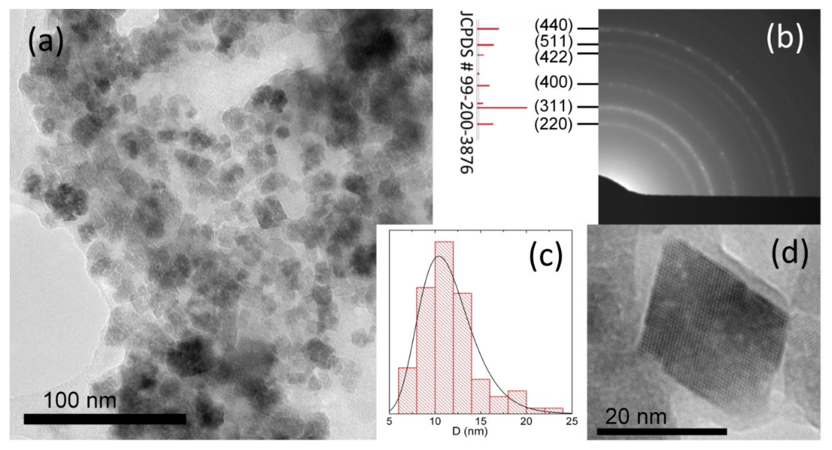
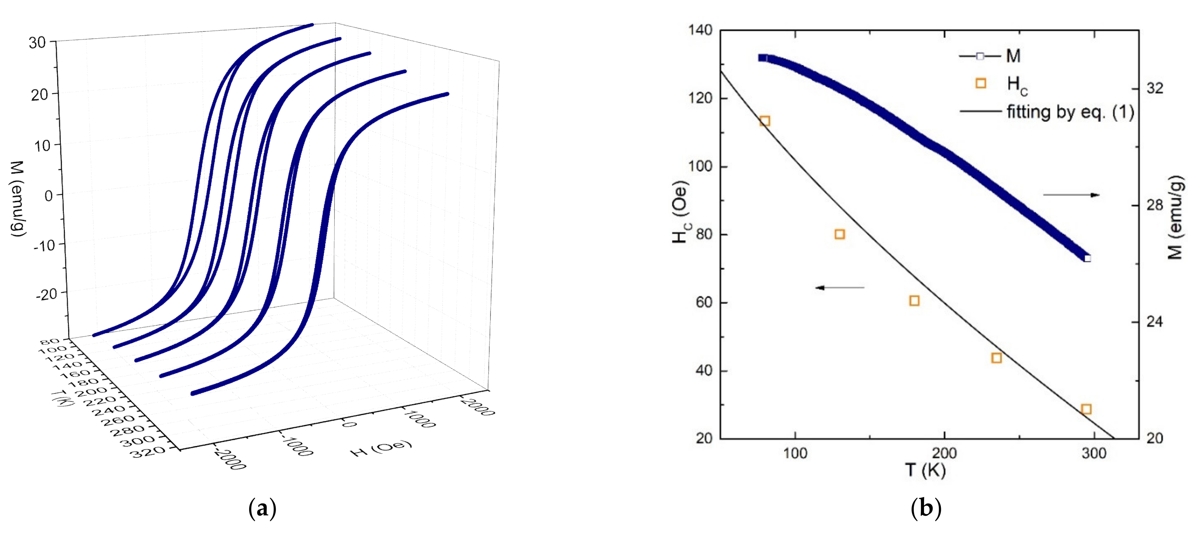
2.1. Physical Properties of Starch-MNPs
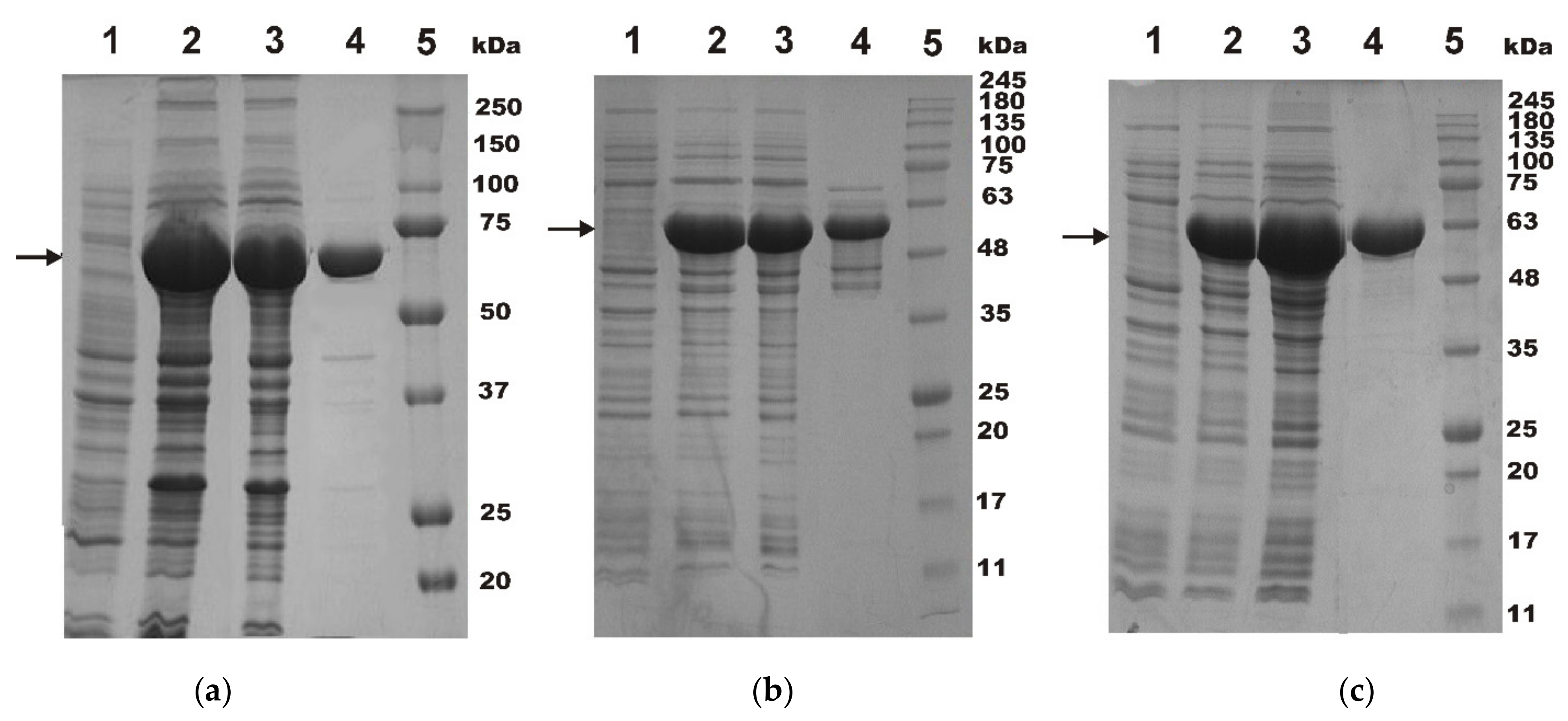
2.2. The Use of Starch-MNPs for One-Step Affine Purification of the MBP-Fused Recombinant Proteins
3. Materials and Methods
3.1. Synthesis and Characterization of Nanoparticles
3.2. MBP Hybrid Protein Expression
3.3. MBP Hybrid Proteins Purification by the Magnetic Nanoparticles
3.4. Starch-MNPs Recycling
4. Conclusions
Author Contributions
Funding
Institutional Review Board Statement
Informed Consent Statement
Data Availability Statement
Acknowledgments
Conflicts of Interest
References
- Kim, S.-E.; Tieu, M.V.; Hwang, S.Y.; Lee, M.-H. Magnetic particles: Their applications from sample preparations to biosensing platforms. Micromachines 2020, 11, 302. [Google Scholar] [CrossRef] [PubMed] [Green Version]
- Stueber, D.D.; Villanova, J.; Aponte, I.; Xiao, Z.; Colvin, V.L. Magnetic Nanoparticles in Biology and Medicine: Past, Present, and Future Trends. Pharmaceutics 2021, 13, 943. [Google Scholar] [CrossRef] [PubMed]
- Ma, Y.; Chen, T.; Iqbal, M.Z.; Yang, F.; Hampp, N.; Wu, A.; Luo, L. Applications of magnetic materials separation in biological nanomedicine. Electrophoresis 2019, 40, 2011–2028. [Google Scholar] [CrossRef] [PubMed]
- Qian, B.; Zhao, Q.; Ye, X. Ultrasound and magnetic responsive drug delivery systems for cardiovascular application. J. Cardiovasc. Pharmacol. 2020, 76, 414–426. [Google Scholar] [CrossRef]
- Zeng, L.; Wu, D.; Zou, R.; Chen, T.; Zhang, J.; Wu, A. Paramagnetic and superparamagnetic inorganic nanoparticles for T1-weighted magnetic resonance imaging. Curr. Med. Chem. 2018, 25, 2970–2986. [Google Scholar] [CrossRef] [PubMed]
- Fatima, H.; Charinpanitkul, T.; Kim, K.-S. Fundamentals to apply magnetic nanoparticles for hyperthermia therapy. Nanomaterials 2021, 11, 1203. [Google Scholar] [CrossRef]
- Masud, M.K.; Na, J.; Younus, M.; Hossain, M.S.A.; Bando, Y.; Shiddiky, M.J.A.; Yamauchi, Y. Superparamagnetic nanoarchitectures for disease-specific biomarker detection. Chem. Soc. Rev. 2019, 48, 5717–5751. [Google Scholar] [CrossRef]
- Fatima, H.; Kim, K.-S. Magnetic nanoparticles for bioseparation. Korean J. Chem. Eng. 2017, 34, 589–599. [Google Scholar] [CrossRef]
- Elahi, N.; Rizwan, M. Progress and prospects of magnetic iron oxide nanoparticles in biomedical applications: A review. Artif. Organs 2021, 45, 1272–1299. [Google Scholar] [CrossRef]
- Concas, G.; Congiu, F.; Muscas, G.; Peddis, D. Determination of blocking temperature in magnetization and Mössbauer time scale: A functional form approach. J. Phys. Chem. C 2017, 121, 16541–16548. [Google Scholar] [CrossRef]
- Khmelinskii, I.; Makarov, V.I. EPR hyperthermia of S. cerevisiae using superparamagnetic Fe3O4 nanoparticles. J. Therm. Biol. 2018, 77, 55–61. [Google Scholar] [CrossRef] [PubMed]
- Lee, J.-H.; Kim, B.; Kim, Y.; Kim, S.-K. Ultra-high rate of temperature increment from superparamagnetic nanoparticles for highly efficient hyperthermia. Sci. Rep. 2021, 11, 4969. [Google Scholar] [CrossRef] [PubMed]
- Bai, Y.; Cui, Y.; Paoli, G.C.; Shi, C.; Wang, D.; Zhou, M.; Zhang, L.; Shi, X. Synthesis of amino-rich silica-coated magnetic nanoparticles for the efficient capture of DNA for PCR. Colloids Surf. B Biointerfaces 2016, 145, 257–266. [Google Scholar] [CrossRef] [PubMed]
- Tyumentseva, A.V.; Yaroslavtsev, R.N.; Stolyar, S.V.; Saitova, A.T.; Tyutrina, E.S.; Gorbenko, A.S.; Stolyar, M.A.; Olkhovskiy, I.A. Silica-coated iron oxide nanoparticles for DNA isolation for molecular genetic studies in hematology. Genet. Test. Mol. Biomarkers 2021, 25, 611–614. [Google Scholar] [CrossRef] [PubMed]
- Komogortsev, S.V.; Stolyar, S.V.; Chekanova, L.A.; Yaroslavtsev, R.N.; Bayukov, O.A.; Velikanov, D.A.; Volochaev, M.N.; Eroshenko, P.E.; Iskhakov, R.S. Square plate shaped magnetite nanocrystals. J. Magn. Magn. Mater. 2021, 527, 167730. [Google Scholar] [CrossRef]
- Assa, F.; Jafarizadeh-Malmiri, H.; Ajamein, H.; Vaghari, H.; Anarjan, N.; Ahmadi, O.; Berenjian, A. Chitosan magnetic nanoparticles for drug delivery systems. Crit. Rev. Biotechnol. 2017, 37, 492–509. [Google Scholar] [CrossRef]
- Stolyar, S.V.; Krasitskaya, V.V.; Frank, L.A.; Yaroslavtsev, R.N.; Chekanova, L.A.; Gerasimova, Y.V.; Volochaev, M.N.; Bairmani, M.S.; Velikanov, D.A. Polysaccharide-coated iron oxide nanoparticles: Synthesis, properties, surface modification. Mater. Lett. 2021, 284, 128920. [Google Scholar] [CrossRef]
- Kheilkordi, Z.; Mohammadi Ziarani, G.; Mohajer, F.; Badiei, A.; Sillanpää, M. Recent advances in the application of magnetic bio-polymers as catalysts in multicomponent reactions. RSC Adv. 2022, 12, 12672–12701. [Google Scholar] [CrossRef]
- Duplay, P.; Bedouelle, H.; Fowler, A.; Zabin, I.; Saurin, W.; Hofnung, M. Sequences of the malE gene and of its product, the maltose-binding protein of Escherichia coli K12. J. Biol. Chem. 1984, 259, 10606–10613. [Google Scholar] [CrossRef]
- Waugh, D.S. The remarkable solubility-enhancing power of Escherichia coli maltose-binding protein. Postepy Biochem. 2016, 62, 377–382. [Google Scholar] [CrossRef]
- Duong-Ly, K.C.; Gabelli, S.B. Affinity purification of a recombinant protein expressed as a fusion with the maltose-binding protein (MBP) tag. Methods Enzymol. 2015, 559, 17–26. [Google Scholar] [PubMed] [Green Version]
- Kobashigawa, Y.; Namikawa, M.; Sekiguchi, M.; Inada, Y.; Yamauchi, S.; Kimoto, Y.; Okazaki, K.; Toyota, Y.; Sato, T.; Morioka, H. Expression, purification and characterization of CAR/NCOA-1 tethered protein in E. coli using maltose-binding protein fusion tag and gelatinized corn starch. Biol. Pharm. Bull. 2021, 44, 125–130. [Google Scholar] [CrossRef] [PubMed]
- Huang, Y.C.; Chang, H.H.; Mou, Y.; Chi, P.; Chan, J.C.; Luo, S.C. Purification of recombinant nacre-associated mineralization protein AP7 fused with maltose-binding protein. Protein Expr. Purif. 2014, 100, 26–32. [Google Scholar] [CrossRef] [PubMed]
- Park, K.C.; Gaze, D.C.; Collinson, P.O.; Marber, M.S. Cardiac troponins: From myocardial infarction to chronic disease. Cardiovasc. Res. 2017, 113, 1708–1718. [Google Scholar] [CrossRef]
- Gunaldi, M.; Isiksacan, N.; Kocoglu, H.; Okuturlar, Y.; Gunaldi, O.; Topcu, T.; Karabulut, M. The value of serum survivin level in early diagnosis of cancer. J. Cancer Res. Ther. 2018, 14, 570–573. [Google Scholar] [PubMed]
- Riechers, A.; Bosserhoff, A.K. Melanoma inhibitory activity in melanoma diagnostics and therapy—A small protein is looming large. Exp. Dermatol. 2014, 23, 12–14. [Google Scholar] [CrossRef] [PubMed]
- Wei, Y.; Han, B.; Hu, X.; Lin, Y.; Wang, X.; Deng, X. Synthesis of Fe3O4 nanoparticles and their magnetic properties. Procedia Eng. 2012, 27, 632–637. [Google Scholar] [CrossRef] [Green Version]
- Asgari, S.; Fakhari, Z.; Berijani, S. Synthesis and characterization of Fe3O4 magnetic nanoparticles coated with carboxymethyl chitosan grafted sodium methacrylate. J. Nanostructures 2014, 4, 55–63. [Google Scholar]
- Shah, N.; Ahmad, H.; Shah, S.S.; Khan, I. Synthesis and characterization of starch coated natural magnetic iron oxide nanoparticles for the removal of methyl orange dye from water. Lett. Appl. NanoBioScience 2021, 10, 2750–2759. [Google Scholar]
- Pfeiffer, H. Determination of anisotropy field distribution in particle assemblies taking into account thermal fluctuations. Phys. Status Solidi 1990, 118, 295–306. [Google Scholar] [CrossRef]
- Iskhakov, R.S.; Komogortsev, S.V.; Stolyar, S.V.; Prokof’ev, D.E.; Zhigalov, V.S. Structure and magnetic properties of nanocrystalline condensates of iron obtained by pulse plasma evaporation. Phys. Met. Metallogr. 1999, 88, 261–269. [Google Scholar]
- Komogortsev, S.V.; Iskhakov, R.S.; Balaev, A.D.; Okotrub, A.V.; Kudashov, A.G.; Momot, N.A.; Smirnov, S.I. Influence of the inhomogeneity of local magnetic parameters on the curves of magnetization in an ensemble of Fe3C ferromagnetic nanoparticles encapsulated in carbon nanotubes. Phys. Solid State 2009, 51, 2286. [Google Scholar] [CrossRef]
- Pisane, K.L.; Singh, S.; Seehra, M.S. Unusual enhancement of effective magnetic anisotropy with decreasing particle size in maghemite nanoparticles. Appl. Phys. Lett. 2017, 110, 222409. [Google Scholar] [CrossRef] [Green Version]
- Miller, D.M.; Olson, J.S.; Pflugrath, J.W.; Quiocho, F.A. Rates of ligand binding to periplasmic proteins involved in bacterial transport and chemotaxis. J. Biol. Chem. 1983, 258, 13665–13672. [Google Scholar] [CrossRef]
- Lim, M.-C.; Lee, G.-H.; Huynh, D.T.N.; Letona, C.A.M.; Seo, D.-H.; Park, C.-S.; Kim, Y.-R. Amylosucrase-mediated synthesis and self-assembly of amylose magnetic microparticles. RSC Adv. 2015, 5, 36088. [Google Scholar] [CrossRef]
- Velikanov, D.A. Vibration Magnetic Meter. RF Patent for the Invention RU2341810 (C1), 20 December 2008. Bulletin No. 3.. [Google Scholar]
- Krasitskaya, V.V.; Bashmakova, E.E.; Kudryavtsev, A.N.; Vorobyeva, M.A.; Shatunova, E.A.; Frank, L.A. The hybrid protein ZZ–OL as an analytical tool for biotechnology research. Russ. J. Bioorganic Chem. 2020, 46, 1004–1010. [Google Scholar] [CrossRef]


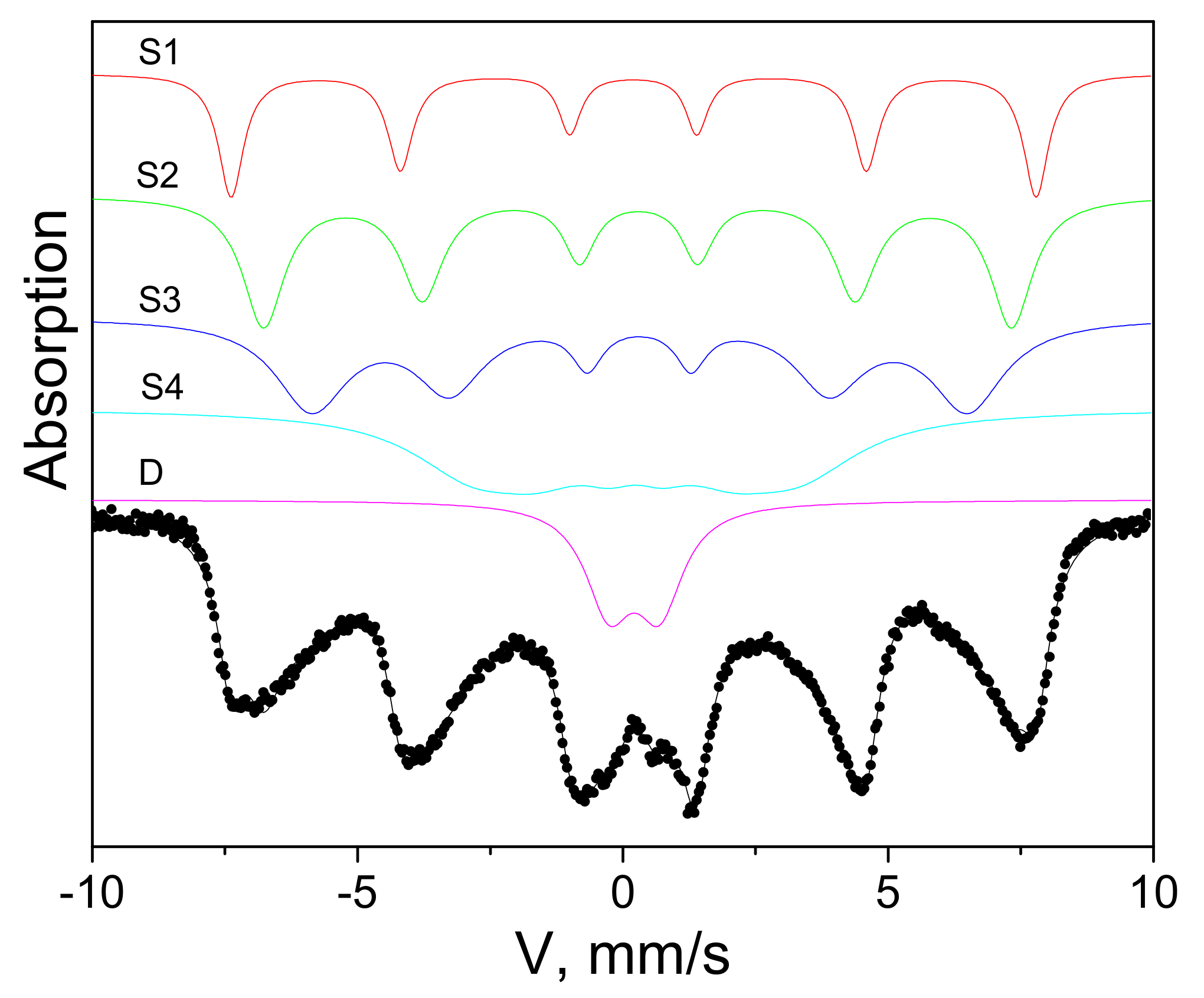
| IS, mm/s | H, kOe | QS, mm/s | W34, mm/s | A, Fract. % | |
|---|---|---|---|---|---|
| 0.34 | 474 | 0.01 | 0.50 | 0.14 | S1 |
| 0.42 | 440 | −0.04 | 0.73 | 0.24 | S2 |
| 0.45 | 386 | 0.01 | 0.67 | 0.26 | S3 |
| 0.37 | 193 | 0.01 | 1.24 | 0.25 | S4 |
| 0.35 | - | 0.96 | 1.08 | 0.11 | D |
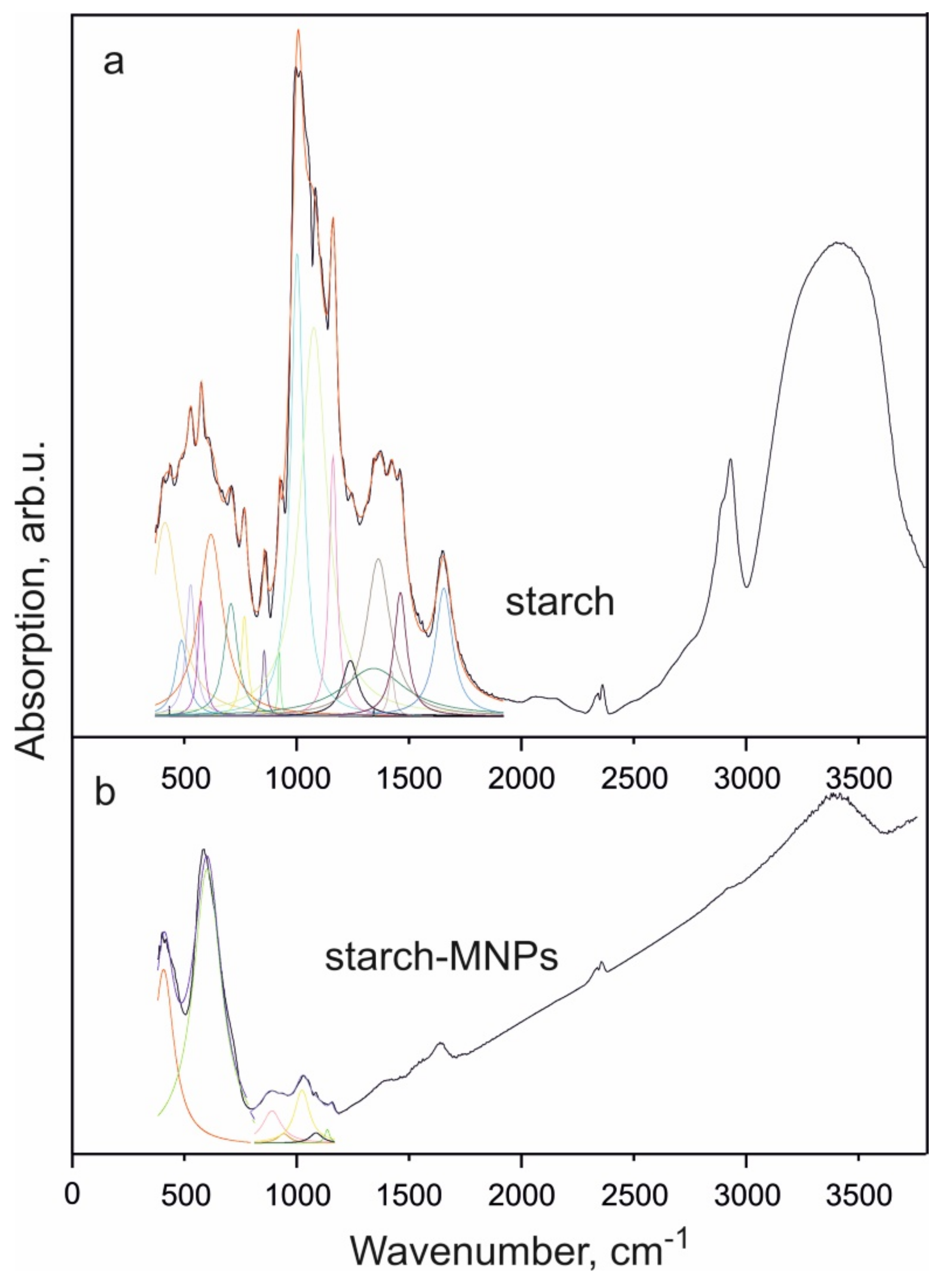
| Range | Absorption Peak, Starch | Absorption Peak, Starch-MNPs | Description |
|---|---|---|---|
| 380–800 | 388 | O–Fe–O | |
| 413 | Vibrations of the pyranose ring and δ-hydroxyl groups | ||
| 432 | |||
| 487 | |||
| 527 | |||
| 573 | Vibrations of the chain C–C–C…– | ||
| 570 | Fe–O | ||
| 617 | Vibrations of the pyranose ring and δ-hydroxyl groups | ||
| 706 | |||
| 767 | |||
| 800–1000 | 855 | C–O in C–O–H | |
| 866 | |||
| 922 | 900 | ||
| 1000–1200 | 1001 | C–O stretching of internal vibrations of C–O bonds (the bands characteristic of polysaccharides are caused by the presence of acetal bonds) | |
| 1025 | |||
| 1075 | |||
| 1092 | |||
| 1150 | |||
| 1161 | |||
| 1200–1500 | 1238 | δ-CH2 groups in CH2OH | |
| 1341 | δ-O–H bonds in CH2OH | ||
| 1368 | δ-bonds of CH2 groups | ||
| 1421 | δ-CH2 groups | ||
| 1461 | δ-OH | ||
| 1500–2000 | 1654 | 1635 | δ-bonds in H–O–H (adsorbed water) |
| 2000–3000 | 2060 | ν-bonds in CH and CH2 groups | |
| 2153 | |||
| 2890 | |||
| 2930 | 2928 | C–H | |
| 3000–4000 | 3406 | 3413 | Internal vibrations of OH groups involved in intermolecular and intramolecular H bonds |
Publisher’s Note: MDPI stays neutral with regard to jurisdictional claims in published maps and institutional affiliations. |
© 2022 by the authors. Licensee MDPI, Basel, Switzerland. This article is an open access article distributed under the terms and conditions of the Creative Commons Attribution (CC BY) license (https://creativecommons.org/licenses/by/4.0/).
Share and Cite
Krasitskaya, V.V.; Kudryavtsev, A.N.; Yaroslavtsev, R.N.; Velikanov, D.A.; Bayukov, O.A.; Gerasimova, Y.V.; Stolyar, S.V.; Frank, L.A. Starch-Coated Magnetic Iron Oxide Nanoparticles for Affinity Purification of Recombinant Proteins. Int. J. Mol. Sci. 2022, 23, 5410. https://doi.org/10.3390/ijms23105410
Krasitskaya VV, Kudryavtsev AN, Yaroslavtsev RN, Velikanov DA, Bayukov OA, Gerasimova YV, Stolyar SV, Frank LA. Starch-Coated Magnetic Iron Oxide Nanoparticles for Affinity Purification of Recombinant Proteins. International Journal of Molecular Sciences. 2022; 23(10):5410. https://doi.org/10.3390/ijms23105410
Chicago/Turabian StyleKrasitskaya, Vasilisa V., Alexander N. Kudryavtsev, Roman N. Yaroslavtsev, Dmitry A. Velikanov, Oleg A. Bayukov, Yulia V. Gerasimova, Sergey V. Stolyar, and Ludmila A. Frank. 2022. "Starch-Coated Magnetic Iron Oxide Nanoparticles for Affinity Purification of Recombinant Proteins" International Journal of Molecular Sciences 23, no. 10: 5410. https://doi.org/10.3390/ijms23105410