Review

Phosphatidic Acid in Plant Hormonal Signaling: From Target Proteins to Membrane Conformations

1
V.P. Kukhar Institute of Bioorganic Chemistry and Petrochemistry National Academy of Sciences of Ukraine, Murmanska Street, 1, 02660 Kiev, Ukraine
2
Sorbonne Universités, Génie Enzymatique et Cellulaire, UMR CNRS 7025, Université de Technologie de Compiègne, CEDEX, 60203 Compiègne, France
3
Institute of Experimental Botany of the Czech Academy of Sciences, Rozvojová 263, 165 00 Prague, Czech Republic
*
Authors to whom correspondence should be addressed.
Int. J. Mol. Sci. 2022, 23(6), 3227; https://doi.org/10.3390/ijms23063227
Submission received: 16 December 2021 / Revised: 24 January 2022 / Accepted: 7 March 2022 / Published: 17 March 2022
(This article belongs to the Special Issue New Insight into Signaling and Autophagy in Plants 2.0)

Abstract

:
Cells sense a variety of extracellular signals balancing their metabolism and physiology according to changing growth conditions. Plasma membranes are the outermost informational barriers that render cells sensitive to regulatory inputs. Membranes are composed of different types of lipids that play not only structural but also informational roles. Hormones and other regulators are sensed by specific receptors leading to the activation of lipid metabolizing enzymes. These enzymes generate lipid second messengers. Among them, phosphatidic acid (PA) is a well-known intracellular messenger that regulates various cellular processes. This lipid affects the functional properties of cell membranes and binds to specific target proteins leading to either genomic (affecting transcriptome) or non-genomic responses. The subsequent biochemical, cellular and physiological reactions regulate plant growth, development and stress tolerance. In the present review, we focus on primary (genome-independent) signaling events triggered by rapid PA accumulation in plant cells and describe the functional role of PA in mediating response to hormones and hormone-like regulators. The contributions of individual lipid signaling enzymes to the formation of PA by specific stimuli are also discussed. We provide an overview of the current state of knowledge and future perspectives needed to decipher the mode of action of PA in the regulation of cell functions.

1. Introduction

Phosphatidic acid (PA) is a minor membrane glycerolipid composed of glycerol-3-phosphate and two fatty acid chains. PA is an intermediate and regulator of glycerolipid metabolism in yeast, animals and plants [1,2,3,4]. Under basal conditions, PA is formed de novo in the endoplasmic reticulum and plastids mainly by acylation of glycerol-3-phosphate catalyzed by specific acyltransferases [5]. PA is also an important lipid second messenger in cells, acting in part through interaction with various effector proteins [6,7]. The level of PA, like that of most phospholipids in cells, is highly dynamic and changes specifically, rapidly (seconds to minutes) and transiently (i.e., eventually reverting to basal level) in response to various extracellular, hormonal or stress stimuli [8,9]. These changes are made possible by various phospholipases, lipid kinases and phosphatases. The generation of signaling PA occurs via the activation of phospholipase D (PLD), which cleaves structural membrane lipids (e.g., phosphatidylcholine) to generate PA and free headgroups (e.g., choline) [10,11,12,13] (Figure 1). Signaling PA can also be produced by the phosphorylation of diacylglycerol (DAG, Figure 1) in a reaction catalyzed by diacylglycerol kinase (DGK) [14,15,16]. The formation of DAG itself is the result of the activity of two classes of phospholipases C: one that hydrolyzes phosphatidylinositol phosphates (phosphatidylinositol-specific phospholipase C, PI-PLC) or one that hydrolyzes structural lipids (phosphatidylcholine-specific or non-specific phospholipase C – PC-PLC or NPC) [17,18]. As a part of lipid turnover, PA can be dephosphorylated back into DAG by lipid phosphate phosphatases (LPP) [19,20].
Each class of enzymes involved in the production of PA is represented by multiple isoforms (isoenzymes) [4,21]. Regulation of their activity by extracellular agonists may represent a specific mechanism to control lipid signaling in cells [9,22]. Changes to activity or abundance of particular isoenzymes acting in PA production may be essential for stress tolerance [23,24] or growth/developmental responses [25,26,27,28].
In cell membranes, PA molecules are not compositionally uniform. They are represented by different molecule classes (molecular species) depending on the length and degree of desaturation of fatty acid moieties they incorporate [29]. Accordingly, PA produced in the course of cell signaling is also heterogeneous. AtPLDα1 activity stimulated by phytohormone abscisic acid (ABA) leads to 34:2-PA, 34:3-PA, 36:3-PA, 36:4-PA, 36:5-PA and 36:6-PA production [30]. The profile of PA molecules generated by AtPLDδ is different: this enzyme additionally generates 34:1-PA and 36:2-PA, but not 36:3-PA and 36:6-PA [31]. Different PA molecular species can affect target proteins in a non-identical way leading to specific physiological consequences. Different PA molecular species bind to proteins acting in different branches of ABA responses with dissimilar affinity: di18:1-PA has been shown to be more active in binding protein phosphatase 2C AtABI1 than di16:0-PA, di18:0-PA and di18:2-PA [32]; 16:0/18:2-PA binds the NADPH oxidase subunit AtRbohD preferentially to 16:0/16:0-PA and 18:0/18:0-PA [30]; the binding of 18:1/18:1-PA, 16:0/18:1-PA and 16:0/18:2-PA to the sphingosine kinases AtSPHK1 and AtSPHK2 is higher than that of 16:0/16:0-PA [33].
The concentration of PA significantly differs depending on plant tissue type, plant developmental phase, treatment condition as well as registration technique. For example, in 32P-labelled Arabidopsis, basal PA levels range from 2% (in germinating seeds and seedlings) to nearly 8% (in leaf peels) of all labeled phospholipids [25]. In the roots of soybean seedlings, the level of PA, as detected by MS, was found to constitute up to 2.5% of total cell lipids [34]. The level of PA in Arabidopsis seedlings detected by GC was found to constitute several mol% [35]. In rice leaves, the level of PA analyzed by mass spectrometry was observed to be in the range of 2.5 to 5 nmol/mg [36].
The general topology of PA signaling in plants seems to be similar to that of animals. Plasma membrane or intracellular receptors activate PA production by directly or indirectly affecting the activity of PA-producing enzymes; then, the formed PA binds target proteins and/or affects membrane conformation. However, some differences exist between PA signaling in animals and plants. The first important difference concerns the interrelationships between DAG and PA. For example, in animals, DAG has several protein targets in comparison with a huge number of PA targets and a broader array of signaling properties that are very distinct from those of DAG. On the other hand, in plants, no DAG targets were found, suggesting that PA is the lipid signal instead of DAG. Although conversion of DAG to PA by the DGK is the major route to terminate DAG signaling in animals, there are several examples indicating that DGKs modulate signaling events by producing PA [37]. The second difference is associated with enzymes of PA production. No NPC gene was found in animals in comparison to plants. Furthermore, the PLD family in plants is more heterogenic in comparison with classical PLDs (PLD1 and PLD2) in animals, and PLD substrate specificity is wider in plants than that in animals [38]. The third difference in PA signaling between animals and plants concerns the pathways of PA metabolic conversions. PA phosphatases from animals can generate DAG from PA produced by PLD, and this newly formed DAG possesses signaling and metabolic function [39]. However, such pathways in plants produce DAG for the synthesis of other lipids (e.g., triacylglycerols, galactolipids), but the role of the pathway in signaling was not discovered in plants. In addition, diacylglycerol pyrophosphate, a phosphorylated form of PA with signaling functions, has been found in plants, but not animals [40]. Such a multifaced role of PA in living organisms suggests an evolutionary significance of its signaling mechanisms.

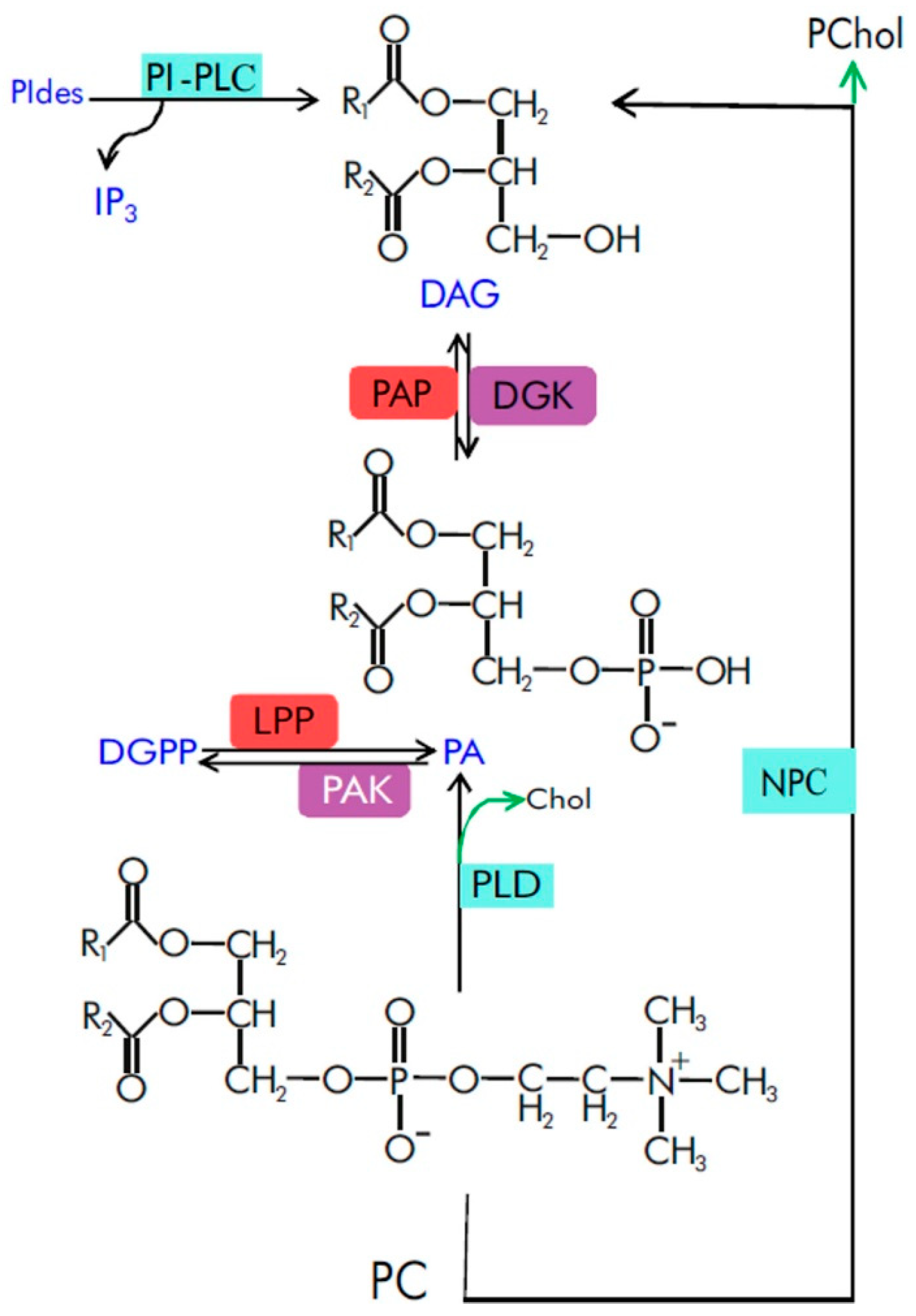
2. Spatiotemporal Characteristics of Phosphatidic Acid as a Signaling Molecule

PA has several properties underlying its signaling functions. PA has a specific pattern of intracellular localization. Under basal conditions, PA has been detected in the plasma membrane (PM) in various plant objects: growing tobacco pollen tubes [41,42], mature trichomes [43], epidermal and meristematic root cells and shoot tissues [44]. However, PA has also been detected in cell membranes other than PM. PA has been detected in the nucleus of Arabidopsis epidermal root cells undergoing cell division [44] and in chloroplast membranes [45]. Interestingly, the level of nuclear PA in Arabidopsis seedlings has been shown to be affected by the activities of lipid signaling enzymes located at the PM, cytosol and endoplasmic reticulum [3]. The level of PA in the PM is highly dynamic even under basal conditions, possibly due to the combination of rapid lateral diffusion of PA and PLD, DGK and LPP activities leading to local accumulation of PA [41]. In response to extracellular stimuli (e.g., abscisic acid), rapid changes in PA signaling, specifically at the PM of plant cells, have been reported [46].
Another important property of PA is its high turnover rate. The rapid turnover of PA contributes to its low level in unstressed cells compared to structural phospholipids. The increase in PA content in response to a stimulus is usually transient [46].
Cells of different organs and tissue types have different capacities to respond to extracellular stimuli. At least in part, this could be due to differences in their basal PA levels. These levels vary depending on the growth stage of the plant [47], type of plant organ [48,49] or developmental phase of the organ [50]. This could explain why the stimulated PA levels were different in plant organs [51] and tissue types [46,52]. The differential capacity for PA signaling at different developmental stages can be illustrated by the high levels of DGK and PLD proteins in developing maize ears [53] and the high basal PA content in cotton fiber cells compared to ovule cells [54]. The expression levels of many PA signal transduction enzymes also differ depending on the plant developmental phase (Figure 2). These differences may thus determine the extent of activation of lipid signal transduction in response to extracellular (or endogenous) stimuli and control its regulatory performance.

3. Effect of PA on Membrane Status

Signaling PA is produced rapidly, transiently and at low concentrations by different pathways in cellular membranes. PA regulatory influence on metabolism is mediated by interactions with target proteins and by inducing effects on membrane functions, charge and conformations.
Phospholipids and enzymes of phospholipid turnover are involved in the control of membrane trafficking [56,57] and the transduction of specific extracellular signals in plants [58,59]. It becomes clear that plant hormone-induced PA accumulation might also be associated with membrane trafficking pathways. This can be illustrated by the fact that n-butanol, an inhibitor of PA formation by PLD, impairs translocation of NON-EXPRESSOR OF PATHOGENESIS-RELATED GENES 1 (NPR1) to the nucleus in response to salicylic acid [60].
Endocytosis and exocytosis induced by PA may be an important part of PA signaling in plants. For example, activation of Brassica napus PLDα1 during compatible pollination has been hypothesized to promote exocytosis required for pollen germination through the production of PA in the PM [61]. This notion is also supported by genetic and pharmacological data suggesting that the production of PA modulates basal cellular membrane trafficking. Suppression of PA production by inhibition of PLD or DGK activity compromised membrane trafficking, disrupted the deposition of cell wall material (pectins) at the pollen tube tip and inhibited pollen tube growth [62]. Inhibition of PLD also blocked the membrane localization of clathrin and affected the localization of PIN2 (an auxin transporter) in root epidermal cells [63]. In dgk4 Arabidopsis plants, a reduced rate of membrane secretion and recycling, as well as inhibited pollen tube growth, were observed [64]. PA and lysophosphatidic acid acyltransferases (AtLPAAT4 and AtLPAAT5) that also generate PA were suggested to be critical for the secretory pathway involved in protein trafficking of the auxin transporter PIN2 and the aquaporin PIP2;7 [65].
A recent study on the functions of PLDδ-family phospholipase in tobacco showed that pollen tubes support a high level of expression of PLDδ genes during growth. For some of the PLDδ isoforms, the unique localization was detected, and the sequences important for binding to the PM were determined [42]. It has been noted that the increased level of PA, formed by the overexpression of NtPLDδ3 isoform, causes specific morphological phenotypes indicating a violation of the balance of vesicular traffic and membrane recirculation [42].
PA and the enzymes that produce it can affect membrane trafficking and endocytosis by interacting with their specific proteins. AtPLDα1 is associated with both microtubules and clathrin-coated vesicles [66], AtECA2 and ANTH domain proteins that recruit clathrin to membranes to facilitate vesicle budding [67], and CHC (Clathrin Heavy Chain) proteins [68]. Other membrane trafficking events regulated by PA include PA-mediated membrane recruitment of NtEXO70A1a and NtEXO70B1—two proteins involved in targeting the vesicle tethering complex (exocyst) for exocytosis [69].
Some of the effects of PA on membrane conformations are local: the accumulation of PA leads to a negative spontaneous curvature that facilitates interactions with specific proteins. Indeed, the binding of the Epsin-like Clathrin Adaptor 1 (AtECA1) to PA was dependent on membrane curvature stress [70]. Hence, binding was achieved only after a certain threshold level of negative curvature stress. This suggests that the binding of AtECA1 requires some degree of hydrophobic interaction with the membrane. PA-binding proteins may take advantage of the presence of bulky hydrophobic residues such as tryptophan to insert themselves into the membrane interface, having a negative spontaneous curvature [71].
Since PA is a component of cell membranes, it interacts not only with proteins but also with other membrane lipids. Studies performed on artificial membranes show that PA binds to phosphatidylcholine (via electrostatic interactions) [72] and stacks with other PA molecules (at low pH or in the presence of divalent cations) [73]. These interactions could be a factor that regulates the affinity of PA towards protein binding and membrane tethering.
PA signaling could be functionally linked to pH changes. The protonation state of PA has been found to depend on the local pH and to affect the efficiency of PA binding to its effector proteins [46].

4. Binding of Phosphatidic Acid to Target Proteins

A number of PA-binding proteins have now been identified. Many of them are thought to play a role in plant signal transduction. However, a detailed characterization of the biological role, as well as the deciphering of the interaction with PA at the molecular level, is available for only some of them (Table 1). Since no PA-recognition motif has yet been identified in plant proteins (as well as animal proteins), it has been proposed that complex structural folds of a protein are involved in PA recognition. PA-binding modules typically consist of an amphipathic α-helix with positively charged amino acids on one side (e.g., lysine-rich sequence motif) and hydrophobic residues on the other [74]. Similar structural folds have been found in proteins involved in plant hormone signaling, suggesting that this may indeed be a common mechanism of PA-protein interaction (Figure 3).
An interaction between a PA and a protein implicates specific amino acid residues of the latter. It has been shown that positively charged polybasic residues (Lys, Arg) [30,75] are involved in PA binding. The negative charges carried by anionic PA thus enable the recruitment of proteins with polybasic regions [44]. In addition, other types of amino acids (e.g., His in the defensin HsAFP1 [76] and Pro in the lipid kinase AtPIPK1 [77]) are suggested to act in the binding of PA. Together, this enables the establishment of electrostatic and hydrophobic interactions as well as hydrogen bonds between PA and a target protein. His, Trp, Ser, Ala and Gly are also commonly present in PA-binding modules of proteins. This is, for example, true for CDeT11-24, a desiccation-related phosphoprotein of Craterostigma plantagineum. The Lys-rich motif of this protein is responsible for the interaction with the PA phosphomonoester moiety, while a region of nonpolar amino acids, seen in a helical wheel projection of the lysine-rich motif of CDeT11-24, may be involved in hydrophobic interactions with the acyl chain of PA [78]. His42 and His51 are thought to contribute to the pH sensitivity of the binding of PA to the Epsin-like Clathrin Adaptor 1 [71]. Non-basic amino acids could play a supporting role in PA binding and cause a change in the PA binding affinity of a protein—this could be an explanation for the differences in PA binding to the NADPH oxidases AtRbohD and AtRbohF [30].
Interestingly, several PA-binding sites are sometimes located in different parts of a protein. It should be noted that multiple basic and hydrophobic amino acid residues have been shown to be important elements of the AtPINOID kinase binding to PA [79]. AtSnRK2.4 contains multiple basic amino acid residues located in different regions, all involved in PA-binding [80]. In the case of the potassium channel OsAKT2, PA was found to bind two adjacent lysine residues in the cytosolic ANK domain [81] and basic residues in the S4 voltage-sensing domain. The cytosol-facing domains of OsAKT2 contribute to the PA-mediated negative regulation of OsAKT2 activity [82]. In addition, the EH1.1 domain of the TPC subunit AtEH1/Pan1 (adaptor complex of clathrin-mediated endocytosis machinery) coordinates the PA molecules by three main regions (residues around K42, residues around K78 and residues located at the C-terminus around K97), whereas the EH1.2 domain interacts with the PA molecules at the region forming the phosphoinositide-binding site and at the region located close to the protein C-terminus. In contrast to EH1.1, EH1.2 binds a higher number of PA molecules [83].
The different affinities of PA species towards binding to target proteins may act as important determinants of PA signal specificity in response to dissimilar stimuli. The role of fatty acid tails in altering the affinity of PA towards a protein is likely determined by the specific structural features of the letter: e.g., the presence of a stretch of hydrophobic residues in the PA-binding region of protein phosphatase 2C AtABI1 and protein kinase AtCTR1 [32,84]; presence of hydrophobic residues at the C-terminus of AtCP, an actin-capping protein [85]; hydrophobic cavity in the structure of AtSPHK1 [86]. PA is a cone-shaped molecule that induces negative spontaneous curvature [87], facilitating “deeper” insertion of proteins into lipid membranes, providing a background for the role of hydrophobic interactions [70]. On the other hand, some proteins, e.g., an MYB transcription factor (AtWER) [88] and glyceraldehyde-3-phosphate dehydrogenases (AtGAPC1 and AtGAPC2) [89] do not show specific preferences for binding to particular PA species. For these proteins, binding is likely determined by the interaction of the phosphomonoester group of PA with basic residues.
Figure 3. PA-binding folds of Arabidopsis proteins involved in plant hormone (abscisic acid) signaling. Shown are amphipathic α-helix projections of a minimal characteristic PA-binding domain obtained with “Heliquest” software [90]. The arrows indicate the hydrophobic moment. Basic and hydrophobic residues are shown in blue and yellow, respectively. PA-binding motifs were obtained from published data: AtRbohD [30], AtABI1 [32], AtRGS1 [91], AtGEF8 [75] and AtSPHK1 [86]. The sequences of PA-binding motifs are as follows: 140-SRELRRVFSRRPSPAVRRFD-159 (AtRbohD), 63-GSESRKVLISRINSPNLNMK-82 (AtABI), 250-QPLLSQISLKKRQNFEFQRM-269 (AtRGS1), 6-ERGLSASKSFNFKRMFDSSS-25 (AtGEF8), 175-KYDGIVCVSGDGILVEVVNG-194 (AtSPHK1).
Ijms 23 03227 g003
Binding to PA is not necessarily associated with a tethering of proteins to PM. It has been shown that the binding of the cytosolic glyceraldehyde-3-phosphate dehydrogenase AtGAPC2 to PA induces proteolytic cleavage of the enzyme [89]. It was found that PA interacts with the transcription factors AtAHL4 (AT-hook motif-containing nuclear localized 4) [3] and Arabidopsis LATE ELONGATED HYPOCOTYL/CIRCADIAN CLOCK ASSOCIATED1 [92] and negatively regulates their DNA-binding ability. For AtAHL4, PA-mediated tethering to the nuclear membrane was excluded as a mechanism of PA action, suggesting a hypothetical possible association of PA with AtAHL4 beyond membranes [3]. PA also binds to the gibberellin receptor OsGID1, promotes its nuclear localization, and mediates the gibberellin response in rice [36].
PA could regulate cell signaling by simultaneous binding to multiple components of the same regulatory pathway. One example is the binding of PA to the potassium channel AtAKT2 [81] and AtPP2CA (protein phosphatase 2CA, negative modulator of AtAKT2 activity) [93]. Similarly, PA binds to AtMKK7/AtMKK9, which phosphorylate PA-binding AtMPK6 [94]; activates AtMKK5, and its downstream targets AtMPK3/AtMPK6 [95]; binds AtPINOID kinase [79], and Arabidopsis D6 PROTEIN KINASE [96], all of which regulate PIN auxin transporter activity within the same pathway. PA has been shown to regulate not only the activity, phosphorylation status and/or localization of AtSnRK2, but also that of its cellular partners: AtSnRK2 enzyme targets (e.g., AtERD14) and negative regulators (AtABI1 and AtSnRK2-interacting calcium sensor) [97].

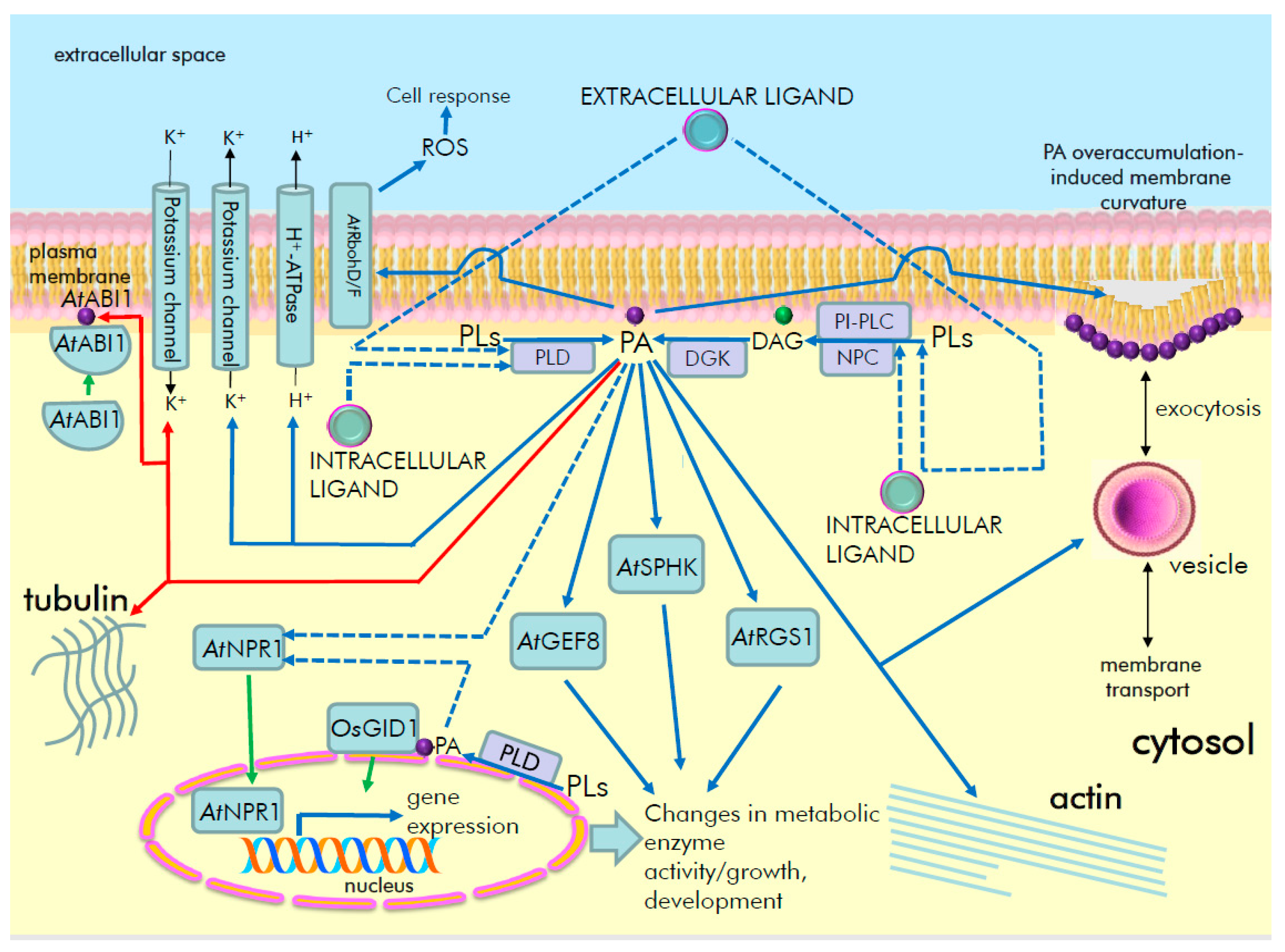
5. Role of PA as a Lipid Second Messenger of Signal Transduction Pathways in Plants

5.1. Phosphatidic Acid Signaling in Hormonal Regulation

PA plays a key role in the regulation of metabolism in plants as well as in other living systems. PA is a mediator in the signal transduction pathways of hormones and other biologically active compounds that ensure plant growth, development and resistance to adverse environmental factors. Regulatory signals originating from hormones such as auxin, brassinosteroids, salicylic acid, abscisic acid, jasmonic acid and gibberellins have been found to implicate PA as a second messenger. PA, which is produced in response to the stimuli, has been found to be involved in genomic and non-genomic signaling events that regulate gene expression, ion transport, cytoskeletal dynamics and metabolic enzyme activities (Figure 4).

5.1.1. Role of Phosphatidic Acid in Auxin Signaling

Genetic studies suggest that the phospholipases AtPI-PLC2 [98] and AtNPC5 [99] play a role in auxin sensing pathways. PA binds to specific signaling proteins involved in the regulation of auxin transport and regulates their activity. For example, PA-binding 3’-phosphoinositide-dependent protein kinase1 (AtPDK1) phosphorylates and regulates PA-binding D6 protein kinase (AtD6PK) to stimulate the activity of PIN auxin efflux carriers [100]. PLD and PA were suggested to be involved in auxin-mediated reversion of the actin bundling induced by the hairpin elicitor [101].

5.1.2. Role of Phosphatidic Acid in Salicylic Acid Signaling

Salicylic acid (SA) activates PA production via DGK [102] and PLD [103,104,105,106] in Arabidopsis thaliana and Capsicum chinense. PLD and PA have been shown to be involved in transcriptome regulation by SA [105,107,108], SA-induced ROS accumulation via the NADPH oxidase AtRbohD [103], SA-induced metabolic changes [109] and nuclear translocation of AtNPR1, an SA signaling regulator [60].

5.1.3. Role of Phosphatidic Acid in Brassinosteroid Signaling

Brassinosteroids (BR) trigger the accumulation of DAG and PA derived from phosphatidylcholine in various plant tissues due to the activation of NPC and DGK [110,111,112]. AtNPC3 and AtNPC4 isozymes are involved in plant sensitivity to BR in terms of root growth and gene expression [110]. Several DGK isoforms contribute to the production of PA in response to BR. Epibrassinolide (EBL) treatment resulted in a two-fold increase in PA in WT Arabidopsis thaliana roots [113]. However, a similar response was reduced in dgk1dgk2, dgk1 and dgk5dgk6 mutant lines. Of all the dgk mutant lines tested, dgk3 plants were characterized by the lowest PA induction in response to EBL treatment compared with WT plants. Pretreatment of plants with BR further increased the levels of DAG and PA during subsequent salt stress. In this context, it has been suggested that DAG and PA are involved in the BR-induced adaptation of plants to high salinity [112]. Moreover, a reduced EBL-stimulated PA production in dgk mutants might be linked to lower germination rates, lower total and alternative respiration under optimal and high salinity conditions [113].

5.1.4. Role of Phosphatidic Acid in Abscisic Acid Signaling

Abscisic acid (ABA) is one of the most important plant hormones with respect to the regulation of plant metabolism (e.g., acting in seed germination, growth responses) and stress resistance (e.g., by controlling stomatal closure). ABA-induced rapid PA generation has been localized to the PM of Arabidopsis thaliana guard cells and cells of the root maturation zone [46]. PA plays a role as a second messenger in ABA-regulated events such as stomata closure and water stress tolerance. Specific PA-binding proteins are known to be involved in ABA signaling in plants. ABA-activated PLD leads to PA production, which inhibits AtRGS1—a regulator of G-protein signaling [91] and activates the guanine nucleotide exchange factor AtGEF8 [75]. Moreover, recruitment of ABI1 to the PM by the leucine-rich receptor-like kinase AtRDK1 in response to ABA requires the binding of PA to the former [114]. PLD and PA are also involved in ABA-induced microtubule depolymerization and calcium spikes in guard cells [115] and HVA1 expression [116].
The functional role of PA in ABA-signaling pathways could be illustrated by in silico analyses, which showed similarities in the gene expression patterns of ABA-treated wild-type plants and untreated rgs1 lines. It is known that AtRGS1 is inhibited by PA as part of the ABA-signaling pathway [84]. Therefore, this signaling event is mimicked in rgs1 plants, at least in part, by the absence of ABA. Comparing the two sets of gene expression data, we found that a number of genes encoding signaling, metabolic or stress-related proteins are regulated in a similar manner (Figure 5). These data suggest that inhibition of AtRGS1 by PA is indeed an important event in the ABA response in Arabidopsis.

5.1.5. Role of Phosphatidic Acid in Jasmonic Acid Signaling

Jasmonic acid is a hormone that rapidly activates the production of PA via PI-PLC/DGK and PLD in potato tubers [119], leaves of Brassica napus [120] and cells of C. chinense [102,121]. Jasmonate-induced accumulation of the secondary metabolites [102] and potato tuber cellular growth [119] were found to be downstream of PI-PLC\PLD activity and PA accumulation.

5.1.6. Role of Phosphatidic Acid in Gibberellin Signaling

Gibberellins were also suggested to interact with PA signaling. In rice, among several PLD isoforms tested, only OsPLDα6 was found to be an important positive regulator of gibberellin sensitivity in terms of modulation of growth responses. Results from lipidomic analysis indicate that phosphatidylethanolamine and phosphatidylserine are the main sources of PA in response to gibberellin. In rice protoplasts, gibberellin induces OsPLDα6 translocation from cytosol to nuclei. PA formed by OsPLDα6 activity positively mediates gibberellin signaling via PA binding to gibberellin receptor OsGID1, promotion of its nuclear localization and degradation of the DELLA protein SLENDER RICE1, a negative regulator of the hormone signaling [36]. In contrast, the level of suppressor protein SLENDER RICE1 was decreased in npc6 rice mutants treated with gibberellin, but drastically increased in OsNPC6 over-expressing plants [122]. Therefore, OsPLDα6 and PA are positive players of early events of gibberellin signaling in plants.
Hormones can also modulate PA signaling by controlling gene expression of PA-producing enzymes (Figure 6). Although the enzymes of PA turnover are well known, controlling the expression level of their encoding genes could still have a significant impact on the resulting signaling function. Thus, downregulation of AtNPC6 expression following treatment with ABA could be a mechanism that attenuates the signaling response (Figure 6). Conversely, upregulation of AtPLDδ, AtPLDζ2 and AtDGK1 by the same stimuli could mean that the abundance of these isoenzymes is important to fulfill the latter steps in the ABA signaling pathway. On the other hand, cytokinins weakly affect PLD gene expression (Figure 6), but the role of PLD and PA as mediators of cytokinin-regulated gene expression was found [123,124].

5.2. Phosphatidic Acid as a Signal Transducer for Other Biologically Active Substances

Some hormone-like substances also trigger PA signaling. It has been shown that treatment with polyamines activates AtPLDα1 [125], AtPLDδ and DGK [52] in Arabidopsis seedlings and also PLD and DGK in C. arabica cells [126], leading to a rapid PA production. This response was dependent on the type of polyamines tested and their charge. For example, putrescine triggered higher AtPLDα1 activation in vitro compared to spermine and spermidine [125], while in Arabidopsis seedlings, spermine, thermospermine and spermidine induced higher PA accumulation compared to putrescine and diaminopropane [52]. Cellular uptake of spermine by the PM localized amino acid transporter Resistant to Methyl Viologen 1 (AtRMV1) is required for triggering PA production [52]. As a port of cell response to polyamines, AtPLDα1-produced PA was shown to trigger H2O2 accumulation in guard cells [125], while AtPLDδ-produced PA triggers potassium ion efflux in the root elongation zone of Arabidopsis [53].
SCOOP12—an active endogenous plant peptide (phytocytokine)—has been shown to rapidly stimulate the production of PA via the PI-PLC/DGK pathway in Arabidopsis cells [127]. During the self-incompatibility response of Brassica napus triggered by the interaction of SP11 peptide with SRK (S-locus receptor kinase), PLDα1 was found to be a target of ARC1-mediated ubiquitination. Subsequent degradation of PLDα1 thus impedes the production of PA, which itself is required for pollination. It has been proposed that activation of the PLDα1 isoform by an unknown ligand during compatible pollination would lead to PM remodeling supported by the production of PA to promote exocytosis required for pollen germination [61]. Other data indirectly suggest a possible role of PA pathway enzymes in peptide signaling. AtPI-PLC2 has been found to interact with AtPXL1—a receptor for the peptide AtCLE41 [128]. Moreover, some peptide receptors might be co-expressed with PA-producing enzymes, as suggested by in silico analysis (see Table 2). This could indicate a possible functional link between certain peptide receptors and a signal lipid production.
PA signaling may also interact with signaling pathways of other bioactive substances. Both AtPLDα1 and AtPLDδ are involved in hydrogen sulfide signaling leading to stomatal closure in Arabidopsis. Hydrogen sulfide-induced H2O2 accumulation in guard cells was specifically dependent on AtPLDα1 but not on AtPLDδ. In contrast, hydrogen sulfide treatment led to PA accumulation in an AtPLDδ-dependent manner. Interestingly, hydrogen sulfide in rbohD lines did not induce PA accumulation in guard cells [129]. RbohD is an enzyme known to be regulated by PA [30], but in hydrogen sulfide signaling it, apparently, acts upstream of PLD-generated PA.
Extracellular PbrS-RNase, which is a proteinaceous female determinant of pear self-incompatibility, induces rapid PA accumulation that depends on PbrPLDδ1 expression in pollen tubes. PA thus prevents depolymerization of the actin cytoskeleton induced by PbrS-RNase and delays self-incompatibility signaling [130].
Biologically active substances can also modulate PA signaling by controlling gene expression of PA-producing enzymes (Figure 6). Consistent with this, the complex regulation of multiple PI-PLC and NPC genes by melatonin in soybean [131] supports the notion that bioregulators define an appropriate expression pattern of lipid signal transduction enzymes.
Figure 6. Meta-analysis of the expression pattern of PA-producing signaling enzymes (PLD, PI-PLC, NPC, DGK) in plants in response to phytohormones. Transcriptomic data were collected from the “Genevestigator” database [55] according to the outputs of classical array experiments (“AT_AFFY_ATH1” platform). Shown here is a heat-map of the relative values of gene expression differences between hormone-treated and mock-treated samples. All statistical data were implemented and provided by the “Genevestigator” database. Color saturation corresponds to the specific level of upregulation (red) and downregulation (green) of gene expression. Genes and corresponding AGI used: AtPLDα1 (At3g15730), AtPLDα2 (At1g52570), AtPLDα3 (At5g25370), AtPLDε (At1g55180), AtPLDβ1 (At2g42010), AtPLDβ2 (At4g00240), AtPLDγ1 (At4g11850), AtPLDγ2 (At4g11830), AtPLDγ3 (At4g11840), AtPLDδ (At4g35790), AtPLDζ1 (At3g16785), AtPLDζ2 (At3g05630); AtPI-PLC1 (At5g58670), AtPI-PLC2 (At3g08510), AtPI-PLC3 (At4g38530), AtPI-PLC4 (At5g58700), AtPI-PLC5 (At5g58690), AtPI-PLC7 (At3g55940), AtPI-PLC8 (At3g47290), AtPI-PLC9 (At3g47220), AtPI-PLC10 (At4G34920), AtPI-PLC11 (At3G19310); AtNPC1 (At1g07230), AtNPC2 (At2g26870), AtNPC3 (At3g03520), AtNPC4 (At3g03530), AtNPC5 (At3g03540), AtNPC6 (At3g48610); AtDGK1 (At5g07920), AtDGK2 (At5g63770), AtDGK3 (At2g18730), AtDGK4 (At5g57690), AtDGK5 (At2g20900), AtDGK6 (At4g28130), AtDGK7 (At4g30340). Gene of AtPI-PLC6 (At2g40116) was not represented in the corresponding platform. A total of 15 experiments were used for the analysis. Abbreviations, short description of experimental conditions (time of treatment, concentrations, tissue types, genotypes) and experiment ID codes for each set of experimental data (from figure left to right) are as follows: ABA, abscisic acid, 10 μM, 3 h, seedlings, Col-0 (AT-00110); ACC, 1-aminocyclopropane-1-carboxylic acid, 10 μM, 3 h, seedlings, Col-0 (AT-00110); ethylene, 5 ppm, 3 h, petiole samples, Col-0 (AT-00013); BA, 6-benzylaminopurine, 10 μM, 1 h, shoot samples, Col-0 (AT-00351); zeatin, 1 μM, 3 h, seedlings, Col-0 (AT-00110); BL, brassinolide, 10 nM, 3 h, seedlings, Col-0 (AT-00110); 24-eBL, 24-epibrassinolide, 0.1 μM, 3 h, dark, seedling sample, Col (AT-00650); GA, gibberellic acid 3, 1 μM, 3 h, seedlings, Col-0 (AT-00110); IAA, indole-acetic acid, 1 μM, 3 h, seedlings, Col-0 (AT-00110); NAA, naphthaleneacetic acid (a synthetic auxin), 10 μM, 2 h, leaf disc samples, Col-0 (AT-00392); MeJa, methyl jasmonate, 10 μM, 3 h, seedlings, Col-0 (AT-00110); OPDA, 12-oxo-phytodienoic acid, 75 μM, 4 h, seedlings, Col-0 (AT-00293); SA, salicylic acid, 2 mM, 1 day, seedlings, Col-0 (AT-00320); RALF, Rapid Alkalinization Factor 1 (peptide, AtRALF), 1 μM, 30 min, seedling sample, Col (AT-00679); strigolactone, GR24, 1 μM, 90 min, max3-9 (a strigolactone deficient mutant) whole plant samples (AT-00404). Original data resources: GSE14961/E-GEOD-14961 datasets for salicylic acid-regulated gene expression (AT-00320), [132,133,134,135,136,137,138,139] for other hormones/biologically active substances.
Ijms 23 03227 g006

6. The Most Common Methods for the Studying of Phosphatidic Acid Signaling in Plants

The detection of transient and minute changes in PA production in plants requires a specific methodological strategy. Current approaches for the measuring of PA content include radioisotope labeling, chromatographic techniques and mass-spectrometric methods [140,141]. PA changes are often rapid, requiring appropriate timing and rigorous handling, extraction and determination of PA, which are time-consuming and expensive.
Recent insights into the nature of protein-phospholipid interactions have enabled the development of genetically-encoded fluorescent molecular probes that can specifically interact with various phospholipids and allow their visualization in living cells. To this end, PA-specific probes containing a PA-binding domain are fused with fluorescent proteins and expressed in plant cells. For example, Potocky et al. [41] constructed a PA biosensor consisting of a Spo20p-PA-binding domain fused with a fluorescent YFP protein that reflects the dynamics of PA in transiently transformed tobacco pollen tubes, as shown by confocal laser scanning microscopy [41]. Subsequently, Li et al. developed a Förster resonance energy transfer technology (FRET)-based PA-specific biosensor PAleon that monitored and visualized the concentration and dynamics of PA in plant cells under abiotic stress [46]. These PA biosensors have helped to reveal the spatiotemporal dynamics of PA in plants. Similarly, DAG, PS, PIP2 and PIP lipids biosensors have also been successfully used in plant cells [142]. These biosensors can be used for real-time monitoring of lipid dynamics and providing new insights into lipid metabolism and signaling in plants.
The PA content in plant tissue extracts can also be measured using a PA enzyme-linked immunosorbent assay (ELISA) with an anti-PA antibody, where the PA content is calculated using a standard curve [143]. In addition, a fluorometrically-coupled enzyme assay for measuring PA is also known. It involves PA deacylation by added lipase and further oxidation of PA-derived glycerol-3-phosphate to generate hydrogen peroxide. The latter is subsequently used for the peroxidase-catalyzed conversion of Amplex Red reagent to a fluorescent resorufin. The fluorescence intensity of resorufin is then measured using a fluorescence microplate reader [144].

7. Conclusions

Despite the well-documented role of PA as a biologically active molecule, we still lack an understanding of the underlying mechanisms. In particular, it is still not clear how the enzymes of PA production in the PM are rapidly activated (usually within minutes) in response to hormones. Not all hormone receptors are located in the PM, so a spatial relationship can be inferred; subsequent studies will be required to produce a more complete outlook.
Deciphering the role of PA in the regulation of multiple cellular activities remains to be a challenge for future research. For example, under nutrient limitation, cells induce autophagy that delivers cellular cargoes for degradation as a part of the nutrient recycling mechanism. The role of PA in plant autophagy could be inferred from data on plant [145] and animal [146] systems. In Arabidopsis, AtPLDε has been found to promote autophagy in plant response to nitrogen deficiency. Physiological analysis indicates that AtPLDε acts to delay senescence in response to nitrogen deficiency. The transcript level of AtPLDε was increased in these stress conditions, while analysis of AtPLDε-GFP cellular distribution indicated that nitrate starvation increased protein levels of AtPLDε [145] and promoted its association with intracellular membranes. Co-immunoprecipitation and bimolecular fluorescence complementation analysis indicate that AtPLDε interacts with AtATG8, an autophagy-related protein that is essential for the biogenesis, cargo recruitment and maturation of autophagosomes. Phosphatidylethanolamine (PE) lipidation of AtATG8 is necessary to its functions; an incubation of affinity-purified protein AtPLDε with membrane proteins in plants expressing GFP-AtATG8 indicated that AtPLDε delipidates AtATG8-PE complex. Results of immunoblotting suggested that during nitrogen starvation, AtPLDε increases the abundance of the AtATG8-PE complex. Genetic analysis of GFP-AtATG8-labeled AtPLDε-mutated plants demonstrated that AtPLDε promotes autophagosome formation in response to nitrogen deprivation. Monitoring of the levels of AtATG1a and AtNBR1 (NEIGHBOR OF BRCA1) proteins that are known to be degraded by autophagy under stress suggested that AtPLDε increases their autophagic degradation under nitrogen deficiency [145]. Therefore, PLD and PA could play a multifaced role in plant autophagy.
The mechanisms and specificity of binding of PA to proteins are also not fully understood. Despite a proposed mechanism primarily involving a phosphomonoester group of PA (hydrogen bond switch model), a role for the variable fatty acid tails of PA in protein binding is apparent. More experimental evidence needs to be collected before the full PA-binding properties of proteins can be considered.
Another unsolved problem is to link the signaling function of PA with a metabolic one. PA is a critical intermediate in lipid turnover and plays a role in recycling orthophosphate into sink tissues, thus minimizing phosphate starvation [147,148]. Furthermore, a basal PA deficiency in pldα1 apparently leads to altered abundances of proteins involved in various housekeeping activities [149,150].
Moreover, not only the increase in PA content might have a regulatory effect. The rapid decrease of basal PA by abscisic acid and salicylic acid in Arabidopsis cells [107] raises the question of whether it also has a signaling function.
An intriguing fact is the PA’s ability to bind transcription factors and negatively regulate their DNA binding [3,92]. This suggests that the abundance of PA in the nuclear membrane may be a factor controlling the transcriptome through a molecular interface yet to be identified.

Author Contributions

Y.K., S.K., Y.B., I.P., E.R., J.M. and V.K. wrote the manuscript. All authors have read and agreed to the published version of the manuscript.

Funding

This work was supported by grants from NAS Ukraine (N 13-03-20/21; 2.1.10.32-20) and the Ministry of Education, Youth and Sports of the Czech Republic (European Regional Development Fund—Project ‘Centre for Experimental Plant Biology’ No. CZ.02.1.01/0.0/0.0/16_019/0000738).

Conflicts of Interest

The authors declare no conflict of interest.

References

  1. Kwiatek, J.M.; Carman, G.M. Yeast phosphatidic acid phosphatase Pah1 hops and scoots along the membrane phospholipid bilayer. J. Lipid Res. 2020, 61, 1232–1243. [Google Scholar] [CrossRef] [PubMed]
  2. Zhukovsky, M.A.; Filograna, A.; Luini, A.; Corda, D.; Valente, C. Phosphatidic acid in membrane rearrangements. FEBS Lett. 2019, 593, 2428–2451. [Google Scholar] [CrossRef] [PubMed] [Green Version]
  3. Cai, G.; Kim, S.-C.; Li, J.; Zhou, Y.; Wang, X. Transcriptional regulation of lipid catabolism during seedling establishment. Mol. Plant 2020, 13, 984–1000. [Google Scholar] [CrossRef]
  4. Zhang, G.; Yang, J.; Chen, X.; Zhao, D.; Zhou, X.; Zhang, Y.; Wang, X.; Zhao, J. Phospholipase D- and phosphatidic acid-mediated phospholipid metabolism and signaling modulate symbiotic interaction and nodulation in soybean (Glycine max). Plant J. 2021, 106, 142–158. [Google Scholar] [CrossRef]
  5. Lavell, A.A.; Benning, C. Cellular organization and regulation of plant glycerolipid metabolism. Plant Cell Physiol. 2019, 60, 1176–1183. [Google Scholar] [CrossRef]
  6. Pokotylo, I.; Kravets, V.; Martinec, J.; Ruelland, E. The phosphatidic acid paradox: Too many actions for one molecule class? Lessons from plants. Progress Lipid Res. 2018, 71, 43–53. [Google Scholar] [CrossRef]
  7. Li, J.; Wang, X. Phospholipase D and phosphatidic acid in plant immunity. Plant Sci. 2019, 279, 45–50. [Google Scholar] [CrossRef]
  8. Meijer, H.J.G.; van Himbergen, J.A.J.; Musgrave, A.; Munnik, T. Acclimation to salt modifies the activation of several osmotic stress-activated lipid signalling pathways in Chlamydomonas. Phytochemistry 2017, 135, 64–72. [Google Scholar] [CrossRef]
  9. Yao, Y.; Li, J.; Lin, Y.; Zhou, J.; Zhang, P.; Xu, Y. Structural insights into phospholipase D function. Prog. Lipid Res. 2021, 81, 101070. [Google Scholar] [CrossRef]
  10. Kocourková, D.; Krčková, Z.; Pejchar, P.; Kroumanová, K.; Podmanická, T.; Daněk, M.; Martinec, J. Phospholipase Dα1 mediates the high-Mg2+ stress response partially through regulation of K+ homeostasis. Plant Cell Environ. 2020, 43, 2460–2475. [Google Scholar] [CrossRef]
  11. Kolesnikov, Y.S.; Nokhrina, K.P.; Kretynin, S.V.; Volotovski, I.D.; Martinec, J.; Romanov, G.A.; Kravets, V.S. Molecular structure of phospholipase D and regulatory mechanisms of its activity in plant and animal cells. Biochemistry 2012, 77, 1–14. [Google Scholar] [CrossRef] [PubMed] [Green Version]
  12. Kocourková, D.; Kroumanová, K.; Podmanická, T.; Daněk, M.; Martinec, J. Phospholipase Dα1 acts as a negative regulator of high Mg2+-induced leaf senescence in Arabidopsis. Front. Plant Sci. 2021, 12, 770794. [Google Scholar] [CrossRef] [PubMed]
  13. Zhang, Y.; Gao, Z.; Hu, M.; Pan, Y.; Xu, X.; Zhang, Z. Delay of ripening and senescence in mango fruit by 6-benzylaminopurine is associated with inhibition of ethylene biosynthesis and membrane lipid catabolism. Postharvest Biol. Technol. 2022, 185, 111797. [Google Scholar] [CrossRef]
  14. Arisz, S.A.; Testerink, C.; Munnik, T. Plant PA signaling via diacylglycerol kinase. Biochim. Biophys. Acta 2009, 1791, 869–875. [Google Scholar] [CrossRef]
  15. Yuan, S.; Kim, S.-C.; Deng, X.; Hong, Y.; Wang, X. Diacylglycerol kinase and associated lipid mediators modulate rice root architecture. New Phytol. 2019, 223, 261–276. [Google Scholar] [CrossRef]
  16. Kue Foka, I.C.; Ketehouli, T.; Zhou, Y.; Li, X.-W.; Wang, F.-W.; Li, H. The emerging roles of diacylglycerol kinase (DGK) in plant stress tolerance, growth, and development. Agronomy 2020, 10, 1375. [Google Scholar] [CrossRef]
  17. Nakamura, Y.; Ngo, A.H. Non-specific phospholipase C (NPC): An emerging class of phospholipase C in plant growth and development. J. Plant Res. 2020, 133, 489–497. [Google Scholar] [CrossRef]
  18. Sagar, S.; Singh, A. Emerging role of phospholipase C mediated lipid signaling in abiotic stress tolerance and development in plants. Plant Cell Rep. 2021, 40, 2123–2133. [Google Scholar] [CrossRef]
  19. Craddock, C.P.; Adams, N.; Kroon, J.T.M.; Bryant, F.M.; Hussey, P.J.; Kurup, S.; Eastmond, P.J. Cyclin-dependent kinase activity enhances phosphatidylcholine biosynthesis in Arabidopsis by repressing phosphatidic acid phosphohydrolase activity. Plant J. 2017, 89, 3–14. [Google Scholar] [CrossRef] [Green Version]
  20. Su, W.; Raza, A.; Zeng, L.; Gao, A.; Lv, Y.; Ding, X.; Cheng, Y.; Zou, X. Genome-wide analysis and expression patterns of lipid phospholipid phospholipase gene family in Brassica napus L. BMC Genom. 2021, 22, 548. [Google Scholar] [CrossRef]
  21. Zhou, Z.; Yang, Y.; Shan, W.; Zhang, H.; Wei, W.; Kuang, J.; Chen, J.; Lu, W. Ethylene attenuates chilling injury of banana fruit via the MabHLH060/183 module in controlling phosphatidic acid formation genes. Postharvest Biol. Technol. 2022, 183, 111724. [Google Scholar] [CrossRef]
  22. Takáč, T.; Novák, D.; Šamaj, J. Recent advances in the cellular and developmental biology of phospholipases in plants. Front. Plant Sci. 2019, 10, 362. [Google Scholar] [CrossRef] [PubMed]
  23. Tan, Y.; Wang, L. MpDGK2, a novel diacylglycerol kinase from Malus prunifolia, confers drought stress tolerance in transgenic Arabidopsis. Plant Mol. Biol. Report. 2020, 38, 452–460. [Google Scholar] [CrossRef]
  24. Rodas-Junco, B.A.; Racagni-Di-Palma, G.E.; Canul-Chan, M.; Usorach, J.; Hernández-Sotomayor, S.M.T. Link between lipid second messengers and osmotic stress in plants. Int. J. Mol. Sci. 2021, 22, 2658. [Google Scholar] [CrossRef]
  25. Zhang, Q.; van Wijk, R.; Shahbaz, M.; Roels, W.; Schooten, B.V.; Vermeer, J.E.M.; Zarza, X.; Guardia, A.; Scuffi, D.; García-Mata, C.; et al. Arabidopsis phospholipase C3 is involved in lateral root initiation and ABA responses in seed germination and stomatal closure. Plant Cell Physiol. 2018, 59, 469–486. [Google Scholar] [CrossRef]
  26. Zhu, L.; Dou, L.; Shang, H.; Li, H.; Yu, J.; Xiao, G. GhPIPLC2D promotes cotton fiber elongation by enhancing ethylene biosynthesis. iScience 2021, 24, 102199. [Google Scholar] [CrossRef]
  27. Kong, X.-X.; Mei, J.-W.; Zhang, J.; Liu, X.; Wu, J.-Y.; Wang, C.-L. Turnover of diacylglycerol kinase 4 by cytoplasmic acidification induces vacuole morphological change and nuclear DNA degradation in the early stage of pear self-incompatibility response. J. Integr. Plant Biol. 2021, 63, 2123–2135. [Google Scholar] [CrossRef]
  28. Zhang, Y.; Liu, R.; Zhou, Y.; Wang, S.; Zhang, B.; Kong, J.; Zheng, S.; Yang, N. PLDα1 and GPA1 are involved in the stomatal closure induced by oridonin in Arabidopsis thaliana. Funct. Plant Biol. 2021, 48, 1005–1016. [Google Scholar] [CrossRef]
  29. Jia, Y.; Li, W. Characterisation of lipid changes in ethylene-promoted senescence and its retardation by suppression of phospholipase Dδ in Arabidopsis leaves. Front. Plant Sci. 2015, 6, 1045. [Google Scholar] [CrossRef] [Green Version]
  30. Zhang, Y.; Zhu, H.; Zhang, Q.; Li, M.; Yan, M.; Wang, R.; Wang, L.; Welti, R.; Zhang, W.; Wang, X. Phospholipase Dα1 and phosphatidic acid regulate NADPH oxidase activity and production of reactive oxygen species in ABA-mediated stomatal closure in Arabidopsis. Plant Cell 2009, 21, 2357–2377. [Google Scholar] [CrossRef] [Green Version]
  31. Guo, L.; Devaiah, S.P.; Narasimhan, R.; Pan, X.; Zhang, Y.; Zhang, W.; Wang, X. Cytosolic glyceraldehyde-3-phosphate dehydrogenases interact with phospholipase Dδ to transduce hydrogen peroxide signals in the Arabidopsis response to stress. Plant Cell 2012, 24, 2200–2212. [Google Scholar] [CrossRef] [PubMed] [Green Version]
  32. Zhang, W.; Qin, C.; Zhao, J.; Wang, X. Phospholipase Dα1-derived phosphatidic acid interacts with ABI1 phosphatase 2C and regulates abscisic acid signaling. Proc. Natl. Acad. Sci. USA 2004, 101, 9508–9513. [Google Scholar] [CrossRef] [PubMed] [Green Version]
  33. Guo, L.; Mishra, G.; Taylor, K.; Wang, X. Phosphatidic acid binds and stimulates Arabidopsis sphingosine kinases. J. Biol. Chem. 2011, 286, 13336–13345. [Google Scholar] [CrossRef] [PubMed] [Green Version]
  34. Wei, F.; Fanella, B.; Guo, L.; Wang, X. Membrane glycerolipidome of soybean root hairs and its response to nitrogen and phosphate availability. Sci. Rep. 2016, 6, 36172. [Google Scholar] [CrossRef] [PubMed] [Green Version]
  35. Vismans, G.; van der Meer, T.; Langevoort, O.; Schreuder, M.; Bouwmeester, H.; Peisker, H.; Dörman, P.; Ketelaar, T.; van der Krol, A. Low-phosphate induction of plastidal stromules is dependent on strigolactones but not on the canonical strigolactone signaling component MAX2. Plant Physiol. 2016, 172, 2235–2244. [Google Scholar] [CrossRef] [Green Version]
  36. Cao, H.; Gong, R.; Yuan, S.; Su, Y.; Lv, W.; Zhou, Y.; Zhang, Q.; Deng, X.; Tong, P.; Liang, S.; et al. Phospholipase Dα6 and phosphatidic acid regulate gibberellin signaling in rice. EMBO Rep. 2021, 22, e51871. [Google Scholar] [CrossRef] [PubMed]
  37. Sakane, F.; Hoshino, F.; Murakami, C. New era of diacylglycerol kinase, phosphatidic acid and phosphatidic acid-binding protein. Int. J. Mol. Sci. 2020, 21, 6794. [Google Scholar] [CrossRef]
  38. McDermott, M.I.; Wang, Y.; Wakelam, M.J.O.; Bankaitis, V.A. Mammalian phospholipase D: Function, and therapeutics. Prog. Lipid Res. 2020, 78, 101018. [Google Scholar] [CrossRef]
  39. Lutkewitte, A.J.; Finck, B.N. Regulation of signaling and metabolism by lipin-mediated phosphatidic acid phosphohydrolase activity. Biomolecules 2020, 10, 1386. [Google Scholar] [CrossRef]
  40. Graber, Z.; Owusu Kwarteng, D.; Lange, S.M.; Koukanas, Y.; Khalifa, H.; Mutambuze, J.W.; Kooijman, E.E. The electrostatic basis of diacylglycerol pyrophosphate-protein interaction. Cells 2022, 11, 290. [Google Scholar] [CrossRef]
  41. Potocky, M.; Pleskot, R.; Pejchar, P.; Vitale, N.; Kost, B.; Zarsky, V. Live-cell imaging of phosphatidic acid dynamics in pollen tubes visualized by Spo20p-derived biosensor. New Phytol. 2014, 203, 483–494. [Google Scholar] [CrossRef] [PubMed]
  42. Pejchar, P.; Sekereš, J.; Novotný, O.; Žárský, V.; Potocký, M. Functional analysis of phospholipase Dδ family in tobacco pollen tubes. Plant J. 2020, 103, 212–226. [Google Scholar] [CrossRef] [PubMed]
  43. Kubátová, Z.; Pejchar, P.; Potocký, M.; Sekereš, J.; Žárský, V.; Kulich, I. Arabidopsis trichome contains two plasma membrane domains with different lipid compositions which attract distinct EXO70 subunits. Int. J. Mol. Sci. 2019, 20, 3803. [Google Scholar] [CrossRef] [PubMed] [Green Version]
  44. Platre, M.P.; Noack, L.C.; Doumane, M.; Bayle, V.; Simon, M.L.A.; Maneta-Peyret, L.; Fouillen, L.; Stanislas, T.; Armengot, L.; Pejchar, P.; et al. A combinatorial lipid code shapes the electrostatic landscape of plant endomembranes. Dev. Cell 2018, 45, 465–480.e11. [Google Scholar] [CrossRef] [PubMed] [Green Version]
  45. Hertle, A.P.; García-Cerdán, J.G.; Armbruster, U.; Shih, R.; Lee, J.J.; Wong, W.; Niyogi, K.K. A Sec14 domain protein is required for photoautotrophic growth and chloroplast vesicle formation in Arabidopsis thaliana. Proc. Natl. Acad. Sci. USA 2020, 117, 9101–9111. [Google Scholar] [CrossRef] [Green Version]
  46. Li, W.; Song, T.; Wallrad, L.; Kudla, J.; Wang, X.; Zhang, W. Tissue-specific accumulation of pH-sensing phosphatidic acid determines plant stress tolerance. Nat. Plants 2019, 5, 1012–1021. [Google Scholar] [CrossRef]
  47. Wang, Y.-S.; Yao, H.-Y.; Xue, H.-W. Lipidomic profiling analysis reveals the dynamics of phospholipid molecules in Arabidopsis thaliana seedling growth. J. Integr. Plant Biol. 2016, 58, 890–902. [Google Scholar] [CrossRef]
  48. Devaiah, S.P.; Roth, M.R.; Baughman, E.; Li, M.; Tamura, P.; Jeannotte, R.; Welti, R.; Wang, X. Quantitative profiling of polar glycerolipid species from organs of wild-type Arabidopsis and a phospholipase Dalpha1 knockout mutant. Phytochemistry 2006, 67, 1907–1924. [Google Scholar] [CrossRef]
  49. Yunus, I.S.; Cazenave-Gassiot, A.; Liu, Y.-C.; Lin, Y.-C.; Wenk, M.R.; Nakamura, Y. Phosphatidic acid is a major phospholipid class in reproductive organs of Arabidopsis thaliana. Plant Signal. Behav. 2015, 10, e1049790. [Google Scholar] [CrossRef] [Green Version]
  50. Nakamura, Y.; Teo, N.Z.; Shui, G.; Chua, C.H.; Cheong, W.F.; Parameswaran, S.; Koizumi, R.; Ohta, H.; Wenk, M.R.; Ito, T. Transcriptomic and lipidomic profiles of glycerolipids during Arabidopsis flower development. New Phytol. 2014, 203, 310–322. [Google Scholar] [CrossRef]
  51. Arisz, S.A.; van Wijk, R.; Roels, W.; Zhu, J.-K.; Haring, M.A.; Munnik, T. Rapid phosphatidic acid accumulation in response to low temperature stress in Arabidopsis is generated through diacylglycerol kinase. Front. Plant Sci. 2013, 4, 1. [Google Scholar] [CrossRef] [PubMed] [Green Version]
  52. Zarza, X.; Shabala, L.; Fujita, M.; Shabala, S.; Haring, M.A.; Tiburcio, A.F.; Munnik, T. Extracellular spermine triggers a rapid intracellular phosphatidic acid response in Arabidopsis, involving PLDδ activation and stimulating ion flux. Front. Plant Sci. 2019, 10, 601. [Google Scholar] [CrossRef] [PubMed]
  53. Hu, X.; Wang, H.; Li, K.; Wu, Y.; Liu, Z.; Huang, C. Genome-wide proteomic profiling reveals the role of dominance protein expression in heterosis in immature maize ears. Sci. Rep. 2017, 7, 16130. [Google Scholar] [CrossRef] [PubMed] [Green Version]
  54. Liu, G.-J.; Xiao, G.-H.; Liu, N.-J.; Liu, D.; Chen, P.-S.; Qin, Y.-M.; Zhu, Y.-X. Targeted lipidomics studies reveal that linolenic acid promotes cotton fiber elongation by activating phosphatidylinositol and phosphatidylinositol monophosphate biosynthesis. Mol. Plant 2015, 8, 911–921. [Google Scholar] [CrossRef] [PubMed] [Green Version]
  55. Hruz, T.; Laule, O.; Szabo, G.; Wessendorp, F.; Bleuler, S.; Oertle, L.; Widmayer, P.; Gruissem, W.; Zimmermann, P. Genevestigator V3: A reference expression database for the meta-analysis of transcriptomes. Adv. Bioinform. 2008, 2008, 420747. [Google Scholar] [CrossRef]
  56. Stenzel, I.; Ischebeck, T.; Vu-Becker, L.H.; Riechmann, M.; Krishnamoorthy, P.; Fratini, M.; Heilmann, I. Coordinated localization and antagonistic function of NtPLC3 and PI4P 5-Kinases in the subapical plasma membrane of tobacco pollen tubes. Plants 2020, 9, 452. [Google Scholar] [CrossRef] [Green Version]
  57. Han, X.; Shi, Y.; Liu, G.; Guo, Y.; Yang, Y. Activation of ROP6 GTPase by phosphatidylglycerol in Arabidopsis. Front. Plant Sci. 2018, 9, 347. [Google Scholar] [CrossRef]
  58. de Jong, F.; Munnik, T. Attracted to membranes: Lipid-binding domains in plants. Plant Physiol. 2021, 185, 707–723. [Google Scholar] [CrossRef]
  59. Hempel, F.; Stenzel, I.; Heilmann, M.; Krishnamoorthy, P.; Menzel, W.; Golbik, R.; Helm, S.; Dobritzsch, D.; Baginsky, S.; Lee, J.; et al. MAPKs influence pollen tube growth by controlling the formation of phosphatidylinositol 4,5-bisphosphate in an apical plasma membrane domain. Plant Cell 2017, 29, 3030–3050. [Google Scholar] [CrossRef] [Green Version]
  60. Janda, M.; Šašek, V.; Chmelařová, H.; Andrejch, J.; Nováková, M.; Hajšlová, J.; Burketová, L.; Valentová, O. Phospholipase D affects translocation of NPR1 to the nucleus in Arabidopsis thaliana. Front. Plant Sci. 2015, 6, 59. [Google Scholar] [CrossRef] [Green Version]
  61. Scandola, S.; Samuel, M.A. A flower-specific phospholipase D is a stigmatic compatibility factor targeted by the self-incompatibility response in Brassica napus. Curr. Biol. 2019, 29, 506–512.e4. [Google Scholar] [CrossRef] [Green Version]
  62. Pleskot, R.; Pejchar, P.; Bezvoda, R.; Lichtscheidl, I.; Wolters-Arts, M.; Marc, J.; Žárský, V.; Potocký, M. Turnover of phosphatidic acid through distinct signaling pathways affects multiple aspects of pollen tube growth in tobacco. Front. Plant Sci. 2012, 3, 54. [Google Scholar] [CrossRef] [PubMed] [Green Version]
  63. Galvan-Ampudia, C.S.; Julkowska, M.M.; Darwish, E.; Gandullo, J.; Korver, R.A.; Brunoud, G.; Haring, M.A.; Munnik, T.; Vernoux, T.; Testerink, C. Halotropism Is a Response of Plant Roots to Avoid a Saline Environment. Curr. Biol. 2013, 23, 2044–2050. [Google Scholar] [CrossRef] [PubMed] [Green Version]
  64. Vaz Dias, F.; Serrazina, S.; Vitorino, M.; Marchese, D.; Heilmann, I.; Godinho, M.; Rodrigues, M.; Malhó, R. A role for diacylglycerol kinase 4 in signalling crosstalk during Arabidopsis pollen tube growth. New Phytol. 2019, 222, 1434–1446. [Google Scholar] [CrossRef] [PubMed]
  65. Wattelet-Boyer, V.; Le Guédard, M.; Dittrich-Domergue, F.; Maneta-Peyret, L.; Kriechbaumer, V.; Boutté, Y.; Bessoule, J.-J.; Moreau, P. Lysophosphatidic acid acyltransferases: A link with intracellular protein trafficking in Arabidopsis root cells? J. Exp. Bot. 2021, 73, 1327–1343. [Google Scholar] [CrossRef] [PubMed]
  66. Novák, D.; Vadovič, P.; Ovečka, M.; Šamajová, O.; Komis, G.; Colcombet, J.; Šamaj, J. Gene expression pattern and protein localization of Arabidopsis phospholipase D alpha 1 revealed by advanced light-sheet and super-resolution microscopy. Front. Plant Sci. 2018, 9, 371. [Google Scholar] [CrossRef] [PubMed]
  67. Kaneda, M.; van Oostende-Triplet, C.; Chebli, Y.; Testerink, C.; Bednarek, S.Y.; Geitmann, A. Plant AP180 N-Terminal Homolog proteins are involved in clathrin-dependent endocytosis during pollen tube growth in Arabidopsis thaliana. Plant Cell Physiol. 2019, 60, 1316–1330. [Google Scholar] [CrossRef] [PubMed]
  68. McLoughlin, F.; Arisz, S.A.; Dekker, H.L.; Kramer, G.; De Koster, C.G.; Haring, M.A.; Munnik, T.; Testerink, C. Identification of novel candidate phosphatidic acid-binding proteins involved in the salt-stress response of Arabidopsis thaliana roots. Biochem. J. 2013, 450, 573. [Google Scholar] [CrossRef] [PubMed] [Green Version]
  69. Sekereš, J.; Pejchar, P.; Šantrůček, J.; Vukašinović, N.; Žárský, V.; Potocký, M. Analysis of exocyst subunit EXO70 family reveals distinct membrane polar domains in tobacco pollen tubes. Plant Physiol. 2017, 173, 1659–1675. [Google Scholar] [CrossRef] [PubMed] [Green Version]
  70. Putta, P.; Rankenberg, J.; Korver, R.A.; van Wijk, R.; Munnik, T.; Testerink, C.; Kooijman, E.E. Phosphatidic acid binding proteins display differential binding as a function of membrane curvature stress and chemical properties. Biochim. Biophys. Acta (BBA)-Biomembr. 2016, 1858, 2709–2716. [Google Scholar] [CrossRef]
  71. Putta, P.; Creque, E.; Piontkivska, H.; Kooijman, E.E. Lipid-protein interactions for ECA1 an N-ANTH domain protein involved in stress signaling in plants. Chem. Phys. Lipids 2020, 231, 104919. [Google Scholar] [CrossRef] [PubMed]
  72. Kulig, W.; Korolainen, H.; Zatorska, M.; Kwolek, U.; Wydro, P.; Kepczynski, M.; Róg, T. Complex behavior of phosphatidylcholine–phosphatidic acid bilayers and monolayers: Effect of acyl chain unsaturation. Langmuir 2019, 35, 5944–5956. [Google Scholar] [CrossRef] [PubMed]
  73. Santos, H.A.F.; Vila-Viçosa, D.; Teixeira, V.H.; Baptista, A.M.; Machuqueiro, M. Constant-pH MD simulations of DMPA/DMPC lipid bilayers. J. Chem. Theory Comput. 2015, 11, 5973–5979. [Google Scholar] [CrossRef] [PubMed]
  74. Tanguy, E.; Kassas, N.; Vitale, N. Protein-phospholipid interaction motifs: A focus on phosphatidic acid. Biomolecules 2018, 8, 20. [Google Scholar] [CrossRef] [Green Version]
  75. Cao, C.; Wang, P.; Song, H.; Jing, W.; Shen, L.; Zhang, Q.; Zhang, W. Phosphatidic acid binds to and regulates guanine nucleotide exchange factor 8 (GEF8) activity in Arabidopsis. Funct. Plant Biol. 2017, 44, 1029–1038. [Google Scholar] [CrossRef]
  76. Cools, T.L.; Vriens, K.; Struyfs, C.; Verbandt, S.; Ramada, M.H.S.; Brand, G.D.; Bloch, C.; Koch, B.; Traven, A.; Drijfhout, J.W.; et al. The antifungal plant defensin HsAFP1 is a phosphatidic acid-interacting peptide inducing membrane permeabilization. Front. Microbiol. 2017, 8, 2295. [Google Scholar] [CrossRef] [Green Version]
  77. Im, Y.J.; Davis, A.J.; Perera, I.Y.; Johannes, E.; Allen, N.S.; Boss, W.F. The N-terminal membrane occupation and recognition nexus domain of Arabidopsis phosphatidylinositol phosphate kinase 1 regulates enzyme activity. J. Biol. Chem. 2007, 282, 5443–5452. [Google Scholar] [CrossRef] [Green Version]
  78. Petersen, J.; Eriksson, S.K.; Harryson, P.; Pierog, S.; Colby, T.; Bartels, D.; Röhrig, H. The lysine-rich motif of intrinsically disordered stress protein CDeT11-24 from Craterostigma plantagineum is responsible for phosphatidic acid binding and protection of enzymes from damaging effects caused by desiccation. J. Exp. Bot. 2012, 63, 4919–4929. [Google Scholar] [CrossRef] [Green Version]
  79. Wang, P.; Shen, L.; Guo, J.; Jing, W.; Qu, Y.; Li, W.; Bi, R.; Xuan, W.; Zhang, Q.; Zhang, W. Phosphatidic acid directly regulates PINOID-dependent phosphorylation and activation of the PIN-FORMED2 auxin efflux transporter in response to salt stress. Plant Cell 2019, 31, 250–271. [Google Scholar] [CrossRef] [Green Version]
  80. Julkowska, M.M.; McLoughlin, F.; Galvan-Ampudia, C.S.; Rankenberg, J.M.; Kawa, D.; Klimecka, M.; Haring, M.A.; Munnik, T.; Kooijman, E.E.; Testerink, C. Identification and functional characterization of the Arabidopsis Snf1-related protein kinase SnRK2.4 phosphatidic acid-binding domain. Plant Cell Environ. 2015, 38, 614–624. [Google Scholar] [CrossRef] [Green Version]
  81. Shen, L.; Tian, Q.; Yang, L.; Zhang, H.; Shi, Y.; Shen, Y.; Zhou, Z.; Wu, Q.; Zhang, Q.; Zhang, W. Phosphatidic acid directly binds with rice potassium channel OsAKT2 to inhibit its activity. Plant J. 2020, 102, 649–665. [Google Scholar] [CrossRef] [PubMed]
  82. Shen, L.; Yang, L.; Zhang, W. Multiple basic amino acid residues contribute to phosphatidic acid-mediated inhibition of rice potassium channel OsAKT2. Plant Signal. Behav. 2020, 15, 1789818. [Google Scholar] [CrossRef] [PubMed]
  83. Yperman, K.; Papageorgiou, A.C.; Merceron, R.; De Munck, S.; Bloch, Y.; Eeckhout, D.; Jiang, Q.; Tack, P.; Grigoryan, R.; Evangelidis, T.; et al. Distinct EH domains of the endocytic TPLATE complex confer lipid and protein binding. Nat. Commun. 2021, 12, 3050. [Google Scholar] [CrossRef] [PubMed]
  84. Testerink, C.; Larsen, P.B.; van der Does, D.; van Himbergen, J.A.J.; Munnik, T. Phosphatidic acid binds to and inhibits the activity of Arabidopsis CTR1. J. Exp. Bot. 2007, 58, 3905–3914. [Google Scholar] [CrossRef] [PubMed] [Green Version]
  85. Pleskot, R.; Pejchar, P.; Žárský, V.; Staiger, C.J.; Potocký, M. Structural insights into the inhibition of actin-capping protein by interactions with phosphatidic acid and phosphatidylinositol (4,5)-bisphosphate. PLoS Comput. Biol. 2012, 8, e1002765. [Google Scholar] [CrossRef] [PubMed] [Green Version]
  86. Pandit, S.; Dalal, V.; Mishra, G. Identification of novel phosphatidic acid binding domain on sphingosine kinase 1 of Arabidopsis thaliana. Plant Physiol. Biochem. 2018, 128, 178–184. [Google Scholar] [CrossRef]
  87. Zimmerberg, J.; Kozlov, M.M. How proteins produce cellular membrane curvature. Nat. Rev. Mol. Cell Biol. 2005, 7, 9–19. [Google Scholar] [CrossRef]
  88. Yao, H.; Wang, G.; Guo, L.; Wang, X. Phosphatidic acid interacts with a MYB transcription factor and regulates its nuclear localization and function in Arabidopsis. Plant Cell 2013, 25, 5030–5042. [Google Scholar] [CrossRef] [Green Version]
  89. Kim, S.-C.; Guo, L.; Wang, X. Phosphatidic acid binds to cytosolic glyceraldehyde-3-phosphate dehydrogenase and promotes its cleavage in Arabidopsis. J. Biol. Chem. 2013, 288, 11834–11844. [Google Scholar] [CrossRef] [Green Version]
  90. Gautier, R.; Douguet, D.; Antonny, B.; Drin, G. HELIQUEST: A web server to screen sequences with specific α-helical properties. Bioinformatics 2008, 24, 2101–2102. [Google Scholar] [CrossRef]
  91. Roy Choudhury, S.; Pandey, S. Phosphatidic acid binding inhibits RGS1 activity to affect specific signaling pathways in Arabidopsis. Plant J. 2017, 90, 466–477. [Google Scholar] [CrossRef] [PubMed] [Green Version]
  92. Kim, S.-C.; Nusinow, D.A.; Sorkin, M.L.; Pruneda-Paz, J.; Wang, X. Interaction and regulation between lipid mediator phosphatidic acid and circadian clock regulators. Plant Cell 2019, 31, 399–416. [Google Scholar] [CrossRef] [PubMed] [Green Version]
  93. Qu, Y.; Song, P.; Hu, Y.; Jin, X.; Jia, Q.; Zhang, X.; Chen, L.; Zhang, Q. Regulation of stomatal movement by cortical microtubule organization in response to darkness and ABA signaling in Arabidopsis. Plant Growth Regul. 2018, 84, 467–479. [Google Scholar] [CrossRef]
  94. Shen, L.; Zhuang, B.; Wu, Q.; Zhang, H.; Nie, J.; Jing, W.; Yang, L.; Zhang, W. Phosphatidic acid promotes the activation and plasma membrane localization of MKK7 and MKK9 in response to salt stress. Plant Sci. 2019, 287, 110190. [Google Scholar] [CrossRef]
  95. Zhou, Y.; Zhou, D.M.; Yu, W.W.; Shi, L.L.; Zhang, Y.; Lai, Y.X.; Huang, L.P.; Qi, H.; Chen, Q.F.; Yao, N.; et al. Phosphatidic acid modulates MPK3- and MPK6-mediated hypoxia signaling in Arabidopsis. Plant Cell 2021, 34, 889–909. [Google Scholar] [CrossRef]
  96. Barbosa, I.C.R.; Shikata, H.; Zourelidou, M.; Heilmann, M.; Heilmann, I.; Schwechheimer, C. Phospholipid composition and a polybasic motif determine D6 PROTEIN KINASE polar association with the plasma membrane and tropic responses. Development 2016, 143, 4687–4700. [Google Scholar] [CrossRef] [Green Version]
  97. Klimecka, M.; Bucholc, M.; Maszkowska, J.; Krzywińska, E.; Goch, G.; Lichocka, M.; Szczegielniak, J.; Dobrowolska, G. Regulation of ABA-non-activated SNF1-related protein kinase 2 signaling pathways by phosphatidic acid. Int. J. Mol. Sci. 2020, 21, 4984. [Google Scholar] [CrossRef]
  98. Chen, X.; Li, L.; Xu, B.; Zhao, S.; Lu, P.; He, Y.; Ye, T.; Feng, Y.-Q.; Wu, Y. Phosphatidylinositol-specific phospholipase C2 functions in auxin-modulated root development. Plant Cell Environ. 2018, 42, 1441–1457. [Google Scholar] [CrossRef]
  99. Peters, C.; Kim, S.-C.; Devaiah, S.; LI, M.; Wang, X. Non-specific phospholipase C5 and diacylglycerol promote lateral root development under mild salt stress in Arabidopsis. Plant Cell Environ. 2014, 37, 2002–2013. [Google Scholar] [CrossRef]
  100. Tan, S.; Zhang, X.; Kong, W.; Yang, X.-L.; Molnár, G.; Vondráková, Z.; Filepová, R.; Petrášek, J.; Friml, J.; Xue, H.-W. The lipid code-dependent phosphoswitch PDK1–D6PK activates PIN-mediated auxin efflux in Arabidopsis. Nat. Plants 2020, 6, 556–569. [Google Scholar] [CrossRef]
  101. Chang, X.; Riemann, M.; Liu, Q.; Nick, P. Actin as Deathly Switch? How Auxin Can Suppress Cell-Death Related Defence. PLoS ONE 2015, 10, e0125498. [Google Scholar] [CrossRef] [PubMed] [Green Version]
  102. Altúzar-Molina, A.R.; Muñoz-Sánchez, J.A.; Vázquez-Flota, F.; Monforte-González, M.; Racagni-Di Palma, G.; Hernández-Sotomayor, S.M.T. Phospholipidic signaling and vanillin production in response to salicylic acid and methyl jasmonate in Capsicum chinense J. cells. Plant Physiol. Biochem. 2011, 49, 151–158. [Google Scholar] [CrossRef] [PubMed]
  103. Kalachova, T.; Iakovenko, O.; Kretinin, S.; Kravets, V. Involvement of phospholipase D and NADPH-oxidase in salicylic acid signaling cascade. Plant Physiol. Biochem. 2013, 66, 127–133. [Google Scholar] [CrossRef] [PubMed]
  104. Rodas-Junco, B.A.; Muñoz-Sánchez, J.A.; Vázquez-Flota, F.; Hernández-Sotomayor, S.M.T. Salicylic-acid elicited phospholipase D responses in Capsicum chinense cell cultures. Plant Physiol. Biochem. 2015, 90, 32–37. [Google Scholar] [CrossRef]
  105. Krinke, O.; Flemr, M.; Vergnolle, C.; Collin, S.; Renou, J.-P.; Taconnat, L.; Yu, A.; Burketová, L.; Valentová, O.; Zachowski, A.; et al. Phospholipase D Activation Is an Early Component of the Salicylic Acid Signaling Pathway in Arabidopsis Cell Suspensions. Plant Physiol. 2009, 150, 424. [Google Scholar] [CrossRef] [Green Version]
  106. Rainteau, D.; Humbert, L.; Delage, E.; Vergnolle, C.; Cantrel, C.; Maubert, M.-A.; Lanfranchi, S.; Maldiney, R.; Collin, S.; Wolf, C.; et al. Acyl chains of phospholipase D transphosphatidylation products in Arabidopsis cells: A study using multiple reaction monitoring mass spectrometry. PLoS ONE 2012, 7, e41985. [Google Scholar] [CrossRef] [Green Version]
  107. Kalachova, T.; Puga-Freitas, R.; Kravets, V.; Soubigou-Taconnat, L.; Repellin, A.; Balzergue, S.; Zachowski, A.; Ruelland, E. The inhibition of basal phosphoinositide-dependent phospholipase C activity in Arabidopsis suspension cells by abscisic or salicylic acid acts as a signalling hub accounting for an important overlap in transcriptome remodelling induced by these hormones. Env. Exp. Bot. 2016, 123, 37–49. [Google Scholar] [CrossRef]
  108. Ruelland, E.; Pokotylo, I.; Djafi, N.; Cantrel, C.; Repellin, A.; Zachowski, A. Salicylic acid modulates levels of phosphoinositide dependent-phospholipase C substrates and products to remodel the Arabidopsis suspension cell transcriptome. Front. Plant Sci 2014, 5, 608. [Google Scholar] [CrossRef] [Green Version]
  109. Rodas-Junco, B.A.; Cab-Guillen, Y.; Muñoz-Sanchez, J.A.; Vázquez-Flota, F.; Monforte-Gonzalez, M.; Hérnandez-Sotomayor, S.M.T. Salicylic acid induces vanillin synthesis through the phospholipid signaling pathway in Capsicum chinense cell cultures. Plant Signal. Behav. 2013, 8, e26752. [Google Scholar] [CrossRef] [Green Version]
  110. Wimalasekera, R.; Pejchar, P.; Holk, A.; Martinec, J.; Scherer, G.F.E. Plant phosphatidylcholine-hydrolyzing phospholipases C NPC3 and NPC4 with roles in root development and brassinolide signaling in Arabidopsis thaliana. Mol. Plant 2010, 3, 610–625. [Google Scholar] [CrossRef]
  111. Pokotylo, I.V.; Kretynin, S.V.; Khripach, V.A.; Ruelland, E.; Blume, Y.B.; Kravets, V.S. Influence of 24-epibrassinolide on lipid signalling and metabolism in Brassica napus. Plant Growth Regul. 2014, 73, 9–17. [Google Scholar] [CrossRef]
  112. Derevyanchuk, M.; Kretynin, S.; Iakovenko, O.; Litvinovskaya, R.; Zhabinskii, V.; Martinec, J.; Blume, Y.; Khripach, V.; Kravets, V. Effect of 24-epibrassinolide on Brassica napus alternative respiratory pathway, guard cells movements and phospholipid signaling under salt stress. Steroids 2017, 117, 16–24. [Google Scholar] [CrossRef] [PubMed]
  113. Derevyanchuk, M.; Kretynin, S.; Kolesnikov, Y.; Litvinovskaya, R.; Martinec, J.; Khripach, V.; Kravets, V. Seed germination, respiratory processes and phosphatidic acid accumulation in Arabidopsis diacylglycerol kinase knockouts – The effect of brassinosteroid, brassinazole and salinity. Steroids 2019, 147, 28–36. [Google Scholar] [CrossRef] [PubMed]
  114. Kumar, D.; Kumar, R.; Baek, D.; Hyun, T.-K.; Chung, W.S.; Yun, D.-J.; Kim, J.-Y. Arabidopsis thaliana RECEPTOR DEAD KINASE1 functions as a positive regulator in plant responses to ABA. Mol. Plant 2017, 10, 223–243. [Google Scholar] [CrossRef] [Green Version]
  115. Jiang, Y.; Wu, K.; Lin, F.; Qu, Y.; Liu, X.; Zhang, Q. Phosphatidic acid integrates calcium signaling and microtubule dynamics into regulating ABA-induced stomatal closure in Arabidopsis. Planta 2014, 239, 565–575. [Google Scholar] [CrossRef]
  116. Uwase, G.; Enrico, T.; Chelimo, D.; Keyser, B.; Johnson, R. Measuring gene expression in bombarded barley aleurone layers with increased throughput. J. Vis. Exp. 2018, 133, 56728. [Google Scholar] [CrossRef]
  117. Pandey, S.; Wang, R.-S.; Wilson, L.; Li, S.; Zhao, Z.; Gookin, T.E.; Assmann, S.M.; Albert, R. Boolean modeling of transcriptome data reveals novel modes of heterotrimeric G-protein action. Mol. Syst. Biol. 2010, 6, 372. [Google Scholar] [CrossRef]
  118. Bates, G.W.; Rosenthal, D.M.; Sun, J.; Chattopadhyay, M.; Peffer, E.; Yang, J.; Ort, D.R.; Jones, A.M. A comparative study of the Arabidopsis thaliana guard-cell transcriptome and its modulation by sucrose. PLoS ONE 2012, 7, e49641. [Google Scholar] [CrossRef]
  119. Cenzano, A.; Cantoro, R.; Racagni, G.; De Los Santos-Briones, C.; Hernández-Sotomayor, T.; Abdala, G. Phospholipid and phospholipase changes by jasmonic acid during stolon to tuber transition of potato. Plant Growth Regul. 2008, 56, 307. [Google Scholar] [CrossRef]
  120. Profotová, B.; Burketová, L.; Novotná, Z.; Martinec, J.; Valentová, O. Involvement of phospholipases C and D in early response to SAR and ISR inducers in Brassica napus plants. Plant Physiol. Biochem. 2006, 44, 143–151. [Google Scholar] [CrossRef]
  121. Muñoz-Sánchez, J.A.; Altúzar-Molina, A.; Hérnandez-Sotomayor, S.M.T. Phospholipase signaling is modified differentially by phytoregulators in Capsicum chinense J. cells. Plant Signal. Behav. 2012, 7, 1103–1105. [Google Scholar] [CrossRef] [PubMed] [Green Version]
  122. Yang, D.; Liu, X.; Yin, X.; Dong, T.; Yu, M.; Wu, Y. Rice non-specific phospholipase C6 is involved in mesocotyl elongation. Plant Cell Physiol. 2021, 62, 985–1000. [Google Scholar] [CrossRef] [PubMed]
  123. Amini, A.; Glévarec, G.; Andreu, F.; Reverdiau, P.; Rideau, M.; Crèche, J. Effects of phosphatidic acid on cytokinin signal transduction in periwinkle cells. J. Plant Growth Regul. 2008, 27, 394–399. [Google Scholar] [CrossRef]
  124. Kravets, V.S.; Kolesnikov, Y.S.; Kretynin, S.V.; Getman, I.A.; Romanov, G.A. Rapid activation of specific phospholipase(s) D by cytokinin in Amaranthus assay system. Physiol. Plant 2010, 138, 249–255. [Google Scholar] [CrossRef]
  125. Qu, Y.; An, Z.; Zhuang, B.; Jing, W.; Zhang, Q.; Zhang, W. Copper amine oxidase and phospholipase D act independently in abscisic acid (ABA)-induced stomatal closure in Vicia faba and Arabidopsis. J. Plant Res. 2014, 127, 533–544. [Google Scholar] [CrossRef]
  126. Echevarría-Machado, I.; Ramos-Díaz, A.; Brito-Argáez, L.; Racagni-Di Palma, G.; Loyola-Vargas, V.M.; Hernández-Sotomayor, S.M.T. Polyamines modify the components of phospholipids-based signal transduction pathway in Coffea arabica L. cells. Plant Physiol. Biochem. 2005, 43, 874–881. [Google Scholar] [CrossRef]
  127. Gully, K.; Pelletier, S.; Guillou, M.-C.; Ferrand, M.; Aligon, S.; Pokotylo, I.; Perrin, A.; Vergne, E.; Fagard, M.; Ruelland, E.; et al. The SCOOP12 peptide regulates defense response and root elongation in Arabidopsis thaliana. J. Exp. Bot. 2019, 70, 1349–1365. [Google Scholar] [CrossRef] [Green Version]
  128. Jones, A.M.; Xuan, Y.; Xu, M.; Wang, R.-S.; Ho, C.-H.; Lalonde, S.; You, C.H.; Sardi, M.I.; Parsa, S.A.; Smith-Valle, E.; et al. Border control—a membrane-linked interactome of Arabidopsis. Science 2014, 344, 711–716. [Google Scholar] [CrossRef]
  129. Scuffi, D.; Nietzel, T. Hydrogen sulfide increases production of NADPH oxidase-dependent hydrogen peroxide and phospholipase D-derived phosphatidic acid in guard cell signaling. Plant Physiol. 2018, 176, 2532–2542. [Google Scholar] [CrossRef] [Green Version]
  130. Chen, J.; Wang, P.; de Graaf, B.H.J.; Zhang, H.; Jiao, H.; Tang, C.; Zhang, S.; Wu, J. Phosphatidic acid counteracts S-RNase signaling in pollen by stabilizing the actin cytoskeleton. Plant Cell 2018, 30, 1023–1039. [Google Scholar] [CrossRef]
  131. Chen, Z.-F.; Ru, J.-N.; Sun, G.-Z.; Du, Y.; Chen, J.; Zhou, Y.-B.; Chen, M.; Ma, Y.-Z.; Xu, Z.-S.; Zhang, X.-H. Genomic-wide analysis of the PLC family and detection of GmPI-PLC7 responses to drought and salt stresses in soybean. Front. Plant Sci. 2021, 12, 631470. [Google Scholar] [CrossRef] [PubMed]
  132. Robert-Seilaniantz, A.; MacLean, D.; Jikumaru, Y.; Hill, L.; Yamaguchi, S.; Kamiya, Y.; Jones, J.D.G. The microRNA miR393 re-directs secondary metabolite biosynthesis away from camalexin and towards glucosinolates. Plant J. 2011, 67, 218–231. [Google Scholar] [CrossRef] [PubMed]
  133. Goda, H.; Sasaki, E.; Akiyama, K.; Maruyama-Nakashita, A.; Nakabayashi, K.; Li, W.; Ogawa, M.; Yamauchi, Y.; Preston, J.; Aoki, K.; et al. The AtGenExpress hormone and chemical treatment data set: Experimental design, data evaluation, model data analysis and data access. Plant J. Cell Mol. Biol. 2008, 55, 526–542. [Google Scholar] [CrossRef]
  134. Van Zanten, M.; Basten Snoek, L.; Van Eck-Stouten, E.; Proveniers, M.C.G.; Torii, K.U.; Voesenek, L.A.C.J.; Peeters, A.J.M.; Millenaar, F.F. Ethylene-induced hyponastic growth in Arabidopsis thaliana is controlled by ERECTA. Plant J. 2010, 61, 83–95. [Google Scholar] [CrossRef]
  135. Argyros, R.D.; Mathews, D.E.; Chiang, Y.-H.; Palmer, C.M.; Thibault, D.M.; Etheridge, N.; Argyros, D.A.; Mason, M.G.; Kieber, J.J.; Schaller, G.E. Type B Response Regulators of Arabidopsis play key roles in cytokinin signaling and plant development. Plant Cell 2008, 20, 2102–2116. [Google Scholar] [CrossRef] [Green Version]
  136. Gupta, A.; Singh, M.; Laxmi, A. Multiple interactions between glucose and brassinosteroid signal transduction pathways in Arabidopsis are uncovered by whole-genome transcriptional profiling. Plant Physiol. 2015, 168, 1091–1105. [Google Scholar] [CrossRef] [Green Version]
  137. Mueller, S.; Hilbert, B.; Dueckershoff, K.; Roitsch, T.; Krischke, M.; Mueller, M.J.; Berger, S. General detoxification and stress responses are mediated by oxidized lipids through TGA transcription factors in Arab. Plant Cell 2008, 20, 768–785. [Google Scholar] [CrossRef] [Green Version]
  138. Haruta, M.; Sabat, G.; Stecker, K.; Minkoff, B.B.; Sussman, M.R. A peptide hormone and its receptor protein kinase regulate plant cell expansion. Science 2014, 343, 408–411. [Google Scholar] [CrossRef] [Green Version]
  139. Mashiguchi, K.; Sakaki, E.; Shimada, Y.; Nagae, M.; Ueno, K.; Nakano, T.; Yoneyama, K.; Suzuki, Y.; Asami, T. Feedback-regulation of strigolactone biosynthetic genes and strigolactone-regulated genes in Arabidopsis. Biosci. Biotechnol. Biochem. 2009, 73, 2460–2465. [Google Scholar] [CrossRef] [Green Version]
  140. Welti, R.; Li, W.; Li, M.; Sang, Y.; Biesiada, H.; Zhou, H.E.; Rajashekar, C.B.; Williams, T.D.; Wang, X. Profiling membrane lipids in plant stress responses. Role of phospholipase D alpha in freezing-induced lipid changes in Arabidopsis. J. Biol. Chem. 2002, 277, 31994–32002. [Google Scholar] [CrossRef] [Green Version]
  141. Noack, L.C.; Bayle, V.; Armengot, L.; Rozier, F.d.r.; Mamode-Cassim, A.; Stevens, F.D.; Caillaud, M.-C.c.; Munnik, T.; Mongrand, S.b.; Pleskot, R.; et al. A nanodomain-anchored scaffolding complex is required for the function and localization of phosphatidylinositol 4-kinase alpha in plants. Plant Cell 2021, 34, 302–332. [Google Scholar] [CrossRef] [PubMed]
  142. Mamode Cassim, A.; Gouguet, P.; Gronnier, J.; Laurent, N.; Germain, V.; Grison, M.; Boutté, Y.; Gerbeau-Pissot, P.; Simon-Plas, F.; Mongrand, S. Plant lipids: Key players of plasma membrane organization and function. Prog. Lipid Res. 2019, 73, 1–27. [Google Scholar] [CrossRef] [PubMed]
  143. Wang, T.; Hu, M.; Yuan, D.; Yun, Z.; Gao, Z.; Su, Z.; Zhang, Z. Melatonin alleviates pericarp browning in litchi fruit by regulating membrane lipid and energy metabolisms. Postharvest Biol. Technol. 2020, 160, 111066. [Google Scholar] [CrossRef]
  144. Wong, A.; Donaldson, L.; Portes, M.T.; Eppinger, J.; Feijó, J.A.; Gehring, C. Arabidopsis DIACYLGLYCEROL KINASE4 is involved in nitric oxide-dependent pollen tube guidance and fertilization. Development 2020, 147. [Google Scholar] [CrossRef]
  145. Yao, S.; Peng, S.; Wang, X. Phospholipase Dε interacts with autophagy-related-protein 8 and promotes autophagy in Arabidopsis response to nitrogen deficiency. Plant J. 2022. [Google Scholar] [CrossRef]
  146. Holland, P.; Knævelsrud, H.; Søreng, K.; Mathai, B.J.; Lystad, A.H.; Pankiv, S.; Bjørndal, G.T.; Schultz, S.W.; Lobert, V.H.; Chan, R.B.; et al. HS1BP3 negatively regulates autophagy by modulation of phosphatidic acid levels. Nat. Commun. 2016, 7, 13889. [Google Scholar] [CrossRef]
  147. Su, Y.; Li, M.; Guo, L.; Wang, X. Different effects of phospholipase Dζ2 and non-specific phospholipase C4 on lipid remodeling and root hair growth in Arabidopsis response to phosphate deficiency. Plant J. Cell Mol. Biol. 2018, 94, 315–326. [Google Scholar] [CrossRef] [Green Version]
  148. Wang, Y.; Duran, H.G.S.; van Haarst, J.C.; Schijlen, E.G.W.M.; Ruyter-Spira, C.; Medema, M.H.; Dong, L.; Bouwmeester, H.J. The role of strigolactones in P deficiency induced transcriptional changes in tomato roots. BMC Plant Biol. 2021, 21, 349. [Google Scholar] [CrossRef]
  149. Takáč, T.; Šamajová, O.; Vadovič, P.; Pechan, T.; Šamaj, J. Shot-gun proteomic analysis on roots of Arabidopsis pldα1 mutants suggesting the involvement of PLDα1 in mitochondrial protein import, vesicular trafficking and glucosinolate biosynthesis. Int. J. Mol. Sci. 2018, 20, 82. [Google Scholar] [CrossRef] [Green Version]
  150. Takáč, T.; Pechan, T.; Šamajová, O.; Šamaj, J. Proteomic analysis of Arabidopsis pldα1 mutants revealed an important role of phospholipase D alpha 1 in chloroplast biogenesis. Front. Plant Sci. 2019, 10, 89. [Google Scholar] [CrossRef] [Green Version]
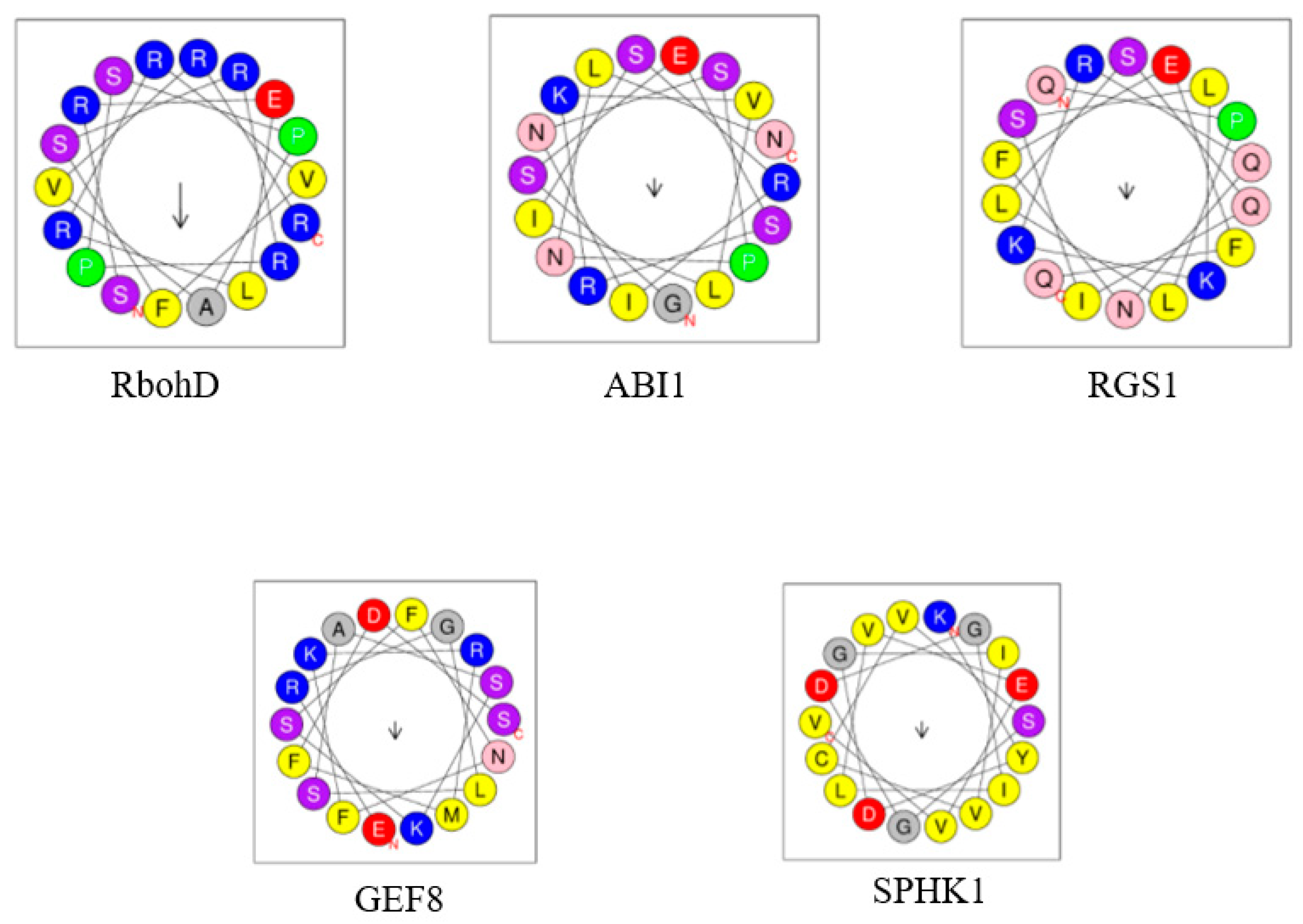
Figure 1. A diagram representing key enzymatic steps of PA production involved in cell signaling. Phosphatidylcholine (PC) is shown as the principal substrate for PLD-generated PA. Signaling lipid mediators and their active products are highlighted in dark blue. Their non-signaling by-products or lipid precursors are shown in black. Phospholipases involved in DAG and PA metabolism are shown in light blue rectangles. Lipid kinases that phosphorylate DAG and PA are shown in violet rectangles. Lipid phosphatases that dephosphorylate DGPP and PA are shown in light red rectangles. Black arrows indicate pathways that generate signaling lipids; green arrows indicate pathways that generate by-products. Note that no PAK-coding gene is currently identified in Arabidopsis. Chol, choline; DGK, diacylglycerol kinase; DGPP, diacylglycerol pyrophosphate; IP3, inositol 1,4,5-trisphosphate; LPP, lipid phosphate phosphatase; NPC, nonspecific phospholipase C; PAK, phosphatidic acid kinase; PAP, phosphatidic acid phosphatase; PChol, phosphocholine; PC, phosphatidylcholine; PIdes, phosphoinositides, PI-PLC, phosphatidylinositol-specific phospholipase C; PLD, phospholipase D; R1, R2, fatty acids.
Ijms 23 03227 g001
Figure 2. Expression levels of genes encoding enzymes of PA signaling at different developmental stages of Arabidopsis thaliana obtained using “Genevestigator” database [55]. Data were collected from data based on classical array experiments (“AT_AFFY_ATH1” platform). The relative values of gene expression (percent of expression potential, log2 scale) are shown. Percent of expression potential denotes the relative transcript abundance given as percentage of the level in the developmental stage where the gene is expressed at maximum level. Color saturation corresponds to the specific level of gene expression. White color signifies an absence of data. Number of samples: germinated seed (515), seedling (2781), young rosette (830), developed rosette (2219), bolting (358), young flower (720), developed flower (1003), flowers and siliques (274), mature siliques (93), senescence (18). Gene identifiers are as follows: PLDα1 (At3g15730), PLDα2 (At1g52570), PLDα3 (At5g25370), PLDε (At1g55180), PLDβ1 (At2g42010), PLDβ2 (At4g00240), PLDγ1 (At4g11850), PLDγ2 (At4g11830), PLDγ3 (At4g11840), PLDδ (At4g35790), PLDζ1 (At3g16785), PLDζ2 (At3g05630); PI-PLC1 (At5g58670), PI-PLC2 (At3g08510), PI-PLC3 (At4g38530), PI-PLC4 (At5g58700), PI-PLC5 (At5g58690), PI-PLC6 (At2g40116), PI-PLC7 (At3g55940), PI-PLC8 (At3g47290), PI-PLC9 (At3g47220), PI-PLC10 (At4G34920), PI-PLC11 (At3G19310); NPC1 (At1g07230), NPC2 (At2g26870), NPC3 (At3g03520), NPC4 (At3g03530), NPC5 (At3g03540), NPC6 (At3g48610); DGK1 (At5g07920), DGK2 (At5g63770), DGK3 (At2g18730), DGK4 (At5g57690), DGK5 (At2g20900), DGK6 (At4g28130), DGK7 (At4g30340). Abbreviations of Arabidopsis developmental stages: G, germinated seed; S, seedling; YR, young rosette; DR, developed rosette; B, bolting; YF, young flower; DF, developed flower; FS, flowers and siliques; MS, mature siliques; S, senescence.
Ijms 23 03227 g002
Figure 4. Schematic representation of the PA signaling in plants. Hypothetical receptors lead to activation of lipid signaling enzymes. Key examples of PA-regulated signaling events in hormonal regulation are shown (for details, see the main text); also shown here are PA-induced changes of membrane curvature and subsequent membrane transport events. Headgroups of DAG in a membrane are shown in green; headgroups of PA in crimson; and headgroups of other lipids in pink color. ABI1, ABSCISIC ACID-INSENSITIVE 1, At, Arabidopsis thaliana, DAG, diacylglycerol, DGK, diacylglycerol kinase, GEF8, RopGEF8, guanine nucleotide exchange factor 8, GID, GIBBERELLIN INSENSITIVE DWARF1, NPC, non-specific phospholipase C, NPR1, NONEXPRESSOR OF PATHOGENESIS-RELATED GENES 1, Os, Oryza sativa, PA, phosphatidic acid, PI-PLC, phosphatidylinositol-specific phospholipase C; PLD, phospholipase D, PLs, phospholipids (e.g., phosphatidylcholine, phosphatidylethanolamine), RbohD/F, respiratory burst oxidase protein D/F, RGS1, Regulator of G protein Signaling 1, ROS, reactive oxygen species, SPHK, sphingosine kinase.
Ijms 23 03227 g004
Figure 5. A set of similarly regulated genes in ABA-treated Col-0 Arabidopsis plants and in mock-treated rgs1-2 knockout mutants obtained using “Genevestigator” database [55]. Experiments ID used to mine the data are as follows: ABA, AT-00433; rgs1-2, AT-00530. Changes in gene expression (log2-ratio) are color-coded according to the provided scale. Statistical treatment of the data was implemented by “Genevestigator” database. Original data resources [117,118].
Ijms 23 03227 g005
Table 1. Plant PA-binding proteins with characterized mechanism of PA binding and identified role in cell signaling.
PA TargetPA-Binding SiteEffect on ActivityRole in SignalingLipid Signaling PathwayNotes (PA Binding Specificity)Refs.
AtRbohD (At5g47910), AtRbohF (At1g64060), respiratory burst oxidase homolog protein D and FArg149, Arg150, between EF-hands and N-terminus (in cytosolic region), in the vicinity of phosphorylation motif (AtRbohD); residues 105 to 341 (AtRbohF)Stimulation at PM (AtRbohD)ABA signaling, ROS generation, ROS-dependent NO production, stomatal closureAtPLDα1di18:1PA, di18:2PA, 16:0–18:1 PA, 16:0–18:2PA, 18:0–18:2 PA), but not to di16:0PA, di18:0PA; AtRbohD has higher affinity to PA than AtRbohF[60]
AtSPHK1 (At4g21540), AtSPHK2 (At4g21534), sphingosine kinasesVSGDGI motif (AtSPHK1)Stimulation by promoting substrate bindingABA signaling, AtPLDα1 activation (for amplification of ABA response), stomatal closure, seed germination, root elongationAtPLDα1Strong binding to 18:1/18:1PA, 16:0/18:1PA, 16:0/18:2PA[71,72]
AtRopGEF8 (At3g24620), guanine nucleotide exchange factor 8Lys13, Lys18 in AtGEF8 PRONE domain and N-terminusStimulation of AtGEF8 activity towards small GTPase ROP7ABA signaling activation di18:2PA, di18:1PA, 16:0–18:1PA, 16:0–18:2PA, 16:0–18:1PA, 18:0–18:2PA[61]
AtRGS1 (At3g26090), regulator of G-protein signalingLys259 in RGS domainInhibition of GTPase-accelerating protein activity of AtRGS1ABA signaling activation, inhibition of seed germination, root growth and stomatal responses--[73]
AtMAP65-1 (At5g55230), a microtubule-associated proteinLys and Arg from 53KRK55 and 61KSR63 sequences, Ser-428 in 428SK429 sequence, out of microtubule binding or phosphorylation regionsPromotion of AtMAP65-1-induced microtubule-bundling activitySalt stress signaling enhances tubulin polymerization involving AtMAP65-1 increases salt tolerance by microtubule stabilizationAtPLDα116:0–18:1PA, 16:0–18:2PA, 18:0–18:1PA, 18:0–18:2PA, but not di18:0PA[74]
AtMPK6 (At2g43790), a mitogen-activated protein kinase 6-StimulationSalt stress signaling, salt tolerance by AtMPK6-dependent phosphorylation of AtSOS1AtPLDα1Significantly to di18:1PA, di18:2PA, 16:0–18:1PA, 16:0–18:2 PA, 18:0–18:2PA[75]
AtMKK7 (At1g18350), AtMKK9 (At1g73500), a mitogen-activated protein kinase kinases-Activation, translocation to PMSalt stress signaling, salt tolerance, maximization of the signal transduction efficiencyAtPLDα1Strongly to di18:1PA, 16:0–18:1PA, 16:0–18:2PA, 18:0–18:2PA[76]
AtMPK3 (At3g45640)/ AtMPK6 (At2g43790), a mitogen-activated protein kinases -Stimulation, interaction with and phosphorylation of hypoxia-related transcription factor RAP2/12Hypoxia stress signaling, translocation to nucleus and stimulation of hypoxia-related transcription factor RAP2/12, feedback inhibition of PA production, tolerance to submergenceAtPLDα1, AtPLDδ-[77]
AtPINOID (At2g34650), a protein kinaseLys119 to Lys121Stimulation, tethering to PMSalt stress signaling, salt tolerance, enhanced AtPINOID-dependent AtPIN2 phosphorylation and PM localization, activation of PIN2-mediated auxin efflux and redistributionAtPLDα1, AtPLDδdi18:1PA, di18:2PA, 16:0–18:1PA, 16:0–18:2PA, 18:0–18:1PA, 18:0-18:2PA[65]
AtSnRK2.4 (At1g10940) -Phosphorylation of SnRK2 targets, affects its interaction with PA and subcellular localization --16:0/18:1 PA[78]
OsAKT2 (Os05g35410), a potassium channelArg190/Lys191, Arg644/Arg645, Arg755/Arg756 in ANK domain, cytoplasmic domain, S4 voltage sensorInhibitionSeedlings growth under short-day conditions-Strongly to 16:0–18:2PA, much weaker to di16:0PA, di18:0PA, di18:1PA, di18:2PA, 16:0–18:1PA, 18:0–18:1PA, 18:0–18:2 PA[67,79]
OsGID1 (Os05g33730), a gibberellin receptorArg79, Arg82Tethering to nucleusGibberellin signaling, promotion of OsSLR1 (a suppressor DELLA protein) degradationOsPLDα6 [80]
Abbreviations: ABA, abscisic acid, Arg, arginine (R), Lys, lysine (K), NO, nitric oxide, PM, plasma membrane, PP2C, protein phosphatase 2C, ROS, reactive oxygen species.
Table 2. Co-expression analysis of Arabidopsis thaliana genes coding selected peptide receptors and enzymes of PA signaling performed in silico using “Genevestigator” database [55]. All statistical data were implemented and provided by “Genevestigator” database; 1031 samples, 25 anatomical parts, 691 perturbations (AT_mRNASeq_ARABI), 10615 samples, 3243 perturbations, 7 developmental stages (AT_AFFY_ATH1) were analyzed. Pearson’s correlation coefficient (score) was used for statistical analysis of gene co-expression. Database dataset denotes aggregated expression data according to a specific category (samples, anatomy, development, perturbation).
Bioactive PeptideReceptorCo-Expressed PA-Metabolizing EnzymePlatformDatabase DatasetStatistical Notes (Score)
PSKPSKR1PLDζ1AT_mRNASeq_ARABIanatomy0.9
PSK PSKR1 PLDγ1AT_AFFY_ATH1 perturbation 0.69
PSK PSKR1 PLDγ1AT_AFFY_ATH1sample0.69
CEP1CEPR1PI-PLC3AT_mRNASeq_ARABIanatomy 0.89
CLE25BAM2NPC6AT_AFFY_ATH1sample0.68
PIP1/2 RLK7DGK5AT_mRNASeq_ARABIperturbation 0.85
PIP1/2 RLK7 PLDγ1AT_mRNASeq_ARABIsample0.79
PEPPEPR1DGK1AT_AFFY_ATH1sample0.56
PEPPEPR2DGK5AT_mRNASeq_ARABIsample0.64
EPF2TMMNPC2AT_AFFY_ATH1development0.9
LURE1 MIDS1PI-PLC6AT_mRNASeq_ARABI perturbation0.61
LURE1 MIK2PLDγ1AT_mRNASeq_ARABI perturbation0.82
LURE1PRK8PLDα2AT_AFFY_ATH1sample0.82
LURE1PRK3DGK4AT_mRNASeq_ARABIperturbation 0.74
LURE1PRK5DGK4AT_mRNASeq_ARABIperturbation 0.74
GRIM REAPERPRK5DGK4AT_mRNASeq_ARABIperturbation 0.74
ESF1SSPDGK4AT_AFFY_ATH1sample0.75
RALF1BAK1PLDγ1AT_AFFY_ATH1 perturbation0.73
RALF1 BAK1PLDγ1AT_mRNASeq_ARABIsample0.8
RALF1 BAK1PLDγ1AT_mRNASeq_ARABIanatomy0.85
Ralf4/19 BUPS1DGK6AT_AFFY_AGsample0.9
Ralf4/19 BUPS1 PI-PLC6AT_mRNASeq_ARABIsample0.92
Ralf4/19BUPS2PI-PLC6AT_mRNASeq_ARABI sample0.95
Ralf1/17/23/
32/33
FERONIANPC1AT_mRNASeq_ARABI sample0.7
Ralf1/17/23/
32/33
FERONIAPLDα1AT_mRNASeq_ARABIanatomy0.88
Ralf1/17/23/
32/33
FERONIAPLDα1AT_mRNASeq_ARABIsample0.79
Ralf1/17/23/
32/33
FERONIAPLDα1AT_AFFY_ATH1sample0.5
Ralf4/19/34ANXUR2PLDα2AT_AFFY_AGRONOMICSsample0.83
CLE45SKM1PLDεAT_mRNASeq_ARABI anatomy0.61
CLE45SKM1 PI-PLC5AT_mRNASeq_ARABI anatomy0.57
CLE42PXL2PLDδAT_mRNASeq_ARABIperturbation0.71
TPD1EMS1PLDεAT_AFFY_AGRONOMICSperturbation 0.82
Abbreviations: BAK1, BRI1-associated kinase 1; BAM, BARELY ANY MERISTEM; BUPS, BUDDHA’S PAPER SEAL; CEP, C-TERMINALLY ENCODED PEPTIDE; CEPR, C-TERMINALLY ENCODED PEPTIDE RECEPTOR; CLE, CLAVATA3/EMBRYO SURROUNDING REGION; DGK, diacylglycerol kinase; EMS1, EXCESS MICROSPOROCYTES1; EPF2, EPIDERMAL PATTERNING FACTOR 2; ESF1, EMBRYO SURROUNDING FACTOR1; MDIS, MALE DISCOVERER; MIK, MDIS1-INTERACTING RECEPTOR-LIKE KINASE; NPC, non-specific phospholipase C; PEP, PLANT ELICITOR PEPTIDE; PEPR, PLANT ELICITOR PEPTIDE RECEPTOR; PIP, PAMP-INDUCED SECRETED PEPTIDES; PI-PLC, phosphatidylinositol-specific phospholipase C; PLD, phospholipase D; PRK, POLLEN-SPECIFIC RECEPTOR-LIKE KINASE; PSK, PHYTOSULFOKINE; PSRKR, PHYTOSULFOKINE RECEPTOR; PXL, PHLOEM INTERCALATED WITH XYLEM-LIKE; RALF, RAPID ALKALINIZATION FACTOR; RLK, RECEPTOR-LIKE KINASE; SKM, STERILITYREGULATING KINASE MEMBER; SSP, SHORT SUSPENSOR; TMM, TOO MANY MOUTHS; TPD1, TAPETUM DETERMINANT1.
Publisher’s Note: MDPI stays neutral with regard to jurisdictional claims in published maps and institutional affiliations.

