Next Article in Journal
Bladder Cancer Extracellular Vesicles Elicit a CD8 T Cell-Mediated Antitumor Immunity
Next Article in Special Issue
Molecular Mechanism of Induction of Bone Growth by the C-Type Natriuretic Peptide
Previous Article in Journal
Targeting Myotonic Dystrophy Type 1 with Metformin
Previous Article in Special Issue
Cellular and Molecular Alterations Underlying Abnormal Bone Growth in X-Linked Hypophosphatemia
 
 
Correction published on 27 May 2022, see Int. J. Mol. Sci. 2022, 23(11), 6020.
Article

Bone Mass and Osteoblast Activity Are Sex-Dependent in Mice Lacking the Estrogen Receptor α in Chondrocytes and Osteoblast Progenitor Cells

1
Institute of Orthopedic Research and Biomechanics, University Medical Center Ulm, 89081 Ulm, Germany
2
Institute of Comparative Molecular Endocrinology (CME), Ulm University, 89081 Ulm, Germany
*
Author to whom correspondence should be addressed.
Int. J. Mol. Sci. 2022, 23(5), 2902; https://doi.org/10.3390/ijms23052902
Submission received: 16 February 2022 / Revised: 3 March 2022 / Accepted: 4 March 2022 / Published: 7 March 2022 / Corrected: 27 May 2022
(This article belongs to the Special Issue Bone Development and Growth)

Abstract

:
While estrogen receptor alpha (ERα) is known to be important for bone development and homeostasis, its exact function during osteoblast differentiation remains unclear. Conditional deletion of ERα during specific stages of osteoblast differentiation revealed different bone phenotypes, which were also shown to be sex-dependent. Since hypertrophic chondrocytes can transdifferentiate into osteoblasts and substantially contribute to long-bone development, we aimed to investigate the effects of ERα deletion in both osteoblast and chondrocytes on bone development and structure. Therefore, we generated mice in which the ERα gene was inactivated via a Runx2-driven cyclic recombinase (ERαfl/fl; Runx2Cre). We analyzed the bones of 3-month-old ERαfl/fl; Runx2Cre mice by biomechanical testing, micro-computed tomography, and cellular parameters by histology. Male ERαfl/fl; Runx2Cre mice displayed a significantly increased cortical bone mass and flexural rigidity of the femurs compared to age-matched controls with no active Cre-transgene (ERαfl/fl). By contrast, female ERαfl/fl; Runx2Cre mice exhibited significant trabecular bone loss, whereas in cortical bone periosteal and endosteal diameters were reduced. Our results indicate that the ERα in osteoblast progenitors and hypertrophic chondrocytes differentially contributes to bone mass regulation in male and female mice and improves our understanding of ERα signaling in bone cells in vivo.

1. Introduction

Sex steroids are essential for skeletal development and regulate bone mass in both males and females [1]. During puberty, estrogen exerts suppressive effects on bone growth, whereas testosterone promotes periosteal bone formation resulting in greater bone size and a stronger skeleton in males [2]. Highlighting the important functions of both hormones on skeletal maintenance, declining levels of estrogen during menopause, and a reduction of both estrogen and androgens with increasing age in men are associated with bone loss and the development of osteoporosis [3,4,5]. The effects of estrogen on bone are mainly mediated via the two estrogen receptors (ERs), ERα and ERβ, which can both be also activated by androgens after aromatization into estrogens [6,7]. In bone, ERα and ERβ are expressed in chondrocytes, osteoblasts, osteocytes, and osteoclasts [8,9]. It is known that ERα is expressed at higher levels in cortical bone, whereas ERβ is strongly expressed in cancellous bone [10]. Because ERβ modulates the transcriptional activity of ERα and antagonizes many of ERα-driven effects [11,12], a “ying yang” relationship between both receptors is described [13]. ERα has become a major focus of research, since a human case report demonstrated that a point mutation in its gene resulted in osteoporosis and unfused growth plates in a 28-year-old man [14]. Furthermore, case reports from men that suffer from an aromatase deficiency together with preclinical studies demonstrated the importance of estrogens for male skeletal homeostasis [15,16,17,18]. In bone, ERα is mainly mediating the actions of estrogen and is therefore considered as more important for skeletal homeostasis [19,20]. Regarding sex-specific differences of ERα expression, we showed that during bone healing, ERα expression is lower in the fracture callus of male mice, specifically in hypertrophic chondrocytes, shown by immunohistochemical stainings and gene expression analysis of the fractured femurs [21]. Furthermore, in the literature, it is reported that cultured human osteoblasts from men and women respond differentially to estrogen or ERα/ERβ-specific agonists in a sex-specific manner [22,23]. These findings indicate that the expression levels of ERα/ERβ as well as their response to its ligand estrogen might be sex-specific. For this reason, we focused on the sex-specific role of ERα on bone.
The crucial role of ERs in skeletal health and growth was further confirmed by preclinical studies using global ERα- (ERα-KO) or ERβ-knockout (ERβ-KO) mice. In females and males, ERα-KO resulted in shorter long bones, whereas ERβ-KO showed no effects on longitudinal bone growth [16,24,25,26]. Additionally, ERα-KO mice displayed a decrease in cortical bone mineral density (BMD) and an increased trabecular bone volume in both sexes [27]. However, in a mouse line with a global deletion of the ERα, unspecific effects or compensatory mechanisms by the increased serum levels of testosterone [27] or the low expression levels of a chimeric truncated ERα isoform due to alternative splicing [28] cannot be ruled out. Therefore, cell-specific knockout models were generated using the Cre/loxP technology to elucidate the role of ERα on different stages of the osteoblast life cycle (Figure 1, Table 1). Under the control of different promotor genes particularly expressed by the respective cell type, the expression of a Cre-recombinase leads to a deletion of the ERα in this cell type.
Regarding the differentiation timeline of osteoblasts (Figure 1), first, Prx1 is expressed by skeletal stem cells followed by Runx2, which is upregulated in osteoblastic precursors together with Col1a1 [36]. When these precursors further differentiate, osterix (Osx1) is upregulated in pre-osteoblasts which then become mature osteoblasts, expressing high levels of osteocalcin (OC). Dmp1 is mainly expressed by osteocytes. ERα deletion in skeletal stem cells (Prx1-Cre) resulted in a reduction of cortical bone mass and BMD in both sexes, whereas the trabecular bone mass was unchanged [29]. When ERα was deleted in pre-osteoblasts (Osx1-Cre), female mice displayed a reduced cortical thickness. A deletion of the ERα during the matrix maturation phase in osteoblasts via the Col1a1-Cre model [29] had no effect on the bone phenotype, neither in male nor in female mice. However, ERα deletion in mature osteoblasts and osteocytes (OC-Cre) resulted in a reduction of cortical and trabecular bone mass in females but not in males [32,33,34].
Taken together, these studies have established that bone development is regulated by ERα in osteoblastic cells. Thereby, ERα-mediated effects are dependent on the osteoblast differentiation stage and on the sex [32,33,34]. However, these studies did not take into account that hypertrophic chondrocytes can undergo transdifferentiation into osteoblasts [37,38], which essentially contributes to bone formation in long bones [39,40]. Therefore, here we investigated the role of ERα in both osteoblast lineage-specific cells and hypertrophic chondrocytes by using the Runx2-Cre mouse model. The transcription factor Runx2 is essential for driving osteoblastogenesis and is expressed in hypertrophic chondrocytes, eventually transdifferentiating into osteoblasts (Figure 1) [41,42,43]. We crossbred Runx2-Cre with ERα floxed mice (ERαfl/fl) and analyzed the bone phenotype in both male and female mice to provide further insights into the regulatory functions of ERα in bone development.
We analyzed bone strength of male and female 3-month-old ERαfl/fl; Runx2Cre mice by biomechanical testing, bone mass and structure by μCT, and cellular parameters by histology. In male ERαfl/fl; Runx2Cre mice, bone strength and cortical thickness were significantly increased compared to age-matched floxed control littermates with no active Cre-transgene (ERαfl/fl). In addition, the periosteal diameter and the moment of inertia were found to be significantly increased, whereas the ERα knockout had no effect on trabecular bone. By contrast, female ERαfl/fl; Runx2Cre mice exhibited significant trabecular bone loss, whereas the cortical bone was mainly unaffected. Our results indicate a sex-specific role of the ERα in osteoblast progenitors and hypertrophic chondrocytes on bone mass in male and female mice.

2. Results

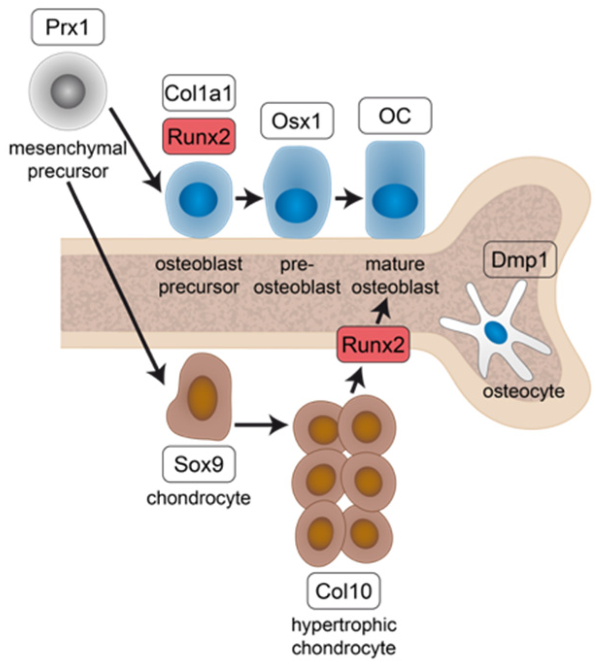
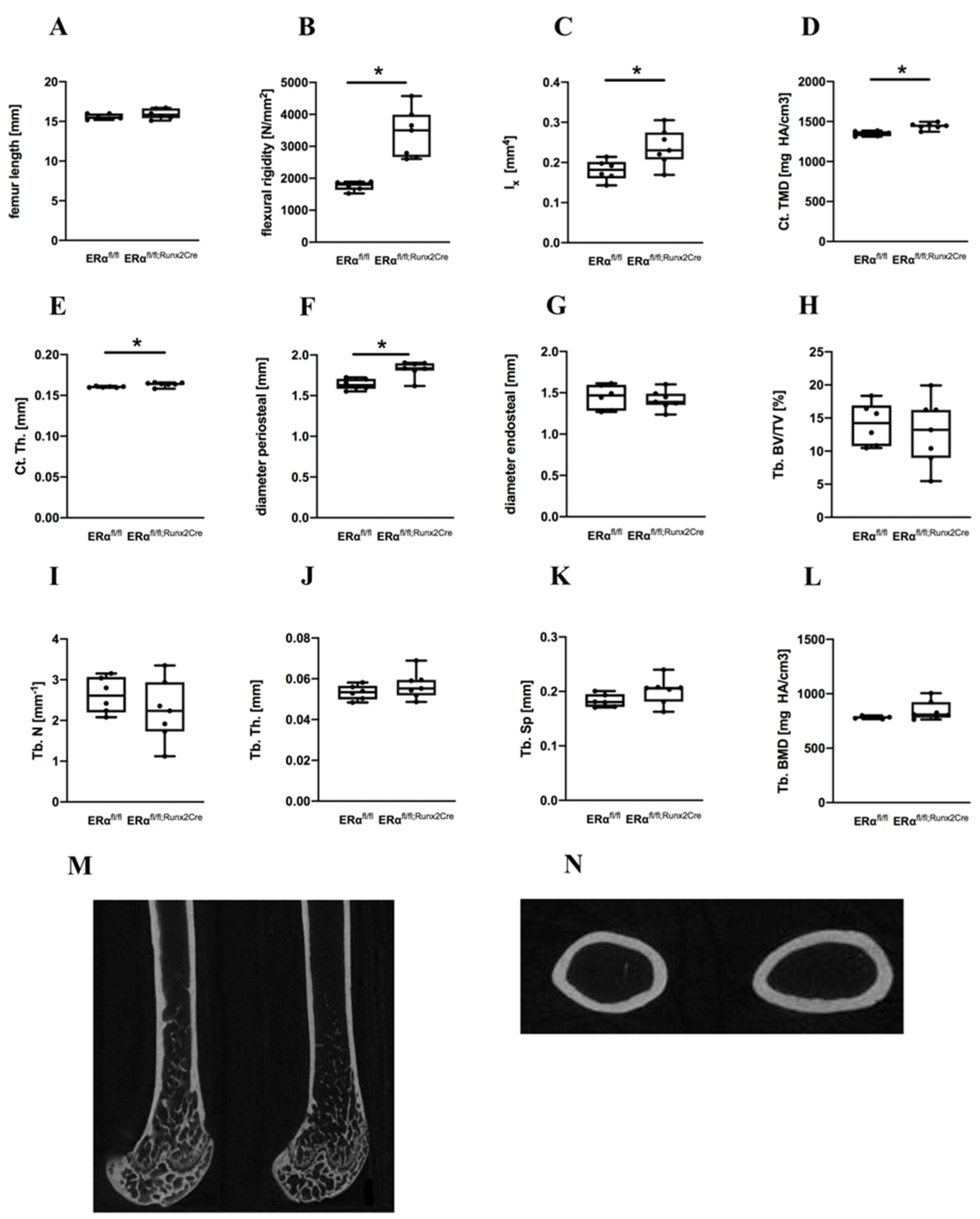
2.1. Influence of ERα Deletion in Osteoblast Progenitors on Bone in Male Mice

The femur lengths of male ERαfl/fl; Runx2Cre mice were not different compared to ERαfl/fl control animals (Figure 2A). To evaluate the flexural rigidity of the femurs and the bone structure of ERαfl/fl; Runx2Cre mice, biomechanical testing and μCT analyses were performed. The ERαfl/fl; Runx2Cre males displayed a significant increased flexural rigidity of the femurs compared to their control littermates (Figure 2B), resulting from a significantly higher moment of inertia and from a significantly increased cortical TMD and thickness (Figure 2C–E). The periosteal bone diameter was significantly increased in ERαfl/fl; Runx2Cre mice, whereas the endosteal diameter was unchanged between both genotypes (Figure 2F,G). In the trabecular compartment, all analyzed bone parameters including BV/TV, trabecular number, thickness, and separation, as well as trabecular BMD, were unaltered (Figure 2H–L).
Histomorphometric analyses of the metaphyseal region demonstrated that trabecular osteoblast number and surface were not affected by the ERα knockout (Figure 3). Similarly, trabecular osteoclast number and surface, as well as cortical osteocyte number, were unaltered (Figure 3). At the growth plate, osteoclast number as well as growth plate thickness were unchanged between both genotypes (Figure S1). Taken together, these results suggest that the deletion of ERα provoked an anabolic effect on the cortical bone of male mice.

2.2. Influence of ER α Deletion in Osteoblast Progenitors on Bone in Female Mice

In contrast to male ERαfl/fl; Runx2Cre mice, the femur lengths, flexural rigidity and the moment of inertia did not significantly differ in female ERαfl/fl; Runx2Cre mice compared to control animals (Figure 4A–C). Periosteal and endosteal diameters of the femurs were significantly reduced in female ERαfl/fl; Runx2Cre mice, whereas the cortical TMD and thickness were unaltered (Figure 4D–G).
In trabecular bone, the lack of the ERα in female mice led to severely reduced BV/TV, BMD, trabecular number, and thickness (Figure 4H–J,L,N and Figure S2 for epiphyseal bone). Consistently, trabecular separation was significantly increased in ERαfl/fl; Runx2Cre animals in comparison to their ERαfl/fl littermates (Figure 4K). Furthermore, a significant reduction of the osteoblast number and surface in the trabecular bone was observed in female ERαfl/fl; Runx2Cre mice, whereas osteoclast number and surface were unaltered (Figure 5A–H). In addition, we found a significantly reduced osteocyte number per bone area in the cortical bone of ERαfl/fl; Runx2Cre animals (Figure 5I). Furthermore, at the growth plate, osteoclast number and growth plate thickness were not affected by the knockout (Figure S1). Osteogenic differentiation experiments of primary osteoblasts cultures isolated from both genotypes revealed a significantly diminished osteogenic differentiation ability of cells derived from ERαfl/fl; Runx2Cre animals shown by a significantly lower gene expression of alkaline phosphatase (AP), osteocalcin (OC), bone sialoprotein (BSP), and runt-related transcription factor-2 (Runx2) in vitro and shown by weaker AP stainings (Figure 5J–Q). Collectively, these findings indicate that female ERαfl/fl; Runx2Cre animals exhibited a reduced trabecular bone mass and altered cortical bone architecture based on a reduced osteoblast differentiation.

3. Discussion

In the present study, we aimed to examine whether ERα signaling in osteoblast lineage-specific cells and hypertrophic chondrocytes are required for bone formation and development. To this end, mice with a deletion of the ERα in both cell types were generated by breeding Runx2-Cre and ERα floxed mice. The Runx2-Cre model allows to study the role of ERα not only in osteoblast progenitor cells directly derived from skeletal stem cells, but also in hypertrophic chondrocytes eventually transdifferentiating into osteoblasts during long bone development.
In male ERαfl/fl; Runx2Cre mice, flexural rigidity and cortical thickness of the femurs were significantly increased, while the trabecular bone mass was not affected.
In contrast to the cortical bone phenotype found by our study, others observed a decrease in cortical bone mass which could be attributed to the used Prx1-Cre model, leading to a deletion of the ERα very early in skeletal stem cells [29]. No effects on either bone mass or composition were observed when using the Col1a1-Cre model to delete the ERα [29]. However, similar to our results, cortical bone mass and biomechanical strength was increased in male mice when ERα was deleted in mature osteoblasts and osteocytes (OC-Cre) [33]. However, these mice also displayed an increased femoral length as well as a significantly higher cancellous BV/TV and greater trabecular thickness at the tibial metaphysis [33]. Most likely, these differences are due to the different Cre model that was used. In other studies that also used the OC-Cre model, no cortical or trabecular changes were found in 16-week-old male mice [34] or in 26-week old mice [31], whereas in 24-week-old male mice, tibial BV/TV and TbN were lower [34]. However, the genetic background of those mice was not reported [34] while in the study of Melville and in our study, C57BL/6 mice were used. The genetic background can strongly influence bone structure and mass [44], which might explain these inconsistent results. A study by Windahl and colleagues analyzed the bone phenotype of male mice with an ERα deletion in osteocytes, showing that the trabecular bone volume was significantly reduced while cortical bone was unaffected [35]. The authors propose that the ERα in osteocytes is necessary to facilitate estrogen signaling that may result in an enhanced osteoblast activity which might explain the reduced trabecular bone phenotype due to a reduced osteoblast activity [35].
To draw conclusions about the role of the ERα on hypertrophic chondrocytes, we compare our results to the data generated under the Col1a1-Cre promotor, because it is known that Col1a1 and Runx2 are simultaneously upregulated in osteoblast precursors. Runx2 is known to induce Col1a1 gene expression in osteoblasts and is further expressed by transdifferentiating hypertrophic chondrocytes [41]. In contrast to the male Col1a1-Cre mice, which did not display any bone phenotype, we observe in our Runx2-Cre animals a significantly increased cortical thickness and a higher flexural rigidity. These differences suggest that in Runx2-Cre mice, the additional deletion of the ERα in hypertrophic chondrocytes might have a crucial impact on the bone phenotype, indicating that the ERα under physiological conditions might have a regulatory role on the transdifferentiation of hypertrophic chondrocytes into osteoblasts and that this process might contribute to cortical bone accrual in male mice. Another reason for the increased cortical thickness might be the involvement of ERα in the estrogen-dependent suppression of periosteal bone mass accrual during bone development [1,5,33,45], which in addition might explain the higher cortical bone mass in male ERαfl/fl; Runx2Cre mice. Furthermore, testosterone might have a stronger positive impact on periosteal bone formation in male mice lacking ERα and possibly result in an increase in cortical thickness via androgen receptors [33]. These findings indicate that ERα mediates its effects on limiting periosteal expansion primarily during the stage of Runx2-positive osteoblast precursor cells, as well as during the OC-positive mature osteoblasts. In trabecular bone, the effect of ERα appears less prominent, because most of the studies do not report an altered trabecular bone microarchitecture in male mice.
ERα deletion in osteoblast progenitor cells and hypertrophic chondrocytes of female mice resulted in significant trabecular bone loss, whereas the bending stiffness of the femurs and the cortical thickness were unaltered.
When ERα deletion occurred at the stage of skeletal stem cells (Prx1-Cre) prior to Runx2 expression, cortical bone mass and BMD were reduced in female mice, whereas the trabecular bone mass was unchanged [29]. Others demonstrated a significantly reduced trabecular bone mass in the tibiae and spine of female Runx2-Cre knockout mice aged 16 weeks, confirming our results [30]. By contrast, in the Col1a1-Cre model, no effect on cortical or trabecular bone was observed [29]. When ERα was deleted at the stage of pre-osteoblasts (Osx1-Cre), female mice displayed a reduced cortical thickness and unchanged trabecular bone parameters. Other studies that have used the OC-Cre model to delete the ERα in mature osteoblasts and osteocytes demonstrated that the trabecular bone mass was significantly reduced in female ERα-KO mice together with an altered cancellous architecture in the proximal tibia, distal femur, and L5 vertebral body [31,32,33,34]. Additionally, they observed a significant decrease in the cortical area and cortical thickness. Interestingly, the ERα on osteocytes was not required for maintaining both trabecular and cortical bone mass in female mice [35].
In comparison to the Col1a1-Cre model, which showed no bone phenotype, our data suggest that the decreased trabecular bone mass of the female ERαfl/fl; Runx2Cre mice results from the reduced number of trabecular osteoblasts that were also found to be significantly reduced in their surface, which is a surrogate for their activity. We hypothesize that the presence of the ERα on hypertrophic chondrocytes might be crucial for the transdifferentiation into osteoblasts that might participate in the formation of trabecular bone. However, future studies are needed to characterize the role of ERα in transdifferentiation of hypertrophic chondrocytes in more detail. Furthermore, we found that primary osteoblasts isolated from female ERαfl/fl; Runx2Cre displayed a significantly reduced differentiation ability. This finding is consistent with one study showing that ERα-deficient osteoblast progenitors from the periosteum have a decreased differentiation potential [29]. In cortical bone of the female ERαfl/fl; Runx2Cre mice, periosteal and endosteal diameters as well as osteocyte numbers were significantly reduced. These findings confirm that the ERα in osteoblast precursors is involved in endosteal bone resorption [46] and explain the reduced endosteal diameter. Regarding the reduced periosteal diameter, it is known that the ERα is important for periosteal bone apposition in female mice [29], resulting in a lower periosteal diameter when ERα is deleted. However, the molecular pathways behind these findings need to be addressed in further studies.
Taken together, the functions of ERα appear to be highly dependent on the maturation status of osteoblasts, while it starts to exert its positive effects on the trabecular bone of female mice at the stage of Runx2-positive osteoblast progenitor cells and is additionally required in mature osteoblasts, but not osteocytes. In cortical bone, ERα might be involved at very early stages of osteoblast maturation (Prx1-Cre) as well as in mature osteoblasts (OC-Cre) in female mice.
When the ERα was specifically deleted in chondrocytes by using a Col2a1-Cre model, no effects on skeletal growth or bone mineral density was reported [47] suggesting that the cartilage-specific ERα might not influence bone metabolism and structure. In our Runx2-Cre model, transdifferentiating chondrocytes and osteoblastic cells are affected by the deletion and we observed a bone phenotype in both males and females, suggesting a sex-specific role of ERα in hypertrophic chondrocytes.
One limitation of our study is that we only analyzed the bone phenotype at the age of 12 weeks, whereas it would be of interest to also include also younger and older mice to investigate bone growth and development in more detail. Furthermore, to study the contribution of estrogen-dependent or -independent signaling to the observed bone phenotype in female ERαfl/fl; Runx2Cre mice, it would be of interest to investigate the bone response to ovariectomy in these mice.
In conclusion, we demonstrated that the ERα in osteoblast progenitors and hypertrophic chondrocytes is crucial for mediating anabolic effects on trabecular bone in female mice, whereas in males it has different effects by limiting the estrogen-dependent periosteal apposition in cortical bone. This suggests a sex-specific role of the ERα that is either promoting or repressing bone apposition at different sites of the long bones. The ERα in hypertrophic chondrocytes might be critically involved in the transdifferentiation towards osteoblasts, thereby regulating bone mass. These findings might be relevant also for human bone development, because case reports have already demonstrated the importance of estrogens and ERα signaling for human skeletal homeostasis [15,16,17,18].

4. Materials and Methods

4.1. Animal Care and Animal Models

All experiments were performed according the ARRIVE guidelines and were approved by the local ethical committee (o.135-9, Regierungspräsidium Tübingen, Germany). Osteoblast progenitor cell-specific ERα-KO mice (Tg(Runx2-cre)1Jtuc x Esr1tm1.2Mma) were generated by crossing Runx2-Cre with ERαfl/fl mice on a C57BL/6 background. ERαfl/fl; Runx2Cre mice were shown to lack the ERα in cells of the osteogenic lineage and in hypertrophic chondrocytes [48]. Age-matched, floxed littermates (ERαfl/fl) with no active Cre-transgene were used as control. All animals were housed in groups of up to five mice per cage with a 14-h light, 10-h dark rhythm and received water ad libitum as well as a standard mouse feed (ssniff R/M-H, V1535-300; Ssniff, Soest, Germany). At the age of 12 weeks, mice were sacrificed using an isoflurane overdose. Mouse genotyping was conducted by lysed tail PCR using the primers: 5′-CCA GGA AGA CTG CCA GAA GG-3′, 5′-TGG CTT GCA GGT ACA GGA G-3′ and 5′-GGA GCT GCC GAG TCA ATA AC-3′ to detect the Cre transgene, whereas the ERα loxP sites were detected using the primers: 5′-TAG GCT TTG TCT CGC TTT CC-3′, 5′-CCC TGG CAA GAT AAG ACA GC-3′ and 5′-AGG AGA ATG AGG TGG CAC AG-3′.

4.2. Biomechanical Testing

To evaluate flexural rigidity of the femora, a non-destructive three-point bending test was performed on freshly prepared samples as described previously [49]. Briefly, an axial load with a maximum of 4 N was applied on the cranial side at the diaphyseal region of the bone using a material-testing machine (1454, Zwick GmbH Co KG, Ulm, Germany). The flexural rigidity was calculated from the slope of the load-deflection curve.

4.3. μCT Analysis

Femora were fixed in 4% phosphate-buffered formaldehyde solution and scanned using a μCT scanning device (Skyscan 1172 v1.5; Skyscan, Kontich, Belgium) at a resolution of 8 μm using a peak voltage of 50 kV and 200 μA. Analyses and calibration steps were performed according to the guidelines of the American Society for Bone and Mineral Research [50] and volumes of interest were chosen as previously described [51]. Briefly, cortical bone was evaluated in a volume of interest (VOI) of 168 μm length within the mid-diaphyseal region. The trabecular bone was assessed 200 μm proximal of the metaphyseal growth plate over a length of 280 μm in the distal femur, excluding the cortex. For assessing the epiphyseal femur, the trabecular bone was evaluated distal from the growth plate by a defined cylindrical VOI.
Within each scan, two phantoms with a defined density of hydroxyapatite (250 and 750 mg hydroxyapatite/cm3) were included to determine the bone mineral density. To distinguish between mineralized and nonmineralized tissue, thresholds for cortical bone (641.9 mg hydroxyapatite/cm3) and trabecular bone (394.8 mg hydroxyapatite/cm3) were used [52]. Analyses were performed by means of Skyscan software (NRecon v1.7.1.0, DataViewer v1.5.1.2, and CTAn v1.17.2.2).

4.4. Histomorphometry

Femora were fixed in 4% phosphate-buffered formaldehyde solution, decalcified using 20% EDTA (pH 7.2 to 7.4) for at least 14 days, and embedded in paraffin. The number and surface of osteoclasts was evaluated using tartrate-resistant alkaline phosphatase (TRAP) staining, whereas osteoblast number and surface were analyzed using Toluidine blue-stained sections. Osteoclasts were defined as TRAP-positive cells with two or more nuclei, directly located on the bone surface with a visible resorption lacunae between the bone surface and the cell. Osteoblasts were identified as cubic-shaped and Toluidine blue-positive cells with visible cytoplasm, directly located on the bone surface. Both osteoclasts and osteoblast parameters were evaluated at the femur metaphysis and normalized to bone surface using the Osteomeasure system (OsteoMeasure system v4.1.0.0; OsteoMetrics, Inc., Decatur, GA, USA). Growth plate thickness was analyzed in Safranin O-stained sections using the Osteomeasure system. Osteoclast activity at the growth plate was assessed within a defined region of 480 μm × 350 μm and osteoclasts were identified as multinucleated cells either laying directly on bone or cartilage tissue. Images were obtained using a Leica microscope (DMI 6000B). All analyses were performed according to American Society for Bone and Mineral Research standards [53].

4.5. Cultivation of Primary Mouse Osteoblasts

Primary osteoblasts were isolated from long bones of 12-week–old mice, as previously described [54]. Briefly, long bones were harvested, and bone marrow was flushed out by centrifugation. Bone diaphyses were cut into pieces and subjected to a 60-min collagenase digestion using 125 U/mL collagenase type VI (Sigma-Aldrich, Taufkirchen, Germany). For osteoblast expansion, bone chips were cultivated in modified minimal essential medium (DMEM; Biochrom, Berlin, Germany) containing, 1% l-glutamine (PAN-Biotech, Aidenbach, Germany), 100 U/mL penicillin/streptomycin, and 0.5% amphotericin B (Fungizone), supplemented with 15% fetal calf serum (all from Gibco, Darmstadt, Germany) at 37 °C under 5% CO2. For the experiments, osteogenic differentiation was induced in the presence of 0.2 mmol/L ascorbate-2-phosphate and 10 mmol/L β-glycerophosphate (both from Sigma-Aldrich). Differentiation medium was replaced twice per week. Alkaline phosphatase stainings were performed by using a commercially available kit (Sigma-Aldrich) to confirm osteogenic differentiation after 14 days. In addition, mRNA samples were obtained after 10 days of osteogenic differentiation. Passage 2 osteoblasts were used for all experiments, which were performed in triplicates at least two times. The SensiFAST SYBR Hi-ROX One-Step Kit (Bioline, Memphis, TN, USA) was used according to the manufacturer’s guidelines to perform quantitative PCR. B2M (F: 5′-ATACGCCTGCAGAGTTAAGCA-3′, R: 5′-TCACAT GTCTCGATCCCAGT-3′) was used as the housekeeping gene. Relative gene expression of AP (F: 5′-GCT GAT CAT TCC CAC GTT TT-3′, R: 5′-GAG CCA GAC CAA AGA TGG AG-3′), OC (F: 5′-CTT GGT GCA CAC CTA GCA GA-3′, R: 5′-ACC TTA TTG CCC TCC TGC TT-3′, BSP (F: 5′-GAA GCA GGT GCA GAA GGA AC-3′, R: 5′-GAA ACC CGT TCA GAA GGA CA-3′), Runx2 (F: 5′-CCA CCA CTC ACT ACC ACA CG-3′, R: 5′-CAC TCT GGC TTT GGG AAG AG-3′), Col1 (F: 5′-GCT GCA TAC ACA ATG GCC TA-3′, R: 5′-TCA AGC ATA CCT CGG GTT TC-3′), and ERα (F: 5′-TCC GGC ACA TGA GTA ACA AA-3′, R: 5′-CCA GGA GCA GGT CAT AGA GG-3′) was calculated using the delta-delta CT method.

4.6. Statistics

Group size was n = 6–8 per group for bone phenotyping. Data were tested for normal distribution using the Shapiro–Wilk test; most data sets were normally distributed. Statistical testing was performed using t-tests with GraphPad Prism 8.4.3 (GraphPad Software, La Jolla, CA, USA). The level of significance was set at p ≤ 0.05. Results are presented as box-and-whisker plots (with median and interquartile range) from maximum to minimum, showing all data points.

Supplementary Materials

The following are available online at https://www.mdpi.com/article/10.3390/ijms23052902/s1.

Author Contributions

Conceptualization, L.S., A.I. and M.H.-L.; methodology, L.S.; software, L.S.; validation, L.S. and J.B.; formal analysis, L.S.; investigation, L.S. and J.B.; resources, A.I. and M.H.-L.; data curation, L.S.; writing—original draft preparation, L.S.; writing—review and editing, A.I. and M.H.-L.; visualization, L.S.; supervision, A.I., J.T. and M.H.-L.; project administration, A.I., J.T. and M.H.-L.; funding acquisition, A.I. and M.H.-L. All authors have read and agreed to the published version of the manuscript.

Funding

This research was funded by the German Research Foundation (DFG, Project-ID HA 8470/1-1; 419) and by the Collaborative Research Center (CRC1149, Project-ID 251293561).

Institutional Review Board Statement

The animal study was protocol was approved by the local.

Informed Consent Statement

Ethical committee Regierungspräsidium Tübingen, Germany (o.135-9, 10 March 2020).

Data Availability Statement

Not applicable.

Acknowledgments

We thank Kristina Karremann, Tina Hieber, Andrea Böhmler, and Iris Baum for excellent technical assistance.

Conflicts of Interest

The authors declare no conflict of interest. The funders had no role in the design of the study; in the collection, analyses, or interpretation of data; in the writing of the manuscript, or in the decision to publish the results.

References

  1. Riggs, B.L.; Khosla, S.; Melton, L.J. Sex steroids and the construction and conservation of the adult skeleton. Endocr. Rev. 2002, 23, 279–302. [Google Scholar] [CrossRef] [PubMed]
  2. Callewaert, F.; Sinnesael, M.; Gielen, E.; Boonen, S.; Vanderschueren, D. Skeletal sexual dimorphism: Relative contribution of sex steroids, GH-IGF1, and mechanical loading. J. Endocrinol. 2010, 207, 127–134. [Google Scholar] [CrossRef] [PubMed]
  3. Vanderschueren, D.; Laurent, M.R.; Claessens, F.; Gielen, E.; Lagerquist, M.; Vandenput, L.; Börjesson, A.E.; Ohlsson, C. Sex Steroid Actions in Male Bone. Endocr. Rev. 2014, 35, 906–960. [Google Scholar] [CrossRef] [PubMed]
  4. Khosla, S. Update on Estrogens and the Skeleton. J. Clin. Endocrinol. Metab. 2010, 95, 3569–3577. [Google Scholar] [CrossRef]
  5. Manolagas, S.C.; O’Brien, C.A.; Almeida, M. The role of estrogen and androgen receptors in bone health and disease. Nat. Rev. Endocrinol. 2013, 9, 699–712. [Google Scholar] [CrossRef]
  6. Callewaert, F.; Boonen, S.; Vanderschueren, D. Sex steroids and the male skeleton: A tale of two hormones. Trends Endocrinol. Metab. 2010, 21, 89–95. [Google Scholar] [CrossRef]
  7. Vanderschueren, D.; Vandenput, L.; Boonen, S.; Lindberg, M.K.; Bouillon, R.; Ohlsson, C. Androgens and Bone. Endocr. Rev. 2004, 25, 389–425. [Google Scholar] [CrossRef]
  8. Almeida, M.; Laurent, M.R.; Dubois, V.; Claessens, F.; O’Brien, C.A.; Bouillon, R.; Vanderschueren, D.; Manolagas, S.C. Estrogens and Androgens in Skeletal Physiology and Pathophysiology. Physiol. Rev. 2017, 97, 135–187. [Google Scholar] [CrossRef]
  9. Manolagas, S.C.; Bellido, T.; Jilka, R.L. Sex Steroids, Cytokines and the Bone Marrow: New Concepts on the Pathogenesis of Osteoporosis. Physiol. Rev. 2007, 191, 187–202. [Google Scholar] [CrossRef]
  10. Bord, S.; Horner, A.; Beavan, S.; Compston, J. Estrogen Receptors? and? Are Differentially Expressed in Developing Human Bone 1. J. Clin. Endocrinol. Metab. 2001, 86, 2309–2314. [Google Scholar] [CrossRef]
  11. Khalid, A.B.; Krum, S.A. Estrogen receptors alpha and beta in bone. Bone 2016, 87, 130–135. [Google Scholar] [CrossRef] [PubMed]
  12. Liu, M.-M.; Albanese, C.; Anderson, C.M.; Hilty, K.; Webb, P.; Uht, R.M.; Price, R.H.; Pestell, R.G.; Kushner, P.J. Opposing action of estrogen receptors α and β on cyclin D1 gene expression. J. Biol. Chem. 2002, 277, 24353–24360. [Google Scholar] [CrossRef] [PubMed]
  13. Lindberg, M.K.; Movérare, S.; Skrtic, S.; Gao, H.; Dahlman-Wright, K.; Gustafsson, J.-A.; Ohlsson, C. Estrogen Receptor (ER)-β Reduces ERα-Regulated Gene Transcription, Supporting a “Ying Yang” Relationship between ERα and ERβ in Mice. Mol. Endocrinol. 2003, 17, 203–208. [Google Scholar] [CrossRef] [PubMed]
  14. Smith, E.P.; Boyd, J.; Frank, G.R.; Takahashi, H.; Cohen, R.M.; Specker, B.; Williams, T.C.; Lubahn, D.B.; Korach, K. Estrogen Resistance Caused by a Mutation in the Estrogen-Receptor Gene in a Man. N. Engl. J. Med. 1994, 331, 1056–1061. [Google Scholar] [CrossRef]
  15. Vanderschueren, D.; Van Herck, E.; Nijs, J.; Ederveen, A.G.H.; De Coster, R.; Bouillon, R. Aromatase Inhibition Impairs Skeletal Modeling and Decreases Bone Mineral Density in Growing Male Rats*. Endocrinology 1997, 138, 2301–2307. [Google Scholar] [CrossRef]
  16. Vidal, O.; Lindberg, M.K.; Hollberg, K.; Baylink, D.J.; Andersson, G.; Lubahn, D.B.; Mohan, S.; Gustafsson, J.Å.; Ohlsson, C. Estrogen receptor specificity in the regulation of skeletal growth and maturation in male mice. Proc. Natl. Acad. Sci. USA 2000, 97, 5474–5479. [Google Scholar] [CrossRef]
  17. Bouillon, R.; Bex, M.; Vanderschueren, D.; Boonen, S. Estrogens Are Essential for Male Pubertal Periosteal Bone Expansion. J. Clin. Endocrinol. Metab. 2004, 89, 6025–6029. [Google Scholar] [CrossRef]
  18. Rochira, V.; Zirilli, L.; Madeo, B.; Aranda, C.; Caffagni, G.; Fabre, B.; Montangero, V.E.; Roldan, E.; Maffei, L.; Carani, C. Skeletal effects of long-term estrogen and testosterone replacement treatment in a man with congenital aromatase deficiency: Evidences of a priming effect of estrogen for sex steroids action on bone. Bone 2007, 40, 1662–1668. [Google Scholar] [CrossRef]
  19. Gennari, L.; Merlotti, D.; Becherini, L.; Martini, G.; De Paola, V.; Calabrò, A.; Nuti, R. Estrogen Receptor Gene Polymorphisms and the Genetics of Osteoporosis: A HuGE Review. Am. J. Epidemiol. 2005, 161, 307–320. [Google Scholar] [CrossRef]
  20. Sims, N.; Clément-Lacroix, P.; Minet, D.; Fraslon-Vanhulle, C.; Gaillard-Kelly, M.; Resche-Rigon, M.; Baron, R. A functional androgen receptor is not sufficient to allow estradiol to protect bone after gonadectomy in estradiol receptor–deficient mice. J. Clin. Investig. 2003, 111, 1319–1327. [Google Scholar] [CrossRef]
  21. Haffner-Luntzer, M.; Fischer, V.; Ignatius, A. Differences in Fracture Healing Between Female and Male C57BL/6J Mice. Front. Physiol. 2021, 12, 2494. [Google Scholar] [CrossRef] [PubMed]
  22. Somjen, D.; Katzburg, S.; Lieberherr, M.; Hendel, D.; Yoles, I. DT56a stimulates gender-specific human cultured bone cells in vitro. J. Steroid Biochem. Mol. Biol. 2006, 98, 90–96. [Google Scholar] [CrossRef] [PubMed]
  23. Somjen, D.; Katzburg, S.; Sharon, O.; Knoll, E.; Hendel, D.; Stern, N. Sex specific response of cultured human bone cells to ERα and ERβ specific agonists by modulation of cell proliferation and creatine kinase specific activity. J. Steroid Biochem. Mol. Biol. 2011, 125, 226–230. [Google Scholar] [CrossRef]
  24. Couse, J.F.; Korach, K.S. Estrogen Receptor Null Mice: What Have We Learned and Where Will They Lead Us? Endocr. Rev. 1999, 20, 358–417. [Google Scholar] [CrossRef] [PubMed]
  25. Vidal, O.; Lindberg, M.; Sävendahl, L.; Lubahn, D.; Ritzen, E.; Gustafsson, J.; Ohlsson, C. Disproportional Body Growth in Female Estrogen Receptor-α-Inactivated Mice. Biochem. Biophys. Res. Commun. 1999, 265, 569–571. [Google Scholar] [CrossRef]
  26. Lindberg, M.K.; Alatalo, S.L.; Halleen, J.M.; Mohan, S.; Gustafsson, J.; Ohlsson, C. Estrogen receptor specificity in the regulation of the skeleton in female mice. J. Endocrinol. 2001, 171, 229–236. [Google Scholar] [CrossRef] [PubMed]
  27. Sims, N.; Dupont, S.; Krust, A.; Clement-Lacroix, P.; Minet, D.; Resche-Rigon, M.; Gaillard-Kelly, M.; Baron, R. Deletion of estrogen receptors reveals a regulatory role for estrogen receptors-β in bone remodeling in females but not in males. Bone 2002, 30, 18–25. [Google Scholar] [CrossRef]
  28. Couse, J.F.; Curtis, S.W.; Washburn, T.F.; Golding, T.S.; Lubahn, D.B.; Korach, K.S.; Lindzey, J.; Smithies, O. Analysis of transcription and estrogen insensitivity in the female mouse after targeted disruption of the estrogen receptor gene. Mol. Endocrinol. 1995, 9, 1441–1454. [Google Scholar] [CrossRef]
  29. Almeida, M.; Iyer, S.; Martin-Millan, M.; Bartell, S.M.; Han, L.; Ambrogini, E.; Onal, M.; Xiong, J.; Weinstein, R.S.; Jilka, R.L.; et al. Estrogen receptor-α signaling in osteoblast progenitors stimulates cortical bone accrual. J. Clin. Investig. 2012, 123, 394–404. [Google Scholar] [CrossRef]
  30. Seitz, S.; Keller, J.; Schilling, A.F.; Jeschke, A.; Marshall, R.P.; Stride, B.D.; Wintermantel, T.; Beil, F.T.; Amling, M.; Schutz, G.; et al. Pharmacological Estrogen Administration Causes a FSH-Independent Osteo-Anabolic Effect Requiring ER Alpha in Osteoblasts. PLoS ONE 2012, 7, e50301. [Google Scholar] [CrossRef]
  31. Rooney, A.M.; Ayobami, O.O.; Kelly, N.H.; Schimenti, J.C.; Ross, F.P.; van der Meulen, M.C. Bone mass and adaptation to mechanical loading are sexually dimorphic in adult osteoblast-specific ERα knockout mice. Bone 2022, 158, 116349. [Google Scholar] [CrossRef] [PubMed]
  32. Melville, K.M.; Kelly, N.H.; Khan, S.; Schimenti, J.C.; Ross, F.P.; Main, R.P.; van der Meulen, M.C.H. Female Mice Lacking Estrogen Receptor-Alpha in Osteoblasts Have Compromised Bone Mass and Strength. J. Bone Miner. Res. 2013, 29, 370–379. [Google Scholar] [CrossRef] [PubMed]
  33. Melville, K.M.; Kelly, N.H.; Surita, G.; Buchalter, D.B.; Schimenti, J.C.; Main, R.P.; Ross, F.P.; Van Der Meulen, M.C.H. Effects of Deletion of ERα in Osteoblast-Lineage Cells on Bone Mass and Adaptation to Mechanical Loading Differ in Female and Male Mice. J. Bone Miner. Res. 2015, 30, 1468–1480. [Google Scholar] [CrossRef]
  34. Määttä, J.A.; Büki, K.G.; Gu, G.; Alanne, M.H.; Vääräniemi, J.; Liljenbäck, H.; Poutanen, M.; Härkönen, P.; Väänänen, K. Inactivation of estrogen receptor α in bone-forming cells induces bone loss in female mice. FASEB J. 2012, 27, 478–488. [Google Scholar] [CrossRef] [PubMed]
  35. Windahl, S.H.; Borjesson, A.E.; Farman, H.H.; Engdahl, C.; Moverare-Skrtic, S.; Sjögren, K.; Lagerquist, M.; Kindblom, J.; Koskela, A.; Tuukkanen, J.; et al. Estrogen receptor- in osteocytes is important for trabecular bone formation in male mice. Proc. Natl. Acad. Sci. USA 2013, 110, 2294–2299. [Google Scholar] [CrossRef] [PubMed]
  36. Miron, R.J.; Zhang, Y.F. Osteoinduction: A review of old concepts with new standards. J. Dent. Res. 2012, 91, 736–744. [Google Scholar] [CrossRef]
  37. Park, J.; Gebhardt, M.; Golovchenko, S.; Perez-Branguli, F.; Hattori, T.; Hartmann, C.; Zhou, X.; Decrombrugghe, B.; Stock, M.; Schneider, H.; et al. Dual pathways to endochondral osteoblasts: A novel chondrocyte-derived osteoprogenitor cell identified in hypertrophic cartilage. Biol. Open 2015, 4, 608–621. [Google Scholar] [CrossRef]
  38. Bruderer, M.; Richards, R.G.; Alini, M.; Stoddart, M.J. Role and regulation of RUNX2 in osteogenesis. Eur. Cell Mater. 2014, 28, 269–286. [Google Scholar] [CrossRef]
  39. Zhou, X.; Von Der Mark, K.; Henry, S.; Norton, W.; Adams, H.; De Crombrugghe, B. Chondrocytes Transdifferentiate into Osteoblasts in Endochondral Bone during Development, Postnatal Growth and Fracture Healing in Mice. PLoS Genet. 2014, 10, e1004820. [Google Scholar] [CrossRef]
  40. Hu, D.P.; Ferro, F.; Yang, F.; Taylor, A.J.; Chang, W.; Miclau, T.; Marcucio, R.S.; Bahney, C.S. Cartilage to bone transformation during fracture healing is coordinated by the invading vasculature and induction of the core pluripotency genes. Development 2017, 144, 221–234. [Google Scholar] [CrossRef]
  41. Ducy, P.; Zhang, R.; Geoffroy, V.; Ridall, A.L.; Karsenty, G. Osf2/Cbfa1: A Transcriptional Activator of Osteoblast Differentiation. Cell 1997, 89, 747–754. [Google Scholar] [CrossRef]
  42. Otto, F.; Thornell, A.P.; Crompton, T.; Denzel, A.; Gilmour, K.C.; Rosewell, I.R.; Stamp, G.W.; Beddington, R.S.; Mundlos, S.; Olsen, B.R.; et al. Cbfa1, a Candidate Gene for Cleidocranial Dysplasia Syndrome, Is Essential for Osteoblast Differentiation and Bone Development. Cell 1997, 89, 765–771. [Google Scholar] [CrossRef]
  43. Komori, T.; Yagi, H.; Nomura, S.; Yamaguchi, A.; Sasaki, K.; Deguchi, K.; Shimizu, Y.; Bronson, R.; Gao, Y.-H.; Inada, M.; et al. Targeted Disruption of Cbfa1 Results in a Complete Lack of Bone Formation owing to Maturational Arrest of Osteoblasts. Cell 1997, 89, 755–764. [Google Scholar] [CrossRef]
  44. Sabsovich, I.; Clark, J.D.; Liao, G.; Peltz, G.; Lindsey, D.P.; Jacobs, C.R.; Yao, W.; Guo, T.-Z.; Kingery, W.S. Bone microstructure and its associated genetic variability in 12 inbred mouse strains: μCT study and in silico genome scan. Bone 2008, 42, 439–451. [Google Scholar] [CrossRef] [PubMed]
  45. Turner, R.T.; Colvard, D.S.; Spelsberg, T.C. Estrogen Inhibition of Periosteal Bone Formation in Rat Long Bones: Down-Regulation of Gene Expression for Bone Matrix Proteins*. Endocrinology 1990, 127, 1346–1351. [Google Scholar] [CrossRef]
  46. Emmanuelle, N.E.; Marie-Cécile, V.; Florence, T.; Jean-François, A.; Françoise, L.; Coralie, F.; Alexia, V. Critical role of estrogens on bone homeostasis in both male and female: From physiology to medical implications. Int. J. Mol. Sci. 2021, 22, 1–18. [Google Scholar]
  47. Börjesson, A.; Lagerquist, M.K.; Liu, C.; Shao, R.; Windahl, S.H.; Karlsson, C.; Sjögren, K.; Movérare-Skrtic, S.; Antal, M.C.; Krust, A.; et al. The role of estrogen receptor α in growth plate cartilage for longitudinal bone growth. J. Bone Miner. Res. 2010, 25, 2690–2700. [Google Scholar] [CrossRef]
  48. Steppe, L.; Krüger, B.T.; Tschaffon, M.E.A.; Fischer, V.; Tuckermann, J.; Ignatius, A.; Haffner-Luntzer, M. Estrogen Receptor α Signaling in Osteoblasts is Required for Mechanotransduction in Bone Fracture Healing. Front. Bioeng. Biotechnol. 2021, 9, 2355. [Google Scholar] [CrossRef]
  49. Röntgen, V.; Blakytny, R.; Matthys, R.; Landauer, M.; Wehner, T.; Göckelmann, M.; Jermendy, P.; Amling, M.; Schinke, T.; Claes, L.; et al. Fracture healing in mice under controlled rigid and flexible conditions using an adjustable external fixator. J. Orthop. Res. 2010, 28, 1456–1462. [Google Scholar] [CrossRef]
  50. Bouxsein, M.L.; Boyd, S.K.; Christiansen, B.A.; Guldberg, R.E.; Jepsen, K.J.; Müller, R. Guidelines for assessment of bone mi-crostructure in rodents using micro-computed tomography. J. Bone Miner. Res. 2010, 25, 1468–1486. [Google Scholar] [CrossRef]
  51. Haffner-Luntzer, M.; Kemmler, J.; Heidler, V.; Prystaz, K.; Schinke, T.; Amling, M.; Kovtun, A.; Rapp, A.E.; Ignatius, A.; Liedert, A. Inhibition of Midkine Augments Osteoporotic Fracture Healing. PLoS ONE 2016, 11, e0159278. [Google Scholar] [CrossRef] [PubMed]
  52. Morgan, E.F.; Mason, Z.D.; Chien, K.B.; Pfeiffer, A.J.; Barnes, G.L.; Einhorn, T.; Gerstenfeld, L.C. Micro-computed tomography assessment of fracture healing: Relationships among callus structure, composition, and mechanical function. Bone 2009, 44, 335–344. [Google Scholar] [CrossRef] [PubMed]
  53. Dempster, D.W.; Compston, J.E.; Drezner, M.K.; Glorieux, F.H.; Kanis, J.A.; Malluche, H.; Meunier, P.J.; Ott, S.M.; Recker, R.R.; Parfitt, A.M. Standardized nomenclature, symbols, and units for bone histomorphometry: A 2012 update of the report of the ASBMR Histomorphometry Nomenclature Committee. J. Bone Miner. Res. 2013, 28, 2–17. [Google Scholar] [CrossRef] [PubMed]
  54. Rapp, A.E.; Bindl, R.; Recknagel, S.; Erbacher, A.; Müller, I.; Schrezenmeier, H.; Ehrnthaller, C.; Gebhard, F.; Ignatius, A. Fracture Healing Is Delayed in Immunodeficient NOD/scid-IL2Rγcnull Mice. PLoS ONE 2016, 11, e0147465. [Google Scholar] [CrossRef]
Figure 1. Schematic representation of osteoblast and chondrocyte lineage commitment from mesenchymal precursors, highlighting the progressive stages of differentiation with their associated characteristic genes (white boxes) that are used to develop osteoblast-specific Cre mouse lines (Table 1). Prx1 = paired-related homeobox 1; Runx2 = runt-related transcription factor 2; Osx1 = osterix; OC = osteocalcin; Dmp1 = dentin matrix protein 1; Sox9 = SRY-Box Transcription Factor 9; Col 10 = collagen type 10.
Ijms 23 02902 g001
Figure 2. Bone-specific parameters of male ERαfl/fl and ERαfl/fl; Runx2Cre mice. (A) Femur length; (B) bending stiffness of the right femur; (C) moment of inertia; (D) cortical tissue mineral density (Ct. TMD); (E) cortical thickness (Ct. Th.); (F) diameter periosteal; and (G) diameter endosteal; (H) trabecular bone volume per tissue volume (Tb. BV/TV) (I) trabecular number (Tb. N). (J) Trabecular thickness (Tb. Th.); (K) trabecular separation (Tb. Sp). (L) Trabecular bone mineral density (Tb. BMD). (M) Representative μCT reconstructions of the right femurs representing the bones of ERαfl/fl mice (left panel) and of ERαfl/fl; Runx2Cre mice (right panel). (N) Representative cross-sectional μCT images of ERαfl/fl mice (left panel) and ERαfl/fl; Runx2Cre mice (right panel). Data are shown as box-and-whisker plot (with median and interquartile range) from maximum to minimum, showing all data points. * Indicates significant effects with p ≤ 0.05.
Ijms 23 02902 g002
Figure 3. Histomorphometrical analysis of male ERαfl/fl and ERαfl/fl; Runx2Cre mice. Representative paraffin sections of femora were stained with TRAP or Toluidine blue and histomorphometrical analysis was performed to determine (A) number of osteoblasts per bone perimeter (N.Ob/B.Pm), (B) osteoblast surface per bone surface (Ob.S/B.Pm), (C) number of osteoclasts per bone perimeter (N.Oc/B.Pm), and (D) osteoclast surface per bone surface (Oc.S/B.Pm). Representative images of TRAP-stained sections from (E) ERαfl/fl mice or (F) ERαfl/fl; Runx2Cre mice as well as Toluidine blue-stained sections from (G) ERαfl/fl mice or (H) ERαfl/fl; Runx2Cre mice. (I) Number of osteocytes per bone perimeter (N.Ot/B.Pm). Scale bar = 50 μm. Data are shown as box-and-whisker plot (with median and interquartile range) from maximum to minimum, showing all data points.
Ijms 23 02902 g003
Figure 4. Bone-specific parameters of female ERαfl/fl and ERαfl/fl; Runx2Cre mice. (A) Femur length; (B) bending stiffness of the right femur; (C) moment of inertia; (D) cortical tissue mineral density (Ct. TMD); (E) cortical thickness (Ct. Th.); (F) diameter periosteal; and (G) diameter endosteal; (H) trabecular bone volume per tissue volume (Tb. BV/TV) (I) trabecular number (Tb. N). (J) Trabecular thickness (Tb. Th.); (K) trabecular separation (Tb. Sp); (L) trabecular bone mineral density (Tb. BMD). (M) Representative μCT reconstructions of the right femora representing the bones of ERαfl/fl mice (left panel) and of ERαfl/fl; Runx2Cre mice (right panel). (N) Representative μCT images of trabecular bone microarchitecture in the distal femurs of ERαfl/fl mice (left panel) and ERαfl/fl; Runx2Cre mice (right panel). (O) Representative cross-sectional μCT images of ERαfl/fl mice (left panel) and ERαfl/fl; Runx2Cre mice (right panel). Data are shown as box-and-whisker plot (with median and interquartile range) from maximum to minimum, showing all data points. * Indicates significant effects with p ≤ 0.05.
Ijms 23 02902 g004
Figure 5. Histomorphometrical analysis of female ERαfl/fl and ERαfl/fl; Runx2Cre mice and differentiation ability of isolated primary osteoblasts. Representative paraffin sections of femurs were stained with TRAP or Toluidine blue and histomorphometrical analysis was performed to determine (A) number of osteoblasts per bone perimeter (N.Ob/B.Pm); (B) osteoblast surface per bone surface (ObS/B.Pm); (C) number of osteoclasts per bone perimeter (N.Oc/B.Pm); and (D) osteoclast surface per bone surface (OcS/B.Pm). Representative images of TRAP-stained sections from (E) ERαfl/fl mice or (F) ERαfl/fl; Runx2Cre mice as well as Toluidine blue-stained sections from (G) ERαfl/fl mice or (H) ERαfl/fl; Runx2Cre mice. (I) Number of osteocytes per bone perimeter (N.Ot/B.Pm). Primary osteoblasts were isolated from both ERαfl/fl and ERαfl/fl; Runx2Cre mice and relative AP expression was determined with (J) qPCR and by (P,Q) AP stainings in either differentiation medium at d10 (P) or control medium at d0 (Q). Relative expression of (K) BSP, (L) OC, (M) Runx2, (N) Col1, and (O) ERα was determined with qPCR. Scale bar = 50 μm. Data are shown as box-and-whisker plot (with median and interquartile range) from maximum to minimum, showing all data points. * Indicates significant effects with p ≤ 0.05.
Ijms 23 02902 g005
Table 1. Effects of osteoblast-specific ERα knockout models on cortical and trabecular bone in male and female mice.
Mouse ModelFemalesMales
CorticalTrabecularCorticalTrabecular
Osteoblast lineage
ERα fl/fl Prx1-Cre [29]↓ Ct. Th.↓ Ct. Th.
ERα fl/fl Runx2-Cre [30]N.a.N.a.N.a.
ERα fl/fl Runx2-Cre
ERα fl/fl Col1a1-Cre [29]
ERα fl/fl Osx1-Cre [29] N.a.N.a.
ERα fl/fl OC-Cre [31]
ERα fl/fl OC-Cre [32]N.a.N.a.
ERα fl/fl OC-Cre [33]
C57Bl/6 background
↑ Ct. Ar.
↑ Femoral length
↑ Tb. Th, BV/TV
ERα fl/fl OC-Cre [34]↓ Tb. BV
ERα fl/fl Dmp1-Cre [35]
↓ = reduced; ↑ = increased; ↔ = unaltered; N.a.= not available; Ct. Th. = cortical thickness, Ct. Ar. = cortical area; Tb. Th = trabecular thickness, BV/TV = relative bone volume; Tb. BV = trabecular bone volume.
Publisher’s Note: MDPI stays neutral with regard to jurisdictional claims in published maps and institutional affiliations.

