
Thermodynamic Studies of Interactions between Sertraline Hydrochloride and Randomly Methylated β-Cyclodextrin Molecules Supported by Circular Dichroism Spectroscopy and Molecular Docking Results

 , , and
Abstract
:1. Introduction
2. Results and Discussion
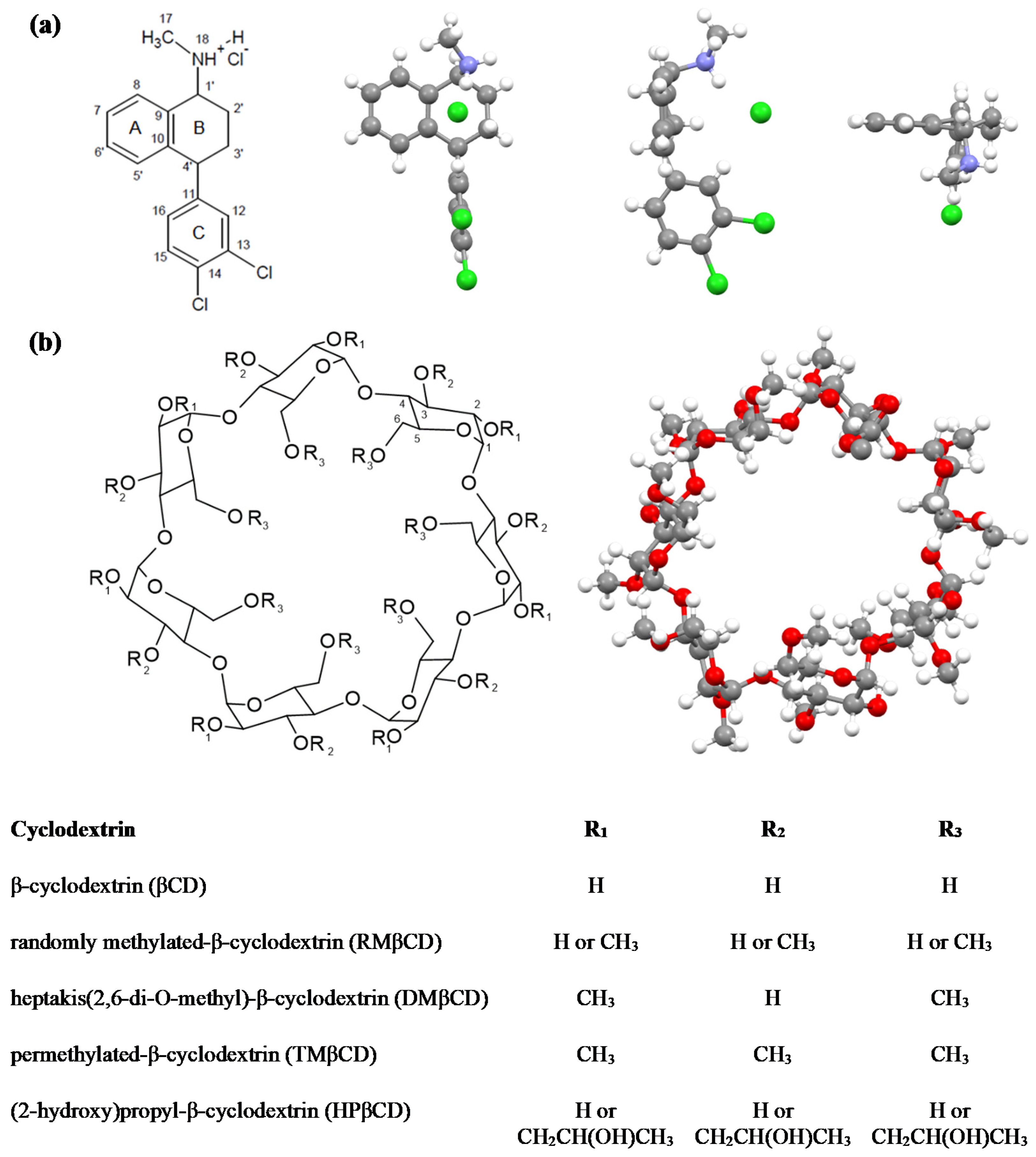
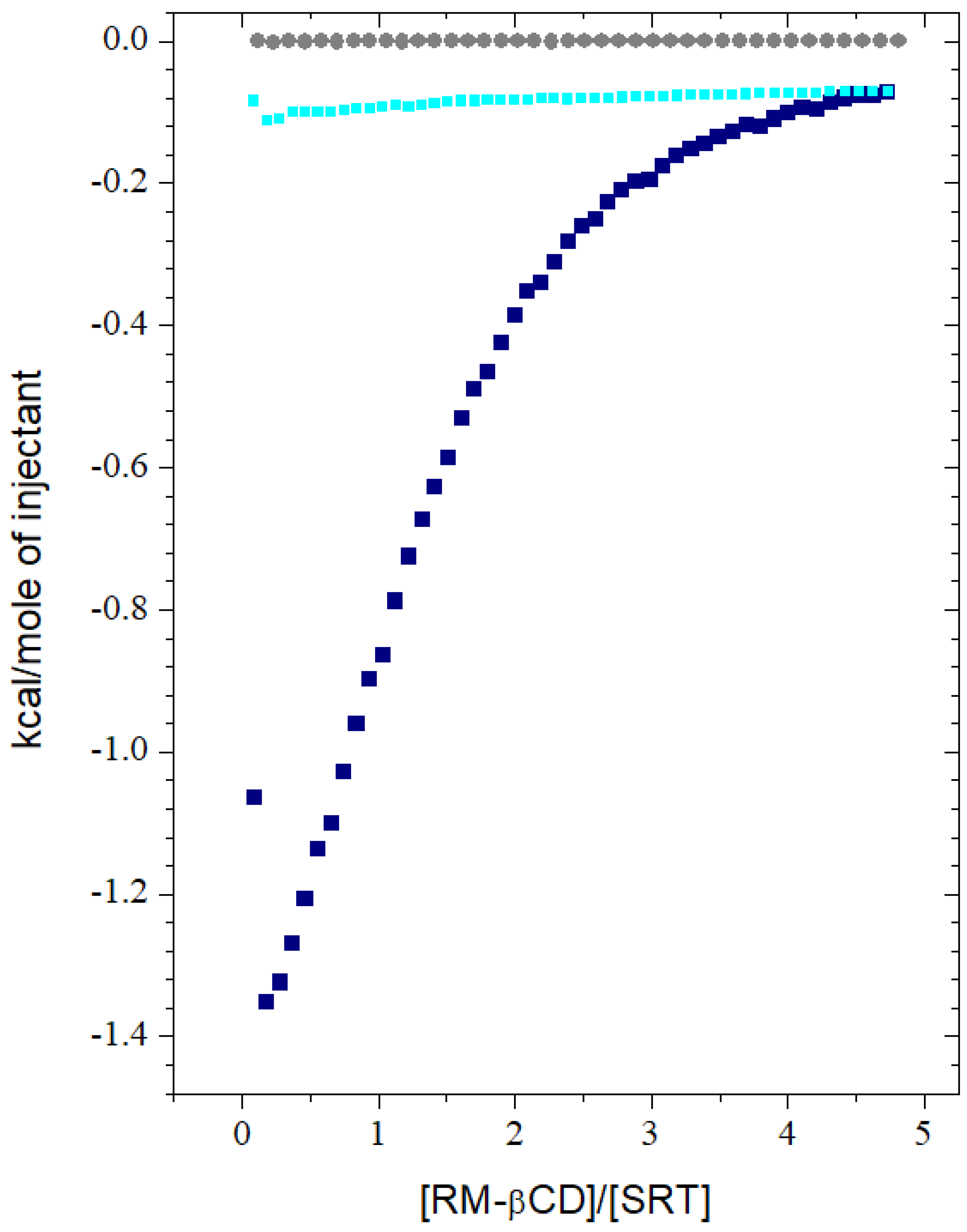
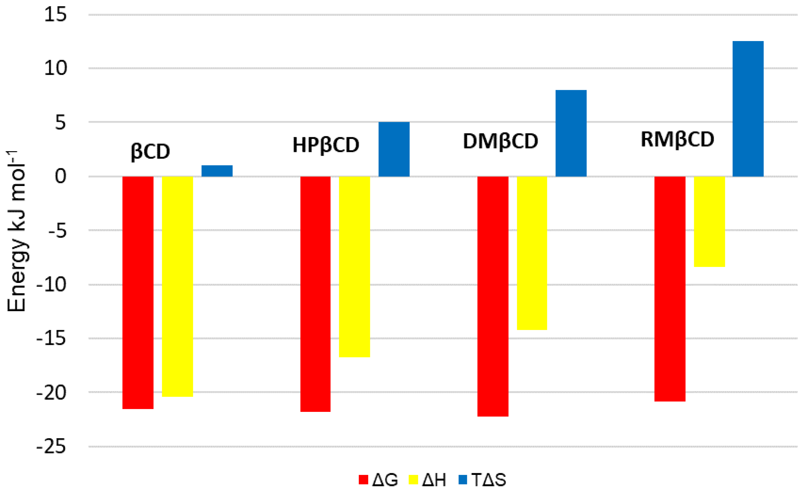
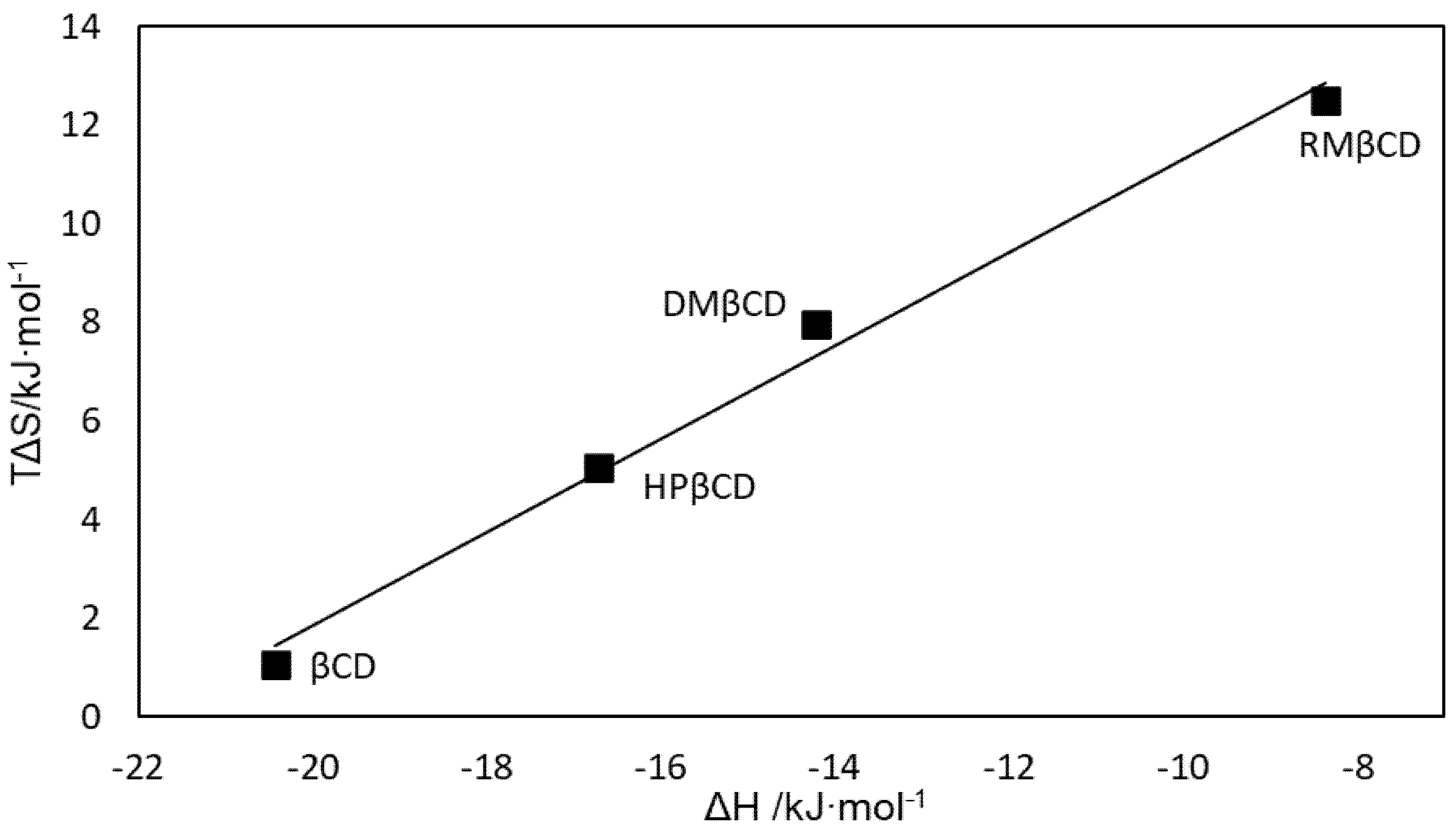
2.1. Isothermal Titration Calorimetry (ITC)
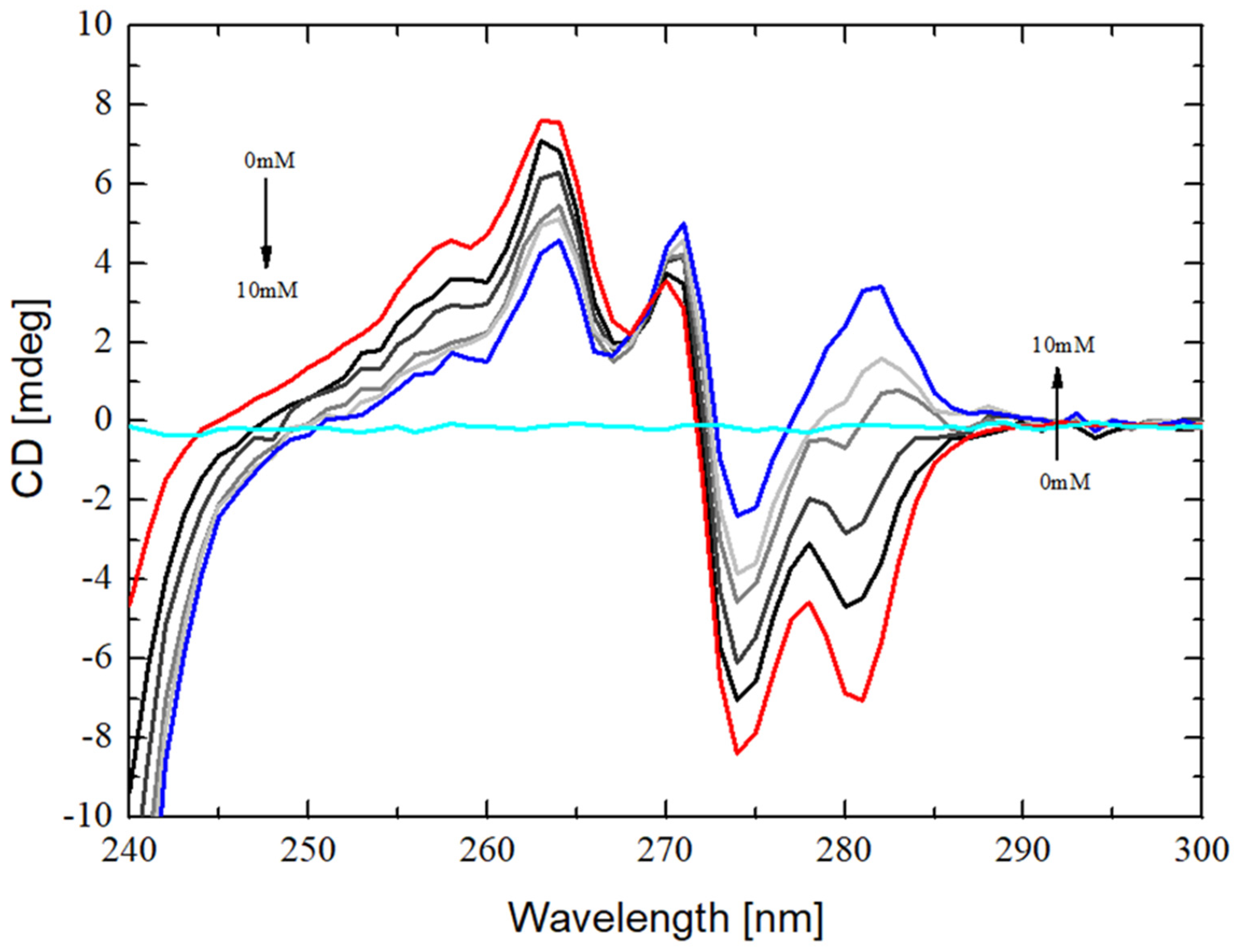
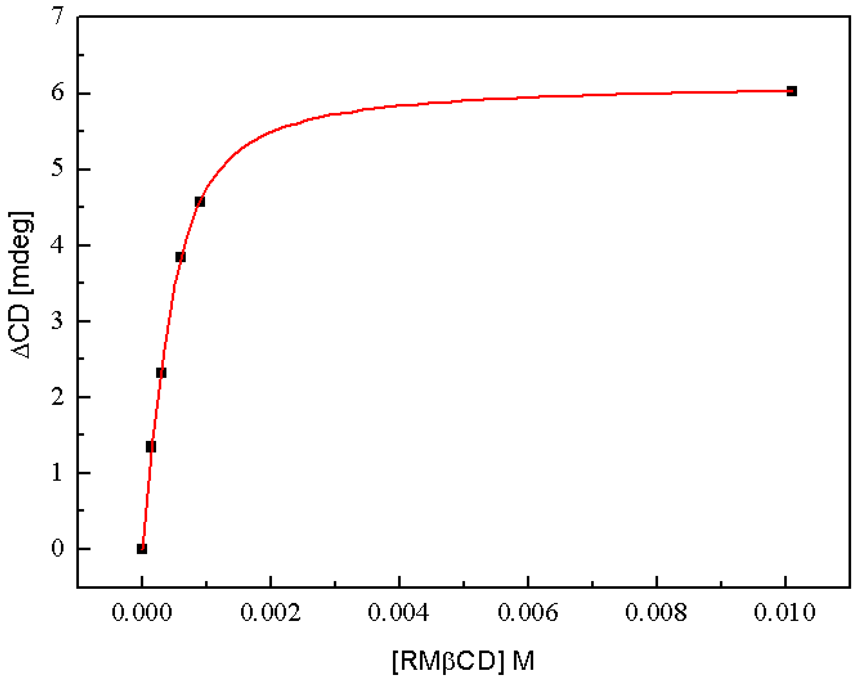
2.2. Circular Dichroism Spectroscopy (CD)
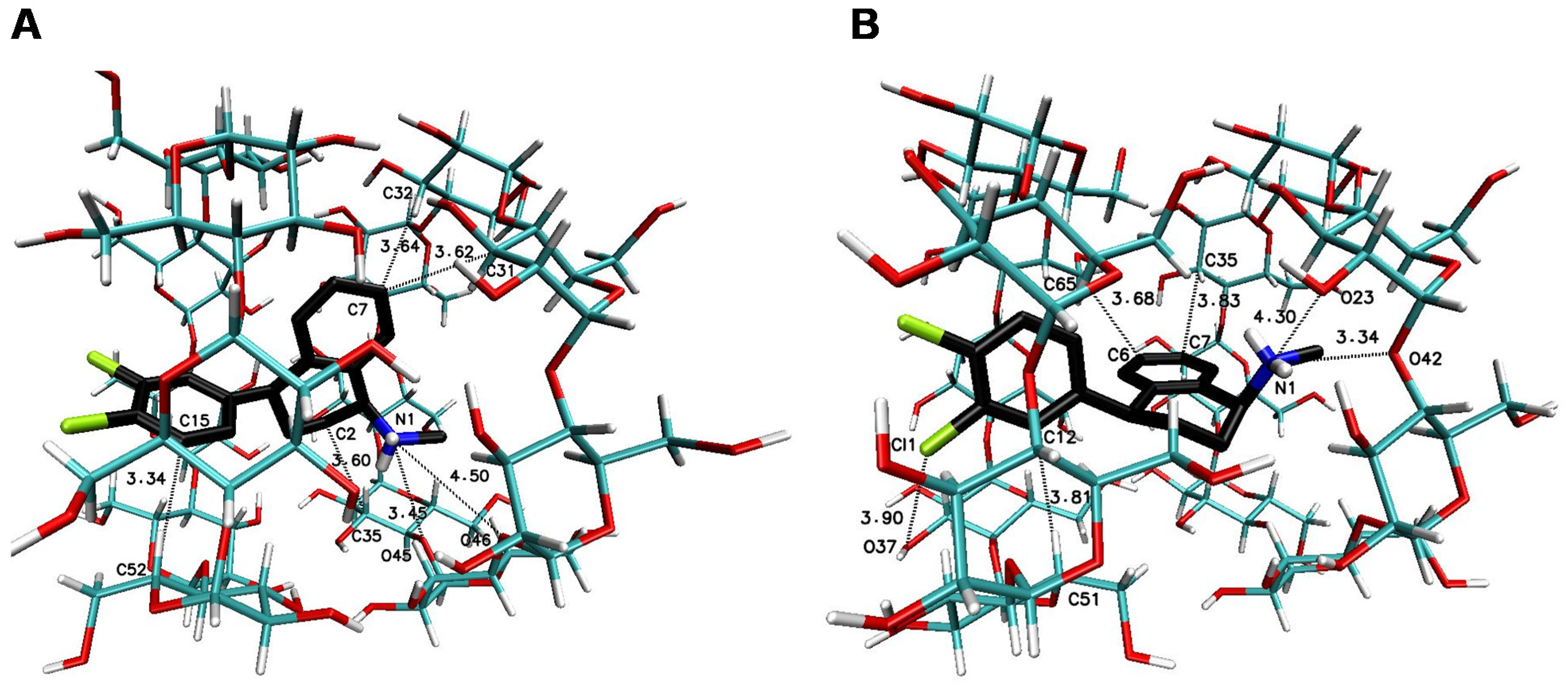
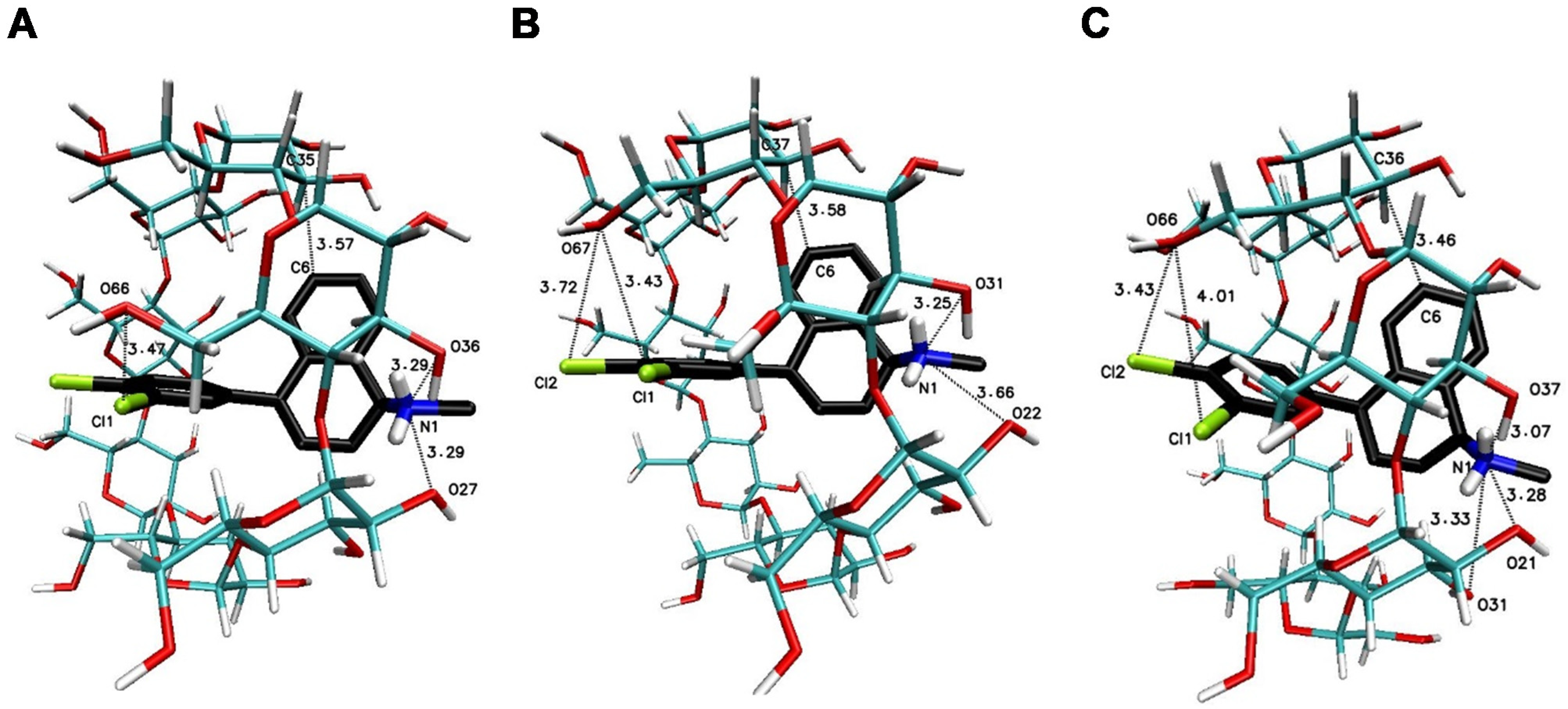
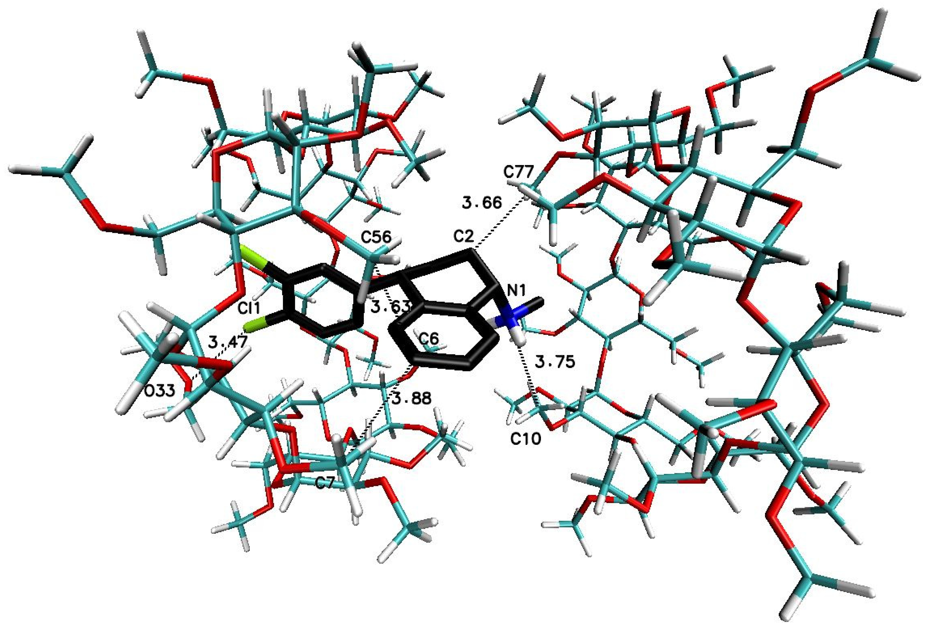
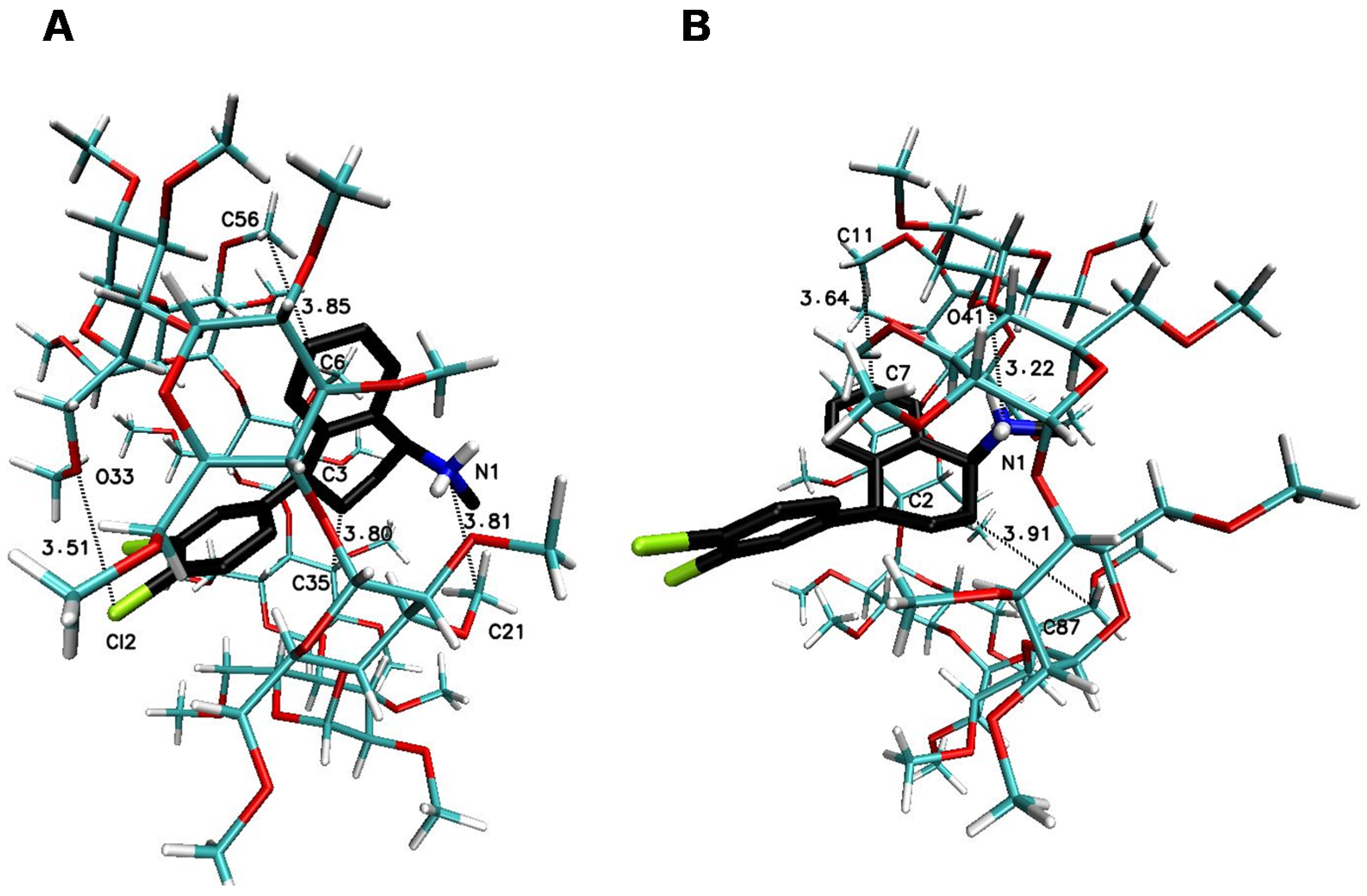
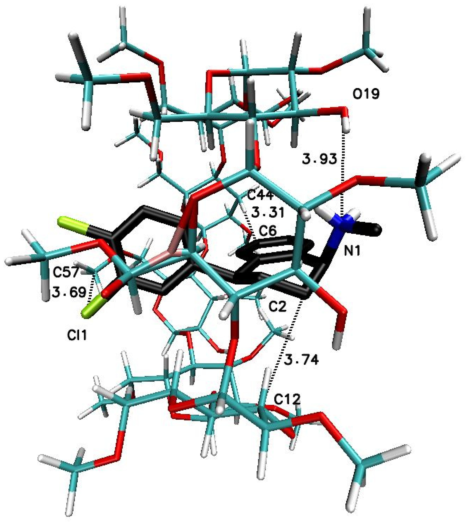
2.3. Molecular Docking (MD)
3. Materials and Methods
3.1. Materials
3.2. Methods
3.2.1. Isothermal Titration Calorimetry (ITC)
- the aqueous solution of the cyclodextrin was added into the pure water placed in the measurement cell and
- the aqueous solution of sertraline hydrochloride was diluted with water injected from the syringe and the heat of the dilution for both stages were registered.
3.2.2. Circular Dichroism (CD) Spectroscopy
3.2.3. Computational Studies
Ligands and Macromolecules Preparation for Molecular Docking
Molecular Docking
- -
- entry from Figure 7 (three molecules of βCD I-II-III): a grid box size of 20 Å × 20 Å × 20 Å centered on the C47 atom (x = −5.017, y = 1.413, z = 0.074);
- -
- entry A from Figure 8 (two molecules of βCD I-II): a grid box size of 20 Å × 20 Å × 20 Å centered on the C45 atom (x = 5.849, y = 3.007, z = −5.646);
- -
- entry B from Figure 8 (two molecules of βCD II-III): a grid box size of 20 Å × 20 Å × 20 Å centered on the C23 atom (x = 4.807, y = 1.076, z = 7.878);
- -
- entry A from Figure 9 (one molecule of βCD I): a grid box size of 20 Å × 20 Å × 20 Å centered on the C45 atom (x = 5.849, y = 3.007, z = −5.646);
- -
- entry B from Figure 9 (one molecule of βCD II): a grid box size of 20 Å × 20 Å × 20 Å centered on the C43 atom (x = 5.243, y = 0.841, z = 1.262);
- -
- entry C from Figure 9 (one molecule of βCD III): a grid box size of 20 Å × 20 Å × 20 Å centered on the C43 atom (x = 4.602, y = −1.221, z = 8.714);
- -
- entry from Figure 10 (two molecules of TMβCD I-II): a grid box size of 20 Å × 20 Å × 20 Å centered on the C11 atom (x = 3.352, y = 6.710, z = 2.402);
- -
- entry A from Figure 11 (two molecules of TMβCD I): a grid box size of 20 Å × 20 Å × 20 Å centered on the C11 atom (x = 3.352, y = 6.710, z = 2.402);
- -
- entry B from Figure 11 (one molecule of TMβCD II): a grid box size of 20 Å × 20 Å × 20 Å centered on the C10 atom (x = 6.775, y = 12.684, z = 10.278);
- -
- entry from Figure 12 (one molecule of RMβCD): a grid box size of 20 Å × 20 Å × 20 Å centered on the C45 atom (x = 2.967, y = 2.155, z = −4.366);
4. Conclusions
Author Contributions
Funding
Institutional Review Board Statement
Informed Consent Statement
Data Availability Statement
Acknowledgments
Conflicts of Interest
References
- Dalla Bella, M.; Szejtli, J. Cyclodextrins. Drugs Future 1983, 8, 391–394. [Google Scholar]
- Szente, L.; Puskás, I.; Sohajda, T.; Varga, E.; Vass, P.; Nagy, Z.K.; Farkas, A.; Várnai, B.; Béni, S.; Hazai, E. Sulfobutylether-beta-cyclodextrin-enabled antiviral remdesivir: Characterization of electrospun- and lyophilized formulations. Carbohydr. Polym. 2021, 264, 118011. [Google Scholar] [CrossRef]
- Sursyakova, V.V.; Levdansky, V.A.; Rubaylo, A.I. Thermodynamic parameters for the complexation of water-soluble betulin derivatives with (2-hydroxypropyl)-β-cyclodextrin determined by affinity capillary electrophoresis. J. Mol. Liq. 2019, 283, 325–331. [Google Scholar] [CrossRef] [Green Version]
- Gao, S.; Liu, Y.; Jiang, J.; Li, X.; Zhao, L.; Fu, Y.; Ye, F. Encapsulation of thiabendazole in hydroxypropyl-β-cyclodextrin nanofibers via polymer-free electrospinning and its characterization. Pest Manag. Sci. 2020, 76, 3264–3272. [Google Scholar] [CrossRef]
- Lin, Z.-Y.; Liu, Y.-X.; Kou, S.-B.; Wang, B.-L.; Shi, J.-H. Characterization of the inclusion interaction of ethinyloestradiol with β-cyclodextrin and hydroxypropyl-β-cyclodextrin: Multi-spectroscopic and molecular modeling methods. J. Mol. Liq. 2020, 311, 113290. [Google Scholar] [CrossRef]
- Briñez-Ortega, E.; DE ALMEIDA, V.L.; Lopes, J.C.D.; Burgos, A.E. Partial inclusion of bis(1,10-phenanthroline) silver(i) salicylate in β-cyclodextrin: Spectroscopic characterization, in vitro and in silico antimicrobial evaluation. An. Acad. Bras. Cienc. 2020, 92, 1–23. [Google Scholar] [CrossRef] [PubMed]
- Xavier-Júnior, F.H.; Tavares, C.T.; Rabello, M.M.; Hernandes, M.Z.; Bezerra, B.P.; Ayala, A.P.; Pessoa, O.D.L.; Ximenes, R.M.; Santos-Magalhães, N.S. Elucidation of the mechanism of complexation between oncocalyxone A and cyclodextrins by isothermal titration calorimetry and molecular modeling. J. Mol. Liq. 2019, 274, 165–172, Corrigendum to 2019, 288, 145. [Google Scholar] [CrossRef]
- Wang, J.; Wang, T.; Hu, Y.; Zhang, X.; Ma, Y.; Lv, H.; Xu, S.; Wang, Y.; Jiang, Z. Fe3+ sensitivity fluorescence sensor from β-cyclodextrin-enhanced Eu3+ luminescence aggregates. J. Mater. Sci. 2021, 56, 10979–10989. [Google Scholar] [CrossRef]
- Sonaimuthu, M.; Balakrishnan, S.B.; Kuppu, S.V.; Veerakanellore, G.B.; Thambusamy, S. Spectral and proton transfer behavior of 1,4-dihydroxylanthraquinone in aqueous and confined media; molecular modelling strategy. J. Mol. Liq. 2018, 259, 186–198. [Google Scholar] [CrossRef]
- Bomzan, P.; Roy, N.; Sharma, A.; Rai, V.; Ghosh, S.; Kumar, A.; Roy, M.N. Molecular encapsulation study of indole-3-methanol in cyclodextrins: Effect on antimicrobial activity and cytotoxicity. J. Mol. Struct. 2021, 1225, 129093. [Google Scholar] [CrossRef]
- Aree, T. Supramolecular complexes of β-cyclodextrin with clomipramine and doxepin: Effect of the ring substituent and component of drugs on their inclusion topologies and structural flexibilities. Pharmaceuticals 2020, 13, 278. [Google Scholar] [CrossRef]
- Li, H.; Chang, S.-L.; Chang, T.-R.; You, Y.; Wang, X.-D.; Wang, L.-W.; Yuan, X.-F.; Tan, M.-H.; Wang, P.-D.; Xu, P.-W.; et al. Inclusion complexes of cannabidiol with β-cyclodextrin and its derivative: Physicochemical properties, water solubility, and antioxidant activity. J. Mol. Liq. 2021, 334, 116070. [Google Scholar] [CrossRef]
- Gaálová, J.; Michel, M.; Bourassi, M.; Ladewig, B.P.; Kasal, P.; Jindřich, J.; Izák, P. Nafion membranes modified by cationic cyclodextrin derivatives for enantioselective separation. Sep. Purif. Technol. 2021, 266, 118538. [Google Scholar] [CrossRef]
- Li, M.; Jiang, Z.; Guo, X.; Di, X.; Yu, J. Enantioseparation and modelling study of six proton pump inhibitors on a novel 3, 5-dichloro-phenylcarbamated β-cyclodextrin chemically bonded chiral stationary phase by high performance liquid chromatography. Microchem. J. 2021, 166, 106211. [Google Scholar] [CrossRef]
- Budryn, G.; Zaczyńska, D.; Pałecz, B.; Rachwał-Rosiak, D.; Belica, S.; Den-Haan, H.; Peña-García, J.; Pérez-Sánchez, H. Interactions of free and encapsulated hydroxycinnamic acids from green coffee with egg ovalbumin, whey and soy protein hydrolysates. LWT-Food Sci. Technol. 2016, 65, 823–831. [Google Scholar] [CrossRef]
- Budryn, G.; Pałecz, B.; Rachwał-Rosiak, D.; Oracz, J.; Zaczyńska, D.; Belica, S.; Navarro-González, I.; Meseguer, J.M.V.; Pérez-Sánchez, H. Effect of inclusion of hydroxycinnamic and chlorogenic acids from green coffee bean in β-cyclodextrin on their interactions with whey, egg white and soy protein isolates. Food Chem. 2015, 168, 276–287. [Google Scholar] [CrossRef]
- Hundre, S.Y.; Karthik, P.; Anandharamakrishnan, C. Effect of whey protein isolate and β-cyclodextrin wall systems on stability of microencapsulated vanillin by spray-freeze drying method. Food Chem. 2015, 174, 16–24. [Google Scholar] [CrossRef]
- Li, X.; Ji, H.; Bai, Y.; Jin, Z. Development of pullulanase mutants to enhance starch substrate utilization for efficient production of β-CD. Int. J. Biol. Macromol. 2021, 168, 640–648. [Google Scholar] [CrossRef]
- Wang, C.; Zou, C.; Cao, Y. Electrochemical and isothermal adsorption studies on corrosion inhibition performance of β-cyclodextrin grafted polyacrylamide for X80 steel in oil and gas production. J. Mol. Struct. 2021, 1228, 129737. [Google Scholar] [CrossRef]
- El-Ghoul, Y. Biological and microbiological performance of new polymer-based chitosan and synthesized amino-cyclodextrin finished polypropylene abdominal wall prosthesis biomaterial. Text. Res. J. 2020, 90, 2690–2702. [Google Scholar] [CrossRef]
- Rekharsky, M.V.; Inoue, Y. Complexation thermodynamics of cyclodextrins. Chem. Rev. 1998, 98, 1875–1917. [Google Scholar] [CrossRef]
- Kumar, R.; Sinha, V.R.; Dahiya, L.; Sarwal, A. Transdermal delivery of duloxetine-sulfobutylether-β-cyclodextrin complex for effective management of depression. Int. J. Pharm. 2021, 594, 120129. [Google Scholar] [CrossRef] [PubMed]
- Urcuk, A.; Karadurmus, L.; Bakirhan, N.K.; Ozkan, S.A. Enhancement of graphene oxide through β-cyclodextrin composite to sensitive analysis of an antidepressant: Sulpiride. Open Chem. 2021, 19, 228–236. [Google Scholar] [CrossRef]
- Aree, T. β-Cyclodextrin Inclusion Complexation With Tricyclic Antidepressants Desipramine and Imipramine: A Structural Chemistry Perspective. J. Pharm. Sci. 2020, 109, 3086–3094. [Google Scholar] [CrossRef] [PubMed]
- Ignaczak, A.; Pałecz, B.; Belica-Pacha, S. Quantum chemical study and isothermal titration calorimetry of β-cyclodextrin complexes with mianserin in aqueous solution. Org. Biomol. Chem. 2017, 15, 1209–1216. [Google Scholar] [CrossRef] [PubMed] [Green Version]
- Belica-Pacha, S.; Małecka, M.; Daśko, M.; Miłowska, K.; Bryszewska, M.; Budryn, G.; Oracz, J.; Pałecz, B. The interaction of heptakis (2,6-di-o-methyl)-β-cyclodextrin with mianserin hydrochloride and its influence on the drug toxicity. Int. J. Mol. Sci. 2021, 22, 9419. [Google Scholar] [CrossRef]
- Majewska, K.; Skwierawska, A.; Kamińska, B.; Prześniak-Welenc, M. Improvement of opipramol base solubility by complexation with β-cyclodextrin. Supramol. Chem. 2018, 30, 20–31. [Google Scholar] [CrossRef]
- Diniz, T.C.; Pinto, T.C.C.; Menezes, P.D.P.; Silva, J.C.; Teles, R.B.D.A.; Ximenes, R.C.C.; Guimarães, A.G.; Serafini, M.R.; Araújo, A.A.D.S.; Quintans Júnior, L.J.; et al. Cyclodextrins improving the physicochemical and pharmacological properties of antidepressant drugs: A patent review. Expert Opin. Ther. Pat. 2018, 28, 81–92. [Google Scholar] [CrossRef]
- Londhe, V.Y.; Deshmane, A.B.; Singh, S.R.; Kulkarni, Y.A. Lurasidone-β-cyclodextrin complexes: Physicochemical characterization and comparison of their antidepressant, antipsychotic activities against that of self microemulsifying formulation. J. Mol. Struct. 2018, 1157, 395–400. [Google Scholar] [CrossRef]
- Belica-Pacha, S.; Miłowska, K.; Ionov, M.; Bryszewska, M.; Buczkowski, A.; Budryn, G.; Oracz, J.; Zaczyńska, D.; Wróblewska, A.; Urbaniak, P.; et al. The impact of β-cyclodextrin on biological and chemical properties of mianserin hydrochloride in aqueous solution. J. Mol. Liq. 2020, 314, 113589. [Google Scholar] [CrossRef]
- Buko, V.; Zavodnik, I.; Lukivskaya, O.; Naruta, E.; Palecz, B.; Belica-Pacha, S.; Belonovskaya, E.; Kranc, R.; Abakumov, V. Cytoprotection of pancreatic β-cells and hypoglycemic effect of 2-hydroxypropyl-β-cyclodextrin: Sertraline complex in alloxan-induced diabetic rats. Chem. Biol. Interact. 2016, 244, 105–112. [Google Scholar] [CrossRef]
- Marzouk, M.A.; Osman, D.A.; Mohamed, O.S. In vitro and in vivo evaluation of taste-masked orodispersible tablets of fluoxetine hydrochloride for the treatment of depression. Drug Dev. Ind. Pharm. 2021, 47, 645–653. [Google Scholar] [CrossRef]
- Stapel, B.; Melzer, C.; von der Ohe, J.; Hillemanns, P.; Bleich, S.; Kahl, K.G.; Hass, R. Effect of SSRI exposure on the proliferation rate and glucose uptake in breast and ovary cancer cell lines. Sci. Rep. 2021, 11, 1250. [Google Scholar] [CrossRef] [PubMed]
- Hayami, T.; Kamiya, N.; Kasahara, K.; Kawabata, T.; Kurita, J.-I.; Fukunishi, Y.; Nishimura, Y.; Nakamura, H.; Higo, J. Difference of binding modes among three ligands to a receptor mSin3B corresponding to their inhibitory activities. Sci. Rep. 2021, 11, 6178. [Google Scholar] [CrossRef]
- Krzyżek, P.; Franiczek, R.; Krzyżanowska, B.; Łaczmański, Ł.; Migdał, P.; Gościniak, G. In Vitro Activity of Sertraline, an Antidepressant, Against Antibiotic-Susceptible and Antibiotic-Resistant Helicobacter pylori Strains. Pathogens 2019, 8, 228. [Google Scholar] [CrossRef] [PubMed] [Green Version]
- Passos, J.J.; De Sousa, F.B.; Lula, I.S.; Barreto, E.A.; Lopes, J.F.; De Almeida, W.B.; Sinisterra, R.D. Multi-equilibrium system based on sertraline and β-cyclodextrin supramolecular complex in aqueous solution. Int. J. Pharm. 2011, 421, 24–33, Erratum in 2013, 444, 201. [Google Scholar] [CrossRef] [PubMed] [Green Version]
- Lović, J.; Lađarević, J.; Trišović, N.; Andrić, F.; Mladenović, A.; Mijin, D.; Vuković, D.; Petrović, S.; Ivić, M.A. Electrochemical determination of sertraline in pharmaceutical formulation and serum using a gold electrode in a pH 8.4 bicarbonate solution. Mon. Chem. 2021, 152, 185–192. [Google Scholar] [CrossRef]
- Lopes, J.F.; Nascimento, C.S.; Anconi, C.P.A.; Santos, H.F.D.; Almeida, W.B.D. Inclusion complex thermodynamics: The β-cyclodextrin and sertraline complex example. J. Mol. Graph. Model. 2015, 62, 11–17. [Google Scholar] [CrossRef]
- Bautista-Renedo, J.M.; Cuevas-Yañez, E.; Reyes-Pérez, H.; Vargas, R.; Garza, J.; González-Rivas, N. Non-covalent interactions between sertraline stereoisomers and 2-hydroxypropyl-ß-cyclodextrin: A quantum chemistry analysis. RSC Adv. 2020, 10, 20202–20210. [Google Scholar] [CrossRef]
- Ogawa, N.; Hashimoto, T.; Furuishi, T.; Nagase, H.; Endo, T.; Yamamoto, H.; Kawashima, Y.; Ueda, H. Solid-state characterization of sertraline base-β-cyclodextrin inclusion complex. J. Pharm. Biomed. Anal. 2015, 107, 265–272. [Google Scholar] [CrossRef]
- Belica, S.; Jeziorska, D.; Urbaniak, P.; Buko, V.U.; Zavodnik, I.B.; Pałecz, B. Calorimetric and spectroscopic characterization of complexes between β-cyclodextrin or heptakis (2,6-di-O-methyl)-β-cyclodextrin and sertraline hydrochloride in aqueous solution. J. Chem. Thermodyn. 2014, 70, 160–167. [Google Scholar] [CrossRef]
- Buko, V.; Palecz, B.; Belica-Pacha, S.; Zavodnik, I. The Supramolecular Complex of Sertraline With Cyclodextrins: Physicochemical and Pharmacological Properties. In Nano-and Microscale Drug Delivery Systems: Design and Fabrication; Elsevier Inc.: Amsterdam, The Netherlands, 2017; pp. 343–356. ISBN 9780323527279. [Google Scholar]
- Xavier-Junior, F.H.; Rabello, M.M.; Hernandes, M.Z.; Dias, M.E.S.; Andrada, O.H.M.S.; Bezerra, B.P.; Ayala, A.P.; Santos-Magalhães, N.S. Supramolecular interactions between β-lapachone with cyclodextrins studied using isothermal titration calorimetry and molecular modeling. J. Mol. Recognit. 2017, 30, e2646. [Google Scholar] [CrossRef]
- Chatziathanasiadou, M.V.; Mavromoustakos, T.; Tzakos, A.G. Unveiling the Thermodynamic Aspects of Drug-Cyclodextrin Interactions Through Isothermal Titration Calorimetry (Book Chapter). Methods Mol. Biol. 2021, 2207, 187–198. [Google Scholar] [CrossRef]
- Cerutti, J.P.; Aiassa, V.; Fernández, M.A.; Longhi, M.R.; Quevedo, M.A.; Zoppi, A. Structural, physicochemical and biological characterization of chloramphenicol multicomponent complexes. J. Mol. Liq. 2021, 331, 115761. [Google Scholar] [CrossRef]
- Ravikumar, K.; Sridhar, B.; Bhanu, M.N. Sertraline hydrochloride form II. Acta Crystallogr. Sect. E Struct. Rep. Online 2006, 62, o565. [Google Scholar] [CrossRef]
- Groom, C.R.; Bruno, I.J.; Lightfoot, M.P.; Ward, S.C. The Cambridge structural database. Acta Crystallogr. Sect. B Struct. Sci. Cryst. Eng. Mater. 2016, 72, 171–179. [Google Scholar] [CrossRef]
- Rysanek, N.; Le Bas, G.; Villain, F.; Tsoucaris, G. Structure of the (1:1:1) complex 2a,2b,2c,2d,2e,2f,3a,3g,6a,6b,6c,6d,6e,6f,6-pentadeca-O-methyl-β-cyclodextrin-1,7-dioxaspiro[5.5]undecane-methanol. Acta Crystallogr. Sect. C 1992, 48, 1466–1471. [Google Scholar] [CrossRef]
- Bouchemal, K.; Mazzaferro, S. How to conduct and interpret ITC experiments accurately for cyclodextrin-guest interactions. Drug Discov. Today 2012, 17, 623–629. [Google Scholar] [CrossRef] [PubMed]
- Saboury, A.A.; Atri, M.S.; Sanati, M.H.; Sadeghi, M. Application of a simple calorimetric data analysis on the binding study of calcium ions by human growth hormone. J. Therm. Anal. Calorim. 2006, 83, 175–179. [Google Scholar] [CrossRef]
- Edwards, P.M. Origin 7.0: Scientific graphing and data analysis software. J. Chem. Inf. Comput. Sci. 2002, 42, 1270. [Google Scholar] [CrossRef]
- MicroCal. ITC Data Analysis in Origin® Tutorial Guide, 7th ed.; MicroCal: Northampton, MA, USA, 2004. [Google Scholar]
- Narczyk, M.; Mioduszewski, Ł.; Oksiejuk, A.; Winiewska-Szajewska, M.; Wielgus-Kutrowska, B.; Gojdź, A.; Cieśla, J.; Bzowska, A. Single tryptophan Y160W mutant of homooligomeric E. coli purine nucleoside phosphorylase implies that dimers forming the hexamer are functionally not equivalent. Sci. Rep. 2021, 11, 11144. [Google Scholar] [CrossRef] [PubMed]
- Freire, E.; Schön, A.; Velazquez-Campoy, A. Chapter 5 Isothermal Titration Calorimetry. In Methods in Enzymology; Academic Press: Cambridge, MA, USA, 2009; pp. 127–155. ISBN 9780123745965. [Google Scholar]
- Inoue, Y.; Liu, Y.; Tong, L.H.; Shen, B.J.; Jin, D. Sen Calorimetric Titration of Inclusion Complexation with Modified β-Cyclodextrins. Enthalpy-Entropy Compensation in Host-Guest Complexation: From Ionophore to Cyclodextrin and Cyclophane. J. Am. Chem. Soc. 1993, 115, 10637–10644. [Google Scholar] [CrossRef]
- Leffler, J.E. The enthalpy-entropy relationship and its implications for organic chemistry. J. Org. Chem. 1955, 20, 1202–1231. [Google Scholar] [CrossRef]
- Schönbeck, C.; Westh, P.; Holm, R. Complexation Thermodynamics of Modified Cyclodextrins: Extended Cavities and Distorted Structures. J. Phys. Chem. B 2014, 118, 10120–10129. [Google Scholar] [CrossRef] [PubMed]
- Schönbeck, C.; Holm, R. Exploring the Origins of Enthalpy–Entropy Compensation by Calorimetric Studies of Cyclodextrin Complexes. J. Phys. Chem. B 2019, 123, 6686–6693. [Google Scholar] [CrossRef] [PubMed]
- Inoue, Y.; Hakushi, T.; Liu, Y.; Tong, L.H. Molecular Design of Crown Ethers. 12. Complexation Thermodynamics of 12- to 16-Crown-4: Thermodynamic Origin of High Lithium Selectivity of 14-Crown-4. J. Org. Chem. 1993, 58, 5411–5413. [Google Scholar] [CrossRef]
- Inoue, Y.; Hakushi, T.; Liu, Y.; Tong, L.H.; Shen, B.J.; Jin, D. Sen Thermodynamics of Molecular Recognition by Cyclodextrins. 1. Calorimetric Titration of Inclusion Complexation of Naphthalenesulfonates with α-, β-, and γ-Cyclodextrins: Enthalpy-Entropy Compensation. J. Am. Chem. Soc. 1993, 115, 475–481. [Google Scholar] [CrossRef]
- Amin Kreaz, R.M.; Novák, C.; Erős, I.; Kata, M. Thermoanalytical studies on complexes of furosemide with β-cyclodextrin derivatives. J. Therm. Anal. Calorim. 1999, 55, 115–122. [Google Scholar] [CrossRef]
- Trandafirescu, C.; Ledeţi, I.; Şoica, C.; Ledeţi, A.; Vlase, G.; Borcan, F.; Dehelean, C.; Coricovac, D.; Racoviceanu, R.; Aigner, Z. Albendazole-cyclodextrins binary systems. J. Therm. Anal. Calorim. 2019, 138, 3039–3054. [Google Scholar] [CrossRef]
- Ishizuka, Y.; Nagawa, Y.; Nakanishi, H.; Kuboyama, A. Circular dichroism spectra of the inclusion complexes of phlorizin in cyclodextrins. J. Incl. Phenom. Mol. Recognit. Chem. 1990, 9, 219–225. [Google Scholar] [CrossRef]
- Otagiri, M.; Ikeda, K.; Uekama, K.; Ito, O.; Hatano, M. Induced circular dichroism of racemic methylcyclohexanones included in β-cyclodextrin. Chem. Lett. 1974, 3, 679–682. [Google Scholar] [CrossRef]
- Kodaka, M. A general rule for circular dichroism induced by a chiral macrocycle. J. Am. Chem. Soc. 1993, 115, 3702–3705. [Google Scholar] [CrossRef]
- Fielding, L. NMR methods for the determination of protein–ligand dissociation constants. Prog. Nucl. Magn. Reson. Spectrosc. 2007, 51, 219–242. [Google Scholar] [CrossRef]
- Chatziefthimiou, S.D.; Yannakopoulou, K.; Mavridis, I.M. β-Cyclodextrin trimers enclosing an unusual organization of guest: The inclusion complex β-cyclodextrin/4-pyridinealdazine. CrystEngComm 2007, 9, 976–979. [Google Scholar] [CrossRef]
- Tsuchiya, Y.; Yamano, A.; Shiraki, T.; Sada, K.; Shinkai, S. Single-crystal structure of porphyrin bicapped with trimethyl-β- cyclodextrins: A novel dye-oriented material. Chem. Lett. 2011, 40, 99–101. [Google Scholar] [CrossRef]
- Xu, X.; Peng, S.; Bao, G.; Zhang, H.; Yin, C. β-cyclodextrin inclusion complexes with vitamin A and its esters: A comparative experimental and molecular modeling study. J. Mol. Struct. 2021, 1223, 129001. [Google Scholar] [CrossRef]
- Zhang, H.; Tan, T.; Hetényi, C.; Lv, Y.; van der Spoel, D. Cooperative Binding of Cyclodextrin Dimers to Isoflavone Analogues Elucidated by Free Energy Calculations. J. Phys. Chem. C 2014, 118, 7163–7173. [Google Scholar] [CrossRef]
- Ferrero, R.; Pantaleone, S.; Delle Piane, M.; Caldera, F.; Corno, M.; Trotta, F.; Brunella, V. On the Interactions of Melatonin/β-Cyclodextrin Inclusion Complex: A Novel Approach Combining Efficient Semiempirical Extended Tight-Binding (xTB) Results with Ab Initio Methods. Molecules 2021, 26, 5881. [Google Scholar] [CrossRef]
- Belica, S.; Sadowska, M.; Stȩpniak, A.; Graca, A.; Pałecz, B. Enthalpy of solution of α- And β-cyclodextrin in water and in some organic solvents. J. Chem. Thermodyn. 2014, 69, 112–117. [Google Scholar] [CrossRef]
- Bertaut, E.; Landy, D. Improving ITC studies of cyclodextrin inclusion compounds by global analysis of conventional and non-conventional experiments. Beilstein J. Org. Chem. 2014, 10, 2630–2641. [Google Scholar] [CrossRef] [Green Version]
- Anderson, T.G.; Tan, A.; Ganz, P.; Seelig, J. Calorimetric Measurement of Phospholipid Interaction with Methyl-β-Cyclodextrin. Biochemistry 2004, 43, 2251–2261. [Google Scholar] [CrossRef] [PubMed]
- Szente, L. Highly soluble cyclodextrin derivatives: Chemistry, properties, and trends in development. Adv. Drug Deliv. Rev. 1999, 36, 17–28. [Google Scholar] [CrossRef]
- Valente, A.J.M.; Carvalho, R.A.; Murtinho, D.; Söderman, O. Molecular Dynamics of Cyclodextrins in Water Solutions from NMR Deuterium Relaxation: Implications for Cyclodextrin Aggregation. Langmuir 2017, 33, 8233–8238. [Google Scholar] [CrossRef] [PubMed]
- Gasteiger, J.; Marsili, M. Iterative partial equalization of orbital electronegativity-a rapid access to atomic charges. Tetrahedron 1980, 36, 3219–3228. [Google Scholar] [CrossRef]
- Xiao, W.; Wang, D.; Shen, Z.; Li, S.; Li, H. Multi-body interactions in molecular docking program devised with key water molecules in protein binding sites. Molecules 2018, 23, 2321. [Google Scholar] [CrossRef] [Green Version]
- Trott, O.; Olson, A.J. Software news and update AutoDock Vina: Improving the speed and accuracy of docking with a new scoring function, efficient optimization, and multithreading. J. Comput. Chem. 2010, 31, 455–461. [Google Scholar]


| n | K/M−1 | ΔH/kJ·mol−1 | TΔS/kJ·mol−1 | ΔG/kJ·mol−1 | |
|---|---|---|---|---|---|
| β-CD a,b | 1.20 a 1.31 b | 5820 a 4999.3 | −20.44 a −15.6 b | 1.06 a 5.5 b | −21.53 a −21.1 b |
| HPβCD c | 1.23 | 6530 | −16.72 | 5.05 | −21.77 |
| DMβCD a | 1.60 | 7960 | −14.20 | 7.96 | −22.19 |
| RMβCD * | 1.26 ± 0.05 | 4520 ± 74 | −8.37 ± 0.07 | 12.49 ± 0.04 | −20.86 ± 0.11 |
| n | K/M−1 | ΔCDmax/mdeg | Reduced χ2 | R2 |
|---|---|---|---|---|
| 1.57 ± 0.11 | 5315 ± 500 | 6.14 ± 0.05 | 0.00197 | 0.99976 |
| Representative Geometry | Crystal Structure Name (Refcode from CSD) | Free Energy of Binding kcal∙mol−1 (kJ∙mol−1) |
|---|---|---|
| Figure 7 | 648855 | −9.2 (−38) |
| (three molecules of βCD I-II-III) | ||
| A | 648855 | −8.6 (−36) |
| Figure 8 | (two molecules of βCD I-II) | |
| B | 648855 | −8.0 (−33) |
| Figure 8 | (two molecules of βCD II-III) | |
| A | 648855 | −5.7 (−24) |
| Figure 9 | (one molecule of βCD I) | |
| B | 648855 | −5.7 (−24) |
| Figure 9 | (one molecule of βCD II) | |
| C | 648855 | −6.0 (−25) |
| Figure 9 | (one molecule of βCD III) | |
| ALIGAE | −7.4 (−31) | |
| Figure 10 | (two molecules of TMβCD I-II) | |
| A | ALIGAE | −6.5 (−27) |
| Figure 11 | (one molecule of TMβCD I) | |
| B | ALIGAE | −5.8 (−24) |
| Figure 11 | (one molecule of TMβCD II) | |
| JOSWOD | −6.3 (−26) | |
| Figure 12 | (one molecule of RMβCD) |
Publisher’s Note: MDPI stays neutral with regard to jurisdictional claims in published maps and institutional affiliations. |
© 2021 by the authors. Licensee MDPI, Basel, Switzerland. This article is an open access article distributed under the terms and conditions of the Creative Commons Attribution (CC BY) license (https://creativecommons.org/licenses/by/4.0/).
Share and Cite
Belica-Pacha, S.; Daśko, M.; Buko, V.; Zavodnik, I.; Miłowska, K.; Bryszewska, M. Thermodynamic Studies of Interactions between Sertraline Hydrochloride and Randomly Methylated β-Cyclodextrin Molecules Supported by Circular Dichroism Spectroscopy and Molecular Docking Results. Int. J. Mol. Sci. 2021, 22, 12357. https://doi.org/10.3390/ijms222212357
Belica-Pacha S, Daśko M, Buko V, Zavodnik I, Miłowska K, Bryszewska M. Thermodynamic Studies of Interactions between Sertraline Hydrochloride and Randomly Methylated β-Cyclodextrin Molecules Supported by Circular Dichroism Spectroscopy and Molecular Docking Results. International Journal of Molecular Sciences. 2021; 22(22):12357. https://doi.org/10.3390/ijms222212357
Chicago/Turabian StyleBelica-Pacha, Sylwia, Mateusz Daśko, Vyacheslav Buko, Ilya Zavodnik, Katarzyna Miłowska, and Maria Bryszewska. 2021. "Thermodynamic Studies of Interactions between Sertraline Hydrochloride and Randomly Methylated β-Cyclodextrin Molecules Supported by Circular Dichroism Spectroscopy and Molecular Docking Results" International Journal of Molecular Sciences 22, no. 22: 12357. https://doi.org/10.3390/ijms222212357