Article

Solitary Fibrous Tumor: Integration of Clinical, Morphologic, Immunohistochemical and Molecular Findings in Risk Stratification and Classification May Better Predict Patient outcome

1
Pathology Department, Instituto Valenciano de Oncología, 46009 Valencia, Spain
2
Patologika Laboratory, Pathology Department, Hospital Quiron-Salud, 46009 Valencia, Spain
3
Zoology Department, University of Valencia, 46009 Valencia, Spain
4
Department of Oncology, Instituto Valenciano de Oncología, 46009 Valencia, Spain
5
Pathology Department, University Hospital “La Fe”, 46009 Valencia, Spain
6
Pathology Department, University of Valencia, 46009 Valencia, Spain
*
Author to whom correspondence should be addressed.
Int. J. Mol. Sci. 2021, 22(17), 9423; https://doi.org/10.3390/ijms22179423
Submission received: 12 July 2021 / Revised: 23 August 2021 / Accepted: 26 August 2021 / Published: 30 August 2021
(This article belongs to the Special Issue Solitary Fibrous Tumors: Diagnosis, Classification and Therapy)

Abstract

:
Although solitary fibrous tumors (SFTs) have an unpredictable evolution, some specific clinicopathologic factors have been associated with the final outcome. We retrieved clinical, pathological and molecular data of 97 patients with a histological diagnosis of SFT and Signal transducer and activator of transcription 6 (STAT6) positivity. We retrospectively studied the pathological factors predictive of recurrence/metastasis and compared them with the clinical outcome. A wide immunohistochemical study and molecular analysis to detect NAB2/STAT6 gene fusion, tumor protein-53 (TP53) and/or (telomerase reverse transcriptase) TERT promotor mutation were performed. The risk of metastasis was calculated using the Demicco risk stratification system (RSS). The results were combined and examined to assess the accuracy of risk stratification and classification. The most common location was in non-extremities; 66% were located in soft tissue or subcutaneous areas and 92.8% in deep locations. On microscopic analysis, 38.1% of tumors revealed hypercellularity with a predominant patternless and/or hemangiopericytic growth pattern; 13.4% had ≥4 mitoses/10HPF; 16.5% showed necrosis, and almost half the tumors showed at least focal myxoid areas. Dedifferentiation was observed in three tumors. Immunomarker expression in SFTs was as follows: CD34 92.9%, CD99 57.1%, Bcl2 67.9%, neuroendocrine markers (at least 1) 25.7%, Desmin 14.3%, CK(AE1/AE3) 3%, Apoptotic Protease Activating Factor (APAF-1) 87% and finally Ki-67 ≥ 10% in 14.4%. The NAB2/STAT6 gene fusion was detected in 50 tumors. After a median follow-up of 90 months, 9.3% recurred, 11.3% metastasized, 10.3% died of disease and 76.2% were free of disease. TERT mutations were detected in 40.6% of the SFTs; the TP53 mutation was detected in 17%, and only 9.3% showed both mutations. According to the Demicco RSS, 6.1%, 11.3% and 82.4% of the tumors were classified as high, intermediate or low-risk of metastasis, respectively. All high-risk tumors had ≥4 mitoses/10HPF, necrosis, Ki-67 ≥ 10, HTER and/or TP53 mutation and poor evolution. The intermediate risk SFTs with worse evolution displayed the HTER mutation. Almost all low-risk tumors had a favorable evolution, although four showed at least one adverse factor (Ki-67 ≥ 10, ≥4 mitoses/10HPF or high tumor size) and had a worse evolution. An integration of clinical, morphologic, immunohistochemical and molecular findings may improve risk stratification and classification and better predict patient outcome. The unfavorable course seems to be more frequent in high-risk SFTs, although it is not exceptional in low-risk SFTs either; hence, a long-term follow-up is required independently of the assigned risk stratification score. The inclusion of molecular findings in risk stratification systems could improve the precision in the classification of SFTs, especially those of intermediate risk. Future studies will be required to determine the most effective way to incorporate molecular analyses into RSS on SFTs. The coexistence of several adverse factors such as ≥4 mitoses/10HPF, necrosis, Ki-67 ≥ 10%, mutations in HTER and/or p53 may suggest a closer clinical follow-up regardless of the histological appearance of the tumor.

1. Introduction

Solitary fibrous tumors (SFTs) are mesenchymal neoplasms that can occur at any location, especially pleural, meningeal or extrapleural sites. Location in limbs is infrequent [1,2,3,4,5,6,7,8,9,10,11,12,13,14,15,16,17,18,19,20,21,22,23,24,25,26,27,28,29,30,31,32,33,34,35,36,37,38,39,40].
The evolution of SFTs is often uncertain [2,3,4,5,6,7,8,9,10]. Although most cases evolve favorably, a small group can progress towards dissemination, generally pulmonary, as well as other locations, and will lead to patient death in the absence of effective treatments [1,2,3,4,5,6,7,8,9,10,11,12,13,14,15,16,17,18,19]. Thus, valid and reproducible tools should be developed in order to design risk-tailored treatment and risk-adjusted follow-up.
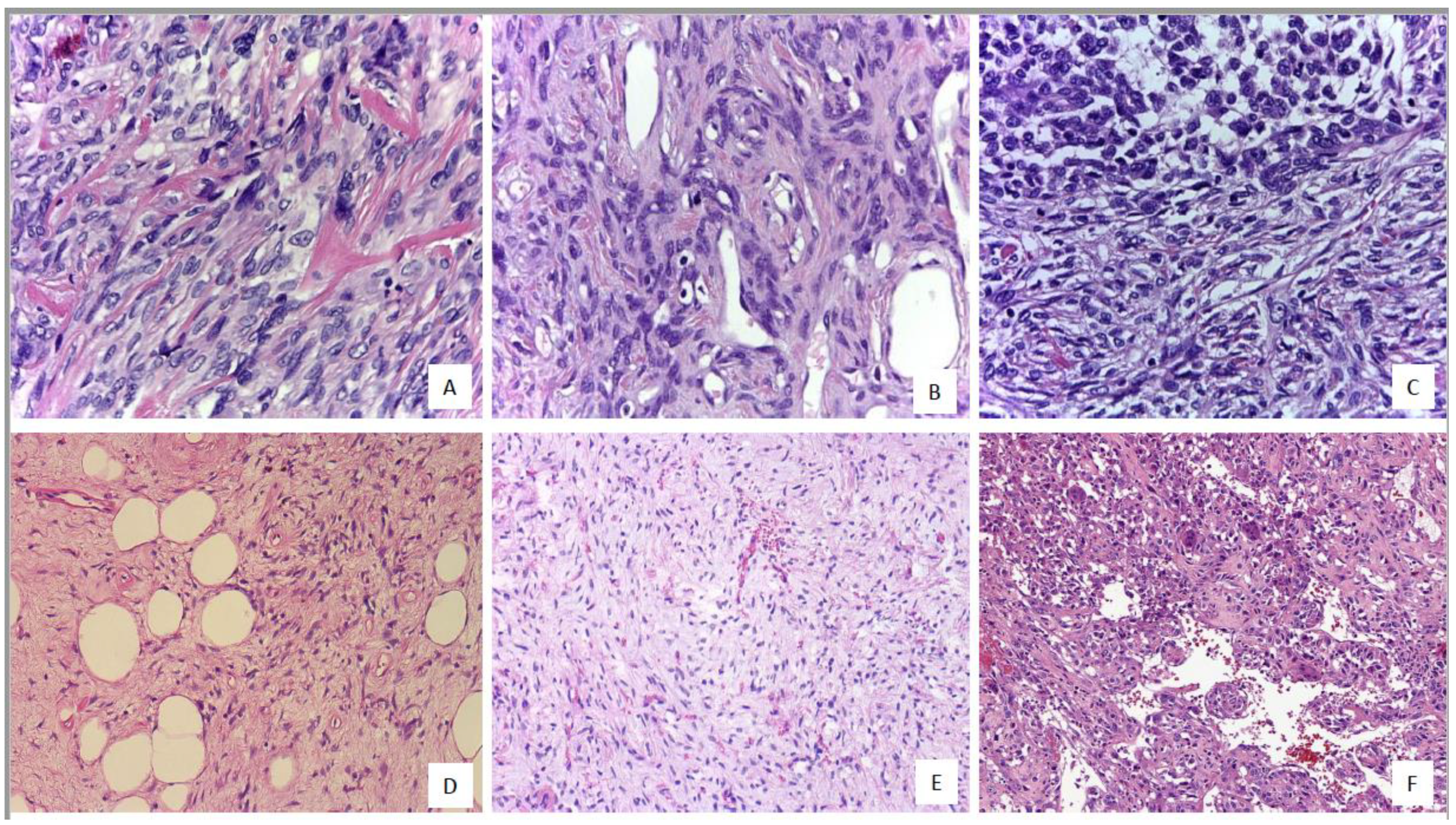
A patternless and hemangiopericytic growth with variably fibrosis and collagenous deposits are characteristic, but SFTs can show many faces, including round cells, giant-cells, myxoid areas, pleomorphic pattern, fat-forming tumors and dedifferentiated forms [1,2,3,4,5,15,16,17,18,19,20,21,22,23,24,25].
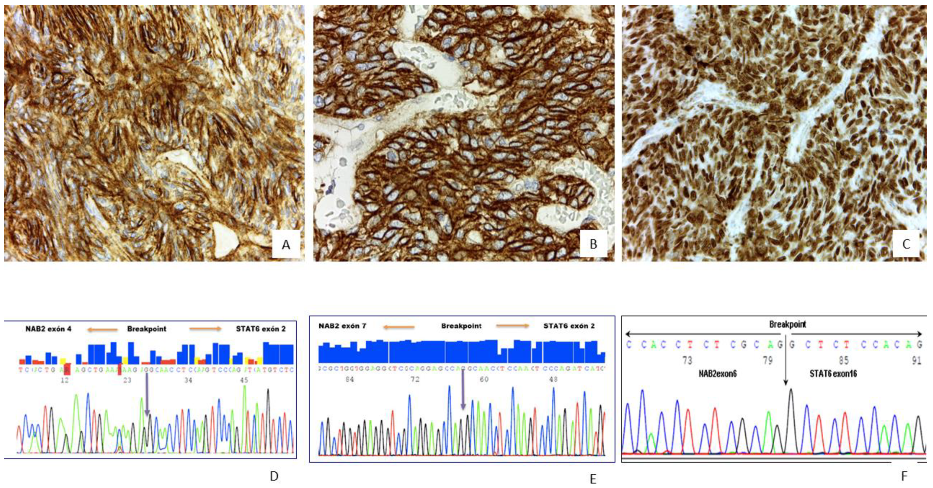
Strong and diffuse nuclear immunoreactivity for Signal transducer and activator of transcription 6 (STAT6) is almost always diagnosed in SFTs, and CD34, Bcl-2 and CD99 immunoexpression is common [1,2,3,4,5,6,7,8,9,10,11,12,13,14,15,16,17,18,19,20,21,22,23,24,25]. Aberrant epithelial, muscular or neuroendocrine marker expression has been described which may lead to confusion with other tumors that share a similar morphology [15,16,17,18,19,20,21,22,23,24,25,36]. The fusion gene NAB2/STAT6 and its variants confirm a diagnosis of SFT in those cases with unconvincing STAT6 immunoreactivity, and specific gene fusions have been related to prognosis and tumor location [6,7,8,9,10,11,12,13,14,26,27,28,29,30,31,32,33,34,35,36,37,38,39,40]. STAT6 nuclear expression is not exclusive to SFTs and has also been reported in dedifferentiated liposarcoma, glioma-associated oncogene homolog 1 (GLI1)-amplified tumor, Kaposi sarcoma and Hodgkin as well non-Hodgkin lymphomas [6,12,13,14,15,16,17,18,19,20,21,22,23,24,25,36,37,38,39,40].
Several risk-stratification systems (RSS) have been described [1,2,3,4,5], although the Demicco et al. RSS seems to be the most widely implemented [1,2,14]. While most SFTs categorized as low-risk by the Demicco system follow an apparently benign course, some cases may have late relapse or metastases; hence, the RSS is not perfectly specific in predicting the evolution in all cases [1,2,14]. In addition, molecular results have not so far been included in any of the current RSS [1,2,14]. Recently, loss of Apoptotic Protease Activating Factor (APAF-1) expression has been associated with poor prognosis in SFTs, but these findings need to be confirmed in larger series [41].
We have previously published a small series of SFTs with at least one histological factor associated with aggressive behavior and explored the correlation between the adverse histological findings and molecular profile with tumor behavior [42]. The main goal of the present study was to confirm our previous results including additional tumors, mainly SFTs categorized as low-risk by Demicco et al. [1,2] and correlate the clinical, histological and immunohistochemical findings, TP53 mutational status and TERT promotor mutational status with clinical outcome.

2. Results

2.1. Clinicopathological and Histopathological Findings

The clinicopathological and follow-up data are shown in Table 1 and Table 2. The histologic findings are shown in Figure 1 and summarized in Table 3. The median follow-up was 90 months. In the microscopic analysis, 38.1% of tumors revealed hypercellularity with a predominant patternless and/or hemangiopericytic growth pattern (Figure 1); 13.4% had ≥4 mitoses/10HPF; 16.5% showed necrosis and myxoid areas were frequent (Figure 1). Dedifferentiation was observed in three tumors (Figure 1). Rare histological patterns were found in sporadic cases (Figure 1).

2.2. Immunohistochemistry Results

The representative status of the immunohistochemical profile is shown in (Figure 2) and summarized in Table 4. All tumors were diffusely positive for STAT6 (Figure 2C), CD34, CD99 (Figure 2A,B) and Bcl2 which were frequent, and neuroendocrine markers (at least 1), Desmin, CK(AE1/AE3) were detected sporadically. APAF-1 immunoexpression was detected in 87% of the SFTs, and Ki-67 ≥ 10 was found in 14.4% of the tumors.

2.3. Molecular Study

Molecular results are summarized in Table 5. NAB2-exon 4/STAT6- exon 2 gene fusion was detected in 35 cases, NAB2-exon 6/STAT6-exon 16/17 in 15 cases (Figure 2D–F). TP53 and TERT promotor status is summarized in Table 5. TERT mutations or TP53 mutations were detected in 40.6% and 17% of the tumors (Figure 3A,B), respectively. A total of 9.3% of the SFTs showed both mutations (Figure 4A,B).

2.4. Risk of Recurrence/Metastasis

The risk stratification system classifications are described in Table 6. Regarding molecular analysis, non-informative results for RNA (NAB2/STAT6 fusion gene) were given in 2 high-risk SFTs, 5 intermediate-risk SFTs and 40 low-risk SFTs. All these cases showed strong and diffuse nuclear STAT6 immunoreactivity. Eleven cases were non-informative for HTER and/or p53 molecular status; these cases were all classified as low-risk of metastasis.
The correlation between clinical and histological findings (mitosis and/or necrosis), the Ki-67 index and molecular status with clinical outcome (metastasis and/or died of disease versus free of disease) in high-risk and intermediate-risk tumors are described in Table 7 and Table 8. The correlation of clinical and histological findings (mitosis and/or necrosis), the Ki-67 index and molecular status with clinical outcome in low-risk tumors with worse evolution is described in Table 9. We did not find any correlation between APAF-1 status and clinical outcome.

3. Discussion

Strong and diffuse nuclear immunoreactivity for STAT6 is a very useful tool for the diagnosis of SFTs, although unexpected epithelial, muscular or neuroendocrine marker expression has also been described in these tumors and may lead to confusion with other neoplasms with hemangiopericytic growth patterns [2,15,16,17,18,19,20,21,22,23,24,25,33,42]. STAT6 expression has also been reported in dedifferentiated liposarcoma and GLI1-amplified tumors; hence, in cases with overlapping morphology and STAT6 immunoreactivity, additional molecular studies are needed to establish a definitive diagnosis [2,15,16,17,18,19,20,21,22,23,24,25,33,34,35,36,37,38,39,40,41,42]. Detection of the specific fusion gene NAB2/STAT6 and its variants confirm a diagnosis of SFT, especially in rare clinical settings, unusual histological findings or unexpected immunohistochemical results. Moreover, specific gene fusions have been related to prognosis and tumor location [7,8,11,12,26,27,28,29,30,31,32,35,36,37,38,39,40,41,42]. Various risk-stratification systems (RSS) have been described, with the Demicco et al. system being the most widely implemented, as recommended in the World Health Organization (WHO) blue book [1,2,3,4,5,14].
Most low risk SFTs categorized by Demicco et al. and other RSS follow an apparently indolent course. However, some of these cases may have late recurrence/relapse and/or metastases leading to uncertainty and skepticism among oncologists regarding the specificity of RSS in correctly predicting evolution in all cases [1,2,3,4,5,14]. Furthermore, molecular results have not been included in any of the present RSS so far [1,2,3,4,5,6,7,8,42].
Regarding histological predictive factors of aggressiveness, high mitotic counts with a general agreement of ≥4/10HPFs represent the strongest predictor of malignant behavior, as confirmed in the present series where all tumors categorized as high-risk and many of the tumors classified as intermediate-risk revealed ≥4/10HPFs [1,2,3,4,5,6,7,8,42]. Likewise, necrosis was present in the same group of tumors (all high-risk and many intermediate-risk).
Of all the immunohistochemical markers applied in this series, only Ki-67 ≥ 10 was associated with poor evolution. Similar to mitosis and necrosis, this finding was also found in all SFTs categorized as high-risk. Ki-67 has only been included in the Diebold et al. RSS [3], and although further confirmation is needed, this factor may represent an additional variable that could provide valuable predictive information on tumor evolution. In the present series, half the tumors categorized as low-risk by the Demicco et al. system but with worse evolution (late recurrences or metastasis) showed Ki-67 ≥ 10.
Recently, loss of APAF-1 immunoexpression has been associated with the progression and poor prognosis in SFTs [41]. APAF-1 inactivation may lead to impaired apoptotic function, and eventually may contribute toward malignant SFT transformation [41]. Nevertheless, this finding needs to be confirmed in larger series. We failed to find this association in the present study, despite some SFTs with aggressive evolution (dedifferentiated tumors) showing loss of APAF-1 expression by immunohistochemistry.
Molecular studies in SFTs have been progressively implemented in many centers for diagnosis and prognosis. SFTs with the most common canonical NAB2 exon 4-STAT6 exon 2 fusion variant are often located in the thorax and are less cellular with abundant fibrosis [8,9,10,11,12,28,29,30,31,32,37,38,39,40]. These tumors have a higher tumor age, larger tumor size, lower mitotic activity and lower recurrence rate [28,29,30,31,32,37,38,39,40]. However, those tumors with NAB2 exon 6-STAT6 exon 16/17 fusion variants typically show a more cellular round to ovoid cell morphology and are often located in the deep soft tissue of the intra-abdominal/retroperitoneal/pelvic region or in central nervous system [28,29,30,31,32,37,38,39,40]. SFTs with NAB2 exon 6-STAT6 exon 16/17 fusion occur in a significantly younger age group, showing higher mitotic activity and a higher recurrence rate [39].
Recently, a new study reported that SFTs with the NAB2 exon 4-STAT6 exon 2 fusion variant show a transcriptional signature enriched for genes involved in DNA binding, gene transcription and nuclear localization, whereas SFTs with the NAB2 exon 6-STAT6 exon 16/17 fusion variants were enriched for genes involved in tyrosine kinase signaling, cell proliferation and cytoplasmic localization [40]. In addition, Georgies T et al. have reported prognostic significance in SFTs depending on STAT6 domain composition [40]. They categorized SFTs as either STAT6-TAD (contained only the transactivation domain of STAT6) or STAT-Full (fusions with most of the STAT6 domain intact). Tumors with STAT6-TAD fusions had a higher mitotic count (p = 0.016) and were associated with poor prognosis [40].
We did not observe any direct association of gene fusion variants with aggressiveness or location of any histologic or phenotypic profile in the present series.
A limitation of the present study was the fact that 48.5% of the samples (47/97) showed non-informative RNA results for the detection of the NAB2/STAT6 fusion gene. Nevertheless, all cases in the present series showed strong and diffuse STAT6 nuclear expression by immunohistochemistry. This finding of strong and diffuse nuclear STAT6 immunoreactivity has previously been implemented in many laboratories as a surrogate for the molecular analysis in solitary fibrous tumors considering the good correlation between the nuclear STAT6 overexpression and the presence of the NAB2/STAT6 fusion gene [1,2,3,4,5,6,7,8,42]. Furthermore, since the NAB2/STAT6 fusion gene represents an intrachromosomal inversion, a non-informative or negative result is not exceptional in gene fusion molecular studies.
Reverse transcription-polymerase chain reaction (RT-PCR) may not be the most suitable method to detect fusion genes in SFTs, and more sensitive techniques such as next generation sequencing (NGS) or RNAseq may have improved the quality of the results. However, these very advanced molecular ancillary tests are also more expensive and not widely available in all pathology labs. RNA analysis failure using RT-PCR could be explained by limited material in the case of a core biopsy or by fixation issues or tissue conservation [1,2,3,4,5,6,7,8,42]. Furthermore, although the present study used the most frequent primers for RT-PCR, they would not necessarily cover the full spectrum of possible gene fusion types in solitary fibrous tumors. Indeed, RNA analysis failure may be considered multifactorial.
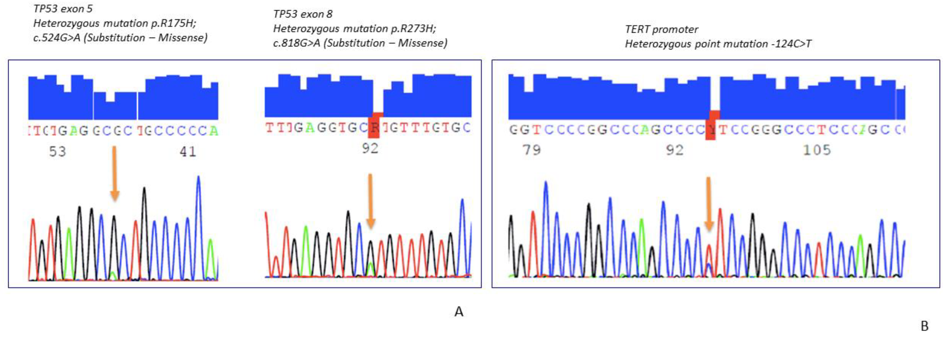
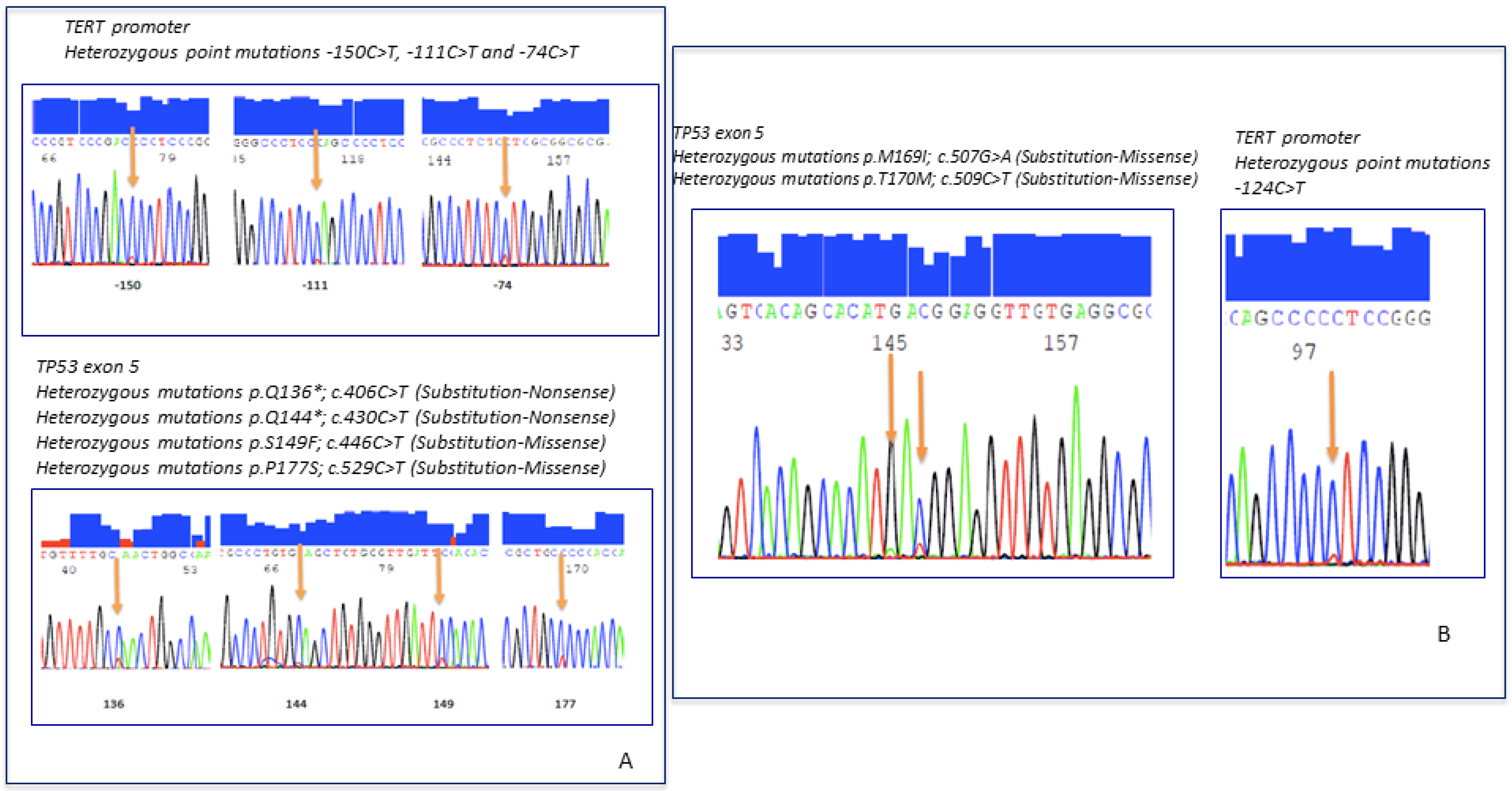
TERT promoter mutations and the TP53 mutation have been associated with malignant behavior in SFTs [7,8,12,35,36,37,38,39,40,41,42]. The overall prevalence of the TP53 mutation in the current series was low, in contrast to the TERT mutation that was found in almost half the tumors. Many of the high-risk tumors had TP53 mutations, HTER mutations or both [7,8,12,35,36,37,38,39,40,41,42]. Likewise, intermediate-risk SFTs with the HTER mutation in the present series showed poor evolution, in line with the previous observation of Demicco et al. where the TERT mutation probably provides no additional prognostic information on tumors already classified as low or high risk [7]. At present, despite the various studies confirming that the HTER promotor and/or TP53 alterations in SFTs are associated with prognosis, no risk stratification system has so far incorporated these molecular factors [1,2,3,4,5,6,7,8]. In the present series, there were only 11 cases with non-informative results for HTER and p53 molecular status, all of which were classified as low-risk of metastasis.
Four RSS have been proposed in the literature that classify SFTs into three (low, intermediate, high-risk) or two categories (low vs. high-risk) depending on the system [1,2,3,4,5,14]. The most commonly implemented is the Demicco et al. RSS which includes both clinical and histological variables [1,2,14]. The Diebold et al. system includes the Ki-67 index, which is not included in the other RSS [3]. Although presumably all dedifferentiated SFTs show other adverse clinical or histological factors, this histological characteristic has not been included in any of the RSS [1,2,3,4,5,14].
In the present series, all SFTs classified as high-risk by Demicco et al. had Ki-67 ≥ 10. We had four cases categorized as low-risk by Demicco et al. but with a worse clinical evolution. Two out these four cases had a Ki-67 index higher than 10 and when applying the Diebold et al. RSS [3], those cases were classified as high-risk tumors; hence, when the systems disagree on risk stratification, the clinical evolution becomes more difficult to predict, which may suggest an imperfect specificity in predicting accurate evolution in some RSS. Incorporating the Ki-67 index might provide prognostic information in some cases independently of the histologic appearance of the tumor. However, Ki-67 results in the present series need to be validated in independent SFT series.
The identification of dedifferentiation in SFTs is very important because new evidence has revealed that antiangiogenics are effective, and their use as a first line of treatment should be considered in SFTs, but not in dedifferentiated SFTs for which chemotherapy is more effective [8].
It would be interesting in the future to study the tumor/stromal tissue interaction, and whether the amount of collagen and/or the type of collagen fibers could have any prognostic significance in these tumors. At present, hypercellularity in solitary fibrous tumors, which usually correlates with a lower amount of collagen, has been correlated in some studies with a somewhat more aggressive evolution in SFTs, although hypercellularity is not necessarily always associated with aggressive evolution [1,2,3,4,5,14].
In conclusion, risk assessment still remains a challenging issue in SFT classification and the final outcome. However, the integration of clinical, morphologic, immunohistochemical and molecular findings may improve risk stratification and classification of SFTs and could guide the clinician when designing risk-adjusted treatment and follow-up. Regardless of the assigned risk stratification score, SFTs may require long-term follow-up considering that low-risk tumors may occasionally show a non-indolent evolution. The inclusion of molecular findings in RSS could improve precision in the classification of SFTs, especially those of intermediate risk. Nevertheless, future studies are required to determine the most effective way to incorporate molecular analyses into RSS on SFTs. The coexistence of several adverse factors such as ≥4 mitoses/10HPF, necrosis, Ki-67 ≥ 10%, mutations in HTER and/or p53 may suggest the need for a closer clinical follow-up regardless of the histological appearance of the tumor.

4. Material and Methods

4.1. Patients and Samples

We collected 97 cases of histologically proven SFTs having strong and diffuse nuclear STAT6 positivity. Formalin-fixed, paraffin-embedded tissue (FFPET) was retrieved from the archives at the Pathology Department, Clinical Hospital, University of Valencia; Hospital Universitari i Politécnic La Fe and Instituto Valenciano de Oncología (IVO) Valencia. This study was conducted in accordance with the principles of the Declaration of Helsinki and approved by the local Ethics Committee (IVO 2018-28). Clinical data (age, gender, tumor site, tumor location, size, tumor depth, treatment) and follow-up data (recurrence, metastases and final outcome) were also retrieved.

4.2. Histopathology

All the available H&E slides were examined by three pathologists (IMP, FG and ALLB) all blinded to the clinical data. In cases with discordant results, a consensus was reached on a multi-head microscope. The following data were retrieved: histological grade: conventional SFT vs. dedifferentiated SFT; predominant tumor cell morphology: round cells, spindle cells, round and spindle cells, others (fat-forming, giant cells, epithelioid, pleomorphic cells); mitotic rate: ≥4/10HPF vs. <4/10HPF; necrosis: yes vs. no; cellularity: high vs. moderate vs. low; nuclear pleomorphism: yes vs. no; fibrosis: yes vs. no; myxoid pattern: yes vs. no.

4.3. Immunohistochemistry

IHC staining was carried out on 3–4 micron-thick FFPET from a single representative block for each primary tumor section. The primary antibodies, source, dilution and staining pattern criteria used are listed in Supplementary Table S1. The reactions were detected using the EnVision system (Dako, Glostrup, Denmark). Staining intensity was graded as negative, weak, moderate or strongly positive. The extent of positive IHC reaction was scored, as previously described [42]. All sections were evaluated independently and read in a blind manner by three pathologists (IM, FG and ALLB). Discordant cases were evaluated at a multi-head microscope to achieve consensus. Standard positive and negative controls were used throughout. The scores by all observers were recorded, and in cases of disagreement, the score was determined by consensus.

4.4. DNA/RNA Isolation, PCR, RT-PCR and Sequencing DNA

Genomic DNA was extracted from formalin-fixed, paraffin-embedded (FFPE) samples using a QIAamp DNA FFPE Tissue Kit (Qiacube automated system, Qiagen, Hilden, Germany) according to the manufacturer’s instructions. DNA quantification was performed using the Nanodrop One (Thermo Scientific, Waltham, MA, USA). PCRs for the TERT promoter and exons 5, 6, 7 and 8 for TP53 were performed in the Proflex PCR System (Applied Biosystems. Waltham, MA, USA). Sequencing reactions were carried out using BigDye v1.1 (Applied Biosystems) and analyzed on a SeqStudio Genetic Analyzer (Applied Biosystems). All sequencing analyses were read on both strands in order to exclude pre-analytical and analytical errors.

4.5. RNA

Total RNA was extracted from formalin-fixed, paraffin-embedded (FFPE) samples using a RNeasy FFPE Kit (Qiacube automated system, Qiagen, Hilden, Germany). We performed PCRs to detect NAB2/STAT6 gene fusions. Primers used are listed in Supplementary Table S2. Sequencing reactions were carried out using BigDye v1.1 (Applied Biosystems) and analyzed in a SeqStudio Genetic Analyzer (Applied Biosystems). All sequencing analyses were read on both strands in order to exclude pre-analytical and analytical errors. Non-informative cases were defined as those with no RT-PCR results.

4.6. Risk of Recurrence/Metastasis

The risk of recurrence/metastasis was calculated using the Demicco scoring system [1,2]. The criteria for the Demicco et al. scoring system are presented in Table 6.

Supplementary Materials

The following are available online at https://www.mdpi.com/article/10.3390/ijms22179423/s1.

Author Contributions

I.M.: conception and design of the study; acquisition and analysis of data; drafting the manuscript and figures; final approval of the full manuscript. M.G.N.M.: acquisition and analysis of molecular data. J.C.: drafting the figures; final approval of the full manuscript. S.N.: drafting the figures; final approval of the full manuscript. F.G.: drafting the figures; final approval of the full manuscript. A.F.: Acquisition and analysis of data; final approval of the full manuscript. J.L.: conception and design of the study; final approval of the full manuscript. A.L.-B.: conception and design of the study; acquisition and analysis of data; drafting the manuscript and figures; final approval of the full manuscript. All authors have read and agreed to the published version of the manuscript.

Funding

This study was funded by Grant from Instituto Valenciano de Oncología, Valencia, Spain.

Institutional Review Board Statement

The study was conducted according to the guidelines of the Declaration of Helsinki, and approved by the Ethics Committee) of Instituto Valenciano de Oncología (IVO 2018-28, approval 8 June 2018).

Informed Consent Statement

Informed consent was obtained from all subjects involved in the study.

Data Availability Statement

Data is contained within the article or Supplementary Material. In addition, any additional data presented in this study are available and would be requested to the corresponding author.

Acknowledgments

The authors thank David Harrison for the English review and editing of the manuscript.

Conflicts of Interest

All authors declare that he/she has no conflict of interest in relation to this study.

References

  1. Demicco, E.G.; Park, M.S.; Araujo, D.M.; Fox, P.S.; Bassett, R.L.; Pollock, R.E.; Lazar, A.J.; Wang, W.L. Solitary fibrous tumor: A clinicopathological study of 110 cases and proposed risk assessment model. Mod. Pathol. 2012, 25, 1298–1306. [Google Scholar] [CrossRef] [Green Version]
  2. Demicco, E.G.; Wagner, M.J.; Maki, R.G.; Gupta, V.; Iofin, I.; Lazar, A.J.; Wang, W.L. Risk assessment in solitary fibrous tumors: Validation and refinement of a risk stratification model. Mod. Pathol. 2017, 30, 1433–1442. [Google Scholar] [CrossRef]
  3. Diebold, M.; Soltermann, A.; Hottinger, S.; Haile, S.R.; Bubendorf, L.; Komminoth, P.; Jochum, W.; Grobholz, R.; Theegarten, D.; Berezowska, S.; et al. Prognostic value of MIB-1 proliferation index in solitary fibrous tumors of the pleura implemented in a new score—A multicenter study. Respir. Res. 2017, 18, 210. [Google Scholar] [CrossRef] [Green Version]
  4. Pasquali, S.; Gronchi, A.; Strauss, D.; Bonvalot, S.; Jeys, L.; Stacchiotti, S.; Hayes, A.; Honore, C.; Collini, P.; Renne, S.L.; et al. Resectable extra-pleural and extra-meningeal solitary fibrous tumours: A multi-centre prognostic study. Eur. J. Surg. Oncol. (EJSO) 2016, 42, 1064–1070. [Google Scholar] [CrossRef]
  5. Salas, S.; Resseguier, N.; Blay, J.Y.; Le Cesne, A.; Italiano, A.; Chevreau, C.; Rosset, P.; Isambert, N.; Soulie, P.; Cupissol, D.; et al. Prediction of local and metastatic recurrence in solitary fibrous tumor: Construction of a risk calculator in a multicenter cohort from the French Sarcoma Group (FSG) database. Ann. Oncol. 2017, 28, 1979–1987. [Google Scholar] [CrossRef]
  6. Yoshida, A.; Tsuta, K.; Ohno, M.; Yoshida, M.; Narita, Y.; Kawai, A.; Asamura, H.; Kushima, R. STAT6 immunohistochemistry is helpful in the diagnosis of solitary fibrous tumors. Am. J. Surg. Pathol. 2014, 38, 552–559. [Google Scholar] [CrossRef] [PubMed]
  7. Demicco, E.G.; Wani, K.; Ingram, D.; Wagner, M.; Maki, R.G.; Rizzo, A.; Meeker, A.; Lazar, A.J.; Wang, W.L. TERT promoter mutations in solitary fibrous tumour. Histopathology 2018, 73, 843–851. [Google Scholar] [CrossRef] [PubMed]
  8. Martin-Broto, J.; Mondaza-Hernandez, J.L.; Moura, D.S.; Hindi, N.A. Comprehensive Review on Solitary Fibrous Tumor: New Insights for New Horizons. Cancers 2021, 13, 2913. [Google Scholar] [CrossRef] [PubMed]
  9. Kim, J.M.; Choi, Y.L.; Kim, Y.J.; Park, H.K. Comparison and evaluation of risk factors for meningeal, pleural, and extrapleural solitary fibrous tumors: A clinicopathological study of 92 cases confirmed by STAT6 immunohistochemical staining. Pathol. Res. Pract. 2017, 213, 619–625. [Google Scholar] [CrossRef]
  10. Lococo, F.; Cusumano, G.; Margaritora, S.; Cardillo, G.; Filosso, P.; Cesario, A. Tapias score for predicting recurrences in resected solitary fibrous tumor of the pleura: Controversial points and future perspectives emerging from an external validation. Chest 2015, 147, e115–e116. [Google Scholar] [CrossRef] [Green Version]
  11. Fritchie, K.; Jensch, K.; Moskalev, E.A.; Caron, A.; Jenkins, S.; Link, M.; Brown, P.D.; Rodriguez, F.J.; Guajardo, A.; Brat, D.; et al. The impact of histopathology and NAB2-STAT6 fusion subtype in classification and grading of meningeal solitary fibrous tumor/hemangiopericytoma. Acta Neuropathol. 2019, 137, 307–319. [Google Scholar] [CrossRef] [Green Version]
  12. Bahrami, A.; Lee, S.; Schaefer, I.M.; Boland, J.M.; Patton, K.T.; Pounds, S.; Fletcher, C.D. TERT promoter mutations and prognosis in solitary fibrous tumor. Mod. Pathol. 2016, 29, 1511–1522. [Google Scholar] [CrossRef] [Green Version]
  13. Gholami, S.; Cassidy, M.R.; Kirane, A.; Kuk, D.; Zanchelli, B.; Antonescu, C.R.; Singer, S.; Brennan, M. Size and location are the most important risk factors for malignant behavior in resected solitary fibrous tumors. Ann. Surg. Oncol. 2017, 24, 3865–3871. [Google Scholar] [CrossRef] [PubMed]
  14. Demicco, E.G.; Fritchie, K.J.; Han, A. Solitary fibrous tumor. In WHO Classification of Tumours of Soft Tissue and Bone, 5th ed.; International Agency for Research on Cancer: Lyon, France, 2020; pp. 104–108. [Google Scholar]
  15. Huang, S.C.; Huang, H.Y. Solitary fibrous tumor: An evolving and unifying entity with unsettled issues. Histol. Histopathol. 2019, 34, 313–334. [Google Scholar] [PubMed]
  16. Smith, S.C.; Gooding, W.E.; Elkins, M.; Patel, R.M.; Harms, P.W.; McDaniel, A.S.; Palanisamy, N.; Uram-Tuculescu, C.; Balzer, B.B.; Lucas, D.R.; et al. Solitary fibrous tumors of the head and neck: A multi-institutional clinicopathologic study. Am. J. Surg. Pathol. 2017, 41, 1642–1656. [Google Scholar] [CrossRef]
  17. Feasel, P.; Al-Ibraheemi, A.; Fritchie, K.; Zreik, R.T.; Wang, W.L.; Demicco, E.; Saeb-Lima, M.; Goldblum, J.R.; Rubin, B.P.; McKenney, J.K.; et al. Superficial Solitary Fibrous Tumor: A Series of 26 Cases. Am. J. Surg. Pathol. 2018, 42, 778–785. [Google Scholar] [CrossRef] [PubMed]
  18. Folpe, A.L.; Devaney, K.; Weiss, S.W. Lipomatous hemangiopericytoma: A rare variant of hemangiopericytoma that may be confused with liposarcoma. Am. J. Surg. Pathol. 1999, 23, 1201–1207. [Google Scholar] [CrossRef]
  19. Guillou, L.; Gebhard, S.; Coindre, J.M. Lipomatous hemangiopericytoma: A fat-containing variant of solitary fibrous tumor? Clinicopathologic, immunohistochemical, and ultrastructural analysis of a series in favor of a unifying concept. Hum. Pathol. 2000, 31, 1108–1115. [Google Scholar] [CrossRef]
  20. Lee, J.C.; Fletcher, C.D.M. Malignant fat-forming solitary fibrous tumor (so-called “lipomatous hemangiopericytoma”): Clinicopathologic analysis of 14 cases. Am. J. Surg. Pathol. 2011, 35, 1177–1185. [Google Scholar] [CrossRef]
  21. Thway, K.; Hayes, A.; Ieremia, E.; Fisher, C. Heterologous osteosarcomatous and rhabdomyosarcomatous elements in dedifferentiated solitary fibrous tumor: Further support for the concept of dedifferentiation in solitary fibrous tumor. Ann. Diagn. Pathol. 2013, 17, 457–463. [Google Scholar] [CrossRef]
  22. de Saint Aubain Somerhausen, N.; Rubin, B.P.; Fletcher, C.D.M. Myxoid solitary fibrous tumor: A study of seven cases with emphasis on differential diagnosis. Mod. Pathol. 1999, 12, 463–471. [Google Scholar]
  23. Collini, P.; Negri, T.; Barisella, M.; Palassini, E.; Tarantino, E.; Pastorino, U.; Gronchi, A.; Stacchiotti, S.; Pilotti, S. High-grade sarcomatous overgrowth in solitary fibrous tumors: A clinicopathologic study of 10 cases. Am. J. Surg. Pathol. 2012, 36, 1202–1215. [Google Scholar] [CrossRef] [PubMed]
  24. Creytens, D.; Ferdinande, L.; Van Dorpe, J. Multifocal cytokeratin expression in a dedifferentiated solitary fibrous tumor with heterologous rhabdomyosarcomatous differentiation: A challenging diagnosis. Int. J. Surg. Pathol. 2018, 26, 423–427. [Google Scholar] [CrossRef] [PubMed]
  25. Lu, C.; Alex, D.; Benayed, R.; Rosenblum, M.; Hameed, M. Solitary fibrous tumor with neuroendocrine and squamous dedifferentiation: A potential diagnostic pitfall. Hum. Pathol. 2018, 77, 175–180. [Google Scholar] [CrossRef]
  26. Subramaniam, M.M.; Lim, X.Y.; Venkateswaran, K.; Shuen, C.S.; Soong, R.; Petersson, F. Dedifferentiated solitary fibrous tumour of the nasal cavity: The first case reported with molecular characterization of a TP53 mutation. Histopathology 2011, 59, 1269–1274. [Google Scholar] [CrossRef]
  27. Robinson, D.R.; Wu, Y.M.; Kalyana-Sundaram, S.; Cao, X.; Lonigro, R.J.; Sung, Y.S.; Chen, C.L.; Zhang, L.; Wang, R.; Su, F.; et al. Identification of recurrent NAB2-STAT6 gene fusions in solitary fibrous tumor by integrative sequencing. Nat. Genet. 2013, 45, 180–185. [Google Scholar] [CrossRef] [Green Version]
  28. Chmielecki, J.; Crago, A.M.; Rosenberg, M.; O’Connor, R.; Walker, S.R.; Ambrogio, L.; Auclair, D.; McKenna, A.; Heinrich, M.C.; Frank, D.A.; et al. Whole-exome sequencing identifies a recurrent NAB2-STAT6 fusion in solitary fibrous tumors. Nat. Genet. 2013, 45, 131–132. [Google Scholar] [CrossRef] [PubMed] [Green Version]
  29. Barthelmess, S.; Geddert, H.; Boltze, C.; Moskalev, E.A.; Bieg, M.; Sirbu, H.; Brors, B.; Wiemann, S.; Hartmann, A.; Agaimy, A.; et al. Solitary fibrous tumors/hemangiopericytomas with different variants of the NAB2-STAT6 gene fusion are characterized by specific histomorphology and distinct clinicopathological features. Am. J. Pathol. 2014, 184, 1209–1218. [Google Scholar] [CrossRef] [PubMed]
  30. Tai, H.C.; Chuang, I.C.; Chen, T.C.; Li, C.F.; Huang, S.C.; Kao, Y.C.; Lin, P.C.; Tsai, J.W.; Lan, J.; Yu, S.C.; et al. NAB2-STAT6 fusion types account for clinicopathological variations in solitary fibrous tumors. Mod. Pathol. 2015, 28, 1324–1335. [Google Scholar] [CrossRef]
  31. Akaike, K.; Kurisaki-Arakawa, A.; Hara, K.; Suehara, Y.; Takagi, T.; Mitani, K.; Kaneko, K.; Yao, T.; Saito, T. Distinct clinicopathological features of NAB2-STAT6 fusion gene variants in solitary fibrous tumor with emphasis on the acquisition of highly malignant potential. Hum. Pathol. 2015, 46, 347–356. [Google Scholar] [CrossRef] [PubMed]
  32. Huang, S.C.; Li, C.F.; Kao, Y.C.; Chuang, I.C.; Tai, H.C.; Tsai, J.W.; Yu, S.C.; Huang, H.Y.; Lan, J.; Yen, S.L.; et al. The clinicopathological significance of NAB2-STAT6 gene fusions in 52 cases of intrathoracic solitary fibrous tumors. Cancer Med. 2016, 5, 159–168. [Google Scholar] [CrossRef] [Green Version]
  33. Doyle, L.A.; Vivero, M.; Fletcher, C.D.M.; Mertens, F.; Hornick, J.L. Nuclear expression of STAT6 distinguishes solitary fibrous tumor from histologic mimics. Mod. Pathol. 2014, 27, 390–395. [Google Scholar] [CrossRef] [PubMed] [Green Version]
  34. Reisenauer, J.S.; Mneimneh, W.; Jenkins, S.; Mansfield, A.S.; Aubry, M.C.; Fritchie, K.J.; Allen, M.S.; Blackmon, S.H.; Cassivi, S.D.; Nichols, F.C.; et al. Comparison of Risk Stratification Models to Predict Recurrence and Survival in Pleuropulmonary Solitary Fibrous Tumor. J. Thorac. Oncol. 2018, 13, 1349–1362. [Google Scholar] [CrossRef] [PubMed] [Green Version]
  35. Wang, W.L.; Gokgoz, N.; Samman, B.; Andrulis, I.L.; Wunder, J.S.; Demicco, E.G. RNA expression profiling reveals PRAME, a potential immunotherapy target, is frequently expressed in solitary fibrous tumors. Mod. Pathol. 2021, 34, 951–960. [Google Scholar] [CrossRef] [PubMed]
  36. Tariq, M.U.; Din, N.U.; Abdul-Ghafar, J.; Park, Y.K. The many faces of solitary fibrous tumor; diversity of histological features, differential diagnosis and role of molecular studies and surrogate markers in avoiding misdiagnosis and predicting the behavior. Diagn Pathol. 2021, 16, 32. [Google Scholar] [CrossRef] [PubMed]
  37. Singh, N.; Collingwood, R.; Eich, M.L.; Robinson, A.; Varambally, S.; Al Diffalha, S.; Harada, S. NAB2-STAT6 Gene Fusions to Evaluate Primary/Metastasis of Hemangiopericytoma/Solitary Fibrous Tumors. Am. J. Clin. Pathol. 2021. [Google Scholar] [CrossRef]
  38. Haller, F.; Schlieben, L.D.; Ferrazzi, F.; Michal, M.; Stöhr, R.; Moskalev, E.A.; Bieg, M.; Bovée, J.V.M.G.; Ströbel, P.; Ishaque, N.; et al. Lipomatous Solitary Fibrous Tumors Harbor Rare NAB2-STAT6 Fusion Variants and Show Up-Regulation of the Gene PPARG, Encoding for a Regulator of Adipocyte Differentiation. Am. J. Pathol. 2021, 21, 1314–1324. [Google Scholar] [CrossRef]
  39. Bieg, M.; Moskalev, E.A.; Will, R.; Hebele, S.; Schwarzbach, M.; Schmeck, S.; Hohenberger, P.; Jakob, J.; Kasper, B.; Gaiser, T.; et al. Gene Expression in Solitary Fibrous Tumors (SFTs) Correlates with Anatomic Localization and NAB2-STAT6 Gene Fusion Variants. Am. J. Pathol. 2021, 191, 602–617. [Google Scholar] [CrossRef]
  40. Georgiesh, T.; Namløs, H.M.; Sharma, N.; Lorenz, S.; Myklebost, O.; Bjerkehagen, B.; Meza-Zepeda, L.A.; Boye, K. Clinical and molecular implications of NAB2-STAT6 fusion variants in solitary fibrous tumour. Pathology 2021, 3025, 00063-5. [Google Scholar] [CrossRef]
  41. Park, H.K.; Yu, D.B.; Sung, M.; Oh, E.; Kim, M.; Song, J.Y.; Lee, M.S.; Jung, K.; Noh, K.W.; An, S.; et al. Molecular changes in solitary fibrous tumor progression. J. Mol. Med. 2019, 97, 1413–1425. [Google Scholar] [CrossRef] [Green Version]
  42. Machado, I.; Morales, G.N.; Cruz, J.; Lavernia, J.; Giner, F.; Navarro, S.; Ferrandez, A.; Llombart-Bosch, A. Solitary fibrous tumor: A case series identifying pathological adverse factors-implications for risk stratification and classification. Virchows Arch. 2020, 476, 597–607. [Google Scholar] [CrossRef] [PubMed]
Figure 1. (A) Soft tissue solitary fibrous tumor (SFT) with spindle/ovoid growth pattern and fibrocollagenous tissue, hematoxylin and eosin (H&E) 40×. (B) SFT with collagenous stromal tissue and focal hemangiopericytoma-like pattern, H&E 40×. (C) Dedifferentiated SFT with abrupt transition between a well-differentiated area with a patternless appearance and dedifferentiated zone with round cell sarcoma appearance, H&E 40×. (D) A lipomatous/fat-forming SFT with mature adipocytes intermixed with spindle-ovoid cells, H&E 20×. (E) Myxoid SFT, H&E 20×. (F) SFT with multinucleated/giant stromal cell, H&E 20×.
Ijms 22 09423 g001
Figure 2. (A) Diffuse and strong CD34 positivity in tumor cells in SFT, 40×. (B) Diffuse (membranous) CD99 immunoreactivity in tumor cells in SFT, 40×. (C) Diffuse and strong nuclear STAT6 positivity in tumor cells of SFT, 40×. (DF) Molecular analysis in three different SFT cases with NAB2/STAT6 gene fusion; the breakpoints were NAB2 exon 4-STAT6 exon 2, NAB2 exon 7-STAT6 exon 2 and NAB2 exon 6-STAT6 exon 16) respectively.
Ijms 22 09423 g002
Figure 3. (A) High-risk SFT with TP53 mutation, (B) Intermediate-risk SFT with HTER mutations.
Ijms 22 09423 g003
Figure 4. (A) Molecular analysis in high-risk SFT shows TP53 and TERT promoter mutations. (B) Dedifferentiated SFT with TP53 and TERT promoter mutations.
Ijms 22 09423 g004
Table 1. Solitary fibrous tumor clinical data (N = 97).
VariablesN%
Age
<555253.6
≥554546.4
Gender
Male4647.4
Female5152.6
Tumor location
Limbs66.2
Not limbs9193.8
Tumor location
Soft tissue and/or subcutaneous tissues6466
Pleural1919.6
Visceral1212.3
Meningeal22.1
Tumor size
<5 cm3839.1
5–<10 cm3637.1
10–<15 cm1212.4
≥15 cm1111.4
Location
Deep9092.8
Superficial77.2
Surgery
Yes9497
No33
Radiotherapy
Yes44
No9396
Chemotherapy
Yes77.2
No9092.8
Table 2. Solitary fibrous tumor. Follow-up data (N = 97). Median follow-up (90 months).
VariablesN%
Recurrence/Relapse
Yes99.3
No8890.7
Metastasis
Yes1111.3
No8688.7
Recurrence/Relapse and metastasis
Yes44.1
No9395.9
Metastasis
Lung981.8
Others218.2
Clinical outcome and current status
Died of disease1010.3
Alive with disease55.3
Alive, free of disease7476.2
Lost to follow-up88.2
Table 3. Solitary fibrous tumor. Histological findings clinical (N = 97).
VariablesN%
Histological pattern
Conventional, no dedifferentiation9497
Dedifferentiated tumor33
Predominant histological pattern
round cells33
spindle cells, patternless5859.9
spindle and round cells1616.5
others (giant-cells, fat-forming, epithelioid, pleomorphic)2020.6
Mitosis
≥4/HPF (high power fields)1313.4
<4/HPF8486.6
Necrosis
Yes1616.5
No8183.5
Cellularity
High3738.1
Moderate3839.1
Low2222.8
Nuclear pleomorphism
Yes2020.6
No7779.4
Myxoid pattern
Yes4445.4
No5354.6
Table 4. Solitary fibrous tumor. Immunohistochemistry (N = 97).
VariablesN%
STAT6
Positive97100
Negative00
Ki-67 index
≥101414.4
<108385.6
CD99
Positive8557.1
Negative1242.9
CD34
Positive9292.9
Negative57.1
Bcl2
Positive8567.9
Negative1232.1
Synaptophysin Chromogranin-A, INSM1 (≥1 positive)
Positive2525.7
Negative7274.3
Desmin, SMA, Myogenin (≥1 positive)
Positive414.3
Negative2485.7
Epithelial markers (CK, EMA)
Positive33
Negative9497
APAF-1
Positive8487
Negative1313
Table 5. Solitary fibrous tumor, molecular results (N = 97).
VariablesN
NAB2/STAT6
Positive NAB2-exon 4/STAT6-exon 235
Positive NAB2-exon 6/STAT6-exon 16/1715
Non informative47
p53
Mutation15 (17%)
WT (wild type)71
Non informative11
HTER
Mutation35 (40.6%)
WT51
Non informative11
p53 and HTER (mutations)
Yes8 (9.3%)
No78
Non informative11
Table 6. Solitary fibrous tumor. Risk stratification system results and Demicco et al. system (N = 97).
Risk Stratitication Assigned by Demicco et al. SystemN%
High-risk SFT (solitary fibrous tumor)66.1
Intermediat-risk SFT1111.3
Low-risk SFT8082.4
Total97100
Demicco et al system, variables scores
Age<550
≥551
Tumor size (cm)<50
5 to <101
10 to <152
≥153
Mitosis (×10 high power fields/HPF)00
1–31
≥42
Necrosis<100
≥101
Final scoreLow0–3
Intermediate4–5
High6–7
Table 7. High-risk solitary fibrous tumor (N = 6).
CasesMitosis ≥ 4NecrosisKi-67 ≥ 10HTERp53Metastasis and/or RecurrenceOutcome
1 WTmutation DOD
2 WTWT AWD
3 mutationmutation DOD
4 mutationmutation DOD
5 mutationmutation DOD
6 mutationWT AWD
Red: yes, WT: wild type, DOD: died of disease, AWD: alive with disease.
Table 8. Intermediate-risk solitary fibrous tumor (N = 11).
CasesMitosis ≥ 4NecrosisKi-67 ≥ 10HTERp53Metastasis and/or RecurrenceOutcome
1 no10mutationWT DOD
2nono<5mutationWT DOD
3nono<5mutationWT LFU
4no 5mutationWT LFU
5 no10mutationWT DOD
6no <5mutationWT AWD
7no <5WTWTnoNED
8 5mutationWT DOD
9 10WTmutationnoDOC
10 no10mutationWTnoNED
11nono<5WTmutationnoNED
Red: yes, DOD: died of disease, WT: wild type, AWD: alive with disease, NED: no evidence of disease, LFU: lost to follow-up, DOC: death from other causes.
Table 9. Low-risk solitary fibrous tumor with poor evolution (N = 4).
CasesAgeTumor SizeMitosis ≥ 4NecrosisDesdKi67%HTERp53Metastasis and/or RecurrenceOutcome
12510nonoyes25WTWT AWD
25124nonono5WTWT DOD
342417nono70WTWT DOD
45285nono5WTWT AWD
Red: yes and/or adverse factor, DOD: died of disease, AWD: alive with disease.
Publisher’s Note: MDPI stays neutral with regard to jurisdictional claims in published maps and institutional affiliations.

