Article

OTX2 Homeoprotein Functions in Adult Choroid Plexus

1
Centre for Interdisciplinary Research in Biology (CIRB), Collège de France, CNRS UMR7241, INSERM U1050, Labex MemoLife, PSL University, 75005 Paris, France
2
Genomics Core Facility, Institut de Biologie de l’ENS (IBENS), Département de Biologie, École Normale Supérieure, CNRS, INSERM, PSL University, 75005 Paris, France
3
Laboratoire de Spectrométrie de Masse Protéomique, Centre de Recherche, Institut Curie, CEDEX 05, 75248 Paris, France
4
Institute of Neurosciences, Chinese Academy of Sciences, 320 Yue Yang Road, Shanghai 200031, China
*
Author to whom correspondence should be addressed.
These authors contributed equally.
Int. J. Mol. Sci. 2021, 22(16), 8951; https://doi.org/10.3390/ijms22168951
Submission received: 19 July 2021 / Revised: 12 August 2021 / Accepted: 17 August 2021 / Published: 19 August 2021
(This article belongs to the Special Issue Choroid Plexus: Novel Functions for an Old Structure 2.0)

Abstract

:
The choroid plexus is an important blood barrier that secretes cerebrospinal fluid, which essential for embryonic brain development and adult brain homeostasis. The OTX2 homeoprotein is a transcription factor that is critical for choroid plexus development and remains highly expressed in adult choroid plexus. Through RNA sequencing analyses of constitutive and conditional knockdown adult mouse models, we reveal putative functional roles for OTX2 in adult choroid plexus function, including cell signaling and adhesion, and show that OTX2 regulates the expression of factors that are secreted into the cerebrospinal fluid, notably transthyretin. We also show that Otx2 expression impacts choroid plexus immune and stress responses, and affects splicing, leading to changes in the mRNA isoforms of proteins that are implicated in the oxidative stress response and DNA repair. Through mass spectrometry analysis of OTX2 protein partners in the choroid plexus, and in known non-cell-autonomous target regions, such as the visual cortex and subventricular zone, we identify putative targets that are involved in cell adhesion, chromatin structure, and RNA processing. Thus, OTX2 retains important roles for regulating choroid plexus function and brain homeostasis throughout life.

1. Introduction

The choroid plexus (ChP) epithelium is located in the brain ventricles and secretes cerebrospinal fluid (CSF) containing molecules that regulate embryonic brain development and adult brain homeostasis [1]. The ventricular system includes the two lateral ventricles (LVs) in each cerebral hemisphere, the central third ventricle of the forebrain diencephalon, and the central fourth ventricle (4V) in the hindbrain. This system is interconnected, allowing for CSF flow throughout, and is also connected via the 4V with the central canal of the spinal cord. The OTX2 homeoprotein transcription factor is critical for ChP embryonic development and functions [2]. Interestingly, temporal and spatial heterogeneity is evident, as the role of OTX2 evolves during development and differs between ChPs. For example, in late embryonic development, OTX2 is required for the maintenance of the 4V ChP, but not LV ChP [2]. Indeed, embryonic LV and 4V ChP show distinct gene expression patterns [3,4], suggesting different signaling properties. In the adult, OTX2 is still strongly expressed in the ChP [5], but its role has not been thoroughly investigated.
Homeoproteins are transcription factors that are important for embryonic development, and adult homeostasis and cell survival, and several homeoproteins have functions beyond transcription, including translation regulation, DNA repair, and signal transduction [6,7,8]. While several studies have explored the molecular partners and transcriptional targets of OTX2, they were typically restricted to embryonic contexts [9,10,11,12,13]. In the adult mouse, recent analyses of OTX2 protein and DNA targets have focused on the retina [14,15], visual cortex [16,17], and ventral tegmental area [18]. These studies revealed targets that are implicated in transcription, epigenetics, signal transduction, and homeostasis, and confirmed that OTX2 not only binds multiple sites across DNA, but also interacts with the machinery for RNA processing, export, and translation. To examine the role of OTX2 in adult ChP, we use a mouse model for constitutive heterozygous Otx2 knockdown, and a model for the ChP-specific conditional knockdown of Otx2. Through transcriptomic analysis of LV and 4V ChPs, we reveal dysregulation of cell adhesion and membrane proteins, secreted factors, signaling factors, immune response, and oxidative stress response. Through mass spectrometry analysis of OTX2 partners in ChP and non-cell-autonomous target regions [19,20], including the ventricular–subventricular zone (SVZ), rostral migratory stream (RMS), and visual cortex (VCx), we identified putative targets that are involved in cell adhesion, chromatin structure, and RNA processing. We also performed splice variant analysis and confirmed, by acute viral shRNA-Otx2 knockdown in the ChP of adult wildtype mice, that OTX2 can regulate the isoform distribution of genes involved in stress response and DNA repair. Taken together, our findings suggest that OTX2 has direct roles in ChP signaling, barrier, and surveillance functions.

2. Results and Discussion

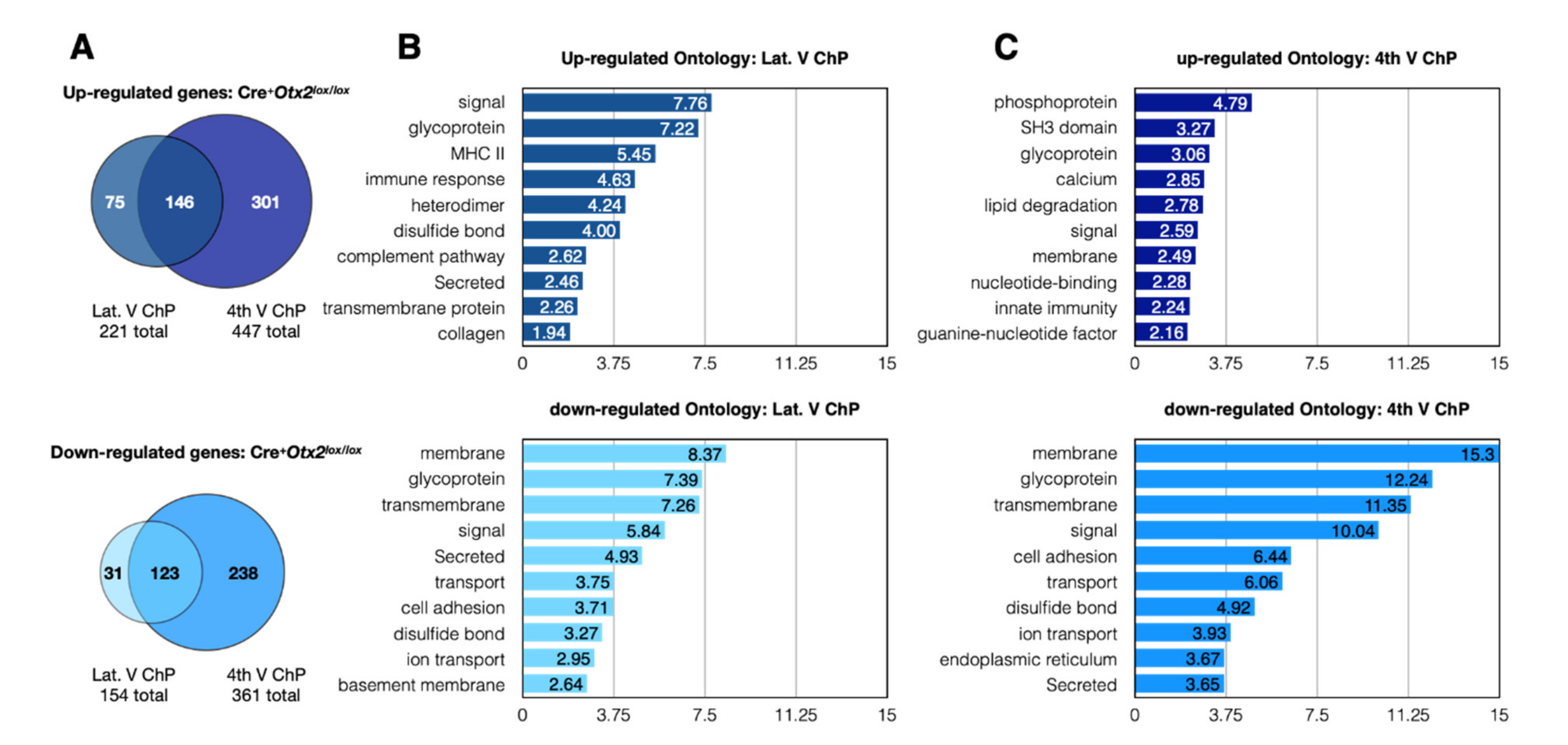
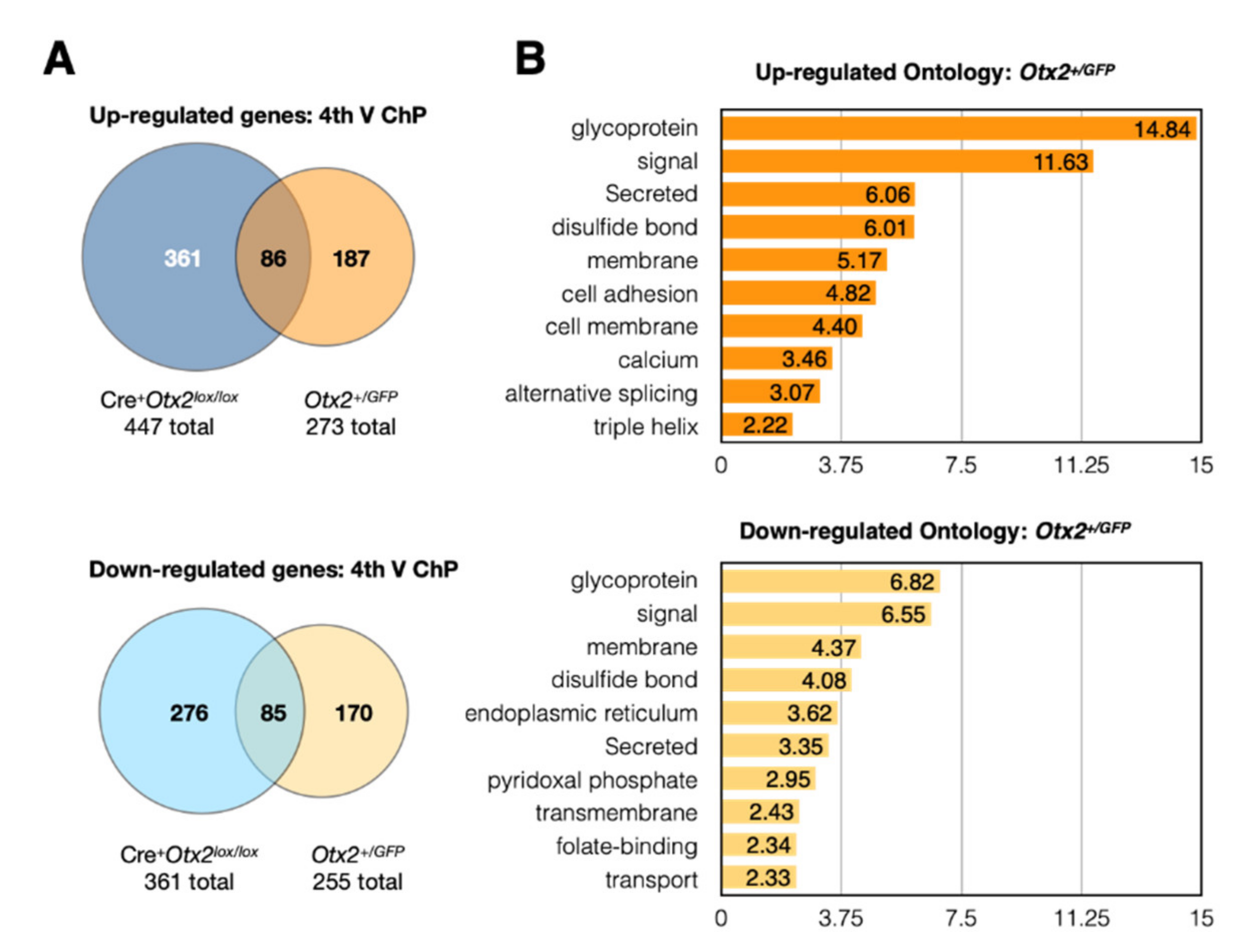
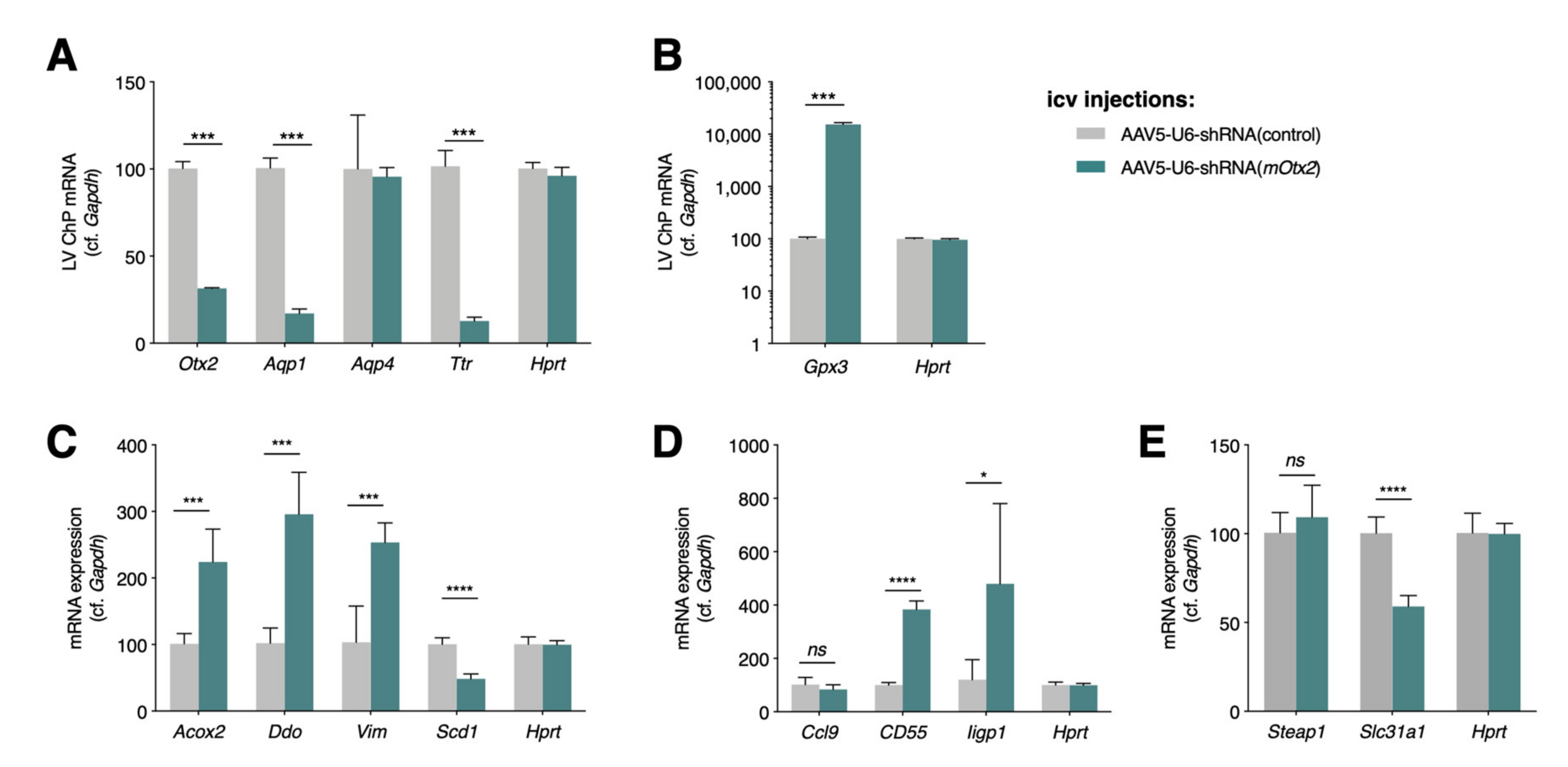
2.1. Conditional and Constitutive Knockdown of Otx2 in Adult ChP

OTX2 is a key regulator of ChP and brain development, but its role in adult ChP is not well known. To gain an insight into its adult ChP functions, we performed RNA sequencing analysis with two mouse models. The first consisted of 3-month-old Otx2lox/lox mice for the conditional knockdown of Otx2, specifically in the ChP, through intracerebroventricular (icv) injections of Cre-Tat recombinant protein, which leads to a ~50% reduction in (mRNA) Otx2 and a >70% reduction in OTX2 protein levels [5]. The ChPs from LV and 4V were dissected separately from both Cre-Tat-injected (Cre+Otx2lox/lox) and control vehicle-injected mice (Veh+Otx2lox/lox). While the bilateral stereotaxic injections of vehicle or Cre-Tat are performed only in the lateral ventricles, we have previously shown, with this protocol, that the level of Otx2 knockdown in 4V ChP is proportional to that in LV ChP [19]. Indeed, we found a 48% decrease in (mRNA) Otx2 (exon 2) in the LV ChP (vehicle, 7924 mean reads; Cre-Tat 4143 mean reads), and a 39% decrease in 4V ChP (vehicle, 6898 mean reads; Cre-Tat 4169 mean reads). The second model consisted of Otx2+/GFP mice as a constitutive heterozygous knockout mutant, with ~50% Otx2 protein levels compared to the wildtype [21]. For this model, given the significant overlap in gene expression changes in the LV and 4V of Cre+Otx2lox/lox mice (see below), only the 4V ChPs were dissected and pooled from 3-month-old wildtype and mutant mice. We found a 38% decrease in (mRNA) Otx2 (exon 2) in 4V ChP (wildtype, 6399 mean reads; Otx2+/GFP 3957 mean reads).
The transcriptomics analysis of adult ChP showed highly expressed genes that are involved in energy metabolism, protein signaling, solute transport, cell adhesion, the cytoskeleton, and chaperone activity (Table 1). While not in the same order of gene expression level, this list compares favorably with those obtained from other ChP transcriptomics studies [4,22,23]. The conditional adult mouse knockdown of Otx2 led to significant changes in the expression of 375 genes in LV ChP and 808 genes in 4V ChP (p-adj < 0.05). The top ten upregulated and downregulated genes show a range of functions, including solute transport, signaling, immune response, and trafficking (Table 2). While there is a significant overlap in the altered gene expression between the ChPs (Figure 1A), the 4V ChP seems to be more susceptible to loss of Otx2 activity. The response to Otx2 knockdown results in a rather even distribution of upregulation (522 genes) and downregulation (392 genes) when grouping both 4V and LV ChPs. However, ontological analysis reveals that the downregulated genes in both ChPs show higher levels of enrichment in significantly altered classes, suggesting that upregulated genes have more broadly distributed functions (Figure 1B,C). Interestingly, both ChPs have similar ontology enrichment in downregulated genes, indicating that Otx2 is generally important for the expression of membrane proteins, glycoproteins, signaling proteins, and cell adhesion proteins. While some of these functions are recapitulated in the upregulated genes, there is much more heterogeneity between the LV and 4V ChP. The LV ChP shows more immune response ontology, while the 4V ChP shows more signaling-related ontology. This suggests that conditional knockdown of Otx2 leads to altered ChP barrier function and ChP signaling, and can impact immune responses.
The constitutive heterozygote Otx2+/GFP adult mice showed significant expression changes in 528 genes of the 4V ChP (p-adj < 0.05), which is comparatively less than for conditional Otx2 knockdown in the 4V ChP (Figure 2A). Given that fewer genes are deregulated in this constitutive model, this suggests that compensatory mechanisms for countering reduced OTX2 levels were activated during development. The changes in gene expression were relatively balanced between upregulation (273 genes) and downregulation (255 genes), and ontology analysis revealed shared terms, including glycoprotein, signal, membrane-related, and secreted proteins (Figure 2B). The upregulated genes are also enriched for cell adhesion and alternative splicing, while downregulated genes are enriched for trafficking and transport. This suggests that brain-wide and life-long knockdown of Otx2 leads to altered ChP signaling, barrier functions, and brain homeostasis.
We hypothesized that genes that were deregulated in both conditional and constitutive models could be either direct targets of OTX2 transcription regulation or targets of important OTX2-dependent pathways. Comparison of gene expression changes in 4V ChP of these two models revealed an overlap of more than 80 genes, in both the upregulated and downregulated repertoires (Figure 2A). This represented about half of the identified expression changes in Otx2+/GFP mice, but less than a third of the changes in the conditional model. When genes from LV ChP conditional Otx2 knockdown are included in the analysis, we identified 42 genes that are globally upregulated and 34 genes that are globally downregulated (Table 3). To determine whether this list contains direct OTX2 transcription targets, we compared it with OTX2 chromatin immunoprecipitation experiments that were previously performed in mouse embryonic brain [9] or adult retina [14]. However, we found almost no overlap, with only Ttr being a common target. This suggests that the transcription-related activity of OTX2 is different in the adult choroid plexus and/or that these deregulated genes are downstream targets of OTX2-dependent pathways. It will be necessary to perform ChIPseq or CUT&RUN analysis of adult choroid plexus, to distinguish between these possibilities. Taken together, our analysis identifies new potential functions for Otx2 in the adult brain. We found upregulation of immune factors, specifically in the conditional Otx2 loss-of-function model, and deregulation of genes involved in cellular adhesion, trafficking, signaling, and secretion, in both knockdown models, suggesting altered ChP function and disruption of the ChP barriers.

2.2. Altered Expression of ChP Secreted Factors

Given that our various ontology analyses often evoked secreted factors, we focused on ChP factors secreted in CSF and implicated in embryonic and/or adult neurogenesis (Table 4), which is one of the identified functions of adult ChP [19,23,24]. The factors implicated in embryonic neurogenesis include SHH, BMPs, and WNTs [25]. While Shh expression was not observed (mean reads < 1) in either ChPs of the wildtype mice, which is consistent with published data [3], the 4V ChP of Otx2+/GFP mice (but not the ChPs of conditional Otx2 ChP knockdown mice) showed a significant increase in Shh expression. Between the various Bmp and Wnt family genes, only Bmp7 and Wnt2b were differentially expressed in Cre+Otx2lox/lox mice, as compared to Veh+Otx2lox/lox mice. Canonical Wnt signaling is perturbed in embryos with Otx2 4V ChP knockdown, which was attributed to the dysregulation of Wnt modulators, including Rspo1, Sfrp2, Sostdc1, Tgm2, and Wnt4, and to the increased levels of WNT4 and TGM2 in the CSF of mutant mice [2]. While Rspo1, Sfrp2, and Wnt4 were very poorly expressed and unchanged in both the LV and 4V ChP of Cre+Otx2lox/lox adult mice, the expression of Sfrp1, Sostdc1, and Tgm2 were significantly changed in 4V ChP (Table 4), suggesting adult OTX2 retains some embryonic functions. To further explore this hypothesis, we compared our RNA sequencing analysis of 4V ChP Cre+Otx2lox/lox to the previous microarray analysis of embryonic Otx2 knockdown, specifically in the hindbrain ChP, performed by Götz and colleagues [2]. They found 340 significantly (FDR < 10%, >2-fold change) expressed genes, with 135 genes upregulated and 225 genes downregulated. Compared to our adult knockdown, there was less than a 20% overlap with upregulated genes (24 of 135 genes) and an almost 30% overlap with downregulated genes (62 of 225 genes). Some of these genes are found among the top ten dysregulated 4V genes (Table 2) and the globally altered genes (Table 3), and they have functions related to cell adhesion, trafficking, and secretion. Given that Otx2 knockdown experiments in late embryonic development showed that OTX2 is necessary for 4V, but not LV, ChP maintenance [2], our results suggest that adult Otx2 expression could retain this maintenance function in 4V ChP.
We have previously shown that OTX2 that is secreted into the CSF from the ChP, can regulate adult neurogenesis non-cell autonomously, by transferring into the astrocytes in the SVZ and RMS, thereby affecting neuroblast migration [19]. This study also showed that ChP Otx2 knockdown in Cre+Otx2lox/lox adult mice, which will impact both cell- and non-cell-autonomous activity, also led to significantly reduced SVZ neurogenesis, suggesting that OTX2 may regulate secreted factors that are implicated in adult neurogenesis, through cell-autonomous effects in the ChP. IGF2 and SLIT1/2 regulate both embryonic and adult neurogenesis [24,26,27,28]. While Slit1 is not expressed and Slit3 is only weakly expressed in adult ChP, Slit2 is highly expressed, but shows no significant change in expression in the ChP, with reduced Otx2. Admittedly, there is a trend towards increased Slit2 expression and we cannot exclude the potential for biological relevance. Igf2 was significantly downregulated, more than 2-fold, in all the ChPs, upon Otx2 knockdown. However, there was a concomitant downregulation in Igfbp2, which can inhibit IGF2, suggesting that the level of IGF2 activity could be maintained through the compensatory reduction in inhibiting factors. Other factors influencing adult neurogenesis include amphiregulin (AREG) [29], FGF2 [30,31,32], and TGF-α [33], yet we found no significant change in their expression (and no detectible expression of Areg). Finally, other factors show more change in gene expression after acute Otx2 knockdown compared to constitutive knockdown. TGF-β negatively regulates adult neurogenesis [34], and Tgf-β2 is downregulated in both the ChPs of Cre+Otx2lox/lox mice (Table 4). Taken together, these minimal or compensatory changes in specific secreted signaling factors suggest that Otx2 expression in the ChP could have only a minor cell-autonomous role in regulating adult neurogenesis. This hypothesis is consistent with similar levels of decrease in adult neurogenesis, observed with both this ChP Otx2 knockdown model and the non-cell-autonomous-only OTX2 knockdown mouse model [19].

2.3. Altered Expression of Immune and Stress Factors

Given the altered expression of homeostasis and stress response-related factors in both the ChPs of conditional Otx2 knockdown mice, we turned to the viral expression of shRNA-Otx2 in LV and 4V ChPs. Intracerebroventricular-injected AAV5 results in ChP-specific expression [35,36], and provides a tool to acutely affect Otx2 expression in any mouse model. We validated this model by qPCR analysis, which showed a 69% decrease in (mRNA) Otx2 and a concomitant very large decrease in the expression of a known direct transcriptional target, transthyretin (Ttr) (Figure 3A). Comparing models, the decrease in (mRNA) Ttr in Cre+Otx2lox/lox mice was 45%, while it was 87% in the shRNA-Otx2 mice, which suggests that viral knockdown provides a more robust effect. TTR, the most highly expressed protein in ChP (Table 1), is secreted into CSF and transports thyroxin and retinol-binding protein, and has a role in regulating cognition and memory, psychological health, and emotions (for a recent review, see [37]), suggesting that OTX2 levels can potentially impact similar brain functions. Furthermore, the downregulation of aquaporin 1 (Aqp1) (Table 3, Figure 3A) was also confirmed, with Aqp4 as a negative control, suggesting that OTX2 can also regulate CSF water homeostasis.
In keeping with roles in brain homeostasis and surveillance, we also chose targets from ontology analysis (Figure 1), in functions related to oxidative stress, immune response, and metal ion transport. A surprising finding was the over 100-fold increase in glutathione peroxidase 3 (Gpx3), an extracellular enzyme that catalyzes the reduction in peroxidases and protects cells from oxidative damage, suggesting that a loss of OTX2 has a significant impact on cell physiology (Figure 3B). Other compensatory mechanisms against oxidative stress include decreased fatty acid oxidation (Scd1), increased peroxisome function (Acox2, Ddo), for countering oxidative stress and inflammation [38], and changes in the structural cell response (Vim) [39] (Figure 3C). Concerning the immune response (Figure 3D), we tested a complement activation factor (CD55), an inflammatory response chemokine (Ccl9), and an innate immune response factor (Iigp1). The direction of change in the expression of all of these factors, upon acute viral Otx2 knockdown, was consistent with the constitutive and conditional mouse models. It remains unclear whether the loss of OTX2 provokes oxidative stress, and thus indirect activation of genes such as Gpx3, or whether OTX2 regulates the genes involved in reactive oxygen species signaling and/or stress response. Finally, given their role in brain homeostasis, we also quantified factors related to metal ion transport (Steap1 and Slc31a) (Figure 3E), which had altered expression in the conditional Cre+Otx2lox/lox mice. Only Slc31a, which transports copper ion, had concomitant reduced expression upon Otx2 knockdown. These findings suggest that ChP function is greatly impacted by Otx2 expression level, opening the question of whether Otx2 overexpression in ChP would also deregulate homeostasis and illicit immune responses in a wildtype context or, on the contrary, rescue deficits in homeostasis in an aged or diseased animal.

2.4. Otx2 Protein Interactions

To further analyze OTX2 function in adult ChP, we performed several OTX2 co-immunoprecipitation (co-IP) experiments with mass spectrometry (MS) analysis, to identify potential protein partners. We previously discovered that OTX2 protein is secreted by the ChP into CSF, and accumulates non-cell autonomously in SVZ and RMS astrocytes [19] and VCx parvalbumin cells [5,40]. The identification of alternate protein partners in cell-autonomous and non-cell-autonomous contexts would suggest that OTX2 takes on specific roles after transferring between cells. To test this hypothesis, and to reinforce ChP analysis, we also performed OTX2 co-IP on lysates from adult mouse SVZ, RMS, and VCx.
We used three criteria to obtain a list of potential OTX2 protein interactions in the four brain structures (Table 5), as follows: (i) unique proteins with three or more peptides identified exclusively in OTX2 compared to IgG co-IP samples (unique protein, ≥3 peptides); (ii) proteins identified with three or more peptides in OTX2 co-IP samples and having a relative peptide difference greater than 50% compared to IgG co-IP (selected protein, ≥50% rel. ∆); and (iii) all small proteins (≤25 kDa) exclusive to OTX2 co-IP samples, regardless of peptide number (unique small protein, ≤25 kDa), given that they have fewer identifiable MS peptides. These lists were used for comparison between structures and for ontology analysis. We generated a list of 60 high-confidence protein partners of OTX2 in ChP that were common to all three ChP samples (Table 6). These proteins cover several functions, including cell adhesion, cell trafficking, cell signaling, metabolism, RNA binding, RNA processing, transcription, chromatin structure, and DNA repair. Interestingly, more than 10% (eight proteins) belong to the “tier 1” proteins identified in stress granules [41], which are involved in translational control and post-transcriptional regulation. Although this functional class was not identified by KEGG pathway analysis (see below), this is likely due to the absence of annotation, given the only recent emergence of updated comprehensive inventories of stress granule proteins. Thus, we can only hypothesize that OTX2 interacts with these granules, although this putative function is given weight by the presence of the PAX1 homeoprotein among the “tier 1” proteins, by the in vivo interaction between the EMX2 homeoprotein with the translation initiation factor eIF4E [42], and by the involvement of the PROX1 homeoprotein in liquid–liquid phase separation [43], which also underlies stress granule assembly [41]. Also of note are the putative partners MECP2 and MOV10, given that OTX2 has been shown to regulate MECP2 foci in the postnatal mouse visual and auditory cortex [16], and that the EN1 homeoprotein is involved in LINE-1 regulation [44].
Few OTX2 partners have been biochemically and functionally validated. One key partner during embryogenesis is MEIS2, which is a co-activator for mesencephalon specification [45]. Meis2 is expressed in ChP at low levels (170 mean reads in 4V ChP; 31 mean reads in LV ChP), as compared to OTX2 (5954 mean reads in 4V ChP and 6829 mean reads in LV ChP), and appears not to be a major partner of OTX2 in ChP. TLE4 is another identified protein partner of OTX2 during development, and allows repression of mesencephalon fate [46]. Despite its expression in ChP (798 mean reads in 4V ChP and 689 mean reads in LV ChP), TLE4 was not identified in our OTX2 co-IP, although this could be due to TLE4 being under the limit of detection or having peptides that are too hydrophobic or hydrophilic for MS detection. The potential absence of TLE4 suggests that OTX2 protein interactions depend strongly on the cell type and developmental context.
To identify novel OTX2 protein partners that are ubiquitous throughout the brain, we compared the lists from the four brain structures, as follows: ChP (pooled LV and 4V), SVZ, RMS, and VCx. Few high-confidence proteins (selected protein ≥ 50% rel. ∆), 14 in total, were common to the three non-cell-autonomous structures (Table 7). Of these 14 common proteins, 5 were also identified in the ChP. Interestingly, these top-ranked proteins include FIG4, VAC14, and PIKFYVE, which play a role in phosphatidylinositol(3,5)bisphosphate [PI(3,5)P2] regulation, in multivesicular body (MVB) biogenesis, and in endosome autophagy and trafficking [47], suggesting that OTX2 plays a role in vesicle transport or is carried via MVBs. Given that MVBs can give rise to extracellular vesicles, interaction with OTX2 may reinforce its role in regulating the pathways of extracellular protein expression that are identified in our RNA sequencing analysis.
To identify potential differences between cell-autonomous and non-cell-autonomous partners, we performed KEGG pathway analysis on OTX2 protein partners, for all structures individually (Table 8). No dramatic differences were found between the structures, suggesting conserved roles of OTX2 in cell-autonomous and non-cell-autonomous OTX2 target structures. Common to nearly all structures are metabolic pathways, RNA transport, oxidative phosphorylation, RNA processing, and spliceosome. Pathways that are specific to the ChP pertain to the maintenance of tight junctions, protein processing, and actin cytoskeleton regulation. Downregulation of the cell adhesion class was also identified in the RNAseq analysis of conditional Cre+Otx2-lox mice (Figure 1), suggesting a direct involvement of OTX2 both in gene regulation and cellular functions for cell-autonomous ChP maintenance. Of the 14 proteins in common between VCx, SVZ, and RMS, eight of them are involved in RNA processing, suggesting a novel function for OTX2. Although the spliceosome pathway was also enriched in ChP, these proteins stand out for their involvement in the U5 snRNP complex, exon junction complex, or mRNA export complex, whereas the spliceosome proteins identified in the ChP are either splicing co-factors, part of the SMN complex, or part of the U2 snRNP complex. Interestingly, OTX2 has been shown to bind the initiation factor eIF4e in GST pull-down experiments [42], while other homeoproteins have been shown to bind translation machinery [6,42,48] that is implicated in RNA export, transport, and translation. Taking the high-confidence partners together with KEGG pathway analysis, cell-autonomous OTX2 is likely implicated in the regulation of genomic landscape, the regulation and processing of RNA, the trafficking of signals, and the maintenance of cellular adhesion, while non-cell-autonomous OTX2 is more implicated in the processing of RNA.

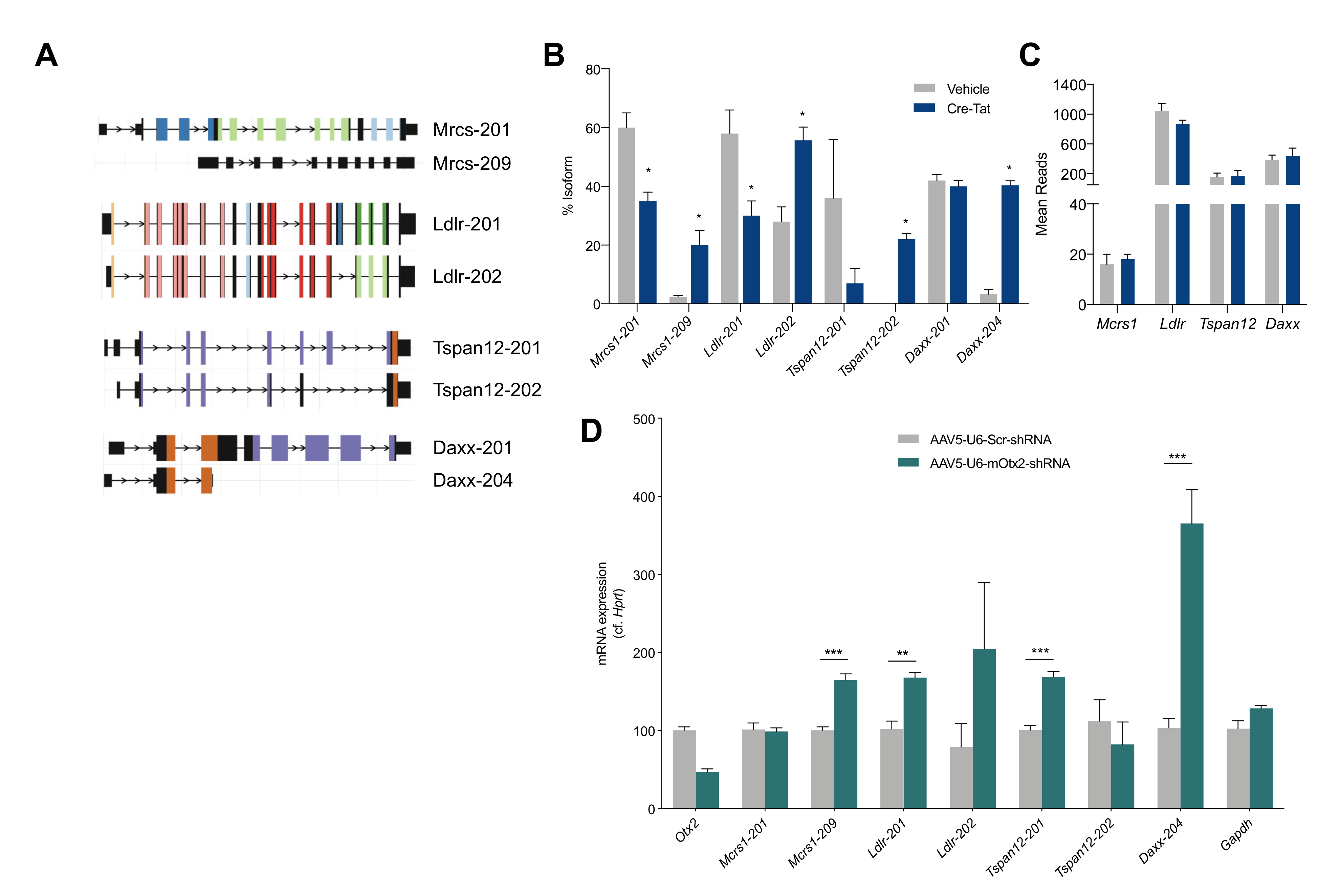
2.5. Splice Variant Analysis

Given the high confidence of OTX2 interaction with spliceosome pathway proteins, we extended the analysis of our transcriptomic data of LV ChP from Cre+Otx2lox/lox mice to measure changes in splice variants. The isoform usage was found to be significantly changed in the coding transcripts for only four genes (Mcrs, Ldlr, Tspan12, and Daxx), and generally for only two isoforms among the splice variants (Figure 4A,B). These genes showed no change in overall expression upon Otx2 knockdown (Figure 4C). Through acute Otx2 knockdown by the viral expression of shRNA-Otx2, we confirmed a significant increase in the expression of only the Mcrs-209 and Daxx-204 isoforms, as other isoforms either did not change significantly or changed in the opposite direction (Figure 4D). Interestingly, MCRS and DAXX interact within a protein complex with various nuclear functions, including transcription regulation, chromatin remodeling, and DNA repair. Further research is needed to determine the functional consequences of these changes in the distribution of transcript isoforms.
Homeoproteins have been postulated to regulate transcript splicing. The PAX6 homeoprotein has been shown to modulate splicing machinery, such that changes in Pax6 expression alter the population of tenascin-C splice variants without changing the total tenascin-C expression [49], while the CDX2 homeoprotein interacts with splicing machinery [50]. Regarding OTX2, its protein interactome in the adult retina revealed putative RNA processing partners, such as SFPQ and U2AF [15], while in the ChP and non-cell-autonomous structures, we found the potential partners ACIN1, DDX41, DDX46, HNRPLL, and PRRC2A, RBM25, RBM39, SF3A1, SF3B1, SNRNP200, and SRRM2 (Table 6 and Table 7). It remains to be determined whether OTX2 controls ChP splicing activity through direct interaction with splicing factors and/or by regulating their expression.

3. Materials and Methods

3.1. Animal Ethics Statement

All animal procedures, including housing, were carried out in accordance with the recommendations of the European Economic Community (2010/63/UE) and the French National Committee (2013/118). For surgical procedures, animals were anesthetized with xylazine (Rompun 2%, 5 mg/kg) and ketamine (Imalgene 500, 80 mg/kg). For biochemical analysis, mice either underwent transcardial perfusion or were sacrificed by cervical elongation.

3.2. Animals and Stereotaxic Surgery

Otx2lox/lox mice were kindly donated by T. Lamonerie [51] and Otx2+/GFP mice by A. Simeone [52]. Three-month-old Otx2lox/lox mice were injected with Cre-Tat or vehicle as described in [19] and housed for 15 days after surgery. The following serotype 5 adeno-associated viruses (AAV5) were purchased from Vector Biolabs (Malvern, PA, USA): AAV5-CMV-EGFP-U6-shRNA (control); and AAV5-CMV-EGFP-U6-shRNA (mOtx2). High-titer AAV5 (~1013 GC/mL) were injected (2 µL per mouse) bilaterally into the LV (coordinates from bregma: x, −0.58 mm; y, ±1.28 mm; z, −2 mm) with a 10 µL Hamilton syringe (0.2 µL/min). Virus-injected mice were housed for 3 weeks after surgery. Animals were an equal mix of males and females. The Otx2+/GFP mice, littermates, and the injected Otx2lox/lox mice underwent transcardial perfusion with 20 mL saline phosphate buffer, and ChPs were dissected and processed for biochemical analysis. Virus-injected mice were sacrificed by cervical elongation for ChP extraction.

3.3. Quantitative PCR Analysis

Total RNA from LV and 4V ChPs was extracted by using the RNeasy lipid tissue mini kit (Qiagen, Courtaboeuf, France) with DNA removal. Total RNA (10 to 20 ng) was retrotranscribed by using the QuantiTect reverse transcription kit (Qiagen, Courtaboeuf, France). Quantitative PCR (qPCR) analyses of cDNA (diluted at 1/10) were performed in triplicate with a LightCycler 480 II (Roche, Meylan, France) using the SYBR Green I master mix (Roche, Meylan, France). Gene-to-Hprt or gene-to-Gapdh ratios were determined by the 2−ΔΔCt method. For Otx2 expression analysis, expression was compared to mean expression of vehicle-injected mice of the same experiment.

3.4. RNA Sequencing Analysis

For analysis of conditional knockdown mice, the RNA was extracted separately from LV and 4V ChPs of Cre-Tat and vehicle-injected mice. A small sample of each ChP was tested by qPCR to ensure Cre-Tat samples had less than 50% Otx2 expression on average compared to control mice. Duplicate samples were prepared by pooling ChP lysates from 2 × 5 Cre-Tat-injected mice and from 2 × 4 vehicle-injected mice. For analysis of constitutive knockout mice, the RNA was extracted from pooled 4V ChPs of four 3-month-old Otx2+/GFP and five wildtype littermates, with duplicate samples (n = 2) of each genotype. PolyA+ mRNA purification, mRNA sequencing with technical replicates, and data normalization and quantification was performed by the Genomic Paris Center (IBENS, Paris, France) using Illumina HiSeq 1500 (Illumina, Evry, France).

3.5. Isoform Analysis

Raw reads were processed with FASTP [53] using standard parameters, and then pseudo-aligned on mm10 GENCODE transcriptome using salmon [54]. The quantified transcriptome was then imported in R using the IsoformSwitchAnalyzeR package [55,56] with dIFcutoff = 0.15. Isoform switch test was performed using DEXseq [57,58] in IsoformSwitchAnalyzeR. Gene coding potential, secondary structures, signal peptides, and protein domains were analyzed with CPAT [59], Net-Surf2 [60], SignalP [61], and Pfam [62], respectively.

3.6. Protein Co-Immunoprecipitation

For each ChP co-IP experiment, ChP from LV and 4V were pooled from four 3-month-old mice and were lysed with 1 mL lysis buffer (100 mM Tris pH 7.5, 1 mM EDTA, 100 mM NaCl, 1% NP40, 1 mM MgCl2, 1X protease/phosphatase inhibitor (Roche, Meylan, France)) containing 1µL of benzonase (Roche, Meylan, France). ChP were dissociated using 26G syringe and incubated 30 min on ice. Tubes were centrifuged at 21,000× g for 10 min and supernatant was recovered and divided in two. Each half was incubated with 44 µg of either anti-OTX2 (ab21990, Abcam, Paris, France) or anti-IgG (ab27478, Abcam, Paris, France) antibodies coupled to magnetic beads (10 mg/mL with 9.5 µg of antibody per mg of beads, Dynabeads antibody coupling kit, Invitrogen, Vilnius, Lithuania) in lysis buffer at 4 °C on rotating wheel overnight. Using magnetic separation, beads were washed 5 times in 1 mL of cold lysis buffer. Pelleted beads were eluted in 20 µL of laemmli buffer by heating 5 min at 95 °C, and then stored at −20 °C.
For SVZ, RMS, and VCx, tissue was pooled from ten 3-month-old wildtype mice and were lysed by trituration (pipette and 26G syringe) in 10 µL lysis buffer II (20 mM Tris pH 8, 120 mM NaCl, 1% NP40, 1 mM MgCl2, 5% glycerol, 1X protease/phosphatase inhibitor) per mg of tissue supplemented with 1 µL of benzonase per 1mL of lysis buffer II. Samples were processed as described above with 25 µL of antibody-coupled beads. Pelleted beads were eluted in 30 µL of laemmli buffer.

3.7. Mass Spectrometry Analysis

Proteomics analyses were performed by the Protein Mass Spectrometry Laboratory (Institut Curie, Paris, France). Eluted samples in laemmli were processed and resulting peptides were analyzed by nano-LC-MS/MS using an Ultimate 3000 system (Dionex, Thermo Fisher Scientific, Paris, France) coupled to an Orbitrap Fusion mass spectrometer (Q-OT-qIT, Thermo Fisher Scientific, Paris, France). Data were acquired using Xcalibur software and the resulting Mascot files (v2.5.1) were further processed by using myProMS software (v3.9) [63]. Percolator [64] was used for FDR calculations set to 1% peptide level. For ChP proteomics, three experiments were performed (N = 3). For SVZ, RMS and VCx proteomics, one experiment was performed (N = 1).

3.8. Ontology Analysis

Genes with >10 mean reads in at least one of the ChP samples were selected for ontology analysis. Differentially expressed gene lists were generated by using threshold of p-adj < 0.05. Ontology term enrichment and KEGG pathways were analyzed with DAVID Bioinformatic resource v6.7 [65,66] and ontology terms were plotted as -log10 scale of the enrichment p-values. UniProt (access date 21 August 2020, http://www.uniprot.org) was used for obtaining functional classes (Table 1, Table 3, Table 6 and Table 7). Gene list comparisons and Venn diagram data were generated with web-based tools (http://www.bioinformatics.lu/venn.php).

4. Conclusions

The ChP has barrier functions for controlling what gets in and out of the brain, and homeostasis functions for controlling brain metabolites in the CSF. The data in this present study allow us to go a step further, by imparting important ChP endocrine functions that are putatively regulated by the cell-autonomous and non-cell-autonomous activities of OTX2. Indeed, in addition to regulating the expression and post-transcriptional modification of genes encoding signaling and hormone-transport proteins that are secreted into the CSF, such as Igf2 and Ttr, OTX2 itself is secreted by the ChP and exerts essential non-cell-autonomous activities, such as the regulation of cerebral cortex plasticity or that of adult neurogenesis [5,19,67]. Transcriptomic analysis of different genetic Otx2 loss-of-function models, including conditional knockdown, specifically in the ChP, coupled with proteomic analysis, are the first steps towards a better understanding of the molecular biology of this traveling transcription factor in and out of its main cerebral source.

Author Contributions

Conceptualization, A.P. (Anabelle Planques), V.O.M. and A.A.D.N.; methodology, A.P. (Anabelle Planques), V.O.M., C.B. (Clémence Bernard), C.B. (Corinne Blugeon), F.D., V.M. and A.A.D.N.; software, D.B. and L.J.; validation, V.O.M., D.B., L.J., V.M. and A.A.D.N.; formal analysis, A.P. (Anabelle Planques), V.O.M. and A.A.D.N.; investigation, A.P. (Anabelle Planques), V.O.M., D.B., C.B. (Clémence Bernard), C.B. (Corinne Blugeon), F.D. and V.M.; resources, D.L. and A.P. (Alain Prochiantz).; data curation, A.P. (Anabelle Planques), V.O.M., D.B., L.J., V.M. and A.A.D.N.; writing—original draft preparation, A.A.D.N.; writing—review and editing, A.P. (Anabelle Planques), V.O.M., V.M., A.P. (Alain Prochiantz) and A.A.D.N.; visualization, A.A.D.N.; supervision, D.L., A.P. (Alain Prochiantz) and A.A.D.N.; project administration, A.A.D.N.; funding acquisition, A.P. (Alain Prochiantz). All authors have read and agreed to the published version of the manuscript.

Funding

This work was supported by the Région Ile-de-France DIM Cerveau et Pensée (to A.P. (Anabelle Planques)) and by the European Research Council (ERC-2013-ADG-339379 to A.P. (Alain Prochiantz)). The IBENS Genomics Core Facility was supported by the France Génomique national infrastructure, funded as part of the “Investissements d’Avenir” program managed by the Agence Nationale de la Recherche (ANR-10-INBS-09).

Institutional Review Board Statement

This research (project No. 00704.02) was approved by Ethics Committee n° 59 of the French Ministry for Research and Higher Education.

Informed Consent Statement

This work did not involve human subjects.

Data Availability Statement

RNA sequencing data are available with GEO accession GSE157386. Mass spectrometry data are available through the ProteomeXchange Consortium via the PRIDE repository with identifier PXD021244.

Acknowledgments

We wish to thank Namsuk Kim for help with tissue preparation.

Conflicts of Interest

The authors declare no conflict of interest.

References

  1. Fame, R.M.; Lehtinen, M.K. Emergence and Developmental Roles of the Cerebrospinal Fluid System. Dev. Cell 2020, 52, 261–275. [Google Scholar] [CrossRef]
  2. Johansson, P.A.; Irmler, M.; Acampora, D.; Beckers, J.; Simeone, A.; Götz, M. The transcription factor Otx2 regulates choroid plexus development and function. Development 2013, 140, 1055–1066. [Google Scholar] [CrossRef] [Green Version]
  3. Lun, M.P.; Johnson, M.B.; Broadbelt, K.G.; Watanabe, M.; Kang, Y.J.; Chau, K.F.; Springel, M.W.; Malesz, A.; Sousa, A.M.M.; Pletikos, M.; et al. Spatially heterogeneous choroid plexus transcriptomes encode positional identity and contribute to regional CSF production. J. Neurosci. 2015, 35, 4903–4916. [Google Scholar] [CrossRef]
  4. Dani, N.; Herbst, R.H.; McCabe, C.; Green, G.S.; Kaiser, K.; Head, J.P.; Cui, J.; Shipley, F.B.; Jang, A.; Dionne, D.; et al. A cellular and spatial map of the choroid plexus across brain ventricles and ages. Cell 2021, 184, 3056–3074.e21. [Google Scholar] [CrossRef]
  5. Spatazza, J.; Lee, H.H.C.; Di Nardo, A.A.; Tibaldi, L.; Joliot, A.; Hensch, T.K.; Prochiantz, A. Choroid-Plexus-Derived Otx2 Homeoprotein Constrains Adult Cortical Plasticity. Cell Rep. 2013, 3, 1815–1823. [Google Scholar] [CrossRef] [Green Version]
  6. Rezsohazy, R. Non-transcriptional interactions of Hox proteins: Inventory, facts, and future directions. Dev. Dyn. 2014, 243, 117–131. [Google Scholar] [CrossRef]
  7. Di Nardo, A.A.; Fuchs, J.; Joshi, R.L.; Moya, K.L.; Prochiantz, A. The physiology of homeoprotein transduction. Physiol. Rev. 2018, 98, 1943–1982. [Google Scholar] [CrossRef] [PubMed]
  8. Di Nardo, A.A.; Joliot, A.; Prochiantz, A. Homeoprotein transduction in neurodevelopment and physiopathology. Sci. Adv. 2020, 6, eabc6374. [Google Scholar] [CrossRef] [PubMed]
  9. Hoch, R.V.; Lindtner, S.; Price, J.D.; Rubenstein, J.L.R. OTX2 Transcription Factor Controls Regional Patterning within the Medial Ganglionic Eminence and Regional Identity of the Septum. Cell Rep. 2015, 12, 482–494. [Google Scholar] [CrossRef] [Green Version]
  10. Rohde, K.; Hertz, H.; Rath, M.F. Homeobox genes in melatonin-producing pinealocytes: Otx2 and Crx act to promote hormone synthesis in the mature rat pineal gland. J. Pineal Res. 2019, 66, e12567. [Google Scholar] [CrossRef] [PubMed]
  11. Zhang, J.; Zhang, M.; Acampora, D.; Vojtek, M.; Yuan, D.; Simeone, A.; Chambers, I. OTX2 restricts entry to the mouse germline. Nature 2018, 562, 595–599. [Google Scholar] [CrossRef]
  12. Matsuda, K.; Mikami, T.; Oki, S.; Iida, H.; Andrabi, M.; Boss, J.M.; Yamaguchi, K.; Shigenobu, S.; Kondoh, H. ChIP-seq analysis of genomic binding regions of five major transcription factors in mouse epiblast stem cells that highlights a central role for ZIC2. Development 2017, 144, 1948–1958. [Google Scholar] [CrossRef] [PubMed] [Green Version]
  13. Acampora, D.; Omodei, D.; Petrosino, G.; Garofalo, A.; Savarese, M.; Nigro, V.; di Giovannantonio, L.G.; Mercadante, V.; Simeone, A. Loss of the Otx2-Binding Site in the Nanog Promoter Affects the Integrity of Embryonic Stem Cell Subtypes and Specification of Inner Cell Mass-Derived Epiblast. Cell Rep. 2016, 15, 2651–2664. [Google Scholar] [CrossRef] [PubMed] [Green Version]
  14. Samuel, A.; Housset, M.; Fant, B.; Lamonerie, T. Otx2 ChIP-seq reveals unique and redundant functions in the mature mouse retina. PLoS ONE 2014, 9, e89110. [Google Scholar] [CrossRef] [Green Version]
  15. Fant, B.; Samuel, A.; Audebert, S.; Couzon, A.; El Nagar, S.; Billon, N.; Lamonerie, T. Comprehensive interactome of Otx2 in the adult mouse neural retina. Genesis 2015, 53, 685–694. [Google Scholar] [CrossRef]
  16. Apulei, J.; Kim, N.; Testa, D.; Ribot, J.; Morizet, D.; Bernard, C.; Jourdren, L.; Blugeon, C.; di Nardo, A.A.; Prochiantz, A. Non-cell Autonomous OTX2 Homeoprotein Regulates Visual Cortex Plasticity Through Gadd45b/g. Cereb. Cortex 2019, 29, 2384–2395. [Google Scholar] [CrossRef]
  17. Sakai, A.; Nakato, R.; Ling, Y.; Hou, X.; Hara, N.; Iijima, T.; Yanagawa, Y.; Kuwano, R.; Okuda, S.; Shirahige, K.; et al. Genome-wide target analyses of Otx2 homeoprotein in postnatal cortex. Front. Neurosci. 2017, 11, 307. [Google Scholar] [CrossRef]
  18. Peña, C.J.; Kronman, H.G.; Walker, D.M.; Cates, H.M.; Bagot, R.C.; Purushothaman, I.; Issler, O.; Loh, Y.-H.E.; Leong, T.; Kiraly, D.D.; et al. Early life stress confers lifelong stress susceptibility in mice via ventral tegmental area OTX2. Science 2017, 356, 1185–1188. [Google Scholar] [CrossRef] [PubMed] [Green Version]
  19. Planques, A.; Oliveira Moreira, V.; Dubreuil, C.; Prochiantz, A.; Di Nardo, A.A. OTX2 Signals from the Choroid Plexus to Regulate Adult Neurogenesis. eNeuro 2019, 6. [Google Scholar] [CrossRef] [PubMed] [Green Version]
  20. Sugiyama, S.; Di Nardo, A.A.; Aizawa, S.; Matsuo, I.; Volovitch, M.; Prochiantz, A.; Hensch, T.K. Experience-Dependent Transfer of Otx2 Homeoprotein into the Visual Cortex Activates Postnatal Plasticity. Cell 2008, 134, 508–520. [Google Scholar] [CrossRef] [PubMed] [Green Version]
  21. Bernard, C.; Kim, H.T.; Torero Ibad, R.; Lee, E.J.; Simonutti, M.; Picaud, S.; Acampora, D.; Simeone, A.; Di Nardo, A.A.; Prochiantz, A.; et al. Graded Otx2 activities demonstrate dose-sensitive eye and retina phenotypes. Hum. Mol. Genet. 2014, 23, 1742–1753. [Google Scholar] [CrossRef] [Green Version]
  22. Baruch, K.; Deczkowska, A.; David, E.; Castellano, J.M.; Miller, O.; Kertser, A.; Berkutzki, T.; Barnett-Itzhaki, Z.; Bezalel, D.; Wyss-Coray, T.; et al. Aging-induced type I interferon response at the choroid plexus negatively affects brain function. Science 2014, 346, 89–93. [Google Scholar] [CrossRef] [PubMed] [Green Version]
  23. Silva-Vargas, V.; Maldonado-Soto, A.R.; Mizrak, D.; Codega, P.; Doetsch, F. Age-Dependent Niche Signals from the Choroid Plexus Regulate Adult Neural Stem Cells. Cell Stem Cell 2016, 19, 643–652. [Google Scholar] [CrossRef] [PubMed] [Green Version]
  24. Lehtinen, M.K.; Zappaterra, M.W.; Chen, X.; Yang, Y.J.; Hill, A.D.; Lun, M.; Maynard, T.; Gonzalez, D.; Kim, S.; Ye, P.; et al. The Cerebrospinal Fluid Provides a Proliferative Niche for Neural Progenitor Cells. Neuron 2011, 69, 893–905. [Google Scholar] [CrossRef] [PubMed] [Green Version]
  25. Bjornsson, C.S.; Apostolopoulou, M.; Tian, Y.; Temple, S. It takes a village: Constructing the neurogenic niche. Dev. Cell 2015, 32, 435–446. [Google Scholar] [CrossRef] [Green Version]
  26. Nguyen-Ba-Charvet, K.T.; Picard-Riera, N.; Tessier-Lavigne, M.; Baron-Van Evercooren, A.; Sotelo, C.; Chédotal, A. Multiple Roles for Slits in the Control of Cell Migration in the Rostral Migratory Stream. J. Neurosci. 2004, 24, 1497–1506. [Google Scholar] [CrossRef] [PubMed] [Green Version]
  27. Sawamoto, K.; Wichterle, H.; Gonzalez-Perez, O.; Cholfin, J.A.; Yamada, M.; Spassky, N.; Murcia, N.S.; Garcia-Verdugo, J.M.; Marin, O.; Rubenstein, J.L.R.; et al. New neurons follow the flow of cerebrospinal fluid in the adult brain. Science 2006, 311, 629–632. [Google Scholar] [CrossRef]
  28. Ziegler, A.N.; Schneider, J.S.; Qin, M.; Tyler, W.A.; Pintar, J.E.; Fraidenraich, D.; Wood, T.L.; Levison, S.W. IGF-II promotes stemness of neural restricted precursors. Stem Cells 2012, 30, 1265–1276. [Google Scholar] [CrossRef] [Green Version]
  29. Falk, A.; Frisén, J. Amphiregulin is a mitogen for adult neural stem cells. J. Neurosci. Res. 2002, 69, 757–762. [Google Scholar] [CrossRef] [PubMed]
  30. Jin, K.; Sun, Y.; Xie, L.; Batteur, S.; Mao, X.O.; Smelick, C.; Logvinova, A.; Greenberg, D.A. Neurogenesis and aging: FGF-2 and HB-EGF restore neurogenesis in hippocampus and subventricular zone of aged mice. Aging Cell 2003, 2, 175–183. [Google Scholar] [CrossRef]
  31. Douet, V.; Kerever, A.; Arikawa-Hirasawa, E.; Mercier, F. Fractone-heparan sulphates mediate FGF-2 stimulation of cell proliferation in the adult subventricular zone. Cell Prolif. 2013, 46, 137–145. [Google Scholar] [CrossRef]
  32. Hayamizu, T.F.; Chan, P.T.; Johanson, C.E. FGF-2 immunoreactivity in adult rat ependyma and choroid plexus: Responses to global forebrain ischemia and intraventricular FGF-2. Neurol. Res. 2001, 23, 353–358. [Google Scholar] [CrossRef] [PubMed]
  33. Tropepe, V.; Craig, C.G.; Morshead, C.M.; Van Kooy, D. Der Transforming growth factor-α null and senescent mice show decreased neural progenitor cell proliferation in the forebrain subependyma. J. Neurosci. 1997, 17, 7850–7859. [Google Scholar] [CrossRef]
  34. Wachs, F.P.; Winner, B.; Couillard-Despres, S.; Schiller, T.; Aigner, R.; Winkler, J.; Bogdahn, U.; Aigner, L. Transforming growth factor-β1 is a negative modulator of adult neurogenesis. J. Neuropathol. Exp. Neurol. 2006, 65, 358–370. [Google Scholar] [CrossRef] [PubMed] [Green Version]
  35. Watson, D.J.; Passini, M.A.; Wolfe, J.H. Transduction of the choroid plexus and ependyma in neonatal mouse brain by vesicular stomatitis virus glycoprotein-pseudotyped lentivirus and adeno-associated virus type 5 vectors. Hum. Gene Ther. 2005, 16, 49–56. [Google Scholar] [CrossRef]
  36. Arnaud, K.; Moreira, V.O.; Vincent, J.; Dallerac, G.; le Poupon, C.; Richter, M.; Müller, U.; Rondi-Reig, L.; Prochiantz, A.; di Nardo, A. Choroid plexus APP regulates adult brain proliferation and animal behavior. bioRxiv 2019. [Google Scholar] [CrossRef] [Green Version]
  37. Sharma, M.; Khan, S.; Rahman, S.; Singh, L.R. The Extracellular Protein, Transthyretin Is an Oxidative Stress Biomarker. Front. Physiol. 2019, 10, 5. [Google Scholar] [CrossRef]
  38. Terlecky, S.R. Peroxisomes, oxidative stress, and inflammation. World J. Biol. Chem. 2012, 3, 93. [Google Scholar] [CrossRef]
  39. De Pablo, Y.; Nilsson, M.; Pekna, M.; Pekny, M. Intermediate filaments are important for astrocyte response to oxidative stress induced by oxygen-glucose deprivation and reperfusion. Histochem. Cell Biol. 2013, 140, 81–91. [Google Scholar] [CrossRef]
  40. Kim, N.; Acampora, D.; Dingli, F.; Loew, D.; Simeone, A.; Prochiantz, A.; Di Nardo, A.A. Immunoprecipitation and mass spectrometry identify non-cell autonomous Otx2 homeoprotein in the granular and supragranular layers of mouse visual cortex. F1000Research 2014, 3, 178. [Google Scholar] [CrossRef] [PubMed] [Green Version]
  41. Youn, J.Y.; Dyakov, B.J.A.; Zhang, J.; Knight, J.D.R.; Vernon, R.M.; Forman-Kay, J.D.; Gingras, A.C. Properties of Stress Granule and P-Body Proteomes. Mol. Cell 2019, 76, 286–294. [Google Scholar] [CrossRef]
  42. Nédélec, S.; Foucher, I.; Brunet, I.; Bouillot, C.; Prochiantz, A.; Trembleau, A. Emx2 homeodomain transcription factor interacts with eukaryotic translation initiation factor 4E (eIF4E) in the axons of olfactory sensory neurons. Proc. Natl. Acad. Sci. USA 2004, 101, 10815–10820. [Google Scholar] [CrossRef] [Green Version]
  43. Liu, X.; Shen, J.; Xie, L.; Wei, Z.; Wong, C.; Li, Y.; Zheng, X.; Li, P.; Song, Y. Mitotic Implantation of the Transcription Factor Prospero via Phase Separation Drives Terminal Neuronal Differentiation. Dev. Cell 2020, 52, 277–293.e8. [Google Scholar] [CrossRef] [PubMed]
  44. Blaudin de Thé, F.; Rekaik, H.; Peze-Heidsieck, E.; Massiani-Beaudoin, O.; Joshi, R.L.; Fuchs, J.; Prochiantz, A. Engrailed homeoprotein blocks degeneration in adult dopaminergic neurons through LINE-1 repression. EMBO J. 2018, 37, e97374. [Google Scholar] [CrossRef]
  45. Agoston, Z.; Schulte, D. Meis2 competes with the Groucho co-repressor Tle4 for binding to Otx2 and specifies tectal fate without induction of a secondary midbrain-hindbrain boundary organizer. Development 2009, 136, 3311–3322. [Google Scholar] [CrossRef] [Green Version]
  46. Heimbucher, T.; Murko, C.; Bajoghli, B.; Aghaallaei, N.; Huber, A.; Stebegg, R.; Eberhard, D.; Fink, M.; Simeone, A.; Czerny, T. Gbx2 and Otx2 Interact with the WD40 Domain of Groucho/Tle Corepressors. Mol. Cell. Biol. 2007, 27, 340–351. [Google Scholar] [CrossRef] [Green Version]
  47. McCartney, A.J.; Zhang, Y.; Weisman, L.S. Phosphatidylinositol 3,5-bisphosphate: Low abundance, high significance. BioEssays 2014, 36, 52–64. [Google Scholar] [CrossRef] [PubMed] [Green Version]
  48. Topisirovic, I.; Kentsis, A.; Perez, J.M.; Guzman, M.L.; Jordan, C.T.; Borden, K.L.B. Eukaryotic Translation Initiation Factor 4E Activity Is Modulated by HOXA9 at Multiple Levels. Mol. Cell. Biol. 2005, 25, 1100–1112. [Google Scholar] [CrossRef] [PubMed] [Green Version]
  49. Von Holst, A.; Egbers, U.; Prochiantz, A.; Faissner, A. Neural stem/progenitor cells express 20 tenascin C isoforms that are differentially regulated by Pax6. J. Biol. Chem. 2007, 282, 9172–9181. [Google Scholar] [CrossRef] [Green Version]
  50. Balbinot, C.; Vanier, M.; Armant, O.; Nair, A.; Penichon, J.; Soret, C.; Martin, E.; Saandi, T.; Reimund, J.M.; Deschamps, J.; et al. Fine-tuning and autoregulation of the intestinal determinant and tumor suppressor homeobox gene CDX2 by alternative splicing. Cell Death Differ. 2017, 24, 2173–2186. [Google Scholar] [CrossRef] [Green Version]
  51. Fossat, N.; Chatelain, G.; Brun, G.; Lamonerie, T. Temporal and spatial delineation of mouse Otx2 functions by conditional self-knockout. EMBO Rep. 2006, 7, 824–830. [Google Scholar] [CrossRef] [Green Version]
  52. Acampora, D.; di Giovannantonio, L.G.; di Salvio, M.; Mancuso, P.; Simeone, A. Selective inactivation of Otx2 mRNA isoforms reveals isoform-specific requirement for visceral endoderm anteriorization and head morphogenesis and highlights cell diversity in the visceral endoderm. Mech. Dev. 2009, 126, 882–897. [Google Scholar] [CrossRef]
  53. Chen, S.; Zhou, Y.; Chen, Y.; Gu, J. Fastp: An ultra-fast all-in-one FASTQ preprocessor. Bioinformatics 2018, 34, i884–i890. [Google Scholar] [CrossRef] [PubMed]
  54. Patro, R.; Duggal, G.; Love, M.I.; Irizarry, R.A.; Kingsford, C. Salmon provides fast and bias-aware quantification of transcript expression. Nat. Methods 2017, 14, 417–419. [Google Scholar] [CrossRef] [PubMed] [Green Version]
  55. Vitting-Seerup, K.; Sandelin, A. IsoformSwitchAnalyzeR: Analysis of changes in genome-wide patterns of alternative splicing and its functional consequences. Bioinformatics 2019, 35, 4469–4471. [Google Scholar] [CrossRef]
  56. Vitting-Seerup, K.; Sandelin, A. The landscape of isoform switches in human cancers. Mol. Cancer Res. 2017, 15, 1206–1220. [Google Scholar] [CrossRef] [PubMed] [Green Version]
  57. Anders, S.; Reyes, A.; Huber, W. Detecting differential usage of exons from RNA-seq data. Genome Res. 2012, 22, 2008–2017. [Google Scholar] [CrossRef]
  58. Ritchie, M.E.; Phipson, B.; Wu, D.; Hu, Y.; Law, C.W.; Shi, W.; Smyth, G.K. Limma powers differential expression analyses for RNA-sequencing and microarray studies. Nucleic Acids Res. 2015, 43, e47. [Google Scholar] [CrossRef] [PubMed]
  59. Wang, L.; Park, H.J.; Dasari, S.; Wang, S.; Kocher, J.P.; Li, W. CPAT: Coding-potential assessment tool using an alignment-free logistic regression model. Nucleic Acids Res. 2013, 41, e74. [Google Scholar] [CrossRef] [PubMed]
  60. Klausen, M.S.; Jespersen, M.C.; Nielsen, H.; Jensen, K.K.; Jurtz, V.I.; Sønderby, C.K.; Sommer, M.O.A.; Winther, O.; Nielsen, M.; Petersen, B.; et al. NetSurfP-2.0: Improved prediction of protein structural features by integrated deep learning. Prot. Struct. Funct. Bioinform. 2019, 87, 520–527. [Google Scholar] [CrossRef] [Green Version]
  61. Almagro Armenteros, J.J.; Tsirigos, K.D.; Sønderby, C.K.; Petersen, T.N.; Winther, O.; Brunak, S.; von Heijne, G.; Nielsen, H. SignalP 5.0 improves signal peptide predictions using deep neural networks. Nat. Biotechnol. 2019, 37, 420–423. [Google Scholar] [CrossRef] [PubMed]
  62. Finn, R.D.; Bateman, A.; Clements, J.; Coggill, P.; Eberhardt, R.Y.; Eddy, S.R.; Heger, A.; Hetherington, K.; Holm, L.; Mistry, J.; et al. Pfam: The protein families database. Nucleic Acids Res. 2014, 42, D222–D230. [Google Scholar] [CrossRef] [PubMed] [Green Version]
  63. Poullet, P.; Carpentier, S.; Barillot, E. myProMS, a web server for management and validation of mass spectrometry-based proteomic data. Proteomics 2007, 7, 2553–2556. [Google Scholar] [CrossRef]
  64. Spivak, M.; Weston, J.; Bottou, L.; Käll, L.; Noble, W.S. Improvements to the percolator algorithm for peptide identification from shotgun proteomics data sets. J. Proteome Res. 2009, 8, 3737–3745. [Google Scholar] [CrossRef] [Green Version]
  65. Huang, D.W.; Sherman, B.T.; Lempicki, R.A. Systematic and integrative analysis of large gene lists using DAVID bioinformatics resources. Nat. Protoc. 2009, 4, 44–57. [Google Scholar] [CrossRef] [PubMed]
  66. Huang, D.W.; Sherman, B.T.; Lempicki, R.A. Bioinformatics enrichment tools: Paths toward the comprehensive functional analysis of large gene lists. Nucleic Acids Res. 2009, 37, 1–13. [Google Scholar] [CrossRef] [Green Version]
  67. Vincent, C.; Gilabert-Juan, J.; Gibel-Russo, R.; Alvarez-Fischer, D.; Krebs, M.-O.; Le Pen, G.; Prochiantz, A.; Di Nardo, A.A. Non-cell-autonomous OTX2 transcription factor regulates anxiety-related behavior in the mouse. Mol. Psychiatry 2021, 1–12. [Google Scholar] [CrossRef]
Figure 1. Changes in gene expression after conditional Otx2 knockdown in choroid plexus. (A) Venn diagrams of the number of up- or down-regulated genes (p-adj < 0.05) from RNAseq analysis of ChP of Cre-Tat icv-injected Otx2lox/lox mice (Cre+Otx2lox/lox) compared to vehicle-injected mice. The diagrams compare the overlap between lateral ventricle (Lat. V ChP) and fourth ventricle (4th V ChP) differentially regulated genes; (B) Ontology analysis of differentially regulated genes in lateral ventricle ChP after conditional Otx2 knockdown; (C) Ontology analysis of differentially regulated genes in fourth ventricle ChP after conditional Otx2 knockdown.
Ijms 22 08951 g001
Figure 2. Gene expression in choroid plexus of Otx2+/GFP mice. (A) Venn diagrams of the number of up- or down-regulated genes (p-adj < 0.05) to compare overlap between fourth ventricle (4th V ChP) changes in Otx2+/GFP mice and in Cre-Tat icv-injected Otx2lox/lox mice (Cre+Otx2lox/lox); (B) Ontology analysis of differentially regulated genes in Otx2+/GFP mice.
Ijms 22 08951 g002
Figure 3. OTX2 regulates choroid plexus expression of oxidative stress, immune system, and metal transport genes. Quantitative PCR analysis of lateral ventricle (LV) ChP gene expression in wildtype mice after viral expression of shRNA against mouse (mRNA) Otx2 (shRNA-mOtx2) compared to control shRNA. (A) Analysis of control genes to validate shRNA-Otx2 activity; (B) Otx2 knockdown induces high expression of Gpx3; (C) Analysis of select genes involved in oxidative stress response; (D) Analysis of select genes involved in immune system response; (E) Analysis of select genes involved in metal ion transport (all values: mean ± SEM; n = 5; t-test; * p < 0.05, *** p < 0.001, **** p < 0.0001).
Ijms 22 08951 g003
Figure 4. Analysis of splice variations induced by Otx2 knockdown in choroid plexus. (A) Selected isoforms of genes of interest. Locus length is in arbitrary units. Colors represent different protein domains within a given gene; (B) Isoform usage, shown as % of total isoforms, in lateral ventricle (LV) ChP from vehicle and Cre-Tat icv-injected Otx2lox/lox mice; (C) Mean reads from transcriptomic analysis of LV ChP from vehicle and Cre-Tat icv-injected Otx2lox/lox mice; (D) Quantitative PCR analysis of LV ChP isoform expression in wildtype mice after viral expression of shRNA against mouse (mRNA) Otx2 (shRNA-Otx2) (all values: mean ± SEM; n = 5; t-test; * p < 0.05, ** p < 0.01, *** p < 0.001).
Ijms 22 08951 g004
Table 1. Top 50 genes expressed in lateral ventricle (LV) and fourth ventricle (4V) choroid plexus (ChP). Significantly different expression between structures is indicated in bold.
Gene SymbolFunctionMean Reads
Combined
Mean Reads
4V ChP
Mean Reads
LV ChP
Fold Change
LV vs. 4V
p-adj
TtrT4 and retinol transport2,623,5412,348,3042,898,7791.20.4700
Enpp2Extracellular signaling593,932612,769575,0940.941
Malat1RNA processing112,726157,39568,0580.430.1482
ApoeLipid transport64,48254,60074,3651.40.7231
Trpm3Cation channel39,62746,15033,1050.720.1606
BsgCell adhesion49,35441,70157,0071.40.5023
KlCell signaling45,04440,66749,4221.20.4792
Abhd2Lipid metabolism41,09436,40545,7831.30.2413
AY036118Hemopoiesis25,99233,90318,0810.531
Slc4a10Solute transport30,24230,56329,9200.981
PsapTrophic, metabolism33,69528,76838,6231.30.0646
Igfbp2IGF-binding25,33628,55622,1170.770.4457
Hspa5ER chaperone25,42624,96025,8911.01
F5Hemostasis25,75824,67226,8431.10.9813
Slc12a2Solute transport23,86924,27923,4590.971
CtsdAPP processing28,20023,67232,7281.40.0423
PrlrHormone receptor27,45222,72832,1761.40.7565
Atp1a1Ion transport22,89622,06723,7241.11
CluExtracellular chaperone26,74721,75031,7441.50.0323
AppCell adhesion, signaling19,29021,19717,3830.820.4130
Cntn1Cell adhesion25,88821,01730,7591.50.0175
Atp2b3Ion transport20,58019,74521,4151.11
Ahcyl2Solute transport20,72619,42722,0251.10.8161
Igf2Growth hormone24,97818,97330,9841.60.0123
Hsp90b1Chaperone18,81618,51419,1171.01
Sptbn1Cytoskeleton18,88318,27619,4901.11
CpeProhormone processing17,18217,44916,9160.971
Car12Metabolism18,52917,27719,7821.10.7979
Clic6Ion channel16,95316,43217,4731.11
Strip2Cytoskeleton14,77616,18713,3660.830.4745
Timp3Collagenase inhibitor18,53015,56221,4991.40.0292
Itpr1ER Ca2+ release16,25215,54616,9571.10.9741
Kcne2Potassium channel13,86115,41612,3050.80.8581
Cgnl1Cell adhesion15,18315,29215,0730.991
Gpm6aMembrane structure22,45615,18129,7322.00.0000
Slc4a2Solute carrier15,50114,91216,0901.11
Atp5a1Metabolism15,92214,76617,0781.20.7456
Nsg2Vesicle trafficking15,58714,66916,5061.10.8949
Zbtb20Transcription factor12,60014,60310,5970.730.5636
Stk39Stress response14,41714,50714,3260.991
Tmem72 14,82114,34315,2981.11
Cab39lCell polarity15,43814,31116,5651.20.8214
Nedd4Ubiquitination15,83714,27217,4021.20.4738
Macf1Cytoskeleton12,70414,02711,3820.810.5999
Vat1l 14,58913,98315,1961.11
Hsp90ab1Chaperone14,36813,75614,9791.11
CalrChaperone13,88913,60614,1711.01
Htr2cSerotonin receptor13,45313,34313,5641.01
Slc5a3Solute transport11,64813,28110,0150.750.0914
Sptan1Cytoskeleton, Secretion11,91313,16110,6650.810.3292
Table 2. Top 10 differentially expressed genes in choroid plexus of Cre+Otx2lox/lox mice. Genes in fourth ventricle with * are also deregulated upon embryonic Otx2 knockdown in the hindbrain choroid plexus [2].
Gene SymbolMean Reads VehicleMean Reads Cre-TatFold Changep-adj
Upregulated in lateral ventricle ChP
Igkv1-1350.633610.0000
Slc1a62.182390.0000
Mup51234119340.0000
Gpx32957229250.0000
Ighv1-671.636220.0000
Saa32.247220.0000
Tnfrsf11b6.3116180.0000
Cacnb324325130.0000
Ndnf891098120.0000
Gm48413.844120.0000
Downregulated in lateral ventricle ChP
Ngfr44483−5.30.0000
Nrn11924496−3.90.0000
B3galt210931−3.60.0007
Dazl20760−3.40.0000
Itga1018454−3.40.0000
Slc26a71670533−3.10.0000
Steap12224735−3.00.0000
Defb11473169−2.80.0000
Entpd324087−2.80.0001
Ccl92037774−2.60.0000
Upregulated in 4th ventricle ChP
9030619P08Rik0.0028.6infinite0.0000
Tmigd10.0033.1infinite0.0000
A730020M07Rik1.376610.0000
Gpx318110,780590.0000
Cacnb318778430.0000
Mup5261053410.0000
Fmod *2868688300.0000
Slitrk64.7125270.0000
Ndnf842070250.0000
Adcy810233230.0000
Downregulated in 4th ventricle ChP
Ngfr684159−4.30.0000
Steap1 *1225326−3.80.0000
Elfn110428−3.70.0000
Gnmt12233−3.70.0000
Gm2265014140−3.50.0000
Mir4489528−3.40.0002
Igf2os8926−3.40.0005
B3galt214144−3.20.0000
Slc26a7843271−3.10.0000
Crhr2504164−3.10.0000
Table 3. Choroid plexus genes with significant expression changes in both Otx2 knockdown models, including lateral ventricle and fourth ventricle from Cre+Otx2lox/lox mice and fourth ventricle from Otx2+/GFP mice. Genes with * are also deregulated upon embryonic Otx2 knockdown in the hindbrain choroid plexus [2].
Up-RegulatedFunctionDownregulatedFunction
Adora1Adenosine receptorAqp1 *Osmotic gradient
Arrb1Receptor signalingAtp2b4Ion transport
Atp1a2 *Ion transportB3galt2Glycosylation
Cadm1Cell adhesionElfn1Signaling cascade
Cd55Complement cascadeEntpd3
Cfap46 Fam132aGlucose uptake
Chn2Signaling cascadeIgf2Growth factor
Col11a1Collagen II fibrilsIns2Glucose uptake
Col1a2Collagen I fibrilsKalrnSignaling cascade
Edn3VasoconstrictionKlhl36Ubiquitination
Eva1aCell deathMapk9Cell signaling
Fam211b Myo5b *Cell trafficking
Fgf1Growth factorMyrip *Cell trafficking
Flrt1FGF signalingNav3Immune response
Fmod *Collagen I and II fibrilsOtx2Transcription factor
GdaMetabolismPcnx *
Gpx3Oxidative stressPitpnm1 *Cytoskeleton
Hopx *Chromatin structurePomgnt1Metabolism
LaynHyaluronan receptorRcn1ER regulation
Lrrc18SpermatogenesisScg5Cell secretion
Mapk10Cell signalingSfrp1Wnt signaling
Matn2Extracellular matrixSlc29a4Cation transport
Megf11 *Cell adhesionSlc2a12 *Glucose transport
Mlc1Osmotic gradientSlc35f1Solute transport
Mup5Pheromone activitySlc41a2Magnesium transport
NdnfCell adhesion, growthStra6Retinol transport
Ndrg3 Tbc1d2Cell adhesion
Pi15Protease inhibitorTbcdCytoskeleton
Plin4Adipocyte formationThumpd3
Rufy4AutophagyTmem255b
Sel1l3 Tmprss11aCellular senescence
Sema5a *Cell adhesionTspan33 *Notch signaling
Shisal1 TtrRetinol and T4 transport
Smrp1Cilia functionWdr17 *
Sncg *Neurofilament network
Sned1 *
Sorcs2Signaling cascade
Sorl1Cell trafficking
Sulf2Extracellular matrix
Tm4sf1
VimCell filaments
Vwa5b1
Table 4. Expression of secreted factors in Otx2 knockdown experiments.
GeneChoroid PlexusMean Reads, Control (Either Veh or Wildtype)Mean Reads, Knockdown (Either Cre-Tat or Otx2+/GFP)Fold Changep-adj
Bmp7Otx2lox/lox LV512570901.40.1845
Otx2lox/lox 4V254740361.60.0014
Otx2+/GFP 4V221522611.01
Wnt2bOtx2lox/lox LV10666.60.0002
Otx2lox/lox 4V12665.40.0001
Otx2+/GFP 4V29351.21
Tgm2Otx2lox/lox LV2764221.50.2496
Otx2lox/lox 4V1382651.90.0040
Otx2+/GFP 4V3884041.01
Sfrp1Otx2lox/lox LV853645110.530.0000
Otx2lox/lox 4V512127260.530.0000
Otx2+/GFP 4V408720010.490.0392
Sostdc1Otx2lox/lox LV862744290.510.0000
Otx2lox/lox 4V541227930.520.0076
Otx2+/GFP 4V893284581.11
ShhOtx2lox/lox LV01.4infinite1
Otx2lox/lox 4V0.74.66.61
Otx2+/GFP 4V064infinite0.0000
Slit2Otx2lox/lox LV230226741.21
Otx2lox/lox 4V347148031.40.7159
Otx2+/GFP 4V432360991.40.6487
Fgf2Otx2lox/lox LV1401230.881
Otx2lox/lox 4V64841.30.9878
Otx2+/GFP 4V67661.01
AregOtx2lox/lox LV00.9infinite1
Otx2lox/lox 4VNANANANA
Otx2+/GFP 4V00NANA
Tgf-αOtx2lox/lox LV214014250.670.0620
Otx2lox/lox 4V13579990.740.1611
Otx2+/GFP 4V104016731.60.2163
Tgf-β2Otx2lox/lox LV923347470.510.0000
Otx2lox/lox 4V375521690.580.0000
Otx2+/GFP 4V332425630.770.3602
Igf2Otx2lox/lox LV42,33619,1440.450.0008
Otx2lox/lox 4V18,71780870.430.0000
Otx2+/GFP 4V42,54217,1970.400.0000
Igfbp2Otx2lox/lox LV30,19520,9920.700.3255
Otx2lox/lox 4V28,16613,2350.470.0003
Otx2+/GFP 4V47,52921,6600.460.0000
Table 5. Summary of protein lists and criteria for identification of putative OTX2 protein partners.
List NameChoroid PlexusSVZRMSVisual Cortex
Total proteins OTX24814113824252644
Total proteins IgG3602177622742667
Unique proteins OTX2 (≥3 peptides)392174029
Unique proteins IgG (≥3 peptides)591392225
Selected proteins OTX2 (≥50% rel. ∆)65367537
Unique small proteins Otx2 (≤25 kDa)182316848
Total OTX2 partners1195 of 481452 of 1138180 of 2425109 of 2644
Table 6. High-confidence OTX2 putative protein partners in choroid plexus identified by MS analysis. Proteins indicated in bold are unique for OTX2 co-IP, proteins with * are “tier 1” stress granule proteins.
ProteinFunctionProteinFunction
ABCF1 *TranslationMAP4Cytoskeleton
ACOT11Lipid metabolismMCM3APRNA export
AGO1 *RNA silencingMECP2Transcription, epigenetics
APCCell adhesionMLYCDMetabolism
ARHGEF6TraffickingMOV10 *RNA and LINE-1 silencing
ARHGEF7Trafficking, cell adhesionMSI2 *Translation
ARVCFCell adhesionMYCBP2Transcription
CDH2Cell adhesionNFATC2Signaling
CDH3Cell adhesionPIKFYVETrafficking
CHD4Cell adhesionPITPNM2Trafficking
CTNNA1Cell adhesionPOLDIP3Translation
CTNNA2Cell adhesionPRRC2A *RNA splicing, stress granule
CTNNB1Cell adhesionRBM39RNA splicing
DDX41RNA splicingRHOT1Mitochondrial trafficking
EDC4 *RNA processingRPL19
EPB41L5Cell adhesionRPL21Translation
ERBINSignalingRPL22
FIG4TraffickingRPL29Translation
FMNL3CytoskeletonRPL35Translation
GIT1Trafficking, cell adhesionRPL36A
GIT2TraffickingSRRM2RNA splicing
GJA1Gap junctionSTRAP *Stress response
GPAMMetabolismTJP2Cell adhesion
GTPBP1RNA processingTMPONuclear membrane
HNRPLL *RNA splicingTNS2Signaling
ILF2TranscriptionTRPV4Osmotic sensitivity
KIFAP3Chromosome structureVAC14Trafficking
LBRMetabolismVRK3Signaling
LIG3DNA repairWDR70
MAP1ACytoskeletonZFRRNA export
Table 7. Peptide number comparisons of OTX2 putative protein partners common to non-cell-autonomous structures. Cell-autonomous partners are indicated in bold.
ProteinFunctionSVZRMSVCxChP
Otx2 co-IPIgG co-IPOtx2 co-IPIgG co-IPOtx2 co-IPIgG co-IPOtx2 co-IPIgG co-IP
ACIN1mRNA splicing30111164
ACOT11Lipid metabolism405081347
ARCN1Protein transport317241
DDX46mRNA splicing300451343
EIF4A3mRNA translation40102115
FIG4PI(3,5)P2 regulation, MVB200260270430
KCND3Potassium channel60507060
PIKFYVEPI(3,5)P2 regulation, MVB3604205901221
RBM25mRNA splicing50111123
SF3A1mRNA splicing30113136
SF3B1mRNA splicing30220259
SNRNP200mRNA splicing50141314
THOC2mRNA export305030
VAC14PI(3,5)P2 regulation, MVB392493560750
Table 8. List of KEGG pathways associated with putative cell- and non-cell-autonomous OTX2 protein partners.
KEGG PathwaySizep-adj
Choroid plexus (1326 proteins)
Tight junction27 of 1370.0000
Metabolic pathways77 of 11840.0000
Protein processing in ER26 of 1690.0000
RNA transport24 of 1680.0000
Ribosome biogenesis18 or 860.0000
Regulation of actin cytoskeleton27 of 2160.0000
Ribosome19 of 1190.0000
Spliceosome20 of 1380.0000
Oxidative phosphorylation20 of 1470.0000
Axon guidance18 of 1310.0000
SVZ (79 proteins)
Ribosome15 of 1190.0000
Spliceosome15 of 1380.0000
RNA transport4 of 1680.0004
mRNA surveillance2 of 930.0219
Neurotrophin signaling2 of 1310.0297
RMS (219 proteins)
Spliceosome28 of 1380.0000
Oxidative phosphorylation11 of 1470.0000
Metabolic pathways24 of 11840.0000
Alzheimer disease10 of 1880.0000
mRNA surveillance8 of 930.0000
Visual cortex (192 proteins)
Spliceosome17 of 1380.0000
RNA transport7 of 1680.0000
Metabolic pathways14 of 11840.0005
Insulin signaling5 of 1370.0007
Oxidative phosphorylation5 of 1470.0007
Publisher’s Note: MDPI stays neutral with regard to jurisdictional claims in published maps and institutional affiliations.

