
Metabolic Comorbidities in Vitiligo: A Brief Review and Report of New Data from a Single-Center Experience


Abstract
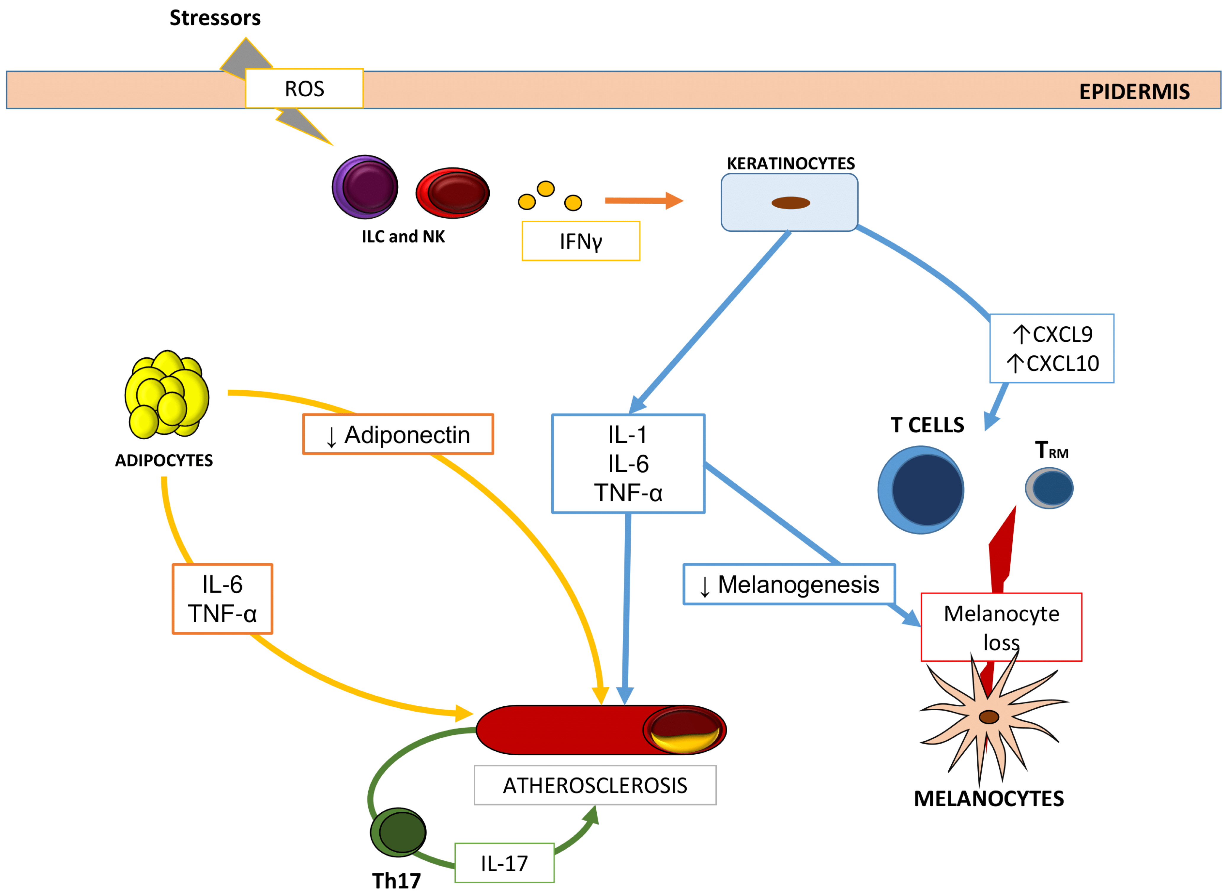
:1. Introduction
2. Metabolic Comorbidities in Vitiligo
3. Conclusions
Author Contributions
Funding
Institutional Review Board Statement
Data Availability Statement
Conflicts of Interest
References
- Goldman, L.; Moraites, R.S.; Kitzmiller, K.W. White Spots in Biblical Times. A Background for the Dermatologist for Participation in Discussions of Current Revisions of the Bible. Arch. Dermatol. 1966, 93, 744–753. [Google Scholar] [CrossRef]
- Kussainova, A.; Kassym, L.; Akhmetova, A.; Glushkova, N.; Sabirov, U.; Adilgozhina, S.; Tuleutayeva, R.; Semenova, Y. Vitiligo and Anxiety: A Systematic Review and Meta-Analysis. PLoS ONE 2020, 15, e0241445. [Google Scholar] [CrossRef] [PubMed]
- Lai, Y.C.; Yew, Y.W.; Kennedy, C.; Schwartz, R.A. Vitiligo and Depression: A Systematic Review and Meta-Analysis of Observational Studies. Br. J. Dermatol. 2017, 177, 708–718. [Google Scholar] [CrossRef]
- Ezzedine, K.; Lim, H.W.; Suzuki, T.; Katayama, I.; Hamzavi, I.; Lan, C.C.; Goh, B.K.; Anbar, T.; Silva de Castro, C.; Lee, A.Y.; et al. Revised Classification/Nomenclature of Vitiligo and Related Issues: The Vitiligo Global Issues Consensus Conference. Pigment Cell. Melanoma Res. 2012, 25, E1–E13. [Google Scholar] [CrossRef] [PubMed] [Green Version]
- Seneschal, J.; Boniface, K.; D’Arino, A.; Picardo, M. An Update on Vitiligo Pathogenesis. Pigment Cell. Melanoma Res. 2021, 34, 236–243. [Google Scholar] [CrossRef] [PubMed]
- Bellei, B.; Pitisci, A.; Ottaviani, M.; Ludovici, M.; Cota, C.; Luzi, F.; Dell’Anna, M.L.; Picardo, M. Vitiligo: A Possible Model of Degenerative Diseases. PLoS ONE 2013, 8, e59782. [Google Scholar] [CrossRef] [Green Version]
- Peters, E.M.; Handjiski, B.; Kuhlmei, A.; Hagen, E.; Bielas, H.; Braun, A.; Klapp, B.F.; Paus, R.; Arck, P.C. Neurogenic Inflammation in Stress-Induced Termination of Murine Hair Growth is Promoted by Nerve Growth Factor. Am. J. Pathol. 2004, 165, 259–271. [Google Scholar] [CrossRef] [Green Version]
- Alkhateeb, A.; Fain, P.R.; Thody, A.; Bennett, D.C.; Spritz, R.A. Epidemiology of Vitiligo and Associated Autoimmune Diseases in Caucasian Probands and their Families. Pigment Cell Res. 2003, 16, 208–214. [Google Scholar] [CrossRef]
- Picardo, M.; Dell’Anna, M.L.; Ezzedine, K.; Hamzavi, I.; Harris, J.E.; Parsad, D.; Taieb, A. Vitiligo. Nat. Rev. Dis. Primers 2015, 1, 15011. [Google Scholar] [CrossRef]
- Le Poole, I.C.; Das, P.K.; van den Wijngaard, R.M.; Bos, J.D.; Westerhof, W. Review of the Etiopathomechanism of Vitiligo: A Convergence Theory. Exp. Dermatol. 1993, 2, 145–153. [Google Scholar] [CrossRef]
- Dell’Anna, M.L.; Ottaviani, M.; Albanesi, V.; Vidolin, A.P.; Leone, G.; Ferraro, C.; Cossarizza, A.; Rossi, L.; Picardo, M. Membrane Lipid Alterations as a Possible Basis for Melanocyte Degeneration in Vitiligo. J. Investig. Dermatol. 2007, 127, 1226–1233. [Google Scholar] [CrossRef]
- Dell’Anna, M.L.; Ottaviani, M.; Kovacs, D.; Mirabilii, S.; Brown, D.A.; Cota, C.; Migliano, E.; Bastonini, E.; Bellei, B.; Cardinali, G.; et al. Energetic Mitochondrial Failing in Vitiligo and Possible Rescue by Cardiolipin. Sci. Rep. 2017, 7, 13663. [Google Scholar] [CrossRef] [PubMed] [Green Version]
- Dell’Anna, M.L.; Ottaviani, M.; Bellei, B.; Albanesi, V.; Cossarizza, A.; Rossi, L.; Picardo, M. Membrane Lipid Defects are Responsible for the Generation of Reactive Oxygen Species in Peripheral Blood Mononuclear Cells from Vitiligo Patients. J. Cell. Physiol. 2010, 223, 187–193. [Google Scholar] [CrossRef] [PubMed]
- Bastonini, E.; Bellei, B.; Filoni, A.; Kovacs, D.; Iacovelli, P.; Picardo, M. Involvement of Non-Melanocytic Skin Cells in Vitiligo. Exp. Dermatol. 2019, 28, 667–673. [Google Scholar] [CrossRef] [Green Version]
- Tulic, M.K.; Cavazza, E.; Cheli, Y.; Jacquel, A.; Luci, C.; Cardot-Leccia, N.; Hadhiri-Bzioueche, H.; Abbe, P.; Gesson, M.; Sormani, L.; et al. Innate Lymphocyte-Induced CXCR3B-Mediated Melanocyte Apoptosis is a Potential Initiator of T-Cell Autoreactivity in Vitiligo. Nat. Commun. 2019, 10, 2178. [Google Scholar] [CrossRef]
- Richmond, J.M.; Bangari, D.S.; Essien, K.I.; Currimbhoy, S.D.; Groom, J.R.; Pandya, A.G.; Youd, M.E.; Luster, A.D.; Harris, J.E. Keratinocyte-Derived Chemokines Orchestrate T-Cell Positioning in the Epidermis during Vitiligo and may Serve as Biomarkers of Disease. J. Investig. Dermatol. 2017, 137, 350–358. [Google Scholar] [CrossRef] [PubMed] [Green Version]
- Harris, J.E.; Harris, T.H.; Weninger, W.; Wherry, E.J.; Hunter, C.A.; Turka, L.A. A Mouse Model of Vitiligo with Focused Epidermal Depigmentation Requires IFN-Γ for Autoreactive CD8+ T-Cell Accumulation in the Skin. J. Investig. Dermatol. 2012, 132, 1869–1876. [Google Scholar] [CrossRef] [Green Version]
- Ho, A.W.; Kupper, T.S. T Cells and the Skin: From Protective Immunity to Inflammatory Skin Disorders. Nat. Rev. Immunol. 2019, 19, 490–502. [Google Scholar] [CrossRef]
- Wang, X.X.; Wang, Q.Q.; Wu, J.Q.; Jiang, M.; Chen, L.; Zhang, C.F.; Xiang, L.H. Increased Expression of CXCR3 and its Ligands in Patients with Vitiligo and CXCL10 as a Potential Clinical Marker for Vitiligo. Br. J. Dermatol. 2016, 174, 1318–1326. [Google Scholar] [CrossRef]
- Maouia, A.; Sormani, L.; Youssef, M.; Helal, A.N.; Kassab, A.; Passeron, T. Differential Expression of CXCL9, CXCL10, and IFN-Γ in Vitiligo and Alopecia Areata Patients. Pigment Cell. Melanoma Res. 2017, 30, 259–261. [Google Scholar] [CrossRef]
- Bertolotti, A.; Boniface, K.; Vergier, B.; Mossalayi, D.; Taieb, A.; Ezzedine, K.; Seneschal, J. Type I Interferon Signature in the Initiation of the Immune Response in Vitiligo. Pigment Cell. Melanoma Res. 2014, 27, 398–407. [Google Scholar] [CrossRef] [PubMed]
- Singh, R.K.; Lee, K.M.; Vujkovic-Cvijin, I.; Ucmak, D.; Farahnik, B.; Abrouk, M.; Nakamura, M.; Zhu, T.H.; Bhutani, T.; Wei, M.; et al. The Role of IL-17 in Vitiligo: A Review. Autoimmun. Rev. 2016, 15, 397–404. [Google Scholar] [CrossRef] [Green Version]
- Speeckaert, R.; Speeckaert, M.; De Schepper, S.; van Geel, N. Biomarkers of Disease Activity in Vitiligo: A Systematic Review. Autoimmun. Rev. 2017, 16, 937–945. [Google Scholar] [CrossRef]
- Dahir, A.M.; Thomsen, S.F. Comorbidities in Vitiligo: Comprehensive Review. Int. J. Dermatol. 2018, 57, 1157–1164. [Google Scholar] [CrossRef]
- Bae, J.M.; Lee, J.H.; Yun, J.S.; Han, B.; Han, T.Y. Vitiligo and Overt Thyroid Diseases: A Nationwide Population-Based Study in Korea. J. Am. Acad. Dermatol. 2017, 76, 871–878. [Google Scholar] [CrossRef] [PubMed]
- Ingordo, V.; Cazzaniga, S.; Raone, B.; Digiuseppe, M.D.; Musumeci, M.L.; Fai, D.; Pellegrino, M.; Pezzarossa, E.; Di Lernia, V.; Battarra, V.C.; et al. Circulating Autoantibodies and Autoimmune Comorbidities in Vitiligo Patients: A Multicenter Italian Study. Dermatology 2014, 228, 240–249. [Google Scholar] [CrossRef]
- Iacovelli, P.; Sinagra, J.L.; Vidolin, A.P.; Marenda, S.; Capitanio, B.; Leone, G.; Picardo, M. Relevance of Thyroiditis and of Other Autoimmune Diseases in Children with Vitiligo. Dermatology 2005, 210, 26–30. [Google Scholar] [CrossRef] [PubMed]
- Yeung, H.; Takeshita, J.; Mehta, N.N.; Kimmel, S.E.; Ogdie, A.; Margolis, D.J.; Shin, D.B.; Attor, R.; Troxel, A.B.; Gelfand, J.M. Psoriasis Severity and the Prevalence of Major Medical Comorbidity: A Population-Based Study. JAMA Dermatol. 2013, 149, 1173–1179. [Google Scholar] [CrossRef] [Green Version]
- Puig, L. Cardiometabolic Comorbidities in Psoriasis and Psoriatic Arthritis. Int. J. Mol. Sci. 2017, 19, 58. [Google Scholar] [CrossRef] [Green Version]
- Powell, S.M.; Ellis, J.P.; Ryan, T.J.; Vickers, H.R. Glucose Tolerance in Lichen Planus. Br. J. Dermatol. 1974, 91, 73–75. [Google Scholar] [CrossRef]
- Saleh, N.; Samir, N.; Megahed, H.; Farid, E. Homocysteine and Other Cardiovascular Risk Factors in Patients with Lichen Planus. J. Eur. Acad. Dermatol. Venereol. 2014, 28, 1507–1513. [Google Scholar] [CrossRef]
- Dreiher, J.; Shapiro, J.; Cohen, A.D. Lichen Planus and Dyslipidaemia: A Case-Control Study. Br. J. Dermatol. 2009, 161, 626–629. [Google Scholar] [CrossRef]
- Arias-Santiago, S.; Buendía-Eisman, A.; Aneiros-Fernández, J.; Girón-Prieto, M.S.; Gutiérrez-Salmerón, M.T.; García-Mellado, V.; Cutando, A.; Naranjo-Sintes, R. Lipid Levels in Patients with Lichen Planus: A Case-Control Study. J. Eur. Acad. Dermatol. Venereol. 2011, 25, 1398–1401. [Google Scholar] [CrossRef]
- Parker, B.; Bruce, I. SLE and Metabolic Syndrome. Lupus 2013, 22, 1259–1266. [Google Scholar] [CrossRef] [PubMed]
- Bartoloni, E.; Baldini, C.; Schillaci, G.; Quartuccio, L.; Priori, R.; Carubbi, F.; Bini, V.; Alunno, A.; Bombardieri, S.; De Vita, S.; et al. Cardiovascular Disease Risk Burden in Primary Sjögren’s Syndrome: Results of a Population-Based Multicentre Cohort Study. J. Intern. Med. 2015, 278, 185–192. [Google Scholar] [CrossRef] [PubMed]
- Ye, Y.M.; Jin, H.J.; Hwang, E.K.; Nam, Y.H.; Kim, J.H.; Shin, Y.S.; Park, H.S. Co-Existence of Chronic Urticaria and Metabolic Syndrome: Clinical Implications. Acta Derm. Venereol. 2013, 93, 156–160. [Google Scholar] [CrossRef] [PubMed] [Green Version]
- Boehncke, W.H. Systemic Inflammation and Cardiovascular Comorbidity in Psoriasis Patients: Causes and Consequences. Front. Immunol. 2018, 9, 579. [Google Scholar] [CrossRef] [PubMed]
- Andersen, Y.M.F.; Egeberg, A.; Gislason, G.H.; Hansen, P.R.; Skov, L.; Thyssen, J.P. Risk of Myocardial Infarction, Ischemic Stroke, and Cardiovascular Death in Patients with Atopic Dermatitis. J. Allergy Clin. Immunol. 2016, 138, 310–312.e3. [Google Scholar] [CrossRef] [PubMed] [Green Version]
- Hashmi, S.; Zeng, Q.T. Role of Interleukin-17 and Interleukin-17-Induced Cytokines Interleukin-6 and Interleukin-8 in Unstable Coronary Artery Disease. Coron. Artery Dis. 2006, 17, 699–706. [Google Scholar] [CrossRef] [PubMed]
- Stelzner, K.; Herbert, D.; Popkova, Y.; Lorz, A.; Schiller, J.; Gericke, M.; Klöting, N.; Blüher, M.; Franz, S.; Simon, J.C.; et al. Free Fatty Acids Sensitize Dendritic Cells to Amplify TH1/TH17-Immune Responses. Eur. J. Immunol. 2016, 46, 2043–2053. [Google Scholar] [CrossRef] [Green Version]
- Sumarac-Dumanovic, M.; Stevanovic, D.; Ljubic, A.; Jorga, J.; Simic, M.; Stamenkovic-Pejkovic, D.; Starcevic, V.; Trajkovic, V.; Micic, D. Increased Activity of Interleukin-23/Interleukin-17 Proinflammatory Axis in Obese Women. Int. J. Obes. 2009, 33, 151–156. [Google Scholar] [CrossRef] [Green Version]
- Zhou, L.; Shi, Y.L.; Li, K.; Hamzavi, I.; Gao, T.W.; Huggins, R.H.; Lim, H.W.; Mi, Q.S. Increased Circulating Th17 Cells and Elevated Serum Levels of TGF-Beta and IL-21 are Correlated with Human Non-Segmental Vitiligo Development. Pigment Cell. Melanoma Res. 2015, 28, 324–329. [Google Scholar] [CrossRef]
- Rork, J.F.; Rashighi, M.; Harris, J.E. Understanding Autoimmunity of Vitiligo and Alopecia Areata. Curr. Opin. Pediatr. 2016, 28, 463–469. [Google Scholar] [CrossRef] [PubMed] [Green Version]
- Rudnicka, L.; Waśkiel-Burnat, A. Systemic Aspects of Alopecia Areata Comment to the Article by Lai and Sinclair. J. Eur. Acad. Dermatol. Venereol. 2021, 35, e214–e215. [Google Scholar] [CrossRef] [PubMed]
- Stochmal, A.; Waśkiel-Burnat, A.; Chrostowska, S.; Zaremba, M.; Rakowska, A.; Czuwara, J.; Rudnicka, L. Adiponectin as a Novel Biomarker of Disease Severity in Alopecia Areata. Sci. Rep. 2021, 11, 13809. [Google Scholar] [CrossRef]
- Grundy, S.M.; Brewer, H.B., Jr.; Cleeman, J.I.; Smith, S.C., Jr.; Lenfant, C.; American Heart Association. National Heart, Lung, and Blood Institute. Definition of Metabolic Syndrome: Report of the National Heart, Lung, and Blood Institute/American Heart Association Conference on Scientific Issues Related to Definition. Circulation 2004, 109, 433–438. [Google Scholar] [CrossRef] [PubMed] [Green Version]
- Eckel, R.H.; Grundy, S.M.; Zimmet, P.Z. The Metabolic Syndrome. Lancet 2005, 365, 1415–1428. [Google Scholar] [CrossRef]
- Alberti, K.G.; Eckel, R.H.; Grundy, S.M.; Zimmet, P.Z.; Cleeman, J.I.; Donato, K.A.; Fruchart, J.C.; James, W.P.; Loria, C.M.; Smith, S.C., Jr.; et al. Harmonizing the Metabolic Syndrome: A Joint Interim Statement of the International Diabetes Federation Task Force on Epidemiology and Prevention; National Heart, Lung, and Blood Institute; American Heart Association; World Heart Federation; International Atherosclerosis Society; and International Association for the Study of Obesity. Circulation 2009, 120, 1640–1645. [Google Scholar]
- Aguilar, M.; Bhuket, T.; Torres, S.; Liu, B.; Wong, R.J. Prevalence of the Metabolic Syndrome in the United States, 2003–2012. JAMA 2015, 313, 1973–1974. [Google Scholar] [CrossRef] [PubMed]
- Bastard, J.P.; Maachi, M.; Van Nhieu, J.T.; Jardel, C.; Bruckert, E.; Grimaldi, A.; Robert, J.J.; Capeau, J.; Hainque, B. Adipose Tissue IL-6 Content Correlates with Resistance to Insulin Activation of Glucose Uptake both in Vivo and in Vitro. J. Clin. Endocrinol. Metab. 2002, 87, 2084–2089. [Google Scholar] [CrossRef]
- Ridker, P.M.; Cushman, M.; Stampfer, M.J.; Tracy, R.P.; Hennekens, C.H. Inflammation, Aspirin, and the Risk of Cardiovascular Disease in Apparently Healthy Men. N. Engl. J. Med. 1997, 336, 973–979. [Google Scholar] [CrossRef] [PubMed]
- Lau, D.C.; Dhillon, B.; Yan, H.; Szmitko, P.E.; Verma, S. Adipokines: Molecular Links between Obesity and Atheroslcerosis. Am. J. Physiol. Heart Circ. Physiol. 2005, 288, H2031–H2041. [Google Scholar] [CrossRef] [Green Version]
- Testa, R.; Olivieri, F.; Bonfigli, A.R.; Sirolla, C.; Boemi, M.; Marchegiani, F.; Marra, M.; Cenerelli, S.; Antonicelli, R.; Dolci, A.; et al. Interleukin-6-174 G > C Polymorphism Affects the Association between IL-6 Plasma Levels and Insulin Resistance in Type 2 Diabetic Patients. Diabetes Res. Clin. Pract. 2006, 71, 299–305. [Google Scholar] [CrossRef]
- Bao, P.; Liu, G.; Wei, Y. Association between IL-6 and Related Risk Factors of Metabolic Syndrome and Cardiovascular Disease in Young Rats. Int. J. Clin. Exp. Med. 2015, 8, 13491–13499. [Google Scholar] [PubMed]
- Azzawi, M.; Hasleton, P. Tumour Necrosis Factor Alpha and the Cardiovascular System: Its Role in Cardiac Allograft Rejection and Heart Disease. Cardiovasc. Res. 1999, 43, 850–859. [Google Scholar] [CrossRef] [Green Version]
- Furukawa, S.; Fujita, T.; Shimabukuro, M.; Iwaki, M.; Yamada, Y.; Nakajima, Y.; Nakayama, O.; Makishima, M.; Matsuda, M.; Shimomura, I. Increased Oxidative Stress in Obesity and its Impact on Metabolic Syndrome. J. Clin. Investig. 2004, 114, 1752–1761. [Google Scholar] [CrossRef]
- Srikanthan, K.; Feyh, A.; Visweshwar, H.; Shapiro, J.I.; Sodhi, K. Systematic Review of Metabolic Syndrome Biomarkers: A Panel for Early Detection, Management, and Risk Stratification in the West Virginian Population. Int. J. Med. Sci. 2016, 13, 25–38. [Google Scholar] [CrossRef] [Green Version]
- Tanacan, E.; Atakan, N. Higher Incidence of Metabolic Syndrome Components in Vitiligo Patients: A Prospective Cross-Sectional Study. An. Bras. Dermatol. 2020, 95, 165–172. [Google Scholar] [CrossRef] [PubMed]
- Ataş, H.; Gönül, M. Increased Risk of Metabolic Syndrome in Patients with Vitiligo. Balkan Med. J. 2017, 34, 219–225. [Google Scholar] [CrossRef] [PubMed]
- Namazi, N.; Amani, M.; Haghighatkhah, H.R.; Noori, E.; Abdollahimajd, F. Increased Risk of Subclinical Atherosclerosis and Metabolic Syndrome in Patients with Vitiligo: A Real Association Or a Coincidence? Dermatol. Ther. 2021, 34, e14803. [Google Scholar] [CrossRef]
- Sharma, Y.K.; Bansal, P.; Menon, S.; Prakash, N. Metabolic Syndrome in Vitiligo Patients among a Semi-Urban Maharashtrian Population: A Case Control Study. Diabetes Metab. Syndr. 2017, 11 (Suppl. 1), S77–S80. [Google Scholar] [CrossRef]
- Karadag, A.S.; Tutal, E.; Ertugrul, D.T. Insulin Resistance is Increased in Patients with Vitiligo. Acta Derm. Venereol. 2011, 91, 541–544. [Google Scholar] [CrossRef]
- Azzazi, Y.; Mostafa, W.Z.; Sayed, K.S.; Alhelf, M.; Safwat, M.; Mahrous, A.; El Lawindi, M.; Ragab, N. Support for Increased Cardiovascular Risk in Non-Segmental Vitiligo among Egyptians: A Hospital-Based, Case-Control Study. Pigment Cell. Melanoma Res. 2021, 34, 598–604. [Google Scholar] [CrossRef]
- Pietrzak, A.; Bartosińska, J.; Dybiec, E.; Chodorowska, G.; Krasowska, D.; Hercogova, J.; Lotti, T. Hepato-Splenic and Lipid Profile Abnormalities--do they Exist in Children Affected with Vitiligo? Acta Dermatovenerol. Croat. 2014, 22, 19–25. [Google Scholar]
- Moretti, S.; Spallanzani, A.; Amato, L.; Hautmann, G.; Gallerani, I.; Fabiani, M.; Fabbri, P. New Insights into the Pathogenesis of Vitiligo: Imbalance of Epidermal Cytokines at Sites of Lesions. Pigment Cell Res. 2002, 15, 87–92. [Google Scholar] [CrossRef]
- Abdallah, M.; El-Mofty, M.; Anbar, T.; Rasheed, H.; Esmat, S.; Al-Tawdy, A.; Fawzy, M.M.; Abdel-Halim, D.; Hegazy, R.; Gawdat, H.; et al. CXCL-10 and Interleukin-6 are Reliable Serum Markers for Vitiligo Activity: A Multicenter Cross-Sectional Study. Pigment Cell. Melanoma Res. 2018, 31, 330–336. [Google Scholar] [CrossRef] [PubMed]
- Boniface, K.; Jacquemin, C.; Darrigade, A.S.; Dessarthe, B.; Martins, C.; Boukhedouni, N.; Vernisse, C.; Grasseau, A.; Thiolat, D.; Rambert, J.; et al. Vitiligo Skin is Imprinted with Resident Memory CD8 T Cells Expressing CXCR3. J. Investig. Dermatol. 2018, 138, 355–364. [Google Scholar] [CrossRef] [PubMed] [Green Version]
- Speeckaert, R.; Mylle, S.; van Geel, N. IL-17A is Not a Treatment Target in Progressive Vitiligo. Pigment Cell. Melanoma Res. 2019, 32, 842–847. [Google Scholar] [CrossRef]
- Sinha, P.K.; Nigam, P.; Swain, J.P. Association of Metabolic Syndrome with Vitiligo- A Case Control Study. J. Evol. Med. Dent. Sci. 2019, 8, 2783–2786. [Google Scholar]
- Rodríguez-Martín, M.; de Paz, N.M.; Mehtani, P.; Ferrer, P.C.; Eliche, M.P.; Martín, B.R.; Sáez, M.; García, M.; Noda, A. Patients with Vitiligo Present Fewer Cardiovascular Risk Factors: Results from a Case-Control Study. J. Eur. Acad. Dermatol. Venereol. 2013, 27, 124–125. [Google Scholar] [CrossRef] [PubMed]
- Pietrzak, A.; Bartosińska, J.; Hercogová, J.; Lotti, T.M.; Chodorowska, G. Metabolic Syndrome in Vitiligo. Dermatol. Ther. 2012, 25 (Suppl. 1), S41–S43. [Google Scholar] [CrossRef] [PubMed]
- Yang, Z.S.; Lin, N.N.; Li, L.; Li, Y. The Effect of TNF Inhibitors on Cardiovascular Events in Psoriasis and Psoriatic Arthritis: An Updated Meta-Analysis. Clin. Rev. Allergy Immunol. 2016, 51, 240–247. [Google Scholar] [CrossRef]
- Maloberti, A.; Vallerio, P.; Triglione, N.; Occhi, L.; Panzeri, F.; Bassi, I.; Pansera, F.; Piccinelli, E.; Peretti, A.; Garatti, L.; et al. Vascular Aging and Disease of the Large Vessels: Role of Inflammation. High Blood Press Cardiovasc. Prev. 2019, 26, 175–182. [Google Scholar] [CrossRef] [PubMed]
- Rosmarin, D.; Pandya, A.G.; Lebwohl, M.; Grimes, P.; Hamzavi, I.; Gottlieb, A.B.; Butler, K.; Kuo, F.; Sun, K.; Ji, T.; et al. Ruxolitinib Cream for Treatment of Vitiligo: A Randomised, Controlled, Phase 2 Trial. Lancet 2020, 396, 110–120. [Google Scholar] [CrossRef]
- Liu, L.Y.; Strassner, J.P.; Refat, M.A.; Harris, J.E.; King, B.A. Repigmentation in Vitiligo using the Janus Kinase Inhibitor Tofacitinib may Require Concomitant Light Exposure. J. Am. Acad. Dermatol. 2017, 77, 675–682.e1. [Google Scholar] [CrossRef] [PubMed]
- Strober, B.E.; Gottlieb, A.B.; van de Kerkhof, P.C.M.; Puig, L.; Bachelez, H.; Chouela, E.; Imafuku, S.; Thaçi, D.; Tan, H.; Valdez, H.; et al. Benefit-Risk Profile of Tofacitinib in Patients with Moderate-to-Severe Chronic Plaque Psoriasis: Pooled Analysis Across Six Clinical Trials. Br. J. Dermatol. 2019, 180, 67–75. [Google Scholar] [CrossRef] [PubMed]


| Variables | Vitiligo Group | Control Group | p |
|---|---|---|---|
| Gender (n, %) | 0.42 | ||
| Female | 488 (58.2%) | 192 (60.9%) | |
| Male | 351 (41.8%) | 124 (39.1%) | |
| Age (mean ± SD, range) | 45.3 ± 15.5 (18–88) | 46.8 (27–67) | |
| FPG (mg/dL) (median ± MAD) | 90 ± 15.8 | 88 ± 4.1 | 0.0003 |
| Total cholesterol (mg/dL) (median ± MAD) | 182 ± 23.7 | 192.5 ± 23.1 | 0.0004 |
| HDL (mg/dL) (median ± MAD) | 58 ± 19.7 | 64 ± 8.1 | 0.0002 |
| LDL (mg/dL) (median ± MAD) | 113 ± 34.1 | 106 ± 9.9 | 0.0001 |
| Triglycerides (mg/dL) (mean ± SD) | 90 ± 141.2 | 77.5 ± 17.3 | 0.0002 |
Publisher’s Note: MDPI stays neutral with regard to jurisdictional claims in published maps and institutional affiliations. |
© 2021 by the authors. Licensee MDPI, Basel, Switzerland. This article is an open access article distributed under the terms and conditions of the Creative Commons Attribution (CC BY) license (https://creativecommons.org/licenses/by/4.0/).
Share and Cite
D’Arino, A.; Picardo, M.; Truglio, M.; Pacifico, A.; Iacovelli, P. Metabolic Comorbidities in Vitiligo: A Brief Review and Report of New Data from a Single-Center Experience. Int. J. Mol. Sci. 2021, 22, 8820. https://doi.org/10.3390/ijms22168820
D’Arino A, Picardo M, Truglio M, Pacifico A, Iacovelli P. Metabolic Comorbidities in Vitiligo: A Brief Review and Report of New Data from a Single-Center Experience. International Journal of Molecular Sciences. 2021; 22(16):8820. https://doi.org/10.3390/ijms22168820
Chicago/Turabian StyleD’Arino, Andrea, Mauro Picardo, Mauro Truglio, Alessia Pacifico, and Paolo Iacovelli. 2021. "Metabolic Comorbidities in Vitiligo: A Brief Review and Report of New Data from a Single-Center Experience" International Journal of Molecular Sciences 22, no. 16: 8820. https://doi.org/10.3390/ijms22168820