
Antibacterial Poly(ε-CL)/Hydroxyapatite Electrospun Fibers Reinforced by Poly(ε-CL)-b-poly(ethylene phosphoric acid)

 ,
,
Abstract
:1. Introduction
2. Results
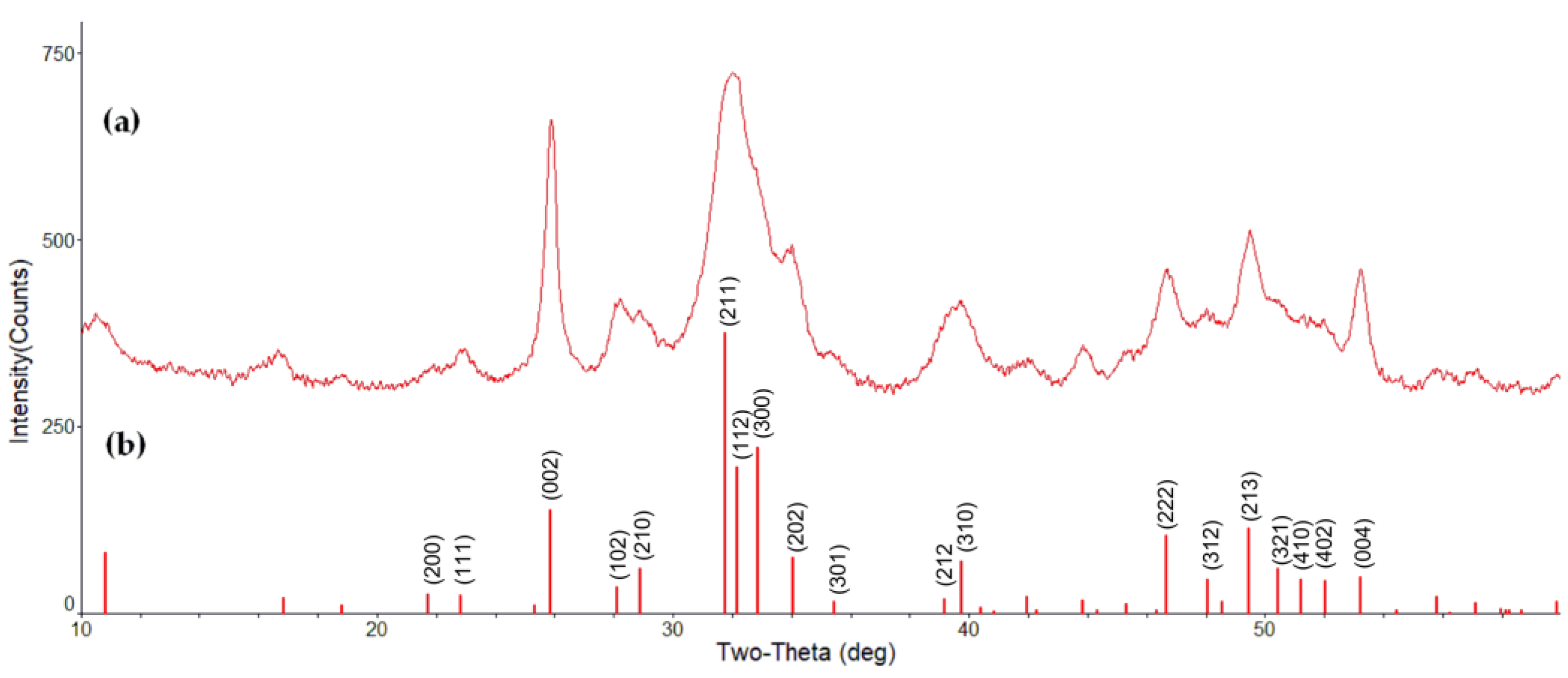
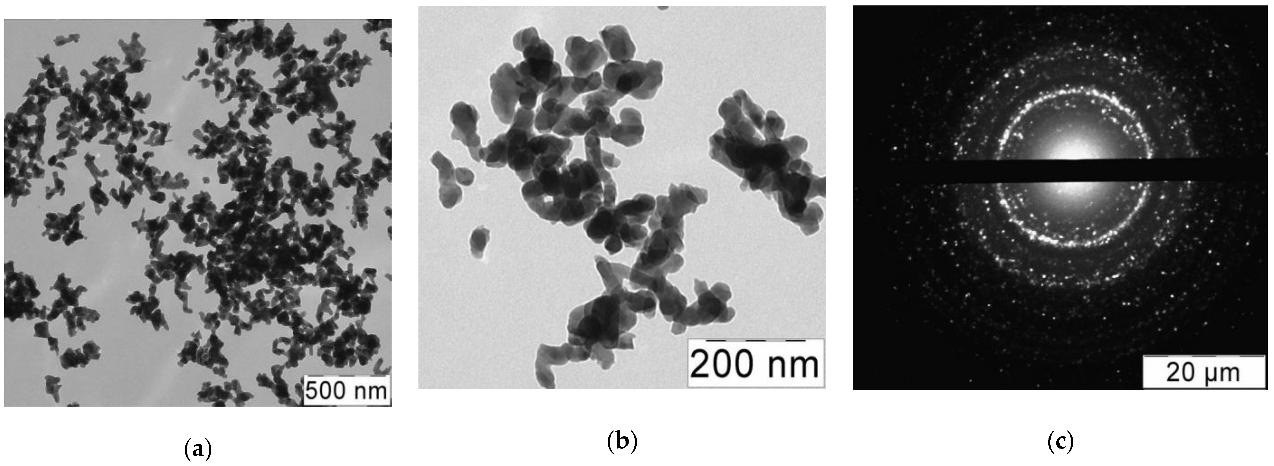
2.1. Preparation and Characterization of Nanosized HAp
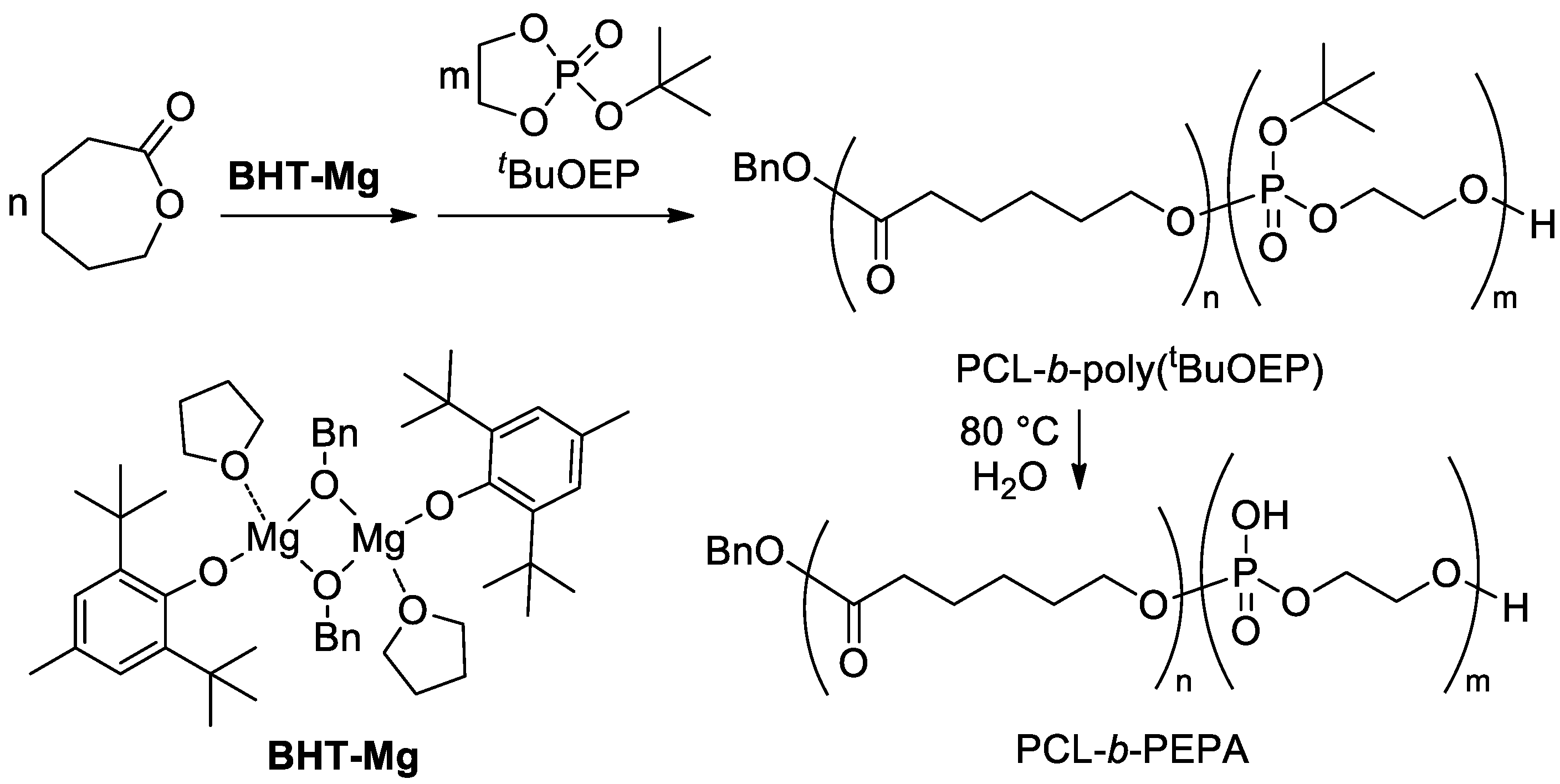
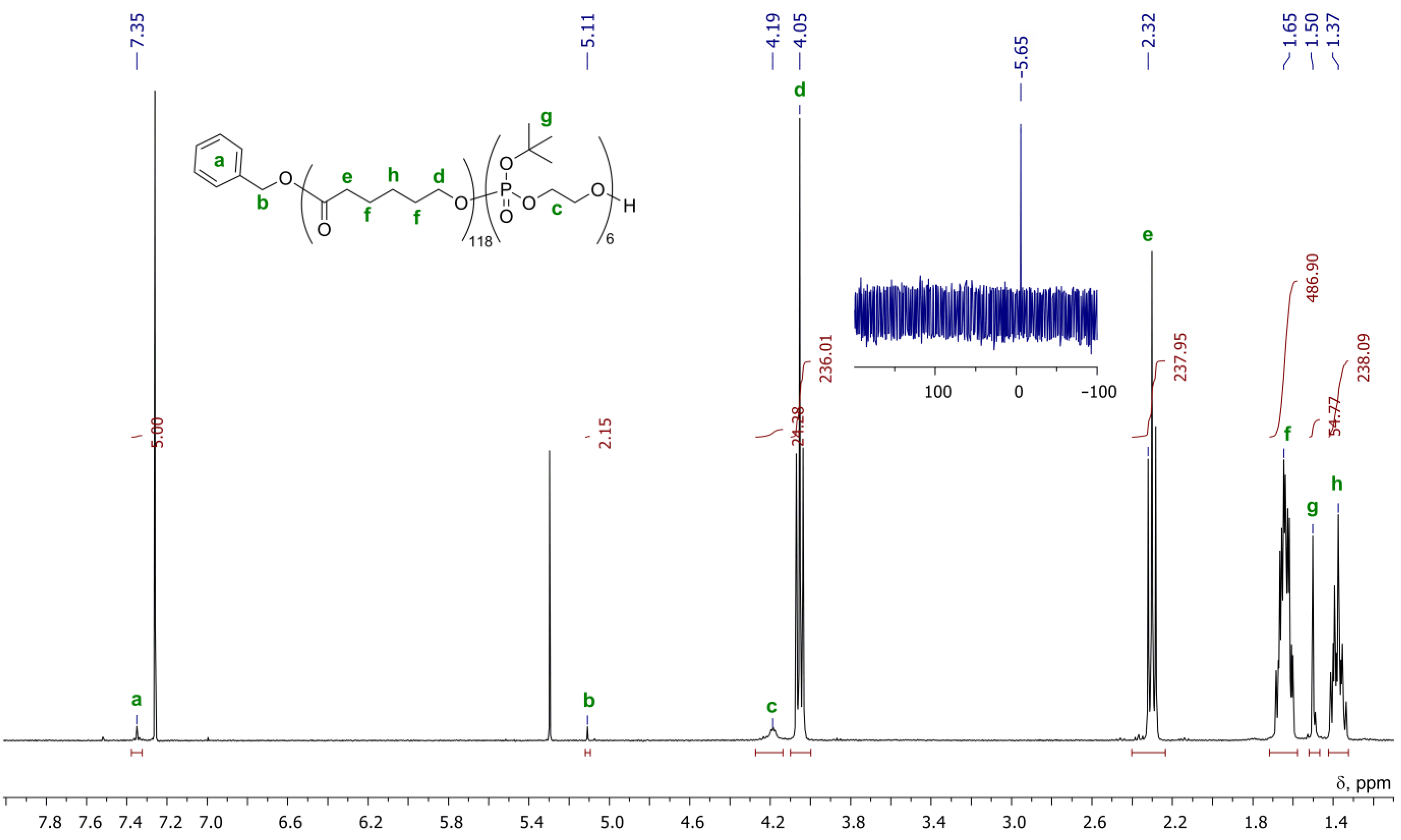
2.2. Synthesis of PCL-b-poly(tBuOEP)
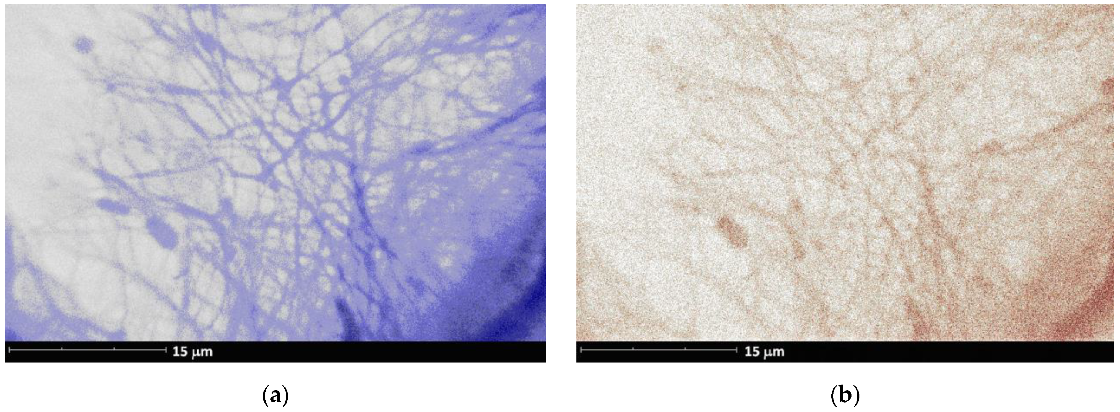
2.3. Preparation and ES Molding of PCL/HAp Composites
2.4. ES Fibrous Films Containing Vancomycin
3. Discussion
4. Materials and Methods
4.1. Preparation and Characterization of HAp Nanoparticles
4.1.1. Reagents and Equipment
4.1.2. Synthesis of HAp Nanoparticles
4.2. Synthesis of PCL-b-poly(tBuOEP)
4.2.1. Reagents and Equipment
4.2.2. Synthesis of PCL-b-poly(tBuOEP)
4.3. Preparation and Study of ES Mats
4.3.1. Reagents and Equipment
4.3.2. Preparation of ES Mats
4.4. Experiments with Van-Loaded HAp Particles and ES Films
4.4.1. Reagents and Equipment
4.4.2. ζ-Potential Measurements
4.4.3. Preparation of Vancomycin-Loaded ES Films
4.4.4. Antibacterial Activity of ES Films
5. Conclusions
Author Contributions
Funding
Institutional Review Board Statement
Informed Consent Statement
Acknowledgments
Conflicts of Interest
References
- Roddy, E.; DeBaun, M.R.; Daoud-Gray, A.; Yang, Y.P.; Gardner, M.J. Treatment of critical-sized bone defects: Clinical and tissue engineering perspectives. Eur. J. Orthop. Surg. Traum. 2018, 28, 351–362. [Google Scholar] [CrossRef]
- Bal, Z.; Kaito, T.; Korkusuz, F.; Yoshikawa, H. Bone regeneration with hydroxyapatite-based biomaterials. Emerg. Mater. 2020, 3, 521–544. [Google Scholar] [CrossRef]
- Bagde, A.D.; Kuthe, A.M.; Quazi, S.; Gupta, V.; Jaiswa, S.; Jyothilal, I.; Lande, S.N.; Nagdeve, S. State of the Art Technology for Bone Tissue Engineering and Drug Delivery. IRBM 2019, 40, 133–144. [Google Scholar] [CrossRef]
- Afewerki, S.; Bassous, N.; Harb, S.; Palo-Nieto, C.; Ruiz-Esparza, G.U.; Marciano, F.R.; Webster, T.J.; Furtado, A.S.A.; Lobo, A.O. Advances in dual functional antimicrobial and osteoinductive biomaterials for orthopaedic applications. Nanomed. Nanotechnol. Biol. Med. 2020, 24, 102143. [Google Scholar] [CrossRef]
- Bezstarosti, H.; Metsemakers, W.J.; van Lieshout, E.M.M.; Voskamp, L.W.; Kortram, K.; McNally, M.A.; Marais, L.C.; Verhofstad, M.H.J. Management of critical-sized bone defects in the treatment of fracture-related infection: A systematic review and pooled analysis. Arch. Orthop. Trauma Surg. 2020. [Google Scholar] [CrossRef]
- Montel, G.; Bonel, G.; Heughebaert, J.C.; Trombe, J.C.; Rey, C. New concepts in the composition, crystallization and growth of the mineral component of calcified tissues. J. Cryst. Growth 1981, 53, 74–99. [Google Scholar] [CrossRef]
- Singh, G.; Singh, R.P.; Jolly, S.S. Customized hydroxyapatites for bone-tissue engineering and drug delivery applications: A review. J. Sol-Gel Sci. Technol. 2020, 94, 505–530. [Google Scholar] [CrossRef]
- Combes, C.; Cazalbou, S.; Rey, C. Apatite Biominerals. Minerals 2016, 6, 34. [Google Scholar] [CrossRef] [Green Version]
- Bohner, M.; Le Gars Santoni, B.; Döbelin, N. β-Tricalcium phosphate for bone substitution: Synthesis and properties. Acta Biomater. 2020, 113, 23–41. [Google Scholar] [CrossRef]
- Lodoso-Torrecilla, I.; van den Beucken, J.J.J.P.; Jansen, J.A. Calcium phosphate cements: Optimization toward biodegradability. Acta Biomater. 2021, 119, 1–12. [Google Scholar] [CrossRef]
- Shi, H.; Zhou, Z.; Li, W.; Fan, Y.; Li, Z.; Wei, J. Hydroxyapatite Based Materials for Bone Tissue Engineering: A Brief and Comprehensive Introduction. Crystals 2021, 11, 149. [Google Scholar] [CrossRef]
- Lett, J.A.; Sagadevan, S.; Fatimah, I.; Hoque, M.E.; Lokanathan, Y.; Léonard, E.; Alshahateet, S.F.; Schirhagl, R.; Oh, W.C. Recent advances in natural polymer-based hydroxyapatite scaffolds: Properties and applications. Eur. Polym. J. 2021, 148, 110360. [Google Scholar] [CrossRef]
- Tang, G.; Liu, Z.; Liu, Y.; Yu, J.; Wang, X.; Tan, Z.; Ye, X. Recent Trends in the Development of Bone Regenerative Biomaterials. Front. Cell Dev. Biol. 2021, 9, 665813. [Google Scholar] [CrossRef] [PubMed]
- Ghavimi, S.A.A.; Allen, B.N.; Stromsdorfer, J.L.; Kramer, J.S.; Li, X.; Ulery, B.D. Calcium and phosphate ions as simple signaling molecules with versatile osteoinductivity. Biomed. Mater. 2018, 13, 055005. [Google Scholar] [CrossRef] [PubMed]
- Shinto, Y.; Uchida, A.; Korkusuz, F.; Araki, N.; Ono, K. Calcium hydroxyapatite ceramic used as a delivery system for antibiotics. J. Bone Jt. Surg. Br. 1992, 74, 600–604. [Google Scholar] [CrossRef] [PubMed] [Green Version]
- Korkusuz, F.; Uchida, A.; Shinto, Y.; Araki, N.; Inoue, K.; Ono, K. Experimental implant-related osteomyelitis treated by antibiotic-calcium hydroxyapatite ceramic composites. J. Bone Jt. Surg. Br. 1993, 75, 111–114. [Google Scholar] [CrossRef]
- Joosten, U.; Joist, A.; Gosheger, G.; Liljenqvist, U.; Brandt, B.; von Eiff, C. Effectiveness of hydroxyapatite-vancomycin bone cement in the treatment of Staphylococcus aureus induced chronic osteomyelitis. Biomaterials 2005, 26, 5251–5258. [Google Scholar] [CrossRef]
- Itokazu, M.; Yang, W.; Aoki, T.; Ohara, A.; Kato, N. Synthesis of antibiotic-loaded interporous hydroxyapatite blocks by vacuum method and in vitro drug release testing. Biomaterials 1998, 19, 817–819. [Google Scholar] [CrossRef]
- Lett, J.A.; Sagadevan, S.; Prabhakar, J.J.; Hamizi, N.A.; Badruddin, I.A.; Johan, M.R.; Marlinda, A.R.; Wahab, Y.A.; Khan, T.M.Y.; Kamangar, S. Drug Leaching Properties of Vancomycin Loaded Mesoporous Hydroxyapatite as Bone Substitutes. Processes 2019, 7, 826. [Google Scholar] [CrossRef] [Green Version]
- Shirtliff, M.E.; Calhoun, J.H.; Mader, J.T. Experimental osteomyelitis treatment with antibiotic-impregnated hydroxyapatite. Clin. Orthop. Relat. Res. 2002, 401, 239–247. [Google Scholar] [CrossRef]
- Yamashita, Y.; Uchida, A.; Yamakawa, T.; Shinto, Y.; Araki, N.; Kato, K. Treatment of chronic osteomyelitis using calcium hydroxyapatite ceramic implants impregnated with antibiotic. Int. Orthop. SICOT 1998, 22, 247–251. [Google Scholar] [CrossRef] [Green Version]
- Siddiqui, H.A.; Pickering, K.L.; Mucalo, M.R.; Siddiqui, H.A.; Pickering, K.L.; Mucalo, M.R. A Review on the Use of Hydroxyapatite-Carbonaceous Structure Composites in Bone Replacement Materials for Strengthening Purposes. Materials 2018, 11, 1813. [Google Scholar] [CrossRef] [Green Version]
- Alizadeh-Osgouei, M.; Li, Y.; Wen, C. A comprehensive review of biodegradable synthetic polymer-ceramic composites and their manufacture for biomedical applications. Bioact. Mater. 2019, 4, 22–36. [Google Scholar] [CrossRef]
- Zhao, D.; Zhu, T.; Li, J.; Cui, L.; Zhang, Z.; Zhuang, X.; Ding, J. Poly(lactic-co-glycolic acid)-based composite bone-substitute materials. Bioact. Mater. 2021, 6, 346–360. [Google Scholar] [CrossRef] [PubMed]
- Lian, X.; Liu, H.; Wang, X.; Xu, S.; Cui, F.; Bai, X. Antibacterial and biocompatible properties of vancomycin-loaded nano-hydroxyapatite/collagen/poly (lactic acid) bone substitute. Prog. Nat. Sci. Mater. Int. 2013, 23, 549–556. [Google Scholar] [CrossRef] [Green Version]
- Morawska-Chochół, A.; Domalik-Pyzik, P.; Chłopek, J.; Szaraniec, B.; Sterna, J.; Rzewuska, M.; Boguń, M.; Kucharski, R.; Mielczarek, P. Gentamicin release from biodegradable poly-l-lactide based composites for novel intramedullary nails. Mater. Sci. Eng. C 2014, 45, 15–20. [Google Scholar] [CrossRef]
- Ramírez-Agudelo, R.; Scheuermann, K.; Gala-García, A.; Monteiro, A.P.F.; Pinzón-García, A.D.; Cortés, M.E.; Sinisterra, R.D. Hybrid nanofibers based on poly-caprolactone/gelatin/hydroxyapatite nanoparticles-loaded Doxycycline: Effective anti-tumoral and antibacterial activity. Mater. Sci. Eng. C 2018, 83, 25–34. [Google Scholar] [CrossRef]
- Macha, I.J.; Cazalbou, S.; Ben-Nissan, B.; Harvey, K.L.; Milthorpe, B. Marine Structure Derived Calcium Phosphate–Polymer Biocomposites for Local Antibiotic Delivery. Mar. Drugs 2015, 13, 666–680. [Google Scholar] [CrossRef] [Green Version]
- Aoki, K.; Saito, N. Biodegradable Polymers as Drug Delivery Systems for Bone Regeneration. Pharmaceutics 2020, 12, 95. [Google Scholar] [CrossRef] [PubMed] [Green Version]
- Mehdi, S.-S. Electrospun Polyhydroxybutyrate/Hydroxyapatite Nanohybrids: Microstructure and Bone Cell Response. J. Mater. Sci. Technol. 2016, 32, 1013–1020. [Google Scholar] [CrossRef]
- Aragon, J.; Navascues, N.; Mendoza, G.; Irusta, S. Laser-treated electrospun fibers loaded with nano-hydroxyapatite for bone tissue engineering. Int. J. Pharm. 2017, 525, 112–122. [Google Scholar] [CrossRef] [Green Version]
- Tanaka, K.; Shiga, T.; Katayama, T. Fabrication of Hydroxyapatite/PLA Composite Nanofibers and Effects of Surface Treatment of Hydroxyapatite on Their Mechanical Properties and Dispersion. J. Soc. Mater. Sci. Jpn. 2018, 67, 291–298. [Google Scholar] [CrossRef] [Green Version]
- Kosowska, K.; Domalik-Pyzik, P.; Krok-Borkowicz, M.; Chłopek, J. Polylactide/Hydroxyapatite Nonwovens Incorporated into Chitosan/Graphene Materials Hydrogels to Form Novel Hierarchical Scaffolds. Int. J. Mol. Sci. 2020, 21, 2330. [Google Scholar] [CrossRef] [Green Version]
- Stastna, E.; Castkova, K.; Rahel, J. Influence of Hydroxyapatite Nanoparticles and Surface Plasma Treatment on Bioactivity of Polycaprolactone Nanofibers. Polymers 2020, 12, 1877. [Google Scholar] [CrossRef] [PubMed]
- Nifant’ev, I.; Bukharova, T.; Dyakonov, A.; Goldshtein, D.; Galitsyna, E.; Kosarev, M.; Shlyakhtin, A.; Gavrilov, D.; Ivchenko, P. Osteogenic Differentiation of Human Adipose Tissue-Derived MSCs by Non-Toxic Calcium Poly(ethylene phosphate)s. Int. J. Mol. Sci. 2019, 20, 6242. [Google Scholar] [CrossRef] [Green Version]
- Nifant’ev, I.E.; Shlyakhtin, A.V.; Bagrov, V.V.; Komarov, P.D.; Tavtorkin, A.N.; Minyaev, M.E.; Kosarev, M.A.; Ivchenko, P.V. Synthesis in aqueous media of poly(ethylene phosphoric acids) by mild thermolysis of homopolymers and block copolymers based on tert-butyl ethylene phosphate. Eur. Polym. J. 2018, 106, 249–256. [Google Scholar] [CrossRef]
- Nifant’ev, I.E.; Shlyakhtin, A.V.; Bagrov, V.V.; Minyaev, M.E.; Churakov, A.V.; Karchevsky, S.G.; Birin, K.P.; Ivchenko, P.V. Mono-BHT heteroleptic magnesium complexes: Synthesis, molecular structure and catalytic behavior in the ring-opening polymerization of cyclic esters. Dalton Trans. 2017, 46, 12132–12146. [Google Scholar] [CrossRef] [Green Version]
- Nifant’ev, I.E.; Shlyakhtin, A.V.; Bagrov, V.V.; Komarov, P.D.; Kosarev, M.A.; Tavtorkin, A.N.; Minyaev, M.E.; Roznyatovsky, V.A.; Ivchenko, P.V. Controlled ring-opening polymerisation of cyclic phosphates, phosphonates and phosphoramidates catalysed by hereroleptic BHT-alkoxy magnesium complexes. Polym. Chem. 2017, 8, 6806–6816. [Google Scholar] [CrossRef]
- Boanini, E.; Gazzano, M.; Bigi, A. Ionic substitutions in calcium phosphates synthesized at low temperature. Acta Biomater. 2010, 6, 1882–1894. [Google Scholar] [CrossRef]
- Li, Y.; Wang, Y.; Li, Y.; Luo, W.; Jiang, J.; Zhao, J.; Liu, C. Controllable Synthesis of Biomimetic Hydroxyapatite Nanorods with High Osteogenic Bioactivity. ACS Biomater. Sci. Eng. 2020, 6, 320–328. [Google Scholar] [CrossRef]
- Khoo, W.; Nor, F.M.; Ardhyananta, H.; Kurniawan, D. Preparation of Natural Hydroxyapatite from Bovine Femur Bones Using Calcination at Various Temperatures. Procedia Manuf. 2015, 2, 196–201. [Google Scholar] [CrossRef] [Green Version]
- Li, Y.; Liu, C. Nanomaterial-based bone regeneration. Nanoscale 2017, 9, 4862–4874. [Google Scholar] [CrossRef]
- Rodríguez-Lugo, V.; Sanchez Hernández, J.; Arellano-Jimenez, M.J.; Hernández-Tejeda, P.H.; Recillas-Gispert, S. Characterization of Hydroxyapatite by Electron Microscopy. Micros. Microanal. 2005, 11, 516–523. [Google Scholar] [CrossRef] [Green Version]
- Agarwal, S.; Wendorff, J.H.; Greiner, A. Use of electrospinning technique for biomedical applications. Polymer 2008, 49, 5603–5621. [Google Scholar] [CrossRef] [Green Version]
- Thenmozhi, S.; Dharmaraj, N.; Kadirvelu, K.; Kim, H.Y. Electrospun nanofibers: New generation materials for advanced applications. Mater. Sci. Eng. B 2017, 217, 36–48. [Google Scholar] [CrossRef]
- Patil, J.V.; Mali, S.S.; Kamble, A.S.; Hong, C.K.; Kim, J.H.; Patil, P.S. Electrospinning: A versatile technique for making of 1D growth of nanostructured nanofibers and its applications: An experimental approach. Appl. Surf. Sci. 2017, 423, 641–674. [Google Scholar] [CrossRef]
- Meireles, A.B.; Corrêa, D.K.; da Silveira, J.V.W.; Millás, A.L.G.; Bittencourt, E.; de Brito-Melo, G.E.A.; González-Torres, L.A. Trends in polymeric electrospun fibers and their use as oral biomaterials. Exp. Biol. Med. 2018, 243, 665–676. [Google Scholar] [CrossRef] [PubMed]
- Xue, J.; Wu, T.; Dai, Y.; Xia, Y. Electrospinning and Electrospun Nanofibers: Methods, Materials, and Applications. Chem. Rev. 2019, 119, 5298–5415. [Google Scholar] [CrossRef] [PubMed]
- Gao, X.; Han, S.; Zhang, R.; Liu, G.; Wu, J. Progress in electrospun composite nanofibers: Composition, performance and applications for tissue engineering. J. Mater. Chem. B 2019, 7, 7075–7089. [Google Scholar] [CrossRef] [PubMed]
- Udomluck, N.; Koh, W.-G.; Lim, D.-J.; Park, H. Recent Developments in Nanofiber Fabrication and Modification for Bone Tissue Engineering. Int. J. Mol. Sci. 2020, 21, 99. [Google Scholar] [CrossRef] [PubMed] [Green Version]
- Nathanael, A.J.; Oh, T.H. Encapsulation of Calcium Phosphates on Electrospun Nanofibers for Tissue Engineering Applications. Crystals 2021, 11, 199. [Google Scholar] [CrossRef]
- Nifant’ev, I.E.; Kosarev, M.A.; Shlyakhtin, A.V.; Tavtorkin, A.N.; Minyaev, M.E.; Ivchenko, P.V. Poly(ε-caprolactone-co-ω-pentadecalactone) electrospun fibers. Mendeleev Commun. 2020, 30, 46–48. [Google Scholar] [CrossRef]
- Liu, C.; Bayer, A.; Cosgrove, S.E.; Daum, R.S.; Fridkin, S.K.; Gorwitz, R.J.; Kaplan, S.L.; Karchmer, A.W.; Levine, D.P.; Murray, B.E.; et al. Clinical Practice Guidelines by the Infectious Diseases Society of America for the Treatment of Methicillin-Resistant Staphylococcus aureus Infections in Adults and Children: Executive Summary. Clin. Infect. Dis. 2011, 52, 285–292. [Google Scholar] [CrossRef] [PubMed]
- World Health Organization. Critically Important Antimicrobials for Human Medicine, 6th rev. 2019. Available online: https://apps.who.int/iris/handle/10665/312266?locale-attribute=en& (accessed on 15 June 2021).
- Makarov, C.; Berdicevsky, I.; Raz-Pasteur, A.; Gotman, I. In vitro antimicrobial activity of vancomycin-eluting bioresorbable β-TCP-polylactic acid nanocomposite material for load-bearing bone repair. J. Mater. Sci. Mater. Med. 2013, 24, 679–687. [Google Scholar] [CrossRef]
- Makarov, C.; Cohen, V.; Raz-Pasteur, A.; Gotman, I. In vitro elution of vancomycin from biodegradable osteoconductive calcium phosphate–polycaprolactone composite beads for treatment of osteomyelitis. Eur. J. Pharm. Sci. 2014, 62, 49–56. [Google Scholar] [CrossRef]
- Makarov, C.; Gotman, I.; Radin, S.; Ducheyne, P.; Gutmanas, E.Y. Vancomycin release from bioresorbable calcium phosphate–polymer composites with high ceramic volume fractions. J. Mater. Sci. 2010, 45, 6320–6324. [Google Scholar] [CrossRef]
- Ravelingien, M.; Mullens, S.; Luyten, J.; D’Hondt, M.; Boonen, J.; De Spiegeleer, B.; Coenye, T.; Vervaet, C.; Remon, J.P. Vancomycin release from poly(d, l-lactic acid) spray-coated hydroxyapatite fibers. Eur. J. Pharm. Biopharm. 2010, 76, 366–370. [Google Scholar] [CrossRef]
- Suchý, T.; Šupová, M.; Klapková, E.; Adamková, V.; Závora, J.; Žaloudková, M.; Rýglová, Š.; Ballay, R.; Denk, F.; Pokorný, M.; et al. The release kinetics, antimicrobial activity and cytocompatibility of differently prepared collagen/hydroxyapatite/vancomycin layers: Microstructure vs. nanostructure. Eur. J. Pharm. Sci. 2017, 100, 219–229. [Google Scholar] [CrossRef] [PubMed]
- Guo, Y.-P.; Yao, Y.-B.; Guo, Y.-J.; Ning, C.-Q. Hydrothermal fabrication of mesoporous carbonated hydroxyapatite microspheres for a drug delivery system. Micropor. Mesopor. Mater. 2012, 155, 245–251. [Google Scholar] [CrossRef]
- Zhitomirsky, I. Cathodic electrodeposition of ceramic and organoceramic materials. Fundamental aspects. Adv. Colloid Interface Sci. 2002, 97, 279–317. [Google Scholar] [CrossRef]
- Sarkar, P.; Nicholson, P.S. Electrophoretic Deposition (EPD): Mechanisms, Kinetics, and Application to Ceramics. J. Am. Ceram. Soc. 1996, 79, 1987–2002. [Google Scholar] [CrossRef]
- Farrokhi-Rad, M.; Shahrabi, T. Effect of suspension medium on the electrophoretic deposition of hydroxyapatite nanoparticles and properties of obtained coatings. Ceram. Int. 2014, 40, 3031–3039. [Google Scholar] [CrossRef]
- Boanini, E.; Gazzano, M.; Bigi, A. Time Course of Zoledronate Interaction with Hydroxyapatite Nanocrystals. J. Phys. Chem. C 2012, 116, 15812–15818. [Google Scholar] [CrossRef]
- Bista, B.; Nakashima, S.; Nikaido, T.; Sadr, A.; Takagaki, T.; Romero, M.J.R.H.; Sato, T.; Tagami, J. Adsorption behavior of methacryloyloxydecyl dihydrogen phosphate on an apatite surface at neutral pH. Eur. J. Oral Sci. 2016, 124, 195–203. [Google Scholar] [CrossRef]
- Kachala, V.V.; Khemchyan, L.L.; Kashin, A.S.; Orlov, N.V.; Grachev, A.A.; Zalesskiy, S.S.; Ananikov, V.P. Target-oriented analysis of gaseous, liquid and solid chemical systems by mass spectrometry, nuclear magnetic resonance spectroscopy and electron microscopy. Russ. Chem. Rev. 2013, 82, 648–685. [Google Scholar] [CrossRef]
- Becker, G.; Wurm, F.R. Breathing air as oxidant: Optimization of 2-chloro-2-oxo-1,3,2-dioxaphospholane synthesis as a precursor for phosphoryl choline derivatives and cyclic phosphate monomers. Tetrahedron 2017, 73, 3536–3540. [Google Scholar] [CrossRef]


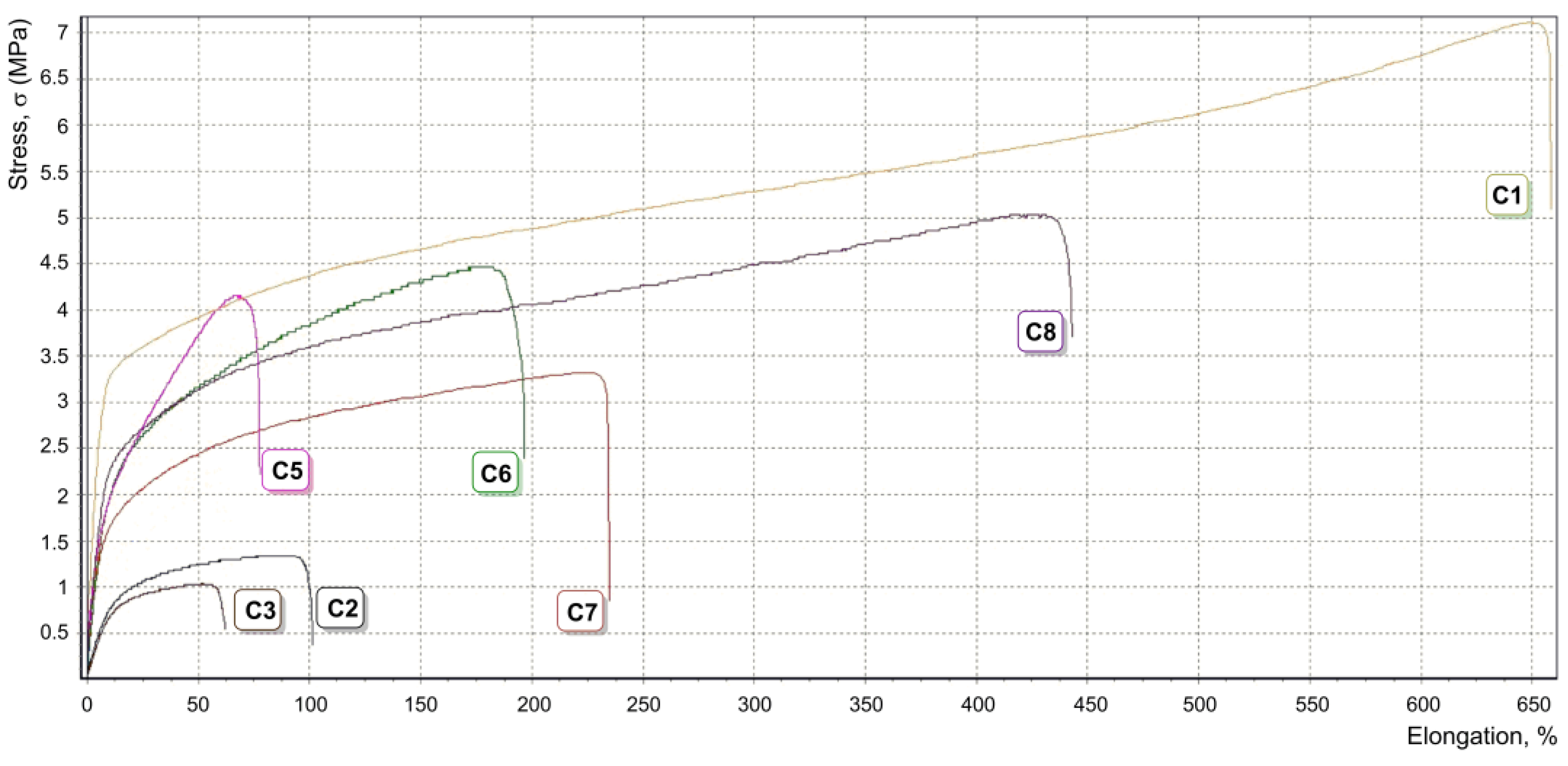
| Entry | PCL, mg | HAp, mg | PCL-b-poly (tBuOEP), mg | Average Cross-Sectional Area, mm2 | Tensile Strength, MPa | Yield Stress σ, MPa | Young’s Modulus E, MPa | Elongation at Break εp, % |
|---|---|---|---|---|---|---|---|---|
| C1 | 1000 | 0 | 0 | 0.66 | 4.5 ± 1.0 | 6.1 ± 1.1 | 24.1 ± 3.4 | 676 ± 28 |
| C2 | 900 | 100 | 0 | 0.50 | 0.44 ± 0.09 | 0.69 ± 0.09 | <7.0 | 98.6 ± 4.1 |
| C3 | 850 | 150 | 0 | 0.65 | 0.54 ± 0.02 | 0.63 ± 0.02 | <7.0 | 65.4 ± 9.5 |
| C4 | 750 | 250 | 0 | 0.68 | n.d. 1 | n.d. | n.d. | n.d. |
| C5 | 825 | 150 | 25 | 0.13 | 4.34 ± 0.69 | 3.30 ± 0.85 | 34.5 ± 6.2 | 79 ± 10 |
| C6 | 800 | 150 | 50 | 0.30 | 2.75 ± 0.63 | 1.63 ± 0.16 | 18.0 ± 2.2 | 213 ± 52 |
| C7 | 775 | 150 | 75 | 0.68 | 1.91 ± 0.54 | 1.48 ± 0.30 | 21.1 ± 5.9 | 287 ± 19 |
| C8 | 925 | 50 | 25 | 0.44 | 3.61 ± 0.95 | 3.1 ± 2.0 | 19.0 ± 5.9 | 437 ± 89 |
| Time, h | [Van·2HCl], mg/mL |
|---|---|
| 0 | 22.5 |
| 1 | 22.0 |
| 2 | 21.6 |
| 3 | 21.5 |
| 5 | 19.6 |
| 24 | 20.9 |
| 48 | 19.7 |
| 120 | 19.2 |
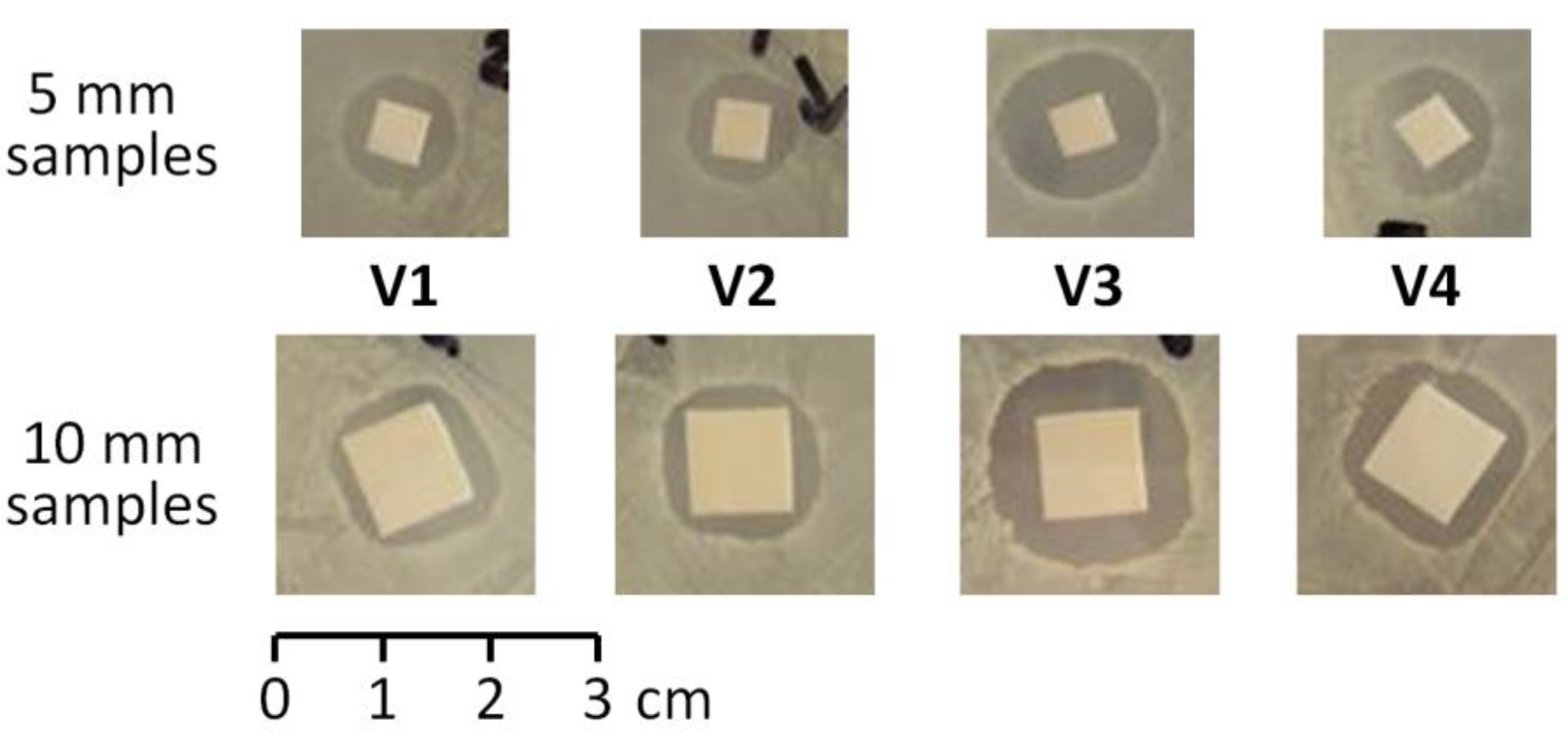
| Sample | ES Fiber Components, wt.% | Note | |||
|---|---|---|---|---|---|
| Van or Van·2HCl | PCL | PCL-b-poly (tBuOEP) | HAp | ||
| V1 | 2.5 | 97.5 | 0 | 0 | Dissolution of both components; ES |
| V2 | 2.5 | 77.5 | 5.0 | 15.0 | Addition of Van + PCL solution to HAp + PCL-b-PEPA suspension; ES |
| V3 | 2.5 | 77.5 | 5.0 | 15.0 | Addition of Van·2HCl + HAp (dried) and PCL solution to PCL-b-PEPA; ES |
| V4 | 0 1 | 80.0 | 5.0 | 15.0 | Exposition of ES film in aq. Van·2HCl |
| Exp. | V1 | V2 | V3 | V4 |
|---|---|---|---|---|
| Inhibition zone diameter (5 × 5 mm sample), 60 mm Petri dishes | ||||
| 1 | 10 | 11 | 15 | 13 |
| 2 | 11 | 10 | 16 | 13 |
| 3 | 11 | 11 | 15 | 12 |
| Average | 10.6 | 10.6 | 15.3 | 12.6 |
| Inhibition zone diameter (10 × 10 mm sample), 90 mm Petri dishes | ||||
| 1 | 15 | 15 | 21 | 17 |
| 2 | 16 | 14 | 21 | 19 |
| 3 | 15 | 15 | 20 | 16 |
| Average | 15.3 | 14.6 | 20.7 | 17.3 |
| Exp. | 1 Day Extract 1 | 2 Day Extract | 3 Day Extract | 3 Day Film |
|---|---|---|---|---|
| V1 | 6 | 0 | 0 | 0 |
| V2 | 9 | 6 | 5 | 7 |
| V3 | 12 | 7 | 5 | 5 |
| V4 | 9 | 0 | 0 | 0 |
Publisher’s Note: MDPI stays neutral with regard to jurisdictional claims in published maps and institutional affiliations. |
© 2021 by the authors. Licensee MDPI, Basel, Switzerland. This article is an open access article distributed under the terms and conditions of the Creative Commons Attribution (CC BY) license (https://creativecommons.org/licenses/by/4.0/).
Share and Cite
Nifant’ev, I.; Gavrilov, D.; Tavtorkin, A.; Chinova, M.; Besprozvannykh, V.; Komarov, P.; Zaitsev, V.; Podoprigora, I.; Ivchenko, P. Antibacterial Poly(ε-CL)/Hydroxyapatite Electrospun Fibers Reinforced by Poly(ε-CL)-b-poly(ethylene phosphoric acid). Int. J. Mol. Sci. 2021, 22, 7690. https://doi.org/10.3390/ijms22147690
Nifant’ev I, Gavrilov D, Tavtorkin A, Chinova M, Besprozvannykh V, Komarov P, Zaitsev V, Podoprigora I, Ivchenko P. Antibacterial Poly(ε-CL)/Hydroxyapatite Electrospun Fibers Reinforced by Poly(ε-CL)-b-poly(ethylene phosphoric acid). International Journal of Molecular Sciences. 2021; 22(14):7690. https://doi.org/10.3390/ijms22147690
Chicago/Turabian StyleNifant’ev, Ilya, Dmitry Gavrilov, Alexander Tavtorkin, Maria Chinova, Victoria Besprozvannykh, Pavel Komarov, Vladimir Zaitsev, Irina Podoprigora, and Pavel Ivchenko. 2021. "Antibacterial Poly(ε-CL)/Hydroxyapatite Electrospun Fibers Reinforced by Poly(ε-CL)-b-poly(ethylene phosphoric acid)" International Journal of Molecular Sciences 22, no. 14: 7690. https://doi.org/10.3390/ijms22147690