Article

Dysregulation of miR-15a-5p, miR-497a-5p and miR-511-5p Is Associated with Modulation of BDNF and FKBP5 in Brain Areas of PTSD-Related Susceptible and Resilient Mice

by
Oriana Maria Maurel
1,†,
Sebastiano Alfio Torrisi
1,†,
Cristina Barbagallo
2,
Michele Purrello
2,
Salvatore Salomone
1,
Filippo Drago
1,
Marco Ragusa
2,*,‡ and
Gian Marco Leggio
1,*,‡
1
Section of Pharmacology, Department of Biomedical and Biotechnological Sciences, University of Catania, 95123 Catania, Italy
2
Section of Biology and Genetics, Department of Biomedical and Biotechnological Sciences, University of Catania, 95123 Catania, Italy
*
Authors to whom correspondence should be addressed.
Shared first authorship.
Shared last authorship.
Int. J. Mol. Sci. 2021, 22(10), 5157; https://doi.org/10.3390/ijms22105157
Submission received: 19 April 2021 / Revised: 6 May 2021 / Accepted: 10 May 2021 / Published: 13 May 2021

Abstract

:
Post-traumatic stress disorder (PTSD) is a neuropsychiatric disorder occurring in susceptible individuals following a traumatic event. Understanding the mechanisms subserving trauma susceptibility/resilience is essential to develop new effective treatments. Increasing evidence suggests that non-coding RNAs, such as microRNAs (miRNAs), may play a prominent role in mediating trauma susceptibility/resilience. In this study, we evaluated the transcriptional expression of two key PTSD-related genes (FKBP5 and BDNF) and the relative targeting miRNAs (miR-15a-5p, miR-497a-5p, miR-511-5p, let-7d-5p) in brain areas of PTSD-related susceptible and resilient mice identified through our recently developed mouse model of PTSD (arousal-based individual screening (AIS) model). We observed lower transcript levels of miR-15a-5p, miR-497a-5p, and miR-511a-5p in the hippocampus and hypothalamus of susceptible mice compared to resilient mice, suggesting that the expression of these miRNAs could discriminate the two different phenotypes of stress-exposed mice. These miRNA variations could contribute, individually or synergically, to the inversely correlated transcript levels of FKBP5 and BDNF. Conversely, in the medial prefrontal cortex, downregulation of miR-15a-5p, miR-511-5p, and let-7d-5p was observed both in susceptible and resilient mice, and not accompanied by changes in their mRNA targets. Furthermore, miRNA expression in the different brain areas correlated to stress-induced behavioral scores (arousal score, avoidance-like score, social memory score and PTSD-like score), suggesting a linear connection between miRNA-based epigenetic modulation and stress-induced phenotypes. Pathway analysis of a miRNA network showed a statistically significant enrichment of molecular processes related to PTSD and stress. In conclusion, our results indicate that PTSD susceptibility/resilience might be shaped by brain-area-dependent modulation of miRNAs targeting FKBP5, BDNF, and other stress-related genes.

1. Introduction

Intense stressful events evoke a plethora of biological responses, including neurochemical cascades, altering the functioning of the sympathetic–adrenal–medullary (SAM) system, the hypothalamic–pituitary–adrenal (HPA) axis, and the immune system. These biological changes can modify the neuronal connectivity, signaling, and remodeling and are associated with the exacerbation or development of neuropsychiatric diseases, such as depression and post-traumatic stress disorder (PTSD) [1,2,3]. Cellular and molecular studies in PTSD and stress-related disorders showed the involvement of a distinct sets of HPA-axis-related genes [4,5,6]. In this context, the roles of FKBP5 (FK506 binding protein 5) and BDNF (brain-derived neurotropic factor) have been reported to be strongly associated to PTSD etiopathogenesis [7,8,9,10]. FKBP5 belongs to the immunophilin protein family and is a co-chaperone component of GR (glucocorticoid receptor) heterocomplex, playing a role in GR regulation through an ultra-short negative feedback loop activity [11]. Thus, glucocorticoids (GCs) increase FKBP5 expression, which in turn decreases GR affinity for GCs [12]. FKBP5 dysregulation has been associated with a maladaptive stress response [13,14] and its increased expression may be related to HPA axis hyperactivity in humans [15]. Previous studies have documented that FKBP5 knock-out mice exhibited a more resilient phenotype, characterized by lower corticosterone levels, a decreased anxiety-like behavior, better behavioral responses following a social defeat stress, tail suspension test, and forced swim test, and enhanced cognitive flexibility in the radial arm water maze [13,16,17,18]. Moreover, FKBP5 expression in infralimbic medial prefrontal cortex (mPFC) (but not in the central prelimbic cortex) increased after fear conditioning in rats and remained elevated even after extinction, supporting its active role in maladaptive stress response [19]. Consistent with these results, Young et al. found increased levels of FKBP5 in the medial orbitofrontal cortex from postmortem brain tissues of PTSD patients, which positively correlated with dendritic spine density [20].
On the other hand, BDNF, the most studied neurotrophin, is involved in neuroprotection, neurogenesis, neurodevelopment, and synaptic plasticity processes required for long-term learning and memory [7,21,22,23,24]. Notwithstanding evidence showing that acute or chronic stress decreased BDNF expression, which could lead to the loss of synaptic function and increased vulnerability to insults [25,26], other studies reported BDNF overexpression resulting in memory impairments in the forebrain of mice [27], as well as in the hippocampus, hypothalamus, and pituitary gland in chronically stressed rats [28]. A progressive BDNF decrease was also demonstrated at different time points after acute social defeat until 24 h after stress, with levels returning to baseline by day 5 post-stress [29]. Taken together, these results suggest a complex relationship between the intensity and duration of stress on BDNF levels, which would depend significantly also on the type of stress and the brain region being studied. The conflicting effects of stressors on BDNF expression and its related phenotypes show the complexity of stress-related behaviors, and could be the results of different BDNF isoforms with dissimilar functions in various brain areas and differential epigenetic and post-transcriptional regulation, depending on the type of neuron and stimulation [30,31,32,33,34]. FKBP5 and BDNF variations are the result of the complex interactions between environment and gene functions through epigenetic modifications such as DNA methylation, histone modifications, and modulation of non-coding RNAs (ncRNAs) [35,36,37], which play a critical role in adaptive and maladaptive processes [38]. Among ncRNAs, microRNAs (miRNAs) seem to play a pivotal role in neural development and function, as well as in the response to stressors [39,40]. The perturbation of cellular homeostasis may interfere with miRNA biogenesis, leading to deregulation of miRNA-controlled pathways and, accordingly, affecting cell susceptibility to stress [41,42]. The role of miRNA-based epigenetic mechanisms may also play a significant role in the onset and the progression of PTSD [43,44], providing new chances to understand its molecular bases. Interestingly, several recent animal and human studies have shown that miRNAs are present in sperm and may be involved in non-Mendelian inheritance of stress behavioral phenotypes [45,46]. Several animal studies reported that specific miRNAs in particular brain regions are involved in various stress-related behaviors, including fear memory consolidation, contextual fear memory, and state-dependent fear [47,48,49,50,51], suggesting that stress-induced alterations of specific miRNAs are not only temporally, but also spatially defined. Since not everyone who experiences intense acute stress develops PTSD, miRNAs have received increasing attention for their potential contribution to susceptibility and resilience to stress. Furthermore, the potential application of miRNAs as biomarkers of vulnerability/resilience to stress is becoming a focus of particular interest, but only few studies have focused on this issue to date [52,53].
In the present study, we evaluated the expression of a set of miRNAs targeting FKBP5 and BDNF, two key PTSD-related genes, in a new PTSD translational animal model [arousal-based individual screening (AIS)] based on post-trauma changes in startle reactivity, following a single, long, and severe traumatic procedure (24 h restraint stress) [54]. We provide evidence that a single traumatic event induced brain-area-specific miRNA alterations, differentially associated with susceptible and resilient phenotypes, and related to behavioral scores.

2. Results

2.1. Selection of miRNAs

BDNF and FKBP5 were used as targets to computationally identify predicted and validated miRNAs, both in human and mouse. By this approach, we found 239 miRNAs targeting BDNF and/or FKBP5 both in human or mouse. We further filtered this list by a manually curated search in biomedical literature to identify potential associations between these miRNAs and stress-related disorders. Finally, we focused our experimental analyses on four miRNAs that targeted both BDNF and FKBP5: miR-15a-5p, miR-497a-5p, miR-511-5p, and let-7d-5p.

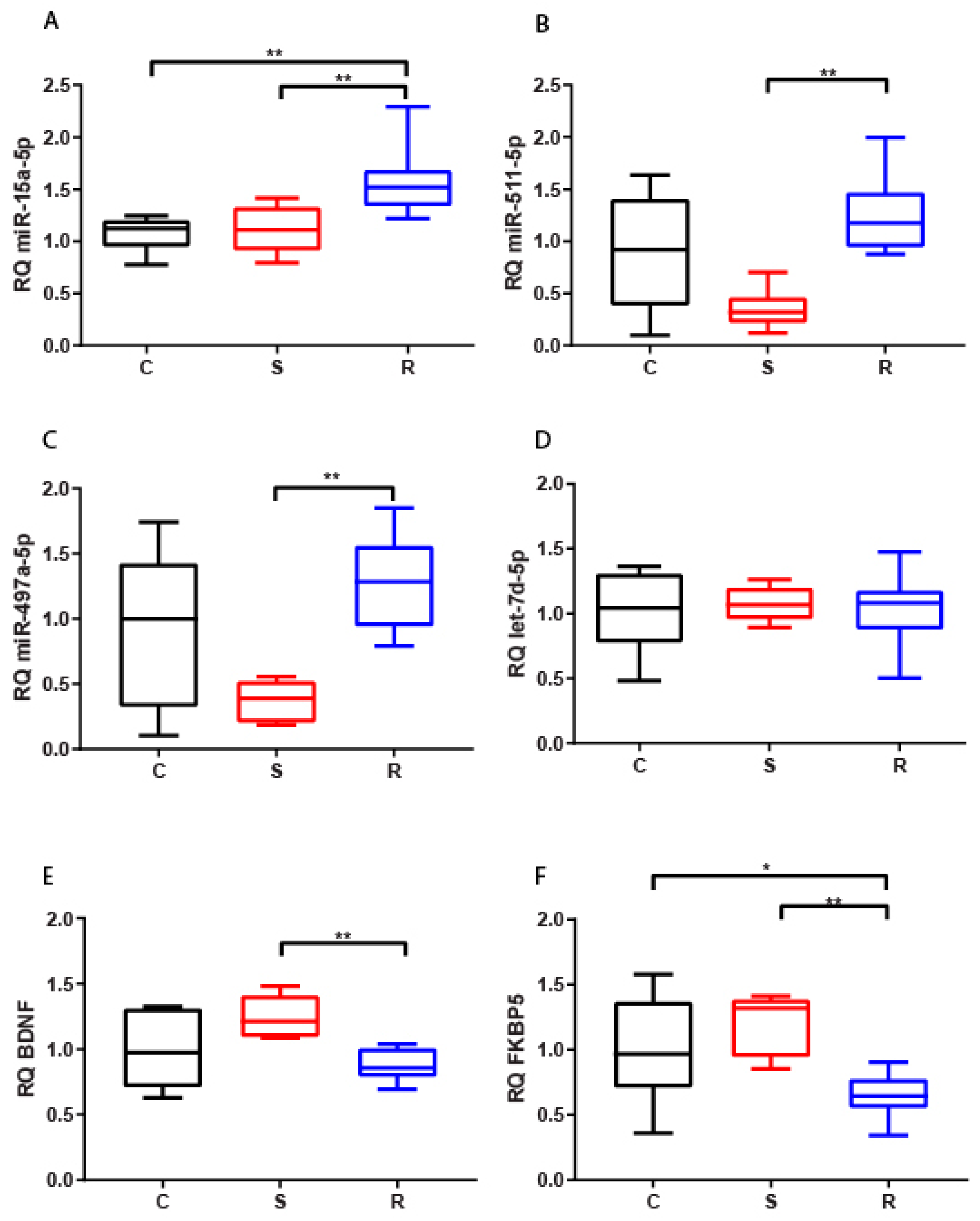
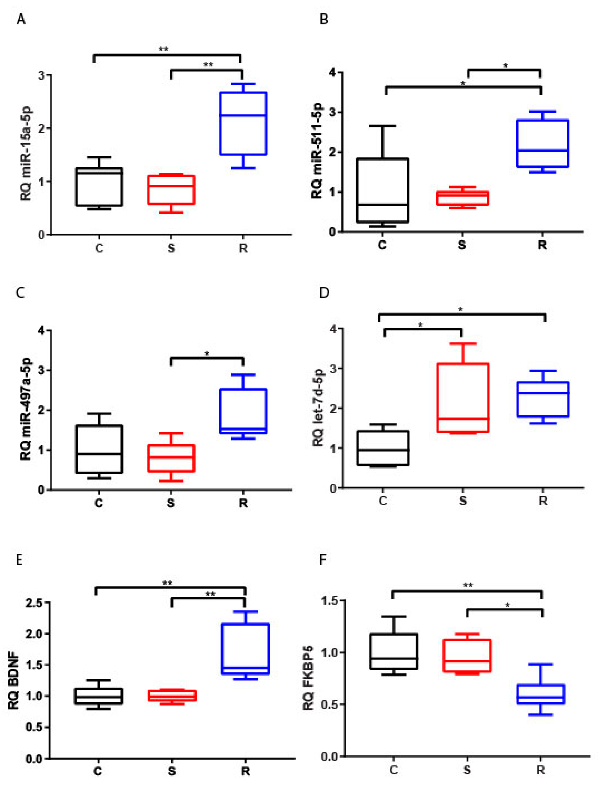
2.2. Expression of miR-15a-5p, let-7d-5p, miR-497a-5p, miR-511-5p, and Their Potential Targets, BDNF and FKBP5

2.2.1. Hippocampus

The expression of miR-15a-5p increased in the hippocampus of resilient mice compared to susceptible and control mice (Figure 1A). In the same area, both miR-511-5p and miR-497a-5p showed reduced levels in susceptible animals with respect to resilient mice (Figure 1B,C). These variations taken together suggest that the expression of these three miRNAs could discriminate the two different phenotypes of stressed mice. In contrast, no significant difference was found for hippocampal expression of let-7d-5p (Figure 1D). We observed a statistically significant reduction for both analyzed potential targets of miRNAs, BDNF and FKBP5, in resilient mice compared to susceptible mice, but also in resilient mice compared to control for FKBP5 (Figure 1E,F).
FKBP5 hippocampal expression showed a significant negative correlation with miR-15a-5p, miR-511-5p, and miR-497a-5p, as expected by functional miRNA:mRNA relationships. In contrast, we found no correlation between expression of BDNF and miRNAs (Table 1).

2.2.2. Medial Prefrontal Cortex

We found a strong reduction of miR-15a-5p, miR-511-5p, and let-7d-5p levels in mPFC in both subpopulations of stressed mice compared to controls, except for miR-497a-5p, whose expression alteration was not statistically significant (Figure 2A–D). These findings suggest that low levels of these miRNAs in this specific brain area are associated with a general reaction to traumatic stress, but not with the discrimination of resilient and susceptible phenotypes. On the other hand, both FKBP5 and BDNF increased their expression in susceptible mice with respect to control and resilient mice (Figure 2E,F).
The inconsistent expression variations between miRNAs and their potential mRNA targets was also demonstrated by calculated correlations. Indeed, we found no significant linear relationship between miRNAs and targets (Table 1).

2.2.3. Hypothalamus

The hypothalamic expression of miR-15a-5p, miR-511-5p, and miR-497a-5p (Figure 3A–C) significantly increased in resilient mice compared to susceptible mice, and also in comparison with control mice for miR-15a-5p and miR-511-5p. However, both stressed groups showed an evident upregulation of let-7d-5p compared to control animals (Figure 3D). We observed a decrease of FKBP5 mRNA expression in resilient mice compared to both susceptible and control mice, whilst an upregulation of BDNF was evident in the same comparisons (Figure 3E,F).
In this brain area, miRNAs showed an opposite trend of deregulation with respect to FKBP5, but not to BDNF, as supported by the significant negative correlations between FKBP5 and miR-15a-5p, miR-511-5p and miR-497a-5p (Table 1).

2.2.4. Evaluation of Synergic Effect of miRNAs on FKBP5 and BDNF Expression

We also investigated whether the four PTSD-related miRNAs may have a synergic effect on regulating FKBP5 and BDNF expression, being both targeted by all the analyzed miRNAs. Different linear regression models were computed, each one including all four miRNAs, three miRNAs, and two miRNAs, considering all possible combinations. For each model, we analyzed all the combinations of miRNAs, considering only those miRNAs showing a negative linear correlation with the target, as previously reported in Table 1. Table 2 lists the models showing the highest R-value for each target in each brain area.
According to R2 values, which represent the proportion of the variance for the dependent variable that may be explained by the independent variables, this analysis suggests that part of FKBP5 or BDNF expression may be explained by the synergic regulation performed by the PTSD-related miRNAs. In particular, as regards FKBP5, more than 41% of its expression in the hippocampus and 51% in the hypothalamus may be explained by variation of miRNAs, with the best result obtained by the combination of all four miRNAs

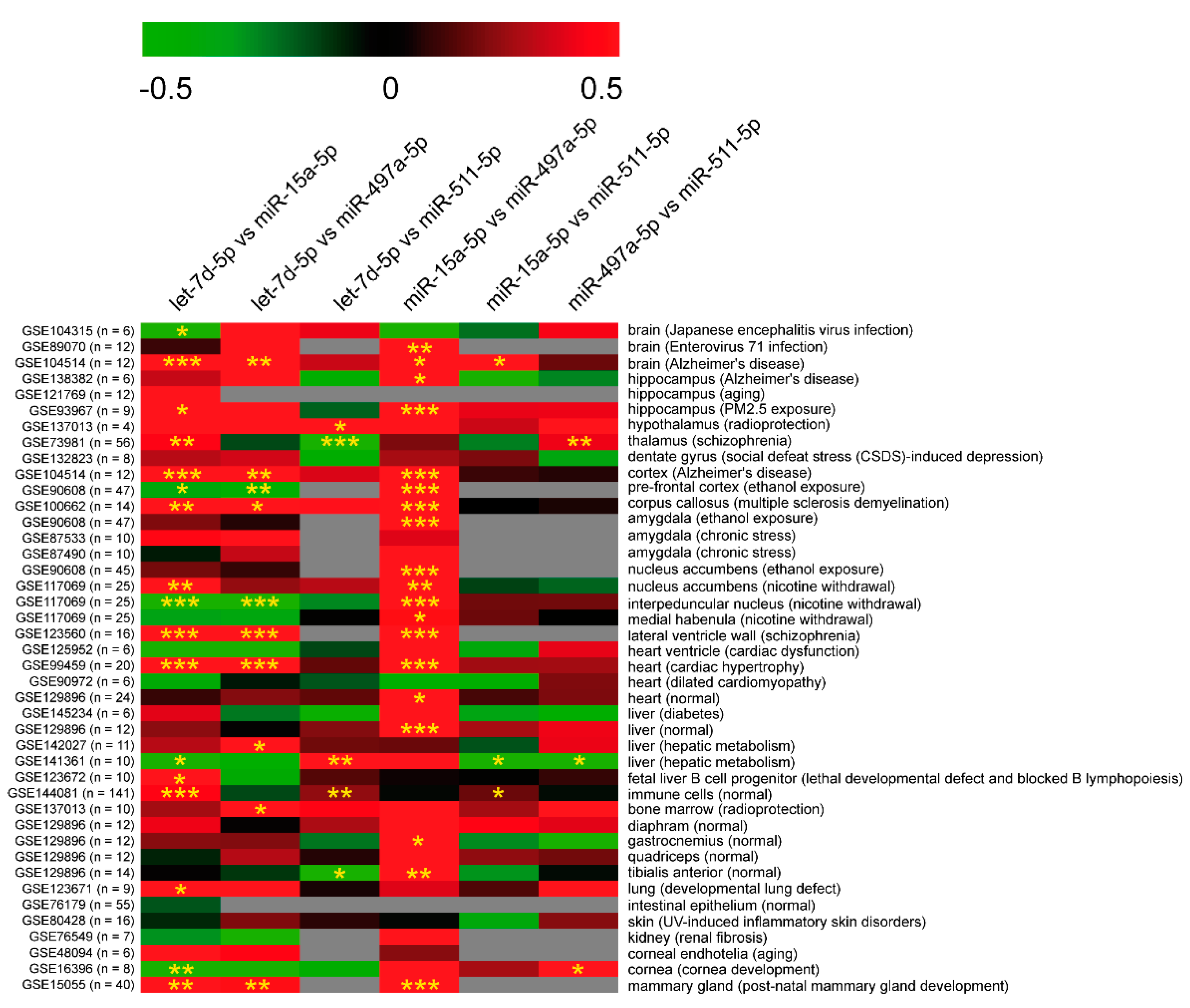
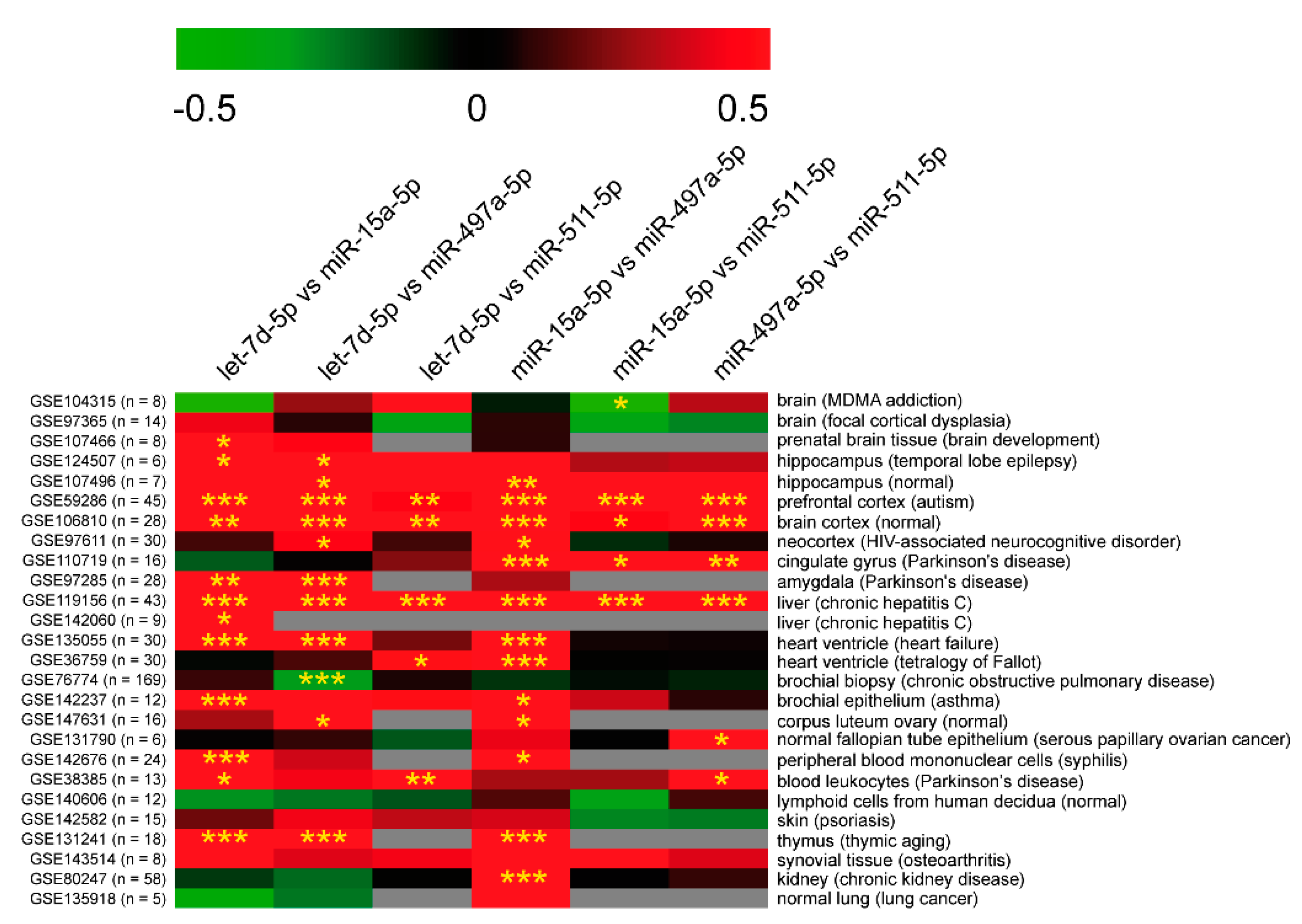
2.2.5. Potential Co-Regulation of PTSD-Related miRNAs

Since the analyzed miRNAs frequently showed similar variation trends in the same brain area in stressed mice, we investigated whether their expressions were statistically correlated with each other in HIP, HT, and, mPFC (Table 3).
In HIP, we found a statistically significant positive correlation between the following miRNAs: miR-15a-5p and let-7d-5p, miR-15a-5p and miR-511-5p, miR-511-5p and miR-497a-5p, and miR-511-5p and let-7d-5p. In contrast, no significant correlation was observed between miR-497a-5p and, respectively, miR-15a-5p and let-7d-5p. In HT, we found a miRNA:miRNA correlation pattern consistent with the one observed in HIP. In fact, miR-15a-5p was positively correlated with miR-511-5p and let-7d-5p, while miR-511-5p showed the same significant relationship with miR-497a-5p and let-7d-5p. No significant linear relationship was observed between miR-497a-5p and miR-15a-5p or let-7d-5p. In mPFC, we found significant positive correlations for the pairs miR-497a-5p:miR-15a-5p and miR-497a-5p:let-7d-5p, and a strong linear relationship between miR-15a-5p and let-7d-5p, as previously observed in HIP. As we found several positive correlations of expression among all miRNAs in all analyzed areas, we corroborated this observation by screening public miRNA expression databases. We retrieved miRNA expression data from different tissues, including brain areas, from mouse and human datasets deposited in the GEO database. Pearson’s correlation coefficients confirmed that expressions of let-7d-5p, miR-15a-5p, miR-497a-5p, and miR-511-5p were frequently co-linear in many mouse and human tissues, especially in the brain, in agreement with results obtained in our PTSD-like model (Figure 4 and Figure 5).
Based on these data, we speculated on the possibility that these miRNAs could share some transcriptional and post-transcriptional regulators. For this purpose, we compared the transcription factors (TFs) binding miRNA promoters and the long non-coding RNAs (lncRNAs) predicted to decoy (bind and sequester) these miRNAs in mouse. This comparative analysis showed that regulatory regions of miR-15a and miR-497a are potentially bound by 27 common TFs, as expected because they belong to the same miRNA family. The genomic loci of all the analyzed miRNAs are characterized by promoter regions that immunoprecipitate with the TFs Cebpa/Cebpb and Spi1 (Table 4).
Moreover, miR-15a-5p, let-7d-5p, miR-511-5p, and miR-497a-5p share RNA-binding sites for several lncRNAs. More specifically, Kcnq1ot1 (KCNQ1 overlapping transcript 1) and Gm4117 are predicted to sequester all the PTSD-related miRNAs (Table 5).

2.2.6. Correlation Analysis Between RNA Expression and Behavioral Scores

Since we observed a dysregulated expression of mRNAs and miRNAs in different brain areas in association with susceptible and resilient phenotypes, we evaluated whether RNA alterations were linearly related to the composite scores of behavioral tests in susceptible and resilient mice, including arousal score, avoidance-like score, social memory score, and PTSD-like score (Table 6).
In HIP, we found a statistically significant inverse correlation between arousal score and expression levels of miR-15a-5p and miR-511-5p and, in contrast, a congruent positive association between the same score and BDNF and FKBP5. These data suggest that the arousal level of mice segregated through the AIS model could be controlled by the hippocampal levels of BDNF and FKBP5, and their targeting miRNAs miR-15a-5p and miR-511-5p. On the other hand, we found no significant correlation between scores and expression of miRNAs and mRNAs in mPFC. These data were consistent with the observation that miRNA dysregulation in mPFC was associated with traumatic stress independently of susceptibility or resilience. Finally, we found a strong anti-correlation between avoidance-like score and hypothalamic expression of both miR-511-5p and BDNF. The expression of miR-497a-5p in the hypothalamus was negatively correlated with arousal, avoidance-like score, and PTSD-like scores. Since we investigated the expression of multiple miRNAs and mRNAs, we evaluated whether their levels could be synergically associated with behavior. Linear regression analysis was performed considering all miRNAs and mRNAs together, or only miRNAs and only mRNAs. Similar to previous linear regression analyses, R2 evaluation showed that miRNAs alone account for 96%, 78%, and 83% of arousal, social, and PTSD-like score, respectively, in the hypothalamus. Moreover, in the hippocampus the combination of miRNAs and mRNAs may affect the arousal score (51%), while mRNAs alone may slightly contribute to it (27%) (Table 7).

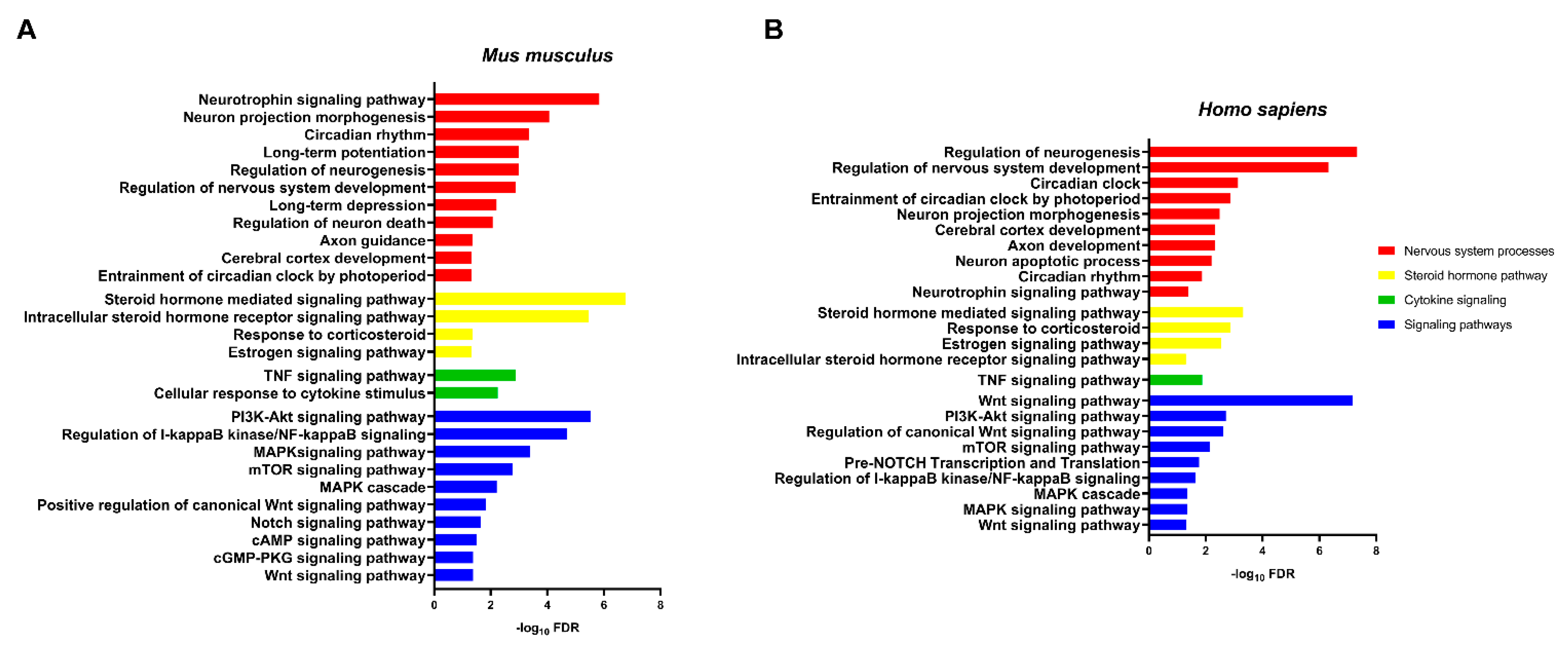
2.2.7. Dysregulation of miRNAs Affects PTSD-Related Pathways

To further characterize the biological meaning of dysregulation of miR-15a-5p, miR-511-5p, miR-497a-5p, and let-7d-5p, gene ontology and pathway enrichment analyses were performed. The network of genes targeted by the investigated miRNAs is involved in several pathways associated with PTSD both in mice and humans. Indeed, we retrieved several terms related to nervous system processes (such as neurotrophin signaling pathway), response to corticosteroid, cytokine signaling, and several signaling pathways previously associated with PTSD (such as MAPK, Wnt, mTOR, Notch signaling pathway) (Figure 6).

3. Discussion

A genetic background is not sufficient alone to properly explain individual differences in sensitivity to stress, given that individual biological diversities are also found within genetically homogeneous populations, such as inbred mice [55]. Life experiences could interact with genetic background to produce long-lasting alterations in coping abilities later in life [56]. For this reason, the development of behavioral and molecular profiling, discriminating susceptible and resilient mice to identify pro-adaptive or maladaptive mechanisms, is fundamental. In the present study, we investigated the expression of two key PTSD-genes, FKBP5 and BDNF, and their targeting miRNAs in a long-term PTSD mouse model, in which a single severe restraint of long duration is able to trigger persistent PTSD-like phenotypes in susceptible mice [54]. Animal studies have shown that dysregulation of miRNA levels in different brain regions can be associated with behavioral alterations [57]. The MiR-15/107 family is highly expressed in brain tissues and comprises multiple highly conserved miRNA members, including miR-497a-5p [58]. Our data would suggest that the consistent variations of miR-15a-5p, miR-497a-5p, and miR-511-5p and their targets in the hippocampus and hypothalamus could potentially promote the resiliency processes by contributing to neuroadaptation [59]. On the other hand, miR-15a-5p, miR-511-5p, and let-7d-5p levels were reduced in the mPFC of all stressed animals, suggesting that these miRNAs are not specifically involved in vulnerability and resiliency phenomena in this brain area, but rather in biological events related to the stress. Moreover, their expression was unrelated to BDNF and FKBP5, suggesting that mRNA modulation is also controlled by other molecular factors in this brain area. Although our molecular and computational data derived from a new mouse model of PTSD, accumulating evidence suggests the involvement of these miRNAs in stress-related disorders, as well as in neurological diseases, thus supporting the results of this study [60]. In this context, Volk et al. showed reduced levels of miR-15a in the amygdala of mice exposed to chronic stress exhibiting an increase of anxiety-like behavior [61]. Others groups confirmed the involvement of miR-511 in the risk of stress-related disorders, including major depression and PTSD, even if its role in susceptibility is still unclear [62,63]. Depressed patients had reduced miR-511 expression in the prefrontal cortex, as well as rats tested in the chronic unpredictable mild stress (CUMS) paradigm [62,63,64]. Some studies showed that miR-497 levels were low in the prefrontal cortex of depressed suicide patients, while significantly increased levels were observed in schizophrenic subjects, suggesting an involvement of miR-497 in psychiatric disorders [65,66,67]. Unlike the other analyzed miRNAs, levels of let-7d-5p did not change in the hippocampus, increased in the hypothalamus and decreased in mPFC in stressed mice. These results may suggest an involvement of let-7d-5p in stress-related disorders, but not in the discrimination of vulnerable and resilient phenotypes. Growing evidence indicates that let-7d may play a role in the neurobiology of psychiatric disorders [68]. Let-7d is largely expressed in the brain and is involved in modulating learning and memory [69,70]; however, little is known about its role in stress-related disorders. Interestingly, its upregulation was reported in the nucleus accumbens in a cocaine-conditioned place preference rat model and, in its circulating form, in animals that had undergone chronic social defeat [52,71]. Specific temporal and spatial expression of miRNAs in the brain is known, but the role of the area specificity in gene regulatory networks has not been satisfactorily addressed yet [70]. Our data, together with those previously published in other models, strongly suggest an involvement of these miRNAs in stress-related disorders; in fact, their activity seems to be brain-area-specific and partially associated with the expression of their potential targets. This last observation would suggest that these miRNAs could also target different mRNAs other than BDNF and FKBP5, as well as that these mRNAs could be regulated by diverse miRNAs or other regulators. Moreover, these PTSD-related miRNAs showed several positive correlations of expression among them in the different brain areas, both in our model and in other human and mouse neuronal and non-neuronal tissues, suggesting that their co-regulation may be due to a common transcriptional or epigenetic control. Indeed, the comparative analysis of miRNA promoters by screening of ENCODE chip-seq experiments showed a potential common regulation by transcription factors Cebpa/Cebpb (CCAAT enhancer binding protein alpha/beta) and Spi1 (transcription factor PU.1) at least for miR-497a-5p and miR-511-5p. Cebpa/b transcriptionally control dozens of genes involved in neuro-differentiation and neurodevelopment in rat hippocampi, and are essential mediators of BDNF/Trk signaling in cortical neurons [72,73]. Although the neuronal role of Spi1 is unknown, its function is critical for viability of brain microglia and its alteration was reported in patients affected by Alzheimer’s disease and major psychiatric disorders [74,75,76]. On the other hand, identification of lncRNAs predicted to bind miR-15a-5p, miR-497a-5p, miR-511-5p, and let-7d-5p showed several common lncRNAs potentially sponging them, thus impairing their function. Among these lncRNAs, Kcnq1ot1 was reported to induce apoptosis or autophagy in neurons after different types of injuries by sequestering different miRNA species [77,78,79]. All these findings would suggest that these miRNAs are part of a complex transcriptional and post-transcriptional machinery that controls fundamental processes in neurons and, consequently, their dysregulation would contribute to molecular cascades induced by neuropsychological stress. Moreover, enrichment analysis of miR-15a-5p, miR-497a-5p, miR-511-5p, and let-7d-5p network showed that such miRNA circuits could affect multiple cellular and molecular processes associated with PTSD pathogenesis [47,80,81,82,83,84,85,86,87,88,89]. Based on these considerations, the inverse correlation between arousal score and hippocampal expression of miR-15a-5p and miR-511-5p is not surprising, as it suggests the existence of a linear relationship between the behavioral arousal score of mice and miRNA levels in the brain that, in turn, coherently influences BDNF and FKBP5 transcripts. Moreover, avoidance-like score in the hypothalamus is negatively correlated with both BDNF and miR-511-5p, hinting that the proportional effects of BDNF on neuronal circuits regulating avoidance-like behaviors are uncoupled by miR-511-5p expression. In the same brain area, miR-497a-5p expression was inversely related to the behavioral arousal, social memory, and PTSD-like scores, suggesting its functional role in PTSD-like phenotypes. Linear regression models showed a synergic effect of the expression of all miRNAs on modulation of behavioral scores in the hypothalamus. This observation, consistent with complex system biology, further indicates that complex phenotypes, such as specific psychiatric behaviors, may be a combined result of expression modulation of different miRNAs, rather than the effect of an individual miRNA [90]. To date, there are only few pre-clinical models based on 24 h restraint stress [91]. Although environmental factors play a predominant role in the individual response to stress, rodent genetic background also modulates stress susceptibility and resilience. Therefore, rodent strain, sex, and biological differences between rats and mice should be considered across experiments to increase reproducibility. For instance, C57Bl/6 strain is reported to be more resilient to stress compared to other strains [92]. There are no comparative studies favoring any acute stress protocol and a single rodent model is not perfectly able to capture human individual complexity [93]. Given that both rats and mice have an important role in the study of neuropsychiatric disorders, it is not surprising that differences in behavior were found, as well as in their molecular biology.
Finally, we believe that the AIS model provides an attractive tool to study the specific role of miRNA-regulated pathways in PTSD susceptibility and resilience. MiRNAs represent a novel class of regulators to molecularly target by using RNA-based therapy. Moreover, even if it is far from our original purpose, we speculate that the miRNAs we found dysregulated could be investigated in body fluids as markers reflecting HPA axis activity and, accordingly, the presence of PTSD phenotype.

4. Materials and Methods

4.1. Animals

Forty-six male C57BL6/J mice (8 weeks, weight 28 ± 2 gr.) were purchased from Charles River Laboratories (Milano, Italy). Mice were group-housed (3–5 per cage) under controlled conditions (12 h light/dark cycle, 2 V2 ± 2 °C, 55 ± 5% humidity, food and water ad libitum) and weighed once a week until the end of each experiment. All experiments were carried out according to EU Directive 2010/63/EU, the Institutional Animal Care and Use Committees of Catania and the Italian Ministry of Health (authorization n.110/ 2019 PR).

4.2. Arousal-Based Individual Screening (AIS), ASR Sessions, Z-Normalization Score, and Behavioral Paradigms

Details regarding the AIS model are described in our previous publication [54]. Briefly, the day before the stress procedure (24 h restraint stress), a pre-trauma acoustic startle reactivity (ASR) session was carried out to measure baseline ASR. The day after, mice were gently put into polyethylene Falcon 50 mL centrifuge tubes and exposed to 24 h of restraint from 3:00 p.m. (3 h before the beginning of the dark phase) to 3:00 p.m. of the next day, without access to food and water. Tubes containing mice were randomly placed in conventional cages, while control mice remained in their home cages in a different room. After 24 h of restraint, mice were suddenly put back in their starting home cages, with free access to food and water. Two other ASR sessions, 14 (ASR 1) and 28 days (ASR 2) after the stress, were given to control and stressed mice in order to assess changes of arousal according to their post-stress ASR changes (% of ASR baseline). To segregate stressed mice into susceptible and resilient subgroups, we created an arousal score by applying a mathematical approach (Z-normalization) to ASR changes. After the segregation, susceptible and resilient mice were housed in the same starting cages. Stressed mice that exhibited an arousal score ≥ 1 were classified as susceptible, while stressed mice with an arousal score < 1 were classified as resilient (Figure S1). We further created other composite scores (avoidance-like score, social memory score, and PTSD-like score) related to other crucial features of PTSD, by z-normalizing data from different behavioral tests (open field (O.F), elevated place maze (E.P.M), 5-trials social memory (SM)) that mice underwent post-segregation, respectively, at days 31, 32, and 35. The described protocol provided 25–35% of susceptible mice and 65–75% of resilient mice. This protocol can be defined as safe and could be conceivably used both in rats and mice [91,94,95,96].

4.3. Selection of miRNAs

We retrieved the miRNAs targeting FKBP5 and BDNF by interpolating the predicted and validated human and murine data from miRecords (http://miRecords.umn.edu/miRecords, accessed on 10 October 2020), miRTarbase (http://miRTarBase.cuhk.edu.cn/, accessed on 10 October 2020) and ENCORI tools (http://starbase.sysu.edu.cn/index.php, accessed on 10 October 2020). Finally, the miRNA list was further filtered by a manually curated search on PubMed, taking into account the dysregulation of these miRNAs in neuropsychiatric phenotypes.

4.4. Tissue Collection

The day after the last behavioral experiment (36 days post-stress), all forty-six mice were sacrificed via cervical dislocation. We used thirty mice to dissect hippocampus and mPFC, and sixteen to dissect hypothalamus according to previously described protocols [97,98].

4.5. Total RNA Extraction

Total RNA was isolated from HIP, HT, and mPFC by using 1 mL Trizol Reagent/50–100 mg of tissue (Invitrogen Life Technologies, Waltham, MA, USA) following the manufacturer’s instructions. Briefly, after homogenization, 5 µg glycogen were added to increase RNA yield. Total RNA concentration and purity for each sample were evaluated by GeneQuant pro spectrophotometer (Biochrom, Cambridge, UK). RNA was stored at −80 °C until cDNA synthesis.

4.6. cDNA Synthesis and RT-qPCR

Single-stranded cDNA was synthesized from total RNA according to the manufacturer’s instructions by using the SuperScript III Reverse Transcriptase kit (Thermo Fisher Scientific, Waltham, MA, USA). We investigated mRNA expression in brain tissues through RT-qPCR by using iTaq Universal SYBR Green Supermix (Bio-Rad, Hercules, CA, USA), according to the manufacturer’s instructions. All RT-qPCR reactions were performed on a 7900HT Fast Real Time PCR System (Applied Biosystems, Life Technologies, Carlsbad, CA, USA). cDNA expression was evaluated through SDS RQ Manager 1.2 software (Applied Biosystems); normalization was performed using GAPDH as reference gene. No template samples were used as negative controls. Relative quantity (RQ) for each transcript was calculated according to the 2−∆∆Ct method [99]. Technical triplicates were performed for each reaction. All RT-PCR reaction were performed in accordance with MIQE guidelines. PCR primers were designed by using the online tool Primer-BLAST (http://www.ncbi.nlm.nih.gov/tools/primer-blast/, accessed on 10 October 2020). Primer sequences are shown in Table 8.

4.7. TaqMan microRNA Assays

Specific single assays were performed for each selected miRNA, exploiting the miRNA-specific reverse transcription (TaqMan microRNA Reverse Transcription Kit; Thermo Fisher Scientific, Waltham, MA, USA) and Real-Time PCR (TaqMan Universal Master Mix II, no UNG; Thermo Fisher Scientific) by using Single TaqMan Assays (Thermo Fischer Scientific), according to the manufacturer’s instructions. RT-qPCRs were performed on a 7900HT Fast Real Time PCR System (Applied Biosystems, Life Technologies). The fold changes were calculated by the 2−∆∆Ct method by using U6 snRNA as endogenous control. No template samples were used as negative control. Technical triplicates were performed for each reaction. All RT-qPCR reactions were performed in accordance with MIQE guidelines.

4.8. Computational Identification of miRNA Regulators

To computationally analyze the potential regulators of miRNAs, namely TFs and lncRNAs, we screened the TransmiR v2.0 (http://www.cuilab.cn/transmir, accessed on 10 October 2020) and LncBase v2.0 (https://diana.e-ce.uth.gr/lncbasev3, accessed on 10 October 2020) databases. From TransmiR, we retrieved the murine TF-miRNA regulations derived from ChIP-seq evidence, while LncBase provided the predicted interactions between miRNAs and lncRNAs in mouse.

4.9. Gene Ontology and Pathway Enrichment Analyses

Gene ontology and pathway enrichment analyses were performed to computationally investigate the biological effects of miRNA deregulation. First, we retrieved experimentally validated miRNA targets (where available) from DIANA Tarbase v8 (http://www.microrna.gr/tarbase, accessed on 10 October /2020); otherwise, predicted targets from microT-CDS (http://www.microrna.gr/webServer, accessed on 10 October 2020) were used. The list of miRNA targets was then submitted to the Database for Annotation, Visualization, and Integrated Discovery (DAVID) online tool (https://david.ncifcrf.gov/, accessed on 10 October 2020), which allowed us to screen different databases (Gene Ontology, Biocarta, KEGG, Reactome). Statistical over-representation was calculated by using Fisher’s exact test, Benjamini and Hochberg FDR Correction, p ≤ 0.05. Pathways showing false discovery rate (FDR) < 0.05 were considered statistically significant.

4.10. Statistical Analysis

Statistical significance of different mRNA and miRNA expression between groups was analyzed by one-way analysis of variance (ANOVA), followed by Tukey’s post hoc test for multiple comparisons. To assess data distribution normality, the D’Agostino-Pearson omnibus normality test was carried out. Expression data (RQ) were shown as a box and whiskers plot (min to max). No animals or samples were excluded from the analysis. Pearson’s correlation coefficients were calculated to statistically evaluate the strength and direction of the linear relationship among miRNA and mRNA expression values (RQ) and behavioral scores. Expression correlations among miRNAs were also investigated in mouse and human datasets from different tissues retrieved from Gene Expression Omnibus (GEO) datasets (https://www.ncbi.nlm.nih.gov/gds, accessed on 10 October 2020). For all analyses described in this section, p-values were two-sided and alpha was set to 0.05. All statistical analyses were performed using GraphPad Prism v7.0 (GraphPad Software, San Diego, CA, USA). To investigate the synergic effects of miRNAs on mRNA expression, linear regression models were built by using SPSS 23. The same statistical approach was used to evaluate the combined effect of expression variation of miRNAs + mRNAs, miRNAs only, and mRNAs only on modulation of behavioral scores.

5. Conclusions

In conclusion, we identified in a new PTSD-like model, brain-area-specific alterations of four miRNAs, potentially regulating two PTSD critical genes, BDNF and FKBP5. MiRNA modulation may underlie the epigenetic mechanisms defining resilience or susceptibility to stress by controlling the expression of BDNF and FKBP5 or other stress-related genes.

Supplementary Materials

The following are available online at https://www.mdpi.com/article/10.3390/ijms22105157/s1, Figure S1: Experimental procedure conceived to shape the AIS model, which identified susceptible and resilient subpopulations.

Author Contributions

Conceptualization, O.M.M. and M.R.; methodology, O.M.M., S.A.T. and M.R.; software, O.M.M. and C.B.; validation, M.R.; formal analysis, O.M.M. and C.B.; investigation, O.M.M., S.A.T. and C.B.; resources, M.R., M.P. and S.S.; data curation, O.M.M., C.B., S.A.T. and M.R.; writing—original draft preparation, O.M.M., C.B. and M.R.; review and editing, M.P., F.D., S.S., G.M.L. and S.A.T.; supervision, F.D., S.S., G.M.L. and M.R.; project administration, F.D., S.S. and G.M.L., funding acquisition, F.D., S.S. and G.M.L. All authors have read and agreed to the published version of the manuscript.

Funding

This research received no external funding.

Institutional Review Board Statement

All experiments were carried out according to EU Directive 2010/63/EU, the Institutional Animal Care and Use Committees of Catania and the Italian Ministry of Health (authorization n.110/ 2019 PR).

Informed Consent Statement

Not applicable.

Data Availability Statement

The data that support the findings of this study are available from the corresponding author upon reasonable request.

Acknowledgments

The authors wish to thank the Scientific Bureau of the University of Catania for language support. This study was supported by “linea intervento 3 Open Access Piano PIACERI 2020-2022” and “Starting Grant 2020” from University of Catania, Italian Ministry of University and Research (PRIN 2017-Prot. 201779W93T to GML and PRIN 2017-Prot. 2017K2NEF4 to FD).

Conflicts of Interest

The authors declare no conflict of interest.

Abbreviations

ASR, acoustic startle reactivity; AIS, arousal-based individual screening; BDNF, brain derived neurotropic factor; BLC, basolateral amygdala complex; C, control; Cebpa, CCAAT/enhancer binding protein alpha; Cebpb, CCAAT/enhancer binding protein beta; CUMS, chronic unpredictable mild stress; DAVID, Database for Annotation, Visualization, and Integrated Discovery; E.P.M., elevated place maze; FDR, false discovery rate; FKBP5, FK506 binding protein 5; GEO, Gene Expression Omnibus; GR, glucocorticoid receptor; HIP, hippocampus; HPA, hypothalamic–pituitary–adrenal axis; HT, hypothalamus; Kcnq1ot1, KCNQ1 overlapping transcript 1; KEGG, Kyoto Encyclopedia of Genes and Genomes; mPFC, medial prefrontal cortex; miRNAs, microRNAs; ncRNAs, non-coding RNAs; O.F., open field; PTSD, post-traumatic stress disorder; R, resilient; SAM, sympathetic–adrenal– medullary system; Sp1, specificity protein 1; S, susceptible; TFs, transcriptional factors; Trk, tropomyosin receptor kinase; 5-trials (SM), 5-trial social memory test.

References

  1. Lupien, S.J.; McEwen, B.S.; Gunnar, M.R.; Heim, C. Effects of stress throughout the lifespan on the brain, behaviour and cognition. Nat. Rev. Neurosci. 2009, 10, 434–445. [Google Scholar] [CrossRef] [PubMed]
  2. McEwen, B.S.; Bowles, N.P.; Gray, J.D.; Hill, M.N.; Hunter, R.G.; Karatsoreos, I.N.; Nasca, C. Mechanisms of stress in the brain. Nat. Neurosci. 2015, 18, 1353–1363. [Google Scholar] [CrossRef] [PubMed]
  3. Juruena, M.F.; Eror, F.; Cleare, A.J.; Young, A.H. The Role of Early Life Stress in HPA Axis and Anxiety. Adv. Exp. Med. Biol. 2020, 1191, 141–153. [Google Scholar] [CrossRef] [PubMed]
  4. Jeanneteau, F.D.; Lambert, W.M.; Ismaili, N.; Bath, K.G.; Lee, F.S.; Garabedian, M.J.; Chao, M.V. BDNF and glucocorticoids regulate cortico-trophin-releasing hormone (CRH) homeostasis in the hypothalamus. Proc. Natl. Acad. Sci. USA 2012, 109, 1305–1310. [Google Scholar] [CrossRef] [Green Version]
  5. Vukojevic, V.; Kolassa, I.-T.; Fastenrath, M.; Gschwind, L.; Spalek, K.; Milnik, A.; Heck, A.; Vogler, C.; Wilker, S.; Demougin, P.; et al. Epigenetic Modification of the Glucocorticoid Receptor Gene Is Linked to Traumatic Memory and Post-Traumatic Stress Disorder Risk in Genocide Survivors. J. Neurosci. 2014, 34, 10274–10284. [Google Scholar] [CrossRef]
  6. White, S.; Acierno, R.; Ruggiero, K.J.; Koenen, K.C.; Kilpatrick, D.G.; Galea, S.; Gelernter, J.; Williamson, V.; McMichael, O.; Vladimirov, V.I.; et al. Association of CRHR1 variants and posttraumatic stress symptoms in hurricane exposed adults. J. Anxiety Disord. 2013, 27, 678–683. [Google Scholar] [CrossRef] [Green Version]
  7. Andero, R.; Ressler, K.J. Fear extinction and BDNF: Translating animal models of PTSD to the clinic. Genes Brain Behav. 2012, 11, 503–512. [Google Scholar] [CrossRef] [Green Version]
  8. Règue, M.; Poilbout, C.; Martin, V.; Franc, B.; Lanfumey, L.; Mongeau, R. Increased 5-HT2C receptor editing predisposes to PTSD-like behaviors and alters BDNF and cytokines signaling. Transl. Psychiatry 2019, 9, 100. [Google Scholar] [CrossRef] [Green Version]
  9. Duman, R.S.; Girgenti, M.J. Molecular and cellular studies of PTSD: Postmortem transcriptome analysis and novel therapeutic targets. J. Neurosci. Res. 2019, 97, 292–299. [Google Scholar] [CrossRef]
  10. Young, D.A.; Inslicht, S.S.; Metzler, T.J.; Neylan, T.C.; Ross, J.A. The effects of early trauma and the FKBP5 gene on PTSD and the HPA axis in a clinical sample of Gulf War veterans. Psychiatry Res. 2018, 270, 961–966. [Google Scholar] [CrossRef]
  11. Binder, E.B. The role of FKBP5, a co-chaperone of the glucocorticoid receptor in the pathogenesis and therapy of affective and anxiety disorders. Psychoneuroendocrinology 2009, 34, 186–195. [Google Scholar] [CrossRef] [PubMed]
  12. Gillespie, C.F.; Phifer, J.; Bradley, B.; Ressler, K.J. Risk and resilience: Genetic and environmental influences on development of the stress response. Depress. Anxiety 2009, 26, 984–992. [Google Scholar] [CrossRef] [PubMed] [Green Version]
  13. Hartmann, J.; Wagner, K.V.; Liebl, C.; Scharf, S.H.; Wang, X.-D.; Wolf, M.; Hausch, F.; Rein, T.; Schmidt, U.; Touma, C.; et al. The involvement of FK506-binding protein 51 (FKBP5) in the behavioral and neuroendocrine effects of chronic social defeat stress. Neuropharmacology 2012, 62, 332–339. [Google Scholar] [CrossRef] [PubMed]
  14. Scharf, S.H.; Liebl, C.; Binder, E.B.; Schmidt, M.V.; Müller, M.B. Expression and Regulation of the Fkbp5 Gene in the Adult Mouse Brain. PLoS ONE 2011, 6, e16883. [Google Scholar] [CrossRef] [Green Version]
  15. Gjerstad, J.K.; Lightman, S.L.; Spiga, F. Role of glucocorticoid negative feedback in the regulation of HPA axis pulsatility. Stress 2018, 21, 403–416. [Google Scholar] [CrossRef] [Green Version]
  16. O’Leary, J.C.; Dharia, S.; Blair, L.J.; Brady, S.; Johnson, A.G.; Peters, M.; Cheung-Flynn, J.; Cox, M.B.; de Erausquin, G.; Weeber, E.J.; et al. A new anti-depressive strategy for the elderly: Ablation of FKBP5/FKBP51. PLoS ONE 2011, 6, e24840. [Google Scholar] [CrossRef]
  17. Sabbagh, J.J.; O’Leary, J.C.; Blair, L.J.; Klengel, T.; Nordhues, B.A.; Fontaine, S.N.; Binder, E.B.; Dickey, C.A. Age-associated epigenetic upregulation of the FKBP5 gene selectively impairs stress resiliency. PLoS ONE 2014, 9, e107241. [Google Scholar] [CrossRef]
  18. Touma, C.; Gassen, N.C.; Herrmann, L.; Cheung-Flynn, J.; Bull, D.R.; Ionescu, I.A.; Heinzmann, J.-M.; Knapman, A.; Siebertz, A.; Depping, A.-M.; et al. FK506 binding protein 5 shapes stress re-sponsiveness: Modulation of neuroendocrine reactivity and coping behavior. Biol. Psychiatry 2011, 70, 928–936. [Google Scholar] [CrossRef]
  19. Criado-Marrero, M.; Silva, R.J.M.; Velazquez, B.; Hernández, A.; Colon, M.; Cruz, E.; Soler-Cedeño, O.; Porter, J.T. Dynamic expression of FKBP5 in the medial prefrontal cortex regulates resiliency to conditioned fear. Learn. Mem. 2017, 24, 145–152. [Google Scholar] [CrossRef]
  20. Young, K.A.; Thompson, P.M.; Cruz, D.A.; Williamson, D.E.; Selemon, L.D. BA11 FKBP5 expression levels correlate with dendritic spine density in postmortem PTSD and controls. Neurobiol. Stress 2015, 2, 67–72. [Google Scholar] [CrossRef] [Green Version]
  21. Minichiello, L. TrkB signalling pathways in LTP and learning. Nat. Rev. Neurosci. 2009, 10, 850–860. [Google Scholar] [CrossRef] [PubMed]
  22. Kowiański, P.; Lietzau, G.; Czuba, E.; Waśkow, M.; Steliga, A.; Moryś, J. BDNF: A Key Factor with Multipotent Impact on Brain Signaling and Synaptic Plasticity. Cell. Mol. Neurobiol. 2018, 38, 579–593. [Google Scholar] [CrossRef]
  23. Duman, R.S.; Monteggia, L.M. A Neurotrophic Model for Stress-Related Mood Disorders. Biol. Psychiatry 2006, 59, 1116–1127. [Google Scholar] [CrossRef]
  24. Notaras, M.; Buuse, M.V.D. Neurobiology of BDNF in fear memory, sensitivity to stress, and stress-related disorders. Mol. Psychiatry 2020, 25, 2251–2274. [Google Scholar] [CrossRef]
  25. Niknazar, S.; Nahavandi, A.; Peyvandi, A.A.; Peyvandi, H.; Akhtari, A.S.; Karimi, M. Comparison of the Adulthood Chronic Stress Effect on Hippocampal BDNF Signaling in Male and Female Rats. Mol. Neurobiol. 2016, 53, 4026–4033. [Google Scholar] [CrossRef]
  26. Kozlovsky, N.; Matar, M.A.; Kaplan, Z.; Kotler, M.; Zohar, J.; Cohen, H. Long-term down-regulation of BDNF mRNA in rat hippo-campal CA1 subregion correlates with PTSD-like behavioural stress response. Int. J. Neuropsychopharmacol. 2007, 10, 741–758. [Google Scholar] [CrossRef] [Green Version]
  27. Cunha, C.; Angelucci, A.; D’Antoni, A.; Dobrossy, M.D.; Dunnett, S.B.; Berardi, N.; Brambilla, R. Brain-derived neurotrophic factor (BDNF) overexpression in the forebrain results in learning and memory impairments. Neurobiol. Dis. 2009, 33, 358–368. [Google Scholar] [CrossRef]
  28. Naert, G.; Ixart, G.; Maurice, T.; Tapia-Arancibia, L.; Givalois, L. Brain-derived neurotrophic factor and hypothalam-ic-pituitary-adrenal axis adaptation processes in a depressive-like state induced by chronic restraint stress. Mol. Cell Neurosci. 2011, 46, 55–66. [Google Scholar] [CrossRef] [PubMed]
  29. Pizarro, J.M.; Lumley, L.A.; Medina, W.; Robison, C.L.; Chang, W.E.; Alagappan, A.; Bah, M.J.; Dawood, M.Y.; Shah, J.D.; Mark, B.; et al. Acute social defeat reduces neurotrophin expression in brain cortical and subcortical areas in mice. Brain Res. 2004, 1025, 10–20. [Google Scholar] [CrossRef]
  30. Tsankova, N.M.; Kumar, A.; Nestler, E.J. Histone Modifications at Gene Promoter Regions in Rat Hippocampus after Acute and Chronic Electroconvulsive Seizures. J. Neurosci. 2004, 24, 5603–5610. [Google Scholar] [CrossRef] [Green Version]
  31. Lubin, F.D.; Roth, T.L.; Sweatt, J.D. Epigenetic Regulation of bdnf Gene Transcription in the Consolidation of Fear Memory. J. Neurosci. 2008, 28, 10576–10586. [Google Scholar] [CrossRef] [PubMed]
  32. Fuchikami, M.; Yamamoto, S.; Morinobu, S.; Takei, S.; Yamawaki, S. Epigenetic regulation of BDNF gene in response to stress. Psychiatry Investig. 2010, 7, 251–256. [Google Scholar] [CrossRef] [Green Version]
  33. Bredy, T.W.; Wu, H.; Crego, C.; Zellhoefer, J.; Sun, Y.E.; Barad, M. Histone modifications around individual BDNF gene promoters in prefrontal cortex are associated with extinction of conditioned fear. Learn. Mem. 2007, 14, 268–276. [Google Scholar] [CrossRef] [Green Version]
  34. Chiaruttini, C.; Vicario, A.; Li, Z.; Baj, G.; Braiuca, P.; Wu, Y.; Lee, F.S.; Gardossi, L.; Baraban, J.M.; Tongiorgi, E. Dendritic trafficking of BDNF mRNA is mediated by translin and blocked by the G196A (Val66Met) mutation. Proc. Natl. Acad. Sci. USA 2009, 106, 16481–16486. [Google Scholar] [CrossRef] [PubMed] [Green Version]
  35. Pfeiffer, J.R.; Mutesa, L.; Uddin, M. Traumatic Stress Epigenetics. Curr. Behav. Neurosci. Rep. 2018, 5, 81–93. [Google Scholar] [CrossRef] [PubMed]
  36. Park, C.; Rosenblat, J.D.; Brietzke, E.; Pan, Z.; Lee, Y.; Cao, B.; Zuckerman, H.; Kalantarova, A.; McIntyre, R.S. Stress, epigenetics and depression: A systematic review. Neurosci. Biobehav. Rev. 2019, 102, 139–152. [Google Scholar] [CrossRef] [PubMed]
  37. Ramo-Fernández, L.; Boeck, C.; Koenig, A.M.; Schury, K.; Binder, E.B.; Gündel, H.; Fegert, J.M.; Karabatsiakis, A.; Kolassa, I.-T. The effects of childhood maltreatment on epigenetic regulation of stress-response associated genes: An intergenerational approach. Sci. Rep. 2019, 9, 1–12. [Google Scholar] [CrossRef] [PubMed] [Green Version]
  38. Yeshurun, S.; Hannan, A.J. Transgenerational epigenetic influences of paternal environmental exposures on brain function and predisposition to psychiatric disorders. Mol. Psychiatry 2019, 24, 536–548. [Google Scholar] [CrossRef] [PubMed]
  39. Rajman, M.; Schratt, G. MicroRNAs in neural development: From master regulators to fine-tuners. Development 2017, 144, 2310–2322. [Google Scholar] [CrossRef] [Green Version]
  40. Follert, P.; Cremer, H.; Béclin, C. MicroRNAs in brain development and function: A matter of flexibility and stability. Front. Mol. Neurosci. 2014, 7, 5. [Google Scholar] [CrossRef] [Green Version]
  41. Olejniczak, M.; Kotowska-Zimmer, A.; Krzyzosiak, W. Stress-induced changes in miRNA biogenesis and functioning. Cell. Mol. Life Sci. 2017, 75, 177–191. [Google Scholar] [CrossRef] [Green Version]
  42. Qiu, L.; Tan, E.K.; Zeng, L. microRNAs and Neurodegenerative Diseases. Adv. Exp. Med. Biol. 2015, 888, 85–105. [Google Scholar] [CrossRef] [PubMed]
  43. Kim, G.S.; Smith, A.K.; Nievergelt, C.M.; Uddin, M. Neuroepigenetics of Post-Traumatic Stress Disorder. Prog. Molecular Biol. Transl. Sci. 2018, 158, 227–253. [Google Scholar] [CrossRef]
  44. Zannas, A.S.; Provençal, N.; Binder, E.B. Epigenetics of Posttraumatic Stress Disorder: Current Evidence, Challenges, and Future Directions. Biol. Psychiatry 2015, 78, 327–335. [Google Scholar] [CrossRef]
  45. Dickson, D.A.; Paulus, J.K.; Mensah, V.; Lem, J.; Saavedra-Rodriguez, L.; Gentry, A.; Pagidas, K.; Feig, L.A. Reduced levels of miRNAs 449 and 34 in sperm of mice and men exposed to early life stress. Transl. Psychiatry 2018, 8, 1–10. [Google Scholar] [CrossRef] [Green Version]
  46. Toth, M. Mechanisms of Non-Genetic Inheritance and Psychiatric Disorders. Neuropsychopharmacology 2014, 40, 129–140. [Google Scholar] [CrossRef] [Green Version]
  47. Dias, B.G.; Goodman, J.V.; Ahluwalia, R.; Easton, A.E.; Andero, R.; Ressler, K.J. Amygdala-Dependent Fear Memory Consolidation via miR-34a and Notch Signaling. Neuron 2014, 83, 906–918. [Google Scholar] [CrossRef] [Green Version]
  48. Griggs, E.M.; Young, E.J.; Rumbaugh, G.; Miller, C.A. MicroRNA-182 Regulates Amygdala-Dependent Memory Formation. J. Neurosci. 2013, 33, 1734–1740. [Google Scholar] [CrossRef] [Green Version]
  49. Vetere, G.; Barbato, C.; Pezzola, S.; Frisone, P.; Aceti, M.; Ciotti, M.; Cogoni, C.; Ammassari-Teule, M.; Ruberti, F. Selective inhibition of miR-92 in hippocampal neurons alters contextual fear memory. Hippocampus 2014, 24, 1458–1465. [Google Scholar] [CrossRef]
  50. Jovasevic, V.; Corcoran, A.K.; Leaderbrand, K.; Yamawaki, N.; Guedea, A.L.; Chen, H.J.; Shepherd, G.M.G.; Radulovic, J. GABAergic mechanisms regulated by miR-33 encode state-dependent fear. Nat. Neurosci. 2015, 18, 1265–1271. [Google Scholar] [CrossRef] [Green Version]
  51. Wang, R.Y.; Phang, R.Z.; Hsu, P.H.; Wang, W.H.; Huang, H.T.; Liu, I.Y. In vivo knockdown of hippocampal miR-132 expression impairs memory acquisition of trace fear conditioning. Hippocampus 2013, 23, 625–633. [Google Scholar] [CrossRef]
  52. Chen, R.J.; Kelly, G.; Sengupta, A.; Heydendael, W.; Nicholas, B.; Beltrami, S.; Luz, S.; Peixoto, L.; Abel, T.; Bhatnagar, S. MicroRNAs as biomarkers of resilience or vul-nerability to stress. Neuroscience 2015, 305, 36–48. [Google Scholar] [CrossRef] [PubMed] [Green Version]
  53. Sillivan, S.E.; Jamieson, S.; de Nijs, L.; Jones, M.; Snijders, C.; Klengel, T.; Joseph, N.F.; Krauskoph, J.; Kleinjans, J.; Vinkers, C.; et al. MicroRNA regulation of persistent stress-enhanced memory. Mol. Psychiatry 2020, 25, 965–976. [Google Scholar] [CrossRef]
  54. Torrisi, S.A.; Lavanco, G.; Maurel, O.M.; Gulisano, W.; Laudani, S.; Geraci, F.; Grasso, M.; Barbagallo, C.; Caraci, F.; Bucolo, C.; et al. A novel arousal-based individual screening reveals susceptibility and resilience to PTSD-like phenotypes in mice. Neurobiol. Stress 2021, 14, 100286. [Google Scholar] [CrossRef] [PubMed]
  55. Hager, T.; Jansen, R.F.; Pieneman, A.W.; Manivannan, S.N.; Golani, I.; Van Der Sluis, S.; Smit, A.B.; Verhage, M.; Stiedl, O. Display of individuality in avoidance behavior and risk assessment of inbred mice. Front. Behav. Neurosci. 2014, 8, 314. [Google Scholar] [CrossRef] [Green Version]
  56. Dirven, B.C.J.; Homberg, J.R.; Kozicz, T.; Henckens, M. Epigenetic programming of the neuroendocrine stress response by adult life stress. J. Mol. Endocrinol. 2017, 59, 11–31. [Google Scholar] [CrossRef] [Green Version]
  57. O’Connor, R.M.; Gururajan, A.; Dinan, T.G.; Kenny, P.J.; Cryan, J.F. All Roads Lead to the miRNome: miRNAs Have a Central Role in the Molecular Pathophysiology of Psychiatric Disorders. Trends Pharmacol. Sci. 2016, 37, 1029–1044. [Google Scholar] [CrossRef]
  58. Wang, S.; Zhu, W.; Xu, J.; Guo, Y.; Yan, J.; Meng, L.; Jiang, C.; Lu, S. Interpreting the MicroRNA-15/107 family: Interaction identification by combining network based and experiment supported approach. BMC Med. Genet. 2019, 20, 96. [Google Scholar] [CrossRef] [PubMed]
  59. Russo, S.J.; Murrough, J.W.; Han, M.-H.; Charney, D.S.; Nestler, E.J. Neurobiology of resilience. Nat. Neurosci. 2012, 15, 1475–1484. [Google Scholar] [CrossRef] [Green Version]
  60. Li, N.; Pan, J.; Liu, W.; Li, Y.; Li, F.; Liu, M. MicroRNA-15a-5p serves as a potential biomarker and regulates the viability and apoptosis of hippocampus neuron in children with temporal lobe epilepsy. Diagn. Pathol. 2020, 15, 46–47. [Google Scholar] [CrossRef]
  61. Volk, N.; Pape, J.C.; Engel, M.; Zannas, A.S.; Cattane, N.; Cattaneo, A.; Binder, E.B.; Chen, A. Amygdalar MicroRNA-15a Is Essential for Coping with Chronic Stress. Cell Rep. 2016, 17, 1882–1891. [Google Scholar] [CrossRef] [PubMed] [Green Version]
  62. Zheng, D.; Sabbagh, J.J.; Blair, L.J.; Darling, A.L.; Wen, X.; Dickey, C.A. MicroRNA-511 Binds to FKBP5 mRNA, Which Encodes a Chaperone Protein, and Regulates Neuronal Differentiation. J. Biol. Chem. 2016, 291, 17897–17906. [Google Scholar] [CrossRef] [Green Version]
  63. Xu, J.; Wang, R.; Liu, Y.; Wang, W.; Liu, D.; Jiang, H.; Pan, F. Short- and long-term alterations of FKBP5-GR and specific microRNAs in the prefrontal cortex and hippocampus of male rats induced by adolescent stress contribute to depression susceptibility. Psychoneuroendocrinology 2019, 101, 204–215. [Google Scholar] [CrossRef]
  64. Yoshino, Y.; Roy, B.; Dwivedi, Y. Differential and unique patterns of synaptic miRNA expression in dorsolateral prefrontal cortex of depressed subjects. Neuropsychopharmacology 2021, 46, 900–910. [Google Scholar] [CrossRef] [PubMed]
  65. Smalheiser, N.R.; Lugli, G.; Rizavi, H.S.; Torvik, V.I.; Turecki, G.; Dwivedi, Y. MicroRNA Expression Is Down-Regulated and Reorganized in Prefrontal Cortex of Depressed Suicide Subjects. PLoS ONE 2012, 7, e33201. [Google Scholar] [CrossRef] [PubMed]
  66. Banigan, M.G.; Kao, P.F.; Kozubek, J.A.; Winslow, A.R.; Medina, J.; Costa, J.; Schmitt, A.; Schneider, A.; Cabral, H.; Cagsal-Getkin, O.; et al. Differential Expression of Exosomal microRNAs in Prefrontal Cortices of Schizophrenia and Bipolar Disorder Patients. PLoS ONE 2013, 8, e48814. [Google Scholar] [CrossRef] [Green Version]
  67. Yadav, S.; Pandey, A.; Shukla, A.; Talwelkar, S.S.; Kumar, A.; Pant, A.B.; Parmar, D. miR-497 and miR-302b Regulate Ethanol-induced Neuronal Cell Death through BCL2 Protein and Cyclin D2*. J. Biol. Chem. 2011, 286, 37347–37357. [Google Scholar] [CrossRef] [Green Version]
  68. Maffioletti, E.; Cattaneo, A.; Rosso, G.; Maina, G.; Maj, C.; Gennarelli, M.; Tardito, D.; Bocchio-Chiavetto, L. Peripheral whole blood microRNA alterations in major depression and bipolar disorder. J. Affect. Disord. 2016, 200, 250–258. [Google Scholar] [CrossRef] [Green Version]
  69. Bahi, A.; Dreyer, J.-L. Lentiviral-mediated let-7d microRNA overexpression induced anxiolytic- and anti-depressant-like behaviors and impaired dopamine D3 receptor expression. Eur. Neuropsychopharmacol. 2018, 28, 1394–1404. [Google Scholar] [CrossRef] [Green Version]
  70. Shu, S.Y.; Qing, D.; Wang, B.; Zeng, Q.-Y.; Chen, Y.-C.; Jin, Y.; Zeng, C.-C.; Bao, R. Comparison of microRNA expression in hippocampus and the marginal division (MrD) of the neostriatum in rats. J. Biomed. Sci. 2013, 20, 9. [Google Scholar] [CrossRef] [Green Version]
  71. Chandrasekar, V.; Dreyer, J.-L. Regulation of MiR-124, Let-7d, and MiR-181a in the Accumbens Affects the Expression, Extinction, and Reinstatement of Cocaine-Induced Conditioned Place Preference. Neuropsychopharmacology 2011, 36, 1149–1164. [Google Scholar] [CrossRef]
  72. Kfoury, N.; Kapatos, G. Identification of neuronal target genes for CCAAT/Enhancer Binding Proteins. Mol. Cell. Neurosci. 2009, 40, 313–327. [Google Scholar] [CrossRef] [Green Version]
  73. Calella, A.M.; Nerlov, C.; Lopez, R.G.; Sciarretta, C.; Halbach, O.V.B.; Bereshchenko, O.; Minichiello, L. Neurotrophin/Trk receptor signaling mediates C/EBPalpha, -beta and NeuroD recruitment to immediate-early gene promoters in neuronal cells and re-quires C/EBPs to induce immediate-early gene transcription. Neural Dev. 2007, 2, 1–22. [Google Scholar] [CrossRef] [Green Version]
  74. Smith, A.M.; Gibbons, H.M.; Oldfield, R.L.; Bergin, P.M.; Mee, E.W.; Faull, R.L.M.; Dragunow, M. The transcription factor PU.1 is critical for viability and function of human brain microglia. Glia 2013, 61, 929–942. [Google Scholar] [CrossRef]
  75. Rustenhoven, J.; Smith, A.M.; Smyth, L.C.; Jansson, D.; Scotter, E.L.; Swanson, M.E.V.; Aalderink, M.; Coppieters, N.; Narayan, P.; Handley, R.; et al. PU.1 regulates Alzheimer’s disease-associated genes in primary human microglia. Mol. Neurodegener. 2018, 13, 1–16. [Google Scholar] [CrossRef]
  76. Zhang, J.; Chang, L.; Pu, Y.; Hashimoto, K. Abnormal expression of colony stimulating factor 1 receptor (CSF1R) and transcription factor PU.1 (SPI1) in the spleen from patients with major psychiatric disorders: A role of brain–spleen axis. J. Affect. Disord. 2020, 272, 110–115. [Google Scholar] [CrossRef]
  77. Yu, S.; Yu, M.; He, X.; Wen, L.; Bu, Z.; Feng, J. KCNQ1OT1 promotes autophagy by regulating miR-200a/FOXO3/ATG7 pathway in cerebral ischemic stroke. Aging Cell 2019, 18, e12940. [Google Scholar] [CrossRef] [PubMed]
  78. Li, M.M.; Liu, X.-H.; Zhao, Y.-C.; Ma, X.-Y.; Zhou, Y.-C.; Zhao, Y.-X.; Liu, X.-Y. Long noncoding RNA KCNQ1OT1 promotes apoptosis in neu-roblastoma cells by regulating miR-296-5p/Bax axis. FEBS J. 2020, 287, 561–577. [Google Scholar] [CrossRef]
  79. Wang, H.J.; Tang, X.L.; Huang, G.; Li, Y.B.; Pan, R.H.; Zhan, J.; Wu, Y.-K.; Liang, J.-F.; Bai, X.-X.; Cai, J. Long Non-Coding KCNQ1OT1 Promotes Oxygen-Glucose-Deprivation/Reoxygenation-Induced Neurons Injury Through Regulating MIR-153-3p/FOXO3 Axis. J. Stroke Cerebrovasc. Dis. 2020, 29, 105126. [Google Scholar] [CrossRef]
  80. Green, C.R.; Corsi-Travali, S.; Neumeister, A. The Role of BDNF-TrkB Signaling in the Pathogenesis of PTSD. J. Depress. Anxiety 2013, 29, 105126. [Google Scholar]
  81. Mahan, A.L.; Ressler, K.J. Fear conditioning, synaptic plasticity and the amygdala: Implications for posttraumatic stress disorder. Trends Neurosci. 2012, 35, 24–35. [Google Scholar] [CrossRef] [Green Version]
  82. Rasmusson, A.M.; Pineles, S.L. Neurotransmitter, Peptide, and Steroid Hormone Abnormalities in PTSD: Biological Endopheno-types Relevant to Treatment. Curr Psychiatry Rep. 2018, 20, 52. [Google Scholar] [CrossRef]
  83. Aliev, G.; Beeraka, N.M.; Nikolenko, V.N.; Svistunov, A.A.; Rozhnova, T.; Kostyuk, S.; Cherkesov, I.; Gavryushova, L.V.; Chekhonatsky, A.A.; Mikhaleva, L.M.; et al. Neurophysiology and Psychopathology Underlying PTSD and Recent Insights into the PTSD Therapies—A Comprehensive Review. J. Clin. Med. 2020, 9, 2951. [Google Scholar] [CrossRef] [PubMed]
  84. Fifield, K.; Hebert, M.; Angel, R.; Adamec, R.; Blundell, J. Inhibition of mTOR kinase via rapamycin blocks persistent predator stress-induced hyperarousal. Behav. Brain Res. 2013, 256, 457–463. [Google Scholar] [CrossRef] [Green Version]
  85. Knox, D.; Della Valle, R.; Mohammadmirzaei, N.; Shultz, B.; Biddle, M.; Farkash, A.; Chamness, M.; Moulton, E. PI3K-Akt Signaling in the Basolateral Amygdala Facilitates Traumatic Stress Enhancements in Fear Memory. Int. J. Neuropsychopharmacol. 2021, 24, 229–238. [Google Scholar] [CrossRef]
  86. Hauger, R.L.; Olivares-Reyes, J.A.; Dautzenberg, F.M.; Lohr, J.B.; Braun, S.; Oakley, R.H. Molecular and cell signaling targets for PTSD pathophysiology and pharmacotherapy. Neuropharmacology 2012, 62, 705–714. [Google Scholar] [CrossRef] [Green Version]
  87. Yehuda, R.; Golier, J.A.; Kaufman, S. Circadian Rhythm of Salivary Cortisol in Holocaust Survivors with and without PTSD. Am. J. Psychiatry 2005, 162, 998–1000. [Google Scholar] [CrossRef]
  88. Gao, J.; Wang, H.; Liu, Y.; Li, Y.Y.; Chen, C.; Liu, L.M.; Wu, Y.-M.; Li, S.; Yang, C. Glutamate and GABA imbalance promotes neuronal apoptosis in hip-pocampus after stress. Med. Sci. Monit. 2014, 20, 499–512. [Google Scholar]
  89. Glover, E.M.; Jovanovic, T.; Norrholm, S.D. Estrogen and Extinction of Fear Memories:Implications for Posttraumatic Stress Disorder Treatment. Biol. Psychiatry 2015, 78, 178–185. [Google Scholar] [CrossRef] [Green Version]
  90. Liufu, Z.; Zhao, Y.; Guo, L.; Miao, G.; Xiao, J.; Lyu, Y.; Chen, Y.; Shi, S.; Tang, T.; Wu, C.-I. Redundant and incoherent regulations of multiple phenotypes suggest microRNAs’ role in stability control. Genome Res. 2017, 27, 1665–1673. [Google Scholar] [CrossRef] [Green Version]
  91. Chu, X.; Zhou, Y.; Hu, Z.; Lou, J.; Song, W.; Li, J.; Liang, X.; Chen, C.; Wang, S.; Yang, B.; et al. 24-hour-restraint stress induces long-term depressive-like phenotypes in mice. Sci. Rep. 2016, 6, 32935. [Google Scholar] [CrossRef] [Green Version]
  92. Mozhui, K.; Karlsson, R.-M.; Kash, T.L.; Ihne, J.; Norcross, M.; Patel, S.; Farrell, M.R.; Hill, E.E.; Graybeal, C.; Martin, K.P.; et al. Strain differences in stress re-sponsivity are associated with divergent amygdala gene expression and glutamate-mediated neuronal excitability. J. Neurosci. 2010, 14, 5357–5367. [Google Scholar] [CrossRef] [PubMed] [Green Version]
  93. Verbitsky, A.; Dopfel, D.; Zhang, N. Rodent models of post-traumatic stress disorder: Behavioral assessment. Transl. Psychiatry 2020, 10, 1–28. [Google Scholar] [CrossRef] [PubMed]
  94. Ellenbroek, B.; Youn, J. Rodent models in neuroscience research: Is it a rat race? Dis. Model. Mech. 2016, 9, 1079–1087. [Google Scholar] [CrossRef] [PubMed] [Green Version]
  95. Alekseeva, I.V.; Abramova, A.Y.; Kozlov, A.Y.; Koplik, E.V.; Pertsov, A.S.; Lyadov, D.A.; Nikenina, E.V. State of Stress-Marker Organs in Rats after a Single Exposure to Long-Term Stress and Treatment with Lipopolysaccharide. Bull. Exp. Biol. Med. 2019, 167, 624–627. [Google Scholar] [CrossRef] [PubMed]
  96. Abramova, A.Y.; Pertsov, S.S.; Alekseeva, I.V.; Nikenina, E.V.; Kozlov, A.Y.; Chekhlov, V.V.; Chukhnina, M.E. Blood Cytokine Concentration in Rats during Antigenic Treatment after a Single Long-Term Stress Exposure. Bull. Exp. Biol. Med. 2020, 168, 713–717. [Google Scholar] [CrossRef] [PubMed]
  97. Spijker, S. Dissection of Rodent Brain Region. In Neuroproteomics; Humana Press: Totowa, NJ, USA, 2011; pp. 13–26. [Google Scholar]
  98. Leggio, G.M.; Camillieri, G.; Platania, C.B.M.; Castorina, A.; Marrazzo, G.; Torrisi, S.A.; Nona, C.N.; D’Agata, V.; Nobrega, J.; Stark, H.; et al. Dopamine D3 Receptor Is Necessary for Ethanol Consumption: An Approach with Buspirone. Neuropsychopharmacology 2014, 39, 2017–2028. [Google Scholar] [CrossRef]
  99. Livak, K.J.; Schmittgen, T.D. Analysis of relative gene expression data using real-time quantitative PCR and the 2(-Delta Delta C(T)) Method. Methods 2001, 25, 402–408. [Google Scholar] [CrossRef]
Figure 1. Expression of miR-15a-5p (A), let-7d-5p (B), miR-497a-5p (C), miR-511-5p (D), BDNF (E), and FKBP5 (F) in hippocampi of control (C = 12), susceptible (S = 7), and resilient (R = 11) mice. * p-value < 0.05, ** p-value < 0.01.
Ijms 22 05157 g001
Figure 2. Expression of miR-15a-5p (A), let-7d-5p (B), miR-497a-5p (C), miR-511-5p (D), BDNF (E), and FKBP5 (F) in mPFCs of control (C = 12), susceptible (S = 7), and resilient (R = 11) mice. * p-value < 0.05, ** p-value < 0.01, *** p-value < 0.001.
Ijms 22 05157 g002
Figure 3. Expression of miR-15a-5p (A), let-7d-5p (B), miR-497a-5p (C), miR-511-5p (D), BDNF (E), and FKBP5 (F) in hypothalami of control (C = 6), susceptible (S = 5), and resilient (R = 5) mice. * p-value < 0.05, ** p-value < 0.01.
Ijms 22 05157 g003
Figure 4. Correlation matrix of miRNA expression from GEO data in Mus musculus. The correlation coefficient is indicated by a color gradient from green (negative correlation) to red (positive correlation), as shown in the colored bar. GEO IDs and number of samples (between brackets) are reported on the left of the matrix. Grey is used for datasets where correlation for the specific pair was impossible to calculate (expression data for at least one of the miRNAs were not available). Tissues and diseases (between brackets) are reported on the left of the matrix. Statistically significant p-values are indicated by asterisks: * p-value < 0.05, ** p-value < 0.01, *** p-value < 0.001.
Ijms 22 05157 g004
Figure 5. Correlation matrix of miRNA expression from GEO data in Homo sapiens. The correlation coefficient is indicated by a color gradient from green (negative correlation) to red (positive correlation), as shown in the colored bar. GEO IDs and number of samples (between brackets) are reported on the left of the matrix. Grey is used for datasets where correlation for the specific pair was impossible to calculate (expression data for at least one of the miRNAs were not available). Tissues and diseases (between brackets) are reported on the left of the matrix. Statistically significant p-values are indicated by asterisks: * p-value < 0.05, ** p-value < 0.01, *** p-value < 0.001.
Ijms 22 05157 g005
Figure 6. Pathway enrichment analysis in Mus Musculus (A) and Homo Sapiens (B). Over-represented biological functions of miR-15a-5p, miR-511-5p, miR-497a-5p, and let-7d-5p were computed by analyzing their validated and predicted targets through the DAVID tool. The over represented pathways are reported on the left of the histograms. Statistical significance is reported as −log10 of FDR of Fisher’s exact test.
Ijms 22 05157 g006
Table 1. Expression correlations among miRNAs and BDNF/FKBP5.
Brain AreaTargetmiR-15a-5pmiR-511-5pmiR-497a-5plet-7d-5p
HIPBDNF−0.09 (0.65)−0.04 (0.82)−0.04 (0.80)−0.18 (0.36)
FKBP5−0.40 (0.03)−0.45 (0.02)−0.43 (0.02)−0.07 (0.69)
HTBDNF0.66 (0.005)0.73 (0.01)0.53 (0.03)0.62 (0.01)
FKBP5−0.69 (0.002)−0.60 (0.01)−0.62 (0.022)−0.38 (0.20)
mPFCBDNF−0.10 (0.60)−0.38 (0.10)−0.01 (0.94)−0.32 (0.09)
FKBP50.03 (0.85)−0.20 (0.29)0.06 (0.76)−0.16 (0.39)
Pearson’s coefficient (r) and p-values (between brackets) are reported. Bold typed values are statistically significant. HIP: Hippocampus; HT: Hypothalamus; mPFC: medial Prefrontal Cortex.
Table 2. Linear regression models built on combinations of 4, 3, or 2 PTSD-related miRNAs.
Brain AreaTargetmiRNA CombinationRR2p-Value
HIPFKBP5let-7d-5p, miR-15a-5p, miR-497a-5p, miR-511-5p0.6570.4310.024
miR-15a-5p, miR-497a-5p, miR-511-5p0.6520.4250.01
miR-497a-5p, miR-511-5p0.6410.4110.004
BDNFlet-7d-5p, miR-15a-5p, miR-497a-5p, miR-511-5p0.1910.0370.936
let-7d-5p, miR-15a-5p, miR-511-5p0.1910.0370.841
let-7d-5p, miR-15a-5p0.1860.0350.656
HTFKBP5let-7d-5p, miR-15a-5p, miR-497a-5p, miR-511-5p0.7490.560.044
miR-15a-5p, miR-497a-5p, miR-511-5p0.7410.5490.019
miR-15a-5p, miR-511-5p0.7190.5170.009
mPFCFKBP5let-7d-5p, miR-15a-5p, miR-497a-5p, miR-511-5p0.4290.1840.322
let-7d-5p, miR-15a-5p, miR-511-5p0.420.1770.206
let-7d-5p, miR-15a-5p0.3230.1050.251
BDNFlet-7d-5p, miR-15a-5p, miR-497a-5p, miR-511-5p0.5970.3570.038
let-7d-5p, miR-15a-5p, miR-511-5p0.5870.3450.019
let-7d-5p, miR-511-5p0.4870.2370.039
For each combination of miRNAs, are reported: R-value, representing the coefficient of correlation; the R2 value, representing the proportion of variance of a dependent variable that may be explained by an (or more) independent variable(s); and the p-value. Bold typed values are statistically significant.
Table 3. Correlation matrix of miRNA expression in the different brain areas.
miRNAsHIPHTmPFC
miR-15a-5plet-7d-5pmiR-511-5pmiR-15a-5plet-7d-5pmiR-511-5pmiR-15a-5plet-7d-5pmiR-511-5p
let-7d-5p0.41 (0.03) 0.55 (0.02) 0.46 (0.01)
miR-511-5p0.38 (0.04)0.39 (0.04) 0.70 (0.002)0.63 (0.008) 0.32 (0.09) 0.18 (0.36)
miR-497a-5p0.25 (0.19)0.35 (0.06)0.64 (0.0003)0.39 (0.12)0.38 (0.13)0.62 (0.01)0.57 (0.001)0.54 (0.003)0.14 (0.46)
Pearson’s coefficient (r) and p-values (between brackets) are reported. Bold typed values are statistically significant.
Table 4. Comparison matrix of TFs shared by miRNAs based on ChIp data.
miRNAsmiR-15alet-7dmiR-497a
let-7dCebpa, Spi1
miR-497aBcl11b, Brd4, Cbfb, Cebpb, Ctcf, Ep300, Erg, Ets1, Hdac2, Ikzf1, Max, Med1, Myc, Nelfe, Nr1d1, Nr3c1, Otx2, Pou5f1, Pparg, Rad21, Rela, Smarca4, Spi1, Suz12, Tfap4, Zfp281, Zfp384Spi1
miR-511Batf, Btaf1, Cebpa, Cebpb, Mafb, Nr3c1, Rela, Spi1, Tcf12Cebpa, Spi1Cebpb, Fli1, Nr3c1, Rela, Spi1, Stat5a
TFs typed in bold are shared by several miRNA pairs.
Table 5. Comparison matrix of lncRNAs predicted to bind PTSD-related miRNAs.
miR-15a-5plet-7d-5pmiR-497a-5p
let-7d-5pGm20605, Kcnq1ot1, Ptprv, Gm4117
miR-497a-5pGm20605, Ptprv, ENSMUSG00000093535, Gm11579, Rab10os, Kcnq1ot1, Gm4117Gm20605, Kcnq1ot1, Ptprv, Gm4117, Mecomos
miR-511-5pKcnq1ot1, ENSMUSG00000093535, Gm4117, Gm11579, ENSMUSG00000085287, Rab10osKcnq1ot1, Gm26856, Gm4117, Zfp950Kcnq1ot1, ENSMUSG00000093535, Gm4117, Gm11579, Rab10os
LncRNAs typed in bold and underlined are shared by all miRNA pairs; lncRNAs typed in bold are shared by several miRNA pairs.
Table 6. Correlations between RNA expression and behavioral scores.
miRNAs/mRNAsHIP
ArousalAvoidanceSocial MemoryPTSD-Like
miR-15a-5p−0.44 (0.02)−0.33 (0.08)−0.12 (0.56)−0.31 (0.11)
let-7d-5p−0.08 (0.68)0.26 (0.20)0.30 (0.13)0.16 (0.42)
miR-511-5p−0.44 (0.02)−0.14 (0.49)−0.10 (0.61)−0.34 (0.08)
miR-497a-5p−0.22 (0.28)−0.12 (0.56)−0.10 (0.61)−0.24 (0.22)
FKBP50.39 (0.04)−0.04 (0.84)−0.09 (0.65)0.11 (0.61)
BDNF0.47 (0.01)−0.02 (0.89)−0.10 (0.62)0.27 (0.18)
HT
ArousalAvoidanceSocial MemoryPTSD-like
miR-15a-5p−0.26 (0.31)−0.27 (0.30)−0.11 (0.66)−0.26 (0.31)
let-7d-5p−0.12 (0.64)−0.20 (0.45)0.09 (0.72)−0.07 (0.79)
miR-511-5p−0.13 (0.62)−0.63 (0.007)−0.31 (0.23)−0.39 (0.12)
miR-497a-5p−0.61 (0.01)−0.57 (0.01)−0.37 (0.15)−0.67 (0.004)
FKBP5−0.02 (0.92)0.22 (0.39)0.27 (0.29)−0.17 (0.51)
BDNF−0.25 (0.33)−0.54 (0.02)−0.37 (0.15)−0.46 (0.07)
mPFC
ArousalAvoidanceSocial MemoryPTSD-like
miR-15a-5p0.09 (0.65)−0.10 (0.60)0.10 (0.61)0.04 (0.83)
let-7d-5p0.11 (0.58)−0.13 (0.53)0.18 (0.38)0.13 (0.52)
miR-511-5p−0.09 (0.67)−0.37 (0.07)−0.09 (0.64)−0.19 (0.34)
miR-497a-5p−0.02 (0.92)−0.27 (0.19)−0.15 (0.45)−0.15 (0.45)
FKBP50.27 (0.18)0.16 (0.41)0.18 (0.37)0.30 (0.12)
BDNF−0.27 (0.19)0.16 (0.43)0.28 (0.16)0.02 (0.89)
Pearson’s coefficient (r) and p-values (between brackets) are reported. Bold typed values are statistically significant.
Table 7. Synergic effects of analyzed RNAs on behavioral scores.
ScoresmiRNAs/mRNAsHIPPOCAMPUSHYPOTHALAMUSmPFC
RR2p-valueRR2p-ValueRR2p-Value
Arousal scoremiRNA + mRNAs0.720.5190.0320.5630.3170.7910.3850.1490.88
MiRNA0.4420.1960.3350.9810.9620.000220.1990.040.937
mRNAs0.5230.2730.030.2740.0750.7610.1030.0110.885
Avoidance-like scoremiRNA + mRNAs0.5290.280.4020.7280.530.4490.5030.2530.632
miRNA0.3490.1220.6060.7110.5060.3040.4880.2380.247
mRNAs0.1330.0180.8220.6880.4730.1060.0380.0010.983
Social memory scoremiRNA + mRNAs0.6910.4770.0580.8840.7820.0940.5410.2930.526
miRNA0.5120.2630.1720.8840.7810.0350.2070.0430.928
mRNAs0.2410.0580.5180.7250.5250.0740.2540.0650.464
PTSD-like scoremiRNA + mRNAs0.6990.4880.050.690.4760.5410.5310.2820.554
miRNA0.5020.2520.1940.9150.8380.0150.330.1090.681
MRNAs0.3470.1210.2440.5410.2930.2980.1620.0260.737
For each model, built on miRNAs+mRNA, only miRNAs, or only mRNAs, the R-value, representing the coefficient of correlation; the R2 value, representing the proportion of variance of a dependent variable that is/may be explained by an (or more) independent variable(s); and the p-value are reported. Bold typed values are statistically significant.
Table 8. PCR primer sequences of mRNAs analyzed in this study.
GeneForward PrimerReverse Primer
FKBP55′-TGAGGGCACCAGTAACAATGG-3′5′-CAACATCCCTTTGTAGTGGACAT-3′
BDNF5′-GTTCGAGAGGTCTGACGACG-3′5′-AGTCCGCGTCCTTATGGTTT-3′
GAPDH5′-AGGTCGGTGTGAACGGATTTG-3′5′-TGTAGACCATGTAGTTGAGGTCA-3′
Publisher’s Note: MDPI stays neutral with regard to jurisdictional claims in published maps and institutional affiliations.

