Article

Electromagnetic Field (EMF) Radiation Alters Estrogen Release from the Pig Myometrium during the Peri-Implantation Period

by
Ewa Monika Drzewiecka
*,
Wiktoria Kozlowska
,
Agata Zmijewska
,
Pawel Jozef Wydorski
and
Anita Franczak
*
Department of Animal Anatomy and Physiology, Faculty of Biology and Biotechnology, University of Warmia and Mazury in Olsztyn, Oczapowskiego 1A, 10-719 Olsztyn, Poland
*
Authors to whom correspondence should be addressed.
Int. J. Mol. Sci. 2021, 22(6), 2920; https://doi.org/10.3390/ijms22062920
Submission received: 22 January 2021 / Revised: 10 March 2021 / Accepted: 11 March 2021 / Published: 13 March 2021
(This article belongs to the Section Molecular Endocrinology and Metabolism)

Abstract

:
An electromagnetic field (EMF) may affect the functions of uterine tissues. This study hypothesized that EMF changes the estrogenic activity of pig myometrium during the peri-implantation period. Tissue was collected on days 15–16 of the gestation and incubated in the presence of EMF (50 and 120 Hz, 2 and 4 h). The cytochrome P450 aromatase type 3 (CYP19A3) and hydroxysteroid 17β dehydrogenase type 4 (HSD17B4) mRNA transcript abundance, cytochrome P450arom (aromatase), and 17β hydroxysteroid dehydrogenase 17βHSD) protein abundance and estrone (E1) and estradiol-17β (E2) release were examined using Real-Time PCR, Western blot and radioimmunoassay. Selected myometrial slices were treated with progesterone (P4) to determine whether it functions as a protector against EMF. CYP19A3 mRNA transcript abundance in slices treated with EMF was less at 50 Hz (2 h) and greater at 120 Hz (2 and 4 h). HSD17B4 mRNA transcript was greater in slices treated with EMF at 120 Hz (2 h). Progesterone diminished EMF-related effects on CYP19A3 and HSD17B4. When P4 was added, EMF had suppressive (50 and 120 Hz, 2 h) or enhancing (50 Hz, 4 h) effects on aromatase abundance. The E1 release was lower after 4 h of EMF treatment at 50 Hz and P4 did not protect myometrial E1 release. In conclusion, EMF alters the synthesis and release of E1 and did not affect E2 release in the myometrium during the peri-implantation period.

Graphical Abstract

1. Introduction

Estrogens are key regulators of female reproduction which affect corpus luteum (CL) lifespan, uterine receptivity, and are involved in embryo-maternal interactions [1,2,3,4,5]. Importantly, a pivotal source of estrogens in the uterus is the endometrium and the myometrium, in which steroidogenic activity depends on the reproductive status of the female [6,7,8]. Interestingly, past studies indicated that uterine steroidogenic activity might also be regulated not only by endocrine factors [9,10,11,12,13,14,15,16,17], but also by physical environmental factors, such as an electromagnetic field (EMF) [18,19,20].
The EMF at frequencies ranging from 50 to 120 Hz is generated by most electrical devices used in an everyday manner [21]. It has been well-documented that EMF at a frequency of 50 Hz and 120 Hz may cause DNA damage, induce structural, morphometric and physiological changes in testes, decrease the rate of fetal development, induce changes in brain protein conformation and generate alterations in signal transduction mechanisms involved in learning capacity and memory processes and induce anti-inflammatory responses [22,23,24,25,26]. In estrous-cyclic pigs, during the mid-luteal phase of the estrous cycle, an EMF at a frequency of 50 and 120 Hz increased myometrial estradiol-17β (E2) release within 4 h of treatment duration [18]. It was also determined that treatment with an EMF (50 Hz for 2 h, 120 Hz for 2 and 4 h) decreases the myometrial release of androstenedione (A4) [19]. Importantly, androgens are substrates for estrogen production [27]; thus, alterations in their production may cause a deregulation in estrogen production. The effect of an EMF on estrogen release from the myometrium has not yet been evaluated. Notably, the peri-implantation period is the most critical period for the success of female reproduction [28], during which the action of estrogens may determine the success of pregnancy [1,2,3,4,5].
Remarkably, in pigs during early pregnancy, estrogens act as anti-luteolytic factors, maintain luteoprotection and support progesterone (P4) production by CLs in early pregnancy [29,30,31]. It has become obvious that P4, which is a substrate for the steroidogenic pathway [8,27], is essential in the uterus to develop uterine receptivity and stimulates the transformation of uterine tissues to create an environment for embryonic development, implantation and placentation [32]. Recommendations are currently being considered for the prescription of P4 for gravid females to avoid early pregnancy complications and to increase endometrial receptivity to implantation [33,34]. Interestingly, P4 in estrous-cyclic females has functions that protect the pig uterine tissues against EMF radiation [18]. Nevertheless, in early pregnant females, P4 is not an obvious protection factor against EMF-related changes in uterine androgen production [19,20].
The present study aimed to determine the effect of an EMF (50 and 120 Hz, 8 militesla (mT), 2- and 4 h duration) treatment on (1) cytochrome P450 aromatase type 3 (CYP19A3) and hydroxysteroid 17β dehydrogenase type 4 (HSD17B4) mRNA transcript abundance, (2) cytochrome P450arom (aromatase) and 17β hydroxysteroid dehydrogenase (17βHSD) protein abundance, and (3) estrone (E1) and estradiol-17β (E2) release from the myometrium of pigs during the peri-implantation period (i.e., days 15 to 16 of gestation). To examine whether the inclusion of P4 may protect the myometrium against the EMF-related effects, selected myometrial slices were incubated in the presence of P4.

2. Results

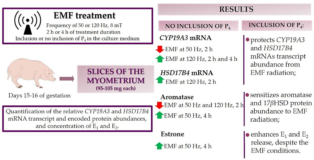
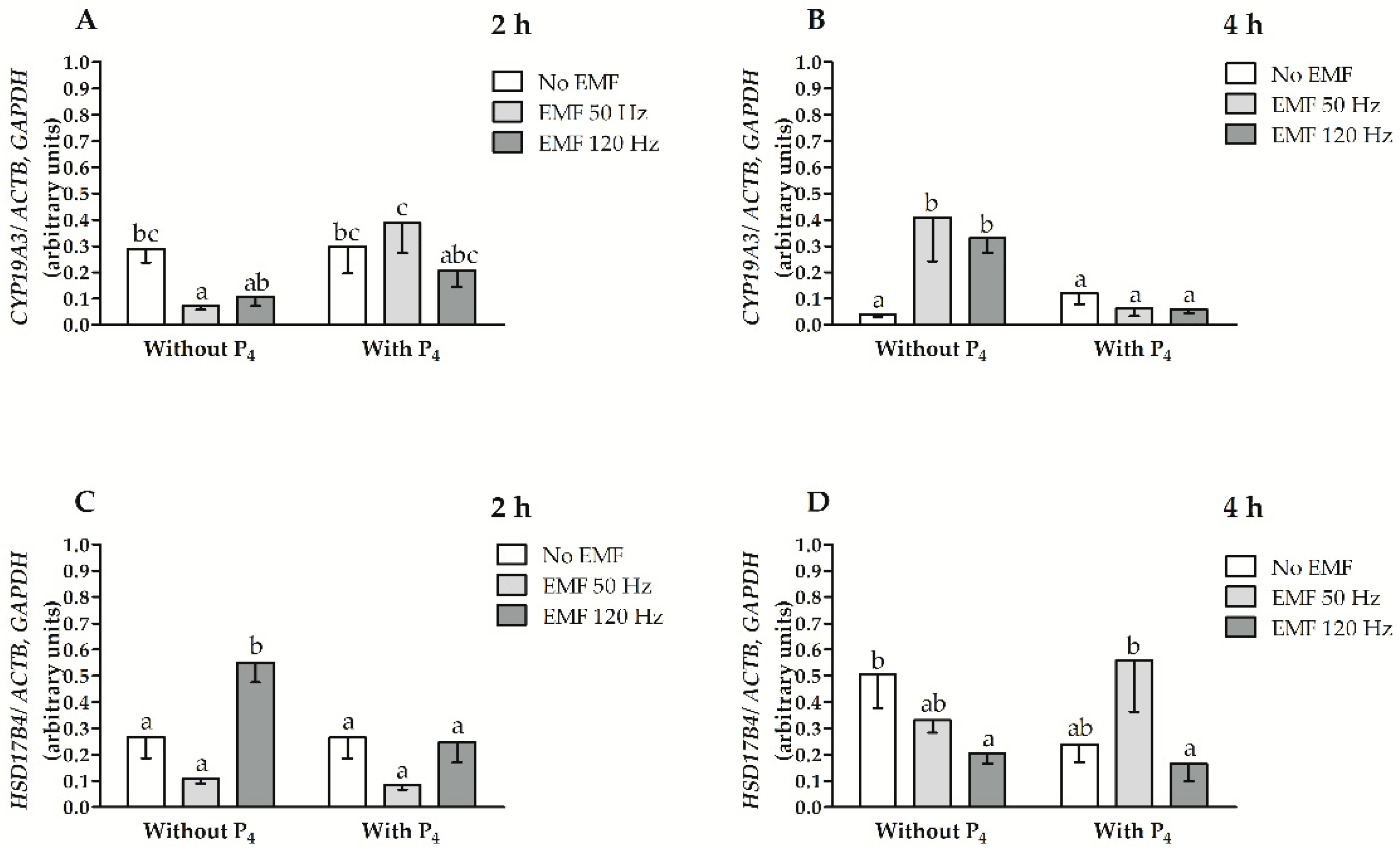
2.1. EMF at 50 and 120 Hz Alter CYP19A3 and HSD17B4 mRNA Transcript Abundance in Myometrial Slices after 2 h and 4 h of In Vitro Incubation in the Presence or Absence of P4

The values for the main effects and interactions among factors affecting CYP19A3 and HSD17B4 mRNA transcript abundances, i.e., treatment with an EMF, duration of EMF treatment, and inclusion of P4 in culture medium are presented in Table 1. The relative abundance of CYP19A3 and HSD17B4 mRNA transcripts was affected by the interaction between an EMF treatment and the treatment duration (p = 0.03, and p = 0.0002, respectively). The relative abundance of CYP19A3 mRNA transcript was also affected by the interaction between the treatment duration with the EMF and P4 inclusion (p = 0.0002) and by the interaction when an EMF treatment was imposed, the treatment duration of the EMF and P4 inclusion in the culture medium (p = 0.002).
After 2 h of treatment duration without the inclusion of P4 in the culture medium, the CYP19A3 mRNA transcript abundance was lower in myometrial slices as a result of treatment with an EMF at a frequency of 50 Hz and was not altered in myometrial slices treated with an EMF at a frequency of 120 Hz (Figure 1A, p ≤ 0.05). After 2 h of treatment when there was the inclusion of P4 in the culture medium, the CYP19A3 mRNA transcript abundance was not altered when treated with an EMF at a frequency of 50 and 120 Hz compared to slices not treated with an EMF (p > 0.05, Figure 1A). When treated with an EMF at a frequency of 50 Hz, the abundance of CYP19A3 mRNA transcript was larger after 2 h of incubation both when there was the inclusion of P4 in the culture medium compared to myometrial slices and when there was no inclusion of P4 in the medium (p ≤ 0.05, Figure 1A). After 4 h of incubation without P4 inclusion in the medium, the abundance of CYP19A3 mRNA transcript was higher when treated with an EMF at 50 Hz and 120 Hz, compared to myometrial slices not treated with an EMF (p ≤ 0.05, Figure 1B). In myometrial slices with P4 inclusion in the culture medium, after 4 h of incubation and imposing an EMF at 50 and 120 Hz, the CYP19A3 mRNA transcript abundance was not altered (p > 0.05, Figure 1B). The abundance of CYP19A3 mRNA transcript in myometrial slices after 4 h of incubation and the imposing of an EMF at 50 and 120 Hz was lower with the inclusion of P4 in the culture medium compared to myometrial slices incubated without P4 (p ≤ 0.05, Figure 1B).
The abundance of HSD17B4 mRNA transcript was greater in myometrial slices after 2 h of incubation without the inclusion of P4 in the medium when treated with an EMF at the frequency of 120 Hz (p ≤ 0.05) and was not altered (p > 0.05) when there was treatment at 50 Hz (Figure 1C). In slices incubated with P4 inclusion in the culture medium after 2 h of incubation and imposing an EMF at 50 and 120 Hz, the abundance of HSD17B4 mRNA transcript was not altered (p > 0.05, Figure 1C). The abundance of HSD17B4 mRNA transcript in myometrial slices after 2 h of incubation without the inclusion of P4 in the culture medium and imposing an EMF at 120 Hz EMF was larger compared to slices incubated with the inclusion of P4 in the culture medium (p ≤ 0.05, Figure 1C). After 4 h of incubation without the inclusion of P4, the HSD17B4 mRNA transcript abundance was lower in slices treated with an EMF at 120 Hz compared to slices not treated with an EMF (p ≤ 0.05, Figure 1D). When there was the inclusion of P4, the abundance of HSD17B4 mRNA transcript was not altered in myometrial slices on which an EMF was imposed for 4 h at 50 and 120 Hz compared to slices not treated with EMF (p > 0.05, Figure 1D). The abundance of HSD17B4 mRNA transcript was similar in myometrial slices both with and without the inclusion of P4 in the culture medium (p > 0.05, Figure 1D).

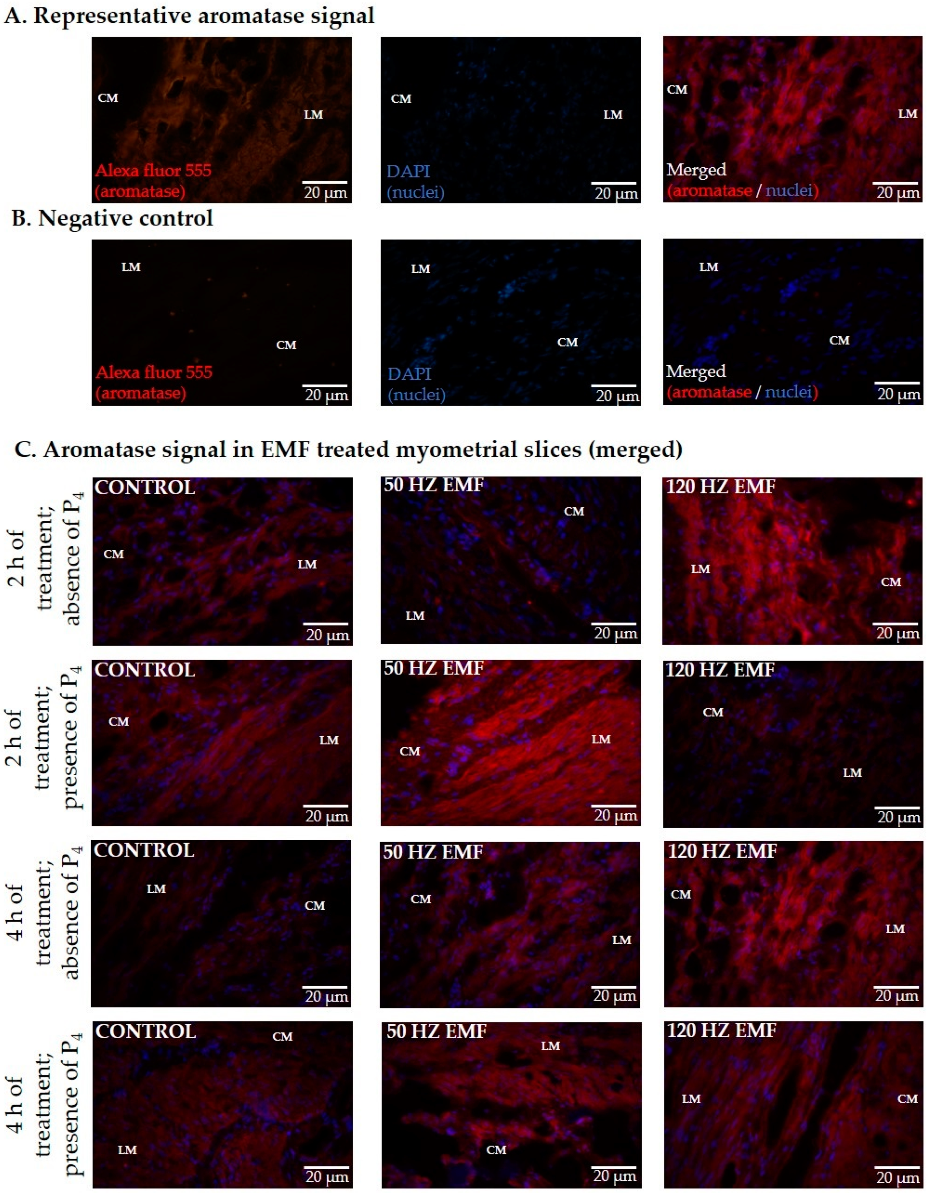
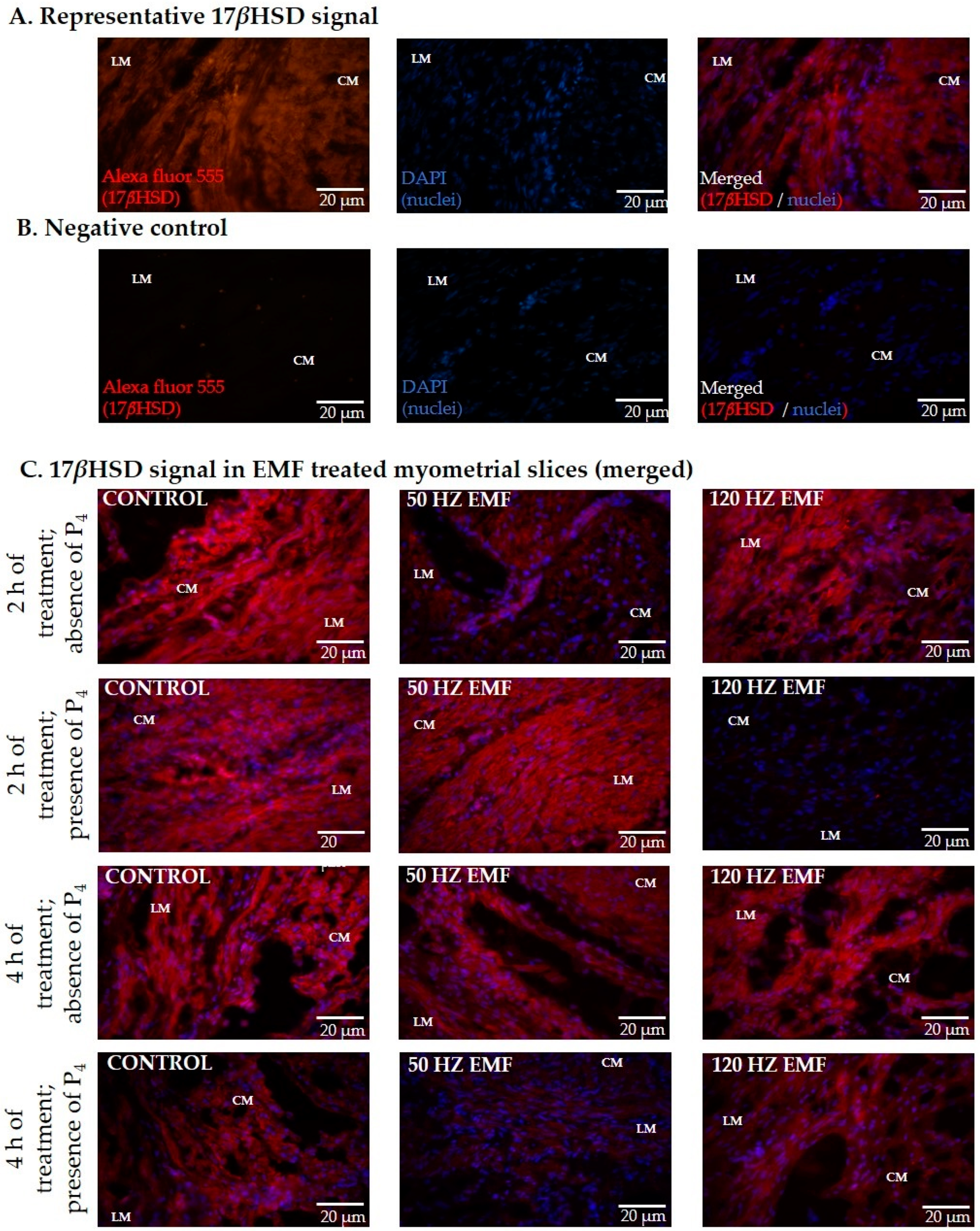
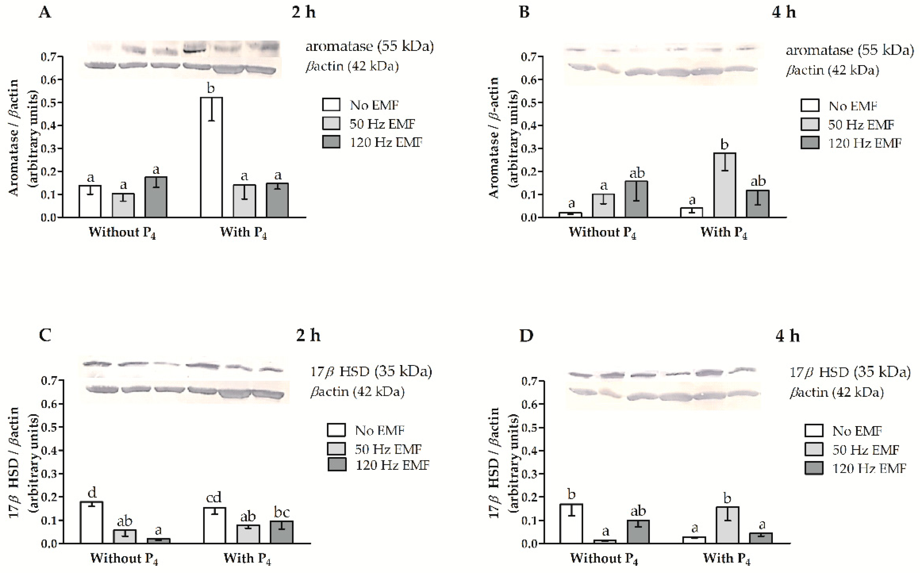
2.2. The Immunolocalization and the Abundance of Aromatase and 17βHSD in Myometrial Slices Treated with EMF at 50 and 120 Hz for 2 h and 4 h with or without the Inclusion of P4

Aromatase and 17βHSD proteins were detected in the longitudinal (LM) and circular (CM) layers of the myometrium incubated for 2 and 4 h with or without treatment with an EMF and with or without the inclusion of P4 in the culture medium. Representative photographs visualizing aromatase and 17βHSD protein immunolocalization are presented in Figure 2 and Figure 3.
The values for the main effects and the interactions among factors affecting aromatase and 17βHSD protein abundances, i.e., treatment with an EMF, duration of EMF treatment, and inclusion of P4 in culture medium are presented in Table 2. Aromatase protein abundance was affected by treatment duration (p = 0.008), P4 inclusion (p = 0.005) and by the interaction between an EMF and the treatment duration (p = 0.00007). The relative abundance of 17βHSD protein was affected by an EMF (p = 0.004). Both aromatase and 17βHSD protein abundance were affected by an interaction that occurred when EMF was imposed and there was P4 inclusion in the culture medium (p = 0.01, and p = 0.001, respectively), and by interaction among treatments with an EMF, treatment duration and the P4 inclusion in the culture medium (both p = 0.007) (Table 2).
In myometrial slices without P4 inclusion in the culture medium after 2 h of incubation and imposing an EMF, the abundance of aromatase was similar to the control (p > 0.05, Figure 4A). In myometrial slices with P4 inclusion in the culture medium after 2 h of incubation and imposing an EMF at 50 and 120 Hz, the abundance of aromatase was less compared to slices not treated with an EMF (p ≤ 0.05, Figure 4A). In slices not treated with an EMF, the relative abundance of aromatase protein in myometrial slices after 2 h of incubation with P4 was larger compared with slices incubated without P4 (p ≤ 0.05, Figure 4A). When the EMF at 50 or 120 Hz was imposed, the abundance of aromatase protein was similar in myometrial slices incubated with and without P4 inclusion after 2 h of incubation (p > 0.05, Figure 4A). After 4 h of incubation without P4 in the culture medium the abundance of aromatase did not differ in myometrial slices treated or not with an EMF (p > 0.05, Figure 4B). In slices incubated with P4, the abundance of aromatase after 4 h of incubation was larger when there was an EMF imposed at 50 Hz (p ≤ 0.05) and was not altered (p > 0.05) when there was an EMF imposed at 120 Hz (Figure 4B). In the myometrial slices treated with EMF at 50 Hz, the aromatase abundance was larger in slices incubated with P4 inclusion (p ≤ 0.05, Figure 4B) compared to slices incubated without P4 (p > 0.05, Figure 4B).
After 2 h of incubation without P4, the abundance of 17βHSD protein was lower in myometrial slices when there was an EMF imposed at 50 and 120 Hz compared to untreated myometrial slices (p ≤ 0.05, Figure 4C). When there was the inclusion of P4, the abundance of 17βHSD was less when there was an EMF imposed at 50 Hz (p ≤ 0.05) and was similar (p > 0.05) when there was an EMF imposed at 120 Hz (Figure 4C). In slices treated with an EMF at 120 Hz, the abundance of 17βHSD protein was larger after 2 h of incubation with P4 compared to slices incubated without P4 (p ≤ 0.05, Figure 4C). After 4 h of incubation without P4 in the culture medium, the 17βHSD abundance was less when there was an EMF imposed at 50 Hz (p ≤ 0.05) and was similar in slices treated with an EMF at 120 Hz (p > 0.05, Figure 4D). When there was the inclusion of P4 in the culture medium, the abundance of 17βHSD was less when an EMF was not imposed, was larger in slices treated with an EMF at 50 Hz, and was similar in slices treated with an EMF at 120 Hz, compared to slices incubated without P4 (p ≤ 0.05, Figure 4B). Full-length blots are provided in Figure S1.

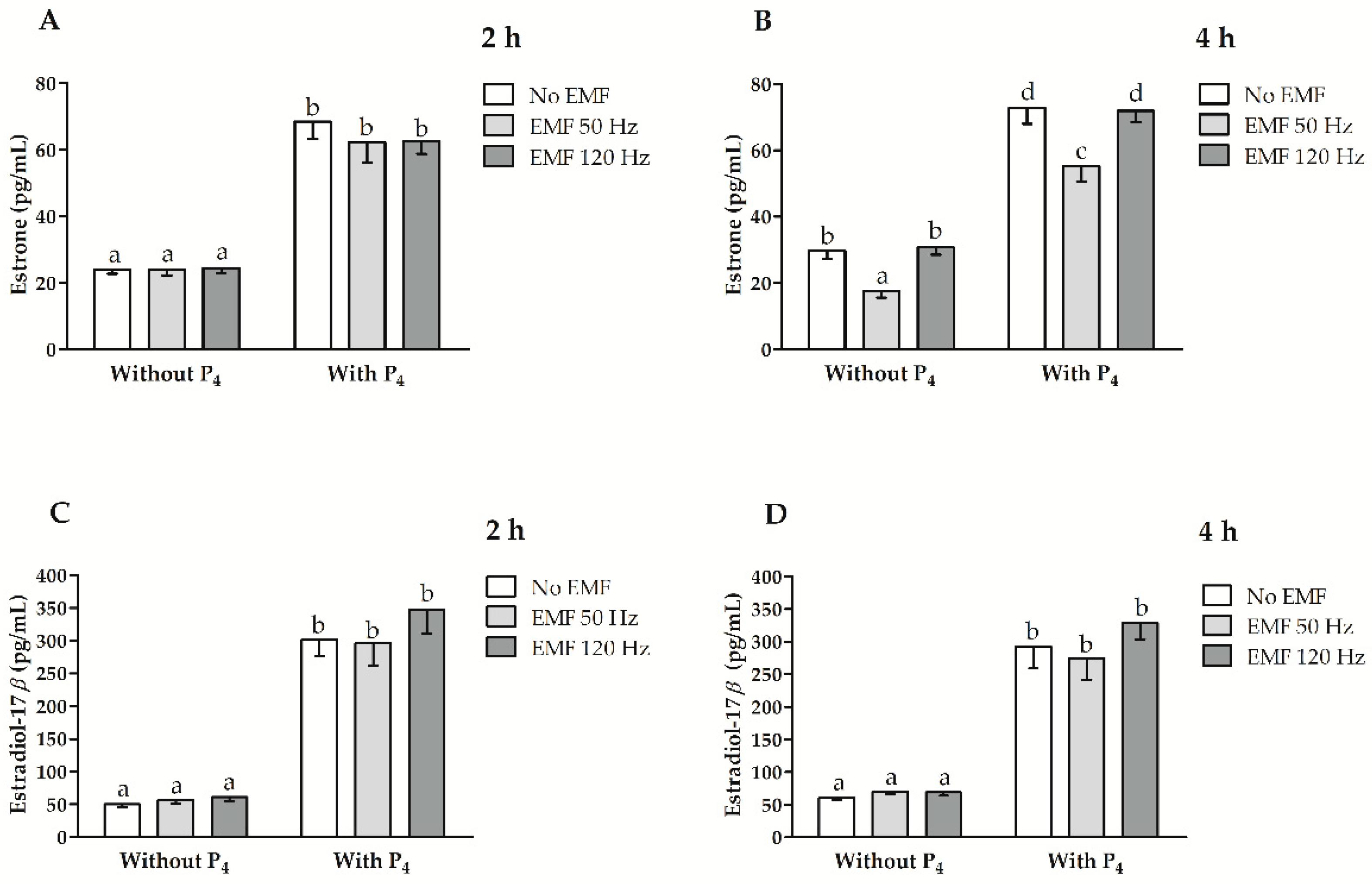
2.3. Effect of EMF at 50 and 120 Hz on E1 and E2 Secretion In Vitro from Myometrial Slices Incubated with or without the Inclusion of P4

The values for the main effects and the interactions among factors affecting E1 and E2 release, i.e., treatment with an EMF, duration of EMF treatment, and inclusion of P4 in culture medium are presented in Table 3. Estrone release was affected by treatment with an EMF (p = 0.00001), P4 inclusion (p < 0.0000001) and the interaction between EMF treatment and EMF treatment duration (p = 0.001). The release of E2 was affected by the P4 inclusion in the culture medium (p < 0.0000001) and the interaction between the treatment duration and the P4 inclusion (p = 0.02).
After 2 h of incubation, the myometrial E1 concentration in the culture medium did not differ for myometrial slices treated with an EMF at 50 and 120 Hz despite the inclusion of P4 in the culture medium (p > 0.05, Figure 5A). After 4 h of incubation with and without P4 inclusion in the culture medium, the concentration of E1 in the culture medium was less for myometrial slices when there was an EMF imposed at 50 Hz and was not altered when there was an EMF imposed at 120 Hz (p ≤ 0.05, Figure 5B). The concentration of E2 release was similar in the culture medium for slices treated with an EMF at 50 and 120 Hz after 2 and 4 h of incubation, despite the inclusion of P4 (p > 0.05, Figure 5C,D). When there was the inclusion of P4, the E1 and E2 concentration in the culture medium was larger than for slices incubated without the inclusion of P4, despite treatment with EMF and the treatment duration with an EMF (p ≤ 0.05, Figure 5A–D).

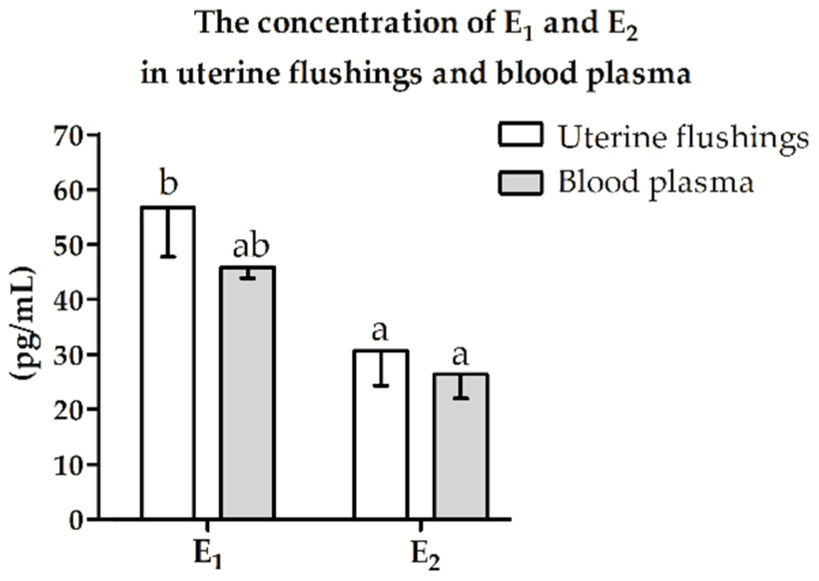
2.4. Estrogen Concentration in Uterine Flushings and Blood Plasma

The concentration of E1 and E2 in uterine flushings and blood plasma did not differ (p > 0.05, Figure 4). In the uterine flushings, the concentration of E1 was higher than the concentration of E2 (p ≤ 0.05, Figure 6). In blood plasma, the concentration of E1 and E2 did not differ (p > 0.05, Figure 6).

3. Discussion

The current study has provided, for the first time, evidence that an EMF treatment induces alterations in estrogen synthesis and release in the myometrium of pigs during the peri-implantation period. Specifically, the abundance of key enzymes involved in the production of estrogens, i.e., aromatase and 17βHSD, was altered in response to an EMF at the frequency of 50 and 120 Hz both on transcript and protein levels, but the effect of EMF treatment was variable due to the treatment duration of EMF, and/or P4 inclusion in the culture medium. Notably, EMF treatment affected the mRNA transcript and protein abundance for these steroidogenic enzymes, but the inductions were inconsistent between the mRNA transcript and protein levels. This observation was not surprising since the transcription and translation processes in Eukaryotes are separated in space and time [35]. Importantly, regardless of the P4 inclusion in the culture medium, myometrial E1 was less in slices treated with an EMF at a frequency of 50 Hz within a longer (4 h) treatment duration of EMF. None of the examined EMF frequencies or treatment durations of EMF affected E2 release from the myometrium.
The mechanism of steroid production is determined by cytochrome P450 17α-hydroxylase/C17-20 lyase (cytochrome P450c17), which is a rate-limiting enzyme controlling the entry of P4 and pregnenolone to the steroidogenic pathway [27,36]. Previously, it was documented that a longer (4 h) treatment duration of the EMF at a frequency of 50 Hz results in a greater cytochrome P450c17 protein abundance, but did not affect A4 release by the myometrium collected from pigs during the peri-implantation period [19]. The EMF radiation at a higher (i.e., 120 Hz) frequency led to an increased concentration of myometrial A4 when there was the inclusion of P4 in the culture medium within a longer (4 h) treatment duration of EMF, which coincided with the greater 3β hydroxysteroid dehydrogenase (3βHSD) abundance in the tissue [19]. The 3βHSD catalyzes 3β-hydroxysteroid dehydrogenation and Δ5 to Δ4 isomerization of pregnenolone and 17α-hydroxypregnenolone, dehydroepiandrosterone and androstenediol into progesterone and 17α-hydroxyprogesterone, A4 and testosterone (T), respectively [37]. Notably, A4 is the most potent and essential androgen in pigs that affects anabolic processes, morphogenesis, cellular proliferation and hyperplasia in the target tissue [38]. Androgens function as substrates for estrogen production in the reaction catalyzed by aromatase [39]. Therefore, the increased abundance of 3βHSD and greater concentration of A4 in the culture medium of myometrial slices incubated for longer treatment duration, when there was an EMF imposed, indicate the potential of an EMF to increase estrogen release in the tissue.
Interestingly, it was found that the basal myometrial abundance of CYP19A3 mRNA transcript is decreased during the peri-implantation period when compared to its abundance in the myometrium collected from estrous-cyclic pigs, but the abundance of the encoded protein is mostly similar in gravid and non-gravid pig myometrium [10]. Thus, the myometrial competence to synthesize estrogens, measured as the abundance of aromatase, on days 15 to 16 of pregnancy and during the respective days of the estrous cycle is similar. The results of the current study indicated that myometrial abundance of CYP19A3 mRNA transcript decreases in response to a relatively short (2 h) duration of EMF treatment at a frequency of 50 Hz, while it increases after a relatively long (4 h) duration of EMF treatment at a frequency of 50 and 120 Hz. Thus, the imposing of an EMF may destabilize the potential of the myometrium to synthesize estrogens. This study evaluated that the interaction among EMF treatment, duration of EMF treatment, and the inclusion of P4 to the culture medium affects the abundance of myometrial CYP19A3 mRNA transcript. Moreover, it was evaluated that the inclusion of P4 to the culture medium diminished all the observed alterations in the abundance of CYP19A3 mRNA transcript that occurred in the presence of EMF. The previous study indicated that the interaction between the EMF presence and the P4 inclusion into the culture medium also affects myometrial CYP19A3 mRNA transcript abundance in pigs during the mid-luteal phase of the estrous cycle [18]. Nevertheless, it was evaluated that when there was an EMF imposed at 50 Hz, P4 functioned as a factor sensitizing myometrial CYP19A3 mRNA transcript abundance to EMF treatment, whereas when there was an EMF imposed at 120 Hz, P4 functioned as a protective factor against EMF radiation [18]. Thus, the function of P4 in the modulation of myometrial CYP19A3 mRNA transcript abundance in estrous-cyclic females depends on the frequency of EMF [18], whereas in early pregnant females P4 may function as a protective factor against alterations evoked by EMF treatment, despite the EMF frequency used (50 or 120 Hz) or the duration of treatment.
Notably, when the myometrium was treated with an EMF at 50 and 120 Hz for 2 or 4 h with no inclusion of P4 in the medium, the abundance of aromatase was not altered. In the presence of P4, the basal myometrial aromatase abundance after 2 h treatment duration was significantly greater than in slices incubated without P4 or with the inclusion of P4 and treated with an EMF at the frequency of 50 and 120 Hz. A significant increase in aromatase abundance in myometrial slices incubated in the medium supplemented with P4, when there was no EMF imposed, may indicate that the presence of a substrate for steroid hormone synthesis induces myometrial production of the enzyme required for estrogen synthesis. It is noteworthy that the previous studies indicated that in the myometrium of pigs during the peri-implantation period, the basal CYP19A3 mRNA is decreased, whereas aromatase abundance is increased [10]. This phenomenon coincided with an increased level of P4 in uterine flushings [40]. Moreover, the results of studies performed on human primary cytotrophoblasts documented that a greater concentration of P4 in the culture medium corresponds to the increased abundance of aromatase in cytotrophoblasts cells [41]. These notions are in an agreement with the current results since the inclusion of P4 to the culture medium leads to a greater abundance of myometrial aromatase protein. Thus, the lack of increased abundance of aromatase in the myometrium treated by the EMF in the presence of P4 may indicate that EMF radiation may disturb the function of the P4 as a substrate for steroid hormone synthesis in the myometrium. Noteworthy, in studies using MCF-7 breast cancer cells, a co-treatment with P4 blocked cyclic adenosine monophosphate and interleukin 1β stimulated aromatase activity [42]. Thus, the relation between P4 presence and the abundance of aromatase might depend on tissue and cell type, but in porcine myometrium, the presence of P4 appears to generally increase aromatase abundance, but the EMF radiation may contribute to the disturbance of the relations between P4 and aromatase.
This study determined that a relatively longer treatment duration (4 h) of EMF leads to apparently different alterations in myometrial aromatase abundance. Specifically, there were no observed differences in basal aromatase abundance in myometrial slices incubated with P4 and not exposed to EMF, compared to slices incubated without P4 included in the culture medium. Interestingly, when EMF at a frequency of 50 Hz was imposed, the aromatase abundance was larger in myometrial tissue when P4 was also provided to the culture medium compared to slices incubated without P4. This phenomenon indicates that in pigs during the peri-implantation period, P4 functions as a factor that sensitizes translation processes in the myometrium to the EMF radiation, resulting in increased aromatase protein synthesis. It cannot be excluded that the increased concentration of aromatase protein could cause a suppression of CYP19A3 mRNA expression. Such a correlation between RNA and protein expression profiles was previously documented in 23 human cell lines [43]. Although the presence of P4 in the culture medium apparently protected against EMF radiation, the transcriptional processes that resulted in the production of myometrial CYP19A3 mRNA transcript, P4 also have the potential to sensitize translation processes to EMF radiation. These changes are likely to impact estrogen production in the myometrium of pigs during the peri-implantation period, potentially affecting CL lifespan, uterine receptivity and embryo-maternal interactions [1,2,3,4,5].
The current study determined that myometrial release of E1 in response to an EMF at 50 Hz (4 h) was lower despite the inclusion or not of P4 in the culture medium. Previously, it was found that in pigs during the peri-implantation period, the basal E1 release by the myometrium is similar to that which occurred in pigs during luteolysis [6]. Since the production of estrogens during early pregnancy in pigs is the result of androgen conversion [44] and A4 is converted to E1 [45], the observed lesser release of E1 in the response to EMF may indicate that the aromatization of A4 to E1 is disturbed as a consequence of EMF radiation at the frequency of 50 Hz. Importantly, this study found no clear relevance between the production of E1 and E2 and the expression levels for steroidogenic enzymes. This phenomenon can be a result of estrogen metabolism documented previously in ovary [46], but it cannot be excluded that it also occurs in uterine tissues. Further studies are needed to evaluate the effect of EMF treatment on estrogen metabolism in the myometrium. Notably, E1 is a less potent estrogen than E2, but, as confirmed within this study, E1 concentration in uterine flushings is higher than the concentration of E2. Taking into account the levels of estrogen production by myometrial slices, it cannot be excluded that the concentration of E1 in the uterine lumen is also the effect of the steroidogenic activity of the endometrium. Moreover, it should be noted that estrogens are considered as important regulators of uterine activity affecting embryo-maternal cross-talk during early pregnancy [47] and, especially E2, may exert a luteotrophic effect [48]. Notably, E2 is still considered as one of the embryonic signals for maternal recognition of pregnancy in pigs [5], but greater than the optimal concentration of E2 in uterine milieu during the peri-implantation period may induce cytotoxic effects in conceptuses [49]. Thus, any factor that could increase E2 production and disturb the slight balance of E1 and E2 in the uterus during the peri-implantation period may be considered as a potent disruptor of pregnancy in pigs.
As indicated above, EMF treatment at a frequency of 50 Hz significantly decreases in vitro production of E1 by myometrial tissue collected from pigs during the peri-implantation period despite the inclusion or not of P4 in the culture medium. Additionally, no EMF treatment-related effects were found on myometrial E2 production. Moreover, the inclusion of P4 does not have functions that protect the pig myometrium against the effect of EMF on E1 release. E1 might be converted to E2 via the activity of 17βHSD [27]. Results from the past study indicate that the expression level of 17βHSD in the endometrium of estrous-cyclic and pregnant pigs corresponds with the enzyme activity [50], and the myometrial activity of 17βHSD is lowered during the peri-implantation period when compared to the foregoing days of early pregnancy [17]. This phenomenon suggests that during the peri-implantation period, the potential of the myometrium to convert E1 to E2 is significantly lower, which may be recognized as a unique protective mechanism against the overproduction of E2 by the tissue. In this context, it is not surprising that EMF influences rather E1 than E2 release, despite the increased abundance of 3βHSD and the concentration of A4 in the culture medium of myometrial slices when there was an EMF imposed [19].
Regarding the results of the current study, the myometrial abundance of HSD17B4 mRNA transcript and encoded protein in the presence of EMF and P4 is not stable and alters dynamically. Specifically, HSD17B4 mRNA transcript abundance is greater in response to EMF treatment at a frequency of 120 Hz after 2 h of EMF treatment duration, but it is lower after 4 h of EMF treatment. The inclusion of P4 to the culture medium diminished the EMF-related effects on transcriptional processes. Furthermore, 17βHSD protein abundance decreased in response to EMF at the frequency of 50 Hz after short (2 h) and long (4 h) treatment duration and at a frequency of 120 Hz after short (2 h) treatment duration without the inclusion of P4 in the culture medium. Noteworthy, the observed increased abundance of HSD17B4 mRNA transcript in myometrial slices exposed to EMF at a frequency of 120 Hz for 2 h is accompanied by low protein concentration. These facts may indicate that the phenomenon of RNA interference or changes in the level of post-translational modifications occurred [43,51]. However, these phenomena require further investigation. Interestingly, when there was an inclusion of P4 in the culture medium and EMF at 50 Hz was imposed, the myometrial 17βHSD protein abundance after relatively short (2 h) treatment duration decreased, but increased after a longer (4 h) duration of EMF treatment. Nevertheless, due to the already significantly lowered abundance of 17βHSD in the myometrium of pigs during the peri-implantation period [17], these changes did not affect the final E2 production despite the various parameters of EMF treatment used within this study. Therefore, it seems that EMF influences the synthesis of estrogens in the myometrium mostly at the level of transcription and translation and influences the E2 synthesis to a lesser extent.

4. Materials and Methods

4.1. Animals and Collection of Myometrial Tissue

Post-pubertal pigs (n = 5, Sus scrofa f. domestica, Polish Landrace × Great White Polish, aged 10 months, weighing 95–110 kg) were observed for estrus behavior in the presence of an intact boar. Pigs were naturally bred twice, i.e., on the first and the second day of the second estrus. The second mating was assigned as the first day of pregnancy. On days 15 to 16 of pregnancy, pigs were slaughtered in the local abattoir using standard procedures (Rozdroże, Poland), provided in the professional slaughterhouse, using humane procedures. All living pigs were handled by the authors. After slaughter, the entire uteri were excised, placed in ice-cold sterile phosphate-buffered saline (PBS, pH = 7.4) supplemented with 3% antibiotic–antimycotic solution (Sigma Aldrich, St Louis, MO, USA). Whole blood was collected into heparinized vials during slaughter. Uteri and blood were transported within 30 min to the Laboratory of Animal Anatomy and Physiology, Department of Biology and Biotechnology, University of Warmia and Mazury in Olsztyn, Olsztyn, Poland, ensuring cooling conditions (4 °C). The stage of pregnancy was confirmed by the morphology of the ovaries and CLs quality [52,53] and the presence and morphology of conceptuses flushed from the uterine horns with sterile saline [54,55]. In the laboratory, blood samples were centrifuged (7500× g, 10 min, 4 °C), and blood plasma was stored at −20 °C for further E1 and E2 concentration analyses. The uteri were flushed with 20 mL of sterile ice-cold phosphate-buffered saline (PBS, pH = 7.4) supplemented with 3% antibiotic—antimycotic solution (Sigma Aldrich, St Louis, MO, USA). Uterine flushings were collected for further determination of E1 and E2 concentrations. Uterine horns were opened longitudinally, the perimetrium was discarded by careful scraping and the myometrial fragments were picked by tweezers. Individual myometrial fragments were washed in an ice-cold PBS (pH = 7.4) supplemented with 3% of the antibiotic–antimycotic solution (Sigma Aldrich, St Louis, MO, USA) and standardized to 95–105 mg-weighting, 2–3-mm-thick fragments used for further in vitro incubation. All myometrial fragments collected from one pig were considered one biological repeat (n).

4.2. In Vitro Incubation and EMF Treatment System

Prepared standardized fragments of the myometrium were placed in 24-well culture dishes and covered with 1 mL of the pre-incubation medium (M199, Sigma Aldrich, St Louis, MO, USA; 0.1% BSA, Carl Roth GmBH + Co KG, Mühlburg, Karlsruhe, Germany; 1% antibiotic–antimycotic solution, Sigma Aldrich, St Louis, MO, USA) and incubated in an EMF exposure system as previously described [19]. Briefly, myometrial tissue slices were first preincubated for 2 h at 37 °C, 95% O2 and 5% CO2 in a water-shaking bath and then incubated in fresh medium (the same composition) or fresh medium supplemented with P4 (10−5 M, SERVA Electrophoresis GmbH, Heidelberg, Germany). The EMF was generated by a Magneris apparatus (Astar, Bielsko-Biala, Poland), ensuring exposition to sinusoidal EMF at a frequency of 50 or 120 Hz and magnetic induction of 8 mT for 2 and 4 h of in vitro incubation. Control fragments of myometrial tissue were not treated with an EMF. The prevention of fields overlapping fields was ensured by a distance (50 cm) separation between water baths in which incubation was held. The rationale of the used EMF parameters (50 and 120 Hz, 8 mT, 2, and 4 h of treatment) has been described previously [18,56]. Moreover, EMF at frequencies of 50 and 120 Hz is classified as an extremely low electromagnetic field, which is common in the environment of living organisms [21], and a magnetic induction at 8 mT is a typical exposure level of the magnetic induction for devices and equipment used for therapeutic purposes or in industry [57]. The graphical presentation of the EMF exposure system was presented previously by Franczak et al. (2020). During the in vitro incubation, the thermal conditions were monitored to exclude the bias of the temperature on the examined parameters. After in vitro incubation, tissue fragments were collected and snap-frozen in liquid nitrogen (−196 °C) and stored at −80 °C for further analysis of mRNA transcript and protein abundances. The samples of the culture medium were collected and stored at −20 °C for further determination of E1 and E2 concentrations.

4.3. Determination of CYP19A3 and HSD17B4 mRNA Transcript Abundance

Total RNA was extracted from myometrial tissue using TRI-Reagent (Sigma Aldrich, Germany) following the standard protocol. The details of RNA extraction and preparation for further analyses were described previously [19]. RNA precipitates were used for analyses that were integral electrophoretically (separation in 1.5% agarose gel) and possessed an optical density ration A260/A280 in the range of 1.8–2.0 (spectrophotometer Tecan, Männedorf, Switzerland). The Real-Time PCR was performed according to the guidelines provided by Bustin et al. (2009) [58]. The amplification was conducted using 4 pg/μL aliquots of extracted RNA, TaqMan®RNA-to- 1-Step Kit (Applied Biosystems, Foster City, CA, USA) and specific TaqMan probes provided by Applied Biosystems (Foster City, CA, USA) listed in Table 4. Amplification was conducted in an AriaMX apparatus (Agilent Technologies, Santa Clara, CA, USA), with a standard thermal profile suggested by the producer. The Ct values were obtained with Aria 1.6 software (Agilent Technologies, Santa Clara, CA, USA) and used for cycle threshold (Ct) calculation. The Ct values of the tested genes were normalized with the geometrical mean of the reference genes and used for 2−∆∆Ct calculation [59].

4.4. Immunodetection of Aromatase and 17βHSD

The immunodetection of aromatase and 17βHSD was performed using immunofluorescence. Firstly, 6-μm-thick cryosections of myometrial tissue were fixed in a 4% paraformaldehyde (P.P.H. STANLAB, Lublin, Poland) for 15 min (room temperature, RT) and subsequently washed 3 × 10 min with Tris-buffered saline containing Tween 20 (TBS-T buffer: 10 mM Tris, 150 mM NaCl, 0.1% Tween 20). To prevent non-specific labeling, the slides were then covered with a blocking buffer containing 20% of a normal donkey serum (EMD Millipore, Billerica, MA, USA) and 0.1% of Triton X-100 (Sigma Aldrich, St Louis, MO, USA) in PBS for 1 h at 4 °C. Next, the slides were washed 3 × 10 min with TBS-T and covered with the primary antibodies rabbit anti-cytochrome P450arom and rabbit anti-17βHSD diluted in TBS-T for 18 h at 4 °C, listed in Table 5. Slices assigned for negative control were covered with TBS-T without primary antibodies. Next, slides were washed 3 × 10 min with TBS-T (RT), covered with donkey anti-rabbit secondary antibodies Alexa Fluor 555 (A32794, Life Technologies, Eugene, OR, USA) diluted in PBS (1:1500) and incubated at 4 °C for 1 h in darkness to prevent photobleaching. Subsequently, the slides were washed in PBS (3 × 5 min) and mounted with Fluoroshield with 4′,6-diamidino-2-phenylindole (DAPI, Sigma Aldrich, St Louis, MO, USA) for further analysis with an epifluorescent BX 51 microscope (Olympus, Tokyo, Japan) under 400-fold magnification. The images were archived with a type DP72 digital camera (Olympus, Tokyo, Japan).

4.5. Determination of Aromatase and 17βHSD Protein Abundance

Total protein was extracted from myometrial tissue as described previously [19]. The 15-µg aliquots of the protein extracts were adjusted to a total volume of 32 µL and mixed with 8 µL of loading buffer containing 6 × Laemmli sample buffer (0.35 M Tris-HCl, pH = 6.8, SDS 10%, glycerol 30%, Dithiothreitol (DTT) 0.6M, bromophenol blue 0.175 M) and 1 M DTT, used in a 1:1 proportion. Protein ladder (PageRuler™ Plus, #26619. Thermo Fisher Scientific, Waltham, MA, USA) was diluted 4 × in water and mixed with the same loading buffer in a 1:5 proportion. Protein aliquots and protein ladder were denatured at 99 °C for 3 min and immediately used for SDS-PAGE electrophoresis (PowerEase 90 W, Life Technologies, Eugene, OR, USA). Samples were concentrated in a 4% stacking gel (15 mA/gel and 40 mV/gel) and then resolved in a 10% running gel (25 mA/gel, 80 mV/gel) in 1 × Tris-glycine-SDS buffer (T7777, Sigma Aldrich, St Louis, MO, USA). The samples were then transferred to nitrocellulose membranes (0.45 µm using a semi-dry transblot apparatus (constant 25 V, 2.4 mA, 12 min, Pierce Power Blot Cassette, Thermo Scientific, Waltham, MA, USA). After transfer, membranes assigned to aromatase and 17βHSD abundance analyses were blocked for 2 h at 4 °C in TBS-T containing 1% of bovine serum albumin fraction V (BSA, Carl Roth GmbH + Co KG, Mühlburg, Karlsruhe, Germany) and membranes assigned to β-actin abundance analysis were blocked for 2 h at RT in TBS-T buffer containing 5% of BSA. After blocking, the membranes were washed for 3 × 10 min in TBS-T buffer and then incubated overnight at 4 °C with primary antibodies (Table 5). Next, the membranes were washed 3 × 5 min in TBS-T buffer and incubated with secondary alkaline phosphatase-conjugated goat anti-rabbit antibodies (sc-2057, Santa Cruz Biotechnology, Dallas, TX, USA) used at a concentration of 0.2 μg/mL for 2 h (for β-actin) or 1.5 h (for aromatase and 17βHSD) at 4 °C or RT, respectively. Subsequently, membranes were washed 3 × 5 min in TBS-T buffer and the immunoreactive bands were visualized using a staining solution containing 2% of NBT/BCIP stock solution in 0.1 M Tris-HCl, pH = 9.5 (20 °C), 0.1 M NaCl, 0.05 M MgCl2 (Merck, Kenilworth, NJ, USA) in darkness for 30 s (for β-actin), 90 s (for aromatase) or 60 s (for 17βHSD). After visualization, the reaction was stopped in deionized water containing 0.5 M of ethylenediaminetetraacetic acid. The blots were subsequently evaluated for further calculations of the optical density of the bands using ImageJ open-source software. The optical density of the detected protein bands was normalized with the optical density of β-actin bands.

4.6. Determination of E1 and E2 Concentrations in Blood Plasma, Uterine Flushings and the Culture Medium

The concentrations of estrogens were determined using radioimmunoassay [60]. The cross-reactivity of antisera against E1 and E2 have been reported previously [61]. The extraction efficiencies of E1 and E2 were 88.55% and 86.55%, respectively. The assay sensitivity was 1 pg and the inter- and intra-assay coefficients of variation were 5.08% and 3.74%, respectively, for E1 and 6.70% and 3.56% for E2, respectively.

4.7. Statistical Analysis and Data Presentation

The results are presented as the mean ± SEM. The differences in mRNA transcript abundance were analyzed using 2−∆∆Ct values. The abundance of proteins was analyzed using mean values of the optical density of detected protein bands normalized with the optical density of β-actin bands values. Each value was then log-transformed for hormone concentration analyses. All data were analyzed using a multi-way analysis of variance (ANOVA) and Fisher’s least significant difference (LSD) post hoc test, with the main factors as follows: 1/treatment with an EMF, which stands for control (no treatment with an EMF) and treatment with an EMF at 50 or 120 Hz; 2/duration of EMF treatment, which stands for 2 and 4 h treatment duration; 3/the inclusion of P4 in medium, which means the inclusion or no inclusion of P4 in the culture medium. The results obtained within 2 and 4 h of incubation were then considered separately and once again analyzed using multi-way ANOVA and Fisher’s LSD post-hoc tests. Statistically significant differences were considered at p ≤ 0.05.

5. Conclusions

In the myometrial tissue of pigs during the peri-implantation period, EMF treatment affects the potential for the synthesis of estrogens. The consequences of EMF radiation in the myometrium may depend on the basal tissue potential for the production of estrogens. The inclusion of P4 in the culture medium diminishes most of the observed EMF treatment-related effects on the level of transcriptional processes that result in the production of mRNA transcripts for these steroidogenic enzymes in the myometrium of pigs. On the contrary, on the level of translation processes that result in protein synthesis, P4 may sensitize myometrium to EMF radiation. Importantly, EMF at a low frequency of 50 Hz decreases myometrial E1 release after a relatively long (4 h) treatment duration, with or without the P4 inclusion and does not alter E2 release. In the pig myometrium, EMF leads to lowered E1 release. Thus, this study provides evidence that EMF can be recognized as a potent disruptor of steroidogenesis in the uterus of females during the peri-implantation period.

Supplementary Materials

The following are available online at https://www.mdpi.com/1422-0067/22/6/2920/s1. Figure S1: Representative full-length Western blot bands showing aromatase, 17βHSD and β-actin proteins in porcine myometrial explants exposed to an electromagnetic field (EMF) at 0, 50 and 120 Hz for 2 or 4 h in the presence or absence of P4.

Author Contributions

Conceptualization, E.M.D., and A.F.; methodology, E.M.D.; software, E.M.D. and W.K.; validation, A.Z.; formal analysis, E.M.D. and W.K.; investigation, E.M.D., and W.K.; resources, E.M.D. and W.K.; data curation, E.M.D., and W.K.; writing—original draft preparation, E.M.D., W.K., A.Z., and A.F.; writing—review and editing, E.M.D., W.K.; A.Z., P.J.W., and A.F.; visualization, E.M.D., W.K. and P.J.W.; supervision, A.F.; project administration, E.M.D. and A.F.; funding acquisition, A.F. All authors have read and agreed to the published version of the manuscript.

Funding

This research was funded by the National Science Centre, Poland, OPUS grant no. 2017/25/B/NZ9/00090.

Institutional Review Board Statement

Ethical review and approval were waived for this study, due to all of the experiments being conducted on animal tissues collected post mortem during regular economic slaughter provided in a professional slaughterhouse. The use of animals was in accordance with the Act of 15th of January 2015 on the Protection of Animals Used for Scientific or Educational Purposes and Directive 2010/63/EU of the European Parliament and the Council of 22nd of September 2010 on the protection of animals used for scientific purposes.

Informed Consent Statement

Not applicable.

Data Availability Statement

All of the data are presented in the study. The raw data used for the preparation of the presented results are available on request from the corresponding author.

Acknowledgments

The authors would like to thank Anna Koziorowska (University of Rzeszow, Poland) for her help in operating the EMF exposure system and Beata Sterbicka for her contribution to RIA analysis.

Conflicts of Interest

The authors declare no conflict of interest. The funders had no role in the design of the study; in the collection, analyses or interpretation of data; in the writing of the manuscript, or in the decision to publish the results.

References

  1. Geisert, R.D.; Renegar, R.H.; Thatcher, W.W.; Roberts, R.M.; Bazer, F.W. Establishment of pregnancy in the pig: I. Interrelationships between preimplantation development of the pig blastocyst and uterine endometrial secretions. Biol. Reprod. 1982, 27, 925–939. [Google Scholar] [CrossRef] [Green Version]
  2. Ziecik, A.; Waclawik, A.; Kaczmarek, M.; Blitek, A.; Jalali, B.M.; Andronowska, A. Mechanisms for the establishment of pregnancy in the Pig. Reprod. Domest. Anim. 2011, 46, 31–41. [Google Scholar] [CrossRef] [PubMed]
  3. Ziecik, A.J.; Przygrodzka, E.; Jalali, B.M.; Kaczmarek, M.M. Regulation of the porcine corpus luteum during pregnancy. Reproduction 2018, 156, 57–67. [Google Scholar] [CrossRef] [PubMed]
  4. Bazer, F.W.; Johnson, G.A. Pig blastocyst-uterine interactions. Differentiation 2014, 87, 52–65. [Google Scholar] [CrossRef] [PubMed]
  5. Waclawik, A.; Kaczmarek, M.M.; Blitek, A.; Kaczynski, P.; Ziecik, A.J. Embryo-maternal dialogue during pregnancy establishment and implantation in the pig. Mol. Reprod. Dev. 2017, 84, 842–855. [Google Scholar] [CrossRef] [PubMed] [Green Version]
  6. Franczak, A. Endometrial and myometrial secretion of androgens and estrone during early pregnancy and luteolysis in pigs. Reprod. Biol. 2008, 8, 213–228. [Google Scholar] [CrossRef]
  7. Franczak, A.; Kotwica, G. Secretion of estradiol-17β by porcine endometrium and myometrium during early pregnancy and luteolysis. Theriogenology 2008, 69, 283–289. [Google Scholar] [CrossRef] [PubMed]
  8. Franczak, A.; Kotwica, G. Androgens and estradiol-17β production by porcine uterine cells: In vitro study. Theriogenology 2010, 73, 232–241. [Google Scholar] [CrossRef]
  9. Franczak, A.; Wojciechowicz, B.; Katwica, G. Novel aspects of cytokine action in porcine uterus—Endometrial and myometrial production of estrone (E1) in the presence of interleukin 1β (IL1β), interleukin 6 (IL6) and tumor necrosis factor (TNFα)—In vitro study. Folia Biol. 2013, 61, 253–261. [Google Scholar] [CrossRef] [PubMed] [Green Version]
  10. Franczak, A.; Wojciechowicz, B.; Kolakowska, J.; Kotwica, G. The effect of interleukin-1β, interleukin-6, and tumor necrosis factor-α on estradiol-17β release in the myometrium: The in vitro study on the pig model. Theriogenology 2014, 81, 266–274. [Google Scholar] [CrossRef]
  11. Smolinska, N.; Dobrzyn, K.; Kiezun, M.; Szeszko, K.; Maleszka, A.; Kaminski, T. Effect of adiponectin on the steroidogenic acute regulatory protein, P450 side chain cleavage enzyme and 3β-hydroxysteroid dehydrogenase gene expression, progesterone and androstenedione production by the porcine uterus during early pregnancy. J. Physiol. Pharmacol. 2016, 67, 443–456. [Google Scholar] [PubMed]
  12. Kiezun, M.; Smolinska, N.; Dobrzyn, K.; Szeszko, K.; Rytelewska, E.; Kaminski, T. The effect of orexin A on CYP17A1 and CYP19A3 expression and on oestradiol, oestrone and testosterone secretion in the porcine uterus during early pregnancy and the oestrous cycle. Theriogenology 2017, 90, 129–140. [Google Scholar] [CrossRef]
  13. Grzesiak, M.; Waszkiewicz, E.; Wojtas, M.; Kowalik, K.; Franczak, A. Expression of vitamin D receptor in the porcine uterus and effect of 1,25(OH)2D3 on progesterone and estradiol-17β secretion by uterine tissues in vitro. Theriogenology 2019, 125, 102–108. [Google Scholar] [CrossRef]
  14. Kisielewska, K.; Rytelewska, E.; Gudelska, M.; Kiezun, M.; Dobrzyn, K.; Szeszko, K.; Bors, K.; Wyrebek, J.; Kaminski, T.; Smolinska, N. The effect of orexin B on steroidogenic acute regulatory protein, P450 side-chain cleavage enzyme, and 3β-hydroxysteroid dehydrogenase gene expression, and progesterone and androstenedione secretion by the porcine uterus during early pregnancy and the est. J. Anim. Sci. 2019, 97, 851–864. [Google Scholar] [CrossRef]
  15. Rytelewska, E.; Kisielewska, K.; Gudelska, M.; Kiezun, M.; Dobrzyn, K.; Bors, K.; Wyrebek, J.; Kaminska, B.; Kaminski, T.; Smolinska, N. The effect of orexin a on the StAR, CYP11A1 and HSD3B1 gene expression, as well as progesterone and androstenedione secretion in the porcine uterus during early pregnancy and the oestrous cycle. Theriogenology 2020, 143, 179–190. [Google Scholar] [CrossRef]
  16. Waszkiewicz, E.M.; Kozlowska, W.; Zmijewska, A.; Franczak, A. Expression of insulin-like growth factor 1 (IGF-1) and epidermal growth factor (EGF) receptors and the effect of IGF-1 and EGF on androgen and estrogen release in the myometrium of pigs—In vitro study. Animals 2020, 10, 915. [Google Scholar] [CrossRef] [PubMed]
  17. Waszkiewicz, E.M.; Zmijewska, A.; Kozlowska, W.; Franczak, A. Effects of LH and FSH on androgen and oestrogen release in the myometrium of pigs during the oestrous cycle and early pregnancy. Reprod. Fertil. Dev. 2020, 32, 1200–1211. [Google Scholar] [CrossRef]
  18. Koziorowska, A.; Waszkiewicz, E.M.; Romerowicz-Misielak, M.; Zglejc-Waszak, K.; Franczak, A. Extremely low-frequency electromagnetic field (EMF) generates alterations in the synthesis and secretion of oestradiol-17β (E2) in uterine tissues: An in vitro study. Theriogenology 2018, 110, 86–95. [Google Scholar] [CrossRef]
  19. Franczak, A.; Waszkiewicz, E.; Kozlowska, W.; Zmijewska, A.; Koziorowska, A. Consequences of electromagnetic field (EMF) radiation during early pregnancy—Androgen synthesis and release from the myometrium of pigs in vitro. Anim. Reprod. Sci. 2020, 218, 106465. [Google Scholar] [CrossRef] [PubMed]
  20. Kozlowska, W.; Drzewiecka, E.; Zmijewska, A.; Koziorowska, A.; Franczak, A. Effects of electromagnetic field (EMF) radiation on androgen synthesis and release from the pig endometrium during the fetal peri-implantation period. Anim. Reprod. Sci. 2021, 226, 106694. [Google Scholar] [CrossRef] [PubMed]
  21. Gajšek, P.; Ravazzani, P.; Grellier, J.; Samaras, T.; Bakos, J.; Thuróczy, G. Review of studies concerning electromagnetic field (EMF) exposure assessment in Europe: Low frequency fields (50 Hz–100 kHz). Int. J. Environ. Res. Public Health 2016, 13, 875. [Google Scholar] [CrossRef] [Green Version]
  22. Koziorowska, A.; Depciuch, J.; Kozioł, K.; Nowak, S.; Lach, K. In vitro study of effects of ELF-EMF on testicular tissues of roe deer (Capreolus capreolus)—FTIR and FT-Raman spectroscopic investigation. Anim. Reprod. Sci. 2020, 213, 106258. [Google Scholar] [CrossRef]
  23. Cao, Y.; Zhang, Y.; Liu, Y. Effects of exposure to extremely low frequency electromagnetic fields on reproduction of female mice and development of offsprings. Zhonghua Lao Dong Wei Sheng Zhi Ye Bing Za Zhi 2006, 24, 468–470. [Google Scholar] [PubMed]
  24. Mansuori, E.; Alihemmati, A.; Mesbahi, A. An overview on the effects of power frequency electromagnetic field exposure on the female reproduction system, pregnancy outcome and fetal development. J. Med. Chem. Sci. 2020, 3, 60–70. [Google Scholar]
  25. Darwish, S.M.; Darwish, A.; Darwish, D.S. An extremely low-frequency magnetic field can affect CREB protein conformation which may have a role in neuronal activities including memory. J. Phys. Commun. 2020, 4, 1–10. [Google Scholar] [CrossRef]
  26. Mahaki, H.; Jabarivasal, N.; Sardanian, K.; Zamani, A. Effects of Various Densities of 50 Hz Electromagnetic Field on Serum IL-9, IL-10, and TNF-α Levels. Int. J. Occup. Environ. Med. 2020, 11, 24–32. [Google Scholar] [CrossRef] [PubMed] [Green Version]
  27. Payne, A.H.; Hales, D.B. Overview of steroidogenic enzymes in the pathway from cholesterol to active steroid hormones. Endocr. Rev. 2004, 25, 947–970. [Google Scholar] [CrossRef] [PubMed]
  28. Lambert, E.; Williams, D.H.; Lynch, P.B.; Hanrahan, T.J.; McGeady, T.A.; Austin, F.H.; Boland, M.P.; Roche, J.F. The extent and timing of prenatal loss in gilts. Theriogenology 1991, 36, 655–665. [Google Scholar] [CrossRef]
  29. Christenson, L.K.; Anderson, L.H.; Ford, S.P.; Farley, D.B. Luteal maintenance during early pregnancy in the pig: Role for prostaglandin E2. Prostaglandins 1994, 47, 61–75. [Google Scholar] [CrossRef]
  30. Waclawik, A.; Jabbour, H.N.; Blitek, A.; Ziecik, A.J. Estradiol-17β, prostaglandin E2 (PGE2), and the PGE2 receptor are involved in PGE2 positive feedback loop in the porcine endometrium. Endocrinology 2009, 150, 3823–3832. [Google Scholar] [CrossRef] [Green Version]
  31. Ziecik, A.J. Old, new and the newest concepts of inhibition of luteolysis during early pregnancy in pig. Domest. Anim. Endocrinol. 2002, 23, 265–275. [Google Scholar] [CrossRef]
  32. Spencer, T.E.; Johnson, G.A.; Burghardt, R.C.; Bazer, F.W. Progesterone and placental hormone actions on the uterus: Insights from domestic animals. Biol. Reprod. 2004, 71, 2–10. [Google Scholar] [CrossRef] [Green Version]
  33. Hoorn, M.-L.; Lashley, L. Effectiveness of progesterone in pregnancy complications. Ned. Tijdschr. Geneeskd. 2019, 163, D4395. [Google Scholar] [PubMed]
  34. Standeven, L.; McEvoy, K.; Osborne, L. Progesterone, Reproduction, and Psychiatric Illness. Best Pract. Res. Clin. Obstet. Gynaecol. 2020, 69, 108–126. [Google Scholar] [CrossRef]
  35. Berg, J.M.; Tymoczko, J.L. 28.2 Eukaryotic Transcription and Translation Are Separated in Space and Time. In Biochemistry; W H Freeman: New York, NY, USA, 2002. [Google Scholar]
  36. Hall, D.P.F. Cytochromes P-450 and the regulation of steroid synthesis. Steroids 1986, 48, 131–196. [Google Scholar] [CrossRef]
  37. Miller, W.L.; Auchus, R.J. The molecular biology, biochemistry, and physiology of human steroidogenesis and its disorders. Endocr. Rev. 2011, 32, 81–151. [Google Scholar] [CrossRef] [Green Version]
  38. Makieva, S.; Saunders, P.T.K.; Norman, J.E. Androgens in pregnancy: Roles in parturition. Hum. Reprod. Update 2014, 20, 542–559. [Google Scholar] [CrossRef] [PubMed] [Green Version]
  39. Simpson, E.R.; Mahendroo, M.S.; Means, G.D.; Kilgore, M.W.; Hinshelwood, M.M.; Graham-LORENCE, S.; Amarneh, B.; Ito, Y.; Fisher, C.R.; Michael, M.D.; et al. Aromatase cytochrome p450, the enzyme responsible for estrogen biosynthesis. Endocr. Rev. 1994, 15, 342–355. [Google Scholar]
  40. Franczak, A.; Zmijewska, A.; Kurowicka, B.; Wojciechowicz, B.; Kotwica, G. Interleukin 1β-induced synthesis and secretion of prostaglandin E2 in the porcine uterus during various periods of pregnancy and the estrous cycle. J. Physiol. Pharmacol. 2010, 61, 733–742. [Google Scholar]
  41. Tang, C.; Pan, Y.; Luo, H.; Xiong, W.; Zhu, H.; Ruan, H.; Wang, J.; Zou, C.; Tang, L.; Iguchi, T.; et al. Hedgehog signaling stimulates the conversion of cholesterol to steroids. Cell. Signal. 2015, 27, 487–497. [Google Scholar] [CrossRef] [PubMed] [Green Version]
  42. Hardy, D.B.; Janowski, B.A.; Chen, C.C.; Mendelson, C.R. Progesterone receptor inhibits aromatase and inflammatory response pathways in breast cancer cells via ligand-dependent and ligand-independent mechanisms. Mol. Endocrinol. 2008, 22, 1812–1824. [Google Scholar] [CrossRef] [PubMed] [Green Version]
  43. Gry, M.; Rimini, R.; Strömberg, S.; Asplund, A.; Pontén, F.; Uhlén, M.; Nilsson, P. Correlations between RNA and protein expression profiles in 23 human cell lines. BMC Genomics 2009, 10, 365. [Google Scholar] [CrossRef] [Green Version]
  44. Bazer, F.W.; Thatcher, W.W. Theory of maternal recognition of pregnancy in swine based on estrogen controlled endocrine versus exocrine secretion of prostaglandin F2α by the uterine endometrium. Prostaglandins 1977, 14, 397–401. [Google Scholar] [CrossRef]
  45. Ryan, K.J. Biochemistry of aromatase: Significance to female reproductive physiology. Cancer Res. 1982, 42, 3342–3344. [Google Scholar]
  46. Mlynarcikova, A.; Fickova, M.; Scsukova, S. Impact of endocrine disruptors on ovarian steroidogenesis. Endocr. Regul. 2014, 48, 201–224. [Google Scholar] [CrossRef] [PubMed]
  47. Nair, R.R.; Verma, P.; Singh, K. Immune-endocrine crosstalk during pregnancy. Gen. Comp. Endocrinol. 2017, 242, 18–23. [Google Scholar] [CrossRef]
  48. Conley, A.J.; Ford, S.P. Direct luteotrophic effect of oestradiol-17β on pig corpora lutea. J. Reprod. Fertil. 1989, 87, 125–131. [Google Scholar] [CrossRef] [PubMed] [Green Version]
  49. Geisert, R.D.; Ross, J.W.; Ashworth, M.D.; White, F.J.; Johnson, G.A.; DeSilva, U. Maternal recognition of pregnancy signal or endocrine disruptor: The two faces of oestrogen during establishment of pregnancy in the pig. Soc. Reprod. Fertil. Suppl. 2006, 62, 131–145. [Google Scholar] [CrossRef] [PubMed]
  50. Wojciechowicz, B.; Kotwica, G.; Zglejc, K.; Waszkiewicz, E.; Franczak, A. Expression of 17β-hydroxysteroid dehydrogenase and the effects of LH, FSH and prolactin on oestrone and 17β-oestradiol secretion in the endometrium of pigs during early pregnancy and the oestrous cycle. Reprod. Fertil. Dev. 2017, 29, 975–984. [Google Scholar] [CrossRef]
  51. De Sousa Abreu, R.; Penalva, L.O.; Marcotte, E.M.; Vogel, C. Global signatures of protein and mRNA expression levels. Mol. Biosyst. 2009, 5, 1512–1526. [Google Scholar] [CrossRef] [Green Version]
  52. Akins, E.L.; Morrissette, M.C. Gross ovarian changes during estrous cycle of swine. Am. J. Vet. Res. 1968, 29, 1953–1957. [Google Scholar] [PubMed]
  53. Ryan, D.P.; Yaakub, H.; Harrington, D.; Lynch, P.B. Follicular development during early pregnancy and the estrous cycle of the sow. Theriogenology 1994, 42, 623–632. [Google Scholar] [CrossRef]
  54. Dantzer, V. Electron microscopy of the initial stages of placentation in the pig. Anat. Embryol. 1985, 172, 281–293. [Google Scholar] [CrossRef]
  55. Oestrup, O.; Hall, V.; Petkov, S.G.; Wolf, X.A.; Hyldig, S.; Hyttel, P. From zygote to implantation: Morphological and molecular dynamics during embryo development in the pig. Reprod. Domest. Anim. 2009, 44, 39–49. [Google Scholar] [CrossRef]
  56. Koziorowska, A.; Pasiud, E.; Fila, M.; Romerowicz-Misielak, M. The impact of electromagnetic field at a frequency of 50 Hz and a magnetic induction of 2.5 mT on viability of pineal cells in vitro. J. Biol. Regul. Homeost. Agents 2016, 30, 1067–1072. [Google Scholar] [PubMed]
  57. Grandolfo, M. Protection of workers from power frequency electric and magnetic fields: A practical guide. In Occupational Safety and Health Series; International Labour Office: Geneva, Switzerland, 1994; p. 69. ISBN 9789221082613. [Google Scholar]
  58. Bustin, S.A.; Benes, V.; Garson, J.A.; Hellemans, J.; Huggett, J.; Kubista, M.; Mueller, R.; Nolan, T.; Pfaffl, M.W.; Shipley, G.L.; et al. The MIQE guidelines: Minimum information for publication of quantitative real-time PCR experiments. Clin. Chem. 2009, 55, 611–622. [Google Scholar] [CrossRef] [PubMed] [Green Version]
  59. Livak, K.J.; Schmittgen, T.D. Analysis of relative gene expression data using real-time quantitative PCR and the 2−ΔΔCT method. Methods 2001, 25, 402–408. [Google Scholar] [CrossRef] [PubMed]
  60. Ciereszko, R.; Renata, C. Radioimmunoassay of Steroid Hormones in Biological Fluids. In Demonstrations and Methods; Przala, J., Ed.; University of Warmia and Mazury Press: Olsztyn, Poland, 2009. [Google Scholar]
  61. Szafrańska, B.; Ziecik, A.; Okrasa, S. Primary antisera against selected steroids or proteins and secondary antisera against gamma-globulins—An available tool for studies of reproductive processes. Reprod. Biol. 2002, 2, 187–204. [Google Scholar]
Figure 1. The relative abundance of cytochrome P450arom (CYP19A3) (A,B) and 17β hydroxysteroid dehydrogenase (HSD17B4) (C,D) mRNA transcript abundance in myometrial slices collected from pigs during days 15–16 of early pregnancy and treated in vitro with EMF at 50 and 120 Hz for 2 h (A,C) and 4 h (B,D) of incubation with or without the inclusion of P4 in the culture medium. Data presented as mean ± SEM. Lower-case letters (a–c) above bars indicate statistically significant differences at p ≤ 0.05 (multi-way ANOVA).
Ijms 22 02920 g001
Figure 2. Randomly selected representative photographs of immunodetection of cytochrome P450arom (aromatase) in porcine myometrium of pigs during the peri-implantation period exposed in vitro to an electromagnetic field (EMF) at the frequency of 50 and 120 Hz, for 2 and 4 h in the presence or absence of P4. The panels show representative aromatase signal (A), negative control (B), and aromatase signal in EMF-treated myometrial slices (merged) (C). Magnification 400×. CM—circular muscle; LM—longitudinal muscle. The nuclei are stained with DAPI (blue) and binding sites are visualized with Alexa Fluor 555 (red).
Ijms 22 02920 g002
Figure 3. Randomly selected representative photographs of immunodetection of 17β-hydroxysteroid dehydrogenase (17βHSD) in porcine myometrium of pigs during the peri-implantation period exposed in vitro to an electromagnetic field (EMF) at 50 and 120 Hz for 2 and 4 h in the presence or absence of P4. The panels show representative 17βHSD signal (A), negative control (B), and 17βHSD signal in EMF-treated myometrial slices (merged) (C). Magnification 400×. CM—circular muscle; LM—longitudinal muscle. The nuclei are stained with DAPI (blue) and binding sites are visualized with Alexa Fluor 555 (red).
Ijms 22 02920 g003
Figure 4. The relative abundance of cytochrome P450arom (aromatase) (A,B) and 17β hydroxysteroid dehydrogenase (17βHSD) (C,D) protein abundances in myometrial slices collected from pigs during days 15–16 of early pregnancy and treated in vitro with an EMF at 50 and 120 Hz for 2 h (A,C) and 4 h (B,D) of incubation with or without the inclusion of P4 in the culture medium. Data are presented as mean ± SEM. Lower-case letters (a–d) above bars indicate statistically significant differences at p ≤ 0.05 (multi-way ANOVA).
Ijms 22 02920 g004
Figure 5. The myometrial release of estrone (A,B) and estradiol-17β (C,D) from myometrial slices collected from pigs during days 15–16 of early pregnancy and treated in vitro with an EMF at 50 and 120 Hz for 2 h (A,C) and 4 h (B,D) of incubation with or without the inclusion of P4 in the culture medium. Data is presented as mean ± SEM. Lower-case letters (a–d) above bars indicate statistically significant differences p ≤ 0.05 (multi-way ANOVA).
Ijms 22 02920 g005
Figure 6. The concentration of estrone (E1) and estradiol-17β (E2) in uterine flushings and blood plasma of pigs during days 15–16 of early pregnancy. Data are presented as mean ± SEM. Lower-case letters (a, b) above bars indicate statistically significant differences p ≤ 0.05 (multi-way ANOVA).
Ijms 22 02920 g006
Table 1. The main factors and the interactions affecting the cytochrome P450arom (CYP19A3) and 17β hydroxysteroid dehydrogenase (HSD17B4) mRNA transcript abundance in porcine myometrium collected from pigs during the peri-implantation period (days 15–16 of pregnancy) and treated in vitro with an EMF at 50 and 120 Hz for 2 and 4 h of incubation with or without the inclusion of P4 (multi-way ANOVA).
CYP19A3 mRNAHSD17B4 mRNA
FactorFpFp
Treatment with an EMF 10.646260.5307050.29700.744938
Duration of EMF treatment 21.783560.1911352.58350.117231
Inclusion of P4 in medium 30.153960.6973791.91350.175597
Treatment with an EMF × Duration of EMF treatment4.011650.02788611.13030.000191
Treatment with an EMF × The inclusion of P4 in medium0.899750.4167113.05230.060373
Duration of EMF treatment × The inclusion of P4 in medium16.939240.0002530.71420.403948
Treatment with an EMF × Duration of EMF treatment × The inclusion of P4 in medium7.470190.0021763.12390.056823
1 Control (no treatment with an EMF) or treatment with an EMF at 50 or 120 Hz; 2 Treatment with an EMF for 2 or 4 h of incubation; 3 The presence or absence of P4 in the culture medium (10−5 M).
Table 2. The main factors and the interactions affecting the aromatase and 17βHSD protein abundance in porcine myometrium collected from pigs during the peri-implantation period (days 15–16 of pregnancy) and treated in vitro with an EMF at 50 and 120 Hz for 2 and 4 h of incubation with or without the inclusion of P4 (multi-way ANOVA).
Aromatase17βHSD
FactorFpFp
Treatment with an EMF 10.37570.6893906.22350.004293
Duration of EMF treatment 27.75950.0083760.48870.488369
Inclusion of P4 in medium 39.13350.0045370.03610.850231
Treatment with an EMF × Duration of EMF treatment12.55240.0000692.75900.074853
Treatment with an EMF × The inclusion of P4 in medium4.89270.0130227.70740.001408
Duration of EMF treatment × The inclusion of P4 in medium1.63800.2085621.50190.227210
Treatment with an EMF × Duration of EMF treatment × The inclusion of P4 in medium5.70700.0069155.65200.006703
1 Control (no treatment with an EMF) or treatment with an EMF at 50 or 120 Hz; 2 Treatment with an EMF for 2 or 4 h of incubation; 3 The presence or absence of P4 in the culture medium (10−5 M).
Table 3. The main factors and the interactions affecting the concentration of estrone and estradiol-17β in the culture medium, released by porcine myometrium collected from pigs during the peri-implantation period (days 15–16 of pregnancy) and treated in vitro with an EMF at 50 and 120 Hz for 2 and 4 h of incubation with or without the inclusion of P4 (multi-way ANOVA).
EstroneEstradiol-17β
FactorFpFp
Treatment with an EMF 112.200.0000122.090.127233
Duration of EMF treatment 20.780.3781352.300.131376
Inclusion of P4 in medium 3453.970.000000773.300.000000
Treatment with an EMF × Duration of EMF treatment6.750.0015360.050.955168
Treatment with an EMF × The inclusion of P4 in medium0.640.5260611.260.287898
Duration of EMF treatment × The inclusion of P4 in medium0.100.7556485.630.019079
Treatment with an EMF × Duration of EMF treatment × The inclusion of P4 in medium1.440.2394920.220.806063
1. Control (no treatment with an EMF) or treatment with an EMF at 50 or 120 Hz; 2. Treatment with an EMF for 2 or 4 h of incubation; 3 The presence or absence of P4 in the culture medium (10−5 M).
Table 4. Taq Man probes used for determination of cytochrome P450arom (CYP19A3) and 17β-hydroxysteroid dehydrogenase (HSD17B4) mRNA transcript abundance in the myometrium of pigs during the peri-implantation period treated in vitro with an electromagnetic field at 50 and 120 Hz for 2 and 4 h in the presence or absence of P4.
Target Gene SymbolTarget Gene NameTaq Man Assay IDs
CYP19A3cytochrome P450aromSs03384905_uH
HSD17B417β-hydroxysteroid dehydrogenaseSs04245958_g1
Reference genes
ACTBβ-actinSs03376081_u1
GAPDHglyceraldehyde 3-phosphate dehydrogenaseSs03374854_g1
Table 5. The primary antibodies used for immunofluorescence and Western blotting analyses.
NameTypeCatalog No./CompanyHostConcentration
Anti-β-actinPrimaryA2066 (Sigma Aldrich)Rabbit2 μg/mL
Anti-aromatasePrimaryA7981 (Sigma Aldrich)Rabbit1 μg/mL
Anti-17βHSDPrimaryOrb137855 (Biorbyt)Rabbit1 μg/mL
Publisher’s Note: MDPI stays neutral with regard to jurisdictional claims in published maps and institutional affiliations.

