Article

Functional Analysis of Differentially Expressed Acetylated Spermatozoal Proteins in Infertile Men with Unilateral and Bilateral Varicocele

by
Manesh Kumar Panner Selvam
1,†,
Luna Samanta
1,2,† and
Ashok Agarwal
1,*
1
American Center for Reproductive Medicine, Cleveland Clinic, Cleveland, OH 44195, USA
2
Redox Biology Laboratory, Department of Zoology, Ravenshaw University, Cuttack 753003, India
*
Author to whom correspondence should be addressed.
These authors contributed equally to this work.
Int. J. Mol. Sci. 2020, 21(9), 3155; https://doi.org/10.3390/ijms21093155
Submission received: 25 February 2020 / Revised: 15 April 2020 / Accepted: 26 April 2020 / Published: 30 April 2020
(This article belongs to the Section Molecular Biology)

Abstract

:
Sperm proteins undergo post-translational modifications, such as phosphorylation, acetylation, and ubiquitination, which in turn play a key role in determining their fertilizing ability. In the current study, we examined the sperm proteome of men with unilateral and bilateral varicocele to identify the key proteins affected by acetylation to gain an insight into the difference in the severity of affected sperm function in the latter. An LTQ-Orbitrap Elite hybrid mass spectrometer system was used to profile the sperm proteome in pooled unilateral and bilateral varicocele patients. Bioinformatics database and tools, such as UniProtKB, Ingenuity Pathway Analysis Software (IPA) and Metacore, were used to identify the differentially expressed proteins (DEPs) involved in the acetylation process. A total of 135 DEPs in the spermatozoa of unilateral and bilateral varicocele patients were found to be affected by acetylation. The majority of these DEPs found were regulated by key transcription factors such as androgen receptor, p53, and NRF2. Furthermore, the DEPs predicted to be affected by the acetylation process were associated with fertilization, acrosome reaction, mitochondrial dysfunction and oxidative stress. Aberrant expression of proteins and their differential acetylation process may affect the normal physiological functions of spermatozoa. Protein–protein interactions identified dysregulation of the proteasome complex in the bilateral varicocele group. Damage to the proteasome complex may result in aggregation of the misfolded proteins, which in turn increase sperm DNA damage and apoptosis in patients with bilateral varicocele.

1. Introduction

The incidence of infertility is about 9% of the total population, and around 20% of cases are contributed only by the male factor [1]. Varicocele is one of the most relevant problems related to male infertility, with a prevalence of 15% of the male population [2] and accounting for 40% to 80% of primary and secondary male factor infertility, respectively [3,4]. The occurrence of unilateral varicocele is more common compared to bilateral varicocele [5]. Poor semen quality with increased seminal oxidative stress and sperm DNA fragmentation (SDF) has been frequently encountered in infertile men with varicocele [6].
Since proteins are considered the functional molecules of the cell, alteration in their expression has deleterious effects on sperm functions. In the post-genomic era, the global proteome is widely used to identify alterations in proteins that can be associated with the etiology of infertility. Although there are only a few studies focused on proteomics/proteins expression and varicocele, it has already been identified that some proteins related to sperm function are altered [7,8,9]. Agarwal et al. examined the proteome of sperm in unilateral and bilateral varicocele samples using liquid chromatography-mass spectrometry (LC-MS) [10]. Key proteins, such as PKAR1A, CCT6B, AK7, TEKT3, TCP11, HSPA2, and ODF2, involved in the stress response, sperm function, and mitochondrial dysfunction were validated and proposed as potential biomarkers for infertile men with unilateral or bilateral varicocele [10]. Besides altered expression, many proteins undergo post-translational modification (PTM), thereby experiencing either a loss their functions or hyperactivation. Either case may impair the cellular function.
Since spermatozoa are transcriptionally and translationally silent [11], the role of PTM in regulating post-testicular maturation and post-ejaculation events cannot be ruled out. Acetylation, a prominent modification of proteins reported in eukaryotic cells, involves the transfer of an acetyl group from acetyl CoA to a free amino group of a target protein [12]. In human spermatozoa, acetylation mainly mediates nuclear histone modification during spermatogenesis [13], with a positive correlation between the acetylation of histones and SDF [12]. Apart from histones, certain important proteins related to sperm function, such as ODF2, TEKT3, GAPDHS, GPX, CABYR, and ACRV1, are acetylated in human sperm [14]. Furthermore, several other acetylated proteins are also involved in cellular signal transduction and the regulation of different biochemical activities [15]. The in-depth analysis by Yu et al. revealed 973 lysine-acetylated sites matching with 456 human sperm proteins, and 671 novel lysine acetylation sites were predicted matching with 205 novel lysine-acetylated proteins [12]. The three conserved motifs (XXXKYXXX, XXXKFXXX, and XXXKHXXX) exhibited by these proteins were annotated to function in multiple metabolic processes, and were localized predominantly in the mitochondrion and cytoplasmic fractions. However, these authors used capacitated and non-capacitated spermatozoa to validate the role of protein acetylation in sperm function. Previous studies by our group demonstrated that the majority of differentially expressed proteins in varicocele patients compared to fertile donors are predominantly of mitochondrial origin and involved in various metabolic functions, including the stress response [10,16]. Similarly, when the proteome profile of unilateral varicocele to bilateral varicocele was compared, ~122 proteins were predicted to have acetylation sites while the principal pathways were predicted to be protein degradation, free radical scavenging, PTMs, mitochondrial function, and apoptosis [16]. Therefore, in the present study, we performed a secondary analysis of the previously obtained sperm proteomic data to identify the proteins susceptible to acetylation and their role in various sperm functions in unilateral and bilateral varicocele patient samples in order to understand the mechanism(s) involved with disease severity in bilateral varicocele.

2. Results

2.1. Semen Analysis and Advanced Sperm Function Tests

Sperm concentration, motility, and normal morphology in both the varicocele group were not significantly different. The seminal reactive oxygen species level was more than the cut-off value of 93 RLU/s/106 sperm whereas the SDF levels was higher in the bilateral varicocele group (Supplementary Table S1).

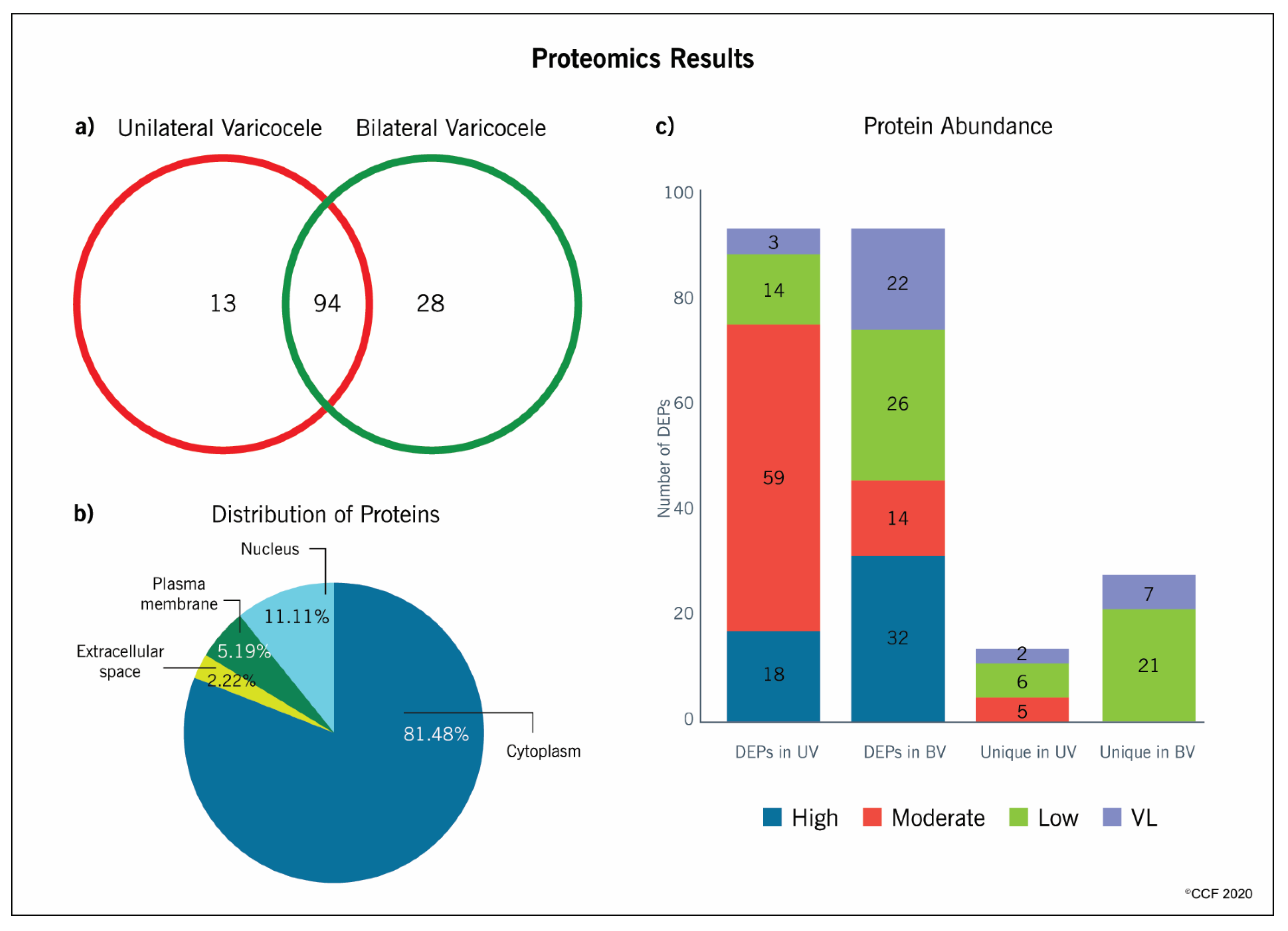
2.2. Proteomic Analysis

Reanalysis of the global proteomics data of spermatozoa from unilateral and bilateral varicocele based on the normalized spectral abundance factor (NSAF) ratio revealed significant differential expression of 135 proteins having susceptible sites for acetylation. Among these, 94 DEPs (62 overexpressed and 32 underexpressed in unilateral varicocele) were common to both groups whereas 13 and 28 proteins were unique to the unilateral and bilateral varicocele group, respectively (Figure 1a). Spectral counts of these DEPs indicated that a majority were moderately abundant (n = 59) and highly abundant (n = 18) in the spermatozoa of men with unilateral varicocele, while in bilateral varicocele, 32 DEPs were highly abundant, but maximum DEPs were either lowly (n = 26) or very lowly (n = 22) abundant in spermatozoa (Figure 1b). Most of these uniquely expressed proteins in both the unilateral and bilateral varicocele groups were present in low and very low abundance levels (Figure 1b).
All the DEPs overexpressed in the spermatozoa of men with bilateral varicocele compared to underexpressed proteins in unilateral varicocele were present in a high abundance except for AKR1B1 and PSMD13 (Table 1). The underexpressed proteins in the bilateral varicocele group compared to overexpressed proteins in unilateral varicocele were either low or very low and moderately abundant except for ENO1 and MDH2, which were highly abundant (Table 2). Certain uniquely expressed proteins in the unilateral group (CAT, PNP, ACCAA2, SELENBP1) were present in moderate abundance. However, the other proteins unique to the bilateral group were either low or very low in abundance (Table 3).

2.3. Distribution Pattern and Functional Annotation of Proteins

In the current study, GO enrichment analysis and Ingenuity Pathway Analysis (IPA) annotation identified that 81.48% and 11.11% of the proteins involved in the acetylation function were present in the cytoplasm and in the nucleus of spermatozoa, respectively (Figure 1c). Functional analysis using advanced databases, such as GO Term Finder, UniProt, and database for annotation, visualization and integrated discovery (DAVID), revealed that a maximum number of proteins involved in the acetylation function had multifunctional properties. Further, annotation analysis revealed that these DEPs were associated with apoptosis or DNA damage, energy metabolism or stress regulation, mitochondrial dysfunction, and sperm function (Supplementary Table S2).

2.4. Classification of DEPs with Predicted Acetylation

All overexpressed DEPs in the bilateral varicocele group were associated with DNA damage, apoptosis, or oxidative stress mechanisms. Moreover, important proteins related to spermatogenesis, sperm function, and mitochondrial dysfunction, such as CLGN, PARK7, HSPA9, HIST1H2BA, DECR1, DLAT, GSR, SDHA, PPP3CA, and SUCLA2, were underexpressed (Supplementary Table S2). Some of the proteins that were highly abundant in the spermatozoa of men diagnosed with both unilateral and bilateral varicocele included ENO1, ACTB, EEF1A1, FASN, HSPA5, HSP90AA1, MYH9, PKM, TUBA3C, and TUBB4B. Nonetheless, proteins, such as CLGN, FTH1, MDH1, MIF, PPP3CA, SUCLA2, and PSMA7, involved in sperm function, apoptosis, and oxidative stress were present in a low and very low abundance in the unilateral and bilateral varicocele group, respectively (Supplementary Table S2).

2.5. Identification of DEPs Involved in Important Networks

Metacore analysis identified that the transcription factors of androgen receptor, p53, and NRF2 are the main proteins affected by the differential expression of proteins with potential acetylation sites (Figure 2). Networks developed using DEPs based on their biological function demonstrated that a majority of proteins were associated with mitochondrial dysfunction and energy metabolism (Figure 3), and oxidative stress (Figure 4). We also noticed proteins were involved in fertilization and the acrosome reaction (Figure 5).

2.6. Protein–Protein Interaction

Further search tool for the retrieval of interacting genes/proteins (STRING) analysis for all DEPs involved in the acetylation process demonstrated key protein–protein interactions in the varicocele condition. These DEPs were involved in regulating sperm functions. Interestingly, the majority of the proteins associated with the proteasome complex were involved in the acetylation process. STRING analysis displayed the DEPs (PSMA1, PSMB3, PSMA6, PSMA4, PSMB4, PSMB2, PSMA2, PSMA5, PSMA3, PSMA7, PSMD13, and PSMB1) present in the proteasome complex by interaction with each other through a binding process (Figure 6).

2.7. Potential Protein Biomarkers and Western Blot Analysis

Functional proteins related to the acetylation process involved in the acrosome reaction and fertilization process, mitochondrial dysfunction, and oxidative stress were listed as potential protein biomarkers to identify PTM defects pertaining to the acetylation process (Table 4). The presence of acetylated proteins (ANXA2, HIST1H2BA, SERPINB6, and SOD1) was demonstrated in acetyl-lysine immunoprecipitated proteins. Further, western blot (WB) analysis revealed SOD1 was significantly underexpressed (p = 0.0143) in the bilateral varicocele group (Figure 7).

3. Discussion

PTMs of sperm proteins are necessary for the normal physiological functions of spermatozoa [17]. Proteomic analysis of sperm proteins was able to detect the maximum number of proteins present in the spermatozoa. Bioinformatic analysis revealed the involvement of these proteins in specific biological, molecular, and cellular pathways. Furthermore, using a bioinformatics approach, sperm proteins involved in important PTMs, such as phosphorylation, methylation, and acetylation, were also predicted. Since acetylation occurs on numerous and diverse proteins and affects many protein functions, such as DNA binding, enzymatic activity, protein stability, protein–protein interaction, and peptide-receptor recognition, we carried out a detailed analysis of DEPs with the potential of acetylation.
Previous studies used enrichment steps to isolate sperm proteins involved in PTMs, such as phosphorylation and acetylation [12,14]. Further, the LC-MS/MS platform was used to identify proteins and peptides. However, it was performed using physiologically normal spermatozoa [12]. Acetylated proteins in the spermatozoa are involved in sperm functions, such as energy metabolism, capacitation, acrosome reaction, sperm–egg recognition, and fusion [12,14]. The fertilization potential of the spermatozoa is compromised in men with varicocele-mediated male infertility. Until now, no studies have investigated the role of acetylation proteins in infertile men with unilateral or bilateral varicocele. In the current study, we used a global proteomic and bioinformatic approach to identify proteins susceptible to acetylation and further validated the presence of key acetylated proteins in sperm from men with unilateral and bilateral varicocele to understand the etiologies of sperm dysfunction in both cases.
In general, the concentration or abundance of proteins with PTMs in spermatozoa are comparatively lower than that of proteins in their native form. More than 1000 lysine acetylation sites were reported in the proteins of capacitated human sperm and were involved in sperm functions, such as sperm motility and the fertilization process [14]. In the current study, we also observed that most of the proteins predicted to be acetylated were present in a low or very low abundance in both groups (Figure 1b). In the human spermatozoa, the majority of these proteins were reported to be acetylated and involved in sperm motility, capacitation, sperm–egg recognition, fusion of sperm–egg plasma, and fertilization [12]. Acetylation is a dynamic process that regulates cellular function, and optimal spatio-temporal acetylation is required for proper functioning of the cell. Even a slight dysregulation in the expression of proteins may affect the acrosome reaction and fertilization potential of the spermatozoa [14].
Proteomic studies in varicocele patients reported aberrant expression of proteins involved in capacitation, acrosome reaction, and fusion of the sperm with the zona-pellucida [10,16]. In the present study, IPA analysis revealed that the identified proteins that carry out acetylation were proteins related to the acrosome reaction and fertilization. Hyperacetylation of histones is an important event during spermatogenesis [15]. Further, histone acetylation facilitates its replacement with protamines for denser packing of the sperm DNA [15]. Our bioinformatics analysis revealed that the differential expression of HIST1H2B is involved in the acetylation process, in both unilateral and bilateral varicocele patients. Acetylation of HIST1H2B in varicocele patients (unilateral and bilateral) was confirmed by WB analysis. Hence, the expressional change in the acetylated HIST1H2B may have a direct impact on the quality of sperm chromatin in the ejaculated spermatozoa. Furthermore, aberrant expression of acetylated HIST1H2B may serve as a marker for sperm chromatin integrity in varicocele patients.
In the current study, the majority of proteins involved in the acetylation process were under the regulation of transcription factors, androgen receptor, p53, and NRF2. The androgen receptor plays an important role in regulating the action of testosterone, which is required for spermatogenesis [18,19]. Further, the expression of proteins involved in oxidative stress, apoptosis, and cell signaling is affected due to the presence of defective androgen receptors [20]. Their presence in human spermatozoa was demonstrated by WB and immunocytochemistry [21], and on the basis of the androgen concentration, they have the ability to modulate the PI3K/AKT pathway to regulate apoptosis in spermatozoa [22]. In varicocele patients, malfunctioning of the androgen receptor may be due to decreased testosterone levels [23]. Androgen receptors are also affected by PTMs, where acetylation specifically induces apoptosis [24,25]. From the network created, it is clear that the majority of the proteins involved in the acetylation process under the regulation of the androgen receptor are underexpressed in patients with bilateral varicocele (Figure 2). This indicates that the damage to spermatogenesis is higher in bilateral varicocele patients as compared to patients with unilateral varicocele.
The transcription factor p53 is involved in the regulation of apoptosis and is reported as a marker of sperm DNA damage [26,27,28]. Augmented expression of p53 along with PARP and Bak was reported in the spermatozoa of varicocele patients, implying a higher incidence of apoptosis in these patients in comparison with normal individuals [29]. In the current study, we noticed that p53 was involved in the regulation of the DEPs associated with the acetylation process. It is not out of context to mention here that intracellular ANXA2 interacts with p53-mediated apoptosis [30]. WB analysis of acetylated ANXA2 further endorsed the role of acetylation by inducing more apoptotic sperm death in bilateral varicocele. In fact, a lower expression of ANXA2 in human sperm is correlated with high levels of SDF [31]. Further, N-terminal acetylation of membrane-bound ANXA2 is required for S100A10 binding for an enhancement of its activity [32]. Thus, the aberrant expression of proteins involved in the acetylation process may trigger p53 transcription factor, which, in turn, activates the apoptosis process in the defective spermatozoa of varicocele patients.
The transcription factor NRF2 regulates the expression of antioxidant enzymes by activation of the antioxidant responsive element (ARE) [33] and was associated with acetylated sperm proteins [34]. In varicocele patients, NRF2 was linked with mitochondrial dysfunction [35]. Validation of the downstream effecter molecule of NRF2, i.e., antioxidant enzyme Cu/Zn-superoxide dismutase (SOD1), further supports the computational findings. WB analysis of immunoprecipitated SOD1 protein present in the network confirms that the proteins identified as acetylated proteins undergo PTM in the spermatozoa of varicocele patients. In fact, the acetylation of lysine residues near the active site has also been implicated in the inhibition of manganese superoxide dismutase (SOD2) while oxidative stress stimulates SIRT3 to deacetylate SOD2, leading to SOD2 activation and reactive oxygen species (ROS) reduction [36,37,38]. Therefore, the differential acetylation of SOD1 in both groups might be due to differences in the incidence of oxidative stress as both the testes in bilateral varicocele experience hypoxia, while in unilateral varicocele, the contra-lateral testis has normal blood flow.
In general, mitochondrial dysfunction and oxidative stress are the main factors affecting the quality of spermatozoa in varicocele pathology. Earlier sperm proteomic studies in varicocele patients revealed mitochondrial dysfunction and underexpression of mitochondrial proteins [10,35]. In the current study, network analysis identified the acetylation of important sperm proteins, such as PRDX1 and SDHA, associated with oxidative stress and mitochondrial dysfunction, respectively (Figure 3 and Figure 4).
Peroxiredoxins play a major role in the regulation of oxidative stress by modulating ROS levels [39]. Cui et al. reported an increased expression of oxidative stress response protein PRDX1 in immature sperm [40]. Acetylation of the PRDX1 protein increases its reducing activity [41]. Our proteomic data revealed that PRDX1 was overexpressed in unilateral varicocele compared to bilateral varicocele. This may be due to the compensatory redox mechanism being activated in the unilateral varicocele condition [42]; alternatively, lower expression of this protein with low levels of acetylation may be responsible for the higher damage in bilateral varicocele patients. In fact, bilateral varicocele patients in general present with severe anomalies in sperm parameters. Hence, the acetylation state of PRDX1 is critical in varicocele patients with oxidative stress-mediated infertility. Unlike PRDX1, SDHA is a mitochondrial protein and a subunit of the succinate dehydrogenase complex that is involved in the tricarboxylic acid cycle (TCA) cycle, electron transport chain, and energy metabolism. The expression of SDHA was dysregulated in asthenozoospermic patients [43]. Hyperacetylation of SDHA decreases its electron-transferring activity [44,45]. In unilateral varicocele patients, we observed the overexpression of SDHA protein in comparison to bilateral varicocele, further validating more functional impairment of spermatozoa in the bilateral varicocele group. Further, WB analysis of the immunoprecipitated proteins ANXA2 and SERPINB6 involved in the same network confirmed the proteins associated with the oxidative stress response are acetylated in the spermatozoa of varicocele patients.
In the current study, seven alpha-type subunits (PSMA1, PSMA2, PSMA3, PSMA4, PSMA5, PSMA6, and PSMA7) and four beta-type subunits (PSMB1, PSMB2, PSMB3, and PSMB4) of the core 20S proteasome were predicted to be potential sites for acetylation and were overexpressed in unilateral varicocele compared to bilateral. Here, 20S core proteasome is the catalytic site responsible for maintenance of protein homeostasis by removing misfolded or damaged proteins that could impair cellular functions, and by removing proteins whose functions are no longer required. Furthermore, lysine acetylation of 20S cardiac proteasome has been reported to augment its activity [46]. It is not out of context to mention here that proteasomes are located in the mammalian sperm acrosome and on the acrosomal surface. Protein kinase A modulates the enzymatic activity of the proteasome during a progesterone-induced acrosome reaction [47]. Furthermore, after capacitation, the acrosomal proteasomes facilitate the degradation of zona pellucida glycoproteins, leading to fertilization. In a recent study, Zigo et al. (2019) showed that both parallel and sequential treatments of ejaculated and capacitated spermatozoa in the absence and presence of a proteasome inhibitor demonstrated putative sperm proteasome-associated sperm proteins in a compartment-specific manner. The investigators further demonstrated that other proteins associated with capacitation, such as P47/lactadherin, ACRBP, ADAM5, and SPINK2, were processed by the proteasome, and proteasomal inhibitors slowed down capacitation-induced reorganization of the outer acrosomal membrane [48]. Therefore, a decline in the expression of these core subunits of 20S proteasome in bilateral varicocele will decrease the chance of fertilization in the spermatozoa of these patients.

4. Conclusions

In this study, we identified sperm DEPs that are able to regulate the protein acetylation process in infertile men with unilateral and bilateral varicocele. Aberrant expression of acetylated proteins associated with oxidative stress and mitochondrial dysfunction affects the fertilization potential of sperm in varicocele patients. In addition, the acetylation status of proteins, such as HIST1H2B, PRDX1, SDHA, and SOD1, can serve as a biomarker for PTM defects pertaining to the acetylation process in the ejaculated spermatozoa of varicocele patients.

5. Materials and Methods

5.1. Patient Characteristics

This study was approved by the Institutional Review Board (IRB) of the Cleveland Clinic (IRB # 17-422), Cleveland, OH, United States. All participants signed an informed written consent form at the Andrology Center, Cleveland Clinic. Semen samples were obtained from 50 varicocele patients (33 unilateral and 17 bilateral). The majority of the varicocele patients were diagnosed with grade 1 or 2 (unilateral = 83.6% and bilateral = 76.4%) (Supplementary Table S1).

5.2. Proteomic Analysis

Proteomic raw data from our previous studies on unilateral [16] and bilateral [10] varicocele were reanalyzed. Experimental details of the global proteome analysis, including sample preparation and LC-MS/MS data analysis, are described in detail in our previous publication [10]. In the present study, we analyzed the functional proteins having potential acetylation sites.

5.3. Database Searching and Protein Identification

Tandem mass spectra were extracted by Proteome Discoverer version 1.4.1.288. Charge state deconvolution and deisotoping were not performed. All MS/MS raw files were analyzed using Mascot (Matrix Science, London, UK; version 2.3.02), Sequest (Thermo Fisher Scientific, San Jose, CA, USA; version 1.4.0.288), and X! Tandem (The GPM, thegpm.org; version CYCLONE (2010.12.01.1). Mascot, Sequest, and X!Tandem were set up to search the human reference with a database (33,292 entries) assuming trypsin as the digestion enzyme. The mass tolerance for the parent ion was set to 10 parts per million (PPM) and for a fragment ion with 1.0 Da.
Scaffold (version 4.0.6.1; Proteome Software Inc., Portland, OR) was used to validate MS/MS-based peptide and protein identifications. Peptide and protein identifications were accepted as previously described [10]. Protein probabilities were assigned by the Protein Prophet (Systems Biology, Seattle, WA, USA) algorithm. Protein annotation was performed using Gene Ontology (GO) terms from National Center for Biotechnology Information (NCBI).

5.4. Quantitative Proteomics

In general, the relative quantity of the proteins was determined by comparing the number of spectra, termed spectral counts (SpCs), used to identify each protein. The total number of mass spectra (SpCs) that matched peptides to a particular protein was used to measure the abundance of proteins in the complex mixture. To overcome the sample-to-sample variation associated with replicate analyses of a sample and the fact that longer proteins tend to have more peptide identifications than shorter proteins, the NSAF approach was applied prior to relative protein quantification. The criteria used to identify and categorize DEPs based on the SpC were explained in our earlier publication [10].

5.5. Large-Scale Identification of Acetylation Sites

The carbamido methylation of cysteine was set as a fixed modification. For the identification of potential PTM sites, protein N-terminal diglycine/acetylation, diglycine/acetylation of lysine, and oxidation of methionine and pyro-glutamination for N-terminal glutamine were set as variable modifications to identify the ubiquitination and acetylation sites in our database search. A maximum of three or six missed cleavages were allowed, setting Trypsin as the proteolytic enzyme. The mass tolerance was set to 3 parts per million (ppm) for peptide masses and 0.8 Da for MS/MS peaks, respectively. In the process of peptide identification, we conducted a decoy database search by Mascot, and applied a filter to satisfy a false positive rate lower than 1%.
Flanking amino acid sequence analysis: For the representation of position weight matrices (PWMs) for (−9) to (+9) amino acid residues surrounding all of the identified lysine ubiquitination and acetylation sites, the probability of the observed amino acid residues, at each position on the flanking sequences of the lysine modification sites, was normalized by the abundance ratio of each amino acid in the RefSeq human protein database. Visualization of the statistically extracted sequence motifs, based on our large-scale lysine modification proteome data, was also performed using motif-X [49,50].
Pathway analysis: The computational analysis for the statistical extraction of canonical pathways was performed using IPA (QIAGEN, Redwood City, CA, USA) [51]. The proteins modified with the ubiquitination and/or acetylation were uploaded into the IPA software (version 2018-2019), and the top canonical pathways associated with the uploaded proteins were listed along with the p-values calculated using a right-tailed Fisher’s exact test.

5.6. Functional Analysis

Functional annotation and enrichment analysis were performed using publicly available bioinformatic annotation tools and databases, such as GO Term Finder, GO Term Mapper, UniProt, Software for researching annotations of proteins (STRAP), and DAVID (http://david.niaid.nih.gov). Proprietary software packages, such as IPA from Ingenuity® Systems, were also used to obtain a consensus-based comprehensive functional context for the list of proteins involved in the acetylation process derived from the proteome.

5.7. Bioinformatic Analysis of Protein–Protein Interaction

STRING was used to display the functional and physical link between the proteins based on the criteria: Neighborhood, gene fusion, occurrence, co-expression, experimental evidences, existing databases, and text mining. This includes all proteins with potential acetylation sites with respect to spermatogenesis, spermatid differentiation or development, sperm motility, capacitation, acrosome reaction, mitochondrial dysfunction, energy metabolism, apoptosis, DNA damage, DNA methylation, and oxidative stress.

5.8. Immunoprecipitation and Western Blotting of Acetylated Proteins

Acetylation of sperm proteins in both unilateral (n = 6) and bilateral varicocele patients (n = 6) was demonstrated using WB. Immunoprecipitation of acetylated proteins was carried out using anti-acetyl Lysine antibody (ab190479, Abcam, USA) followed by WB detection of selected acetylated proteins. The criteria applied for the selection of DEPs involved in the acetylation process were as follows: (i) Proteins involved in the networks; (ii) abundance of the protein must be moderate or high in any one group; and (iii) proteins with a well-described function in the literature. Four proteins (ANXA2, HIST1H2BA, SERPINB6, and SOD1) were chosen for validation by WB in both the unilateral and bilateral varicocele group.
Immunoprecipitated acetylated proteins were first loaded into a 4–15% SDS–PAGE for 2 h at 90 V. The resolved proteins were transferred onto polyvinylidene difluoride (PVDF) membranes and analyzed as described earlier [52]. The expression levels of the WB-validated proteins were normalized against the global acetylated proteins (Supplementary Figure S1) and compared between unilateral and bilateral varicocele using the Mann–Whitney test and p < 0.05 was considered significant. Data analysis was performed using MedCalc Statistical Software (version 17.8; MedCalc Software, Ostend, Belgium).

Supplementary Materials

The following are available online at https://www.mdpi.com/1422-0067/21/9/3155/s1, Supplementary Table S1. Semen parameters in infertile men with unilateral and bilateral varicocele. Supplementary Table S2. List of proteins involved in the acetylation process in the spermatozoa of unilateral and bilateral varicocele infertile men.

Author Contributions

Project administration and study design, A.A.; methodology, data curation, software, M.K.P.S., L.S.; writing—original draft preparation, M.K.P.S.; writing—review and editing, A.A., M.K.P.S., L.S. All authors have read and agreed to the published version of the manuscript.

Funding

Financial support for this study was provided by the American Center for Reproductive Medicine, Cleveland Clinic, Ohio, USA.

Acknowledgments

Belinda Willard, PhD, Director of Proteomic Core Laboratory, Lerner Research Institute assisted with proteomic analysis. The Orbitrap Elite instrument was purchased via an NIH shared instrument grant, 1S10RR031537-01. The authors are grateful to the following for their critical review of our manuscript and helpful suggestions: Ralf Henkel and Kristian Leisegang (University of the Western Cape, South Africa), Rakesh Sharma, Renata Finelli and Saradha Baskaran (Cleveland Clinic, USA).

Conflicts of Interest

The authors declare no conflict of interest.

References

  1. Samanta, L.; Durairajanayagam, D. Introduction. In Proteomics in Human Reproduction: Biomarkers for Millennials; Springer International Publishing: Cham, Switzerland, 2016; pp. 1–8. [Google Scholar]
  2. Marmar, J.L. The pathophysiology of varicoceles in the light of current molecular and genetic information. Hum. Reprod. Update 2001, 7, 461–472. [Google Scholar] [CrossRef] [PubMed]
  3. Dahl, E.V.; Herrick, J.F. A vascular mechanism for maintaining testicular temperature by counter-current exchange. Surg. Gynecol. Obstet. 1959, 108, 697–705. [Google Scholar] [PubMed]
  4. Jarow, J.P.; Coburn, M.; Sigman, M. Incidence of varicoceles in men with primary and secondary infertility. Urology 1996, 47, 73–76. [Google Scholar] [CrossRef]
  5. Baazeem, A.; Belzile, E.; Ciampi, A.; Dohle, G.; Jarvi, K.; Salonia, A.; Weidner, W.; Zini, A. Varicocele and male factor infertility treatment: A new meta-analysis and review of the role of varicocele repair. Eur. Urol. 2011, 60, 796–808. [Google Scholar] [CrossRef]
  6. Esteves, S.C.; Agarwal, A. Afterword to varicocele and male infertility: Current concepts and future perspectives. Asian J. Androl. 2016, 18, 319–322. [Google Scholar] [CrossRef]
  7. Hosseinifar, H.; Sabbaghian, M.; Nasrabadi, D.; Modarresi, T.; Dizaj, A.V.; Gourabi, H.; Gilani, M.A. Study of the effect of varicocelectomy on sperm proteins expression in patients with varicocele and poor sperm quality by using two-dimensional gel electrophoresis. J. Assist. Reprod. Genet. 2014, 31, 725–729. [Google Scholar] [CrossRef] [Green Version]
  8. Hosseinifar, H.; Gourabi, H.; Salekdeh, G.H.; Alikhani, M.; Mirshahvaladi, S.; Sabbaghian, M.; Modarresi, T.; Gilani, M.A. Study of sperm protein profile in men with and without varicocele using two-dimensional gel electrophoresis. Urology 2013, 81, 293–300. [Google Scholar] [CrossRef]
  9. Camargo, M.; Intasqui Lopes, P.; Del Giudice, P.T.; Carvalho, V.M.; Cardozo, K.H.; Andreoni, C.; Fraietta, R.; Bertolla, R.P. Unbiased label-free quantitative proteomic profiling and enriched proteomic pathways in seminal plasma of adult men before and after varicocelectomy. Hum. Reprod. (Oxford, England) 2013, 28, 33–46. [Google Scholar] [CrossRef] [Green Version]
  10. Agarwal, A.; Sharma, R.; Samanta, L.; Durairajanayagam, D.; Sabanegh, E. Proteomic signatures of infertile men with clinical varicocele and their validation studies reveal mitochondrial dysfunction leading to infertility. Asian J. Androl. 2016, 18, 282–291. [Google Scholar] [CrossRef]
  11. Baker, M.A.; Nixon, B.; Naumovski, N.; Aitken, R.J. Proteomic insights into the maturation and capacitation of mammalian spermatozoa. Syst. Biol. Reprod. Med. 2012, 58, 211–217. [Google Scholar] [CrossRef]
  12. Yu, H.; Diao, H.; Wang, C.; Lin, Y.; Yu, F.; Lu, H.; Xu, W.; Li, Z.; Shi, H.; Zhao, S.; et al. Acetylproteomic analysis reveals functional implications of lysine acetylation in human spermatozoa (sperm). Mol. Cell. Proteom. MCP 2015, 14, 1009–1023. [Google Scholar] [CrossRef] [PubMed] [Green Version]
  13. Krejci, J.; Stixova, L.; Pagacova, E.; Legartova, S.; Kozubek, S.; Lochmanova, G.; Zdrahal, Z.; Sehnalova, P.; Dabravolski, S.; Hejatko, J.; et al. Post-Translational Modifications of Histones in Human Sperm. J. Cell. Biochem. 2015, 116, 2195–2209. [Google Scholar] [CrossRef] [PubMed]
  14. Sun, G.; Jiang, M.; Zhou, T.; Guo, Y.; Cui, Y.; Guo, X.; Sha, J. Insights into the lysine acetylproteome of human sperm. J. Proteom. 2014, 109, 199–211. [Google Scholar] [CrossRef] [PubMed]
  15. Pang, A.; Rennert, O. Protein acetylation and spermatogenesis. Reprod. Syst. Sex. Disord. Curr. Res. 2013, (Suppl 1), 5. [Google Scholar] [CrossRef] [Green Version]
  16. Agarwal, A.; Sharma, R.; Durairajanayagam, D.; Cui, Z.; Ayaz, A.; Gupta, S.; Willard, B.; Gopalan, B.; Sabanegh, E. Differential proteomic profiling of spermatozoal proteins of infertile men with unilateral or bilateral varicocele. Urology 2015, 85, 580–588. [Google Scholar] [CrossRef]
  17. Samanta, L.; Swain, N.; Ayaz, A.; Venugopal, V.; Agarwal, A. Post-Translational Modifications in sperm Proteome: The Chemistry of Proteome diversifications in the Pathophysiology of male factor infertility. Biochim. Biophys. Acta 2016, 1860, 1450–1465. [Google Scholar] [CrossRef]
  18. Walker, W.H.; Easton, E.; Moreci, R.S.; Toocheck, C.; Anamthathmakula, P.; Jeyasuria, P. Restoration of Spermatogenesis and Male Fertility Using an Androgen Receptor Transgene. PLoS ONE 2015, 10, e0120783. [Google Scholar] [CrossRef] [Green Version]
  19. O’Hara, L.; Smith, L.B. Androgen receptor roles in spermatogenesis and infertility. Best Pract. Res. Clin. Endocrinol. Metab. 2015, 29, 595–605. [Google Scholar] [CrossRef] [Green Version]
  20. Stanton, P.G.; Sluka, P.; Foo, C.F.; Stephens, A.N.; Smith, A.I.; McLachlan, R.I.; O’Donnell, L. Proteomic changes in rat spermatogenesis in response to in vivo androgen manipulation; impact on meiotic cells. PLoS ONE 2012, 7, e41718. [Google Scholar] [CrossRef] [Green Version]
  21. Solakidi, S.; Psarra, A.M.; Nikolaropoulos, S.; Sekeris, C.E. Estrogen receptors alpha and beta (ERalpha and ERbeta) and androgen receptor (AR) in human sperm: Localization of ERbeta and AR in mitochondria of the midpiece. Hum. Reprod. (Oxford, England) 2005, 20, 3481–3487. [Google Scholar] [CrossRef] [Green Version]
  22. Aquila, S.; Middea, E.; Catalano, S.; Marsico, S.; Lanzino, M.; Casaburi, I.; Barone, I.; Bruno, R.; Zupo, S.; Ando, S. Human sperm express a functional androgen receptor: Effects on PI3K/AKT pathway. Hum. Reprod. (Oxford, England) 2007, 22, 2594–2605. [Google Scholar] [CrossRef] [PubMed] [Green Version]
  23. Alkaram, A.; McCullough, A. Varicocele and its effect on testosterone: Implications for the adolescent. Transl. Androl. Urol. 2014, 3, 413–417. [Google Scholar]
  24. Gioeli, D.; Paschal, B.M. Post-translational modification of the androgen receptor. Mol. Cell. Endocrinol. 2012, 352, 70–78. [Google Scholar] [CrossRef] [PubMed]
  25. Coffey, K.; Robson, C.N. Regulation of the androgen receptor by post-translational modifications. J. Endocrinol. 2012, 215, 221. [Google Scholar] [CrossRef] [PubMed] [Green Version]
  26. Riley, T.; Sontag, E.; Chen, P.; Levine, A. Transcriptional control of human p53-regulated genes. Nat. Rev. Mol. Cell Biol. 2008, 9, 402–412. [Google Scholar] [CrossRef] [PubMed]
  27. Raimondo, S.; Gentile, T.; Cuomo, F.; De Filippo, S.; Aprea, G.E.; Guida, J. Quantitative evaluation of p53 as a new indicator of DNA damage in human spermatozoa. J. Hum. Reprod. Sci. 2014, 7, 212–217. [Google Scholar] [CrossRef] [PubMed]
  28. Raimondo, S.; Gentile, T.; Gentile, M.; Morelli, A.; Donnarumma, F.; Cuomo, F.; De Filippo, S.; Montano, L. p53 Protein Evaluation on Spermatozoa DNA in Fertile and Infertile Males. J. Hum. Reprod. Sci. 2019, 12, 114–121. [Google Scholar] [CrossRef]
  29. Chang, F.W.; Sun, G.H.; Cheng, Y.Y.; Chen, I.C.; Chien, H.H.; Wu, G.J. Effects of varicocele upon the expression of apoptosis-related proteins. Andrologia 2010, 42, 225–230. [Google Scholar] [CrossRef]
  30. Huang, Y.; Jin, Y.; Yan, C.H.; Yu, Y.; Bai, J.; Chen, F.; Zhao, Y.Z.; Fu, S.B. Involvement of Annexin A2 in p53 induced apoptosis in lung cancer. Mol. Cell. Biochem. 2008, 309, 117–123. [Google Scholar] [CrossRef]
  31. Munuce, M.J.; Marini, P.E.; Teijeiro, J.M. Expression profile and distribution of Annexin A1, A2 and A5 in human semen. Andrologia 2019, 51, e13224. [Google Scholar] [CrossRef]
  32. Nazmi, A.R.; Ozorowski, G.; Pejic, M.; Whitelegge, J.P.; Gerke, V.; Luecke, H. N-terminal acetylation of annexin A2 is required for S100A10 binding. Biol. Chem. 2012, 393, 1141–1150. [Google Scholar] [CrossRef] [PubMed]
  33. Ma, Q. Role of nrf2 in oxidative stress and toxicity. Annu. Rev. Pharmacol. Toxicol. 2013, 53, 401–426. [Google Scholar] [CrossRef] [PubMed] [Green Version]
  34. Dias, T.R.; Samanta, L.; Agarwal, A.; Pushparaj, P.N.; Panner Selvam, M.K.; Sharma, R. Proteomic Signatures Reveal Differences in Stress Response, Antioxidant Defense and Proteasomal Activity in Fertile Men with High Seminal ROS Levels. Int. J. Mol. Sci. 2019, 20, 203. [Google Scholar] [CrossRef] [PubMed] [Green Version]
  35. Samanta, L.; Agarwal, A.; Swain, N.; Sharma, R.; Gopalan, B.; Esteves, S.C.; Durairajanayagam, D.; Sabanegh, E. Proteomic Signatures of Sperm Mitochondria in Varicocele: Clinical Use as Biomarkers of Varicocele Associated Infertility. J. Urol. 2018, 200, 414–422. [Google Scholar] [CrossRef] [PubMed]
  36. Qiu, X.; Brown, K.; Hirschey, M.D.; Verdin, E.; Chen, D. Calorie restriction reduces oxidative stress by SIRT3-mediated SOD2 activation. Cell Metab. 2010, 12, 662–667. [Google Scholar] [CrossRef] [PubMed] [Green Version]
  37. Tao, R.; Coleman, M.C.; Pennington, J.D.; Ozden, O.; Park, S.-H.; Jiang, H.; Kim, H.-S.; Flynn, C.R.; Hill, S.; McDonald, W.H. Sirt3-mediated deacetylation of evolutionarily conserved lysine 122 regulates MnSOD activity in response to stress. Mol. Cell 2010, 40, 893–904. [Google Scholar] [CrossRef] [Green Version]
  38. Chen, Y.; Zhang, J.; Lin, Y.; Lei, Q.; Guan, K.L.; Zhao, S.; Xiong, Y. Tumour suppressor SIRT3 deacetylates and activates manganese superoxide dismutase to scavenge ROS. EMBO Rep. 2011, 12, 534–541. [Google Scholar] [CrossRef]
  39. O’Flaherty, C. Peroxiredoxins: Hidden players in the antioxidant defence of human spermatozoa. Basic Clin. Androl. 2014, 24, 4. [Google Scholar] [CrossRef] [Green Version]
  40. Cui, Z.; Sharma, R.; Agarwal, A. Proteomic analysis of mature and immature ejaculated spermatozoa from fertile men. Asian J. Androl. 2016, 18, 735–746. [Google Scholar]
  41. Parmigiani, R.B.; Xu, W.S.; Venta-Perez, G.; Erdjument-Bromage, H.; Yaneva, M.; Tempst, P.; Marks, P.A. HDAC6 is a specific deacetylase of peroxiredoxins and is involved in redox regulation. Proc. Natl. Acad. Sci. USA 2008, 105, 9633–9638. [Google Scholar] [CrossRef] [Green Version]
  42. Swain, N.; Samanta, L.; Agarwal, A.; Kumar, S.; Dixit, A.; Gopalan, B.; Durairajanayagam, D.; Sharma, R.; Pushparaj, P.N.; Baskaran, S. Aberrant Upregulation of Compensatory Redox Molecular Machines May Contribute to Sperm Dysfunction in Infertile Men with Unilateral Varicocele: A Proteomic Insight. Antioxid. Redox Signal. 2019, 32, 504–521. [Google Scholar] [CrossRef]
  43. Nowicka-Bauer, K.; Lepczynski, A.; Ozgo, M.; Kamieniczna, M.; Fraczek, M.; Stanski, L.; Olszewska, M.; Malcher, A.; Skrzypczak, W.; Kurpisz, M.K. Sperm mitochondrial dysfunction and oxidative stress as possible reasons for isolated asthenozoospermia. J. Physiol. Pharmacol. An Off. J. Pol. Physiol. Soc. 2018, 69. [Google Scholar] [CrossRef]
  44. Cimen, H.; Han, M.J.; Yang, Y.; Tong, Q.; Koc, H.; Koc, E.C. Regulation of succinate dehydrogenase activity by SIRT3 in mammalian mitochondria. Biochemistry 2010, 49, 304–311. [Google Scholar] [CrossRef] [PubMed] [Green Version]
  45. Finley, L.W.; Haas, W.; Desquiret-Dumas, V.; Wallace, D.C.; Procaccio, V.; Gygi, S.P.; Haigis, M.C. Succinate dehydrogenase is a direct target of sirtuin 3 deacetylase activity. PLoS ONE 2011, 6, e23295. [Google Scholar] [CrossRef] [PubMed] [Green Version]
  46. Wang, D.; Fang, C.; Zong, N.C.; Liem, D.A.; Cadeiras, M.; Scruggs, S.B.; Yu, H.; Kim, A.K.; Yang, P.; Deng, M.; et al. Regulation of acetylation restores proteolytic function of diseased myocardium in mouse and human. Mol. Cell. Proteom. MCP 2013, 12, 3793–3802. [Google Scholar] [CrossRef] [Green Version]
  47. Kerns, K.; Morales, P.; Sutovsky, P. Regulation of sperm capacitation by the 26S proteasome: An emerging new paradigm in spermatology. Biol. Reprod. 2016, 94, 1–9. [Google Scholar] [CrossRef]
  48. Zigo, M.; Manaskova-Postlerova, P.; Jonakova, V.; Kerns, K.; Sutovsky, P. Compartmentalization of the proteasome-interacting proteins during sperm capacitation. Sci. Rep. 2019, 9, 12583. [Google Scholar] [CrossRef] [Green Version]
  49. Schwartz, D.; Gygi, S.P. An iterative statistical approach to the identification of protein phosphorylation motifs from large-scale data sets. Nat. Biotechnol. 2005, 23, 1391–1398. [Google Scholar] [CrossRef]
  50. Kramer, A.; Green, J.; Pollard, J., Jr.; Tugendreich, S. Causal analysis approaches in Ingenuity Pathway Analysis. Bioinformatics (Oxford, England) 2014, 30, 523–530. [Google Scholar] [CrossRef]
  51. Narushima, Y.; Kozuka-Hata, H.; Koyama-Nasu, R.; Tsumoto, K.; Inoue, J.; Akiyama, T.; Oyama, M. Integrative Network Analysis Combined with Quantitative Phosphoproteomics Reveals Transforming Growth Factor-beta Receptor type-2 (TGFBR2) as a Novel Regulator of Glioblastoma Stem Cell Properties. Mol. Cell. Proteom. MCP 2016, 15, 1017–1031. [Google Scholar] [CrossRef] [PubMed] [Green Version]
  52. Panner Selvam, M.K.; Agarwal, A.; Dias, T.R.; Martins, A.D.; Samanta, L. Presence of Round Cells Proteins do not Interfere with Identification of Human Sperm Proteins from Frozen Semen Samples by LC-MS/MS. Int. J. Mol. Sci. 2019, 20, 314. [Google Scholar] [CrossRef] [PubMed] [Green Version]
Figure 1. (a) Differential expression proteins predicted to be acetylated in unilateral and bilateral varicocele, (b) Abundance of differentially expressed proteins, (c) Distribution pattern of differentially expressed proteins involved in the acetylation process.
Ijms 21 03155 g001
Figure 2. Transcription factors androgen receptor, p53, and NRF2 affected by the differential expression of proteins involved in the acetylation process.
Ijms 21 03155 g002
Figure 3. Interaction network of acetylated proteins associated with mitochondrial dysfunction and energy metabolism.
Ijms 21 03155 g003
Figure 4. Interaction network of acetylated proteins associated with oxidative stress.
Ijms 21 03155 g004
Figure 5. Interaction network of acetylated proteins associated with fertilization and the acrosome reaction.
Ijms 21 03155 g005
Figure 6. STRING analysis showing the interaction among the proteins involved in the proteasome complex.
Ijms 21 03155 g006
Figure 7. Western blot analysis of proteins associated with the acetylation process in unilateral and bilateral varicocele infertile men. (A) Annexin A2 (ANXA2), (B) histone H2B type 1-A (HIST1H2BA), (C) serpin B6 (SERPINB6), (D) superoxide dismutase 1 (SOD1). Results are expressed as mean ± standard error of mean and in fold variation to the unilateral varicocele group.
Ijms 21 03155 g007
Table 1. Comparison of the enrichment of lysine-acetylated functional sperm proteins underexpressed in unilateral varicocele in comparison to bilateral varicocele.
Gene SymbolProtein NameAccession NumberAbundanceNSAF Ratio (UV vs. BV)
UVBV
ACTBactin, cytoplasmic 14501885HH0.55
EEF1A1elongation factor 1-alpha 14503471HH0.56
FASNfatty acid synthase41872631HH0.59
HSPA578 glucose-regulated protein precursor16507237HH0.56
HSP90AA1heat shock protein HSP 90-alpha isoform 1153792590HH0.32
MYH9myosin-912667788HH0.4
PKMpyruvate kinase isozymes M1/M2 isoform c332164775HH0.63
TUBA3Ctubulin alpha-3C/D chain17921993HH0.48
TUBB4Btubulin beta-4B chain5174735HH0.41
ACAT1acetyl-CoA acetyltransferase, mitochondrial precursor (involved in fatty acid metabolism and ketone body formation not lysine-acetylation of proteins)4557237LH0.19
P4HBprotein disulfide-isomerase precursor20070125LH0.31
ALDOAfructose-bisphosphate aldolase A isoform 2342187211MH0.59
ANXA1annexin A14502101MH0.46
ANXA2annexin A2 isoform 150845388MH0.21
ANXA5annexin A54502107MH0.22
ANXA6annexin A6 isoform 171773329MH0.18
ATP5A1ATP synthase subunit alpha, mitochondrial precursor4757810MH0.58
ATP5BATP synthase subunit beta, mitochondrial precursor32189394MH0.3
PDIA3protein disulfide-isomerase A3 precursor21361657MH0.26
HK1hexokinase-1 isoform HKI-ta/tb15991831MH0.5
HSPA8heat shock cognate 71 protein isoform 15729877MH0.37
HSP90AB1heat shock protein HSP 90-beta20149594MH0.42
LCP1plastin-2167614506MH0.65
HSP90B1endoplasmin precursor4507677MH0.27
RUVBL1ruvB-like 14506753MH0.55
HYOU1hypoxia up-regulated protein 1 precursor5453832MH0.17
CCT2T-complex protein 1 subunit beta isoform 15453603MH0.55
RUVBL2ruvB-like 25730023MH0.37
PHGDHD-3-phosphoglycerate dehydrogenase23308577MH0.57
CANXcalnexin precursor10716563VLH0.08
AKR1B1aldose reductase4502049VLL0.12
PSMD1326S proteasome non-ATPase regulatory subunit 13 isoform 1157502193VLL0.05
UV: unilateral varicocele, BV: bilateral varicocele, H: high, M: moderate, L: low, VL: very low.
Table 2. Comparison of the enrichment of lysine-acetylated functional sperm proteins overexpressed in unilateral varicocele in comparison to bilateral varicocele.
Gene SymbolProtein NameAccession NumberAbundanceNSAF Ratio (UV vs. BV)
UVBV
ENO1alpha-enolase isoform 14503571HH2.05
MDH2malate dehydrogenase, mitochondrial precursor21735621HH3.91
ACADVLvery long-chain specific acyl-CoA dehydrogenase, mitochondrial isoform 2 precursor76496475HL10.53
PARK7protein DJ-131543380LL3.78
ACADMmedium-chain specific acyl-CoA dehydrogenase, mitochondrial isoform b precursor187960098ML6.83
ALDH9A14-trimethylaminobutyraldehyde dehydrogenase115387104ML12.28
CALM1calmodulin58218968ML5.22
cpt2carnitine O-palmitoyltransferase 2, mitochondrial precursor4503023ML2.24
HSPA9stress-70 protein, mitochondrial precursor24234688ML2.29
IPO5importin-524797086ML4.17
PSMB1proteasome subunit beta type-14506193ML6.66
SOD2superoxide dismutase [Mn], mitochondrial isoform A precursor67782305ML10.51
DNPEPaspartyl aminopeptidase156416028ML5.45
HIST1H2AAhistone H2A type 1-A25092737ML3.86
HIST1H2BAhistone H2B type 1-A24586679ML2.40
DLDdihydrolipoyl dehydrogenase, mitochondrial precursor91199540HM10.28
FLNBfilamin-B isoform 2105990514HM2.03
GPIglucose-6-phosphate isomerase isoform 218201905HM8.39
SERPINB6serpin B641152086HM4.68
LAP3cytosol aminopeptidase41393561HM8.54
DECR12,4-dienoyl-CoA reductase, mitochondrial precursor4503301MM2.65
DLATdihydrolipoyllysine-residue acetyltransferase component of pyruvate dehydrogenase complex, mitochondrial precursor31711992MM2.36
DLSTdihydrolipoyllysine-residue succinyltransferase component of 2-oxoglutarate dehydrogenase complex, mitochondrial isoform 1 precursor19923748MM2.91
ETFAelectron transfer flavoprotein subunit alpha, mitochondrial isoform a4503607MM2.77
GSRglutathione reductase, mitochondrial isoform 1 precursor50301238MM6.39
PRDX1peroxiredoxin-1320461711MM2.04
SDHAsuccinate dehydrogenase [ubiquinone] flavoprotein subunit, mitochondrial156416003MM2.35
APMAPadipocyte plasma membrane-associated protein24308201MM2.45
ASRGL1l-asparaginase145275200MM1.99
GOT2aspartate aminotransferase, mitochondrial precursor73486658HVL31.16
CLGNcalmegin precursor4758004LVL3.77
FTH1ferritin heavy chain56682959LVL6.51
HAGHhydroxyacylglutathione hydrolase, mitochondrial isoform 1 precursor94538322LVL8.18
MDH1malate dehydrogenase, cytoplasmic isoform 1312283701LVL5.46
MIFmacrophage migration inhibitory factor4505185LVL6.98
PPP3CAserine/threonine-protein phosphatase 2B catalytic subunit alpha isoform isoform 2194688147LVL10.66
RPL560S ribosomal protein L514591909LVL8.34
SUCLA2succinyl-CoA ligase [ADP-forming] subunit beta, mitochondrial precursor11321583LVL46.32
LACTB2beta-lactamase-like protein 27705793LVL7.36
C1QBPcomplement component 1 Q subcomponent-binding protein, mitochondrial precursor4502491MVL7.24
CAPN1calpain-1 catalytic subunit311893363MVL5.51
FBP1fructose-1,6-bisphosphatase 1189083692MVL26.30
SOD1superoxide dismutase [Cu-Zn]4507149MVL5.68
TKTtransketolase205277463MVL11.17
UV: unilateral varicocele, BV: bilateral varicocele, H: high, M: moderate, L: low, VL: very low.
Table 3. Important proteins with enriched lysine acetylation uniquely expressed in the unilateral varicocele (UV) and bilateral varicocele (BV) group.
Gene SymbolProtein NameAccession NumberAbundanceExpression
GSS glutathione synthetase4504169LUnique in UV
PPP5Cserine/threonine-protein phosphatase 5 isoform 15453958LUnique in UV
TALDO1transaldolase5803187LUnique in UV
GNPDA1glucosamine-6-phosphate isomerase 113027378LUnique in UV
CATcatalase4557014MUnique in UV
PNPpurine nucleoside phosphorylase157168362MUnique in UV
SELENB P1selenium-binding protein 116306550MUnique in UV
ACAA23-ketoacyl-CoA thiolase, mitochondrial167614485MUnique in UV
ACTC1actin, alpha cardiac muscle 1 proprotein4885049LUnique in BV
ATP5C1ATP synthase subunit gamma, mitochondrial isoform L (liver) precursor50345988LUnique in BV
HMOX2heme oxygenase 28051608LUnique in BV
PFN2profilin-2 isoform a16753215LUnique in BV
PHBprohibitin4505773LUnique in BV
VDAC1voltage-dependent anion-selective channel protein 14507879LUnique in BV
YWHAB14-3-3 protein beta/alpha21328448LUnique in BV
YWHAG14-3-3 protein gamma21464101LUnique in BV
DNAJA2dnaJ homolog subfamily A member 25031741LUnique in BV
STOML2stomatin-like protein 27305503LUnique in BV
CYCScytochrome c11128019LUnique in BV
MTPNmyotrophin21956645LUnique in BV
7-Sepseptin-7 isoform 2148352329VLUnique in BV
TAGLN2transgelin-24507357VLUnique in BV
H: high, M: moderate, L: low, VL: very low.
Table 4. Potential acetylated protein biomarkers in the unilateral and bilateral varicocele group.
Sperm FunctionDEPsUnilateral VaricoceleBilateral Varicocele
AbundanceExpressionAbundanceExpression
Fertilization and acrosome reactionHIST1H2BModerateOELowUE
Mitochondrial dysfunction and oxidative stressSDHAModerateOEModerateUE
PRDX1ModerateOEModerateUE
SOD1ModerateOEVery lowUE
DEPs: differentially expressed proteins, UE: underexpressed, OE: overexpressed.

