Article

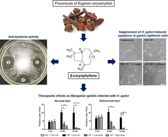
Inhibitory Effects of β-Caryophyllene on Helicobacter pylori Infection In Vitro and In Vivo

1
Department of Biomedical Laboratory Science, College of Health Sciences, Yonsei University, Wonju 26493, Korea
2
Department of Clinical Laboratory Science, College of Medical Sciences, Daegu Haany University, Gyeongsan 38610, Korea
3
Forensic DNA Division, National Forensic Service, Wonju 26460, Korea
4
Department of Biomedical Laboratory Science, Daekyeung University, Gyeongsan 38547, Korea
5
SFC BIO Co., Ltd. 1505-1ho, Daerung-town, 25, Gasan digital 1 ro, Geumcheon-gu 08594, Seoul, Korea
6
College of Pharmacy, Dankook University, 119 Dandae-ro, Cheonan-si, Chungnam 31116, Korea
7
Department of Clinical Laboratory Science, Semyung University, Jaecheon 27136, Korea
*
Authors to whom correspondence should be addressed.
These authors contributed equally to this work as co-first authors.
Int. J. Mol. Sci. 2020, 21(3), 1008; https://doi.org/10.3390/ijms21031008
Submission received: 8 January 2020 / Revised: 29 January 2020 / Accepted: 30 January 2020 / Published: 3 February 2020
(This article belongs to the Section Molecular Pharmacology)

Abstract

:
The human specific bacterial pathogen Helicobacter pylori (H. pylori) is associated with severe gastric diseases, including gastric cancer. Recently, the increasing resistance makes the usage of antibiotics less effectively. Therefore, development of a new antimicrobial agent is required to control H. pylori infection. In the current study, the inhibitory effect of β-caryophyllene on H. pylori growth, as well as the antibacterial therapeutic effect, has been demonstrated. β-caryophyllene inhibited H. pylori growth via the downregulation of dnaE, dnaN, holB, and gyrA and also downregulated virulence factors such as CagA, VacA, and SecA proteins. β-caryophyllene inhibited expression of several T4SS components, so that CagA translocation into H. pylori-infected AGS gastric cancer cells was decreased by β-caryophyllene treatment. β-caryophyllene also inhibited VacA entry through the downregulation of T5aSS. After β-caryophyllene administration on Mongolian gerbils, the immunohistochemistry (IHC) and Hematoxylin&Eosin stains showed therapeutic effects in the treated groups. Hematological data, which was consistent with histological data, support the therapeutic effect of β-caryophyllene administration. Such a positive effect of β-caryophyllene on H. pylori infection potently substantiates the natural compound as being capable of being used as a new antimicrobial agent or functional health food to help patients who are suffering from gastroduodenal diseases due to H. pylori infection.

Graphical Abstract

1. Introduction

Helicobacter pylori (H. pylori) is a Gram-negative, spiral-shaped, microaerophilic bacterium that selectively colonizes in human gastric mucosa [1,2]. Compared with uninfected individuals, H. pylori infected individuals have a 2−8-fold increased risk of developing gastric cancer [3]. For these reasons, H. pylori has been classified as class I carcinogen by the World Health Organization (WHO) [4]. H. pylori-mediated gastric diseases are mostly due to the effect of its virulence factors. Thus, understanding the biological characteristics and mechanisms of their virulence factors might offer a more comprehensive understanding into the pathogenesis of H. pylori infection.
Cytotoxin-associated protein A (CagA) is a highly immunogenic protein that is encoded by the cag pathogenicity island (PAI). The cagPAI genes also encode components of a type IV secretion system (T4SS), which injects CagA protein into host cells [5]. The T4SS apparatus commonly consists of 11 VirB proteins, encoded by the virB1-B11 genes and VirD4 protein [6,7]. Once translocated, CagA dysregulates the homeostatic signal transduction of the gastric epithelial cells, leading the loss of cell polarity, apoptosis, and proliferation, which are involved in chronic inflammation and gastric cancer [8,9].
Vacuolating cytotoxin A (VacA) protein is a known pore-forming secreted toxin found in almost all H. pylori strains [10]. VacA secretion is regulated by the type Va secretion system (T5aSS) and depends on the Sec machinery for transport through the inner membrane [11]. Secretion system subunit protein A (SecA) is an intracellular ATPase that provides the necessary energy for the translocation of H. pylori proteins out of the bacterial plasma membrane [11,12,13]. Following translocation into the host cells, VacA leads multiple cellular alterations, including vacuolation and cell death [14].
DNA replication is the biological process of copying the DNA in all living organisms. Initiation of DNA replication occurs after the binding of the initiator protein DnaA to the AT-rich regions on oriC. DnaA then forms a complex with other nucleoproteins (DnaBC) and ATPs to make the pre-Replicative Complex [15]. DnaB is DNA helicase that progressively unwinds the double stranded DNA in the 5′-3′ direction [16]. H. pylori possesses six DNA polymerase III holoenzyme genes. These include two genes for replicase (dnaE and dnaQ), one for clamp (dnaN) and three for clamp loader (dnaX, holA and holB) [15]. The termination of DNA replication takes place when the helicase DnaB on the leading strands reach a Tus protein [17].
β-caryophyllene is a volatile bicyclic sesquiterpene compound that exists as a mixture of mainly β-caryophyllene and small amounts of α-humulene [18]. It is easily found in the essential oils of many edible plants such as cloves (Eugenia caryophyllata) [19], oregano (Origanum vulgare), and cinnamon (Cinnamomum spp.) [18,20,21]. Recent approval of β-caryophyllene as a food additive and flavoring agent by the Food and Drug Administration in the USA (FDA) and the European Food Safety Authority (EFSA) generated interest among the scientific community to explore its additional therapeutic benefits [22]. Numerous reports showed inhibitory effects of β-caryophyllene against bacteria, virus, and fungi [23,24,25,26].
Recently, natural compounds derived from medicinal plants seem to be an important source of antimicrobial agents. Due to the increasing resistance and the emergence of adverse effects, the usage of antibiotics and antibacterial therapeutics is becoming less effective. Therefore, inhibitory effect of β-caryophyllene on H. pylori growth and its inhibitory mechanisms were investigated in this study. Furthermore, we aimed to discover a new antimicrobial agent to eradicate H. pylori, or to identify a new functional health food that can reduce the virulence of H. pylori in infected gastric cells in vitro and in vivo.

2. Results and Discussion

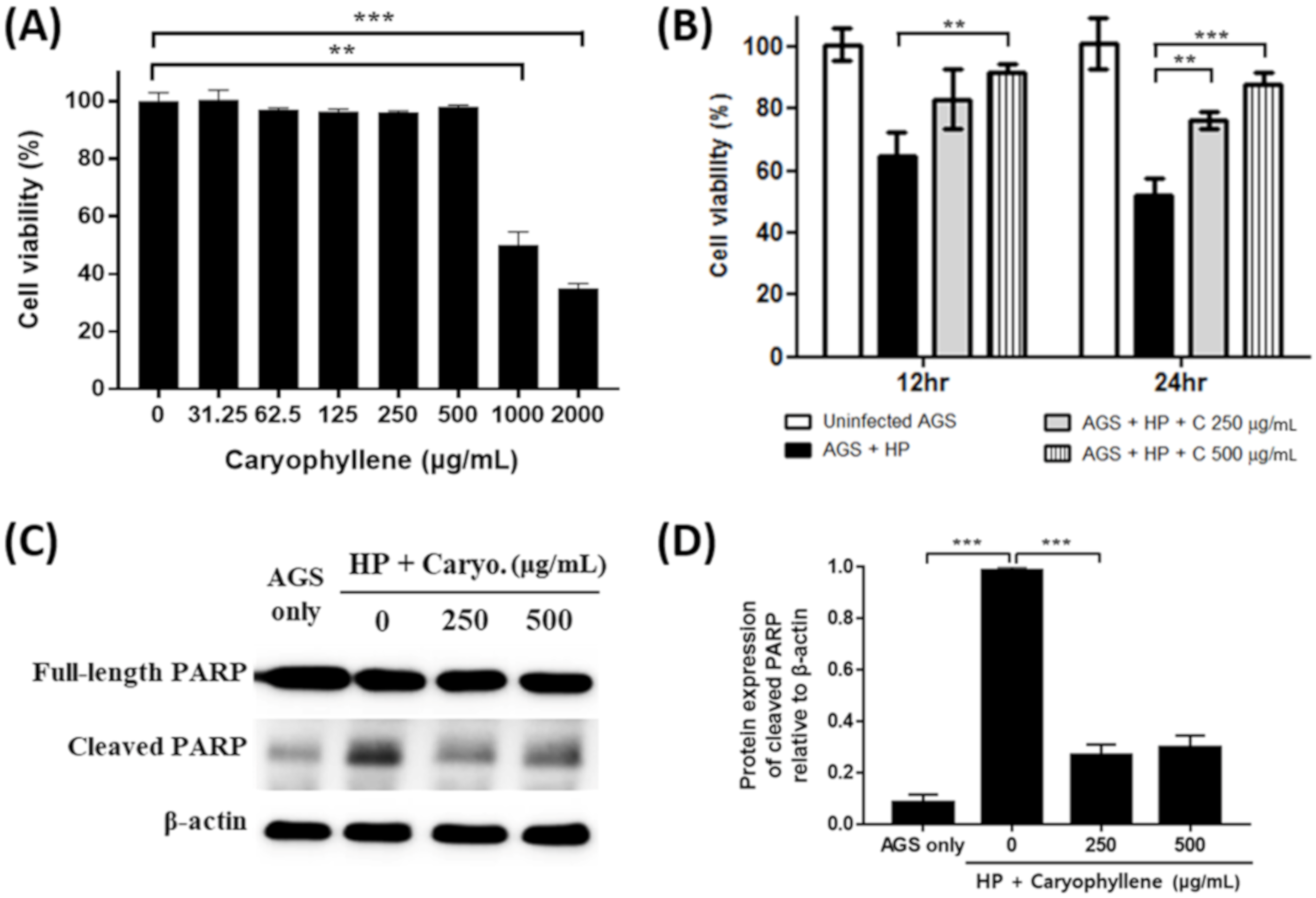
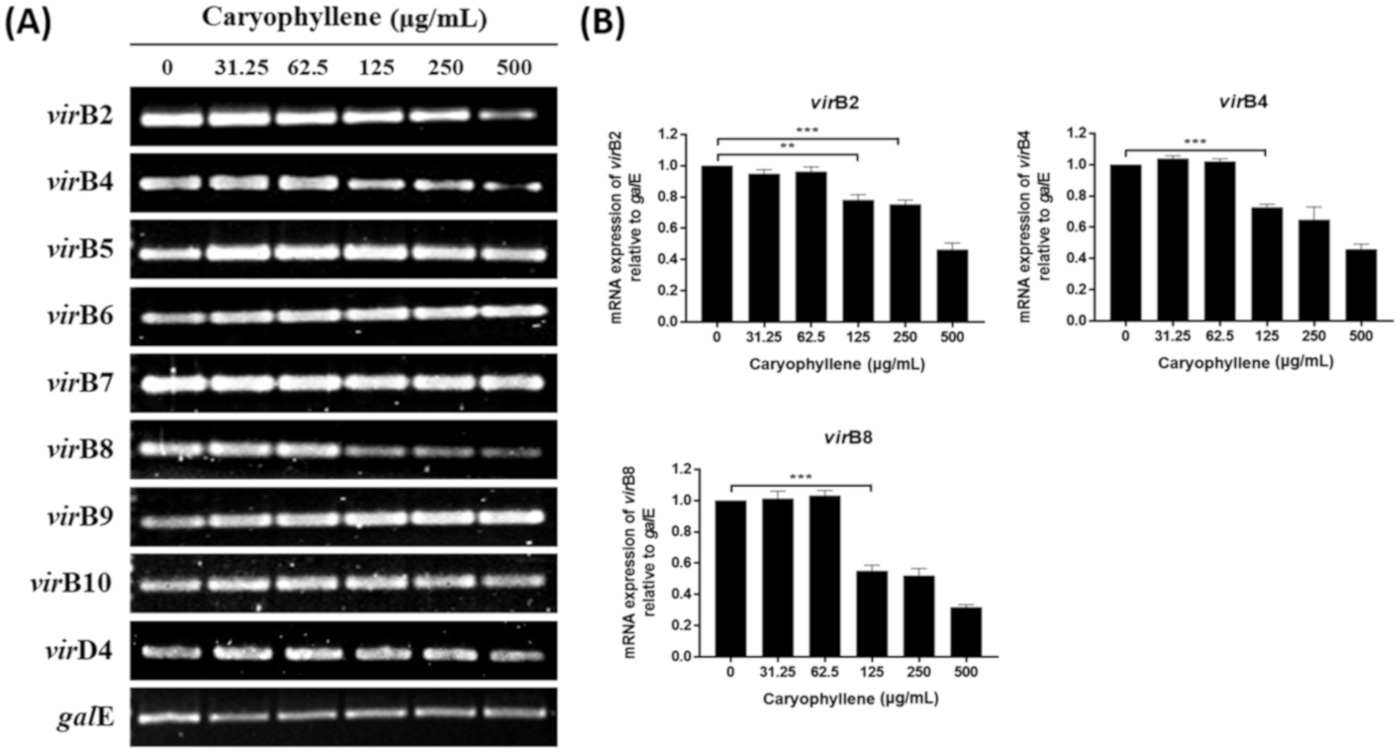
2.1. Inhibitory Effect of β-Caryophyllene on the Growth and Expression of Replication Genes of H. pylori

The inhibitory effect of β-caryophyllene on H. pylori was previously evaluated by a screening test using the disc diffusion assay. Clear inhibitory zones were observed around 10, 50, and 100 μg discs and the diameters of the inhibitory zones were 17, 21, and 23 mm, respectively (Figure 1A). By the broth dilution test, the minimum inhibitory concentration (MIC)MIC of β-caryophyllene against H. pylori (ATCC 49503) was determined to be 1000 μg/mL (Figure 1B). Only one strain was used to confirm the inhibitory effect of β-caryophyllene, and therefore, it will be necessary to it apply to other reference strains or clinical isolates.
To elucidate how β-caryophyllene inhibits the growth of H. pylori, expressions of replication genes of H. pylori were evaluated by reverse transcriptase-polymerase chain reaction (RT-PCR). β-caryophyllene treatment decreased the mRNA expression levels of dnaE, dnaN, holB, and gyrA genes (Figure 1C,D). DnaE is the catalytic α subunit of DNA polymerase III. It has been reported that the mutant dnaN was unable to support E. coli growth [27]. Song et al. have shown that E. coli strains bearing chromosomal knockout of holB gene was not viable [28]. These studies demonstrated that the DnaE, DnaN, and HolB are necessary for cell growth, and thus they are indispensable. DNA gyrase is pivotal for the process of bacterial replication, so that it has received the most attention in developing antibiotics such as novobiocin (the ATP site inhibitor) and fluoroquinolone (the catalytic site inhibitor) [29,30]. Therefore, interruption of bacterial replication via downregulation of dnaE, dnaN, holB, and gyrA genes by β-caryophyllene may explain the inhibitory mechanism of β-caryophyllene against H. pylori growth.

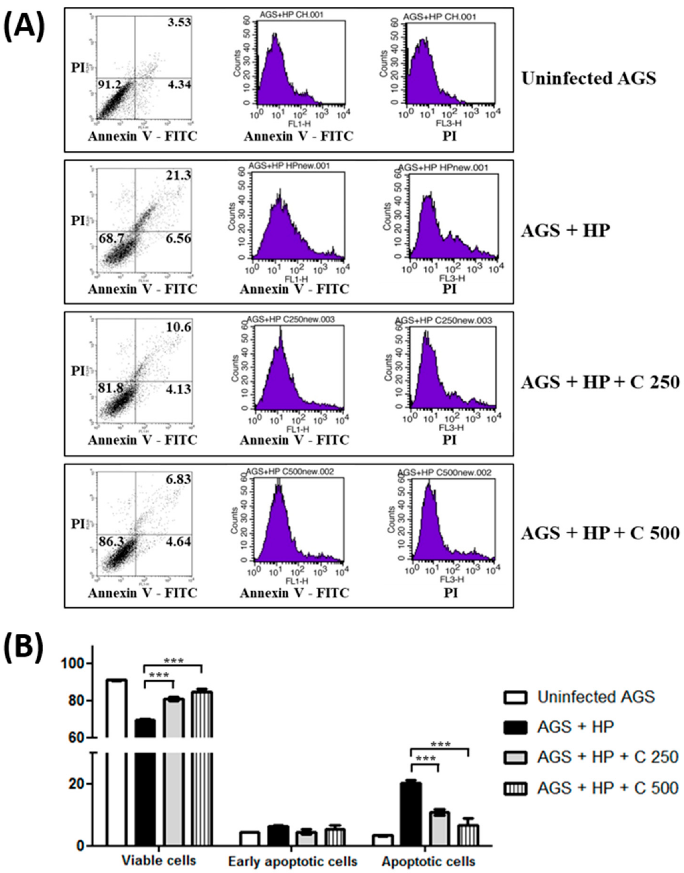
2.2. Suppression of H. pylori-Induced Apoptosis in Gastric Epithelial Cells by β-Caryophyllene

Infection of H. pylori results in deleterious effects on gastric epithelial cells such as the induction of apoptosis, which is closely related to gastric cancer development [31]. Thus, it was evaluated whether β-caryophyllene can alleviate the deleterious effects of H. pylori infection on gastric epithelial cells. β-caryophyllene showed no cytotoxic effect on AGS gastric cancer cells without H. pylori infection up to 500 μg/mL (Figure 2A) and H. pylori infection (200 MOI) reduced cell viability of AGS cells to 51.8% in 24 h (Figure 2B). However, the reduced cell viability was alleviated up to 87.6% by 500 μg/mL β-caryophyllene treatment (Figure 2B).
In the Western blot analysis, poly ADP-ribose polymerase (PARP) was cleaved in AGS cells by H. pylori infection, indicating the induction of apoptosis. The H. pylori-induced PARP cleavage was inhibited by β-caryophyllene treatment (Figure 2C,D). Furthermore, annexin V-FITC stain result also showed that apoptosis induced by H. pylori infection was alleviated in AGS cells by β-caryophyllene treatment (Figure 3A,B). These results collectively suggest that β-caryophyllene inhibited apoptosis and alleviated cell death in AGS cells infected with H. pylori.

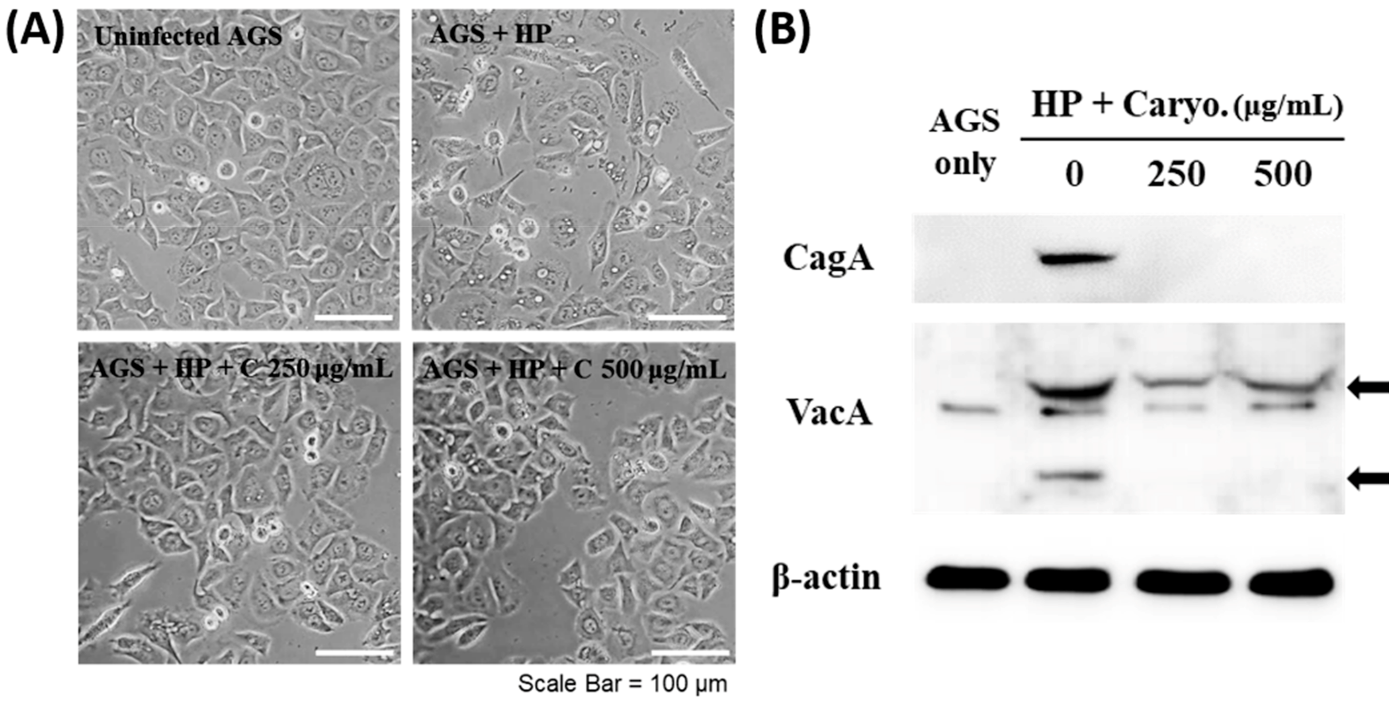
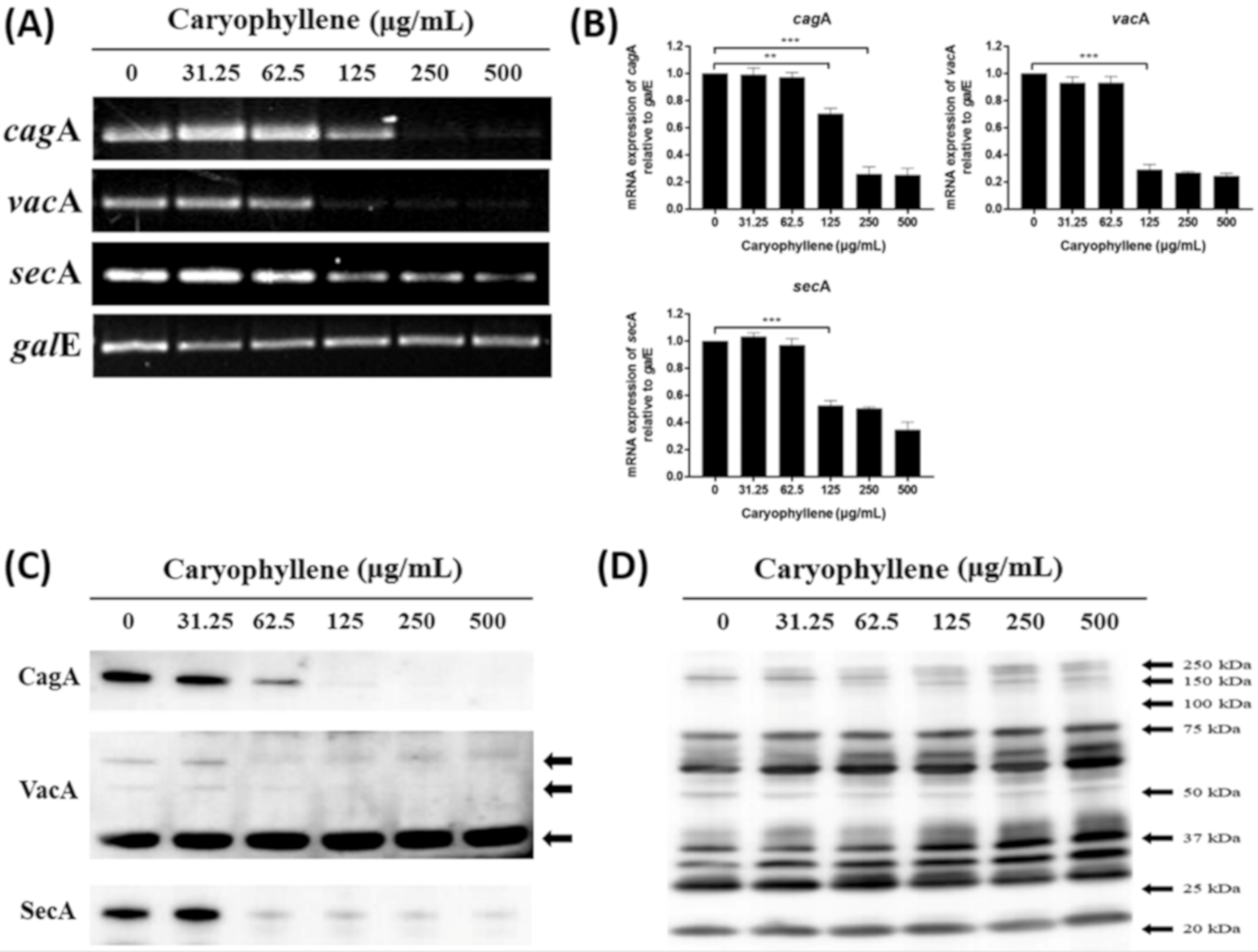
2.3. Inhibitory Effects of β-Caryophyllene on the Translocation of Bacterial CagA and VacA Proteins

Cytoskeletal rearrangement and resultant morphological change so-called hummingbird phenotype is a noted outcome appearing after injection of CagA protein into AGS cells and the accumulation of cytoplasmic vesicles (vacuolation) is induced by VacA translocation. β-caryophyllene treatment diminished the hummingbird phenotype and vacuolation on H. pylori-infected AGS cells dose dependently (Figure 4A) and also inhibited CagA and VacA protein translocation into the H. pylori-infected AGS cells (Figure 4B).
To elucidate the reasons why the translocation of CagA and VacA proteins to AGS cells was inhibited by β-caryophyllene, we investigated the effect of β-caryophyllene on H. pylori directly on the mRNA and protein expressions of CagA and VacA. Both mRNA and protein levels of CagA, VacA and SecA in H. pylori were reduced by β-caryophyllene treatment (Figure 5). Besides, the mRNA expression levels of virB2, virB4, virB8, and T4SS components in H. pylori, were reduced in a β-caryophyllene dose-dependent manner (Figure 6). These results suggest that inhibition of CagA and T4SS expressions by β-caryophyllene might be, in part, associated with the decreased translocation of CagA to AGS cells and downregulated SecA, as well as VacA might be induced VacA translocation to AGS cells. Collectively, these in vitro data provides evidence supporting that β-caryophyllene may helpful in attenuating the deleterious effects such as hummingbird phenotype, vacuolation, and apoptotic cell death induced by H. pylori infection.

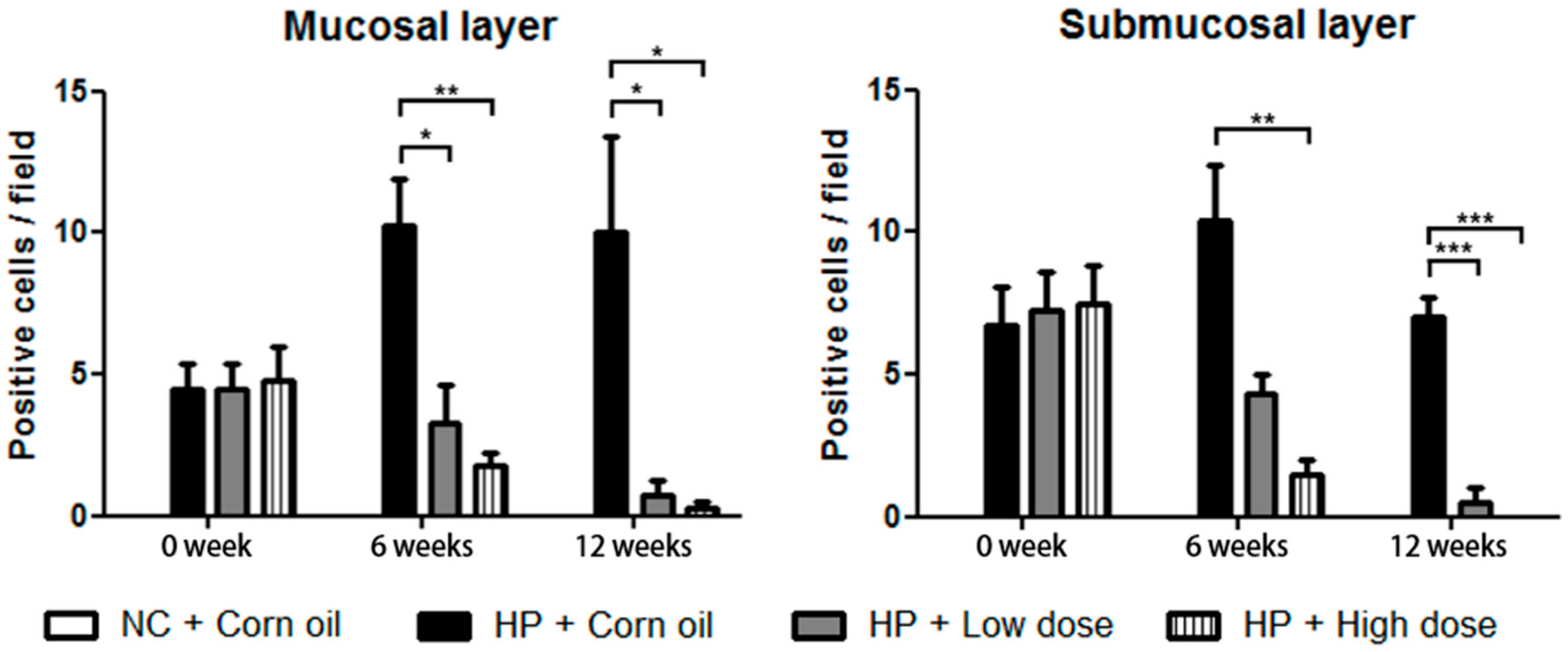
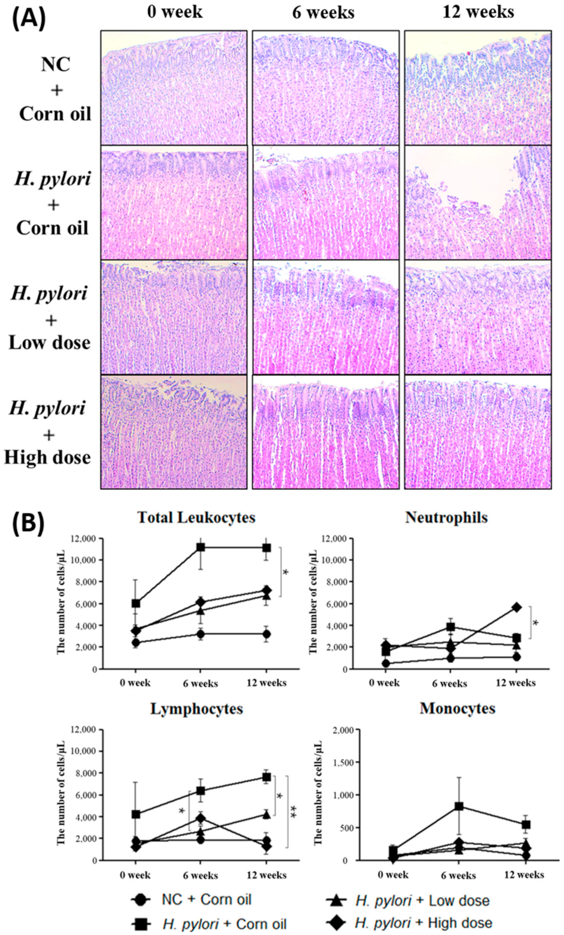
2.4. Therapeutic Efefcts of β-Caryophyllene on Mongolian Gerbils Infected With H. pylori

The in vivo experiments in this study demonstrated the therapeutic effects of β-caryophyllene. Mongolian gerbils in each group were sacrificed at 0 week, 6 weeks, and 12 weeks after beginning the β-caryophyllene treatment. RNA was extracted from stomach of the Mongolian gerbils, and then RT-PCR targeting H. pylori’s 16S rRNA was performed to evaluate the presence of living H. pylori. The result at 0 week showed successful colonization of H. pylori in all the infected groups. The presence of H. pylori was detected in all the H. pylori infected Mongolian gerbils without β-caryophyllene treatment, and the infection lasted until 12 weeks. However, H. pylori was not detected in the groups treated with β-caryophyllene since 6 weeks, indicating the successful eradication of H. pylori by β-caryophyllene (Figure S2).
Furthermore, gastric tissue sections of each group were prepared and presence of H. pylori was also investigated by using the IHC stain. Anti-H. pylori antibody was used to detect the presence of H. pylori. There was no positive signal observed in the uninfected gerbil group (normal control group). In contrast, positive signals for anti-H. pylori antibody were observed abundantly in gastric mucosal and the submucosal layer of β-caryophyllene-untreated group (H. pylori control group), which indicated a marked H. pylori infection in the gastric epithelium of the gerbils. H. pylori were also detected in the β-caryophyllene-treated groups at 0 week, but the positive signals were decreased over time after β-caryophyllene treatment, suggesting that both low doses (100 μg/g) and high doses (500 μg/g) of β-caryophyllene significantly reduced the degree of infection by H. pylori (Figure 7, Figures S3 and S4).

2.5. Inhibitory Effects of β-Caryophyllene on the H. pylori-Induced Inflammation

H&E staining data showed that β-caryophyllene treatment diminishes inflammation in H. pylori-infected stomach tissues (Figure 8A). The reason why inflammatory signs are decreased in β-caryophyllene-treated groups is due to the fact that colonized H. pylori might be eradicated by β-caryophyllene administration. In a long-term in vivo study, Wiedemann et al. reported that early inflammation was observed at 8 weeks and precancerous gastric changes were developed at late time points (32 or 64 weeks) [32]. According to these findings, it might be able to detect more severe inflammatory signs and precancerous changes from the H. pylori control group, if the infection was maintained over 32 or 64 weeks.
In the hematological study, the numbers of total leukocytes in H. pylori control group were vertically increased at 6 weeks and maintained highly at 12 weeks. The numbers of total leukocytes in β-caryophyllene-treated groups, however, were gradually increased after β-caryophyllene administration (Figure 8B). Detailed comparison of hematological changes between β-caryophyllene-untreated group and β-caryophyllene-treated groups also showed that the treatment of β-caryophyllene alleviates H. pylori infection. As neutrophils actively resist bacterial infection, the increased-neutrophil count is commonly found in bacterial infection. However, elevated numbers of neutrophils were relieved by β-caryophyllene administration. This finding implies that bacterial infection is dwindled or bacterial pathogenesis is hindered. In addition to neutrophils, the increased number of lymphocytes and monocytes were lessened at 6 and 12 weeks after β-caryophyllene administration. Contrary to human leukocytes, the composition ratio of lymphocytes in total leukocytes is over 60% in Mongolian gerbils [33,34]. Resulting from the ratio, total changes of leukocytes may be mostly affected by the changes of lymphocytes in Mongolian gerbils. These data correspond with preceded histological data and this certainly supports the therapeutic effect of β-caryophyllene on H. pylori infection. Meanwhile, there may be controversy concerning the neutrophil count data because of the sudden fluctuation in the high dose group at 12 weeks. It is speculated that daily medication with a high dose of β-caryophyllene may lead to the chemical toxicity.
The development of new antimicrobial agents with fewer disadvantages is necessary for the eradication of H. pylori. β-caryophyllene, which is one of the natural compounds that is easily found in the essential oils of many plants [19,20,21]. According to several reports, β-caryophyllene has antibacterial effect on cariogenic bacteria and food-spoilage bacteria [23,24,25]. This study demonstrated the inhibitory effect of β-caryophyllene on H. pylori growth and the protective effect against H. pylori infection, as well as an antibacterial therapeutic effect. The aim of this study was to discover the new antimicrobial agent from nature that may inhibit the H. pylori infection; thus, this study aimed to investigate the effect of β-caryophyllene as one of the natural compounds that possess multiple pharmacological properties [18]. Further studies are required to fully elucidate about the anti-inflammatory and anti-apoptotic mechanism of β-caryophyllene against H. pylori. A previous study from Tambe et al. suggested that β-caryophyllene has a gastric cytoprotective effect [35]. Thus, it would be intriguing to study the anti-inflammatory and anti-apoptotic mechanism of β-caryophyllene during H. pylori infection. Along with the mechanisms studied, the oral LD50 of β-caryophyllene in Mongolian gerbil seems to be necessary to establish.

3. Materials and Methods

3.1. Bacterial Culture and Determination of Antibacterial Activity

H. pylori ATCC 49503 strain (East-asian type: CagA+/VacA+, American Type Culture Collection, Manassas, VA, USA) was grown on Brucella agar plates (Becton-Dickinson, Braintree, MA, USA), supplemented with 10% bovine serum (BRL Life Technologies, Grand Island, NY, USA) at 37 °C for 72 h under a humidified atmosphere with 5% CO2. For disc diffusion method, the number of bacteria in the H. pylori suspension was adjusted to a McFarland scale 2 (6 × 10⁸ cells/mL) on Mueller-Hinton agar (Becton-Dickinson), supplemented with 10% bovine serum and then incubated for 72 h. To determine the minimum inhibitory concentration (MIC) of β-caryophyllene against H. pylori, the number of bacterial particles in the H. pylori suspension was set to McFarland scale 0.5 (1.5 × 10⁸ cells/mL). Various concentrations of β-caryophyllene (7.81–4000 μg/mL) were treated and the bacteria were incubated for 72 h and final optical density (600 nm) of the bacterial suspension was measured by using NanoQuant spectrophotometer (infinite M200, TECAN, Männedorf, Switzerland). For normal control, the same volume of ethanol was administrated to culture media. β-caryophyllene was provided by SFC BIO Co., Ltd. Seoul, in republic of Korea.

3.2. Mammalian Cell Culture

AGS gastric adenocarcinoma cells (ATCC CRL-1739) were cultured in DMEM medium (BRL Life Technologies), supplemented with 10% fetal bovine serum (BRL Life Technologies) and streptomycin-penicillin (100 μg/mL and 100 IU/mL, BRL Life Technologies). Cells were infected with H. pylori as a concentration of 200 multiplicity of infection (MOI) without the addition of antimicrobial agents in media and then treated with β-caryophyllene (250 and 500 μg/mL). For normal control, the equivalent amount of ethanol was administrated to culture media.

3.3. Reverse Transcriptase-Polymerase Chain Reaction (RT-PCR)

H. pylori ATCC 49503 strain was grown in Mueller-Hinton broth (Becton-Dickinson) for 72 h. Cultured H. pylori was washed twice with phosphate-buffered saline (PBS) and total RNA was extracted using Trizol reagent (Invitrogen, Carlsbad, CA, USA), as described in the manufacturer’s instructions. cDNA was synthesized by reverse transcription with random hexamer (Invitrogen) and Moloney murine leukemia virus-reverse transcriptase (MMLV-RT, Invitrogen). Subsequent PCR amplification was performed in a thermocycler, using specific primers.

3.4. Western Blotting

Bacteria and AGS cells were lysed with radio immunoprecipitation assay (RIPA) lysis buffer (Millipore, Billerica, MA, USA) containing a protease inhibitor cocktail (Calbiochem, San Diego, CA, USA) and protein concentrations were determined based on Lowry method. Antibodies to detect CagA, VacA, and β-actin were purchased from Santa Cruz Biotechnology (Dallas, TX, USA) and polyclonal antibody against whole H. pylori (ATCC 49503) and SecA were produced, as previously described [13,36]. Antibodies to detect PARP were purchased from Cell Signaling Technology (Danvers, MA, USA). Anti-H. pylori polyclonal antibody and β-actin were used as an internal control for bacteria and mammalian cell proteins.

3.5. WST Cell Viability Assay Using EZ-Cytox

AGS cells (1 × 10⁴ per well) were plated in 96-well plates. After 24 h, cells were treated with various concentrations of β-caryophyllene. The cells were then incubated for 24 h and subjected to water soluble tetrazolium salt (WST) assay by using EZ-Cytox cell viability assay kit according to manufacturer’s instruction. Ten μL of WST solutions were added to the cultured media and were incubated for 2 h in the CO2 incubator. The absorbance was measured at 450 nm by a spectrophotometer.

3.6. Annexin V and PI Staining

Annexin V and PI staining was performed by using Annexin V-FITC Apoptosis Detection Kit I (Becton-Dickinson) according to the manufacturer’s instruction. The cultured cells were detached with 0.25% trypsin-EDTA, washed twice with cold PBS, and centrifuged at 3000 rpm for 5 min. The cells were resuspended in 500 μL of 1X binding buffer at a concentration of 5 × 10⁵ cells/mL and 5 μL of Annexin V-FITC and 5 μL of PI were added to the cell suspension. The mixture was incubated for 10 min at 37 °C in the dark and analyzed by FACS Caliber flow cytometry (Becton-Dickinson).

3.7. Animal and Experimental Design

Inbred specific pathogen free (SPF) 5 week-old, male and female Mongolian gerbils for mating were purchased from Central Lab Animals, South Korea. Gerbils used in this study were obtained from 10 breeding cages bred in-house. Animals were divided into four groups. “Normal control group” comprised of 7 gerbils that were inoculated with corn oil. “H. pylori control group” comprised of 8 gerbils that were inoculated orogastrically three-times with H. pylori (1 × 10⁹ cells) and were given no further treatment. Group, “High dose” and group, “Low dose” were inoculated with H. pylori (1 × 10⁹ cells) and the administration of β-caryophyllene was orogastrically administrated every day for 12 weeks. β-caryophyllene was diluted with sterile corn oil at two concentrations. Five hundred μg/g was prepared for high dose group, and one hundred μg/g was prepared for low dose group (Figure S1). All gerbils were sacrificed using CO2 euthanasia at different times post-administration (0, 6, 12 weeks). The stomach was excised, opened along the greater curvature. One half was used for a culture study (reisolation) and the extraction of RNA, while the other was used for immunohistochemical and histopathological analyses. The blood samples were taken from all sacrificed gerbils for hematological examination.

3.8. Assessment of Histopathology

H. pylori were detected in infected gastric tissues by immunohistochemistry (IHC) using a rabbit anti-H. pylori antibody (Abcam, Milton, Cambridge, UK). Positive-staining cells were visualized with diaminobenzidine (DAB) (Vectastain Elite ABC Kit for rabbit; Vector Laboratories, Burlingame, CA, USA) and morphometrically analyzed with Leica DM 2500 microscopy and Leica Application Suite software (version 4.4; Leica microsystems, Heerbrugg, Switzerland). The evaluation of H. pylori-positive cells as marker of the infection was performed by counting the H. pylori-positive cells distributed in the non-infected control gastric tissue. Paraffin embedded longitudinal sections of antrum and corpus were stained with hematoxylin and eosin (H&E) and evaluated. It was graded for gastritis and mucosal changes and analyzed by a double blind test according to the grading scheme for rodents [32,37].

3.9. Statistical Analysis

Data in the bar graphs are presented as mean ± standard error of mean (SEM). All the statistical analyses were performed using GraphPad Prism 5.02 software (GraphPad Software, San Diego, CA, USA). All the data were analyzed by unpaired Student’s t-test and p < 0.05 was considered to be statistically significant (* p < 0.05, ** p < 0.01 and *** p < 0.001).

3.10. Ethics Statement

All in vivo experiments and procedures were approved by the Institutional Animal Care and Use Committee (IACUC) of Yonsei University Wonju Campus (approval number: YWC-150612-1, 12 June 2015). All works was conducted in compliance with government regulations, including Welfare Measures for Animal Protection of Ministry of Agriculture, Food and Rural Affairs in republic of Korea.

Supplementary Materials

Supplementary materials can be found at https://www.mdpi.com/1422-0067/21/3/1008/s1. Figure S1. Experimental protocol in vivo. Thirty-one gerbils were divided into four groups. “NC + corn oil” were inoculated corn oil without H. pylori. “H. pylori + corn oil” inoculated with H. pylori (1 × 10⁹ cells) 3 times and were given no further treatment. Group, “H. pylori + low dose” and group, “H. pylori + high dose” were inoculated with H. pylori and β-caryophyllene administration was started 2 weeks after the initial inoculation. The treatment was orogastrically performed during 12-week period; 500 μg/g for high dose group, 100 μg/g for low dose group. All gerbils were sacrificed at specified time of administration (0, 6, 12 weeks). Figure S2. Detection of H. pylori in stomach of Mongolian gerbils using RT-PCR. The stomach tissues from H. pylori infected Mongolian gerbils were collected for RNA extraction then was subjected to RT-PCR. Colonization of H. pylori in stomach of Mongolian gerbils was eradicated after β-caryophyllene administration. H. pylori-16S rRNA was used to determine the presence of infection. NC: NC + corn oil; HP: H. pylori + corn oil; Low: H. pylori + low dose group; High: H. pylori + high dose group; PC: Positive control. Figure S3. Immunohistochemistry of gastric mucosal layer in H. pylori-infected Mongolian gerbils. Gastric tissue sections of each group were prepared and presence of H. pylori was also investigated by using IHC stain. Photomicrograph shows gastric mucosal layer of Mongolian gerbils (×200). Arrows or circles indicate the presence of H. pylori. Positive signals for anti-H. pylori antibody were observed abundantly in gastric mucosal layer of β-caryophyllene-untreated group (H. pylori control group) while there was no positive signal observed in the uninfected gerbil group (Normal control group). H. pylori were also detected in the β-caryophyllene-treated groups at 0 week, but the positive signals were decreased over times after β-caryophyllene treatment suggesting that both low dose and high dose of β-caryophyllene significantly reduced the degree of infection by H. pylori. Figure S4. Immunohistochemistry of gastric submucosal layer in H. pylori-infected Mongolian gerbils. Gastric tissue sections of each group were prepared and presence of H. pylori was also investigated by using IHC stain. Photomicrograph shows gastric mucosal layer of Mongolian gerbils (×200). Arrows indicate the presence of H. pylori. Positive signals for anti-H. pylori antibody were observed abundantly in gastric submucosal layer of β-caryophyllene-untreated group (H. pylori control group) while there was no positive signal observed in the uninfected gerbil group (Normal control group). H. pylori were also detected in the β-caryophyllene-treated groups at 0 week, but the positive signals were decreased over times after β-caryophyllene treatment suggesting that both low dose and high dose of β-caryophyllene significantly reduced the degree of infection by H. pylori.

Author Contributions

H.J.W. and J.Y.Y. have written the manuscript. H.J.W., J.Y.Y., M.H.L., H.W.K. and H.J.K. have carried out all animal studies and performed the analysis. M.P. aided in interpreting the results. S.-k.K. and S.-Y.P. conceived of the original idea. S.-H.K. and J.-B.K. supervised the findings of this work and helped shape the research, analysis and manuscript. All authors have read and agreed to the published version of the manuscript.

Acknowledgments

This work was supported by Korea Institute of Planning and Evaluation for Technology in Food, Agriculture and Forestry (IPET) through Future Innovation Food Technology Development Program, funded by Ministry of Agriculture, Food and Rural Affairs (MAFRA) (119023-3).

Conflicts of Interest

The authors have declared no conflicts of interest.

References

  1. Marshall, B.J.; Warren, J.R. Unidentified curved bacilli in the stomach of patients with gastritis and peptic ulceration. Lancet 1984, 1, 1311–1315. [Google Scholar] [CrossRef]
  2. Warren, J.R.; Marshall, B. Unidentified curved bacilli on gastric epithelium in active chronic gastritis. Lancet 1983, 1, 1273–1275. [Google Scholar] [PubMed]
  3. Vohlonen, I.; Pukkala, E.; Malila, N.; Harkonen, M.; Hakama, M.; Koistinen, V.; Sipponen, P. Risk of gastric cancer in Helicobacter pylori infection in a 15-year follow-up. Scand. J. Gastroenterol. 2016, 51, 1159–1164. [Google Scholar] [CrossRef] [PubMed] [Green Version]
  4. IARC Working Group. Schistosomes, liver flukes and Helicobacter pylori. IARC Working Group on the Evaluation of Carcinogenic Risks to Humans. Lyon, 7–14 June 1994. IARC Monogr. Eval. Carcinog. Risks Hum. 1994, 61, 1–241. [Google Scholar]
  5. Backert, S.; Tegtmeyer, N. Type IV Secretion and Signal Transduction of Helicobacter pylori CagA through Interactions with Host Cell Receptors. Toxins (Basel) 2017, 9. [Google Scholar] [CrossRef] [Green Version]
  6. Kwok, T.; Zabler, D.; Urman, S.; Rohde, M.; Hartig, R.; Wessler, S.; Misselwitz, R.; Berger, J.; Sewald, N.; Konig, W.; et al. Helicobacter exploits integrin for type IV secretion and kinase activation. Nature 2007, 449, 862–866. [Google Scholar] [CrossRef]
  7. Merino, E.; Flores-Encarnacion, M.; Aguilar-Gutierrez, G.R. Functional interaction and structural characteristics of unique components of Helicobacter pylori T4SS. FEBS J. 2017. [Google Scholar] [CrossRef] [Green Version]
  8. Zhang, X.S.; Tegtmeyer, N.; Traube, L.; Jindal, S.; Perez-Perez, G.; Sticht, H.; Backert, S.; Blaser, M.J. A Specific A/T Polymorphism in Western Tyrosine Phosphorylation B-Motifs Regulates Helicobacter pylori CagA Epithelial Cell Interactions. Plos Pathog. 2015, 11. [Google Scholar] [CrossRef] [Green Version]
  9. Glowinski, F.; Holland, C.; Thiede, B.; Jungblut, P.R.; Meyer, T.F. Analysis of T4SS-induced signaling by H. pylori using quantitative phosphoproteomics. Front. Microbiol. 2014, 5. [Google Scholar] [CrossRef] [Green Version]
  10. Sgouras, D.N.; Trang, T.T.; Yamaoka, Y. Pathogenesis of Helicobacter pylori Infection. Helicobacter 2015, 20 (Suppl. 1), 8–16. [Google Scholar] [CrossRef] [Green Version]
  11. Boquet, P.; Ricci, V. Intoxication strategy of Helicobacter pylori VacA toxin. Trends Microbiol. 2012, 20, 165–174. [Google Scholar] [CrossRef] [PubMed]
  12. Leyton, D.L.; Rossiter, A.E.; Henderson, I.R. From self sufficiency to dependence: Mechanisms and factors important for autotransporter biogenesis. Nature Rev. Microbiol. 2012, 10, 213–225. [Google Scholar] [CrossRef] [PubMed]
  13. Kim, S.H.; Woo, H.; Park, M.; Rhee, K.J.; Moon, C.; Lee, D.; Seo, W.D.; Kim, J.B. Cyanidin 3-O-glucoside reduces Helicobacter pylori VacA-induced cell death of gastric KATO III cells through inhibition of the SecA pathway. Int. J. Med. Sci. 2014, 11, 742–747. [Google Scholar] [CrossRef] [PubMed] [Green Version]
  14. Memon, A.A.; Hussein, N.R.; Miendje Deyi, V.Y.; Burette, A.; Atherton, J.C. Vacuolating cytotoxin genotypes are strong markers of gastric cancer and duodenal ulcer-associated Helicobacter pylori strains: A matched case-control study. J. Clin. Microbiol. 2014, 52, 2984–2989. [Google Scholar] [CrossRef] [PubMed] [Green Version]
  15. Nitharwal, R.G.; Verma, V.; Dasgupta, S.; Dhar, S.K. Helicobacter pylori chromosomal DNA replication: Current status and future perspectives. FEBS Lett. 2011, 585, 7–17. [Google Scholar] [CrossRef] [PubMed] [Green Version]
  16. Joo, S.; Chung, B.H.; Lee, M.; Ha, T.H. Ring-shaped replicative helicase encircles double-stranded DNA during unwinding. Nucleic Acids Res. 2019, 47, 11344–11354. [Google Scholar] [CrossRef]
  17. Dewar, J.M.; Walter, J.C. Mechanisms of DNA replication termination. Nat. Rev. Mol. Cell Biol. 2017, 18, 507–516. [Google Scholar] [CrossRef]
  18. Sharma, C.; Al Kaabi, J.M.; Nurulain, S.M.; Goyal, S.N.; Kamal, M.A.; Ojha, S. Polypharmacological Properties and Therapeutic Potential of beta-Caryophyllene: A Dietary Phytocannabinoid of Pharmaceutical Promise. Curr. Pharm. Des. 2016, 22, 3237–3264. [Google Scholar] [CrossRef]
  19. Kamatou, G.P.; Vermaak, I.; Viljoen, A.M. Eugenol--from the remote Maluku Islands to the international market place: A review of a remarkable and versatile molecule. Molecules 2012, 17, 6953–6981. [Google Scholar] [CrossRef]
  20. Ghelardini, C.; Galeotti, N.; Di Cesare Mannelli, L.; Mazzanti, G.; Bartolini, A. Local anaesthetic activity of beta-caryophyllene. Farmaco 2001, 56, 387–389. [Google Scholar] [CrossRef]
  21. De Almeida Borges, V.R.; Ribeiro, A.F.; de Souza Anselmo, C.; Cabral, L.M.; de Sousa, V.P. Development of a high performance liquid chromatography method for quantification of isomers beta-caryophyllene and alpha-humulene in copaiba oleoresin using the Box-Behnken design. J. Chromatogr. B Analyt. Technol. Biomed. Life Sci. 2013, 940, 35–41. [Google Scholar] [CrossRef]
  22. Younis, N.S.; Mohamed, M.E. beta-Caryophyllene as a Potential Protective Agent Against Myocardial Injury: The Role of Toll-Like Receptors. Molecules 2019, 24. [Google Scholar] [CrossRef] [PubMed] [Green Version]
  23. Crevelin, E.J.; Caixeta, S.C.; Dias, H.J.; Groppo, M.; Cunha, W.R.; Martins, C.H.; Crotti, A.E. Antimicrobial Activity of the Essential Oil of Plectranthus neochilus against Cariogenic Bacteria. Evid. Based Complement. Alternat. Med. 2015, 2015, 102317. [Google Scholar] [CrossRef] [PubMed] [Green Version]
  24. Schmidt, E.; Bail, S.; Friedl, S.M.; Jirovetz, L.; Buchbauer, G.; Wanner, J.; Denkova, Z.; Slavchev, A.; Stoyanova, A.; Geissler, M. Antimicrobial activities of single aroma compounds. Nat. Prod. Commun. 2010, 5, 1365–1368. [Google Scholar] [CrossRef] [PubMed] [Green Version]
  25. Venturi, C.R.; Danielli, L.J.; Klein, F.; Apel, M.A.; Montanha, J.A.; Bordignon, S.A.; Roehe, P.M.; Fuentefria, A.M.; Henriques, A.T. Chemical analysis and in vitro antiviral and antifungal activities of essential oils from Glechon spathulata and Glechon marifolia. Pharm. Biol. 2015, 53, 682–688. [Google Scholar] [CrossRef] [PubMed] [Green Version]
  26. Nikolic, M.; Stojkovic, D.; Glamoclija, J.; Ciric, A.; Markovic, T.; Smiljkovic, M.; Sokovic, M. Could essential oils of green and black pepper be used as food preservatives? J. Food Sci. Tech. Mys. 2015, 52, 6565–6573. [Google Scholar] [CrossRef] [Green Version]
  27. Heltzel, J.M.; Scouten Ponticelli, S.K.; Sanders, L.H.; Duzen, J.M.; Cody, V.; Pace, J.; Snell, E.H.; Sutton, M.D. Sliding clamp-DNA interactions are required for viability and contribute to DNA polymerase management in Escherichia coli. J. Mol. Biol. 2009, 387, 74–91. [Google Scholar] [CrossRef] [Green Version]
  28. Song, M.S.; Pham, P.T.; Olson, M.; Carter, J.R.; Franden, M.A.; Schaaper, R.M.; McHenry, C.S. The delta and delta’ subunits of the DNA polymerase III holoenzyme are essential for initiation complex formation and processive elongation. J. Biol. Chem. 2001, 276, 35165–35175. [Google Scholar] [CrossRef] [Green Version]
  29. De Souza Mendes, C.; de Souza Antunes, A.M. Pipeline of Known Chemical Classes of Antibiotics. Antibiotics (Basel) 2013, 2, 500–534. [Google Scholar] [CrossRef] [Green Version]
  30. Luan, G.; Drlica, K. Fluoroquinolone-Gyrase-DNA Cleaved Complexes. Methods Mol. Biol. 2018, 1703, 269–281. [Google Scholar] [CrossRef]
  31. Zhang, R.G.; Duan, G.C.; Fan, Q.T.; Chen, S.Y. Role of Helicobacter pylori infection in pathogenesis of gastric carcinoma. World J. Gastrointest. Pathophysiol. 2016, 7, 97–107. [Google Scholar] [CrossRef] [PubMed]
  32. Wiedemann, T.; Loell, E.; Mueller, S.; Stoeckelhuber, M.; Stolte, M.; Haas, R.; Rieder, G. Helicobacter pylori cag-Pathogenicity island-dependent early immunological response triggers later precancerous gastric changes in Mongolian gerbils. PLoS ONE 2009, 4, e4754. [Google Scholar] [CrossRef] [PubMed] [Green Version]
  33. Dillon, W.G.; Glomski, C.A. The Mongolian gerbil: Qualitative and quantitative aspects of the cellular blood picture. Lab. Anim. 1975, 9, 283–287. [Google Scholar] [CrossRef] [PubMed]
  34. Rosa, R.A.; Glomski, C.A. The Mongolian gerbil (Meriones unguiculatus): Its histological and haematological response to methylcellulose. Lab. Anim. 1981, 15, 131–136. [Google Scholar] [CrossRef] [PubMed] [Green Version]
  35. Tambe, Y.; Tsujiuchi, H.; Honda, G.; Ikeshiro, Y.; Tanaka, S. Gastric cytoprotection of the non-steroidal anti-inflammatory sesquiterpene, beta-caryophyllene. Planta Med. 1996, 62, 469–470. [Google Scholar] [CrossRef] [PubMed]
  36. Kim, S.H.; Park, M.; Woo, H.; Tharmalingam, N.; Lee, G.; Rhee, K.J.; Eom, Y.B.; Han, S.I.; Seo, W.D.; Kim, J.B. Inhibitory effects of anthocyanins on secretion of Helicobacter pylori CagA and VacA toxins. Int. Med. Sci. 2012, 9, 838–842. [Google Scholar] [CrossRef] [Green Version]
  37. Garhart, C.A.; Redline, R.W.; Nedrud, J.G.; Czinn, S.J. Clearance of Helicobacter pylori Infection and Resolution of Postimmunization Gastritis in a Kinetic Study of Prophylactically Immunized Mice. Infect. Immun. 2002, 70, 3529–3538. [Google Scholar] [CrossRef] [Green Version]
Figure 1. Anti-bacterial activity of β-caryophyllene against H. pylori and downregulation of replication-related genes of H. pylori. (A) Growth inhibitory activity of β-caryophyllene against H. pylori was confirmed by a disc diffusion test. Disk 0, control; disk 1, β-caryophyllene 10 μg; disk 2, β-caryophyllene 50 μg; and disk 3, β-caryophyllene 100 μg. (B) Minimal inhibitory concentration of β-caryophyllene against H. pylori was determined by the broth dilution method. Results from triplicate experiments were analyzed by Student’s t-test (** p < 0.001). (C) The mRNA level expression levels of DNA replication machineries. Constitutively expressed galE was used as an internal control. (D) Density of the bands were illustrated as a graph and the results from triplicate experiments were analyzed by Student’s t-test (* p < 0.05, ** p < 0.01 and *** p < 0.001).
Ijms 21 01008 g001
Figure 2. Inhibitory effect of β-caryophyllene on H. pylori-infected AGS gastric cancer cell death. (A) AGS cells were treated with the indicated concentrations of β-caryophyllene for 24 h and cell viability was measured by the water soluble tetrazolium salt (WST) assay. Cell viability of AGS cells was decreased with 1000 μg/mL or higher dose of β-caryophyllene treatment. AGS cells were infected with H. pylori (200 MOI) and treated with β-caryophyllene. (B) After 12 or 24 h, cell viability was measured the WST assay. (C) The cell lysates were assessed by Western blotting to detect a full-length of poly ADP-ribose polymerase (PARP, 116 kDa) and cleaved PARP (89 kDa). β-actin was used as an internal control. (D) The density of the bands are illustrated as a graph. Data in the bar graphs are presented as the mean ± SEM. Data were from the three independent experiments and analyzed by unpaired Student’s t-test (** p < 0.01 and *** p < 0.001).
Ijms 21 01008 g002
Figure 3. Effect of β-caryophyllene on apoptosis of AGS cell infected with H. pylori. AGS cells were infected with H. pylori (200 MOI) and treated with indicated dose of β-caryophyllene (250 and 500 μg/mL) for 24 h. After incubation, the cells were stained with annexin V-FITC and PI and subjected to flow cytometry. β-caryophyllene alleviated apoptosis of AGS cells induced by H. pylori infection. (A) Stained cells were analyzed and illustrated on the quadrant by CellQuestPro software. (B) The percentage of cells in apoptosis was analyzed and illustrated as a graph. Results from triplicate experiments were analyzed by Student’s t-test (*** p < 0.001).
Ijms 21 01008 g003
Figure 4. Inhibitory effect of β-caryophyllene on CagA and VacA translocation into AGS cells by H. pylori. AGS cells were infected with H. pylori (200 MOI) and treated with indicated dose of β-caryophyllene (250 and 500 μg/mL) for 24 h. After incubation, (A) morphological changes were observed with an inverted microscope (×200). (B) The cell lysates were assessed by Western blotting to detect translocated CagA and VacA proteins to AGS cells.
Ijms 21 01008 g004
Figure 5. Expression of virulence factors in H. pylori treated with β-caryophyllene. H. pylori was treated with indicated concentrations of β-caryophyllene in Mueller-Hinton broth for 72 h. (A) The RNA was extracted and subjected to Rt-PCR to detect the expression levels of virulence factors. Constitutively expressed gale was used as an internal control. (B) Density of the bands were illustrated as a graph and the results from triplicate experiments were analyzed by Student’s t-test (** p < 0.01 and *** p < 0.001). (C) The bacterial lysates were assessed by Western blotting to detect CagA, VacA, and SecA protein. (D) Rabbit anti-H. pylori polyclonal antibody was used as an internal control.
Ijms 21 01008 g005
Figure 6. Inhibitory effect of β-caryophyllene on the CagA translocation through T4SS. (A) H. pylori was treated with indicated concentrations of β-caryophyllene and the RNA was extracted. The collected RNA was subjected to reverse transcriptase-polymerase chain reaction (RT-PCR) to detect the expression of T4SS components. The galE was used as an internal control. (B) The density of the bands were illustrated as a graph, and the results from triplicate experiments were analyzed by Student’s t-test (** p < 0.01 and *** p < 0.001).
Ijms 21 01008 g006
Figure 7. Immunohistochemistry of gastric mucosal and submucosal layer in H. pylori-infected Mongolian gerbils. Statistical analysis of data obtained from photomicrograph images (Figures S3 and S4). H. pylori-positive cells were identified by counting the number of cells staining intensely in five fields from each sample and 2−3 gastric tissues were assessed for each group. Data in the bar graphs are presented as the mean ± SEM. Results were analyzed by Student’s t-test (** p < 0.01 and *** p < 0.001).
Ijms 21 01008 g007
Figure 8. H&E staining of gastric mucosal layer and blood leukocyte count in H. pylori-infected Mongolian gerbils. (A) The stomach tissues from H. pylori-infected Mongolian gerbils were collected for H&E stain. Photomicrograph shows gastric mucosal layer of H. pylori-infected Mongolian gerbils (×200). (B) Blood was taken from H. pylori-infected Mongolian gerbils at the specified time after β-caryophyllene administration (0, 6, and 12 weeks) and subjected to blood cell counting. Data in the bar graphs are presented as mean ± SEM. The results were significant (* p < 0.05 and ** p < 0.01), as compared with the H. pylori control. Circle, NC + corn oil group; square, H. pylori + corn oil group; triangle, H. pylori + low dose group; diamond, H. pylori + high dose group.
Ijms 21 01008 g008

