Health Functions and Related Molecular Mechanisms of Tea Components: An Update Review

 ,
,
Abstract
:
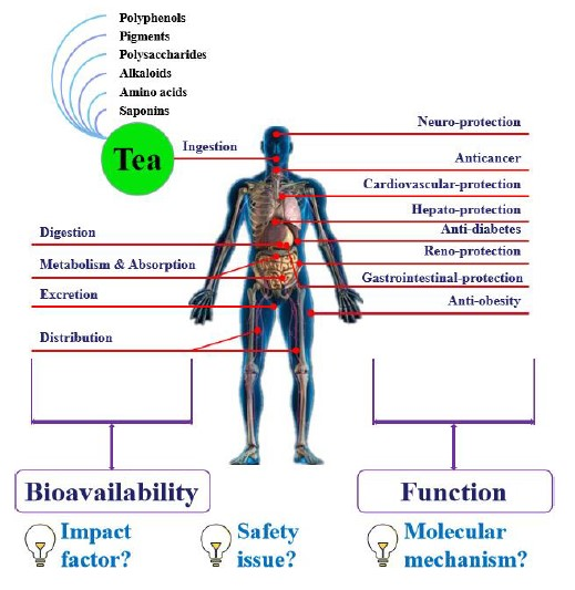
1. Introduction
2. Bioactive Components
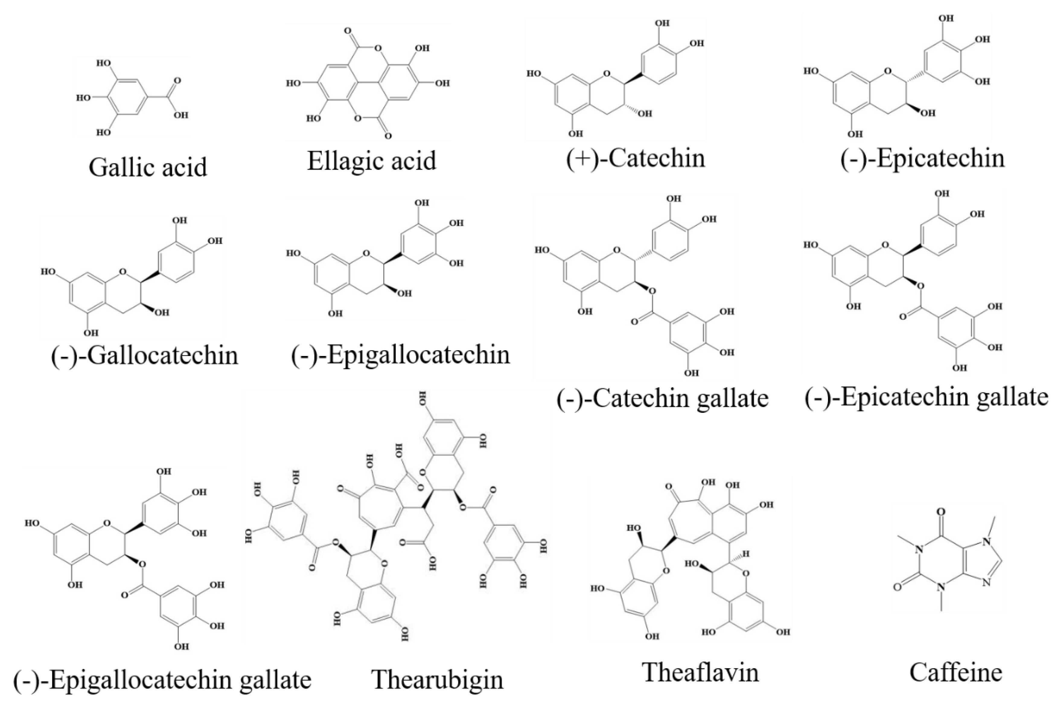
2.1. Polyphenols
2.2. Pigments
2.3. Polysaccharides
2.4. Alkaloids
2.5. Amino Acids
2.6. Saponins
3. Bioavailability
3.1. Bioavailability of Tea Polyphenols
3.2. Strategies to Increase Tea Polyphenol Bioavailability
3.3. Factors That Reduce Tea Polyphenol Bioavailability
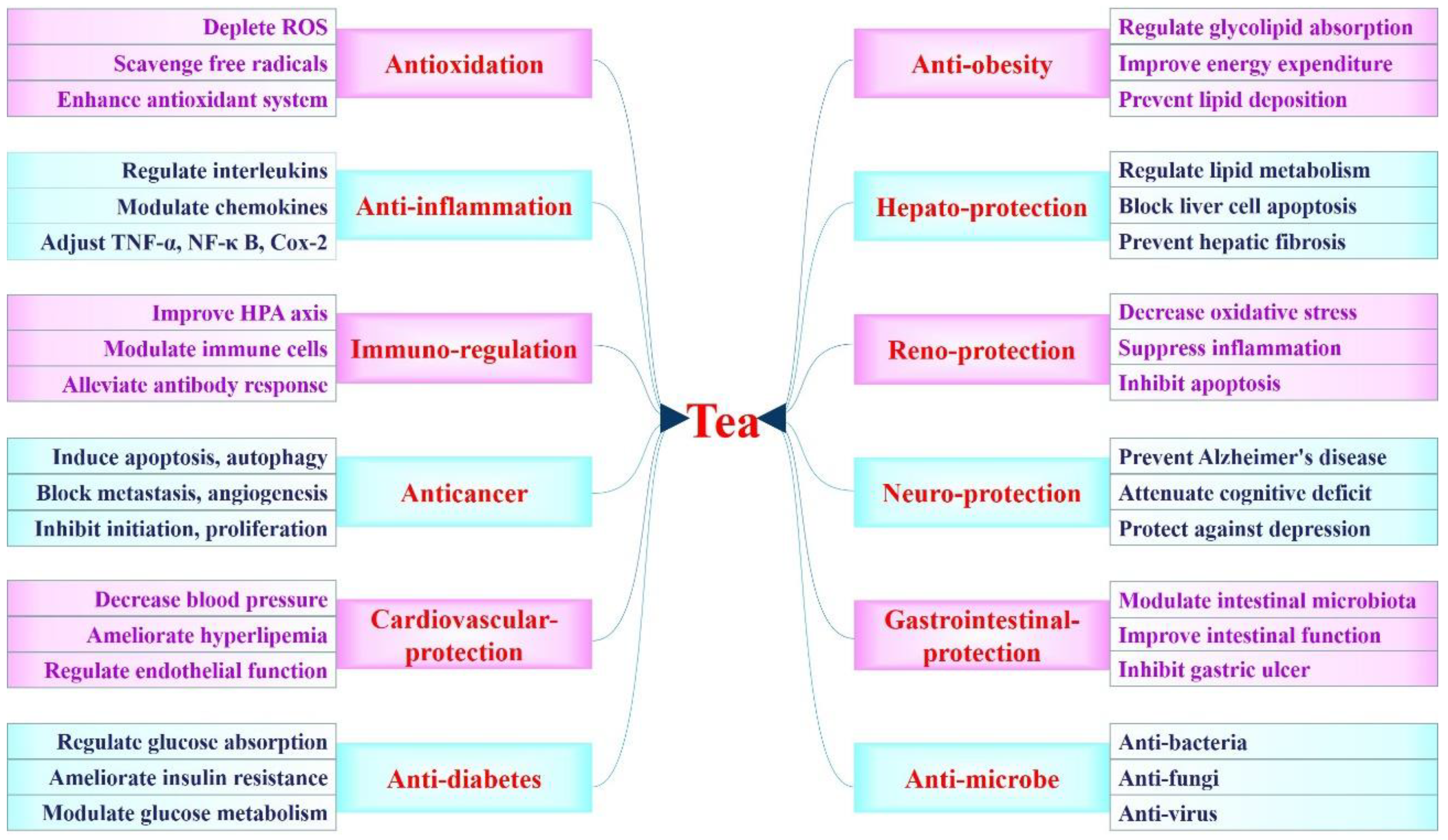
4. Health Functions
4.1. Antioxidant Activity
4.1.1. Antioxidant Activity In Vitro
4.1.2. Antioxidant Activity In Vivo
4.1.3. Antioxidant Activity in Humans
4.2. Anti-Inflammatory Activity
4.2.1. Anti-Inflammatory Activity In Vitro
4.2.2. Anti-Inflammatory Activity In Vivo
4.2.3. Anti-Inflammatory Activity in Humans
4.3. Immuno-Regulatory Activity
4.3.1. Immuno-Regulative Activity In Vitro
4.3.2. Immuno-Regulative Activity In Vivo
4.3.3. Immuno-Regulative Activity in Humans
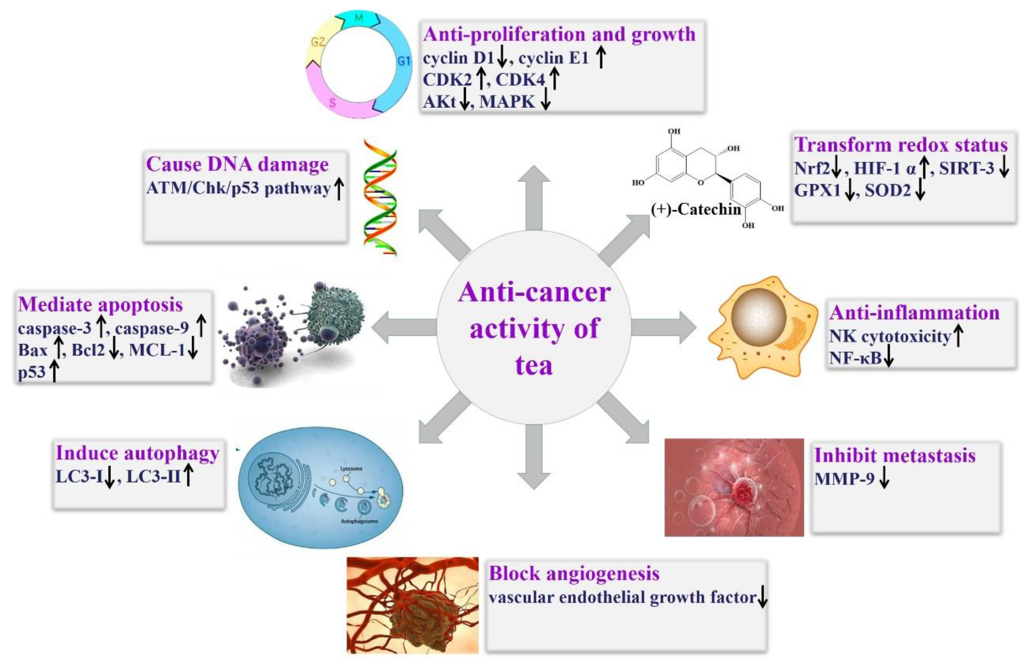
4.4. Anticancer Effect
4.4.1. Anticancer Effect In Vitro
4.4.2. Anticancer Effect In Vivo
4.4.3. Anticancer Effect in Humans
4.4.4. Strategy to Improve Anticancer Effect of Tea and Its Component
4.5. Cardiovascular-Protective Effect
4.5.1. Cardiovascular-Protective Effect In Vitro
4.5.2. Cardiovascular-Protective Effect In Vivo
4.5.3. Cardiovascular-Protective Effect in Humans
4.6. Anti-Diabetic Effect
4.6.1. Anti-Diabetic Effect In Vitro
4.6.2. Anti-Diabetic Effect In Vivo
4.6.3. Anti-Diabetic Effect in Humans
4.7. Anti-Obesity Effect
4.7.1. Anti-Obesity Effect In Vitro
4.7.2. Anti-Obesity Effect In Vivo
4.7.3. Anti-Obesity Effect in Humans
4.8. Hepato-Protective Effect
4.8.1. Hepato-Protective Effect In Vitro
4.8.2. Hepato-Protective Effect In Vivo
4.8.3. Hepato-Protective Effect in Humans
4.9. Other Health Functions
5. Potential Safety Issues
6. Conclusions
Author Contributions
Funding
Conflicts of Interest
References
- Guo, Y.J.; Sun, L.Q.; Yu, B.Y.; Qi, J. An integrated antioxidant activity fingerprint for commercial teas based on their capacities to scavenge reactive oxygen species. Food Chem. 2017, 237, 645–653. [Google Scholar] [CrossRef]
- Islam, S.N.; Farooq, S.; Sehgal, A. Effect of consecutive steeping on antioxidant potential of green, oolong and black tea. Int. J. Food Sci. Technol. 2018, 53, 182–187. [Google Scholar] [CrossRef]
- Kujawska, M.; Ewertowska, M.; Ignatowicz, E.; Adamska, T.; Szaefer, H.; Gramza-Michalowska, A.; Korczak, J.; Jodynis-Liebert, J. Evaluation of safety and antioxidant activity of yellow tea (Camellia sinensis) extract for application in food. J. Med. Food 2016, 19, 330–336. [Google Scholar] [CrossRef]
- Lv, H.P.; Zhang, Y.; Shi, J.; Lin, Z. Phytochemical profiles and antioxidant activities of Chinese dark teas obtained by different processing technologies. Food Res. Int. 2017, 100, 486–493. [Google Scholar] [CrossRef]
- Sanlier, N.; Atik, I.; Atik, A. A minireview of effects of white tea consumption on diseases. Trends Food Sci. Technol. 2018, 82, 82–88. [Google Scholar] [CrossRef]
- Hilal, Y.; Engelhardt, U. Characterisation of white tea - Comparison to green and black tea. J. Consum. Prot. Food Saf. 2007, 2, 414–421. [Google Scholar] [CrossRef]
- Zheng, W.J.; Wan, X.C.; Bao, G.H. Brick dark tea: A review of the manufacture, chemical constituents and bioconversion of the major chemical components during fermentation. Phytochem. Rev. 2015, 14, 499–523. [Google Scholar] [CrossRef]
- Bi, W.; He, C.N.; Ma, Y.Y.; Shen, J.; Zhang, L.H.; Peng, Y.; Xiao, P.G. Investigation of free amino acid, total phenolics, antioxidant activity and purine alkaloids to assess the health properties of non-Camellia tea. Acta Pharm. Sin. B 2016, 6, 170–181. [Google Scholar] [CrossRef] [Green Version]
- Guo, L.; Guo, J.C.; Zhu, W.C.; Jiang, X.R. Optimized synchronous extraction process of tea polyphenols and polysaccharides from Huaguoshan Yunwu tea and their antioxidant activities. Food Bioprod. Process. 2016, 100, 303–310. [Google Scholar] [CrossRef]
- Pan, H.B.; Wang, F.; Rankin, G.O.; Rojanasakul, Y.; Tu, Y.Y.; Chen, Y.C. Inhibitory effect of black tea pigments, theaflavin-3/3’-gallate against cisplatin-resistant ovarian cancer cells by inducing apoptosis and G1 cell cycle arrest. Int. J. Oncol. 2017, 51, 1508–1520. [Google Scholar] [CrossRef] [Green Version]
- Wang, B.; Tu, Y.; Zhao, S.P.; Hao, Y.H.; Liu, J.X.; Liu, F.H.; Xiong, B.H.; Jiang, L.S. Effect of tea saponins on milk performance, milk fatty acids, and immune function in dairy cow. J. Dairy Sci. 2017, 100, 8043–8052. [Google Scholar] [CrossRef] [Green Version]
- Tang, G.Y.; Zhao, C.N.; Xu, X.Y.; Gan, R.Y.; Cao, S.Y.; Liu, Q.; Shang, A.; Mao, Q.Q.; Li, H.B. Phytochemical composition and antioxidant capacity of 30 Chinese teas. Antioxidants 2019, 8, 180. [Google Scholar] [CrossRef] [Green Version]
- Zhao, C.N.; Tang, G.Y.; Cao, S.Y.; Xu, X.Y.; Gan, R.Y.; Liu, Q.; Mao, Q.Q.; Shang, A.; Li, H.B. Phenolic profiles and antioxidant activities of 30 tea infusions from green, black, oolong, white, yellow and dark teas. Antioxidants 2019, 8, 215. [Google Scholar] [CrossRef] [Green Version]
- Di Lorenzo, A.; Curti, V.; Tenore, G.C.; Nabavi, S.M.; Daglia, M. Effects of tea and coffee consumption on cardiovascular diseases and relative risk factors: An update. Curr. Pharm. Design 2017, 23, 2474–2487. [Google Scholar] [CrossRef]
- Gan, R.Y.; Li, H.B.; Sui, Z.Q.; Corke, H. Absorption, metabolism, anticancer effect and molecular targets of epigallocatechin gallate (EGCG): An updated review. Crit. Rev. Food Sci. Nutr. 2018, 58, 924–941. [Google Scholar] [CrossRef]
- Li, Y.C.; Wang, C.; Huai, Q.J.; Guo, F.C.; Liu, L.Y.; Feng, R.N.; Sun, C.H. Effects of tea or tea extract on metabolic profiles in patients with type 2 diabetes mellitus: A meta-analysis of ten randomized controlled trials. Diabetes Metab. Res. 2016, 32, 2–10. [Google Scholar] [CrossRef]
- Ramadan, G.; El-Beih, N.M.; Talaat, R.M.; Abd El-Ghffar, E.A. Anti-inflammatory activity of green versus black tea aqueous extract in a rat model of human rheumatoid arthritis. Int. J. Rheum. Dis. 2017, 20, 203–213. [Google Scholar] [CrossRef]
- Santamarina, A.B.; Carvalho-Silva, M.; Gomes, L.M.; Okuda, M.H.; Santana, A.A.; Streck, E.L.; Seelaender, M.; Do Nascimento, C.; Ribeiro, E.B.; Lira, F.S.; et al. Decaffeinated green tea extract rich in epigallocatechin-3-gallate prevents fatty liver disease by increased activities of mitochondrial respiratory chain complexes in diet-induced obesity mice. J. Nutr. Biochem. 2015, 26, 1348–1356. [Google Scholar] [CrossRef] [Green Version]
- Suzuki, T.; Pervin, M.; Goto, S.; Isemura, M.; Nakamura, Y. Beneficial effects of tea and the green tea catechin epigallocatechin-3-gallate on obesity. Molecules 2016, 21, 1305. [Google Scholar] [CrossRef] [Green Version]
- Cai, Z.Y.; Li, X.M.; Liang, J.P.; Xiang, L.P.; Wang, K.R.; Shi, Y.L.; Yang, R.; Shi, M.; Ye, J.H.; Lu, J.L.; et al. Bioavailability of tea catechins and its improvement. Molecules 2018, 23, 2346. [Google Scholar] [CrossRef] [Green Version]
- Henning, S.M.; Niu, Y.T.; Lee, N.H.; Thames, G.D.; Minutti, R.R.; Wang, H.J.; Go, V.; Heber, D. Bioavailability and antioxidant activity of tea flavanols after consumption of green tea, black tea, or a green tea extract supplement. Am. J. Clin. Nutr. 2004, 80, 1558–1564. [Google Scholar] [CrossRef]
- Kulandaivelu, K.; Mandal, A. Improved bioavailability and pharmacokinetics of tea polyphenols by encapsulation into gelatin nanoparticles. IET Nanobiotechnol. 2017, 11, 469–476. [Google Scholar] [CrossRef]
- Peng, Y.R.; Meng, Q.L.; Zhou, J.; Chen, B.; Xi, J.J.; Long, P.P.; Zhang, L.; Hou, R.Y. Nanoemulsion delivery system of tea polyphenols enhanced the bioavailability of catechins in rats. Food Chem. 2018, 242, 527–532. [Google Scholar] [CrossRef]
- Zou, L.Q.; Liu, W.; Liu, W.L.; Liang, R.H.; Li, T.; Liu, C.M.; Cao, Y.L.; Niu, J.; Liu, Z. Characterization and bioavailability of tea polyphenol nanoliposome prepared by combining an ethanol injection method with dynamic High-Pressure microfluidization. J. Agric. Food Chem. 2014, 62, 934–941. [Google Scholar] [CrossRef]
- Cladiere, M.; Delaporte, G.; Le Roux, E.; Camel, V. Multi-class analysis for simultaneous determination of pesticides, mycotoxins, process-induced toxicants and packaging contaminants in tea. Food Chem. 2018, 242, 113–121. [Google Scholar] [CrossRef]
- Dekant, W.; Fujii, K.; Shibata, E.; Morita, O.; Shimotoyodome, A. Safety assessment of green tea based beverages and dried green tea extracts as nutritional supplements. Toxicol. Lett. 2017, 277, 104–108. [Google Scholar] [CrossRef]
- Gorur, F.K.; Keser, R.; Akcay, N.; Dizman, S.; Okumusoglu, N.T. Radionuclides and heavy metals concentrations in Turkish market tea. Food Control 2011, 22, 2065–2070. [Google Scholar] [CrossRef]
- Sarma, D.N.; Barrett, M.L.; Chavez, M.L.; Gardiner, P.; Ko, R.; Mahady, G.B.; Marles, R.J.; Pellicore, L.S.; Giancaspro, G.I.; Dog, T.L. Safety of green tea extracts - a systematic review by the US Pharmacopeia. Drug Saf. 2008, 31, 469–484. [Google Scholar] [CrossRef]
- Turkozu, D.; Sanlier, N. L-theanine, unique amino acid of tea, and its metabolism, health effects, and safety. Crit. Rev. Food Sci. Nutr. 2017, 57, 1681–1687. [Google Scholar] [CrossRef]
- Luca, V.S.; Stan, A.M.; Trifan, A.; Miron, A.; Aprotosoaie, A.C. Catechins profile, caffeine content and antioxidant activity of camellia sinensis teas commercialized in romania. Med. Sur. J. Revista Medico-Chirurgicala 2016, 120, 457–463. [Google Scholar]
- Satoh, T.; Fujisawa, H.; Nakamura, A.; Takahashi, N.; Watanabe, K. Inhibitory effects of eight green tea catechins on cytochrome p450 1A2, 2C9, 2D6, and 3A4 activities. J. Pharm. Pharm. Sci. 2016, 19, 188–197. [Google Scholar] [CrossRef] [PubMed] [Green Version]
- Yang, H.; Xue, X.J.; Li, H.; Apandi, S.N.; Tay-Chan, S.C.; Ong, S.P.; Tian, E.F. The relative antioxidant activity and steric structure of green tea catechins—A kinetic approach. Food Chem. 2018, 257, 399–405. [Google Scholar] [CrossRef] [PubMed]
- Bai, W.X.; Wang, C.; Wang, Y.J.; Zheng, W.J.; Wang, W.; Wan, X.C.; Bao, G.H. Novel acylated flavonol tetraglycoside with inhibitory effect on lipid accumulation in 3T3-L1 cells from Lu’an GuaPian tea and quantification of flavonoid glycosides in six major processing types of tea. J. Agric. Food Chem. 2017, 65, 2999–3005. [Google Scholar] [CrossRef] [PubMed]
- Chen, G.H.; Lin, Y.L.; Hsu, W.L.; Hsieh, S.K.; Tzen, J. Significant elevation of antiviral activity of strictinin from Pu’er tea after thermal degradation to ellagic acid and gallic acid. J. Food Drug Anal. 2015, 23, 116–123. [Google Scholar] [CrossRef] [PubMed] [Green Version]
- Zielinski, A.; Granato, D.; Alberti, A.; Nogueira, A.; Demiate, I.M.; Haminiuk, C. Modelling the extraction of phenolic compounds and in vitro antioxidant activity of mixtures of green, white and black teas (Camellia sinensis L. Kuntze). J. Food Sci. Technol. Mys. 2015, 52, 6966–6977. [Google Scholar] [CrossRef]
- De Oliveira, C.C.; Calado, V.; Ares, G.; Granato, D. Statistical approaches to assess the association between phenolic compounds and the in vitro antioxidant activity of Camellia sinensis and Ilex paraguariensis Teas. Crit. Rev. Food Sci. Nutr. 2015, 55, 1456–1473. [Google Scholar] [CrossRef]
- Koch, W.; Kukula-Koch, W.; Glowniak, K. Catechin composition and antioxidant activity of black teas in relation to brewing time. J. AOAC Int. 2017, 100, 1694–1699. [Google Scholar] [CrossRef]
- Tang, P.; Shen, D.Y.; Xu, Y.Q.; Zhang, X.C.; Shi, J.; Yin, J.F. Effect of fermentation conditions and plucking standards of tea leaves on the chemical components and sensory quality of fermented juice. J. Chem. 2018, 2018, 4312875. [Google Scholar] [CrossRef]
- Sakakibara, H.; Honda, Y.; Nakagawa, S.; Ashida, H.; Kanazawa, K. Simultaneous determination of all polyphenols in vegetables, fruits, and teas. J. Agric. Food Chem. 2003, 51, 571–581. [Google Scholar] [CrossRef]
- Bhattacharya, U.; Mukhopadhyay, S.; Giri, A.K. Comparative antimutagenic and anticancer activity of three fractions of black tea polyphenols thearubigins. Nutr. Cancer 2011, 63, 1122–1132. [Google Scholar] [CrossRef]
- Weerawatanakorn, M.; Lee, Y.L.; Tsai, C.Y.; Lai, C.S.; Wan, X.C.; Ho, C.T.; Li, S.M.; Pan, M.H. Protective effect of theaflavin-enriched black tea extracts against dimethylnitrosamine-induced liver fibrosis in rats. Food Funct. 2015, 6, 1832–1840. [Google Scholar] [CrossRef] [PubMed]
- Xiao, J.B.; Jiang, H.X. A review on the structure-function relationship aspect of polysaccharides from tea materials. Crit. Rev. Food Sci. Nutr. 2015, 55, 930–938. [Google Scholar] [CrossRef] [PubMed]
- Fan, M.H.; Sun, X.; Qian, Y.L.; Xu, Y.; Wang, D.F.; Cao, Y.P. Effects of metal ions in tea polysaccharides on their in vitro antioxidant activity and hypoglycemic activity. Int. J. Biol. Macromol. 2018, 113, 418–426. [Google Scholar] [CrossRef]
- Park, H.R.; Hwang, D.; Suh, H.J.; Yu, K.W.; Kim, T.Y.; Shin, K.S. Antitumor and antimetastatic activities of rhamnogalacturonan-II-type polysaccharide isolated from mature leaves of green tea via activation of macrophages and natural killer cells. Int. J. Biol. Macromol. 2017, 99, 179–186. [Google Scholar] [CrossRef] [PubMed]
- Sun, L.J.; Warren, F.J.; Gidley, M.J. Soluble polysaccharides reduce binding and inhibitory activity of tea polyphenols against porcine pancreatic α-amylase. Food Hydrocoll. 2018, 79, 63–70. [Google Scholar] [CrossRef] [Green Version]
- Wang, Y.F.; Li, Y.F.; Liu, Y.Y.; Chen, X.Q.; Wei, X.L. Extraction, characterization and antioxidant activities of Se-enriched tea polysaccharides. Int. J. Biol. Macromol. 2015, 77, 76–84. [Google Scholar] [CrossRef]
- Wang, H.J.; Shi, S.S.; Bao, B.; Li, X.J.; Wang, S.C. Structure characterization of an arabinogalactan from green tea and its anti-diabetic effect. Carbohyd. Polym. 2015, 124, 98–108. [Google Scholar] [CrossRef]
- Xu, Y.; Zhang, M.; Wu, T.; Dai, S.D.; Xu, J.L.; Zhou, Z.K. The anti-obesity effect of green tea polysaccharides, polyphenols and caffeine in rats fed with a high-fat diet. Food Funct. 2015, 6, 297–304. [Google Scholar] [CrossRef]
- Yang, X.H.; Huang, M.J.; Qin, C.Q.; Lv, B.Y.; Mao, Q.L.; Liu, Z.H. Structural characterization and evaluation of the antioxidant activities of polysaccharides extracted from Qingzhuan brick tea. Int. J. Biol. Macromol. 2017, 101, 768–775. [Google Scholar] [CrossRef]
- Yuan, C.F.; Li, Z.H.; Peng, F.; Xiao, F.X.; Ren, D.M.; Xue, H.; Chen, T.; Mushtaq, G.; Kamal, M.A. Combination of selenium-enriched green tea polysaccharides and Huo-ji polysaccharides synergistically enhances antioxidant and immune activity in mice. J. Sci. Food Agric. 2015, 95, 3211–3217. [Google Scholar] [CrossRef]
- Li, X.; Liu, G.J.; Zhang, W.; Zhou, Y.L.; Ling, T.J.; Wan, X.C.; Bao, G.H. Novel flavoalkaloids from white tea with inhibitory activity against the formation of advanced glycation end products. J. Agric. Food Chem. 2018, 66, 4621–4629. [Google Scholar] [CrossRef] [PubMed]
- Zhu, Y.C.; Luo, Y.H.; Wang, P.P.; Zhao, M.Y.; Li, L.; Hu, X.S.; Chen, F. Simultaneous determination of free amino acids in Pu-erh tea and their changes during fermentation. Food Chem. 2016, 194, 643–649. [Google Scholar] [CrossRef] [PubMed]
- Song, C.W.; Yu, Q.S.; Li, X.H.; Jin, S.N.; Li, S.; Zhang, Y.; Jia, S.L.; Chen, C.; Xiang, Y.; Jiang, H.L. The hypolipidemic effect of total saponins from kuding tea in high-fat diet-induced hyperlipidemic mice and its composition characterized by UPLC-QTOF-MS/MS. J. Food Sci. 2016, 81, H1313–H1319. [Google Scholar] [CrossRef] [PubMed]
- Yuan, C.X.; Li, Y.; Li, Q.C.; Jin, R.S.; Ren, L.L. Purification of tea saponins and evaluation of its effect on alcohol dehydrogenase activity. Open Life Sci. 2018, 13, 56–63. [Google Scholar] [CrossRef] [Green Version]
- Xu, D.P.; Li, Y.; Meng, X.; Zhou, T.; Zhou, Y.; Zheng, J.; Zhang, J.J.; Li, H.B. Natural antioxidants in foods and medicinal plants: Extraction, assessment and resources. Int. J. Mol. Sci. 2017, 18, 96. [Google Scholar] [CrossRef]
- Jiang, X.D.; Feng, K.J.; Yang, X.P. In vitro antifungal activity and mechanism of action of tea polyphenols and tea saponin against Rhizopus stolonifer. J. Mol. Microb. Biotech. 2015, 25, 269–276. [Google Scholar] [CrossRef] [PubMed]
- Lin, S.; Chen, Y.X.; Bai, Y.; Cai, H.J.; Wei, H.; Tian, H.J.; Zhao, J.W.; Chen, Y.; Yang, G.; Gu, X.J.; et al. Effect of tea saponin-treated host plants on activities of antioxidant enzymes in larvae of the diamondback moth Plutella Xylostella (Lepidoptera: Plutellidae). Environ. Entomol. 2018, 47, 749–754. [Google Scholar] [CrossRef]
- Xu, J.Z.; Yeung, S.; Chang, Q.; Huang, Y.; Chen, Z.Y. Comparison of antioxidant activity and bioavailability of tea epicatechins with their epimers. Br. J. Nutr. 2004, 91, 873–881. [Google Scholar]
- Zhu, M.; Chen, Y.; Li, R.C. Oral absorption and bioavailability of tea catechins. Planta Med. 2000, 66, 444–447. [Google Scholar] [CrossRef]
- Chen, L.S.; Lee, M.J.; Li, H.; Yang, C.S. Absorption, distribution, and elimination of tea polyphenols in rats. Drug Metab. Dispos. 1997, 25, 1045–1050. [Google Scholar]
- Sun, H.Y.; Chen, Y.H.; Cheng, M.; Zhang, X.; Zheng, X.J.; Zhang, Z.C. The modulatory effect of polyphenols from green tea, oolong tea and black tea on human intestinal microbiota in vitro. J. Food Sci. Technol. Mys. 2018, 55, 399–407. [Google Scholar] [CrossRef] [PubMed]
- De Oliveira, D.M.; Sampaio, G.R.; Pinto, C.B.; Catharino, R.R.; Bastos, D. Bioavailability of chlorogenic acids in rats after acute ingestion of mat, tea (Ilex paraguariensis) or 5-caffeoylquinic acid. Eur. J. Nutr. 2017, 56, 2541–2556. [Google Scholar] [CrossRef] [PubMed]
- Del Rio, D.; Stalmach, A.; Calani, L.; Crozier, A. Bioavailability of coffee chlorogenic acids and green tea flavan-3-ols. Nutrients 2010, 2, 820–833. [Google Scholar] [CrossRef] [PubMed]
- Del Rio, D.; Calani, L.; Cordero, C.; Salvatore, S.; Pellegrini, N.; Brighenti, F. Bioavailability and catabolism of green tea flavan-3-ols in humans. Nutrition 2010, 26, 1110–1116. [Google Scholar] [CrossRef] [PubMed]
- Calani, L.; Del Rio, D.; Callegari, M.L.; Morelli, L.; Brighenti, F. Updated bioavailability and 48 h excretion profile of flavan-3-ols from green tea in humans. Int. J. Food Sci. Nutr. 2012, 63, 513–521. [Google Scholar] [CrossRef]
- Shahrzad, S.; Aoyagi, K.; Winter, A.; Koyama, A.; Bitsch, I. Pharmacokinetics of gallic acid and its relative bioavailability from tea in healthy humans. J. Nutr. 2001, 131, 1207–1210. [Google Scholar] [CrossRef]
- Wiseman, S.; Mulder, T.; Rietveld, A. Tea flavonoids: Bioavailability in vivo and effects on cell signaling pathways in vitro. Antioxid. Redox Sign. 2001, 3, 1009–1021. [Google Scholar] [CrossRef]
- Catterall, F.; King, L.J.; Clifford, M.N.; Ioannides, C. Bioavailability of dietary doses of H-3-labelled tea antioxidants (+)-catechin and (-)-epicatechin in rat. Xenobiotica 2003, 33, 743–753. [Google Scholar] [CrossRef]
- Del Rio, D.; Calani, L.; Scazzina, F.; Jechiu, L.; Cordero, C.; Brighenti, F. Bioavailability of catechins from ready-to-drink tea. Nutrition 2010, 26, 528–533. [Google Scholar] [CrossRef]
- Zhao, D.Y.; Shah, N.P. Concomitant ingestion of lactic acid bacteria and black tea synergistically enhances flavonoid bioavailability and attenuates D-galactose-induced oxidative stress in mice via modulating glutathione antioxidant system. J. Nutr. Biochem. 2016, 38, 116–124. [Google Scholar] [CrossRef]
- Choi, E.H.; Lee, D.Y.; Kim, S.; Chung, J.O.; Choi, J.K.; Joo, K.M.; Jeong, H.W.; Kim, J.K.; Kim, W.G.; Shim, S.M. Influence of flavonol-rich excipient food (onion peel and Dendropanax morbifera) on the bioavailability of green tea epicatechins in vitro and in vivo. Food Funct. 2017, 8, 3664–3674. [Google Scholar] [CrossRef] [PubMed]
- Kale, A.; Gawande, S.; Kotwal, S.; Netke, S.; Roomi, W.; Ivanov, V.; Niedzwiecki, A.; Rath, M. Studies on the effects of oral administration of nutrient mixture, quercetin and red onions on the bioavailability of epigallocatechin gallate from green tea extract. Phytother. Res. 2010, 241, S48–S55. [Google Scholar] [CrossRef] [PubMed]
- Lambert, J.D.; Hong, J.G.; Kim, D.H.; Mishin, V.M.; Yang, C.S. Piperine enhances the bioavailability of the tea polyphenol (-)-epigallocatechin-3-gallate in mice. J. Nutr. 2004, 134, 1948–1952. [Google Scholar] [CrossRef] [PubMed] [Green Version]
- Peters, C.M.; Green, R.J.; Janle, E.M.; Ferruzzi, M.G. Formulation with ascorbic acid and sucrose modulates catechin bioavailability from green tea. Food Res. Int. 2010, 43, 95–102. [Google Scholar] [CrossRef] [PubMed] [Green Version]
- Wang, P.W.; Heber, D.; Henning, S.M. Quercetin increased bioavailability and decreased methylation of green tea polyphenols in vitro and in vivo. Food Funct. 2012, 3, 635–642. [Google Scholar] [CrossRef] [PubMed] [Green Version]
- Naumovski, N.; Blades, B.L.; Roach, P.D. Food inhibits the oral bioavailability of the major green tea antioxidant epigallocatechin gallate in humans. Antioxidants 2015, 4, 373–393. [Google Scholar] [CrossRef]
- Chow, H.; Hakim, I.A.; Vining, D.R.; Crowel, J.A.; Ranger-Moore, J.; Chew, W.M.; Celaya, C.A.; Rodney, S.R.; Hara, Y.; Alberts, D.S. Effects of dosing condition on the oral bioavailability of green tea catechins after single-dose administration of Polyphenon E in healthy individuals. Clin. Cancer Res. 2005, 11, 4627–4633. [Google Scholar] [CrossRef] [Green Version]
- Egert, S.; Tereszczuk, J.; Wein, S.; Muller, M.J.; Frank, J.; Rimbach, G.; Wolffram, S. Simultaneous ingestion of dietary proteins reduces the bioavailability of galloylated catechins from green tea in humans. Eur. J. Nutr. 2013, 52, 281–288. [Google Scholar] [CrossRef]
- James, K.D.; Forester, S.C.; Lambert, J.D. Dietary pretreatment with green tea polyphenol, (-)-epigallocatechin-3-gallate reduces the bioavailability and hepatotoxicity of subsequent oral bolus doses of (-)-epigallocatechin-3-gallate. Food Chem. Toxicol. 2015, 76, 103–108. [Google Scholar] [CrossRef] [Green Version]
- Zeng, L.; Luo, L.Y.; Li, H.J.; Liu, R.H. Phytochemical profiles and antioxidant activity of 27 cultivars of tea. Int. J. Food Sci. Nutr. 2017, 68, 525–537. [Google Scholar] [CrossRef]
- Liu, S.M.; Huang, H.H. Assessments of antioxidant effect of black tea extract and its rationals by erythrocyte haemolysis assay, plasma oxidation assay and cellular antioxidant activity (CAA) assay. J. Funct. Foods 2015, 18, 1095–1105. [Google Scholar] [CrossRef]
- Peluso, I.; Manafikhi, H.; Raguzzini, A.; Longhitano, Y.; Reggi, R.; Zanza, C.; Palmery, M. The peroxidation of leukocytes index ratio reveals the prooxidant effect of green tea extract. Oxid. Med. Cell Longev. 2016, 2016, 9139731. [Google Scholar] [CrossRef] [PubMed] [Green Version]
- Fei, T.Y.; Fei, J.; Huang, F.; Xie, T.P.; Xu, J.F.; Zhou, Y.; Yang, P. The anti-aging and anti-oxidation effects of tea water extract in Caenorhabditis elegans. Exp. Gerontol. 2017, 97, 89–96. [Google Scholar] [CrossRef] [PubMed]
- Bartikova, H.; Skalova, L.; Valentova, K.; Matouskova, P.; Szotakova, B.; Martin, J.; Kvita, V.; Bousova, I. Effect of oral administration of green tea extract in various dosage schemes on oxidative stress status of mice in vivo. Acta Pharmaceut. 2015, 65, 65–73. [Google Scholar] [CrossRef] [PubMed] [Green Version]
- Jowko, E.; Dlugolecka, B.; Makaruk, B.; Cieslinski, I. The effect of green tea extract supplementation on exercise-induced oxidative stress parameters in male sprinters. Eur. J. Nutr. 2015, 54, 783–791. [Google Scholar] [CrossRef] [PubMed] [Green Version]
- Megow, I.; Darvin, M.E.; Meinke, M.C.; Lademann, J. A randomized controlled trial of green tea beverages on the in vivo radical scavenging activity in human skin. Skin Pharmacol. Phys. 2017, 30, 225–233. [Google Scholar] [CrossRef]
- Venkatakrishnan, K.; Chiu, H.F.; Cheng, J.C.; Chang, Y.H.; Lu, Y.Y.; Han, Y.C.; Shen, Y.C.; Tsai, K.S.; Wang, C.K. Comparative studies on the hypolipidemic, antioxidant and hepatoprotective activities of catechin-enriched green and oolong tea in a double-blind clinical trial. Food Funct. 2018, 9, 1205–1213. [Google Scholar] [CrossRef]
- Hamer, M. The beneficial effects of tea on immune function and inflammation: A review of evidence from in vitro, animal, and human research. Nutr. Res. 2007, 27, 373–379. [Google Scholar] [CrossRef]
- Cyboran, S.; Strugala, P.; Wloch, A.; Oszmianski, J.; Kleszczynska, H. Concentrated green tea supplement: Biological activity and molecular mechanisms. Life Sci. 2015, 126, 1–9. [Google Scholar] [CrossRef]
- Ben Lagha, A.; Grenier, D. Black tea theaflavins attenuate Porphyromonas gingivalis virulence properties, modulate gingival keratinocyte tight junction integrity and exert anti-inflammatory activity. J. Periodontal Res. 2017, 52, 458–470. [Google Scholar] [CrossRef]
- Fechtner, S.; Singh, A.; Chourasia, M.; Ahmed, S. Molecular insights into the differences in anti-inflammatory activities of green tea catechins on IL-1 beta signaling in rheumatoid arthritis synovial fibroblasts. Toxicol. Appl. Pharm. 2017, 329, 112–120. [Google Scholar] [CrossRef] [PubMed]
- Liu, L.X.; Wu, X.Q.; Zhang, B.C.; Yang, W.; Li, D.L.; Dong, Y.Q.; Yin, Y.J.; Chen, Q. Protective effects of tea polyphenols on exhaustive exercise-induced fatigue, inflammation and tissue damage. Food Nutr. Res. 2017, 61, 1333390. [Google Scholar] [CrossRef] [PubMed] [Green Version]
- Scoparo, C.T.; de Souza, L.M.; Rattmann, Y.D.; Kiatkoski, E.C.; Dartora, N.; Iacomini, M. The protective effect of green and black teas (Camellia sinensis) and their identified compounds against murine sepsis. Food Res. Int. 2016, 83, 102–111. [Google Scholar] [CrossRef]
- Shamekhi, Z.; Amani, R.; Habibagahi, Z.; Namjoyan, F.; Ghadiri, A.; Malehi, A.S. A randomized, double-blind, placebo-controlled clinical trial examining the effects of green tea extract on systemic lupus erythematosus disease activity and quality of life. Phytother. Res. 2017, 31, 1063–1071. [Google Scholar] [CrossRef] [PubMed]
- Zhang, T.; Li, L.; Liu, Y.H.; Zhong, D.Q.; Tao, Y.; Jiang, X.J.; Xu, Z.Q. Effect of coffee and green tea on executive ability and plasma levels of inflammatory factors in soldiers with 48-hour total sleep deprivation. Int. J. Clin. Exp. Med. 2016, 9, 19354–19362. [Google Scholar]
- Suzuki, K.; Takahashi, M.; Li, C.Y.; Lin, S.P.; Tomari, M.; Shing, C.M.; Fang, S.H. The acute effects of green tea and carbohydrate coingestion on systemic inflammation and oxidative stress during sprint cycling. Appl. Physiol. Nutr. Metab. 2015, 40, 997–1003. [Google Scholar] [CrossRef]
- Wu, D.Y. Green tea EGCG, T-cell function, and T-cell-mediated autoimmune encephalomyelitis. J. Investig. Med. 2016, 64, 1213–1219. [Google Scholar] [CrossRef]
- Joshi, R.; Rana, A.; Kumar, V.; Kumar, D.; Padwad, Y.S.; Yadav, S.K.; Gulati, A. Anthocyanins enriched purple tea exhibits antioxidant, immunostimulatory and anticancer activities. J. Food Sci. Technol. Mys. 2017, 54, 1953–1963. [Google Scholar] [CrossRef]
- Sharma, R.; Sharma, A.; Kumari, A.; Kulurkar, P.M.; Raj, R.; Gulati, A.; Padwad, Y.S. Consumption of green tea epigallocatechin-3-gallate enhances systemic immune response, antioxidative capacity and HPA axis functions in aged male swiss albino mice. Biogerontology 2017, 18, 367–382. [Google Scholar] [CrossRef]
- Kim, Y.H.; Won, Y.S.; Yang, X.; Kumazoe, M.; Yamashita, S.; Hara, A.; Takagaki, A.; Goto, K.; Nanjo, F.; Tachibana, H. Green tea catechin metabolites exert immunoregulatory effects on CD4+ T cell and natural killer cell activities. J. Agric. Food Chem. 2016, 64, 3591–3597. [Google Scholar] [CrossRef]
- Sil, S.; Bhandari, K.; Gupta, P.; Ghosh, R.; Mitra, A.; Ghosh, B.C.; Ghosh, T. Protective effects of black tea-TV 25 on the cognitive impairments and some peripheral immune responses in intracerebroventricular colchicine injected rats. Orient. Pharm. Exp. Med. 2018, 18, 39–50. [Google Scholar] [CrossRef]
- Ahmed, S.T.; Lee, J.W.; Mun, H.S.; Yang, C.J. Effects of supplementation with green tea by-products on growth performance, meat quality, blood metabolites and immune cell proliferation in goats. J. Anim. Physiol. Anim. Nutr. 2015, 99, 1127–1137. [Google Scholar] [CrossRef] [PubMed]
- EI-Desouky, W.; Hanafi, A.; Abbas, M.M. Radioprotective effect of green tea and grape seed extracts mixture on gamma irradiation induced immune suppression in male albino rats. Int. J. Radiat. Biol. 2017, 93, 433–439. [Google Scholar] [CrossRef] [PubMed]
- Yusni Husni, T.T.; Achmad, T.H. Aktivitas activity of green tea (Camellia sinensis (L) o. Kuntze) polyphenols as immunomodulator through response of suppression immunoglobulin e (IgE) in allergic rhinitis. Majalah Kedokteran Bandung-Mkb-Bandung Med. J. 2015, 47, 160–166. [Google Scholar] [CrossRef] [Green Version]
- Krstic, M.; Stojadinovic, M.; Smiljanic, K.; Stanic-Vucinic, D.; Velickovic, T.C. The anticancer activity of green tea, coffee and cocoa extracts on human cervical adenocarcinoma HeLa cells depends on both pro-oxidant and anti-proliferative activities of polyphenols. RSC Adv. 2015, 5, 3260–3268. [Google Scholar] [CrossRef]
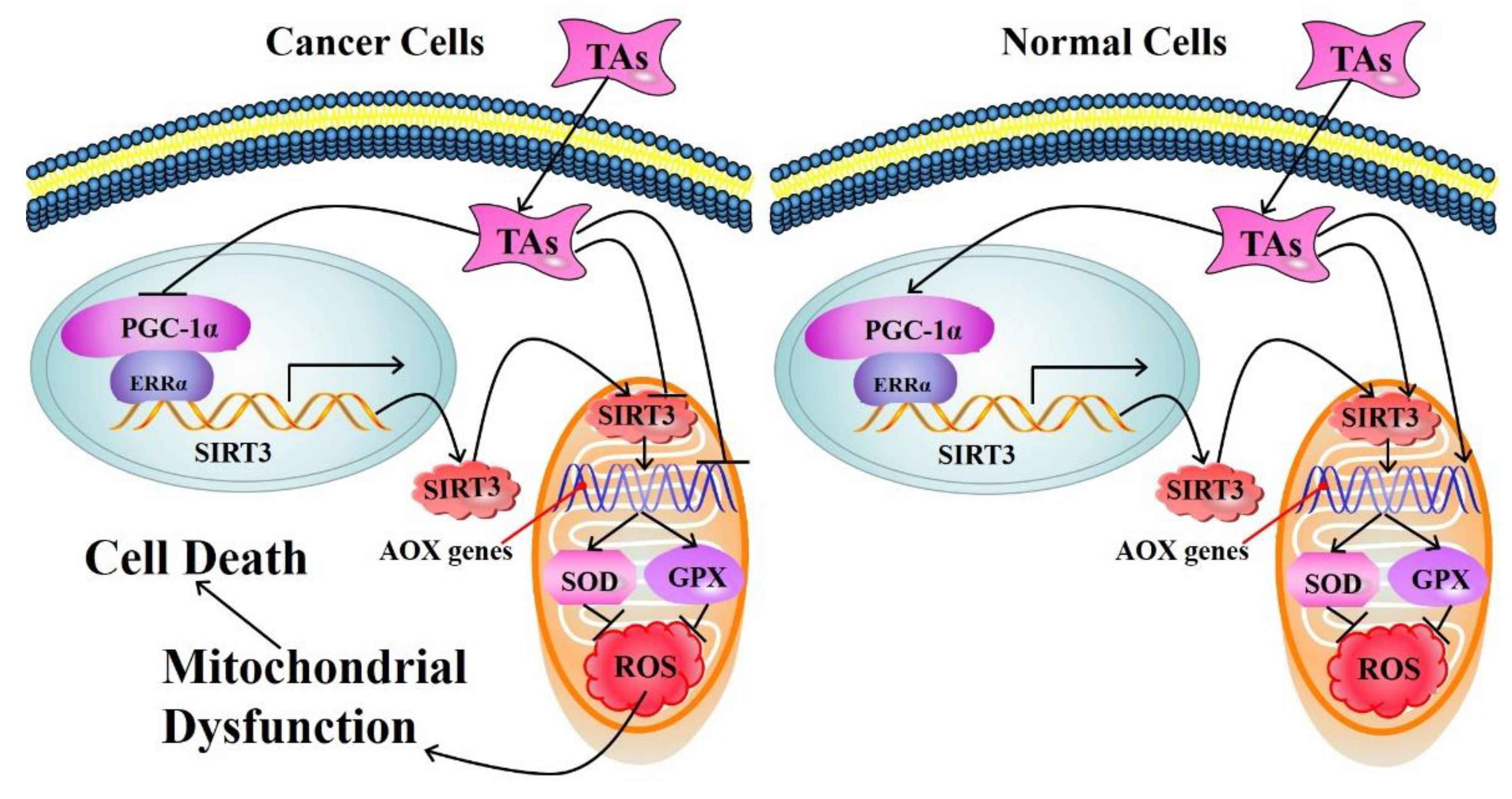
- Tao, L.; Park, J.Y.; Lambert, J.D. Differential prooxidative effects of the green tea polyphenol, (-)-epigallocatechin-3-gallate, in normal and oral cancer cells are related to differences in sirtuin 3 signaling. Mol. Nutr. Food Res. 2015, 59, 203–211. [Google Scholar] [CrossRef] [PubMed]
- Calgarotto, A.K.; Maso, V.; Franchi, G.C.; Nowill, A.E.; Latuf, P.; Vassallo, J.; Saad, S. Antitumor activities of quercetin and green tea in xenografts of human leukemia HL60 cells. Sci. Rep. 2018, 8, 3459. [Google Scholar] [CrossRef] [Green Version]
- Torello, C.O.; Shiraishi, R.N.; Della Via, F.I.; de Castro, T.; Longhini, A.L.; Santos, I.; Bombeiro, A.L.; Silva, C.; Queiroz, M.; Rego, E.M.; et al. Reactive oxygen species production triggers green tea-induced anti-leukaemic effects on acute promyelocytic leukaemia model. Cancer Lett. 2018, 414, 116–126. [Google Scholar] [CrossRef]
- El-Missiry, M.A.; Abdraboh, M.E.; Othman, A.I.; Abdeen, A.M.; Mohsen, N. Apoptogenic effect of green tea polyphenon-60 against ehrlich ascites carcinoma cells in swiss albino mice. Res. J. Pharm. Biol. Chem. Sci. 2016, 7, 3146–3156. [Google Scholar]
- Adami, G.R.; Tangney, C.C.; Tang, J.L.; Zhou, Y.; Ghaffari, S.; Naqib, A.; Sinha, S.; Green, S.J.; Schwartz, J.L. Effects of green tea on miRNA and microbiome of oral epithelium. Sci. Rep. 2018, 8, 5873. [Google Scholar] [CrossRef]
- Lassed, S.; Deus, C.M.; Djebbari, R.; Zama, D.; Oliveira, P.J.; Rizvanov, A.A.; Dahdouh, A.; Benayache, F.; Benayache, S. Protective effect of green tea (Camellia sinensis (L.) kuntze) against prostate cancer: From in vitro data to algerian patients. Evid. Based Compl. Altern. 2017, 2017, 1691568. [Google Scholar] [CrossRef] [PubMed] [Green Version]
- Micali, S.; Territo, A.; Pirola, G.M.; Ferrari, N.; Sighinolfi, M.C.; Martorana, E.; Navarra, M.; Bianchi, G. Effect of green tea catechins in patients with high-grade prostatic intraepithelial neoplasia: Results of a short-term double-blind placebo controlled phase II clinical trial. Archivio Italiano Di Urologia E Andrologia 2017, 89, 197–202. [Google Scholar] [CrossRef] [PubMed] [Green Version]
- Wang, J.Y.; Liu, W.; Chen, Z.Q.; Chen, H.X. Physicochemical characterization of the oolong tea polysaccharides with high molecular weight and their synergistic effects in combination with polyphenols on hepatocellular carcinoma. Biomed. Pharmacother. 2017, 90, 160–170. [Google Scholar] [CrossRef] [PubMed]
- Alshatwi, A.A.; Periasamy, V.S.; Athinarayanan, J.; Elango, R. Synergistic anticancer activity of dietary tea polyphenols and bleomycin hydrochloride in human cervical cancer cell: Caspase-dependent and independent apoptotic pathways. Chem. Biol. Interact. 2016, 247, 1–10. [Google Scholar] [CrossRef] [PubMed]
- Singh, M.; Bhatnagar, P.; Mishra, S.; Kumar, P.; Shukla, Y.; Gupta, K.C. PLGA-encapsulated tea polyphenols enhance the chemotherapeutic efficacy of cisplatin against human cancer cells and mice bearing Ehrlich ascites carcinoma. Int. J. Nanomed. 2015, 10, 6789–6809. [Google Scholar] [CrossRef] [Green Version]
- Mukherjee, S.; Ghosh, S.; Das, D.K.; Chakraborty, P.; Choudhury, S.; Gupta, P.; Adhikary, A.; Dey, S.; Chattopadhyay, S. Gold-conjugated green tea nanoparticles for enhanced anti-tumor activities and hepatoprotection—Synthesis, characterization and in vitro evaluation. J. Nutr. Biochem. 2015, 26, 1283–1297. [Google Scholar] [CrossRef] [PubMed]
- Tsai, Y.J.; Chen, B.H. Preparation of catechin extracts and nanoemulsions from green tea leaf waste and their inhibition effect on prostate cancer cell PC-3. Int. J. Nanomed. 2016, 11, 1907–1926. [Google Scholar]
- Nakachi, K.; Matsuyama, S.; Miyake, S.; Suganuma, M.; Imai, K. Preventive effects of drinking green tea on cancer and cardiovascular disease: Epidemiological evidence for multiple targeting prevention. Biofactors 2000, 13, 49–54. [Google Scholar] [CrossRef]
- Serban, C.; Sahebkar, A.; Antal, D.; Ursoniu, S.; Banach, M. Effects of supplementation with green tea catechins on plasma C-reactive protein concentrations: A systematic review and meta-analysis of randomized controlled trials. Nutrition 2015, 31, 1061–1071. [Google Scholar] [CrossRef]
- Ueshima, H. Explanation for the Japanese paradox: Prevention of increase in coronary heart disease and reduction in stroke. J. Atheroscler. Thromb. 2007, 14, 278–286. [Google Scholar] [CrossRef] [Green Version]
- Zeng, L.; Yan, J.N.; Luo, L.Y.; Zhang, D.Y. Effects of Pu-erh tea aqueous extract (PTAE) on blood lipid metabolism enzymes. Food Funct. 2015, 6, 2008–2016. [Google Scholar] [CrossRef] [PubMed]
- Yang, T.; Koo, M. Inhibitory effect of Chinese green tea on endothelial cell-induced LDL oxidation. Atherosclerosis 2000, 148, 67–73. [Google Scholar] [CrossRef]
- Shibu, M.A.; Kuo, C.H.; Chen, B.C.; Ju, D.T.; Chen, R.J.; Lai, C.H.; Huang, P.J.; Viswanadha, V.P.; Kuo, W.W.; Huang, C.Y. Oolong tea prevents cardiomyocyte loss against hypoxia by attenuating p-JNK mediated hypertrophy and enhancing P-IGF1R, p-akt, and p-Bad(ser136) activity and by fortifying NRF2 antioxidation system. Environ. Toxicol. 2018, 33, 220–233. [Google Scholar] [CrossRef] [PubMed]
- Garcia, M.L.; Pontes, R.B.; Nishi, E.E.; Ibuki, F.K.; Oliveira, V.; Sawaya, A.; Carvalho, P.O.; Nogueira, F.N.; Franco, M.D.; Campos, R.R.; et al. The antioxidant effects of green tea reduces blood pressure and sympathoexcitation in an experimental model of hypertension. J. Hypertens. 2017, 35, 348–354. [Google Scholar] [CrossRef]
- Szulinska, M.; Stepien, M.; Kregielska-Narozna, M.; Suliburska, J.; Skrypnik, D.; Bak-Sosnowska, M.; Kujawska-Luczak, M.; Grzymislawska, M.; Bogdanski, P. Effects of green tea supplementation on inflammation markers, antioxidant status and blood pressure in NaCl-induced hypertensive rat model. Food Nutr. Res. 2017, 61, 1295525. [Google Scholar] [CrossRef] [Green Version]
- Li, S.B.; Li, Y.F.; Mao, Z.F.; Hu, H.H.; Ouyang, S.H.; Wu, Y.P.; Tsoi, B.; Gong, P.; Kurihara, H.; He, R.R. Differing chemical compositions of three teas may explain their different effects on acute blood pressure in spontaneously hypertensive rats. J. Sci. Food Agric. 2015, 95, 1236–1242. [Google Scholar] [CrossRef]
- Xu, P.; Ying, L.; Wu, J.; Kong, D.D.; Wang, Y.F. Safety evaluation and antihyperlipidemia effect of aqueous extracts from fermented puerh tea. Food Funct. 2016, 7, 2667–2674. [Google Scholar] [CrossRef]
- Seo, D.B.; Jeong, H.W.; Kim, Y.J.; Kim, S.; Kim, J.; Lee, J.H.; Joo, K.; Choi, J.K.; Shin, S.S.; Lee, S.J. Fermented green tea extract exhibits hypolipidaemic effects through the inhibition of pancreatic lipase and promotion of energy expenditure. Brit. J. Nutr. 2017, 117, 177–186. [Google Scholar] [CrossRef] [Green Version]
- Nakamura, M.; Miura, S.; Takagaki, A.; Nanjo, F. Hypolipidemic effects of crude green tea polysaccharide on rats, and structural features of tea polysaccharides isolated from the crude polysaccharide. Int. J. Food Sci. Nutr. 2017, 68, 321–330. [Google Scholar] [CrossRef]
- Mao, Y.; Wei, B.Y.; Teng, J.W.; Xia, N.; Zhao, M.M.; Huang, L.; Ye, Y. Polysaccharides from Chinese Liupao dark tea and their protective effect against hyperlipidemia. Int. J. Food Sci. Technol. 2018, 53, 599–607. [Google Scholar] [CrossRef]
- Li, Y.F.; Chang, Y.Q.; Deng, J.; Li, W.X.; Jian, J.; Gao, J.S.; Wan, X.; Gao, H.; Kurihara, H.; Sun, P.H.; et al. Prediction and evaluation of the lipase inhibitory activities of tea polyphenols with 3D-QSAR models. Sci. Rep. 2016, 6, 34387. [Google Scholar] [CrossRef] [Green Version]
- Leung, F.P.; Yung, L.M.; Ngai, C.Y.; Cheang, W.S.; Tian, X.Y.; Lau, C.W.; Zhang, Y.; Liu, J.; Chen, Z.Y.; Bian, Z.X.; et al. Chronic black tea extract consumption improves endothelial function in ovariectomized rats. Eur. J. Nutr. 2016, 55, 1963–1972. [Google Scholar] [CrossRef] [Green Version]
- Jang, H.J.; Ridgeway, S.D.; Kim, J.A. Effects of the green tea polyphenol epigallocatechin-3-gallate on high-fat diet-induced insulin resistance and endothelial dysfunction. Am. J. Physiol.-Endoc. Metab. 2013, 305, E1444–E1451. [Google Scholar] [CrossRef]
- Potenza, M.A.; Marasciulo, F.L.; Tarquinio, M.; Tiravanti, E.; Colantuono, G.; Federici, A.; Kim, J.A.; Quon, M.J.; Montagnani, M. EGCG, a green tea polyphenol, improves endothelial function and insulin sensitivity, reduces blood pressure, and protects against myocardial I/R injury in SHR. Am. J. Physiol. Endoc. Metab. 2007, 292, E1378–E1387. [Google Scholar] [CrossRef]
- Nogueira, L.D.; Neto, J.; Klein, M.; Sanjuliani, A.F. Short-term effects of green tea on blood pressure, endothelial function, and metabolic profile in obese prehypertensive women: A crossover randomized clinical trial. J. Am. Coll. Nutr. 2017, 36, 108–115. [Google Scholar] [CrossRef]
- Woodward, K.A.; Hopkins, N.D.; Draijer, R.; de Graaf, Y.; Low, D.A.; Thijssen, D. Acute black tea consumption improves cutaneous vascular function in healthy middle-aged humans. Clin. Nutr. 2018, 37, 242–249. [Google Scholar] [CrossRef]
- Grassi, D.; Draijer, R.; Schalkwijk, C.; Desideri, G.; D’Angeli, A.; Francavilla, S.; Mulder, T.; Ferri, C. Black tea increases circulating endothelial progenitor cells and improves flow mediated dilatation counteracting deleterious effects from a fat load in hypertensive patients: A randomized controlled study. Nutrients 2016, 8, 727. [Google Scholar] [CrossRef]
- Oyama, J.; Maeda, T.; Kouzuma, K.; Ochiai, R.; Tokimitsu, I.; Higuchi, Y.; Sugano, M.; Makino, N. Green tea catechins improve human forearm endothelial dysfunction and have antiatherosclerotic effects in smokers. Circ. J. 2010, 74, 578–588. [Google Scholar] [CrossRef] [Green Version]
- Lorenz, M.; Rauhut, F.; Hofer, C.; Gwosc, S.; Muller, E.; Praeger, D.; Zimmermann, B.F.; Wernecke, K.D.; Baumann, G.; Stangl, K.; et al. Tea-induced improvement of endothelial function in humans: No role for epigallocatechin gallate (EGCG). Sci. Rep. 2017, 7, 2279. [Google Scholar] [CrossRef]
- Jochmann, N.; Lorenz, M.; von Krosigk, A.; Martus, P.; Bohm, V.; Baumann, G.; Stangl, K.; Stangl, V. The efficacy of black tea in ameliorating endothelial function is equivalent to that of green tea. Br. J. Nutr. 2008, 99, 863–868. [Google Scholar] [CrossRef]
- Samavat, H.; Newman, A.R.; Wang, R.W.; Yuan, J.M.; Wu, A.H.; Kurzer, M.S. Effects of green tea catechin extract on serum lipids in postmenopausal women: A randomized, placebo-controlled clinical trial. Am. J. Clin. Nutr. 2016, 104, 1671–1682. [Google Scholar] [CrossRef] [PubMed]
- Orem, A.; Alasalvar, C.; Kural, B.V.; Yaman, S.; Orem, C.; Karadag, A.; Pelvan, E.; Zawistowski, J. Cardio-protective effects of phytosterol-enriched functional black tea in mild hypercholesterolemia subjects. J. Funct. Foods 2017, 31, 311–319. [Google Scholar] [CrossRef]
- Troup, R.; Hayes, J.H.; Raatz, S.K.; Thyagarajan, B.; Khaliq, W.; Jacobs, D.R.; Key, N.S.; Morawski, B.M.; Kaiser, D.; Bank, A.J.; et al. Effect of black tea intake on blood cholesterol concentrations in individuals with mild hypercholesterolemia: A Diet-controlled randomized trial. J. Acad. Nutr. Diet 2015, 115, 264-U1122. [Google Scholar] [CrossRef] [PubMed] [Green Version]
- Oh, J.; Jo, S.H.; Kim, J.S.; Ha, K.S.; Lee, J.Y.; Choi, H.Y.; Yu, S.Y.; Kwon, Y.I.; Kim, Y.C. 1 Selected tea and tea pomace extracts inhibit intestinal α-Glucosidase activity in vitro and postprandial hyperglycemia in vivo. Int. J. Mol. Sci. 2015, 16, 8811–8825. [Google Scholar] [CrossRef] [PubMed]
- Satoh, T.; Igarashi, M.; Yamada, S.; Takahashi, N.; Watanabe, K. Inhibitory effect of black tea and its combination with acarbose on small intestinal α-glucosidase activity. J. Ethnopharmacol. 2015, 161, 147–155. [Google Scholar] [CrossRef] [PubMed]
- Deng, Y.T.; Lin-Shiau, S.Y.; Shyur, L.F.; Lin, J.K. Pu-erh tea polysaccharides decrease blood sugar by inhibition of α-glucosidase activity in vitro and in mice. Food Funct. 2015, 6, 1539–1546. [Google Scholar] [CrossRef]
- Li, S.Q.; Chen, H.X.; Wang, J.; Wang, X.M.; Hu, B.; Lv, F.N. Involvement of the PI3K/Akt signal pathway in the hypoglycemic effects of tea polysaccharides on diabetic mice. Int. J. Biol. Macromol. 2015, 81, 967–974. [Google Scholar] [CrossRef]
- Han, M.M.; Zhao, G.S.; Wang, Y.J.; Wang, D.X.; Sun, F.; Ning, J.M.; Wan, X.C.; Zhang, J.S. Safety and anti-hyperglycemic efficacy of various tea types in mice. Sci. Rep. 2016, 6, 31703. [Google Scholar] [CrossRef] [Green Version]
- Lee, J.E.; Kang, S.J.; Choi, S.H.; Song, C.H.; Lee, Y.J.; Ku, S.K. Fermentation of green tea with 2% aquilariae lignum increases the anti-diabetic activity of green tea aqueous extracts in the high fat-fed mouse. Nutrients 2015, 7, 9046–9078. [Google Scholar] [CrossRef] [Green Version]
- Mahmoud, F.; Haines, D.; Al-Ozairi, E.; Dashti, A. Effect of black tea consumption on intracellular cytokines, regulatory T cells and metabolic biomarkers in type 2 diabetes patients. Phytother. Res. 2016, 30, 454–462. [Google Scholar] [CrossRef]
- Jiao, H.; Hu, G.H.; Gu, D.Y.; Ni, X.L. Having a promising efficacy on type II diabetes, it’s definitely a green tea time. Curr. Med. Chem. 2015, 22, 70–79. [Google Scholar] [CrossRef] [PubMed]
- Suraphad, P.; Suklaew, P.O.; Ngamukote, S.; Adisakwattana, S.; Makynen, K. The effect of isomaltulose together with green tea on glycemic response and antioxidant capacity: A single-blind, crossover study in healthy subjects. Nutrients 2017, 9, 464. [Google Scholar] [CrossRef] [PubMed] [Green Version]
- De Amorim, L.; Vaz, S.R.; Cesario, G.; Coelho, A.; Botelho, P.B. Effect of green tea extract on bone mass and body composition in individuals with diabetes. J. Funct Foods 2018, 40, 589–594. [Google Scholar] [CrossRef]
- Roberto, B.S.; Macedo, G.A.; Macedo, J.A.; Martins, I.M.; Nakajima, V.M.; Allwood, J.W.; Stewart, D.; McDougall, G.J. Immobilized tannase treatment alters polyphenolic composition in teas and their potential anti-obesity and hypoglycemic activities in vitro. Food Funct. 2016, 7, 3920–3932. [Google Scholar] [CrossRef]
- Lao, W.G.; Tan, Y.; Jin, X.L.; Xiao, L.D.; Kim, J.; Qu, X.Q. Comparison of cytotoxicity and the anti-adipogenic effect of green tea polyphenols with epigallocatechin-3-gallate in 3T3-L1 preadipocytes. Am. J. Chin. Med. 2015, 43, 1177–1190. [Google Scholar] [CrossRef]
- Choi, J.Y.; Kim, Y.J.; Ryu, R.; Cho, S.J.; Kwon, E.Y.; Choi, M.S. Effect of green tea extract on systemic metabolic homeostasis in diet-induced obese mice determined via RNA-Seq transcriptome profiles. Nutrients 2016, 8, 640. [Google Scholar] [CrossRef] [Green Version]
- Nam, M.; Choi, M.S.; Choi, J.Y.; Kim, N.; Kim, M.S.; Jung, S.; Kim, J.; Ryu, D.H.; Hwang, G.S. Effect of green tea on hepatic lipid metabolism in mice fed a high-fat diet. J. Nutr. Biochem. 2018, 51, 1–7. [Google Scholar] [CrossRef]
- Liu, Z.B.; Chen, Z.C.; Guo, H.W.; He, D.P.; Zhao, H.R.; Wang, Z.Y.; Zhang, W.; Liao, L.; Zhang, C.; Ni, L. The modulatory effect of infusions of green tea, oolong tea, and black tea on gut microbiota in high-fat-induced obese mice. Food Funct. 2016, 7, 4869–4879. [Google Scholar] [CrossRef]
- Cheng, M.; Zhang, X.; Zhu, J.Y.; Cheng, L.; Cao, J.X.; Wu, Z.F.; Weng, P.F.; Zheng, X.J. A metagenomics approach to the intestinal microbiome structure and function in high fat diet-induced obesity mice fed with oolong tea polyphenols. Food Funct. 2018, 9, 1079–1087. [Google Scholar] [CrossRef]
- Wang, L.; Zeng, B.H.; Zhang, X.J.; Liao, Z.L.; Gu, L.H.; Liu, Z.W.; Zhong, Q.P.; Wei, H.; Fang, X. The effect of green tea polyphenols on gut microbial diversity and fat deposition in C57BL/6J HFA mice. Food Funct. 2016, 7, 4956–4966. [Google Scholar] [CrossRef]
- Chen, I.J.; Liu, C.Y.; Chiu, J.P.; Hsu, C.H. Therapeutic effect of high-dose green tea extract on weight reduction: A randomized, double-blind, placebo-controlled clinical trial. Clin. Nutr. 2016, 35, 592–599. [Google Scholar] [CrossRef] [PubMed]
- Taghizadeh, M.; Farzin, N.; Taheri, S.; Mahlouji, M.; Akbari, H.; Karamali, F.; Asemi, Z. The effect of dietary supplements containing green tea, capsaicin and ginger extracts on weight loss and metabolic profiles in overweight women: A randomized double-blind placebo-controlled clinical trial. Ann. Nutr. Metab. 2017, 70, 277–285. [Google Scholar] [CrossRef] [PubMed]
- Roberts, J.D.; Roberts, M.G.; Tarpey, M.D.; Weekes, J.C.; Thomas, C.H. The effect of a decaffeinated green tea extract formula on fat oxidation, body composition and exercise performance. J. Int. Soc. Sport Nutr. 2015, 12, 1. [Google Scholar] [CrossRef] [PubMed] [Green Version]
- Afzalpour, M.E.; Ghasemi, E.; Zarban, A. Effects of 10 weeks of high intensity interval training and green tea supplementation on serum levels of Sirtuin-1 and peroxisome proliferator-activated receptor gamma co-activator 1-α in overweight women. Sci. Sport. 2017, 32, 82–90. [Google Scholar] [CrossRef]
- Gahreman, D.; Heydari, M.; Boutcher, Y.; Freund, J.; Boutcher, S. The effect of green tea ingestion and interval sprinting exercise on the body composition of overweight males: A randomized trial. Nutrients 2016, 8, 510. [Google Scholar] [CrossRef] [Green Version]
- Meng, X.; Li, Y.; Li, S.; Gan, R.Y.; Li, H.B. Natural products for prevention and treatment of chemical-induced liver injuries. Compr. Rev. Food Sci. Food Saf. 2018, 17, 472–495. [Google Scholar] [CrossRef] [Green Version]
- Xiao, M.L.; Chen, G.D.; Zeng, F.F.; Qiu, R.; Shi, W.Q.; Lin, J.S.; Cao, Y.; Li, H.B.; Ling, W.H.; Chen, Y.M. Higher serum carotenoids associated with improvement of non-alcoholic fatty liver disease in adults: A prospective study. Eur. J. Nutr. 2018, 58, 721–730. [Google Scholar] [CrossRef]
- Zhang, J.J.; Meng, X.; Li, Y.; Zhou, Y.; Xu, D.P.; Li, S.; Li, H.B. Effects of melatonin on liver injuries and diseases. Int. J. Mol. Sci. 2017, 18, 673. [Google Scholar] [CrossRef] [Green Version]
- Zhang, Y.J.; Zhou, T.; Wang, F.; Zhou, Y.; Li, Y.; Zhang, J.J.; Zheng, J.; Xu, D.P.; Li, H.B. The effects of Syzygium samarangense, Passiflora edulis and Solanum muricatum on alcohol-induced liver injury. Int. J. Mol. Sci. 2016, 17, 1616. [Google Scholar] [CrossRef] [Green Version]
- Zhou, T.; Zhang, Y.J.; Xu, D.P.; Wang, F.; Zhou, Y.; Zheng, J.; Li, Y.; Zhang, J.J.; Li, H.B. Protective effects of lemon juice on alcohol-induced liver injury in mice. Biomed. Res. Int. 2017, 2017, 7463571. [Google Scholar] [CrossRef]
- Braud, L.; Battault, S.; Meyer, G.; Nascimento, A.; Gaillard, S.; de Sousa, G.; Rahmani, R.; Riva, C.; Armand, M.; Maixent, J.M.; et al. Antioxidant properties of tea blunt ROS-dependent lipogenesis: Beneficial effect on hepatic steatosis in a high fat-high sucrose diet NAFLD obese rat model. J. Nutr. Biochem. 2017, 40, 95–104. [Google Scholar] [CrossRef] [PubMed] [Green Version]
- Rangi, S.; Dhatwalia, S.K.; Bhardwaj, P.; Kumar, M.; Dhawan, D.K. Evidence of similar protective effects afforded by white tea and its active component ‘EGCG’ on oxidative-stress mediated hepatic dysfunction during benzo(a)pyrene induced toxicity. Food Chem. Toxicol. 2018, 116, 281–291. [Google Scholar] [CrossRef] [PubMed]
- Reddyvari, H.; Govatati, S.; Matha, S.K.; Korla, S.V.; Malempati, S.; Pasupuleti, S.R.; Bhanoori, M.; Nallanchakravarthula, V. Therapeutic effect of green tea extract on alcohol induced hepatic mitochondrial DNA damage in albino wistar rats. J. Adv. Res. 2017, 8, 289–295. [Google Scholar] [CrossRef] [PubMed]
- Li, W.F.; Huang, D.; Gao, A.N.; Yang, X.B. Stachyose increases absorption and hepatoprotective effect of tea polyphenols in high fructose-fed mice. Mol. Nutr. Food Res. 2016, 60, 502–510. [Google Scholar] [CrossRef] [PubMed]
- Hu, W.Y.; Ma, X.H.; Zhou, W.Y.; Li, X.X.; Sun, T.T.; Sun, H. Preventive effect of Silibinin in combination with Pu-erh tea extract on non-alcoholic fatty liver disease in ob/ob mice. Food Funct. 2017, 8, 1105–1115. [Google Scholar] [CrossRef] [PubMed]
- Singh, D.P.; Singh, J.; Boparai, R.K.; Zhu, J.H.; Mantri, S.; Khare, P.; Khardori, R.; Kondepudi, K.K.; Chopra, K.; Bishnoi, M. Isomalto-oligosaccharides, a prebiotic, functionally augment green tea effects against high fat diet-induced metabolic alterations via preventing gut dysbacteriosis in mice. Pharmacol. Res. 2017, 123, 103–113. [Google Scholar] [CrossRef] [PubMed]
- Pezeshki, A.; Safi, S.; Feizi, A.; Askari, G.; Karami, F. The effect of green tea extract supplementation on liver enzymes in patients with nonalcoholic fatty liver disease. Int. J. Prev. Med. 2016, 7, 131. [Google Scholar]
- Delwing-Dal Magro, D.; Roecker, R.; Junges, G.M.; Rodrigues, A.F.; Delwing-de Lima, D.; Da Cruz, J.; Wyse, A.; Pitz, H.S.; Zeni, A. Protective effect of green tea extract against proline-induced oxidative damage in the rat kidney. Biomed. Pharmacother. 2016, 83, 1422–1427. [Google Scholar] [CrossRef]
- Lv, J.; Feng, M.; Zhang, L.L.; Wan, X.; Zeng, Y.C.; Liang, P.F.; Xu, A.P. Protective effect of epigallocatechin gallate, a major constituent of green tea, against renal ischemia-reperfusion injury in rats. Int. Urol. Nephrol. 2015, 47, 1429–1435. [Google Scholar] [CrossRef]
- Veljkovic, M.; Pavlovic, D.R.; Stojiljkovic, N.; Ilic, S.; Petrovic, A.; Jovanovic, I.; Radenkovic, M. Morphological and morphometric study of protective effect of green tea in gentamicin-induced nephrotoxicity in rats. Life Sci. 2016, 147, 85–91. [Google Scholar] [CrossRef]
- Wang, H.D.; Li, D.Y.; Hu, Z.Z.; Zhao, S.M.; Zheng, Z.J.; Li, W. Protective effects of green tea polyphenol against renal injury through ROS-mediated JNK-MAPK pathway in lead exposed rats. Mol. Cells 2016, 39, 508–513. [Google Scholar] [CrossRef] [PubMed] [Green Version]
- Xie, X.; Yi, W.J.; Zhang, P.W.; Wu, N.N.; Yan, Q.Q.; Yang, H.; Tian, C.; Xiang, S.Y.; Du, M.Y.; Assefa, E.G.; et al. Green tea polyphenols, mimicking the effects of dietary restriction, ameliorate high-fat diet-induced kidney injury via regulating autophagy flux. Nutrients 2017, 9, 497. [Google Scholar] [CrossRef] [PubMed] [Green Version]
- Ye, T.; Zhen, J.H.; Du, Y.; Zhou, J.K.; Peng, A.; Vaziri, N.D.; Mohan, C.; Xu, Y.; Zhou, X.J. Green tea polyphenol (-)-epigallocatechin-3-gallate restores nrf2 activity and ameliorates crescentic glomerulonephritis. PLoS ONE 2015, 10, e0119543. [Google Scholar]
- Arab, H.; Mahjoub, S.; Hajian-Tilaki, K.; Moghadasi, M. The effect of green tea consumption on oxidative stress markers and cognitive function in patients with Alzheimer’s disease: A prospective intervention study. Casp. J. Int. Med. 2016, 7, 188–194. [Google Scholar]
- Cai, S.X.; Yang, H.; Wen, B.B.; Zhu, K.; Zheng, X.; Huang, J.A.; Wang, Y.Z.; Liu, Z.H.; Tu, P.F. Inhibition by microbial metabolites of Chinese dark tea of age- related neurodegenerative disorders in senescence- accelerated mouse prone 8 (SAMP8) mice. Food Funct. 2018, 9, 5455–5462. [Google Scholar] [CrossRef] [PubMed]
- Di Lorenzo, A.; Nabavi, S.F.; Sureda, A.; Moghaddam, A.H.; Khanjani, S.; Arcidiaco, P.; Nabavi, S.M.; Daglia, M. Antidepressive-like effects and antioxidant activity of green tea and GABA green tea in a mouse model of post-stroke depression. Mol. Nutr. Food Res. 2016, 60, 566–579. [Google Scholar] [CrossRef]
- Schimidt, H.L.; Garcia, A.; Martins, A.; Mello-Carpes, P.B.; Carpes, F.P. Green tea supplementation produces better neuroprotective effects than red and black tea in Alzheimer-like rat model. Food Res. Int. 2017, 100, 442–448. [Google Scholar] [CrossRef]
- Teng, J.; Zhou, W.; Zeng, Z.; Zhao, W.F.; Huang, Y.H.; Zhang, X. Quality components and antidepressant-like effects of GABA green tea. Food Funct. 2017, 8, 3311–3318. [Google Scholar] [CrossRef]
- Mathiyazahan, D.B.; Thenmozhi, A.J.; Manivasagam, T. Protective effect of black tea extract against aluminium chloride-induced Alzheimer’s disease in rats: A behavioural, biochemical and molecular approach. J. Funct. Foods 2015, 16, 423–435. [Google Scholar] [CrossRef]
- Qi, G.Y.; Mi, Y.S.; Liu, Z.G.; Fan, R.; Qiao, Q.L.; Sun, Y.L.; Ren, B.; Liu, X.B. Dietary tea polyphenols ameliorate metabolic syndrome and memory impairment via circadian clock related mechanisms. J. Funct. Foods 2017, 34, 168–180. [Google Scholar] [CrossRef]
- Foster, M.T.; Gentile, C.L.; Cox-York, K.; Wei, Y.R.; Wang, D.; Estrada, A.L.; Reese, L.; Miller, T.; Pagliassotti, M.J.; Weir, T.L. Fuzhuan tea consumption imparts hepatoprotective effects and alters intestinal microbiota in high saturated fat diet-fed rats. Mol. Nutr. Food Res. 2016, 60, 1213–1220. [Google Scholar] [CrossRef] [PubMed]
- Scoparo, C.T.; Souza, L.M.; Dartora, N.; Sassaki, G.L.; Santana, A.P.; Werner, M.; Borato, D.G.; Baggio, C.H.; Iacomini, M. Chemical characterization of heteropolysaccharides from green and black teas (Camellia sinensis) and their anti-ulcer effect. Int. J. Biol. Macromol. 2016, 86, 772–781. [Google Scholar] [CrossRef] [PubMed]
- Wang, Y.L.; Xu, A.Q.; Liu, P.; Li, Z.J. Effects of Fuzhuan brick-tea water extract on mice infected with E. coli O157:H7. Nutrients 2015, 7, 5309–5326. [Google Scholar] [CrossRef] [PubMed] [Green Version]
- Yang, J.N.; Zhou, W.Y.; Gu, Y.R.; Dai, J.W.; Li, X.X.; Tai, P.; Li, Y.C.; Ma, X.H.; Zhang, Y.Y. Protective effect of Pu-erh tea extracts against ethanol-induced gastric mucosal damage in rats. Biomed. Rep. 2018, 8, 335–342. [Google Scholar] [CrossRef] [PubMed]
- Yi, R.; Wang, R.; Sun, P.; Zhao, X. Antioxidant-mediated preventative effect of dragon-pearl tea crude polyphenol extract on reserpine-induced gastric ulcers. Exp. Therap. Med. 2015, 10, 338–344. [Google Scholar] [CrossRef] [PubMed] [Green Version]
- Lu, X.J.; Liu, J.X.; Zhang, N.S.; Fu, Y.H.; Zhang, Z.C.; Li, Y.X.; Wang, W.Q.; Li, Y.Y.; Shen, P.; Cao, Y.G. Ripened Pu-erh tea extract protects mice from obesity by modulating gut microbiota composition. J. Agric. Food Chem. 2019, 67, 6978–6994. [Google Scholar] [CrossRef] [PubMed]
- Liu, J.H.; Hao, W.J.; He, Z.Y.; Kwek, E.; Zhao, Y.M.; Zhu, H.Y.; Liang, N.; Ma, K.Y.; Lei, L.; He, W.S.; et al. Beneficial effects of tea water extracts on the body weight and gut microbiota in C57BL/6J mice fed with a high-fat diet. Food Funct. 2019, 10, 2847–2860. [Google Scholar] [CrossRef]
- Zhang, X.; Zhang, M.; Ho, C.T.; Guo, X.J.; Wu, Z.F.; Weng, P.F.; Yan, M.D.; Cao, J.X. Metagenomics analysis of gut microbiota modulatory effect of green tea polyphenols by high fat diet-induced obesity mice model. J. Funct. Foods 2018, 46, 268–277. [Google Scholar] [CrossRef]
- Chen, G.J.; Xie, M.H.; Dai, Z.Q.; Wan, P.; Ye, H.; Zeng, X.X.; Sun, Y. Kudingcha and fuzhuan brick tea prevent obesity and modulate gut microbiota in high-fat diet fed mice. Mol. Nutr. Food Res. 2018, 62, 1700485. [Google Scholar] [CrossRef]
- Zhou, J.; Tang, L.L.; Shen, C.L.; Wang, J.S. Green tea polyphenols modify gut-microbiota dependent metabolisms of energy, bile constituents and micronutrients in female Sprague-Dawley rats. J. Nutr. Biochem. 2018, 61, 68–81. [Google Scholar] [CrossRef]
- Chen, G.J.; Xie, M.H.; Wan, P.; Chen, D.; Dai, Z.Q.; Ye, H.; Hu, B.; Zeng, X.X.; Liu, Z.H. Fuzhuan brick tea polysaccharides attenuate metabolic syndrome in high-fat diet induced mice in association with modulation in the gut microbiota. J. Agric. Food Chem. 2018, 66, 2783–2795. [Google Scholar] [CrossRef] [PubMed]
- Ma, H.; Zhang, B.W.; Hu, Y.Z.; Wang, J.; Liu, J.M.; Qui, R.B.; Lv, S.W.; Wang, S. Correlation analysis of intestinal redox state with the gut microbiota reveals the positive intervention of tea polyphenols on hyperlipidemia in high fat diet fed mice. J. Agric. Food Chem. 2019, 67, 7325–7335. [Google Scholar] [CrossRef] [PubMed]
- Barroso, H.; Ramalhete, R.; Domingues, A.; Maci, S. Inhibitory activity of a green and black tea blend on Streptococcus mutans. J. Oral Microbiol. 2018, 10, 1481322. [Google Scholar] [CrossRef] [PubMed] [Green Version]
- Chen, M.; Zhai, L.; Arendrup, M.C. In vitro activity of 23 tea extractions and epigallocatechin gallate against Candida species. Med. Mycol. 2015, 53, 194–198. [Google Scholar] [CrossRef] [Green Version]
- Gopal, J.; Muthu, M.; Paul, D.; Kim, D.H.; Chun, S. Bactericidal activity of green tea extracts: The importance of catechin containing nano particles. Sci. Rep. 2016, 6, 19710. [Google Scholar] [CrossRef]
- Li, Z.P.; Summanen, P.H.; Downes, J.; Corbett, K.; Komoriya, T.; Henning, S.M.; Kim, J.; Finegold, S.M. Antimicrobial activity of pomegranate and green tea extract on propionibacterium acnes, propionibacterium granulosum, staphylococcus aureus and staphylococcus epidermidis. J. Drugs Dermatol. 2015, 14, 574–578. [Google Scholar]
- Pandey, R.; Ter Beek, A.; Vischer, N.; Smelt, J.; Kemperman, R.; Manders, E.; Brul, S. Quantitative analysis of the effect of specific tea compounds on germination and outgrowth of Bacillus subtilis spores at single cell resolution. Food Microbiol. 2015, 45, 63–70. [Google Scholar] [CrossRef]
- Randazzo, W.; Falco, I.; Aznar, R.; Sanchez, G. Effect of green tea extract on enteric viruses and its application as natural sanitizer. Food Microbiol. 2017, 66, 150–156. [Google Scholar] [CrossRef]
- Gurusubramanian, G.; Rahman, A.; Sarmah, M.; Ray, S.; Bora, S. Pesticide usage pattern in tea ecosystem, their retrospects and alternative measures. J. Environ. Biol. 2008, 29, 813–826. [Google Scholar]
- Isomura, T.; Suzuki, S.; Origasa, H.; Hosono, A.; Suzuki, M.; Sawada, T.; Terao, S.; Muto, Y.; Koga, T. Liver-related safety assessment of green tea extracts in humans: A systematic review of randomized controlled trials. Eur. J. Clin. Nutr. 2016, 70, 1221–1229. [Google Scholar] [CrossRef] [Green Version]
- Zhao, Z.J.; Hu, X.C.; Liu, Q.J. Recent advances on the fungi of Pu-erh ripe tea. Int. Food Res. J. 2015, 22, 1240–1246. [Google Scholar]
- Nkansah, M.A.; Opoku, F.; Ackumey, A.A. Risk assessment of mineral and heavy metal content of selected tea products from the Ghanaian market. Environ. Monit. Assess. 2016, 188, 332. [Google Scholar] [CrossRef] [PubMed]
- Zhang, J.; Yang, R.D.; Chen, R.; Peng, Y.S.; Wen, X.F.; Gao, L. Accumulation of heavy metals in tea leaves and potential health risk assessment: A case study from Puan county, Guizhou province, China. Int. J. Environ. Res. Public Health 2018, 15, 133. [Google Scholar] [CrossRef] [PubMed] [Green Version]
- Li, L.H.; Fu, Q.L.; Achal, V.; Liu, Y.L. A comparison of the potential health risk of aluminum and heavy metals in tea leaves and tea infusion of commercially available green tea in Jiangxi, China. Environ. Monit. Assess. 2015, 187, 228. [Google Scholar] [CrossRef]
- Chen, H.P.; Wang, Q.H.; Jiang, Y.; Wang, C.P.; Yin, P.; Liu, X.; Lu, C.Y. Monitoring and risk assessment of 74 pesticide residues in Pu-erh tea produced in Yunnan, China. Food Addit. Contam. B 2015, 8, 56–62. [Google Scholar] [CrossRef]
- Huo, F.F.; Tang, H.; Wu, X.; Chen, D.Z.; Zhao, T.; Liu, P.; Li, L. Utilizing a novel sorbent in the solid phase extraction for simultaneous determination of 15 pesticide residues in green tea by GC/MS. J. Chromatogr. B 2016, 1023, 44–54. [Google Scholar] [CrossRef]
- Feng, J.; Tang, H.; Chen, D.Z.; Li, L. Monitoring and risk assessment of pesticide residues in tea samples from china. Hum. Ecol. Risk Assess. 2015, 21, 169–183. [Google Scholar] [CrossRef]
- Li, J.X.; Sun, M.Y.; Chang, Q.Y.; Hu, X.Y.; Kang, J.; Fan, C.L. Determination of pesticide residues in teas via QuEChERS combined with dispersive liquid-liquid microextraction followed by gas chromatography-tandem mass spectrometry. Chromatographia 2017, 80, 1447–1458. [Google Scholar] [CrossRef]
- Cao, Y.L.; Tang, H.; Chen, D.Z.; Li, L. A novel method based on MSPD for simultaneous determination of 16 pesticide residues in tea by LC-MS/MS. J. Chromatogr. B 2015, 998, 72–79. [Google Scholar] [CrossRef]
- Martinez-Dominguez, G.; Romero-Gonzalez, R.; Frenich, A.G. Multi-class methodology to determine pesticides and mycotoxins in green tea and royal jelly supplements by liquid chromatography coupled to Orbitrap high resolution mass spectrometry. Food Chem. 2016, 197, 907–915. [Google Scholar] [CrossRef]
- Haas, D.; Pfeifer, B.; Reiterich, C.; Partenheimer, R.; Reck, B.; Buzina, W. Identification and quantification of fungi and mycotoxins from Pu-erh tea. Int. J. Food Microbiol. 2013, 166, 316–322. [Google Scholar] [CrossRef] [PubMed]
- Liu, Z.; Liu, D.Y.; Cheng, J.G.; Mei, S.; Fu, Y.; Lai, W.Q.; Wang, Y.; Xu, Y.H.; Vo, T.D.; Lynch, B.S. Lipid-soluble green tea extract: Genotoxicity and subchronic toxicity studies. Regul. Toxicol. Pharm. 2017, 86, 366–373. [Google Scholar] [CrossRef] [PubMed]
- Sur, T.K.; Chatterjee, S.; Hazra, A.K.; Pradhan, R.; Chowdhury, S. Acute and sub-chronic oral toxicity study of black tea in rodents. Indian J. Pharmacol. 2015, 47, 167–172. [Google Scholar] [PubMed] [Green Version]
- Dostal, A.M.; Samavat, H.; Bedell, S.; Torkelson, C.; Wang, R.W.; Swenson, K.; Le, C.; Wu, A.H.; Ursin, G.; Yuan, J.M.; et al. The safety of green tea extract supplementation in postmenopausal women at risk for breast cancer: Results of the Minnesota Green Tea Trial. Food Chem. Toxicol. 2015, 83, 26–35. [Google Scholar] [CrossRef] [PubMed] [Green Version]
- Dabanovic, V.; Soskic, M.; Durovic, D.; Mugosa, B. Investigation of heavy metals content in selected tea brands marketed in podgorica, montenegro. Int. J. Pharm. Sci. Res. 2016, 7, 4798–4804. [Google Scholar]
- Peng, C.Y.; Zhu, X.H.; Hou, R.Y.; Ge, G.F.; Hua, R.M.; Wan, X.C.; Cai, H.M. Aluminum and heavy metal accumulation in tea leaves: An interplay of environmental and plant factors and an assessment of exposure risks to consumers. J. Food Sci. 2018, 83, 1165–1172. [Google Scholar] [CrossRef] [PubMed]
- Yang, Y.; Liu, Y.; Huang, C.F.; de Silva, J.; Zhao, F.J. Aluminium alleviates fluoride toxicity in tea (Camellia sinensis). Plant Soil 2016, 402, 179–190. [Google Scholar] [CrossRef]
- Li, H.S.; Huang, C.Q.; Jiang, Q.T.; Zhong, Y.Y.; Zhu, X.Y.; Chen, X.M.; Mo, W.M. Determination of five neonicotinoid pesticide residues in tea by online clean-up-liquid chromatouraphy-tandem mass spectrometry. Chin. J. Chromatogr. 2016, 34, 263–269. [Google Scholar] [CrossRef]
- Yadav, S.; Rai, S.; Srivastava, A.K.; Panchal, S.; Patel, D.; Sharma, V.; Jain, S.; Srivastava, L. Determination of pesticide and phthalate residues in tea by QuEChERS method and their fate in processing. Environ. Sci. Pollut. R 2017, 24, 3074–3083. [Google Scholar] [CrossRef]
- Chen, H.P.; Pan, M.L.; Liu, X.; Lu, C.Y. Evaluation of transfer rates of multiple pesticides from green tea into infusion using water as pressurized liquid extraction solvent and ultra-performance liquid chromatography tandem mass spectrometry. Food Chem. 2017, 216, 1–9. [Google Scholar] [CrossRef]
- Cho, S.K.; Abd El-Aty, A.M.; Rahman, M.M.; Choi, J.H.; Shim, J.H. Simultaneous multi-determination and transfer of eight pesticide residues from green tea leaves to infusion using gas chromatography. Food Chem. 2014, 165, 532–539. [Google Scholar] [CrossRef] [PubMed]
- Witczak, A.; Abdel-Gawad, H.; Zalesak, M.; Pohorylo, A. Tracking residual organochlorine pesticides (OCPs) in green, herbal, and black tea leaves and infusions of commercially available tea products marketed in Poland. Food Addit. Contam. A 2018, 35, 479–486. [Google Scholar] [CrossRef] [PubMed]
- Chen, H.P.; Hao, Z.X.; Wang, Q.H.; Jiang, Y.; Pan, R.; Wang, C.; Liu, X.; Lu, C.Y. Occurrence and risk assessment of organophosphorus pesticide residues in Chinese tea. Hum. Ecol. Risk Assess. 2016, 22, 28–38. [Google Scholar] [CrossRef]
- Jayabalan, R.; Malbasa, R.V.; Loncar, E.S.; Vitas, J.S.; Sathishkumar, M. A review on kombucha TeaMicrobiology, composition, fermentation, beneficial effects, toxicity, and tea fungus. Compr. Rev. Food Sci. Food Saf. 2014, 13, 538–550. [Google Scholar] [CrossRef]
- Li, Q.; Chai, S.; Li, Y.D.; Huang, J.A.; Luo, Y.; Xiao, L.Z.; Liu, Z.H. Biochemical components associated with microbial community shift during the pile-fermentation of primary dark tea. Front. Microbiol. 2018, 9, 1509. [Google Scholar] [CrossRef]
- Hu, J.; Webster, D.; Cao, J.; Shao, A. The safety of green tea and green tea extract consumption in adults—Results of a systematic review. Regul. Toxicol. Pharm. 2018, 95, 412–433. [Google Scholar] [CrossRef]
- Maeda-Yamamoto, M.; Nishimura, M.; Kitaichi, N.; Nesumi, A.; Monobe, M.; Nomura, S.; Horie, Y.; Tachibana, H.; Nishihira, J. A randomized, placebo-controlled study on the safety and efficacy of daily ingestion of green tea (Camellia sinensis L.) cv. “Yabukita” and “Sunrouge” on eyestrain and blood pressure in healthy adults. Nutrients 2018, 10, 569. [Google Scholar] [CrossRef] [Green Version]
- Kasai, N.; Nakatsubo, G. Size-exclusion chromatography of tea tannins and intercepting potentials of peptides for the inhibition of trypsin-caseinolytic activity by tea tannins. J. Agric. Food Chem. 2006, 54, 5149–5156. [Google Scholar] [CrossRef]
- Kondo, M.; Hirano, Y.; Ikai, N.; Kita, K.; Jayanegara, A.; Yokota, H. Assessment of anti-nutritive activity of tannins in tea by-products based on in vitro rumen fermentation. Asian Australas. J. Anim. Sci. 2014, 27, 1571–1576. [Google Scholar] [CrossRef] [Green Version]
- Zheng, Q.M.; Han, C.Y.; Zhong, Y.M.; Wen, R.S.; Zhong, M. Effects of dietary supplementation with green tea waste on growth, digestive enzyme and lipid metabolism of juvenile hybrid tilapia, Oreochromis niloticus x O-aureus. Fish Physio. Biochem. 2017, 43, 361–371. [Google Scholar] [CrossRef]
- Tan, Y.Q.; Chang, S.K.C. Digestive enzyme inhibition activity of the phenolic substances in selected fruits, vegetables and tea as compared to black legumes. J. Funct. Foods 2017, 38, 644–655. [Google Scholar] [CrossRef]
- Liu, C.; He, W.J.; Chen, S.S.; Chen, J.; Zeng, M.M.; Qin, F.; He, Z.Y. Interactions of digestive enzymes and milk proteins with tea catechins at gastric and intestinal pH. Int. J. Food Sci. Technol. 2017, 52, 247–257. [Google Scholar] [CrossRef] [Green Version]
- Fan, F.S. Iron deficiency anemia due to excessive green tea drinking. Clin. Case Rep. 2016, 4, 1053–1056. [Google Scholar] [CrossRef] [PubMed]
- Besral, B.; Meilianingsih, L.; Sahar, J. The effect of drinking tea to the anemia among elderly in bandung. Makara J. Heal. Res. 2007, 11, 38–43. [Google Scholar] [CrossRef]
- Marouani, N.; Chahed, A.; Hedhili, A.; Hamdaoui, M.H. Both aluminum and polyphenols in green tea decoction (Camellia sinensis) affect iron status and hematological parameters in rats. Eur. J. Nutr. 2007, 46, 453–459. [Google Scholar] [CrossRef] [PubMed]
- Ruenwongsa, P.; Pattanavibag, S. Imparement of acetylcholine synthesis in thianmine-deficent rats developed by prolonged tea consumption. Life Sci. 1984, 34, 365–370. [Google Scholar] [CrossRef]
- Mohanpuria, P.; Kumar, V.; Yadav, S.K. Tea caffeine: Metabolism, functions, and reduction strategies. Food Sci. Biotechnol. 2010, 19, 275–287. [Google Scholar] [CrossRef]


| Phytochemicals | Gongmei Tea | Dianqing Tea | Junshan Yinzhen Tea | Fenghuang Shuixian Tea | Yichang Congou Tea | Fuzhuan Brick Tea |
|---|---|---|---|---|---|---|
| White Tea | Green Tea | Yellow Tea | Oolong Tea | Black Tea | Dark Tea | |
| Catechin | ND | 1.37 | 1.32 | ND | ND | 4.93 |
| EC | ND | 6.20 | 5.97 | 1.58 | 0.74 | 10.36 |
| GC | ND | 2.74 | 1.86 | 2.51 | ND | 5.54 |
| EGC | 8.42 | 13.66 | 13.09 | 31.25 | ND | 23.43 |
| CG | ND | 0.35 | ND | ND | ND | ND |
| ECG | 3.14 | 30.49 | 35.40 | 8.44 | 3.51 | 10.88 |
| GCG | ND | 1.45 | ND | ND | 0.51 | 0.93 |
| EGCG | 6.01 | 50.78 | 59.35 | 36.70 | 3.80 | 10.89 |
| Galli acid | 2.18 | 0.94 | 1.43 | 3.28 | 3.55 | 3.10 |
| Chlorogenic acid | ND | ND | 0.37 | ND | 0.19 | 0.28 |
| Ellagic acid | ND | 1.88 | 2.14 | 1.88 | 2.61 | 2.21 |
| Kaempferol-3-G | 0.50 | 1.05 | 1.61 | 1.19 | 1.45 | 1.00 |
| Theaflavine | ND | ND | ND | ND | 0.56 | 0.48 |
| Caffeine | 27.47 | 41.46 | 39.76 | 34.77 | 41.63 | 27.08 |
| Ingredients | Dosages | Microbes | Effects | References |
|---|---|---|---|---|
| Polyphenols from green, oolong, and black teas | 1% (w/v) in medium | Bacterium | Modulate intestinal flora, induce the proliferation of Bifidobacterium spp., and Lactobacillus/Enterococcus spp., and inhibit Bacteroides-Prevotella and Clostridium histolyticum. | [61] |
| Green tea | MIC: 400 μg/mL | Bacterium | Anti-bacterial activities against Propionibacterium acnes, P. granulosum, Staphylococcus aureus, and S. epidermidis | [206] |
| Green and black tea blend | MIC and MBC: 12.5 mg/mL | Bacterium | Anti-bacterial and bactericidal activities against Streptococcus mutans. | [203] |
| Green tea extract | Bacterium | Bactericidal activity against Streptococcus mutans. | [205] | |
| 23 tea extractions | MIC: 0.078–0.156 mg/mL | Fungus | Anti-fungal activities against Candida glabrata, C. albicans and C. parapsilosis. | [204] |
| Tea polyphenols, tea saponins and their combination | IC50: 1.66–2.92 mg/mL | Fungus | Inhibit the growth of Rhizopus stolonifer by inducing H2O2 production, leading to cell membrane oxidative damage and intracellular constituent leakage. | [56] |
| Tea gallic acid, GCG, Teavigo (>90% EGCG), and theaflavin-3,3′- digallate | 15, 30, 60, 120 μmol/L gallic acid and 2.5, 5, 10, 20, 40 μmol/L GCG, Teavigo, and theaflavin- 3,3′-digallate | Fungus | Inhibit germination and outgrowth of Bacillus subtilis spores. | [207] |
| Green tea extract | 0.5, 5 and 10 mg/mL | Virus | Inhibit enteric viruses including murine norovirus and hepatitis A virus. | [208] |
| Pu-erh tea ellagic acid | IC50: 6 μmol/L | Virus | Anti-viral activities against human influenza virus A/Puerto Rico/8/34. | [34] |
| Ingredients | Dosages | Subjects | Categories | Effects and Molecular Mechanisms | References |
|---|---|---|---|---|---|
| Green tea extract | 1 mL/100 g BW, 1w | Rats | Reno-protection | Protect against proline-induced oxidative damage in the kidney. | [178] |
| Green tea extract | 300 mg/kg BW, 15 d | Rats | Reno-protection | Ameliorate nephrotoxicity induced by gentamicin, by decreasing oxidative stress and lipid peroxidation in the kidney. | [180] |
| Polyphenols from green tea | 20 or 50 mg/kg BW, 60 d | Wistar rats | Reno-protection | Protect against Pb-induced renal dysfunction and intoxication, by reducing Pb concentration and accumulation in kidney, suppressing apoptosis, scavenging ROS, inhibiting ROS-mediated ERK/JNK/p38 pathway and downstream cytokines. | [181] |
| Polyphenols from green tea | 200 mg/kg BW, 18 d | Wistar rats | Reno-protection | Ameliorate high-fat diet-induced kidney injury, by regulating autophagy-lysosome related proteins (LC3-II, Beclin-1, p62, cathepsin B, cathepsin D, and LAMP-1) and elevating AMPK phosphorylation. | [182] |
| EGCG from green tea | 50 mg/kg BW, once | SD rats | Reno-protection | Alleviate renal ischemia-reperfusion injury, by suppressing inflammation and cell apoptosis via regulating expression of TNF-α, IL-1 β, IL-6, Bax, and caspase-3. | [179] |
| EGCG from green tea | 50 mg/kg BW, 3 w | 129/svJ mice | Reno-protection | Ameliorate crescentic glomerulonephritis, by restoring Nrf2 activity and PPAR and SIRT1 levels, and decreasing p-Akt, p-JNK, p-ERK1/2, and p-P38. | [183] |
| Microbial metabolites of Chinese dark tea | 10 mg/kg BW, 14 w | SAMP8 mice | Neuro-protection | Protect against age-related neurodegenerative disorders, by down-regulating the formation of 4-HNE and ubiquitinated protein aggregates and the Aβ metabolic pathway, increasing endogenous anti-oxidant capacity, relieving cell hypoxia, and reducing the rate of neuronal apoptosis. | [185] |
| Black tea | 1.5% in drinking water, 60 d | Wistar rats | Neuro-protection | Protect against AD induced by AlCl3, attenuated cognitive deficits, by improving beta-amyloid 1–42, acetylcholinesterase, TBARS, GSH, SOD, CAT, GPX, Bax, Bc1-2, cyto c, and caspases-3/8/9 in hippocampus and cortex. | [189] |
| Green tea | 1333 mg/mL in drinking water, 8 w | Rats | Neuro-protection | Protect against AD, avoided memory deficits, by preventing oxidative stress and damage in the hippocampus. | [187] |
| Green tea | 2 g/2 pills/d, 2 m | Patients with AD | Neuro-protection | The benefit to cognitive function, by enhancing anti-oxidant system. | [184] |
| Polyphenols form green tea | 2 g/L in drinking water, 8 w | C57BL/6J mice | Neuro-protection | Ameliorate memory impairment, by reversing the relatively shallow daily oscillations of circadian clock genes transcription and protein expression in both liver and hypothalamus. | [190] |
| GABA from green tea | 0.83, 1.67, or 3.33 g/kg BW, 15 d | Mice | Neuro-protection | Reduce depression, by modulating GABAergic neurotransmission of cerebral cortex via up-regulating the expression of GABA(A) receptor α 1. | [188] |
| GABA green tea | 50 and 100 mg/kg BW, 7 d | Balb/c mice | Neuro-protection | Reduce depression in post-stroke depressive mice, by reducing oxidative stress via improving endogenous anti-oxidant system. | [186] |
| Pu-erh tea | 0.50, 1.00, or 1.50 g/kg BW, 14 d | SD rats | Gastrointestinal-protection | Ameliorate gastric ulcer, by decreasing the activity of myeloperoxidase and the concentration of asymmetric arginine in gastric mucosal homogenate. | [194] |
| Hetero-polysaccharides from green and black teas | Wistar rats | Gastrointestinal-protection | Ameliorate gastric ulcer, by protecting gastric mucosa, reducing gastric lesions, and maintaining gastric mucus. | [192] | |
| Polyphenols from dragon pearl tea | 50, 100, or 200 mg/kg BW, 4 w | Mice | Gastrointestinal-protection | Ameliorate gastric ulcer, by improving stomach acidity conditions, altering serum levels of SOD, GPX, CAT, MDA, and lipid peroxidation, increasing the mRNA expression levels of epidermal growth factor, epidermal growth factor receptor, vascular endothelial growth factor, and vascular endothelial growth factor receptor 1, and reducing gastrin expression levels. | [195] |
| Fuzhuan brick-tea | 200 mg/kg BW, 8 w | Rats | Gastrointestinal-protection | Improve the intestinal function of high-fat diet-fed to rats, by increasing two Lactobacillus spp in intestinal microbiota. | [191] |
| Fuzhuan brick-tea | 1, 10, or 20 g/kg BW, 10 d | Kunming mice | Gastrointestinal-protection | Regulate colonic microbiota, increased species diversity in Lactobacillus, Bacteroides, and Clostridium cluster IV. | [193] |
| Ripped Pu-erh tea extract | 0.1%, 0.2%, or 0.4% in tap water, 8 w | Male C57BL/6N mice | Decrease weight gain, fat accumulation, adipose inflammation, and metabolic endotoxemia while improving the intestinal barrier integrity, by modulating gut microbiota composition (decreasing the Firmicutes/Bacteroidetes ratio). | [196] | |
| Water extracts of green, oolong, and black teas | 1% in drinking water, 28 w | C57BL/6J mice | Gut microbiota modulation | Reduce gain in weight, hepatic lipid, and white adipose tissue weight and plasma level of LPS, increase production of short-chain fatty acids, by regulating gut microbiota composition (decreasing the relative abundance of family Rikenellaceae and Desulfovibrionaceae and changing the abundance of key operational taxonomic units including Alistipes, Rikenella, Ruminiclostridium, and Acetatifactor). | [197] |
| Polyphenols from green tea | 0.1% in diet, 8 w | C57BL/6J mice | Gut microbiota modulation | Ameliorate the obesity-induced gut dysbiosis, decrease the Firmicutes/Bacteroidetes ratio. | [198] |
| Extract of Fuzhuan brick-tea | 400 mg/kg BW, 8 w | C57BL/6J mice | Gut microbiota modulation | Improve oxidative injury, inflammation, lipid metabolism, and obesity, by enhancing the diversity of gut microbiota, reducing the Firmicutes/Bacteroidetes ratio, and enhancing the relative abundance of Bifidobacteriaceae. | [199] |
| Polysaccharides from Fuzhuan brick tea | 200, 400, or 800 mg/kg BW, 8 w | C57BL/6 mice | Gut microbiota modulation | Increase phylogenetic diversity of gut microbiota, restore the HFD-induced increases in relative abundances of Erysipelotrichaceae, Coriobacteriaceae, and Streptococcaceae. | [201] |
| Polyphenols from green tea | 0.5% and 1.5% in drinking water, 3 or 6 m | SD rats | Gut microbiota modulation | Modify gut-microbiota dependent metabolisms of energy, bile constituents, and micronutrients | [200] |
| Tea polyphenols | 100, 200, or 400 mg/kg BW, 12 w | C57BL/6 mice | Gut microbiota modulation | Ameliorate hyperlipidemia, improve the expression levels of hepatic lipid metabolism genes, and modulate gut microbiota, by modulating intestinal redox state. | [202] |
| Subjects | Ingredients | Dosages | Health Functions | Mechanisms | References |
|---|---|---|---|---|---|
| 60 male sprinters | Green tea extract | 2 capsules × times/d, 2 × 4 w, with a 4-week washout period | Antioxidation | Prevent oxidative stress, by increasing total antioxidant capacity and decreasing MDA level of blood plasma. | [85] |
| 60 mildly hyper-cholesterolemic subjects | Catechin- enriched green and oolong tea | 2 × 300 mL/d, 12 w | Antioxidation | Improve GSH, SOD, CAT, GPx, and GR, and decrease lipid peroxidation. | [87] |
| 32 participants | Benifuuki and Yabukita green tea | 3 cups/d, 2 w | Antioxidation | Protect against cutaneous oxidative stress, by increasing the radical scavenging activity of the skin. | [86] |
| 68 SLE patients | Green tea extract | 1000 mg/2 capsules/d, 12 w | Anti-inflammation | Improve the SLE disease as well as the corresponding vitality and general health. | [94] |
| 45 male soldiers | Green tea | 12 g tea leaves/d | Anti-inflammation | Decrease plasma levels of IL-6 and NF-κB in soldiers with sleep deprivation. | [95] |
| 9 well-trained male cyclists | Green tea and carbohydrate | Acute ingestion | Anti-inflammation | Did not evidently improve inflammatory biomarkers during sprint cycling in athletes in comparison to carbohydrates. | [96] |
| 16 tobacco smokers | Green tea | 5 × 1 cup/d, 4 w | Anticancer | Reduce the risk of oral carcinogenesis, by modulating oral bacteria. | [110] |
| 70 Algerian prostate cancer patients and 120 age-matched healthy subjects | Green tea | 5 cups/2 g tea leaves/d, 6 m | Anticancer | Prevent prostate cancer initiation or delay its progression. | [111] |
| 60 high-grade prostate intraepithelial neoplasia patients | Green tea catechins | 600 mg/d, 6 and 12 m | Anticancer | Show a non-significant improvement in lower urinary tract symptoms and a better quality of life with very limited adverse effects. | [112] |
| 20 obese prehypertensive women | Green tea extract | 500 mg/3 capsules/d, 4 w | Cardiovascular-protection | Reduce blood pressure. | [135] |
| 20 healthy participants | Black tea | 200 mL/d, 1 w | Cardiovascular-protection | Enhance the cutaneous vascular response to gradual local heating to 42 °C, by activating endothelium-derived chemical mediators like NO. | [136] |
| 19 hypertensive patients | Black tea | With 150 mg polyphenols, twice/d, 8 d | Cardiovascular-protection | Protect blood vessels, by augmenting the amount of circulating angiogenic cells and blocking endothelial dysfunction. | [137] |
| 30 healthy male smokers | Green tea catechins | 580 mg/d, 2 w | Cardiovascular-protection | Improve human forearm endothelial dysfunction, and anti-atherosclerosis. | [138] |
| 50 healthy men | Green tea | equivalent to 200 mg EGCG/d | Cardiovascular-protection | Improve the endothelial function in humans in terms of flow-mediated dilation. | [139] |
| 936 postmenopausal women | Green tea extracts | 1315 mg catechins/4 capsules/d, 12 m | Cardiovascular-protection | Reduce blood TC and LDL-C levels, particularly in subjects with increased baseline TC level. | [141] |
| 99 mild hyper-cholesterolemia subjects | Functional black tea | with 2 g phytosterols, once/d, 4 w | Cardiovascular-protection | Reduce the TC, LDL-C, and apolipoprotein B levels, as well as oxidative stress index, increase adiponectin and tissue-plasminogen activator, and improve total antioxidant status. | [142] |
| 57 borderline hypercholesterolemic individuals | Black tea | 5 cups/d, 4 w | Cardiovascular-protection | Show no significant alteration on the lipid profile. | [143] |
| 30 T2DM patients | Black tea | 1 or 3 cups (200 or 600 mL)/d, 12 w | Anti-diabetes | Reduce HbA1c level and help to decrease the risk of suffering from TD2M. | [150] |
| 15 healthy subjects | Green tea | 400 mL/visit, 5 visits with a two-week washout period | Anti-diabetes | Suppress postprandial plasma glucose and insulin concentration. | [152] |
| 35 diabetic subjects | Green tea extract | 1120 mg/d, 10 and 20 w | Anti-diabetes | Improve glycemic control and prevent osteoporosis in diabetic patients | [153] |
| 102 women with central obesity | Green tea extracts | 856.8 mg/d, 12 w | Anti-obesity | Reduce body weight, waist circumference, and plasma TC and LDL levels, probably by inhibiting ghrelin secretion and increasing adiponectin levels. | [161] |
| 50 overweight women | Mixture of extracts | 125 mg green tea, 25 mg capsaicin, and 50 mg ginger extracts/d, 8 w | Anti-obesity | Reduce the weight, BMI, plasma GSH level, and insulin metabolism markers. | [162] |
| 30 non-athlete overweight females | green tea | 500 mg/3 tablets/d with high- intensity interval training, 10 w | Anti-obesity | Reduce body weight, BMI, and the undesirable consequence of overweight, by augmenting the levels of SIRT-1 and PPAR γ co-activator 1-α. | [164] |
| 48 overweight males | Green tea extracts | 250 mg/3 capsules/d, with interval sprinting exercise, 12 w | Anti-obesity | Decrease body and abdominal fat, and increase total lean mass in overweight males | [165] |
| 80 participants with NAFLD | Green tea extract | 500 mg/d, 90 d | Hepato- protection | Decrease the levels of liver enzymes including ALT, AST, and ALP | [177] |
| 60 mildly hypercholesterolemic subjects | Catechin- enriched green and oolong teas | 2 × 300 mL/d, 12 w | Hepato- protection | Decrease body weight, BMI, fat, lipid peroxidation, and lipid profiles (TG, TC, LDL-C, and HDL-C), and improve GSH, SOD, CAT, GPX, and GR in the liver. | [87] |
| Samples | Location | Safety Categories | Specific Safety Items | Remarks | References |
|---|---|---|---|---|---|
| 15 teas | Ghana | Heavy metal | Fe, Cu, Zn, Pb, As, and Cd | Below the maximum permissible limits by WHO and USP. | [212] |
| 26 teas | Guizhou, China | Pb, Cu, As, Hg, Cd, and Cr | Below the standard limit values in China. | [213] | |
| 26 green teas | Jiangxi, China | Cd, Cr, Pb, and Cu | Cu content (31.48 mg/kg) in one sample exceeded the maximum allowable levels (30 mg/kg) for tea. | [214] | |
| 100 Pu-erh teas | Yunnan, China | Pesticide | 74 pesticides | 11 pesticides were detected, below the Chinese maximum residual levels. | [215] |
| 6 teas | Different regions of China | Lindane, Parathion-Methyl, Methidathion, Fenitrothion, Fenthion, Fenpropathrin, Endosulfan sulfate, α-Endosulfan, β-Endosulfan, P,P’-DDE, O,P’-DDT, P,P’-DDD, P,P’-DDT, Bifenthrin, Permethrin | Below the MRLs by European Union. | [216] | |
| 223 teas | Yunnan, Zhejiang, and Fujian, China | 32 pesticides | Residue levels in 39 samples exceeded the MRLs by European Union. | [217] | |
| 24 teas | Beijing, China | 15 classes of pesticides | Chlorpyrifos (145.1 μg/kg) and α-HCH (22.2 μg/kg) in green tea exceeded the European Union MRLs (100 and 20 μg/kg, respectively). | [218] | |
| 8 teas | Beijing, China | Methomyl, Dimethoate, Propoxur, Carbaryl, Pirimicarb, Malathion, Fenitrothion, Kresoxim-methyl, Bifenthrin, Chlorpyrifos, Fenpropathrin, Lambda-cyhalothrin, Cypermethrin, Deltamethrin, Fenvalerate, Carbosulfan | Methomyl (197.45 ng/g vs. 100 ng/g) in black tea exceeded the MRLs by European Union. | [219] | |
| 18 teas | Almería, Spain | Mycotoxin | Aflatoxins | The aflatoxin B1 (5.4 mu g/kg) was found in one of the green tea samples. | [220] |
| 36 Pu-erh teas | Yunnan, China | Aflatoxins, fumonisins, and ochratoxins | Ochratoxin A was detected in 4 of 36 teas (11.1%). | [221] | |
| Green tea extract | Toxicity | Mutagenic and genotoxic toxicity | No targeted effects were observed. | [222] | |
| Black tea extract | Acute and sub-chronic toxicity | No targeted effects were observed. | [223] | ||
| Green tea extract | Gastrointestinal symptom | 6.7% of the participants experienced alanine aminotransferase elevations, with 1.3% experiencing alanine aminotransferase-related serious adverse events. | [224] |
© 2019 by the authors. Licensee MDPI, Basel, Switzerland. This article is an open access article distributed under the terms and conditions of the Creative Commons Attribution (CC BY) license (http://creativecommons.org/licenses/by/4.0/).
Share and Cite
Tang, G.-Y.; Meng, X.; Gan, R.-Y.; Zhao, C.-N.; Liu, Q.; Feng, Y.-B.; Li, S.; Wei, X.-L.; Atanasov, A.G.; Corke, H.; et al. Health Functions and Related Molecular Mechanisms of Tea Components: An Update Review. Int. J. Mol. Sci. 2019, 20, 6196. https://doi.org/10.3390/ijms20246196
Tang G-Y, Meng X, Gan R-Y, Zhao C-N, Liu Q, Feng Y-B, Li S, Wei X-L, Atanasov AG, Corke H, et al. Health Functions and Related Molecular Mechanisms of Tea Components: An Update Review. International Journal of Molecular Sciences. 2019; 20(24):6196. https://doi.org/10.3390/ijms20246196
Chicago/Turabian StyleTang, Guo-Yi, Xiao Meng, Ren-You Gan, Cai-Ning Zhao, Qing Liu, Yi-Bin Feng, Sha Li, Xin-Lin Wei, Atanas G. Atanasov, Harold Corke, and et al. 2019. "Health Functions and Related Molecular Mechanisms of Tea Components: An Update Review" International Journal of Molecular Sciences 20, no. 24: 6196. https://doi.org/10.3390/ijms20246196