Article

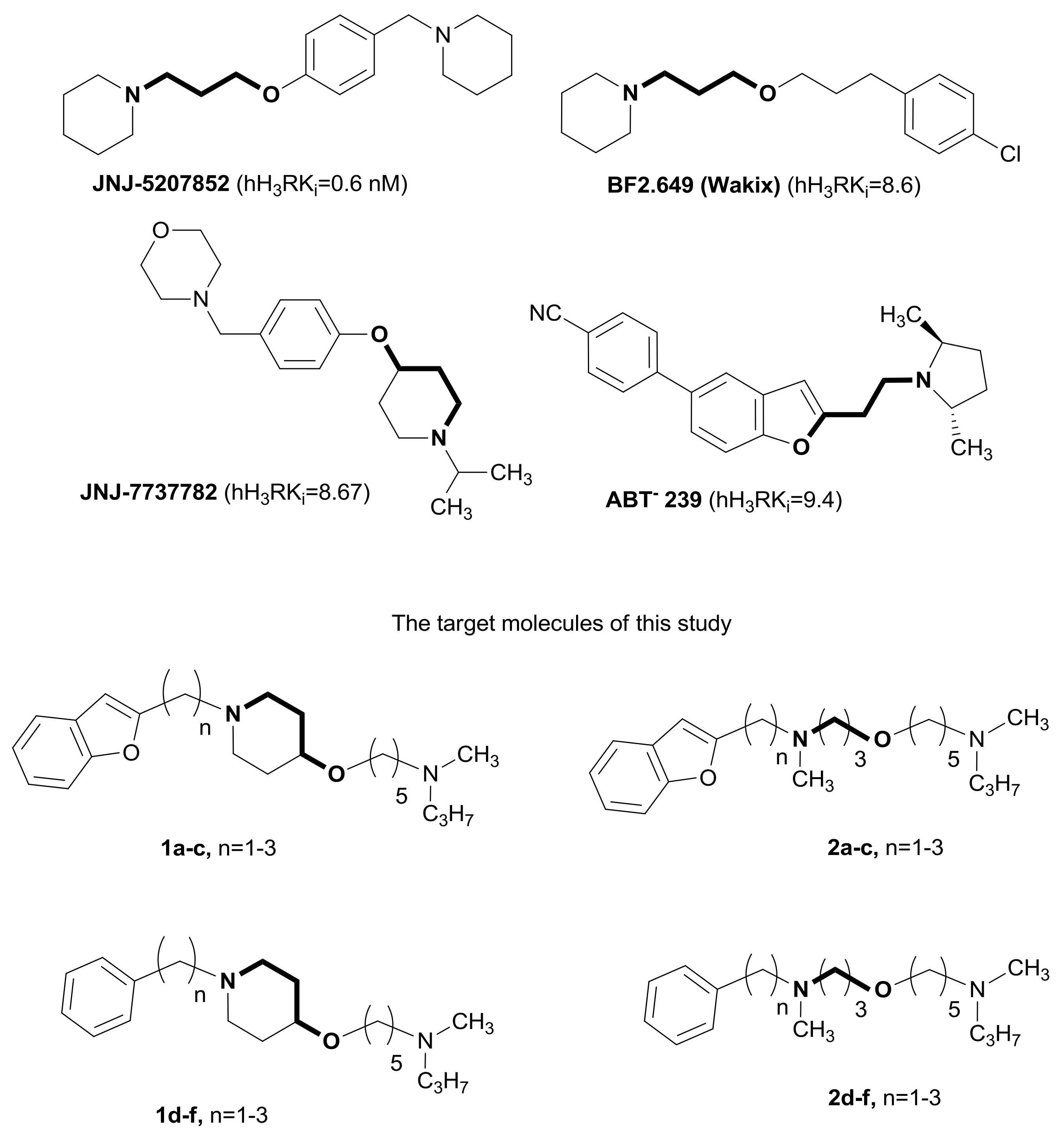
4-Hydroxypiperidines and Their Flexible 3-(Amino)propyloxy Analogues as Non-Imidazole Histamine H3 Receptor Antagonist: Further Structure–Activity Relationship Exploration and In Vitro and In Vivo Pharmacological Evaluation

by
Beata Olszewska
1,*,
Anna Stasiak
2,
Daniel McNaught Flores
3,
Wiesława Agnieszka Fogel
2,
Rob Leurs
3 and
Krzysztof Walczyński
1,*
1
Department of Synthesis and Technology of Drugs, Medical University of Lodz, Muszyńskiego Street 1, 90-145 Łódź, Poland
2
Department of Hormone Biochemistry, Medical University of Lodz, Żeligowskiego Street 7/9, 90-752 Łódź, Poland
3
Amsterdam Institute of Molecules, Medicines & Systems, Division of Medicinal Chemistry, Vrije Universiteit Amsterdam, De Boelelaan 1108, 1081 HZ Amsterdam, The Netherlands
*
Authors to whom correspondence should be addressed.
Int. J. Mol. Sci. 2018, 19(4), 1243; https://doi.org/10.3390/ijms19041243
Submission received: 9 March 2018 / Revised: 5 April 2018 / Accepted: 17 April 2018 / Published: 19 April 2018
(This article belongs to the Section Molecular Toxicology)

Abstract

:
Presynaptic histamine H3 receptors (H3R) act as auto- or heteroreceptors controlling, respectively, the release of histamine and of other neurotransmitters in the central nervous system (CNS). The extracellular levels of several neurotransmitters are enhanced by H3R antagonists, and there is a great interest for potent, brain-penetrating H3 receptor antagonists/inverse agonists to compensate for the neurotransmitter deficits present in various neurological disorders. We have shown that 1-[(benzylfuran-2-yl)methyl]piperidinyl-4-oxyl- and benzyl- derivatives of N-propylpentan-1-amines exhibit high in vitro potencies toward the guinea pig H3 receptor (jejunum), with pA2 = 8.47 and 7.79, respectively (the reference compound used was thioperamide with pA2 = 8.67). Furthermore, following the replacement of 4-hydroxypiperidine with a 3-(methylamino)propyloxy chain, the pA2 value for the first group decreased, whereas it increased for the second group. Here, we present data on the impact of elongating the aliphatic chain between the nitrogen of 4-hydroxypiperidine or 3-(methylamino)propan-1-ol and the lipophilic residue. Additionally, the most active compound in this series of non-imidazole H3 receptor antagonists/inverse agonists, i.e., ADS-003, was evaluated for its affinity to the recombinant rat and human histamine H3 receptors transiently expressed in HEK-293T cells. It was shown that ADS-003, given parenterally for 5 days, reduced the food intake of rats, as well as changed histamine and noradrenaline concentrations in the rats’ brain in a manner and degree similar to the reference H3 antagonist Ciproxifan.

1. Introduction

The histamine H3 receptors were first identified in 1983 in the rat brain by J.-Ch. Schwartz and coworkers [1]. The presence of H3 receptors was confirmed in the human brain a few years later [2]. The cDNA encoding the human H3 receptor was successfully cloned and functionally expressed by Lovenberg et al. [3]. Histamine H3 receptors predominantly have a presynaptic localization in histaminergic or other neurons and they modulate, through a negative feedback mechanism, the biosynthesis and release of histamine as autoreceptors [4] or the release of various other neurotransmitters, including norepinephrine [5], dopamine [6], serotonin [7], gamma-aminobutyric acid (GABA) [8], glutamate [9], and acetylcholine [10], acting as heteroreceptors. Consequently, it has been observed that administration of H3-antagonists to the CNS enhances neurotransmission and improves cognition and attention in relevant animal models of CNS diseases [11]. Therefore, H3-antagonists/inverse agonists have been proposed for the treatment of cognitive disorders, such as attention-deficit hyperactivity disorder (ADHD) [12] and Alzheimer’s disease [13] as well as of memory and learning deficits [10]. They may also be useful in epilepsy [14], schizophrenia [15], and obesity [16].
The discovery of thioperamide [17], the prototype of the H3 antagonist, led to the development of a wide range of imidazole-containing ligands [18]. Although many of them have found utility as pharmacological tools, the presence of an imidazole ring greatly limited brain penetration [19] and also introduced the potential for cytochrome P450 interactions [20]. For these reasons, efforts have been directed toward the design and synthesis of non-imidazole H3 antagonists with a good binding affinity, CNS penetration ability, and reduced/no potential for cytochrome P450 inhibition. A number of such antagonists with high selectivity and specificity have since been reported [21].
The marine natural product aplysamine-1 patented by the Harbor Branch Oceanographic Institution as a weak histamine H3 receptor antagonist [22], possesses the characteristic 3-aminopropan-1-ol functionality in its structure [23]. This moiety has successfully been used by several laboratories for the development of non-imidazole histamine H3 receptor antagonists and resulted in a number of highly potent and selective compounds, for example, JNJ-5207852 [24] and Pitolisant BF2.649 (Wakix) [25], the potent and selective H3R antagonist, which was approved by the European Medicine Agency (EMA) in March 2016 for the treatment of the orphan disease narcolepsy with and without cataplexy (Figure 1). Later on, the successful replacement of the highly flexible 3-aminopropyloxy link with the 4-phenoxypiperidine moiety JNJ-7737782 [26] (Figure 1) or the partially rigid 2-aminoethylbenzofuran substructure ABT-239 [27] (Figure 1) was demonstrated.
Previously, our laboratory has described several non-imidazole piperazine- [28,29] and 4-hydroxypiperidine-based histamine H3 antagonists with a moderate to pronounced affinity for the receptor [30,31]. The structure–activity relationship (SAR) of 4-hydroxypiperidines series showed that the most potent compounds, under in vitro screening conditions, were the benzofuranylpiperidinyloxy 1a (ADS-003) (pA2 = 8.47; Figure 1) [31] (for reference, thioperamide pA2 = 8.67) and benzyl 1d (pA2 = 7.79; Figure 1) derivatives [30]. None of the compounds showed any activity following binding to the histamine H1 receptor. Two analogues of the compounds 1a and 1d, i.e., 2a and 2d (Figure 1), in which the 4-hydroxypiperidine ring was replaced by a flexible 3-(methylamino)propyloxy chain, were synthesized and pharmacologically evaluated in vitro. In the case of derivatives carrying a (benzofuran-2-yl)methyl substituent (1a), a drastic reduction in potency was observed (2a, pA2 = 6.23; Figure 1) [31]. For compounds bearing a benzyl substituent, an inverse relationship was observed—5-{3-[benzyl(methyl)amino]propoxy}-N-methyl-N-propylpentane-1-amine 2d (pA2 = 8.06; Scheme 2) expressed higher potency than its 4-hydroxypiperidine analogue 2a (pA2 = 7.79; Scheme 1) [31].
In continuation of our search for new highly active and selective non-imidazole histamine H3 receptor antagonists, the first step of this study aimed to clarify the significant difference between the potencies of the 4-hydroxypiperidine derivatives and their 3-(methylamino)propyloxy analogues, bearing ω-(benzofuran-2-yl)alkyl and ω-phenylakyl moieties, respectively. We synthesized and in vitro pharmacologically [32] evaluated a series of derivatives in which the aliphatic chain between the nitrogen of the 4-hydroxypiperidine or the 3-(methylamino)propan-1-ol and the lipophilic residue was elongated by two to three methylene groups (compounds 1b,c,e,f, and 2b,c,e,f; Figure 1, respectively). This was to check whether the earlier observed tendency would still be kept, or if it was an exceptional case only for short methyl chain. We also wanted to study how the elongation of the alkyl chain would affect the potency for the H3 receptor. Additionally, the affinity of ADS-003—the most active compound that we have so far synthesized in this series—was estimated for the recombinant rH3R and hH3R (respectively), transiently expressed in HEK-293T cells. This derivative has also been proven to cross the blood–brain barrier; given parenterally for 5 days, ADS-003 reduced the food intake by rats as well as changed the cerebral histamine and noradrenaline concentrations in these animals in a manner and degree similar to the reference H3 antagonist—Ciproxifan.

2. Results and Discussion
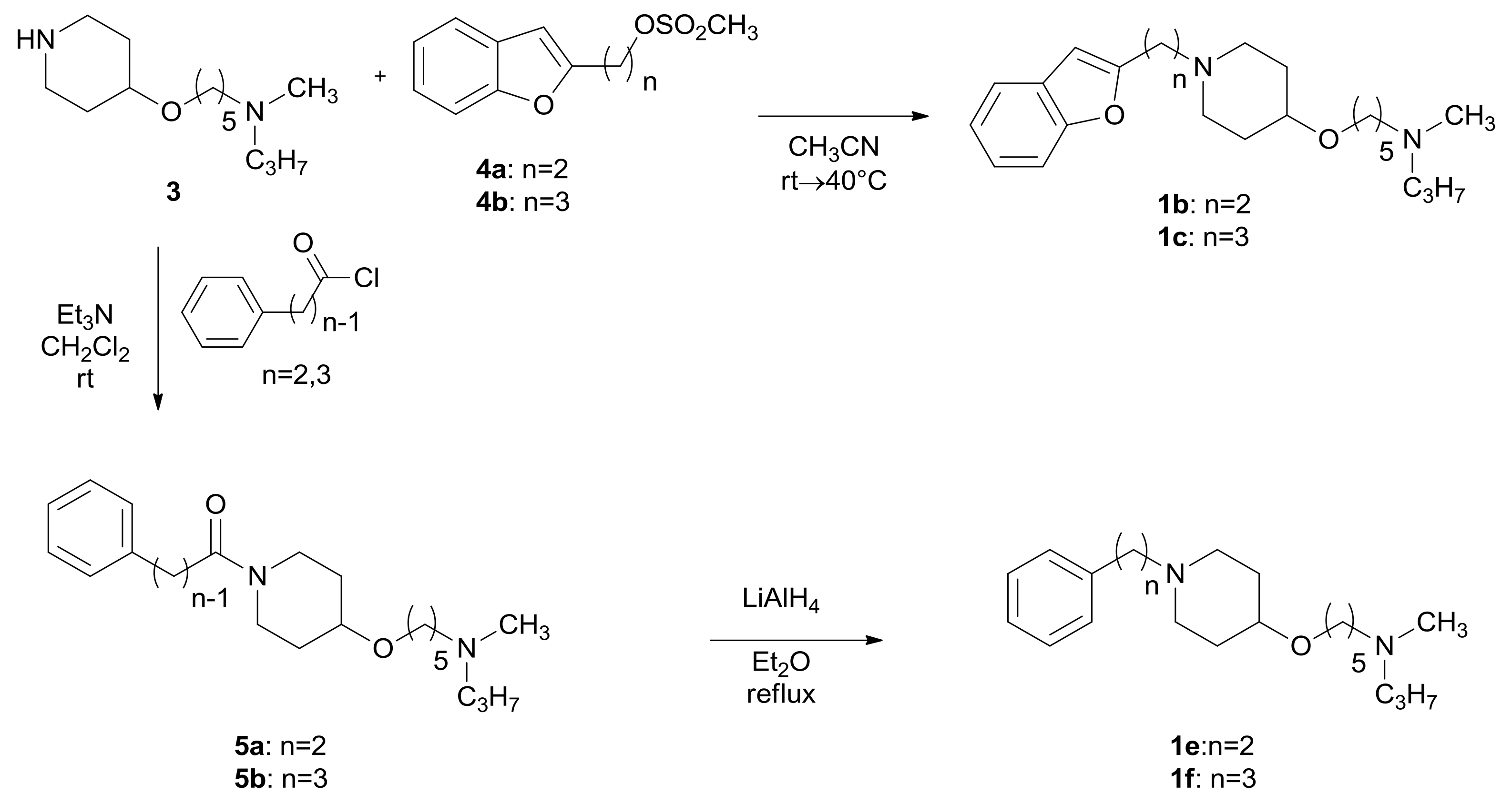
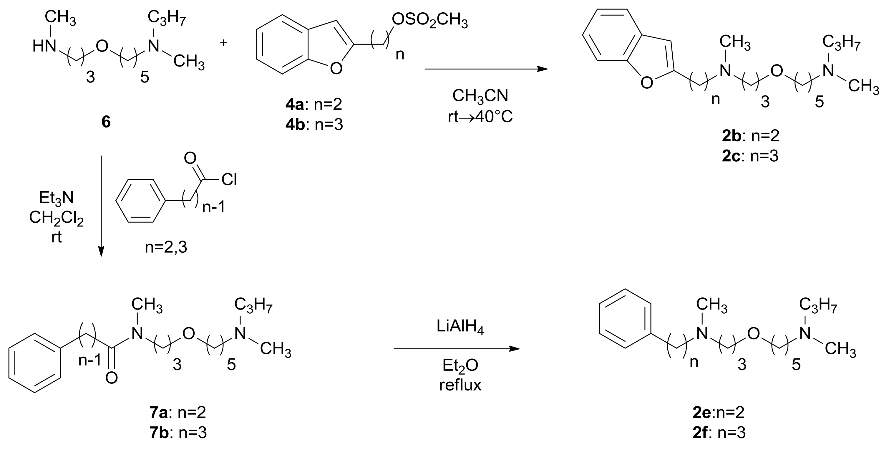
2.1. Chemistry

The general synthetic procedures used in this study are illustrated in Scheme 1 and 2. The key intermediates for all novel synthesized 4-hydroxypiperidines (1b,c,e,f; Scheme 1) and 3-(methylamino)propan-1-ols (2b,c,e,f; Scheme 2) were N-methyl-5-[(piperidin-4-yl)oxy]-N-propylpentan-1-amine (3) and N-methyl-5-[3-(methylamino)propoxy-N-propylpentane-1-amine (6), respectively. Both mentioned intermediates were prepared by hydrogenation of their corresponding benzyl derivatives with a catalytic amount of palladium in charcoal in ethanol [31].
The ω-(benzofuran-2-yl)alkyl derivatives of 4-hydoxypiperidine (1b,c; Scheme 1) and 3-(methyl)aminopropan-1-ol (2b,c; Scheme 2) were obtained from compounds 3 or 6 by alkylation with the corresponding ω-(benzofuran-2-yl)alkyl methanesulfonate in acetonitrile followed by purification by column chromatography.
The ω-phenylalkyl derivatives of 4-hydoxypiperidine (1e,f; Scheme 1) and 3-(methyl)aminopropan-1-ol (2e,f; Scheme 2) were obtained from compound 3 and 6 by a two-step synthesis: (1) acylation with the corresponding ω-phenylalkyl acid chloride in dry dichloromethane in the presence of triethylamine (TEA) to amides 5a,b (Scheme 1) and 7a,b (Scheme 2), (2) reduction of amides with LiAlH4 in dry ethyl ether to compounds 1e,f (Scheme 1) and compounds 2e,f (Scheme 2), respectively. Each step was followed by purification by column chromatography.
The synthesis of the required intermediates 4a,b (Scheme 3) was carried out by first treating the 2-iodophenol with the appropriate ω-alkyn-1-ol in the presence of palladium acetate, copper(I) iodide, and triphenylphosphine in dry triethylamine [33], followed by mesylation of the hydroxyl group by methanesulfonyl chloride in pyridine to yield the corresponding methanesulfonate with a 2 and 3 methylene linker. The detailed synthetic procedure and analytical data for compounds 8a,b and 4a,b are shown in Supplementary Material A (Section 1).

2.2. Pharmacology

2.2.1. In Vitro Pharmacological Studies

H3 Antagonistic Activity for Compounds 1af, and 2af

All newly synthesized compounds were converted into their dihydrogenoxolates salts and in vitro evaluated as H3 receptor antagonists against H3 agonist-induced inhibition of the electrically evoked contraction of the guinea pig jejunum [32].
The potencies of compounds 1b,c, 1e,f, 2b,c, and 2e,f, are reported in Table 1, as well as the previously described data for compounds 1a, [31] 1d [32], 2a [32], and 2d [32]. Derivatives 1b,c, 1e,f, 2b,c, and 2e,f, showed moderate to weak antagonist activity at H3-receptor.
With the aim of clarifying the significant difference between the change in the potency of derivatives 1a/2a (pA2 = 8.27/6.23) carrying a 2-methylbenzofuranyl substituent versus their benzyl-analogues 1d/2d (pA2 = 7.79/8.06), we compared the homologues pairs of compounds 1b/2b versus 1e/2e, and 1c/2c versus 1f/2f (Table 1). It was found that 1b (pA2 = 7.26) had a higher activity than its 3-(methylamino)propyloxy analogue 2b (pA2 = 6.32), as also observed for derivatives 1a/2a. In contrast, in the homologues pair 1e/2e, the 3-(methylamine)propan-1-ol derivative 2e (pA2 = 6.72) with the phenylethyl moiety showed antagonistic activity at the same level as its 4-hydroxy piperidine analogue 1e (pA2 = 6.67). In contrast to the above results, further elongation of the alkyl chain to three methylene groups led to a decrease of potency for both pairs 1c/2c versus 1f/2f. Derivative 1c (pA2 = 7.11), again, showed a higher potency than its 3-(methylamino)propyloxy analogue 2c (pA2 = 6.37), but the phenylpropyl derivative of 4-hydroxypiperidine 1f (pA2 = 7.51) had a higher potency than its analogue 2f (pA2 = 6.79), which is in opposition to the previously obtained results for derivatives 1d/2d.
The differences were observed within the 1ac and 1df series. While the previously reported 4-hydroxypiperidine derivative bearing a 2-benzofuranylmethyl moiety 1a showed a high potency, the newly synthesized compounds 1b,c, where the aliphatic chain between the piperidine nitrogen and the benzofuranyl residue is elongated from 2 to 3 methylene groups, resulted in a decrease of potency (pA2 = 7.26 and 7.11, respectively). A similar effect was observed when the 2-benzofuranylmethyl substituent was replaced by a ω-phenylakyl one (compounds 1df). In this series, derivative 1d (pA2 = 7.79) showed a high potency, but an increase in the alkyl chain length to 2 methylene groups resulted in a decrease of antagonist activity for compound 1e (pA2 = 6.67), and the activity increased again on further lengthening to 3 methylene groups (1f; pA2 = 7.51).
In the series of derivatives 2ac, bearing a ω-(benzofuran-2-yl)alkyl substituent—analogues of compounds 1ac—only a weak activity (pA2 ranging from 6.23 to 6.37), independent of the alkyl chain length, was observed. Elongation of the alkyl chain from 2 to 3 methylene groups in the analogue series of compounds 1e,f i.e., 2e,f resulted in a drastic reduction of potency (pA2 = 6.72 and 6.79, respectively) in comparison to the parent compound 2d (pA2 = 8.06).
Representative graphs of the antagonism by ADS-003 (1a) and thioperamide of the inhibitory effect of R-(−)-α-methylhistamine (R-α-MH) on the electrically induced contraction of guinea pig ileum strips are shown in Supplementary Material B (Section 2.2.1; Figure S2).

H1 Antagonistic Activity for Compounds 1b and 1f

The compounds 1b and 1f, possessing the highest potency for the H3 receptors, were also tested for H1 antagonistic effects in vitro, using standard methods [34]. Derivatives 1b and 1f did not show any antagonistic activity for the H1-receptor (pA2 < 4; for pyrilamine pA2 = 9.37).

2.2.2. Histamine H3 Receptor Affinity

The affinity, based on the SARs obtained for both series (compounds: 1af and 2af), of the most active compound 1a (ADS-003) was evaluated by measuring the displacement curve of [3H]-Nα-methylhistamine from the rat (rH3R) and human histamine H3 receptor (hH3R) in HEK-293Tcell membranes, as described by Bonger [35].

Saturation of Rat and Human H3 Receptors

The saturation of rat and human H3 receptors were carried out as described previously [36].
A representative graph of saturation of rat and human H3R can be found in Supplementary Material B (Section 2.2.2; Figure S3).
The analysis of the [3H]-Nα-MH saturation binding yielded, at rH3R, a KD value of 2.72 ± 0.34 nM and a Bmax value of 2715 ± 445 fmol/mg protein and, at hH3R, a KD value of 0.9 ± 0.08 nM and a Bmax value of 632 ± 52 fmol/mg protein.

Competition Binding of H3 Receptor Ligands

The affinity of ADS-003 (1a), histamine, and thioperamide was determined—the reference compounds were evaluated by measuring the displacement curves of [3H]-Nα-methylhistamine binding to rat and human histamine H3 receptor expressed in HEK-293T membranes. Derivative ADS-003 (1a) possessed a high nanomolar affinity for the rat H3R, with a pKi value of 7.9 ± 0.1, similar to thioperamide (pKi value 7.9 ± 0.1) and slightly higher than histamine (pKi = 7.3 ± 0.1). A significantly lower affinity was observed for ADS-003 (1a) for the human H3R, with pKi 6.6 ± 0.1, in comparison with the pKi of thioperamide (7.2 ± 0.1) and the pKi of histamine (7.7 ± 0.1). Representative graphs of competition binding of H3R ligands to rat and human H3 receptor are shown in Supplementary Material B (Section 2.2.2; Figures S4 and S5, respectively).

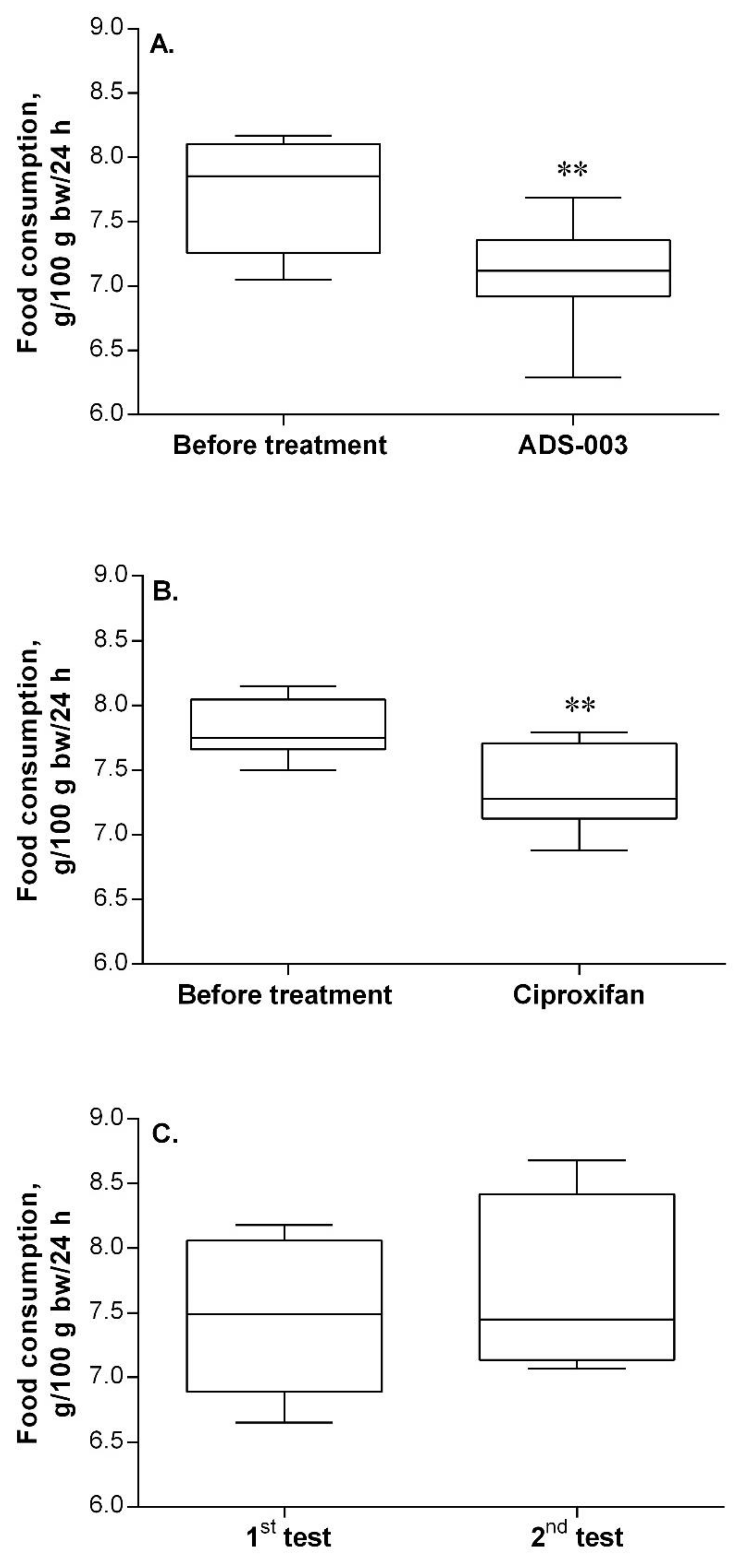
2.2.3. Verification of In Vivo Activity for Compound ADS-003 (1a)

Finally, compound ADS-003 (1a) was subjected to an in vivo evaluation of its impact on brain neurotransmitter systems. This assessment concerned:
-
The effects of the compound on the feeding behavior of rats after its repeated peripheral administration. Given that the compound enters the CNS and blocks the H3R, it should release histamine. Histamine, in turn, acting via H1R, would induce loss of appetite, i.e., the food intake of rats would decrease,
-
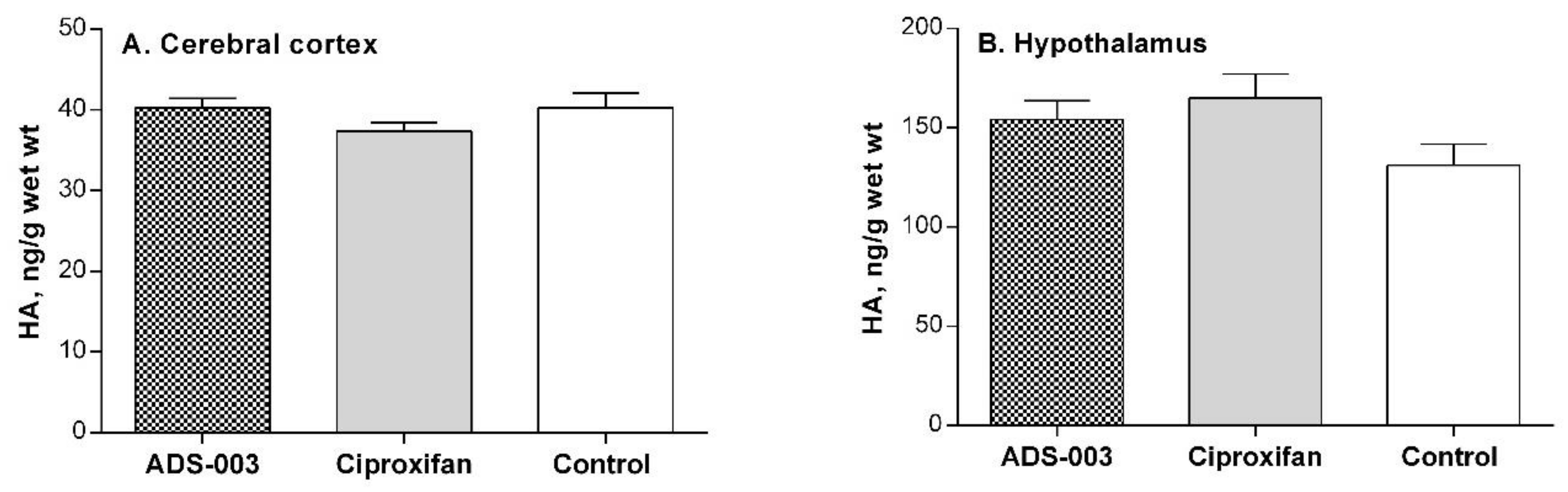
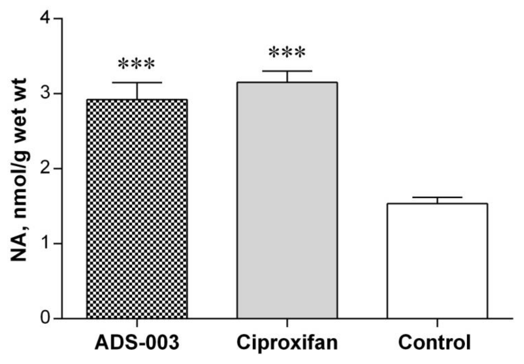
The influence on the cerebral amine neurotransmitter concentrations, as well as the activity of the monoamine oxidases A and B and histamine N-methyltransferase. The latter was accomplished by postmortem analyses of the brain tissues of the treated rats.
To evaluate the central effects of peripheral administration of ADS-003 (1a) to rats, the feeding behavior of rats was monitored after drug administration. The neuronal histaminergic system is known to be one of the regulatory systems in food intake. Studies in various animal models have convincingly shown that histamine H1R and H3R receptors play an important role in this respect. Activation of both histamine receptors is a critical part of the diurnal rhythm of the food consumption regulatory mechanism, as well as in energy intake and expenditure [37,38,39,40,41]. In a comprehensive study done in rats, it was demonstrated that centrally infused histamine or H1 receptor agonists invariably decreased food intake, as did the strategies leading either to an enhanced release of hypothalamic histamine by the blocking the H3 receptor or to an increase of available histamine by inhibiting its degradation. The opposite, i.e., hyperphagia, was seen for H1 antagonists [38]. Compatible with the earlier reported experimental data, it was assumed that, if an H3 receptor antagonist subcutaneously injected into rats crossed the blood–brain barrier (BBB), it would affect the animal food consumption [37]. As presented in Figure 2, the treatment of rats with either of the two H3R antagonists ADS-003 (3 mg/kg s.c.) and Ciproxifan (3 mg/kg s.c.) over a period of 5 days evoked a statistically significant reduction in the amount of consumed food by rats compared to the pre-treatment period.
In in vivo studies, Ciproxifan was used as a reference instead of thioperamide, because the latter demonstrated lower bioavailability due to restricted brain penetration [42].
There was no difference in the efficacy of ADS-003 (1a) and Ciproxifan (the reference compound). These results suggest that ADS-003 crosses the BBB, and its potency at H3R is similar to that of Ciproxifan.

Post-Mortem Biochemical Analysis of the Brain Tissues of ADS-003 (1a)-Treated Rats

Postmortem biochemical analysis of the brain tissues of ADS-003-treated rats quantified the brain concentration of histamine, serotonin, dopamine, noradrenaline, and the activities of monoamine oxidase (MAO)-A, MAO-B, and HNMT. As shown in Figure 3, the histamine concentration in the hypothalamus, where histaminergic cell bodies are located, showed a tendency to increase—which could be explained by the stimulation of the amine synthesis following its release by H3R blockade with 1a (ADS-003) or Ciproxifan to replenish vesicular stores. Yet, one-way ANOVA and Tukey’s multiple comparisons test showed no statistically significant differences. Similarly, no changes were found in the histamine levels in the cerebral cortex of the treated rats (Figure 3).
On the other hand, both H3R antagonists caused a significant increase in noradrenaline levels in the cerebral cortex (Figure 4).
There were no changes in serotonin and dopamine concentration.
The increase in tissue NA is compatible with previous data reporting an inhibitory control exerted by H3 histamine receptors on NA neuronal function in the cortex [43,44]. The fact that both histamine H3 receptor antagonists, Ciproxifan and ADS-003, enhanced the tissue levels of NA in a similar manner strengthens this idea.
Using sensitive isotopic assays, neither changes in monoamine oxidase A and B nor in histamine N-methyltransferase in the brain tissues of rats were observed (Table 2).

3. Materials and Methods

3.1. Chemistry

3.1.1. General Methods

All melting points (mp) were measured in open capillaries in an electrothermal apparatus and are uncorrected. The 1H NMR spectra were recorded in CDCl3 as a solvent in a 600 MHz spectrometer, a Bruker Avance III spectrometer at ambient temperature. The chemical shifts are reported in ppm on scale downfield from tetramethylsilane (TMS ) as internal standard, and the signal patterns are indicated as follows: s = singlet, d = doublet, t = triplet, m = multiplet, br = broad; * exchangeable by D2O; a number of protons, and J approximate coupling constant in Hertz. The 13C NMR spectra were recorded in a 600 MHz spectrometer, a Bruker Avance III (150 MHz). Elemental analyses (C, H, N) for all compounds were measured in Perkin Elmer Series II CHNS/O Analyzer 2400 and agreed with the theoretical values within ±0.4%. TLC data were obtained with Merck silica gel 60F254 aluminum sheets. For flash column chromatography using silica gel, 60 Å, 50 μm (J. T. Baker B. V.), the same solvent system as for TLC, was used. All obtained final free bases were treated with methanolic oxalic acid, and the dihydrogenoxolates were precipitated with dry diethyl ether and crystallized twice from ethanol. All dihydrogenoxolates were obtained as white crystalline solids.

Chemicals

The reagents 2-iodophenol, palladium acetate, copper(I) iodide, triphenylphosphine, 3-butyn-1-ol, 4-pentyn-1-ol, methanesulfonyl chloride, lithium aluminum hydride, and all solvents were purchased from Sigma-Aldrich (Saint Louis, MO, USA) and Alfa Aesar (Haverhill, MA, USA) and were used without any purification.

3.1.2. General Procedure for the Preparation of Compounds 1b,c and 2b,c

To a solution of the corresponding amines 3 or 6 (1.26 mmol) in acetonitrile (5 mL), the appropriate methanesulfonate 4a or 4b (1.05 mmol) was added. The reaction mixture was stirred at 40 °C for 24 h. After completion of the reaction, water was added. The mixture was extracted with dichloromethane (3 × 20 mL), and the organic layer was dried over MgSO4 and filtered. The solvent was evaporated, and the residue was purified by silica gel-flash column chromatography to yield a sticky oil.
5-{{1-[2-(1-Benzofuran-2-yl)ethyl]piperidin-4-yl}oxy}-N-methyl-N-propylpentan-1-amine (1b): (119 mg, 29.0%): Rf = 0.49 (CH2Cl2/MeOH/NH3(aq) 8:1:1%); 1H NMR (600 MHz, CDCl3): δ = 0.90 (t, J = 7.4 Hz, 3H, NCH2CH2CH3), 1.34–1.38 (m, 2H, H-3), 1.49–1.65 (m, 10H, H-2, H-4, NCH2CH2CH3, 2xCH2pip), 1.87–1.92 (m, 2H, CH2pip), 2.28 (s, 3H, CH3), 2.36 (t, J = 7.8 Hz, 2H, H-5), 2.41 (t, J = 7.7 Hz, 2H, NCH2CH2CH3), 2.76 (t, J = 7.4 Hz, 2H, C8H5OCH2CH2N), 2.81–2.83 (m, 2H, CH2pip), 2.97 (t, J = 7.4 Hz, 2H, C8H5OCH2CH2N), 3.27–3.31 (m, 1H, CHpip), 3.42 (t, J = 6.5 Hz, 2H, H-1), 6.42 (s, 1H, CHfuran), 7.15–7.21 (m, 2H, C6H4), 7.39 (d, J = 7.6 Hz, 1H, C6H4), 7.46 ppm (d, J = 7.2 Hz, 1H, C6H4); 13C NMR (150 MHz, CDCl3): δ = 12.04 (NCH2CH2CH3), 20.08 (NCH2CH2CH3), 24.41 (C-3), 26.73 (C8H5OCH2CH2N), 29.90 (CH2pip), 30.19 (C-4), 31.08 (C-2), 42.05 (CH3), 51.28 (CH2pip), 56.59 (C8H5OCH2CH2N), 57.59 (C-5), 59.61 (NCH2CH2CH3), 67.93 (C-1), 75.06 (CHpip), 102.66 (Cfuran), 110.94, 120.47, 122.66, 123.43,129.12, 154.85 (C6H4), 157.83 ppm (Cfuran).
Anal. calcd for dihydrogenoxolate (C24H38N2O2 2C2H2O4): C 59.35, H 7.47, N 4.94; found: C 59.08, H 7.83, N 5.07; mpdihydrogenoxolate = 133.2–134.9 °C.
5-{{1-[3-(1-Benzofuran-2-yl)propyl]piperidin-4-yl}oxy}-N-methyl-N-propylpentan-1-amine (1c): (226 mg, 54.0%): Rf = 0.38 (CH2Cl2/MeOH/NH3(aq) 8:1:1%); 1H NMR (600 MHz, CDCl3): δ = 0.88 (t, J = 7.4 Hz, 3H, NCH2CH2CH3), 1.31–1.36 (m, 2H, H-3), 1.45–1.51 (m, 4H, NCH2CH2CH3, CH2pip), 1.55–1.62 (m, 6H, H-2, H-4, CH2pip), 1.87–1.89 (m, 2H, CH2pip), 1.91–1.94 (m, 2H, C8H5OCH2CH2CH2N), 2.20 (s, 3H, CH3), 2.28 (t, J = 7.7 Hz, 2H, H-5), 2.32 (t, J = 7.8 Hz, 2H, NCH2CH2CH3), 2.39 (t, J = 7.6 Hz, 2H, C8H5OCH2CH2CH2N), 2.74–2.80 (m, 4H, CH2pip, C8H5OCH2CH2CH2N) 3.24–3.27 (m, 1H, CHpip), 3.42 (t, J = 6.6 Hz, 2H, H-1), 6.37 (s, 1H, CHfuran), 7.14–7.20 (m, 2H, C6H4), 7.38 (d, J = 7.8 Hz, 1H, C6H4), 7.46 ppm (d, J = 7.2 Hz, 1H, C6H4); 13C NMR (150 MHz, CDCl3): δ = 12.15 (NCH2CH2CH3), 20.60 (NCH2CH2CH3), 24.46 (C8H5OCH2CH2CH2N), 25.49 (C-3), 26.64 (C8H5OCH2CH2CH2N), 27.32 (C-4), 30.29 (CH2pip), 31.65 (C-2), 42.49 (CH3), 51.56 (CH2pip), 57.95 (C8H5OCH2CH2CH2N and C-5), 60.06 (NCH2CH2CH3), 67.98 (C-1), 74.06 (CHpip), 102.66 (Cfuran), 110.94, 120.47, 122.66, 123.43, 129.12, 154.85 (C6H4), 157.83 ppm (Cfuran).
Anal. calcd for dihydrogenoxolate (C25H40N2O2 2C2H2O4 0.5 H2O): C 59.07, H 7.69, N 4.75; found: C 59.05, H 7.75, N 4.80; mpdihydrogenoxolate = 157–159 °C.
5-{3-{[2-(Benzofuran-2-yl)ethyl](methyl)amino}propoxy}-N-methyl-N-propylpentan-1-amine (2b): (109 mg, 28.0%): Rf = 0.51 (CH2Cl2/MeOH/NH3(aq) 8:1:1%);1H NMR (600 MHz, CDCl3): δ = 0.88 (t, J = 7.4 Hz, 3H, NCH2CH2CH3), 1.31–1.36 (m, 2H, H-6), 1.43–1.50 (m, 4H, H-7, NCH2CH2CH3), 1.54–1.59 (m, 2H, H-5), 1.72–1.77 (m, 2H, H-2), 2.19 (s, 3H, CH3), 2.27 (t, J = 7.6 Hz, 2H, NCH2CH2CH3), 2.29–2.32 (m, 5H, H-8, CH3), 2.49 (t, J = 7.2 Hz, 2H, H-1), 2.76–2.79 (m, 2H, C8H5OCH2CH2N), 2.91–2.95 (m, 2H, C8H5OCH2CH2N), 3.36 (t, J = 6.7 Hz, 2H, H-4), 3.42 (t, J = 6.5 Hz, 2H, H-3), 6.41 (s, 1H, CHfuran), 7.13–7.20 (m, 2H, C6H4), 7.38–7.40 (m, 1H, C6H4), 7.45–7.47 ppm (m, 1H, C6H4); 13C NMR (150 MHz, CDCl3): δ = 12.15 (NCH2CH2CH3), 20.65 (C-7), 24.44 (C-6), 26.92 (NCH2CH2CH3), 27.37 (C-2), 27.92 (C8H5OCH2CH2N), 29.94 (C-5), 42.33, 42.49 (2× CH3), 54.58 (C-1), 55.83 (C8H5OCH2CH2N), 58.00 (NCH2CH2CH3), 60.10 (C-8), 69.23 (C-4), 71.13 (C-3), 102.64 (Cfuran), 110.91, 120.43, 122.62, 123.36, 129.17, 154.87 (C6H4), 158.00 ppm (Cfuran).
Anal. calcd for dihydrogenoxolate (C23H38N2O2 2C2H2O4·0.5 H2O): C 57.54, H 7.69, N 4.97; found: 5 C 7.37, 7.95 C H, N 5.02; mpdihydrogenoxolate= 144.5–145.7 °C.
5-{3-{[3-(Benzofuran-2-yl)propyl](methyl)amino}propoxy}-N-methyl-N-propylpentan-1-amine (2c): (134 mg, 34.0%): Rf = 0.45; (CH2Cl2/MeOH/NH3(aq) 8:1:1%); 1H NMR (600 MHz, CDCl3): δ = 0.88 (t, J = 7.4 Hz, 3H, NCH2CH2CH3), 1.30–1.35 (m, 2H, H-6), 1.44–1.50 (m, 4H, H-7, NCH2CH2CH3), 1.55–1.59 (m, 2H, H-5), 1.71–1.76 (m, 2H, H-2), 1.88–1.93 (m, 2H, C8H5OCH2CH2CH2N), 2.19 (s, 3H, CH3), 2.23 (s, 3H, CH3), 2.28 (t, J = 7.6 Hz, 2H, NCH2CH2CH3), 2.31 (t, J = 7.6 Hz, 2H, H-8), 2.41–2.43 (m, 4H, H-3, C8H5OCH2CH2CH2N), 2.78 (t, J = 7.4 Hz, 2H, C8H5OCH2CH2CH2N), 3.38 (t, J = 6.7 Hz, 2H, H-4), 3.41–3.44 (m, 2H, H-1), 6.37 (s, 1H, CHfuran), 7.14–7.20 (m, 2H, C6H4), 7.39 (d, J = 8.1 Hz, 1H, C6H4), 7.46 ppm (d, J = 8.1 Hz, 1H, C6H4); 13C NMR (150 MHz, CDCl3): δ = 12.09 (C-11), 20.39 (C-7), 24.39 (C-6), 25.67 (C-10), 26.50 (C-2), 27.83 (C8H5OCH2CH2CH2N), 29.90 (C8H5OCH2CH2CH2N and C-5), 42.30, 42.40 (2× CH3), 54.80 (C8H5OCH2CH2CH2N), 57.19 (C-1), 57.83 (C-9), 59.89 (C-8), 69.33 (C-3), 71.08 (C-4), 102.16 (Cfuran), 110.92, 120.39, 112.60, 123.30, 129.20, 154.89 (C6H4), 159.51 ppm (Cfuran).
Anal. calcd for dihydrogenoxolate (C24H40N2O2·2C2H2O4 0.5 H2O): C 58.22, H 7.85, N 4.85; found: C 58.61, H 8.30, N 4.97; mpdihydrogenoxolate = 106.0–108.0 °C.

3.1.3. General Procedure for the Preparation of Compounds 5a,b and 7a,b

The corresponding amines 3 or 6 (2.6 mmol) and Et3N (3.12 mmol) were dissolved in dichloromethane and cooled to 0°C. After flushing with argon, 2-phenylacetyl chloride (3.12 mmol) or 3-phenylpropanoyl chloride (3.12 mmol) was slowly added to the mixture, and the reaction was stirred at room temperature for 12 h. After completion of the reaction, an aqueous solution of K2CO3 was added. The reaction mixture was extracted with dichloromethane (3 × 20 mL), and the organic layer was dried over MgSO4 and filtered. The solvent was evaporated, and the residue was purified by silica gel-flash column chromatography to yield a sticky oil.
1-{4-{{5-[Methyl(propyl)amino]pentyl}oxy}piperidin-1-yl}-2-phenylethanone (5a): (818 mg, 87.0%): Rf = 0.42 (CH2Cl2/MeOH/NH3(aq) 8:1:1%); 1H NMR (600 MHz, CDCl3): δ = 0.89 (t, J = 7.3 Hz, 2H, NCH2CH2CH3), 1.30–1.35 (m, 3H, H-3, CHpip), 1.46–1.49 (m, 4H, NCH2CH2CH3, H-4), 1.53–1.57 (m, 4H, H-2, CH2pip), 1.74–1.80 (m, 1H, CHpip), 2.21 (s, 3H, CH3), 2.28 (t, J = 7.6 Hz, 2H, H-5), 2.32 (t, J = 7.6 Hz, 2H, NCH2CH2CH3), 3.17–3.21 (m, 1H, CHpip), 3.31–3.43 (m, 4H, H-1, CHpip, CHpip), 3.60–3.65 (m, 1H, CHpip), 3.73 (s, 2H, C6H5CH2C(O)), 3.88–3.93 (m, 1H, CHpip), 7.21–7.24 (m, 3H, C6H5), 7.29–7.32 ppm (m, 2H, C6H5); 13C NMR (150 MHz, CDCl3): δ = 12.09 (NCH2CH2CH3), 20.40 (NCH2CH2CH3), 24.40 (C-3), 30.17 (C-4), 30.84 (C-2), 31.65 (CH2pip), 39.32 (CH2pip), 41.39 (C6H5CH2C(O)), 43.65 (CH3), 57.83 (C-5), 59.92 (NCH2CH2CH3), 68.25 (C-1), 73.98 (CHpip), 126.93, 128.75, 128.92, 135.52 (C6H5), 169.50 ppm (C=O).
1-{4-{{5-[Methyl(propyl)amino]pentyl}oxy}piperidin-1-yl}-3-phenylpropan-1-one (5b): (815 mg, 84.0%): Rf = 0.45 (CH2Cl2/MeOH/NH3(aq 8:1:1%)); 1H NMR (600 MHz, CDCl3): δ = 0.88 (t, J = 7.4 Hz, 3H, NCH2CH2CH3), 1.33–1.37 (m, 2H, H-3), 1.45–1.53 (m, 6H, H-4, NCH2CH2CH3, CH2pip), 1.54–1.60 (m, 2H, H-2), 1.69–1.72 (m, 1H, CHpip), 1.76–1.80 (m, 1H, CHpip), 2.21 (s, 3H, CH3), 2.27–2.34 (m, 4H, H-5, NCH2CH2CH3), 2.62 (t, J = 8.2 Hz, 2H, C6H5CH2CH2C(O)), 2.96 (t, J = 7.7 Hz, 2H, C6H5CH2CH2C(O)), 3.15–3.18 (s, 1H, CHpip), 3.30–3.34 (s, 1H, CHpip), 3.40–3.48 (m, 3H, H-1, CHpip), 3.57–3.61 (m, 1H, CHpip), 3.88–3.92 (m, 1H, CHpip), 7.18–7.22 (m, 3H, C6H5), 7.26–7.29 ppm (m, 2H, C6H5); 13C NMR (150 MHz, CDCl3): δ = 12.15 (NCH2CH2CH3), 20.61 (NCH2CH2CH3), 24.46 (C-3), 30.25 (C-2 and C-4), 30.91 (CH2pip), 31.88 (C6H5CH2CH2C(O)), 35.33 (C6H5CH2CH2C(O)), 29.19 (CH2pip), 43.03 (CH3), 57.98 (C-5), 60.09 (NCH2CH2CH3), 68.28 (C-1), 74.03 (CHpip), 126.36, 128.65, 128.72 (C6H5), 141.66 (C6H5), 170.72 ppm (C=O).
N-Methyl-N-{3-{{5-[methyl(propyl)amino]pentyl}oxy}propyl}-2-phenylacetamide (7a): (382 mg, 42.0%): Rf = 0.48 (CH2Cl2/MeOH/NH3(aq) 8:1:1%); 1H NMR (600 MHz, CDCl3): δ = 0.86 (t, J = 7.4 Hz, 3H, NCH2CH2CH3), 1.29–1.38 (m, 2H, H-6), 1.47–1.52 (m, 4H, H-5, NCH2CH2CH3), 1.55–1.62 (m, 2H, H-7), 1.72–1.76 (m, 1H, H-2), 1.77–1.82 (m, 1H, H-2), 2.22 (s, 1.5H, CH3), 2.23 (s, 1.5H, CH3), 2.29–2.37 (m, 4H, H-8, NCH2CH2CH3), 2.92 (s, 1.5H, CH3), 2.97 (s, 1.5H, CH3), 3.34–3.46 (m, 6H, H-1, H-3, H-4), 3.69 (s, 1H, C6H5CH2C(O)), 3.74 (s, 1H, C6H5CH2C(O)), 7.20–7.32 ppm (m, 5H, C6H5); 13C NMR (150 MHz, CDCl3): δ = 11.74 and 11.78 (NCH2CH2CH3), 19.39 and 19.58 (NCH2CH2CH3), 24.07 and 24.13 (C-6), 25.90 and 26.25 (C-7), 27.67 and 28.56 (C-2), 29.49 and 29.63 (C-5), 33.35 and 36.18 (CH3), 40.48 and 41.37 (CH3), 41.43 and 41.61 (C6H5CH2C(O)), 45.59 and 47.21 (C-1), 57.09 and 57.23 (C-8), 59.02 and 59.23 (NCH2CH2CH3), 67.17 and 68.35 (C-3), 70.73 and 70.93 (C-4), 126.71, 126.74, 128.63, 128.67, 128.82, 128.89, 129.01, 129.29, 135.19, 135.63 (C6H5), 170. 91 and 171.20 ppm (C=O).
N-Methyl-N-{3-{{5-[methyl(propyl)amino]pentyl}oxy}propyl}-3-phenylpropanamide (7b): (705 mg, 75.0%): Rf = 0.70 (CH2Cl2/MeOH/NH3(aq) 8:1:1%); 1H NMR (600 MHz, CDCl3): δ = 0.88 (t, J = 7.3 Hz, 3H, NCH2CH2CH3), 1.26–1.36 (m, 2H, H-6), 1.42–1.59 (m, 6H, H-5, H-7, NCH2CH2CH3), 1.72–1.80 (m, 2H, H-2), 2.19 (s, 3H, CH3), 2.25–2.33 (m, 4H, H-8, NCH2CH2CH3), 2.59 (t, J = 7.7 Hz, 1H, C6H5CH2CH2C(O)), 2.65 (t, J = 7.7 Hz, 1H, C6H5CH2CH2C(O)), 2.91 (s, 3H, CH3), 2.97 (q, J = 6.7 Hz, 2H, C6H5CH2CH2C(O)), 3.32–3.36 (m, 3H, H-1, H-3), 3.37–3.41 (m, 2H, H-4), 3.43 (t, J = 6.9 Hz, 1H, H-3), 7.17–7.28 ppm (m, 5H, C6H5); 13C NMR (150 MHz, CDCl3): δ = 12.14 (NCH2CH2CH3), 20.61 (NCH2CH2CH3), 24.37 and 24.42 (C-6), 27.34 (C-7), 27.98 and 28.82 (C-2), 29.87 and 29.92 (C-5), 31.56 and 31.83 (C6H5CH2CH2C(O)), 34.94 (CH3), 35.70 and 35.89 (C6H5CH2CH2C(O)), 42.47 (CH3), 45.69 and 46.88 (C-1), 57.94 and 57.96 (C-8), 60.07 and 60.09 (NCH2CH2CH3), 67.26 and 68.59 (C-3), 71.17 and 71.29 (C-4), 126.25, 126.28, 128.62, 128.65, 141,76, 141.80 (C6H5), 172.19 and 172.50 ppm (C=O).

3.1.4. General Procedure for the Preparation of Compounds 1e,f and 2e,f

To a vigorous stirred solution of the corresponding amides 5a,b or 7a,b (1.0 mmol) in 250 mL of anhydrous diethyl ether, LiAlH4 (2.5 mmol) was added portionwise. The mixture was stirred at reflux for 2 h, cooled to room temperature, and quenched by a dropwise addition of water (0.5 mL). The suspension was stirred for 30 min and filtered. The filter cake was washed with diethyl ether. The solvent was evaporated, and the residue was purified by silica gel-flash column chromatography (eluent: CH2Cl2/MeOH/NH3(aq) 8:1:1%) to yield a sticky oil.
N-Methyl-5-[(1-phenethylpiperidin-4-yl)oxy]-N-propylpentan-1-amine (1e): (252 mg, 72.0%): Rf = 0.32 (CH2Cl2/MeOH/NH3(aq) 8:1:1%) = 0.32; 1H NMR (600 MHz, CDCl3): δ = 0.89 (t, J = 7.3 Hz, 3H, NCH2CH2CH3), 1.32–1.37 (m, 2H, H-3), 1.45–1.51 (m, 4H, H-4, NCH2CH2CH3), 1.56–1.65 (m, 4H, H-2, CH2pip), 1.87–1.92 (m, 2H, CH2pip), 2.18–2.22 (m, 5H, CH3, CH2pip), 2.28 (t, J = 7.4 Hz, 2H, NCH2CH2CH3), 2.32 (t, J = 7.3 Hz, 2H, H-5), 2.57 (t, J = 8.6 Hz, 2H, C6H5CH2CH2), 2.78–2.84 (m, 4H, C6H5CH2CH2, CH2pip), 3.26–3.31 (m, 1H, CHpip), 3.43 (t, J = 6.6 Hz, 2H, H-1), 7.17–7.19 (m, 3H, C6H5), 7.25–7.28 ppm (m, 2H, C6H5); 13C NMR (150 MHz, CDCl3): δ = 12.17 (NCH2CH2CH3), 20.65 (NCH2CH2CH3), 24.49 (C-3), 27.37 (C-4). 30.32 (C-2), 31.67 (CH2pip), 34.16 (C6H5CH2CH2), 42.54 (CH3), 51.53 (CH2pip), 58.03 (C-5), 60.12 (C6H5CH2CH2), 60.77 (NCH2CH2CH3), 68.02 (C-1), 75.01 (CHpip), 126.19, 128.57, 128.92, 140.80 ppm (C6H5).
Anal. calcd for dihydrogenoxolate (C22H38N2O 2C2H2O4): C 59.30, H 8.04, N 5.32; found: C 58.86, H 8.44, N 5.38; mpdihydrogenoxolate= 131.0–133.0 °C.
N-Methyl-5-{[1-(3-phenylpropyl)piperidin-4-yl]oxy}-N-propylpentan-1-amine (1f): (342 mg, 95.0%): Rf = 0.45 (CH2Cl2/MeOH/NH3(aq) 8:1:1% CH2Cl2/MeOH/NH3(aq)); 1H NMR (600 MHz, CDCl3): δ = 0.88 (t, J = 7.3 Hz, 2H, NCH2CH2CH3), 1.31–1.36 (m, 2H, H-3), 1.44–1.50 (m, 4H, H-4, NCH2CH2CH3), 1.55–1.61 (m, 4H, H-2, CH2pip), 1.81 (t, J = 7.6 Hz, 2H, C6H5CH2CH2CH2), 1.85–1.88 (m, 2H, CH2pip), 2.07–2.11 (m, 2H, CH2pip), 2.20 (s, 3H, CH3), 2.27 (t, J = 7.6 Hz, 2H, H-5), 2.30–2.35 (m, 4H, NCH2CH2CH3, C6H5CH2CH2CH2), 2.62 (t, J = 7.6 Hz, 2H, C6H5CH2CH2CH2), 2.72–2.76 (m, 2H, CH2pip), 3.23–3.28 (m, 1H, CHpip), 3.41 (t, J = 6.6 Hz, 2H, H-1), 7.15–7.18 (m, 3H, C6H5), 7.25–7.28 ppm (m, 2H, C6H5); 13C NMR (150 MHz, CDCl3): δ = 12.16 (NCH2CH2CH3), 20.66 (NCH2CH2CH3), 24.47 (C-3), 27.37 (C-4), 29.11 (C6H5CH2CH2CH2), 30.31 (C-2), 31.68 (CH2pip), 34.02 (C6H5CH2CH2CH2), 42.53 (CH3), 51.57 (CH2pip), 58.03 (C6H5CH2CH2CH2), 58.21 (NCH2CH2CH3), 60.12 (C-5), 67.98 (C-1), 75.28 (CHpip), 125.91, 128.48, 128.61, 142.49 ppm (C6H5).
Anal. calcd for dihydrogenoxolate (C23H40N2O 2C2H2O4 0.5 H2O): C 59.00, H 8.25, N 5.10; found: C 58.90, H 8.42, N 5.20; mpdihydrogenoxolate = 134.5–137.8 °C.
N-Methyl-5-{3-[methyl(phenethyl)amino]propoxy}-N-propylpentan-1-amine (2e): (264 mg, 80.0%): Rf = 0.48 (CH2Cl2/MeOH/NH3(aq) 8:1:1%); 1H NMR (600 MHz, CDCl3): δ = 0.88 (t, J = 7.4 Hz, 3H, NCH2CH2CH3), 1.31–1.36 (m, 2H, H-6), 1.45–1.49 (m, 4H, H-7, NCH2CH2CH3), 1.55–1.59 (m, 2H, H-5), 1.72–1.77 (m, 2H, H-2), 2.19 (s, 3H, CH3), 2.58–2.32 (m, 7H, H-8, NCH2CH2CH3, CH3), 2.48 (t, J = 7.3 Hz, 2H, H-1), 2.59–2.62 (m, 2H, C6H5CH2CH2), 2.75–2.78 (m, 2H, C6H5CH2CH2), 3.39 (t, J = 6.7 Hz, 2H, H-4), 3.42 (t, J = 6.5 Hz, 2H, H-3), 7.18–7.28 ppm (m, 5H, C6H5); 13C NMR (150 MHz, CDCl3): δ = 12.18 (NCH2CH2CH3), 20.67 (NCH2CH2CH3), 24.46 (C-6), 27.40 (C-2), 2.89 (C-7), 29.96 (C-5), 34.08 (C6H5CH2CH2), 42.47 and 42.53 (2× CH3), 54.66 (C-1), 58.03 (C6H5CH2CH2), 59.86 (C-8), 60.13 (NCH2CH2CH3), 69.36 (C-3), 71.14 (C-4), 126.14, 128.55, 128.92, 140.88 ppm (C6H5).
Anal. calcd for dihydrogenoxolate (C21H38N2O 2C2H2O4 0.5 H2O): C 57.34, H 8.28, N 5.35; found: C 57.32, H 8.53, N 5.40; mpdihydrogenoxolate = 110.0–112.0 °C.
N-Methyl-5-(3-(methyl(3-phenylpropyl)amino)propoxy)-N-propylpentan-1-amine (2f): (175 mg, 50.0%): Rf = 0.63 (CH2Cl2/MeOH/NH3(aq) 8:1:1%) = 0.63. 1H NMR (600 MHz, CDCl3): δ = 0.88 (t, J = 7.4 Hz, 3H, NCH2CH2CH3), 1.30–1.36 (m, 2H, H-6), 1.43–1.49 (m, 4H, H-7, NCH2CH2CH3), 1.55–1.60 (m, 2H, H-5), 1.69–1.72 (m, 2H, H-2), 1.76–1.80 (m, 2H, C6H5CH2CH2CH2), 2.19 (s, 3H, CH3), 2.20 (s, 3H, CH3), 2.26 (t, J = 7.6 Hz, 2H, NCH2CH2CH3), 2.30 (t, J = 7.5 Hz, 2H, H-8), 2.36 (t, J = 7.3 Hz, 2H, H-1), 2.39 (t, J = 7.1 Hz, 2H, C6H5CH2CH2CH2), 2.62 (t, J = 7.7 Hz, 2H, C6H5CH2CH2CH2), 3.39 (t, J = 6.7 Hz, 2H, H-3), 3.43 (t, J = 6.5 Hz, 2H, H-4), 7.15–7.18 (m, 2H, C6H5), 7.25–7.27 ppm (m, 3H, C6H5); 13C NMR (150 MHz, CDCl3): δ = 12.17 (NCH2CH2CH3), 20.71 (NCH2CH2CH3), 24.44 (C-6), 27.44 (C-7), 27.88 (C-2), 29.31 (C6H5CH2CH2CH2), 29.96 (C-5), 33.94 (C6H5CH2CH2CH2), 42.44 and 42.55 (2× CH3), 54.80 (C6H5CH2CH2CH2), 57.53 (C-1), 58.05 (C-8), 60.16 (NCH2CH2CH3), 69.44 (C-4), 71.14 (C-3), 125.89, 128.49, 128.61, 142.64 ppm (C6H5).
Anal. calcd for dihydrogenoxolate (C22H40N2O 2C2H2O4): C 59.07, H 8.39, N 5.30; found: C 58.84, H 8.80, N 5.39; mpdihydrogenoxolate = 104.5–107.0 °C.
1H and 13C NMR spectral data of final compounds 1b,c,e,f and 2b,c,e,f can be found in Supplementary Material A (Section 3).

3.2. In Vitro Pharmacology

3.2.1. H3 Antagonistic Activity for Compounds 1af and 2af

In the first step, all the obtained compounds were tested for their H3 antagonistic effects in vitro, following standard methods, using the electrically contracting guinea pig jejunum [32].
Male guinea pigs weighing 300–400 g were sacrificed, and a portion of the small intestine, 20–50 cm proximal to the ileocaecal valve (jejunum), was removed and placed in Krebs buffer (composition (mM) NaCl 118; KCl 5.6; MgSO4 1.18; CaCl2 2.5; NaH2PO4 1.28; NaHCO3 25; glucose 5.5; indomethacin (1 × 10⁶mol/L)). Whole jejunum segments (2 cm) were prepared and mounted between two platinum electrodes (4 mm apart) in 20 mL Krebs buffer, continuously gassed with 95% O2:5% CO2, and maintained at 37 °C. the contractions were recorded isotonically under 1.0 g tension with a Hugo Sachs Hebel–Messvorsatz (Tl-2)/HF-modem (Hugo Sachs Elektronik, Hugstetten, Germany) connected to a pen recorder. The equilibration lasted for one hour with washings every 10 min. The muscle segments were then stimulated at a maximum between 15 and 20 Volts, continuously at a frequency of 0.1 Hz for a duration of 0.5 msec, with rectangular wave electrical pulses, delivered by a Grass Stimulator S-88 (Grass Instruments Co., Quincy, MA, USA). After 30 min of stimulation and 5 minutes before adding (R)-α-methylhistamine, pyrilamine (1 × 10⁵mol/L concentration in organ bath) was added, and then cumulative concentration–response curves (half-log increments) of (R)-α-methylhistamine, an H3-agonist, were recorded until no further change in the responses was found. Five minutes before adding the tested compounds, the pyrilamine (1 × 10⁵mol/L concentration in an organ bath) was added. The antagonists were preincubated for 20 min during the stimulation, before the preparation were challenged with (R)-α-methylhistamine. Antagonist potency was determined by the construction of a Schild plot [34], using three different concentrations of the antagonist. The potency of an antagonist is expressed by its pA2 value. The pA2 values were compared with those indicating the potency of thioperamide.

3.2.2. H1 Antagonistic Activity for Compounds 1b and 1f

In addition, the compounds 1b and 1f with the highest potency at the H3 receptors were also tested for H1 antagonistic effects in vitro, using standard methods [34]. The potencies of the aforementioned ligands were determined for the guinea pig ileum histamine H3 receptors as described previously [36], using histamine as a competing agonist.
The pA2 values were calculated according to Arunlakshana and Schild [34]. The pA2 values were compared with those of pyrilamine.

Chemicals

Thioperamide maleate, (R)-α-methylhistamine dihydrochloride, indomethacin, pyrilamine maleate, and histamine dihydrochloride were purchased from Sigma Aldrich (Saint Louis, MO, USA).

3.2.3. Antagonist Binding to Rat rH3R and Human hH3R

Cell Culture and Transfection

Human Embryonic Kidney cells (HEK293T) were cultured in DMEM supplemented with 10% Fetal Bovine Serum, 100 IU·mL−1 penicillin, and 100 μg·mL−1 streptomycin at 37 °C and 5% CO2. The day prior to transfection, 2 million cells were seeded in 10 cm dishes. Approximately 4 million cells were transfected by the polyethyleneimine (PEI) method with 5 μg of cDNA in a ratio of 1:4 (DNA/PEI). Briefly, 0.5 μg of pcDNA3-rH3R or pcDNA3.1-hH3R and 4.5 μg of empty plasmid (pcDNA3.1) were mixed with 20 μg of 25 kDa linear PEI in 500 μL of 150 mM NaCl and incubated for 30 min at 22 °C. Meanwhile, the medium in the 10 cm dishes was replaced with fresh culture medium, and the transfection mix was subsequently added dropwise to the cells, which were incubated for 48 h at 37 °C and 5% CO2.

Crude Membrane Extracts

Forty-eight hours after transfection, the cells were washed with ice-cold phosphate buffered saline (PBS) and scrapped, and the homogenate was centrifuged for 10 min at ~2000× g, 4 °C. The supernatant was aspirated, and the cell pellets were resuspended in 1 ml ice-cold PBS and centrifuged again under the same conditions, aspirating the supernatant. The membranes were stored at −20 °C until further use.

[3H]-Nα-Methylhistamine Binding

The affinities of the derivative ADS-003 were determined for rat and human histamine H3 receptors (445 isoforms) as described previously [36], using [3H]-NαMH as a competing radioligand.

Chemicals

Dubelcco’s Modified Eagles Medium (DMEM), Phosphate Buffered Saline (PBS), Trizma Base, polyethyleneimine solution (50%, PEI) were purchased from Sigma Aldrich (Saint Louis, MO, USA). Fetal Bovine Serum (FBS, Bodinco BV, Alkmaar, The Netherlands), Penicillin/Streptomycin (streptomycin 10,000 IU·mL−1; penicillin 10,000 μg·mL−1, Thermo Fischer Scientific, (p/a Perbio Science BVBA, Etten-Leur, The Netherlands) , linear 25 kDa polyethyleneimine (PEI, Polysciences, Warrington, PA, USA), [3H]-N-α-methylhistamine (specific activity 79.7 Ci/mmol, Perkin Elmer, (Waltham, MA, USA), thioperamide (Abcam, Cambridge, UK) histamine (TCI, Nihonbashi-honcho, Chuo-ku, Tokyo, Japan).

3.3. Verification of In Vivo Activity of Compound ADS-003 (1a)

All animal experimental procedures were in accordance with EU directives and local ethical regulations. Male Wistar rats (260–300 g) were used. The animals were maintained under standard laboratory conditions (liquid and food available ad libitum, 12 h light–dark cycle). For feeding behavior examination, the rats were placed individually in metabolic cages (TecniplastGazzada, Buguggiate, Italy) and kept there throughout the entire test. Pharmacotherapy was preceded by a control period of 4 days aimed to determine the basal feed and water consumptions as well as urine excretion. The rats (n = 8 per group) were randomly given the H3R antagonists ADS-003 or Ciproxifan, the latter serving as a reference compound [40]. Control rats were treated with an equivalent volume of physiological saline. The compounds, dissolved in distilled water with DMSO (7%, v/v), were administered subcutaneously at a dose of 3 mg/kg of body mass for 5 consecutive days, always during the morning hours, following the record of feeding parameters. The dose of ADS-003 had been determined on the basis of a thorough characterization in vitro. The recorded volumes of consumed food are expressed in g or ml per 100 g of body weight or in ml per 24 h, respectively. The final results are given as means, with SEM calculated for each 24 h period, computed from a four-day (before treatment) or a five-day (treatment) monitoring

3.3.1. Post-Mortem Biochemical Analyses

Following the behavioral study, the rats were sacrificed, and their brains were collected. From each brain, the cerebral cortex and hypothalamus were quickly dissected according to the Glowinski and Iversen method [37]. The samples were immediately frozen in liquid nitrogen and kept at −70 °C until assayed.
The tissue concentration of histamine (HA) was measured by radioisotopic assay according to Taylor and Snyder [45], while catecholamine (dopamine, DA; noradrenaline, NA) and serotonin (5-HT) concentrations were measured by radioimmunoassays using RIA kits (DIAsourceImmunoAssays S.A., Nivelles, Belgium). The amine concentrations were expressed as ng/g wet weight (HA) or nmol/g wet weight (DA, NA, 5-HT).
Monoamine oxidase A and B (EC 1.4.3.4; MAO A and B) activities were estimated in cerebral homogenates with radioassays, using serotonin (fine conc. 200 μM) and β-phenylethylamine (fine conc. 20 μM), as well as specific inhibitors—clorgyline and deprenyl (0.3 μM each), respectively [46]. Histamine N-methyltransferase (EC 2.1.1.8; HMNT) activity was determined radioenzymatically, as described by Taylor and Snyder [45], by measurements of radioactive Nτ-methylhistamine formed in a transmethylation reaction catalyzed by the enzyme, as previously described; S-Adenosyl-l-(methyl-14C)-Methionine was used as a donor of the methyl group.
The enzyme activities are expressed as pmol/min/mg protein. Protein concentration was analyzed according to Lowry’s method [47].

Chemicals

Adenosyl-l-methionine, S [methyl-14C] (specific activity 50 μCi) and β-phenylethylamine hydrochloride [ethyl-1-14C] (specific activity 250 μCi) were purchased from ARC.

4. Conclusions

The highest in vitro potency as an H3 receptor antagonist for both series was observed for the (benzylfuran-2-yl)methyl derivative of 4-hydroxypiperidine 1a (ADS-003) and the benzyl derivative of 3-(methylamino)propan-1-ol 2d. Compared to 1a and 2d, the in vitro potency was found to decrease with increasing chain length (1af; 2df), with the exception of the series 2ac, where only weak potency was observed, independent of the alkyl chain length.
In competition radioligand binding studies to the rat histamine H3 receptor, compound ADS-003 (pKi = 7.9 ± 0.1) showed nanomolar affinity at the same level as the reference compound thioperamide (pKi = 7.9 ± 0.1) and a slightly higher affinity than histamine (pKi = 7.3 ± 0.1). A significantly lower affinity was observed for ADS-003 for the human H3R, with pKi 6.6 ± 0.1, in comparison with the pKi of thioperamide (7.2 ± 0.1) and the pKii of histamine (7.7 ± 0.1).
ADS-003, given parenterally for 5 days, reduced the food intake of rats as well as changed rats’ brain amine concentrations in a manner and degree similar to those observed for the reference H3 antagonist Ciproxifan. These results indicate that the compound crosses the blood–brain barrier and acts also as an H3R antagonist in vivo in the rat brain.

Supplementary Materials

Supplementary materials can be found at https://www.mdpi.com/1422-0067/19/4/1243/s1.

Acknowledgments

This study was supported by departmental sources of the Medical University of Lodz grant numbers 503/3-016-01/503-31-001; 503/5-087-02/503-01 and COST Action CA 15135. The authors would like to thank Mieczyslaw Wośko, president of a pharmaceutical company Polfarmex SA, for providing financial support for the purchase of the necessary reagents for in vivo studies and also to thank H. Stark, from Heinrich-Heine-Universität Düsseldorf, Germany, for kindly donating Ciproxifan, the reference compound for in vivo study.

Author Contributions

Krzysztof Walczyński was responsible for the supervision and development of the whole project. Beata Olszewska performed the chemical syntheses of the newly synthesized compounds and performed preliminary pharmacological studies in vitro, both at H3 and H1 receptor. AnnaStasiak performed the extended pharmacological studies in vivo, elaborated and described the results. Daniel McNaught Flores performed the hH3 and rH3 binding affinity test, elaborated and described the results. Agnieszka Fogel coordinated the advanced pharmacological studies in vivo and interpreted the obtained results. Rob Leurs coordinated the hH3 and rH3 binding affinity test and interpreted the obtained results.

Conflicts of Interest

The authors have declared no conflict of interest.

References

  1. Arrang, J.-M.; Garbarg, M.; Schwartz, J.-C. Auto-inhibition of brain histamine release mediated by a novel class (H3) of histamine receptor. Nature 1983, 302, 832–837. [Google Scholar] [CrossRef] [PubMed]
  2. Arrang, J.-M.; Devaux, B.; Chodkiewicz, J.-P.; Schwartz, J.-C. H3 receptors control release of histamine in human brain. J. Neurochem. 1988, 51, 105–108. [Google Scholar] [CrossRef] [PubMed]
  3. Lovenberg, T.W.; Roland, B.L.; Wilson, S.J.; Jiang, X.; Pyati, J.; Huvar, A.; Jackson, M.R.; Erlander, M.G. Cloning and functional expression of the human histamine H3 receptor. Mol. Pharmacol. 1999, 55, 1101–1107. [Google Scholar] [CrossRef] [PubMed]
  4. Gomez-Ramirez, J.; Ortiz, J.; Blanco, I. Presynaptic H3 autoreceptors modulate histamine synthesis through cAMP pathway. Mol. Pharmacol. 2002, 61, 239–245. [Google Scholar] [CrossRef] [PubMed]
  5. Blandizzi, C.; Toghetti, M.; Colucci, R.; Del Tacca, M. Histamine H3 receptors mediate inhibition of noradrenaline release from intestinal sympathetic nerves. Br. J. Pharmacol. 2000, 129, 1387–1396. [Google Scholar] [CrossRef]
  6. Schlicker, E.; Fink, K.; Detzner, M.; Göthert, M.J. Histamine inhibits dopamine release in the mouse striatum via presynaptic H3 receptors. Neural Transm. Gen. Sect. 1993, 93, 1–10. [Google Scholar] [CrossRef]
  7. Schlicker, E.; Betz, R.; Göthert, M. Histamine H3 receptor-mediated inhibition of serotonin release in the rat brain cortex. Naunyn-Schmiedeberg’s Arch. Pharmacol. 1988, 337, 588–590. [Google Scholar] [CrossRef]
  8. Bergquist, F.; Ruthven, A.; Ludwig, M.; Dutia, M.B. Histaminergic and glycinergic modulation of GABA release in the vestibular nuclei of normal and labyrinth ctomised rat. J. Physiol. 2006, 577, 857–868. [Google Scholar] [CrossRef] [PubMed]
  9. Garduño-Torres, B.; Treviño, M.; Gutierrez, R.; Arias-Montaño, J.A. Pre-synaptic histamine H3 receptors regulate glutamate, but not GABA release in rat thalamus. Neuropharmacology 2007, 52, 527–535. [Google Scholar] [CrossRef] [PubMed]
  10. Blandina, P.; Giorgetti, M.; Bartolini, L.; Cecchi, M.; Timmerman, H.; Leurs, R.; Pepeu, G.; Giovannini, M.G. Inhibition of cortical acetylcholine release and cognitive performance by histamine H3 receptor activation in rats. Br. J. Pharmacol. 1996, 119, 1656–1664. [Google Scholar] [CrossRef] [PubMed]
  11. Hancock, A.A.; Fox, G.B. Cognitive enhancing effects of drugs that target histamine receptors. In Cognition-Enhancing Drugs Milestones in Drug Therapy; Buccafusco, J.J., Ed.; Birkhäuser Verlag: Basel, Switzerland, 2004; pp. 97–114. [Google Scholar]
  12. Onodera, K.; Miyazaki, S.; Imaizumi, M.; Stark, H.; Schunack, W. Improvement by FUB 181, a novel histamine H3-receptor antagonist, of learning and memory in the elevated plus-maze test in mice. Naunyn Schmiedeberg’s Arch. Pharmacol. 1998, 357, 508–513. [Google Scholar] [CrossRef]
  13. Passani, M.B.; Cangioli, I.; Bocciottini, L.; Mannaioni, P.F. Thioperamide, and cimetidine modulate acetylcholine release from the amygdala of freely moving rats. Inflamm. Res. 2000, 49, S43–S44. [Google Scholar] [CrossRef] [PubMed]
  14. Bhowmik, M.; Khanam, R.; Vohora, D. Histamine H3 receptor antagonists in relation to epilepsy and neurodegeneration: A systemic consideration of recent progress and perspectives. Br. J. Pharmacol. 2012, 167, 1398–1414. [Google Scholar] [CrossRef] [PubMed]
  15. Pillot, C.; Ortiz, J.; Héron, A.; Ridray, S.; Schwartz, J.-C.; Arrang, J.-M. Ciproxifan, a histamine H3-receptor antagonist/inverse agonist, potentiates neurochemical and behavioral effects of haloperidol in the rat. J. Neurosci. 2002, 22, 7272–7280. [Google Scholar] [CrossRef]
  16. Takahashi, K.; Suwa, H.; Ishikawa, T.; Kotani, H.J. Targeted disruption of H3 receptors results in changes in brain histamine tone leading to an obese phenotype. J. Clin. Investig. 2002, 110, 1791–1799. [Google Scholar] [CrossRef] [PubMed]
  17. Van der Goot, H.; Timmerman, H. Selective ligands as tools to study histamine receptors. Eur. J. Med. Chem. 2000, 35, 5–20. [Google Scholar] [CrossRef]
  18. Stark, H.; Kathmann, M.; Schlicker, E.; Schunack, W.; Schlegel, B.; Sippl, W. Medicinal chemical and pharmacological aspects of imidazole-containing histamine H3 receptor antagonists. Mini-Rev. Med. Chem. 2004, 4, 965–977. [Google Scholar] [CrossRef] [PubMed]
  19. Liedtke, S.; Flau, K.; Kathmann, M.; Schlicker, E.; Stark, H.; Meier, G.; Schunack, W. Replacement of imidazole by a piperidine moiety differentially affects the potency of histamine H3-receptor antagonists. Naunyn-Schmiedeberg’s Arch. Pharmacol. 2003, 367, 43–50. [Google Scholar] [CrossRef] [PubMed]
  20. Yang, R.; Hey, J.A.; Aslanian, R.; Rizzo, C.A. Coordination of histamine H3 receptor antagonists with human adrenal cytochrome P450 enzymes. Pharmacology 2002, 66, 128–135. [Google Scholar] [CrossRef] [PubMed]
  21. Kuhne, S.; Wijtmans, M.; Lim, H.D.; Leurs, R.; de Esch, I.J.P. Several down, a few to go: Histamine H3 receptor ligands making the final push towards the market? Expert Opin. Investig. Drugs 2011, 20, 1629–1648. [Google Scholar] [CrossRef] [PubMed]
  22. Pomponi, S.A.; Gullo, V.P.; Horan, A.C.; Patel, M.G. Coval SUS (Harbor Branch Oceanographic Institution). U.S. Patent 5352707, 4 October 1994. [Google Scholar]
  23. Swanson, D.M.; Wilson, S.J.; Boggs, J.D.; Xiao, W.; Apodaca, R.; Barbier, A.J.; Lovenberg, T.W.; Carruthers, N.I. Aplysamine-1 and related analogs as histamine H3 receptor antagonists. Bioorg. Med. Chem. Lett. 2006, 16, 897–900. [Google Scholar] [CrossRef] [PubMed]
  24. Apodaca, R.; Dvorak, C.A.; Xiao, W.; Barbier, A.J.; Boggs, J.D.; Wilson, S.J.; Lovenberg, T.W.; Carruthers, N.I. A new class of diamine-based human histamine H3 receptor antagonists: 4-(Aminoalkoxy)benzylamines. J. Med. Chem. 2003, 46, 3938–3944. [Google Scholar] [CrossRef] [PubMed]
  25. Ligneau, X.; Perrin, D.; Landais, L.; Camelin, J.C.; Calmels, T.P.; Berrebi-Bertrand, I.; Lecomte, J.M.; Parmentrier, R.; Anaclet, C.; Lin, J.S.; et al. BF2.649 [1-{3-[3-(4 Chlorophenyl)propoxy]propyl}piperidine, hydrochloride], a nonimidazole inverse agonist/antagonist at the human histamine H3 receptor. J. Pharmacol. Exp. Ther. 2007, 320, 365–375. [Google Scholar] [CrossRef] [PubMed]
  26. Dvorak, C.A.; Apodaca, R.; Barbier, A.J.; Berridge, C.W.; Wilson, S.J.; Boggs, J.D.; Xiao, W.; Lovenberg, T.W.; Carruthers, N.I. 4-phenoxypiperidines: Potent, conformationally restricted, non-imidazolehistamine H3 antagonists. J. Med. Chem. 2005, 48, 2229–2238. [Google Scholar] [CrossRef] [PubMed]
  27. Cowart, M.D.; Bennani, Y.L.; Faghih, R.; Gfesser, G.A.; Black, L.A. Abbott Laboratories, USA. U.S. Patent 20020183309, 12 May 2002. [Google Scholar]
  28. Staszewski, M.; Walczyński, K. Synthesis and preliminary pharmacological investigation of new N-substituted-N-[ω-(ω-phenoxy-alkylpiperazin-1-yl)alkyl]guanidines as non-imidazole histamine H3 antagonists. Arch. Pharm. Chem. Life Sci. 2012, 345, 431–443. [Google Scholar] [CrossRef] [PubMed]
  29. Staszewski, M.; Walczyński, K. 1-Phenoxyalkyl-4-[(N,N-disubstitutedamino)alkyl]-piperazine derivatives as non-imidazole histamine H3-antagonists. Med. Chem. Res. 2013, 22, 1287–1304. [Google Scholar] [CrossRef]
  30. Masłowska-Lipowicz, I.; Figlus, M.; Zuiderveld, O.P.; Walczyński, K. New 1-benzyl-4-hydroxypiperidine derivatives as non-imidazole histamine H3 receptor antagonists. Arch. Pharm. 2008, 341, 762–773. [Google Scholar] [CrossRef] [PubMed]
  31. Masłowska-Lipowicz, I.; Walczyński, K. Structure-activity relationships of new 1-substitutedmethyl-4-[5-(N-methyl-N-propylamino)pentyloxy]piperidines and selected 1-[(N-substituted-N-methyl)-3-propyloxy]-5-(N-methy-l-N-propyl)-pentanediamines as H3-antagonists. Chem. Biol. Drug Des. 2014, 83, 106–118. [Google Scholar] [CrossRef] [PubMed]
  32. Vollinga, R.C.; Zuiderveld, O.P.; Scheerens, H.; Bast, A.; Timmerman, H. A simple and rapid in vitro test system for the screening of histamine H3 ligands. Methods Find. Exp. Clin. Pharmacol. 1992, 105, 747. [Google Scholar]
  33. Sibasish, P.; Sankha, P.; Surajit, S. Synthesis of new series of iboga analogues. Tetrahedron Lett. 2011, 52, 6166–6169. [Google Scholar] [CrossRef]
  34. Arunlakshana, O.; Schild, H.O. Some quantitative uses of drug antagonists. Br. J. Pharmacol. 1959, 14, 48–55. [Google Scholar] [CrossRef]
  35. Bongers, G.; Sallmen, T.; Passani, M.B.; Mariottini, C.; Wendelin, D.; Lozada, A.; van Marle, A.; Navis, M.; Blandina, P.; Bakker, R.A.; et al. The Akt/GSK-3b axis as a new signaling pathway of the histamine H3 receptor. J. Neurochem. 2007, 103, 248–258. [Google Scholar] [CrossRef] [PubMed]
  36. Guryn, R.; Staszewski, M.; Stasiak, A.; McNaught Flores, D.; Fogel, W.A.; Leurs, R.; Walczyński, K. Non-imidazole histamine H3 ligands. Part VII. Synthesis, in vitro and in vivo characterization of 5-substituted-2-thiazol-4-n-propylpiperazines. Molecules 2018, 23, 326. [Google Scholar] [CrossRef] [PubMed]
  37. Glowinski, J.; Iversen, L.L. Regional studies of catecholamines in the rat Brain-I. The disposition of [3H]norepinephrine, [3H]dopamine and [3H]dopa in various region of the brain. J. Neurochem. 1966, 13, 655–669. [Google Scholar] [CrossRef] [PubMed]
  38. Clineschmidt, B.V.; Lotti, V.J. Histamine: Intraventricular injection suppresses ingestive behavior of the cat. Arch. Int. Pharmacodyn. Ther. 1973, 206, 288–298. [Google Scholar] [PubMed]
  39. Masaki, T.; Chiba, S.; Yasuda, T.; Noguchi, H.; Kakuma, T.; Watanabe, T.; Sakata, T.; Yoshimatsu, H. Involvement of hypothalamic histamine H1 receptor in the regulation of feeding rhythm and obesity. Diabetes 2004, 53, 2250–2260. [Google Scholar] [CrossRef] [PubMed]
  40. Provensi, G.; Coccurello, R.; Umehara, H.; Munari, L.; Giacovazzo, G.; Galeotti, N.; Nosi, D.; Gaetani, S.; Romano, A.; Moles, A.; et al. Satiety factor oleoylethanolamide recruits the brain histaminergic system to inhibit food intake. Proc. Natl. Acad. Sci. USA 2014, 111, 11527–11532. [Google Scholar] [CrossRef] [PubMed]
  41. Provensi, G.; Blandina, P.; Passani, M.B. The histaminergic system as a target for the prevention of obesity and metabolic syndrome. Neuropharmacology 2016, 106, 3–12. [Google Scholar] [CrossRef] [PubMed]
  42. Ligneau, X.; Lin, J.-S.; Vanni-Mercier, G.; Jouvet, M.; Muir, J.L.; Ganellin, C.R.; Stark, H.; Elz, S.; Schunack, W.; Schwartz, J.-C. Neurochemical and behavioral effects of ciproxifan, a potent histamine H3-receptor antagonist. JPET 1998, 297, 658–666. [Google Scholar]
  43. Schlicker, E.; Werthwein, S.; Zentner, J. Histamine H3 receptor-mediated inhibition of noradrenaline release in the human brain. Fundam. Clin. Pharmacol. 1999, 13, 120–122. [Google Scholar] [CrossRef] [PubMed]
  44. Flik, G.; Folgering, J.H.; Cremers, T.I.; Westerink, B.H.; Dremencov, E. Interaction between brain histamine and serotonin, norepinephrine, and dopamine systems: In vivo microdialysis and electrophysiology study. J. Mol. Neurosci. 2015, 56, 320–328. [Google Scholar] [CrossRef] [PubMed]
  45. Taylor, K.M.; Snyder, S.H. Isotopic microassay of histamine, histidine, histidine decarboxylase and histamine methyltransferase in brain tissue. J. Neurochem. 1972, 19, 1343–1358. [Google Scholar] [CrossRef] [PubMed]
  46. Fowler, C.J.; Tipton, K.F. Deamination of 5-hydroxytryptamine by both forms of monoamine oxidase in the rat brain. J. Neurochem. 1982, 38, 733–736. [Google Scholar] [CrossRef] [PubMed]
  47. Lowry, O.H.; Rosebrough, N.J.; Farr, A.L.; Randall, R.J. Protein measurement with the folin phenol reagent. J. Biol. Chem. 1951, 193, 265–275. [Google Scholar] [PubMed]
Figure 1. Representative non-imidazole H3-histamine receptor antagonists containing the characteristic 3-aminopropan-1-ol functionality, its rigid analogues, and the target molecules of this study.
Ijms 19 01243 g001
Scheme 1. Synthetic routes to compounds 1b,c,e,f.
Ijms 19 01243 sch001
Scheme 2. Synthetic routes to compounds 2b,c,e,f.
Ijms 19 01243 sch002
Scheme 3. Synthesis of ω-(1-benzofuran-2-yl)alkyl methanesulfonates 4a,b.
Ijms 19 01243 sch003
Figure 2. The effect of ADS-003 (s.c. 3 mg/kg/daily for 5 days), the newly synthesized histamine H3 receptor antagonist (A), and of the reference compound Ciproxifan (s.c. 3 mg/kg/daily for 5 days; B) on food intake. Consumption by untreated rats (C). The median (the line in the middle of the box) and the range of values (whiskers) are given for eight rats. Paired t-test: ** p < 0.01 versus before treatment.
Ijms 19 01243 g002
Figure 3. Cerebral histamine concentration in rats subchronically treated with Ciproxifan and the newly synthesized ADS-003 histamine H3 receptor antagonist. The median (the line in the middle of the box) and the range of values (whiskers) are given for eight rats. Paired t-test: p < 0.05 versus “before treatment” for eight rats. Paired t-test, p < 0.05, p < 0.01 versus “before treatment”.
Ijms 19 01243 g003
Figure 4. The concentration of noradrenaline (NA) in the cerebral cortex of rats subchronically treated with the newly synthesized ADS-003 histamine H3 receptor antagonist or with Ciproxifan. The values are means ± SEM for four–nine rats. One-way ANOVA and Tukey’s multiple: *** p < 0.05, three symbols: p < 0.001.
Ijms 19 01243 g004
Table 1. H3 antagonistic potency of 4-hydroxypiperidine derivatives (1af) and their 3-(methyl-amino)propyloxyanalogues (2af) as tested in the in vitro test system in the guinea pig jejunum.
Cpd.Code Cpd.StructurepA2 (sem) aN (caviae)
1a [31]ADS-003 Ijms 19 01243 i0018.27 (0.05) (8.47) *12 (4)
1bADS-013 Ijms 19 01243 i0027.26 (0.25)12 (4)
1cADS-014 Ijms 19 01243 i0037.11 (0.12)12 (4)
1d [30]ADS-009 Ijms 19 01243 i0047.79 (0.06)12 (4)
1eADS-015 Ijms 19 01243 i0056.67 (0.12)9 (4)
1fADS-016 Ijms 19 01243 i0067.51 (0.03)8 (4)
2a [31]ADS-012 Ijms 19 01243 i0076.23 (0.12)12 (4)
2bADS-017 Ijms 19 01243 i0086.32 (0.52)5 (4)
2cADS-018 Ijms 19 01243 i0096.37 (0.01)6 (4)
2d [31]ADS-011 Ijms 19 01243 i0108.06 (0.05)12 (4)
2eADS-019 Ijms 19 01243 i0116.72 (0.06)6 (4)
2fADS-020 Ijms 19 01243 i0126.79 (0.01)6 (4)
Thioperamide8.44 (0.26)36 (12)
a sem—standard error of the mean; N—number of different animal preparations; (caviae)—number of animals; * pA2 value obtained previously [31]; for Thioperamide *, pA2 H3 = 8.65, (sem; 0.07); N (caviae)—36 (12).
Table 2. The effect of subchronic administration of ADS-003 (s.c. 3 mg/kg/daily for 5 days) or Ciproxifan (s.c. 3 mg/kg/daily for 5 days) on cerebral MAOs and HNMT activities.
GroupMAO-AMAO-BHMNT
pmol/min/mg Proteinpmol/min/mg Protein
CTXCTXCTXHTH
ADS-0031536 ± 501065 ± 4844.41 ± 1.0641.09 ± 0.80
Ciproxifan1654 ± 311049 ± 5646.90 ± 3.5951.76 ± 5.67
Control1506 ± 291054 ± 1945.97 ± 1.2839.81 ± 1.94
The values are given as means ± SEM for four–eight rats. The drugs were administered subcutaneously (s.c.) at a dose of 3 mg/kg of body mass for 5 consecutive days. MAO—monoamine oxidase, HNMT—histamine N-methyltransferase, CTX—cerebral cortex, HTH—hypothalamus. one-way ANOVA and Tukey’s multiple comparisons test showed no statistically significant differences.

