Fucaceae: A Source of Bioactive Phlorotannins


Department of Chemistry & Organic Chemistry, Natural Products and Food Stuffs Research Unit (QOPNA), University of Aveiro, Aveiro 3810-193, Portugal
*
Author to whom correspondence should be addressed.
Int. J. Mol. Sci. 2017, 18(6), 1327; https://doi.org/10.3390/ijms18061327
Submission received: 29 April 2017
/
Revised: 14 June 2017
/
Accepted: 15 June 2017
/
Published: 21 June 2017
(This article belongs to the Special Issue Biological Activity of Natural Secondary Metabolite Products)
Abstract
:Fucaceae is the most dominant algae family along the intertidal areas of the Northern Hemisphere shorelines, being part of human customs for centuries with applications as a food source either for humans or animals, in agriculture and as remedies in folk medicine. These macroalgae are endowed with several phytochemicals of great industrial interest from which phlorotannins, a class of marine-exclusive polyphenols, have gathered much attention during the last few years due to their numerous possible therapeutic properties. These compounds are very abundant in brown seaweeds such as Fucaceae and have been demonstrated to possess numerous health-promoting properties, including antioxidant effects through scavenging of reactive oxygen species (ROS) or enhancement of intracellular antioxidant defenses, antidiabetic properties through their acarbose-like activity, stimulation of adipocytes glucose uptake and protection of β-pancreatic cells against high-glucose oxidative stress; anti-inflammatory effects through inhibition of several pro-inflammatory mediators; antitumor properties by activation of apoptosis on cancerous cells and metastasis inhibition, among others. These multiple health properties render phlorotannins great potential for application in numerous therapeutical approaches. This review addresses the major contribution of phlototannins for the biological effects that have been described for seaweeds from Fucaceae. In addition, the bioavailability of this group of phenolic compounds is discussed.
Keywords:
seaweeds; algae; Fucaceae; phlorotannins; bioactivities; antioxidant; antidiabetes; anti-inflammatory; antitumor; bioavailability1. Introduction
Fucaceae is a family of brown algae containing five subordinate taxa currently recognized, including Ascophyllum, Fucus, Pelvetia, Pelvetiopsis and Silvetia (Figure 1), which dominate the biomass in the intertidal areas of many cold and warm temperate regions in the Northern Hemisphere, being distributed along the Northeast-Atlantic coastlines, from the White Sea to the south of the Canary Islands, and the Northwest-Atlantic, from south Greenland to North Carolina, as well as along the Northeast-Pacific coastline, extending from Alaska to California [1,2]. Fucus is undoubtedly the most prominent genus from this family. It currently comprises 66 taxonomically accepted species, which are characterized by a greenish brown trisected thallus, i.e., a structure consisting of a holdfast, a small stipe and flattened dichotomously-branched blades with terminal receptacles that swell during the reproductive season. The blades usually have a central-thickened area called the midrib, and in some species, such as F. vesiculosus, air bladders can be found to keep them floating in a vertical position when submerged [3]. This is also the most widely-distributed genus from Fucaceae being scattered throughout all of the regions covered by this family [4,5]. The most well-known species of this genus is F. vesiculosus, that commonly dominates the shallow macroalgae communities growing on high salinity waters from 0.5–4 m in depth and forming large belts that constitute the habitats for species-rich epiphytic and epibenthic communities [6,7].
Ascophyllum and Pelvetia are two monotypic genera, i.e., each comprises solely one species, namely A. nodosum and P. canaliculata, respectively, and are both exclusive to the North-Atlantic, although the latter is only found in the European coastlines [1,5,8,9,10]. As the most tolerant species to the exposure conditions, P. canaliculata forms a zone at the upper region of the shore, sometimes growing among coarse grass and other longshore angiosperms [11].
On the other hand, Pelvetiopsis and Silvetia are two genera from Fucaceae that are exclusive to the North-Pacific, the former distributed from south Canada to north California, while the latter covers the west coast of North America and has also been reported to occur in the Japan, China and Korea coastlines [10,12,13]. Silvetia species were originally classified as members of Pelvetia; however, owing to differences in oogonium structures and rDNA sequences, the new genus was created in 1999 [14]. Due to the lack of scientific interest in these two genera, little is known about them.
The use of Fucaceae, alongside other seaweeds, has long been part of human activities with applications in the most varied fields. Historically, Ascophyllum, Fucus, Pelvetia and Silvetia have been harvested and used as a food source for humans, typically in countries from Far East Asia, where seaweed consumption is part of their culture. Furthermore, although with less incidence, some Fucus species have also been consumed as foods in coastal countries of Western Europe and Alaska [15]. In the Azores Islands, the swollen receptacles of F. spiralis are a popular delicacy, known as sea lupines and eaten fresh [16].
Besides being used as food, Ascophyllum sp. and Fucus spp. have been used for distinct purposes over the centuries. Note that these seaweeds are often known as kelp, which is the name of the alkaline ashes produced from brown algae and used as an alkali agent for soap, paper and glass production, dying and in linen bleaching during the eighteenth–nineteenth centuries [17,18]. Later in the 1940s, A. nodosum was the most important feedstock for the business of alginate production in countries such as Ireland, Scotland and Norway, which were the principal suppliers of this phycocolloid [18,19]. However, because this species is relatively costly to harvest and it has a lower extract quality compared to other species, its use for this purpose has dramatically decreased during the recent years and been replaced by more attractive and versatile seaweeds including Laminaria hyperborea and Lessonia spp. [20]. Nevertheless, due to their combination of macro- and micro-nutrients, as well as the presence of natural plant growth hormones and other biostimulants, A. nodosum and, to a lesser extent, Fucus spp. continue to be used as biofertilizers [21,22,23,24], animal nutrition [25,26,27] and pest control [28,29,30,31]. Indeed, nowadays, A. nodosum has found its major application in the fertilizers, animal feed and phytopharmaceuticals industries, it being possible to find a series of A. nodosum-based products, such Acadian®, Agri-Gro Ultra, Alg-A-Mic, Maxicrop, Nitrozime, Soluble Seaweed Extract, Stimplex®, Tasco® and several others currently available on the market.
In turn, the current most popular application of Fucus spp. is for the treatment of goiter, i.e., the swelling of thyroid, and thyroid-related complications caused by iodine deficits. In fact, F. vesiculosus along with Laminaria sp. were the original sources of iodine, found in 1811 by Bernard Courtois [32]. This element was further described by Moro and Basile [33] as the most important active principle of F. vesiculosus, since it is essential for the production of thyroid hormones, which in turn are responsible for the increase of the metabolism in most tissues and consequently raise the basal metabolic rate [32]. Because of that, F. vesiculosus supplements are commonly used not only for the treatment of goiter, but also for treating obesity [34]. F. vesiculosus has also been commonly used for the treatment of rheumatoid arthritis, asthma, atherosclerosis, psoriasis and skin diseases, as well as several other complications [35,36,37]. Likewise, Ascophyllum and Pelvetia are endowed with several medicinal properties including antioxidant [38,39], anticoagulant [40,41], anti-inflammatory [41,42], antitumor [43,44] and antidiabetic [45,46,47], among others. In addition, these Fucaceae can be currently found in the ingredient labels of a dozen cosmetic products used as antiaging, anti-wrinkle, anti-photoaging, slimming, moisturizing and skin-whitening agents [48,49].
Among the various Fucaceae secondary metabolites, one can detach the importance of the phlorotannins, i.e., a class of phenolic compounds that is found exclusively in marine organisms, particularly in brown macroalgae [50]. These are very hydrophilic compounds, consisting of dehydro-oligomers or dehydro-polymers formed through C–C and/or C–O–C oxidative coupling of phloroglucinol (1,3,5-trihydroxybenzene) monomeric units, which are biosynthesized through the acetate–malonate pathway [51]. Phlorotannins may be found in a wide range of molecular sizes, comprised between 126 Da and 650 kDa [52], and according to the number of hydroxyl groups and nature of the structural linkages between phloroglucinol units, they can be characterized into four different subclasses: fuhalols and phlorethols (possessing an ether linkage), fucols (possessing an aryl linkage), fucophlorethols (possessing an ether and aryl linkage) and eckols and carmalols (possessing a dibenzodioxin linkage) [53]. From these subclasses, the most commonly found in Fucaceae are fucols and fucophlorethols (Figure 2).
Phlorotannins have been suggested to be multifunctional in brown seaweeds, with putative roles as primary cell wall components also involved in its biosynthesis and defensive mediators against natural enemies, working as herbivore deterrents, digestive inhibitors and as antibacterial and antifouling agents [54,55,56]. Besides, they also contribute to the protection of algae against ultraviolet radiation (UV-B) and may act as chelators of metal ions [57,58,59]. These compounds are known to accumulate mainly in the cell cytoplasm in specialized membrane-bound vesicles named physodes, representing up to 25% of seaweed´s dry weight (DW) [54].
During the last few years, an increasing interest has been paid to these algal metabolites since they have been demonstrated to exert numerous biological activities with potential application in food, pharmaceutical and cosmetic industries, among others. Because of their high abundance in phlorotannins, most studies involving the bioactivities of these phenolic compounds have been performed mainly with Laminariales, particularly those belonging to the Lessoniaceae family including Ecklonia spp. and Eisenia spp., while other algae families, such as Sargassaceae or Fucaceae, which could also represent a good source of these compounds, remain virtually unexploited. In this context, Fucaceae is of particular interest since, contrary to Sargassaceae (with some exceptions), most of the species from this family are considered edible. Therefore, and because of Fucaceae’s abundance in phlorotannins, these seaweeds might be of particular economic interest as they own great potential to be used as natural raw ingredients in foods, nutraceutical or pharmaceutical industries [60]. In this context, this manuscript revises the major biological properties described so far for the Fucaceae family, with special focus on their phlorotannin composition and importance for such effects, hoping to contribute to boost their industrial interest and utilization.
2. Phlorotannins from Fucaceae
Although some authors have reported the presence of phenolic acids and flavonoids in brown algae [61,62], phlorotannins represent their major phenolic constituents and, therefore, also in Fucaceae [63]. In fact, phlorotannins have been reported as the only phenolic compounds in F. vesiculosus [64,65]. According to what was revised by Holdt et al. [60], the highest phlorotannin contents registered for A. nodosum and Fucus sp. are 14% and 12% dry weight, respectively. Nevertheless, Fucaceae phlorotannins are very susceptible to inter-species variations. Connan et al. [66] observed that species such as A. nodosum and F. vesiculosus growing in the mid-tide zone have the highest content in phenolics (about 5.8% dry weight), while those growing in the lower intertidal level, such as F. serratus, have a lower phenolic content (4.3% dry weight), and the species growing at the upper level of the intertidal zone, such as F. spiralis and P. canaliculata, contain the lowest phenolic content (3.9% and 3.4% dry weight, respectively). Moreover, phlorotannins are also subject to significant intra-species variability depending on several factors such as algae size, age, tissue type and environmental factors, including nutrients, light, salinity, water depth and season [67]. The seasonal variations observed by Ragan and Jensen for A. nodosum and F. vesiculosus indicated that the polyphenols content was minimum (approximately 9–10% and 8–10% of dry matter, respectively) at the end of spring, during the period of fertility, and maximum (approximately 12–14% and 11–13% of dry matter, respectively) during the winter [68]. However, contradictory results were later reported, revealing that the phlorotannin peak of these Fucaceae occurs during the summer, matching with the higher solar exposure period and thus agreeing with the UV-protective functions invoked for these compounds [66]. Likewise, in Pavia and Toth’s [67] experiments, the authors observed that the thalli from A. nodosum and F. vesiculosus that had been exposed to sunlight contained higher phlorotannins than the shaded ones. Observations of maximum phlorotannin content in the summer, when the irradiance is highest, has been described for other Fucales as well [69,70,71], and current evidence suggests that the production of phlorotannins by seaweeds is tightly correlated with UV radiation [72,73,74].
Salinity is another parameter considered determinant for phlorotannin concentrations in seaweeds since, according to Pedersen [75], the phenolic content of A. nodosum and F. vesiculosus increases with increasing salinity in their habitats. Further research confirmed that the decrease of the salinity coincided with high exudation of A. nodosum and F. vesiculosus phenolics into the surrounding water, thus resulting in a significant reduction of the phenolic content of these two species [58].
Identification and characterization of phlorotannins from brown algae has been a challenging subject since, in addition to their high susceptibility to oxidation and lack of commercially available standards, the large size and complexity, structural similarity and reactivity with other compounds make them very difficult to isolate and purify from such polymeric mixtures as crude seaweed extracts [76,77]. Therefore, the exact characterization of phlorotannins commonly requires the combination of ultra-performance liquid chromatography (UPLC) (equipped with column technologies capable of resolving extremely polar complex polymer mixtures) with mass spectrometry (MS) and nuclear magnetic resonance (NMR) techniques [78,79]. Nevertheless, a few works have already focused on the phlorotannin profile from Fucus spp., Pelvetia canaliculata and Ascophyllum nodosum in terms of degree of polymerization (DP). In A. nodosum and P. canaliculata, phlorotannins of DP 6–13 were found to be predominant, while F. spiralis was particularly rich in compounds with lower DP (4–6) [79]. Similarly, Steevensz et al. [80] reported that higher DP phlorotannins were observed with more abundance in P. canaliculata > F. vesiculosus > A. nodosum > F. spiralis. Interestingly, phloroglucinol monomers up to 39 units were detected in all of these seaweeds, except P. canaliculata, which contained phlorotannins composing up to 49 monomeric units. This fact has been hypothesized by the authors to be correlated with the higher exposure of this species to extreme conditions, consequently requiring more complex phlorotannin structures for their protection.
In addition to DP studies, some authors were also able to isolate and identify phlorotannins from F. vesiculosus including phloroglucinol (1 in Figure 2), difucol, trifucol and tetrafucols A and B; fucophlorethol, fucodiphlorethol and fucotriphlorethols A and E; trifucodiphlorethol A and trifucotriphlorethol A (4–13, respectively, in Figure 2) [77,81,82,83,84]. More recently, compounds such as hydroxyfuhalol A, difucol/diphlorethol, tetrafucol, fucodiphlorethol, 7-hydroxyeckol and the C–O–C dimer of phloroglucinol (2–4, 6, 9, 14 and 15, respectively, in Figure 2) were identified in A. nodosum extracts, as well [85].
3. Biological Activities
Although phlorotannins have been subject to thorough research focusing on their numerous potential biological activities, the majority of these studies have been performed with extracts from Ecklonia spp. or Eisenia bicyclis [52]. Still, interesting results focusing on phlorotannins extracted from brown algae belonging to Fucaceae, mostly from Fucus spp. and Ascophyllum nodosum, have arisen. Phenolic extracts from these algae have been demonstrated to exhibit various biological activities including antioxidant, anti-inflammatory, antimicrobial, antidiabetic and several others that could be of great interest for the development of new functional and/or therapeutic agents with high value for the food and pharmaceutical industries, thus strengthening the commercial exploitation of such macroalgae.
3.1. Antioxidant Activity
As phenolic compounds, the most characteristic biological effect of phlorotannins is the antioxidant activity. Among four species of brown algae, including Cystoseira nodicaulis, Himanthalia elongata, F. serratus and F. vesiculosus, the latter exhibited a total phenolic content (TPC) of 232.0 μg phloroglucinol equivalents (PE)/mg ethanolic extract, corresponding to the extract with the highest phenolic abundance, and the strongest activity in ferric reducing antioxidant power (FRAP) and 1,1-diphenyl-2-picrylhydrazyl radical (DPPH●) assays (307.3 μg trolox equivalents (TE)/mg extract and IC50 = 4 μg/mL, respectively). In turn, C. nodicaulis ethanolic extract, which yields the lowest phenolic content (89.1 μg PE/mg extract), tendentially revealed the lowest antioxidant activity in these two methods (101.4 μg TE/mg extract and IC50 = 28.0 μg/mL, respectively) [86]. A similar study performed with ten species belonging either to green, red or brown algae revealed that the group of Fucaceae (F. vesiculosus, F. serratus and A. nodosum) gave origin to the richest acetone (70%, v:v) extracts in terms of phenolic content, representing 24.2, 24.0 and 15.9 g PE/100 g extract, respectively. Likewise, these three were the most active antioxidant extracts, revealing EC50 values of 10.7, 11.0 and 18.5 μg/mL, respectively, in DPPH● and oxygen radical antioxidant capacity (ORAC) values of 2.57, 2.55 and 1.42, respectively, against the >25.8 μg/mL and >0.98 mmol TE/g extract observed for the remaining extracts [87]. Nevertheless, although this evidence indicates a strong correlation between antioxidant activity and total phenolic content, this might not always be true in every case. In fact, according to O’Sullivan and co-workers [88], despite that the total phenolic content of the methanolic extract (60%, v:v) from F. vesiculosus only accounted for 2.5 mg gallic acid equivalents (GAE)/g DW, this exhibited an overall antioxidant activity in the FRAP (109.8 μM ascorbic acid/g DW), DPPH● (31.2% radical scavenging) and β-carotene bleaching inhibition (71.2% protection) assays, which was better than the equivalent extracts from A. nodosum (81.0 μM ascorbic acid/g DW, 25.6 and 76.3%, respectively), P. canaliculata (71.5 μM ascorbic acid/g DW, 7.3 and 53.9%, respectively) and F. serratus (113.5 μM ascorbic acid/g DW, 5.5 and 62.2%, respectively), all containing approximately 4 mg GAE/g DW. Although this fact could result from the contribution of non-phenolic compounds present in the extract, it may also suggest that more important than the total phenolics content in the extract is the nature of such compounds. Indeed, when considering the specific activity of phlorotannins, Breton et al. [89] observed that the oligophenols fraction (<2 kDa) from A. nodosum methanol 100% extract revealed an antioxidant index (AI50) values (i.e., the amount of phenols in μg contained in the fraction necessary to obtain 50% of inhibition in the DPPH● assay) below 20 μg, whereas the fraction of >50 kDa phenols exhibited AI50 values of 34 μg, thus evidencing the importance of the molecular weight for the physiological roles and putative function of phlorotannins. Through an electrochemical approach, the specific activity was also found stronger for subfractions of 2–50 kDa and <2 kDa isolated from A. nodosum methanol 100% extracts, rather than subfractions over 50 kDa (AI50 = 0.24, 0.78 and 1.24 × 10³ μM PE/L, respectively), and 1–4-times more active than the corresponding subfractions obtained from the crude methanol 50% extract, indicating differences on phlorotannins activity based on polarity [90]. However, when testing the relationships between the degree of polymerization, molecular size and antioxidant activity of different molecular weight subfractions obtained from F. vesiculosus ethanol 80% extracts, no clear correlations were found, except for the Fe2+ chelating ability, which was greater for the 100–300 and >300 kDa subfractions (47.6 and 45.1%, respectively) than for the 30–100, 5–30 and <5 kDa subfractions (36.6, 33.7 and 25.1%, respectively) [91].
Cérantola et al. [92] showed that fucol and fucophlorethol polymers, both isolated from F. spiralis, presented identical Q50 values (approximately 33 μg), i.e., the amount of compound in μg necessary to obtain 50% of inhibition in DPPH● assay, which in turn were lower than those obtained for ascorbic acid and phloroglucinol (38.2 and 41.7 μg, respectively), thus evidencing higher antioxidant activity than these two reference compounds. More recently, positive results were observed for three phlorotannins isolated from F. vesiculosus, namely trifucodiphlorethol A, trifucotriphlorethol A and fucotriphlorethol A, which revealed good DPPH● scavenging activity (IC50 = 14.4, 13.8 and 10.0 μg/mL, respectively) comparable to that of phloroglucinol (IC50 = 13.2 μg/mL), as well as a potential for scavenging peroxyl radical three-times more strongly than that of trolox in the ORAC assay. Additionally, moderate inhibitory effects towards xanthine oxidase activity were observed for trifucotriphlorethol A [84].
Because of these promising antioxidant effects, Fucaceae seaweeds are endowed with a great potential for the development of novel antioxidant products with high commercial interest for pharmaceutical, nutraceutical, cosmetic and especially food industries. Indeed, the introduction of Fucaceae phlorotannin extracts in food matrixes has already been demonstrated to effectively act as rancidification inhibitors/retarders, thus contributing for the enhancement of their shelf-lives and standing out as good candidates for exploitation as natural food additives. In this context, Honold et al. [93] found that introducing 1.5–2 g/kg of F. vesiculosus ethanol 80% or acetone 70% extracts in fish-oil-mayonnaise resulted in a significant enhancement of the product’s oxidative stability by reducing the hydroperoxides’ formation and lipid oxidation reactions. In a similar study conducted with fish muscle, the addition of 300 mg/kg muscle of oligomeric purified phlorotannin subfractions from F. vesiculosus was capable of inhibiting the lipid peroxidation of the product, demonstrating an effectiveness comparable to that of 100 mg/kg propyl gallate, one of the most potent antioxidant additives in food systems [94]. O’Sullivan et al. [95] also observed that the introduction of 0.5% (w/w) of F. vesiculosus ethanolic extracts into raw milk had promising effects against lipid oxidation of this dairy product, although it was not well accepted from a sensorial perspective due to the green/yellowish color and fishy taste. Further studies conducted by the same research group showed that the incorporation of 0.5% (w/w) A. nodosum or F. vesiculosus extracts (ethanolic 80% and 60%, respectively) into yogurts resulted in good inhibitory effects against lipid oxidation, without affecting the product’s acidity, microbiology or whey separation parameters. Once again, introduction of F. vesiculosus extract was sensorially rejected, while yogurts with A. nodosum extract were generally well accepted by the panelists [96]. The promising antioxidant effects of Fucaceae phenolic extracts have also been demonstrated in cellular models (Table 1).
In Wang et al. work [91], five phlorotannin subfractions from F. vesiculosus (separated by dialysis according to their different molecular weights) produced a decrease in ROS production inversely proportional to the compounds molecular weight in phorbol-12-myristate-13-acetate (PMA)-induced human mononuclear cell primary cultures. Indeed, incubation of Raw 264.7 macrophages with two F. vesiculosus ethanolic extracts (Ext1, 35%, and Ext2, 70%) resulted in the reduction of PMA or lipopolysaccharide (LPS)-stimulated O2●− production, the former showing IC50 values of approximately 38 μg/mL in both assays, while the latter was more effective towards PMA rather than LPS stimulation (IC50 = 31 and 68 μg/mL, respectively) [97]. A. nodosum phlorotannin extract at 0.2% was also shown to significantly reduce the tert-butyl hydroperoxide (t-BHP)-induced ROS production in epithelial cells to levels close to the negative control [38].
Quéguineur et al. [99] further observed that a digested-dialyzed phlorotannin extract (rich in compounds over 1 kDa) from A. nodosum not only caused the reduction of intracellular ROS and lipid peroxidation in t-BHP-induced HepG-2 cells, as also enhanced their endogenous antioxidant defenses by increasing the levels of glutathione (GSH) and the enzyme activities of GSH-peroxidase (GSH-px), GSH-reductase (GSH-red) and GSH-S-transferase (GSH-tr). Augmented levels of GSH (32–39% higher than the control) were observed as well on Caco-2 cells incubated not only in the presence of A. nodosum hydromethanolic extract at 100 μg/mL, but also with those of P. canaliculata, F. vesiculosus and F. serratus. Furthermore, all of these extracts, particularly that of P. canaliculata, could almost completely restore the H2O2-induced depletion of superoxide dismutase (SOD) activity, although only two Fucaceae extracts, namely F. serratus followed by F. vesiculosus, exhibited a reduction of H2O2-induced oxidative damage to DNA (from 63% in control to 53% and 50%, respectively) [88]. The same F. serratus and F. vesiculosus methanolic 50% extracts were posteriorly confirmed to exhibit DNA protective effects in Caco-2 cells treated with H2O2, but not with t-BHP, although both F. serratus alongside with P. canaliculata extracts completely restored the SOD activity that was impaired by the t-BHP stimulation [39]. However, when testing extracts obtained by different procedures, instead of observing DNA protective effect against H2O2-induced oxidative damage, F. serratus (aqueous and ethanolic 80% extracts) and F. vesiculosus (methanolic 60% extract) revealed a decrease of the t-BHP-induced DNA damage of approximately 50% compared to the non-treated Caco-2 cells, most likely due to the extraction of phlorotannins with different polarities. In turn, A. nodosum ethanolic 80% extracts exhibited DNA protective effects either in the presence of H2O2 or t-BHP, while the ethanol 60% extract was only active against H2O2 and the aqueous extract was effective against t-BHP [98]. Hence, overall, the above-mentioned works point out the promising effects of phlorotannins of Fucaceae origin towards distinct oxidative stress events. Still, it is relevant to note a common flaw in all of these studies, i.e., the lack of comparison of the antioxidant activities of these seaweed extracts and/or phlorotannin compounds with that of well-known compounds. The gathering of this information would be helpful to achieve a better comprehension of the actual potential of these compounds.
In vivo experiments conducted by Zaragozá et al. [97] revealed that the feeding of Sprague–Dawley rats with F. vesiculosus phenolic extracts resulted in an increased blood plasma antioxidant activity slightly better than that of phloroglucinol, which is commonly used as a standard compound. In more detail, after a four-week oral treatment of 200 mg/kg body weight/day of F. vesiculosus ethanol 70% extract, the reducing power, paraoxonase 1 (PON-1) activity and O2•− scavenging activity in the plasma were increased by 29%, 33% and 25%, respectively. Phloroglucinol administered in the same conditions also produced positive, although slightly lower effects in these parameters, causing a 31% and 12% increase of reducing power and PON-1 activity, respectively, and no activity against O2•−. The fact that thiobarbituric acid reactive substances (TBARS) were also reduced by 17% in the F. vesiculosus-treated rats and 12% in phloroglucinol-treated group might be a consequence not only the plasma’s increased ability to scavenge free radicals, but also PON-1’s greater hydrolytic activity. Particularly, in the phloroglucinol-treated group, in which no effects were seen on O2•−, the increase of this enzyme activity might be the major cause for TBARS reduction since this enzyme is known for protecting low-density lipoproteins from oxidative modification by ROS and contributing for the degradation of hydrogen peroxide (peroxidase activity) [100].
In several studies, phloroglucinol was proven to display a very pleiotropic role in oxidative stress events. Indeed, this compound was shown to reduce several oxidative stress hallmarks in numerous cell lines, stimulate the intracellular antioxidant defenses including the activation of nuclear factor (erythroid-derived 2)-like 2 (Nrf2) [101,102,103,104,105,106] and even positively contribute for photoprotective effects on skin [107] and improvement of motor functions and oxidative damage in the brain of animal models of Parkinson’s disease [105,108].
3.2. Antidiabetic Activity
In 2012, diabetes mellitus was the direct cause of 1.5 million deaths, reaching an estimated prevalence of approximately 9% among the worldwide adult population in 2014. Moreover, in 2030, it is projected that this disease will be the 7th main cause of death in the world [109]. In the specific case of type 2 diabetes mellitus, the most common therapeutic targets are α-amylase and α-glucosidase, two enzymes responsible for the starch hydrolysis releasing the glucose monomers for subsequent absorption by the small intestine. Therefore, the inhibition of these enzymes reduces the availability of free glucose monomers and consequently decreases the postprandial peak of blood glucose levels [110].
In this context, phenolic extracts from Fucus spp. and particularly from A. nodosum have demonstrated promising effects against these enzymes (Table 2). Per Zhang et al. [45], the inhibitory effects of different fractions from A. nodosum ethanol 50% extracts towards α-glucosidase activity was highly correlated with their phlorotannin content, as the lowest IC50 value (24.0 μg/mL) was observed for the C18 purified ethyl acetate fraction (TPC = 70.2% PE), followed by non-purified ethyl acetate fraction (IC50 = 38.0 μg/mL; TPC = 39.8% PE) and crude ethanol extract (IC50 = 77.0 μg/mL; TPC = 22.5% PE). When comparing the α-glucosidase inhibitory activities of ethanol 96% and acetone 70% extracts from A. nodosum and F. vesiculosus, both rich in phlorotannins, to that of acarbose (i.e., a well-known inhibitor of α-glucosidase and α-amylase currently used as an antidiabetic drug), IC50 values of 8.9 and 0.72 μg/mL, respectively, for the former, and 4.4 and 0.34 μg/mL, respectively, for the latter were obtained, corresponding to an inhibitory activity 160–2000-times stronger than that of acarbose (IC50 = 720 μg/mL) [111]. The methanolic extract of P. siliquosa (currently S. siliquosa), also rich in phlorotannins, was shown to be an effective inhibitor of α-glucosidase, as well [112]. It should be noted that the biological effects of Fucaceae algae towards this enzyme can be significantly affected depending on the harvesting season. In the specific case of A. nodosum, the highest inhibitory activity against α-glucosidase was observed during the summer, more precisely in July, when the authors found the highest phlorotannin accumulation for this species [113].
In addition to the strong inhibitory effect against α-glucosidase, A. nodosum extracts were also proven to display inhibition towards α-amylase [46]. Indeed, an acetonitrile 50% extract from A. nodosum purified in a solid-phase extraction (SPE) column was shown to exert higher inhibitory activity on α-amylase rather than on α-glucosidase, with an IC50 value eight-times lower than that of acarbose (0.8 μg/mL) [114]. Similar results were described for phlorotannin-purified fractions of an extract from F. distichus, which were capable of reducing the activity of both α-glucosidase and α-amylase 126- and 10-times more effectively than the above-mentioned pharmaceutical drug [115]. The aqueous and ethanolic 80% extracts of A. nodosum, F. vesiculosus, F. serratus, F. spiralis and P. canaliculata presented inhibitory properties against these two enzymes comparable to that of acarbose as well, although depending on the extract procedure, some differences could be observed. In particular, for the aqueous extracts, the strongest α-amylase inhibitor was A. nodosum (IC50 = 53.6 μg/mL), followed by F. vesiculosus > P. canaliculata > F. serratus > F. spiralis, while for the ethanol extracts, A. nodosum (IC50 = 44.7 μg/mL) still exhibited the best activity, but the P. canaliculata extract was more active than that of F. vesiculosus. These differences were more evident in the case of α-glucosidase. The aqueous extracts from F. vesiculosus and P. canaliculata exhibited similar inhibitory activity (IC50 approximately 0.3 μg/mL), followed by A. nodosum > F. serratus > F. spiralis, while for the ethanol extract, F. vesiculosus maintained the strongest activity (IC50 = 0.49 μg/mL), but P. canaliculata activity was only followed by that of F. spiralis. It is worth noting that overall, the ethanol extracts were more effective against α-amylase, while the opposite was observable for α-glucosidase. Nevertheless, with the exception of F. spiralis, α-amylase inhibitory profiles of all aqueous and ethanolic extracts were very similar to that of acarbose. Notably, both F. vesiculosus extracts exhibited stronger α-glucosidase inhibitory activity than that of the pharmaceutical drug [47].
In Roy et al. [119], the incubation of α-amylase and α-glucosidase with a commercial mixture of A. nodosum and F. vesiculosus phlorotannin extract resulted in inhibitions of approximately 100% at concentrations below 0.2 μM. Furthermore, the immediate postprandial blood glucose levels of Wistar rats orally treated with this mixture (7.5 mg/kg) were decreased by 90%, and the peak increase of insulin secretion was reduced by 22%. In addition, in a previous study, the oral administration of 200 mg/kg/day of two different A. nodosum phlorotannin extracts (crude ethanol 50% extract and a HP-20 column purified ethanol 50% extract) to streptozotocin-diabetic mice fed with sucrose during four weeks was shown to improve the fasting serum glucose levels and lower the postprandial blood glucose level at the 14th day by 27% and 25% comparing to the diabetic controls [45]. Identical results were described for P. siliquosa (currently S. siliquosa) methanolic 70% extracts, which not only suppressed the enzymatic activities of sucrase and maltase in vitro (IC50 = 2.24 and 2.84 mg/mL), but also reduced the postprandial blood glucose levels in vivo on sucrose-fed Wistar rats orally treated with 1 g/kg body weight of this extract [112].
All these evidences suggest that the extracts from A. nodosum and/or Fucus spp. have great potential to be used either as an anti-diabetic therapeutic approach targeting α-glucosidase and α-amylase and/or as a co-ingredient of already existent pharmaceutical drugs. Indeed, Pantidos et al. [118] demonstrated that the combination of acarbose with a purified phlorotannin-rich fraction from A. nodosum exerted a synergistic inhibitory effect towards these two enzymes, thus allowing one to reduce the concentration of acarbose necessary for obtaining an effective inhibitory activity from 1.0–0.5 μg/mL. Moreover, in a human clinical trial, the single ingestion of 500 mg of a commercial extract mixture from A. nodosum and F. vesiculosus 30 min prior to the consumption of 50 g of carbohydrates was associated with a 12.1% reduction in the insulin incremental area of the curve and a 7.9% increase in insulin sensitivity [120].
Although the Fucaceae phenolic extracts have been mainly described for their acarbose-like effects when evaluating their anti-diabetic effects, other possible mechanisms were also reported. For example, the phenolic-rich ethanol extract from A. nodosum was shown to stimulate the basal glucose uptake into 3T3-L1 adipocytes, thus contributing to the reduction of blood glucose levels and the amelioration of hyperglycemia [45]. Furthermore, phloroglucinol alongside with four purified phlorotannin (from F. vesiculosus acetone 70% extract) fractions demonstrated very effective inhibitory activities against the bovine serum albumin (BSA)-methylglyoxal assay (IC50 = 58 μg/mL for phloroglucinol and approximately 160 μg/mL for algal fractions) and the BSA-glucose assay (IC50 = 68 μg/mL for phloroglucinol and 45–1526 μg/mL for algal fractions) and, therefore, a promising anti-advanced glycated end-products (AGEs) formation activity, i.e., a class of compounds generated by the exposure of proteins and other endogenous molecules to reducing sugars [116]. Due to the high blood glucose levels on diabetic patients, AGEs are produced in concentrations beyond the normal levels, thus leading to pathological consequences that are on the basis of the diabetic complications like retinopathy, nephropathy, neuropathy and cardiomyopathy [121]. Therefore, the ability of phloroglucinol and F. vesiculosus phlorotannins to prevent their formation indicate that they may contribute to the protection against the diabetic-related pathologies. Other studies with phloroglucinol demonstrated that it has the capacity to protect pancreatic β-cells from high glucose-induced oxidative stress and consequent apoptosis [122].
3.3. Anti-Inflammatory Activity
In addition to the previously mentioned biological activities, phlorotannins have also been closely related to the targeting of numerous inflammatory events. Note that inflammation is a complex and coordinated immunological response of the organism to harmful stimuli, consisting of a tightly regulated signaling cascade that is orchestrated by a series of pro-inflammatory mediators including cytokines, chemokines, adhesion molecules, enzymes and others [123]. Among these mediators, one can highlight the importance of tumor necrosis factor-α (TNF-α), whose main function is the activation of nuclear factor-κB [124], which in turn is responsible for the transcription of several genes encoding other pro-inflammatory mediators including TNF-α itself, interleukins (ILs), chemokines, adhesion molecules and key inflammatory enzymes including cyclooxygenase-2 and inducible nitric oxide synthase (COX-2 and iNOS, respectively), which further disseminate the pro-inflammatory stimuli [125]. Therefore, the described anti-inflammatory activities of Fucaceae phlorotannins are based on the screening of their ability to target one or multiple of these mediators (Table 3).
According to Zaragozá et al. [97], the production of NO● (i.e., a pivotal free radical involved in the signaling and pathogenesis of inflammation) in PMA-stimulated RAW 264.7 cells was inhibited in a dose-dependent fashion by a phlorotannin-rich F. vesiculosus ethanol 35% extract (IC50 of 37 μg/mL). Likewise, a phlorotannin extract from A. nodosum was shown to dose-dependently decrease the LPS-induced expression of TNF-α and IL-6 in U937 macrophages [38]. Similar results were later observed in an identical cellular model, thus endorsing the hypothesis that phenolic extracts of this species could act as anti-inflammatory agents by blocking the propagation of the pro-inflammatory stimuli [126]. Bahar and co-workers [42] reported that the treatment of porcine colonic tissues ex vivo either with A. nodosum ethanol 80% or F. serratus aqueous extracts, caused a significant downregulation of the LPS-induced pro-inflammatory genes including IL6, IL8 and TNFA (encoding for the cytokines IL-6, IL-8 and TNF-α, respectively), comparable to that of dexamethasone (i.e., a corticosteroid medication used for the treatment of inflammation and autoimmune diseases). More recently, this research group also observed that the treatment of TNF-α-challenged Caco-2 cells with an ethanol 80% extract of A. nodosum significantly suppressed the expression of several pro-inflammatory genes encoding cytokines (IL8, TNFA, IL1B, IL18 and CSF1), chemokines (CXCL10, CCL5), components of the NF-κB pathway (NFKB2 and IKBKB) and other mediators (PTGS2 and MIF) by more than two-fold compared to the negative control. Further experiments in LPS-stimulated porcine colonic tissue ex vivo revealed that this A. nodosum extract caused the downregulation of immune-related genes, including LYZ, IL8, PTGS2, TLR6, CXCL10, IL6, CXCL11, ICAM, NFKB1 and CXCL2 [127]. Identical results were also reported for a cold water extract of F. vesiculosus, which inhibited the expressions (>2-fold) of the genes IL17A and IL8 (encoding for cytokines), CCL2, CXCL2, CXCL10 and CXCL11 (encoding for chemokines), ICAM1 and VCAM1 (encoding for cell adhesion molecules), TLR4 and TLR7 (encoding for Toll-like receptors), NFKB1 and RELB (encoding for NF-κB components), MAP3K8 and CJUN (encoding for mitogen activated protein kinases and activator protein-1 components, respectively) and PTGS2, C5 and LYZ (encoding for other pro-inflammatory mediators), in the same ex vivo model. Notably, Toll-like receptor 4 (TLR-4) was identified in this study as an important target for the anti-inflammatory effect of this extract [128]. It is interesting to note that dexamethasone (as shown in pig or in rat colonic tissue models) does not seem to interfere with the expression of TLR-4 [129,130]. However, further investigations are still needed in order to understand whether the A. nodosum anti-inflammatory bioactivity mediated through inhibition of TLR-4 expression has any distinct advantage over the inflammatory immune diseases treatments based on dexamethasone.
Such a broad spectrum of anti-inflammatory bioactivity of A. nodosum and F. vesiculosus suggests that there is a great potential for future exploitation of these seaweeds as therapeutic agents for the treatment of inflammatory conditions, particularly those related with mammalian intestine diseases, although further studies, namely in vivo, would be necessary to better evaluate the feasibility of these results.
F. distichus is another example of a Fucaceae with promising anti-inflammatory properties, comparable to those of dexamethasone. Kellogg et al. [131] reported that the fucophlorethols-rich fraction isolated from a methanolic 80% extract of this seaweed was remarkably effective against the expression of an array of inflammatory markers triggered by LPS-stimulation of RAW 264.7 macrophages, showing particular high activity towards COX-2, iNOS, IL-1β, IL-6, TNF-α, intercellular adhesion molecule-1 and TLR-4, reducing their expression to below 10% at 50 μg/mL when comparing to the LPS control. Monocyte chemoattractant protein-1, IL-17 and TLR-9 were also found strongly inhibited, below 60%, for the same concentration. Based on this data and on the fact that fucophlorethols are one of the most abundant phlorotannin groups in Fucaceae, it is possible to suggest that these compounds might be important contributors for the anti-inflammatory activity that has been observed for the phenolic extracts from this family.
3.4. Antitumor Activity
Both oxidative stress and inflammation have long been associated with the development of cancer. The production of ROS, including hydroxyl radical (OH●) and superoxide (O₂●−), and reactive nitrogen species (RNS), such as nitric oxide (NO●) and peroxynitrite (ONOO-), associated with chronic inflammatory states may lead to environments that foster genomic lesions and tumor initiation [132]. In this field, reported data suggest that Fucaceae phlorotannins can exert important chemopreventive and antiproliferative effects against some cancer cell lines (Table 4).
According to Nwosu et al. [114], a purified phlorotannin extract from A. nodosum origin was shown to strongly inhibit the proliferation of colon cancer cells in a dose-responsive manner, with IC50 values of 33 μg/mL. Likewise, an HPLC fraction obtained from an F. vesiculosus acetone extract was reported to have potent anti-proliferative effects on different pancreatic cancer cell lines, showing EC50 values between 17.4 and 28.9 μg/mL. The authors also mentioned that this extract affected only proliferating, but not resting cells through stimulation of cell cycle arrest, which is comparable to the effects of common chemotherapeutic drugs clinically used, such as gemcitabine [133]. Further studies from this research group concluded that the multistep fractionation of F. vesiculosus acetone extract through precipitation, normal phase HPLC and reversed phase HPLC could result in the obtainment of two active fractions (F15/16 and F36/37) against human pancreatic cancer (Panc89) (EC50 of approximately 16 and 47 μg/mL for F15/16 and F36/37, respectively) and human pancreatic cancer PancTu1 (EC50 of approximately 17 and 80 μg/mL for F15/16 and F36/37, respectively), despite that their anti-proliferative effects were far from those of the chemotherapeutic gemcitabine (EC50 = 3.5 ng/mL and 14 ng/mL against Panc89 and PancTu1 cells, respectively) commonly used as a first line treatment for pancreatic cancer [134]. Antitumor activity against HeLa cells was reported for an F. spiralis dichloromethane extract, which reduced their proliferation by 50% at 10.7 μg/mL. However, the phenolic content of this extract was only 13 μg GAE/mg extract, which makes phlorotannins unlikely to contribute for these results [135]. Nevertheless, a phlorotannin extract from this species, particularly abundant in fucophlorethols, was shown to inhibit the activity of hyaluronidase, an enzyme overexpressed in breast cancer, revealing an IC50 of 0.73 mg/mL DW, which was 2–4-times lower than the results observed for three other Fucales, namely Cystoseira nodicaulis, C. usneoides and C. tamariscifolia [136]. Still, one should note that these inhibitory effects are considerably lower when comparing with the IC50 reported for other compounds such as catechin (0.18 mg/mL), epigallocatechin gallate (0.09 mg/mL) or sodium cromoglycate (0.14 mg/mL), known as good inhibitors of this enzyme [137].
Focusing three fucophlorethols isolated from F. vesiculosus, namely trifucodiphlorethol A, trifucotriphlorethol A and fucotriphlorethol A, Parys et al. [84] reported that the good chemopreventive properties of these compounds were due to their capacity to inhibit the activity of aromatase (an enzyme also involved in the carcinogenesis from breast and other estrogen-related cancers) and CYP1A, which is an enzyme belonging to the cytochrome P450 family and known to be involved in carcinogen activation of mutagens derived from cooked food.
These data suggest that A. nodosum and Fucus spp. phenolic compounds could represent possible new agents with therapeutic applications on the treatment of pancreatic and colon cancer, the former being one of the most aggressive cancer entities and the latter one of the most incident cancers worldwide [138]. Still, much work needs to be carried out in order to prove both the efficacy and safety of these agents in vivo.
3.5. Other Biological Activities
The typical phlorotannin profile from brown algal with antimicrobial activity mainly consists of phloroglucinol, eckol and dieckol [139,140]. Fucaceae seaweeds are, however, more prevalent in fucols and fucophlorethols. Yet, some positive results in this field have already been reported. Indeed, Sandsdalen et al. [141] have shown that a fucophlorethol derivative isolated from F. vesiculosus was a potent bactericidal agent against both Gram-positive (Staphylococcus aureus, Staphylococcus epidermidis) and Gram-negative (Escherichia coli, Proteus mirabilis, Pseudomonas aeruginosa) bacteria, reducing their growth by 85% compared to the controls. Likewise, the phlorotannins purified from F. spiralis acetone 70% extract showed antibacterial effects against Gram-positive bacteria, exhibiting minimum inhibitory concentrations of 2 mg/mL for Micrococcus luteus, 2 mg/mL for S. epidermidis, 7.8 mg/mL for S. aureus and Bacillus cereus and 15.6 mg/mL for Enterococcus faecalis, while no activity was observed for the Gram-negative ones [142]. Identical results were observed for an acetone extract from A. nodosum, which also produced more effective inhibition towards Gram-positive (MIC of 0.25 and 0.2 mg/mL for M. luteus and S. aureus, respectively) than Gram-negative (MIC of 0.4 and 0.5 mg/mL for E. coli and Enterococcus aerogenes, respectively) bacteria. Notably, the inhibitory effectiveness of this extract towards Gram-positive and Gram-negative bacteria was respectively 25–30- and 12–15-times stronger than those of ethylparaben, sodium benzoate and potassium sorbate, which are three important bactericides used as food preservatives [143]. On the other hand, Wang et al. [144] observed that a purified phlorotannin extract of A. nodosum origin exhibited strong bactericidal activity against E. coli O157:H7, and a complete eradication of this microorganism was observed after 6 h treatment with extract at 50 μg/mL. Furthermore, in combination with a silver-zeolite, A. nodosum aqueous phenolic-rich extract obtained by alginate precipitation and a series of filtrations resulted in a complete inhibition of the film formation of Streptococcus gordonii alone and altered the film formation of co-cultured Porphyromonas gingivalis and S. gordonii, thus indicating a possible therapeutic approach for preventing and/or treating periodontal diseases [126]. Fungicidal properties were described for F. spiralis phlorotannin-purified acetone 70% extract, which exhibited particular inhibitory effects against the growth of several dermatophytes, revealing MICs of 3.9–31.3 mg/mL DW for Trichophyton rubrum > Epidermophyton floccosum > Trichophyton mentagrophytes = Microsporum canis > Microsporum gypseum, thus being of some interest for the development of skincare products for the treatment of dermatophytosis [145]. In addition, F. vesiculosus was shown to be a rich source of polysaccharides and polyphenols with the capacity to inhibit both HIV-induced syncytium formation and reverse transcriptase activity [146].
Photoprotective activity is another biological property that has been described for phenolics from Fucaceae as well. The phenolic-rich water-soluble fraction from F. vesiculosus and A. nodosum acetone 70% extracts revealed moderate photoprotective effects in vivo, as they could prevent the UV-B-exposed zebrafish embryos from dying, although a big percentage of these embryos presented low-level malformations. Notwithstanding, the number of normal embryos was higher in the presence 0.4 mg PE/mL of F. vesiculosus extract (17%) than in the presence of the same concentration of A. nodosum (8.3%), which is very likely due to the differences in the phenolic profile between species and consequently their different radical scavenging activities [147].
Consumption of Fucaceae seaweeds may also have a significant impact on the control of hypertensive conditions. In this context, a methanolic extract from F. spiralis was reported for its capacity to inhibit angiotensin I-converting enzyme, a key player in the control of blood pressure, by approximately 80%. In addition, the fractionation of this extract according to their molecular weight (<1 kDa, 1–3 kDa and >3 kDa) resulted in distinct inhibition abilities towards the enzyme, being the strongest (almost 90% inhibition) observed for the >3 kDa fraction at 200 μg/mL, which was almost as effective as captopril (i.e., a pharmaceutical anti-hypertensive agent) that caused 97% of inhibition for the same concentration [148]. S. siliquosa ethanol 95% extract has shown good inhibitory effects towards this enzyme as well (45% at 164 μg/mL), although this was considerably less effective than captopril (33% inhibition at 1.6 ng/mL) [149].
Recently, Kellogg et al. [131] observed that the treatment of 3T3-L1 adipocytes with 100 μg/mL of an ethyl acetate fraction that was obtained from a F. distichus crude methanolic extract resulted in a reduction of cellular lipid accumulation to 77.5%. Moreover, the authors further observed that, at 50 μg/mL, a fucophlorethol-rich subfraction obtained by an addition purifying step onto a Sephadex LH-20 column not only reduced lipid accumulation in the same cellular model down to 45.9%, but also had the capacity to inhibit leptin mRNA expression close to 0% and enhance that of adiponectin in 20% compared to the untreated control. These results were even more pronounced than those obtained for dexamethasone, which only caused a 20% reduction of the leptin and negatively interfered with the adiponectin expression, reducing it by approximately 50%. Therefore, these data suggest that F. distichus phlorotannins may exert anti-obesity effects through regulation of lipid metabolism.
It should be noted that phlorotannins are endowed with several other biological properties such as neuroprotective, cardioprotective, antiallergic, anti-arthritis and many others. However, it must also be emphasized that these properties are very frequently reported for phlorotannins from brown algae species such as Ecklonia spp., Eisenia spp. and Ishige okamurae, which are abundant in phlorotannins (such as eckol, dieckol and several derivatives) that have different structural features when compared to the typical phlorotannins found in Fucaceae [52,150,151]. Hence, further studies are necessary in order to clarify the possible targeting of Fucaceae typical phlorotannins in such mechanisms.
4. Bioavailability
Dietary habits are the major source of polyphenols. However, the biological activity of these compounds in vivo is critically influenced by their bioaccessibility, absorption and metabolism [152]. Since very few data concerning the bioavailability of phlorotannins are currently available, it is common to consider that this group of compounds follow an identical behavior to that of plant polyphenols, which are better absorbed in the large intestine after undergoing an extensive transformation by enzymatic activity or colon microbial fermentation [153,154].
Until recently, the bioavailability of phlorotannins was still an unexplored subject. However, the earliest studies in this field are already emerging. A preliminary approach was carried out by Bangoura et al. [155,156], who observed that the phlorotannin concentration in the flesh of abalones was raised after feeding them either with Ecklonia cava or Ecklonia stolonifera, i.e., two brown algae species rich in these compounds.
More recently, Corona and co-workers [85] conducted a study aiming to determine the gastrointestinal stability and bioavailability of a food-grade phlorotannin extract from A. nodosum. In more detail, this extract was submitted to an in vitro gastric and ileal digestion followed by colonic bacteria fermentation and, ultimately, a dialysis filtration to simulate the absorption into the circulation. Through HPLC-MS, the authors could identify 11 compounds in the dialysate, four of them corresponding to hydroxytrifuhalol A, a C–O–C dimer of phloroglucinol, diphlorethol/difucol and 7-hydroxyeckol, which had been previously identified in the crude extract of A. nodosum, and seven new uncharacterized compounds that corresponded to in vitro-absorbed metabolites. Some of these compounds were further detected on urine samples of human volunteers who were administered with a single capsule containing 100 mg of the A. nodosum extract, thus confirming that they were absorbed into the blood circulation in vivo. In plasma, the total level of phlorotannins/metabolites detected varied between 0.011 and 7.757 µg/mL, while in urine, the values ranged between 0.15–33.52 µg/mL, and although some metabolites were found in samples collected at 2–4 h after capsule ingestion, the majority were detected at late time points, indicating that the high molecular weight phlorotannins were poorly absorbed in the upper tract and went through colonic fermentation, which resulted in the formation of lower molecular weight derivatives that were more likely to be absorbed. During the passage through the digestive tract, phenolic compounds are known to undergo extensive modification by glucosidase enzymes, phase I enzymes, including cytochrome P450, and phase II enzymes (glucuronosyltransferases, sulfotransferases) found both in the small intestine and the liver [157]. Indeed, in this work, the authors found that some metabolites were only detectable in blood or urine samples after an enzymatic treatment with glucuronidase or sulfatase, while others were only observable in untreated samples, indicating that these compounds corresponded to the conjugated metabolites. On the other hand, some compounds were detected in samples either with or without enzymatic treatment, which means that these were the unconjugated metabolites [85].
Based on these data, it seems that, similarly to what happens to plant polyphenols, phlorotannins may undergo different modifications during their transit in the gastrointestinal tract, and the resultant metabolites might represent active forms that will pass through the gut barrier and exert their physiological and biological functions in the organism [158,159].
5. Concluding Remarks
In conclusion, Fucaceae seaweeds are a valuable source of phlorotannins, which have drawn much attention during recent years due to their numerous possible therapeutic properties. Common features of phlorotannin extracts from Fucaceae include antioxidant effects through scavenging of ROS or enhancement of intracellular antioxidant defenses, antidiabetic properties through their acarbose-like activity and capacity to increase adipocytes glucose uptake and β-pancreatic cells resistance to high-glucose oxidative stress, anti-inflammatory effects through inhibition of several pro-inflammatory mediators and antitumor properties through activation of apoptosis on cancerous cells and inhibition of metastasis. Other important biological activities have been demonstrated, such as antimicrobial, anti-hypertensive, anti-obesity and photoprotective activities. Besides, the bioavailability of phlorotannins is presently suggested to resemble that of plant tannins, with the majority of these compounds being modified by the gut microflora and the resultant metabolites possibly representing true bioactive forms. In sum, it can be suggested that Fucaceae phlorotannins present powerful and versatile bioactivities that grant them great potential for exploitation as renewable feedstocks for the development of new nutraceutical, cosmetic and pharmaceutical products.
Acknowledgments
Thanks are due to University of Aveiro, Science and Technology Foundation/Ministry of Education and Science (FCT/MEC) for the financial support to the Organic Chemistry, Natural Products and Food Stuffs Research Unit (QOPNA) research Unit (FCT UID/QUI/00062/2013), through national funds and, where applicable, co-financed by the European Regional Development Fund (FEDER), within the Portugal 2020 (PT2020) Partnership Agreement. The authors also acknowledge the funding through the Project Seaweed for Healthier Traditional Products (SHARP), Research & Development Co-promotion No. 3419, supported by European structural and investment funds (FEEI) under the Program “Portugal 2020”. Marcelo D. Catarino and Susana M. Cardoso acknowledge FCT for financial support (PD/BD/114577/2016 and SFRH/BPD/113080/2015 fellowships, respectively).
Conflicts of Interest
The authors declare no conflict of interest.
Abbreviations
| AGEs | Advanced Glycated End-Products |
| BSA | Bovine Serum Albumin |
| CAT | Catalase |
| COX-2 | Cyclooxygenase-2 |
| DP | Degree of Polymerization |
| DPPH● | 1,1-Diphenyl-2-Picrylhydrazyl Radical |
| FRAP | Ferric Reducing Antioxidant Power |
| GAE | Gallic Acid Equivalents |
| GSH | Glutathione |
| GSH-px | Glutathione Peroxidase |
| GSH-red | Glutathione Reductase |
| GSH-tr | Glutathione Transferase |
| HPLC | High Performance Liquid Chromatography |
| IL | Interleukin |
| iNOS | Inducible Nitric Oxide Synthase |
| LPS | Lipopolysaccharide |
| MIC | Minimum Inhibitory Concentration |
| MS | Mass Spectrometry |
| NF-κB | Nuclear Factor-κB |
| NMR | Nuclear Magnetic Spectroscopy |
| Nrf2 | Nuclear Factor (Erythroid-Derived 2)-Like 2 |
| ORAC | Oxygen Radical Absorbance Capacity |
| PE | Phloroglucinol Equivalents |
| PMA | Phorbol-12-Myristate-13-Acetate |
| PON-1 | Paraoxonase-1 |
| RNS | Reactive Nitrogen Species |
| ROS | Reactive Oxygen Species |
| SOD | Superoxide Dismutase |
| SPE | Solid Phase Extraction |
| t-BHP | tert-Butyl Hydroperoxide |
| TBARS | Thiobarbituric Acid Reactive Substances |
| TE | Trolox Equivalents |
| TLR | Toll-Like Receptor |
| TNF-α | Tumor Necrosis Factor-α |
| TPC | Total Phenolic Content |
| UV | Ultraviolet |
| UPLC | Ultra-Performance Liquid Chromatography |
References
- Guiry, M.D. AlgaeBase. World-wide electronic publication, National University of Ireland, Galway. Available online: http://www.algaebase.org (accessed on 31 March 2017).
- GBIF Secretariat. GBIF Backbone Taxonomy—Fucaceae. Available online: http://www.gbif.org/species/9641 (accessed on 3 April 2017).
- Kucera, H.; Saunders, G.W. Assigning morphological variants of Fucus (Fucales, Phaeophyceae) in Canadian waters to recognized species using DNA barcoding. Botany 2008, 86, 1065–1079. [Google Scholar] [CrossRef]
- GBIF Secretariat. GBIF Backbone Taxonomy—Fucus L. Available online: http://www.gbif.org/species/7832266 (accessed on 3 April 2017).
- Jueterbock, A.; Tyberghein, L.; Verbruggen, H.; Coyer, J.A.; Olsen, J.L.; Hoarau, G. Climate change impact on seaweed meadow distribution in the North Atlantic rocky intertidal. Ecol. Evol. 2013, 3, 1356–1373. [Google Scholar] [CrossRef] [PubMed]
- Torn, K.; Krause-Jensen, D.; Martin, G. Present and past depth distribution of bladderwrack (Fucus vesiculosus) in the Baltic Sea. Aquat. Bot. 2006, 84, 53–62. [Google Scholar] [CrossRef]
- Malm, T.; Kautsky, L.; Engkvist, R. Reproduction, recruitment and geographical distribution of Fucus serratus L. in the Baltic Sea. Bot. Mar. 2001, 44, 101–108. [Google Scholar] [CrossRef]
- GBIF Secretariat. GBIF Backbone Taxonomy—Pelvetia Decaisne & Thuret. Available online: http://www.gbif.org/species/3196494 (accessed on 3 April 2017).
- GBIF Secretariat. GBIF Backbone Taxonomy—Ascophyllum Stackhouse. Available online: http://www.gbif.org/species/3196523 (accessed on 3 April 2017).
- Lee, Y.K.; Yoon, H.S.; Motomura, T.; Kim, Y.J.; Boo, S.M. Phylogenetic relationships between Pelvetia and Pelvetiopsis (Fucaceae, Phaeophyta) inferred from sequences of the RuBisCo spacer region. Eur. J. Phycol. 1999, 34, 205–211. [Google Scholar] [CrossRef]
- Lewis, J.R. The Ecology of Rocky Shores; English Universities Press: London, UK, 1964. [Google Scholar]
- GBIF Secretariat. GBIF Backbone Taxonomy—Silvetia E. Serrão, T.O. Cho, S.M. Boo & S.H. Brawley. Available online: http://www.gbif.org/species/3196480 (accessed on 3 April 2017).
- GBIF Secretariat. GBIF Backbone Taxonomy—Pelvetiopsis N.L. Gardener. Available online: http://www.gbif.org/species/3196508 (accessed on 3 April 2017).
- Serrão, E.A.; Alice, L.A.; Brawley, S.H. Evolution of the Fucaceae (Phaeophyceae) inferred from nrDNA-ITS. J. Phycol. 1999, 35, 382–394. [Google Scholar] [CrossRef]
- Pereira, L. Edible Seaweeds of the World; CRC Press: Boca Raton, FL, USA, 2016. [Google Scholar]
- Patarra, R.F.; Paiva, L.; Neto, A.I.; Lima, E.; Baptista, J. Nutritional value of selected macroalgae. J. Appl. Phycol. 2011, 23, 205–208. [Google Scholar] [CrossRef]
- Stansbury, J.; Saunders, P.; Winston, D. Promoting healthy thyroid function with iodine, bladderwrack, guggul and iris. J. Restor. Med. 2012, 1, 83–90. [Google Scholar] [CrossRef]
- Guiry, M.D.; Morrison, L. The sustainable harvesting of Ascophyllum nodosum (Fucaceae, Phaeophyceae) in Ireland, with notes on the collection and use of some other brown algae. J. Appl. Phycol. 2013, 25, 1823–1830. [Google Scholar] [CrossRef]
- Guiry, M.D.; Garbary, D.J. Geographical and taxonomic guide to European seaweeds of economic importance. In Seaweed Resources in Europe: Uses and Potential; Guiry, M.D., Blunden, G., Eds.; John Wiley & Sons: Chichester, UK, 1991. [Google Scholar]
- Bixler, H.J.; Porse, H. A decade of change in the seaweed hydrocolloids industry. J. Appl. Phycol. 2011, 23, 321–335. [Google Scholar] [CrossRef]
- Colapietra, M.; Alexander, A. Effect of foliar fertilization on yield and quality of table grapes. Acta Hortic. 2006, 721, 213–218. [Google Scholar] [CrossRef]
- Bozorgi, H.; Bidarigh, S.; Bakhshi, D. Marine brown alga extract (Ascophyllum nodosum) under foliar spraying of methanol and iron fertilizers on flower tube length of saffron (Crocus sativus L.) in North of Iran. Int. J. Agric. Crop Sci. 2012, 4, 1512–1518. [Google Scholar]
- Sharma, S.H.S.; Lyons, G.; McRoberts, C.; McCall, D.; Carmichael, E.; Andrews, F.; Swan, R.; McCormack, R.; Mellon, R. Biostimulant activity of brown seaweed species from Strangford Lough: Compositional analyses of polysaccharides and bioassay of extracts using mung bean (Vigno mungo L.) and pak choi (Brassica rapa chinensis L.). J. Appl. Phycol. 2012, 24, 1081–1091. [Google Scholar] [CrossRef]
- Shah, M.T.; Zodape, S.T.; Chaudhary, D.R.; Eswaran, K.; Chikara, J. Seaweed sap as an alternative liquid fertilizer for yield and quality improvement of wheat. J. Plant Nutr. 2013, 36, 192–200. [Google Scholar] [CrossRef]
- Anderson, M.J.; Blanton, J.R.; Gleghorn, J.; Kim, S.W.; Johnson, J.W. Ascophyllum nodosum supplementation strategies that improve overall carcass merit of implanted english crossbred cattle. Asian-Australas. J. Anim. Sci. 2006, 19, 1514–1518. [Google Scholar] [CrossRef]
- Evans, F.D.; Critchley, A.T. Seaweeds for animal production use. J. Appl. Phycol. 2014, 26, 891–899. [Google Scholar] [CrossRef]
- Turner, J.L.; Dritz, S.S.; Higgins, J.J.; Minton, J.E. Effects of Ascophyllum nodosum extract on growth performance and immune function of young pigs challenged with Salmonella typhimurium. J. Anim. Sci. 2002, 80, 1947–1953. [Google Scholar] [CrossRef] [PubMed]
- Hankins, S.D.; Hockey, H.P. The effect of a liquid seaweed extract from Ascophyllum nodosum (Fucales, Phaeophyta) on the two-spotted red spider mite Tetranychus urticae. Hydrobiologia 1990, 204, 555–559. [Google Scholar] [CrossRef]
- Jayaraman, J.; Norrie, J.; Punja, Z.K. Commercial extract from the brown seaweed Ascophyllum nodosum reduces fungal diseases in greenhouse cucumber. J. Appl. Phycol. 2011, 23, 353–361. [Google Scholar] [CrossRef]
- Radwan, M.A.; Farrag, S.A.A.; Abu-Elamayem, M.M.; Ahmed, N.S. Biological control of the root-knot nematode, Meloidogyne incognita on tomato using bioproducts of microbial origin. Appl. Soil Ecol. 2012, 56, 58–62. [Google Scholar] [CrossRef]
- Sultana, V.; Baloch, G.N.; Ara, J.; Ehteshamul-Haque, S.; Tariq, R.M.; Athar, M. Seaweeds as an alternative to chemical pesticides for the management of root diseases of sunflower and tomato. J. Appl. Bot. Food Qual. 2011, 84, 162–168. [Google Scholar]
- Küpper, F.C.; Feiters, M.C.; Olofsson, B.; Kaiho, T.; Yanagida, S.; Zimmermann, M.B.; Carpenter, L.J.; Luther, G.W.; Lu, Z.; Jonsson, M.; et al. Commemorating two centuries of iodine research: An interdisciplinary overview of current research. Angew. Chem. Int. Ed. Engl. 2011, 50, 11598–11620. [Google Scholar] [CrossRef] [PubMed]
- Moro, C.O.; Basile, G. Obesity and medicinal plants. Fitoterapia 2000, 71, S73–S82. [Google Scholar] [CrossRef]
- Balch, P.A.; Bell, S. Prescription for Herbal Healing, 2nd ed.; Penguin Group Inc.: London, UK, 2012. [Google Scholar]
- Pereira, L. A review of the nutrient composition of selected edible seaweeds. In Seaweed: Ecology, Nutrient Composition and Medicinal Uses; Pomin, V.H., Ed.; Nova Science Publishers, Inc.: New York, USA, 2011; pp. 15–47. [Google Scholar]
- Ale, M.T.; Mikkelsen, J.D.; Meyer, A.S. Important determinants for fucoidan bioactivity: A critical review of structure-function relations and extraction methods for fucose-containing sulfated polysaccharides from brown seaweeds. Mar. Drugs 2011, 9, 2106–2130. [Google Scholar] [CrossRef] [PubMed]
- Laekeman, G. Assessment Report on Fucus vesiculosus L., Thallus; EMA/HMPC/313675/2012; European Medicines Agency: London, UK, 2014. [Google Scholar]
- Dutot, M.; Fagon, R.; Hemon, M.; Rat, P. Antioxidant, anti-inflammatory, and anti-senescence activities of a phlorotannin-rich natural extract from brown seaweed Ascophyllum nodosum. Appl. Biochem. Biotechnol. 2012, 167, 2234–2240. [Google Scholar] [CrossRef] [PubMed]
- O’Sullivan, A.M.; O’Callaghan, Y.C.; O’Grady, M.N.; Queguineur, B.; Hanniffy, D.; Troy, D.J.; Kerry, J.P.; O’Brien, N.M. Assessment of the ability of seaweed extracts to protect against hydrogen peroxide and tert-butyl hydroperoxide induced cellular damage in Caco-2 cells. Food Chem. 2012, 134, 1137–1140. [Google Scholar] [CrossRef] [PubMed]
- Colliec, S.; Boisson-vidal, C.; Jozefonvicz, J. A low molecular weight fucoidan fraction from the brown seaweed Pelvetia canaliculata. Phytochemistry 1994, 35, 697–700. [Google Scholar] [CrossRef]
- Cumashi, A.; Ushakova, N.A.; Preobrazhenskaya, M.E.; D’Incecco, A.; Piccoli, A.; Totani, L.; Tinari, N.; Morozevich, G.E.; Berman, A.E.; Bilan, M.I.; et al. A comparative study of the anti-inflammatory, anticoagulant, antiangiogenic, and antiadhesive activities of nine different fucoidans from brown seaweeds. Glycobiology 2007, 17, 541–552. [Google Scholar] [CrossRef] [PubMed]
- Bahar, B.; O’Doherty, J.V.; Hayes, M.; Sweeney, T. Extracts of brown seaweeds can attenuate the bacterial lipopolysaccharide-induced pro-inflammatory response in the porcine colon ex vivo. J. Anim. Sci. 2012, 90, 46–48. [Google Scholar] [CrossRef] [PubMed]
- Nakayasu, S.; Soegima, R.; Yamaguchi, K.; Oda, T. Biological activities of fucose-containing polysaccharide ascophyllan isolated from the brown alga Ascophyllum nodosum. Biosci. Biotechnol. Biochem. 2009, 73, 961–964. [Google Scholar] [CrossRef] [PubMed]
- Abu, R.; Jiang, Z.; Ueno, M.; Isaka, S.; Nakazono, S.; Okimura, T.; Cho, K.; Yamaguchi, K.; Kim, D.; Oda, T. Anti-metastatic effects of the sulfated polysaccharide ascophyllan isolated from Ascophyllum nodosum on B16 melanoma. Biochem. Biophys. Res. Commun. 2015, 458, 727–732. [Google Scholar] [CrossRef] [PubMed]
- Zhang, J.; Tiller, C.; Shen, J.; Wang, C.; Girouard, G.S.; Dennis, D.; Barrow, C.J.; Miao, M.; Ewart, H.S. Antidiabetic properties of polysaccharide- and polyphenolic-enriched fractions from the brown seaweed Ascophyllum nodosum. Can. J. Physiol. Pharmacol. 2007, 85, 1116–1123. [Google Scholar] [CrossRef] [PubMed]
- Apostolidis, E.; Lee, C.M. In vitro potential of Ascophyllum nodosum phenolic antioxidant-mediated α-glucosidase and α-amylase inhibition. J. Food Sci. 2010, 75, H97–H102. [Google Scholar] [CrossRef] [PubMed]
- Lordan, S.; Smyth, T.J.; Soler-Vila, A.; Stanton, C.; Paul Ross, R. The α-amylase and α-glucosidase inhibitory effects of Irish seaweed extracts. Food Chem. 2013, 141, 2170–2176. [Google Scholar] [CrossRef] [PubMed]
- Bedoux, G.; Hardouin, K.; Burlot, A.S.; Bourgougnon, N. Bioactive components from seaweeds: Cosmetic applications and future development. In Advances in Botanical Research; Bourgougnon, N., Ed.; Academic Press: London, UK, 2014; Volume 71, pp. 345–378. [Google Scholar]
- European Commission. CosIng Database. Available online: https://ec.europa.eu/growth/sectors/cosmetics/cosing (accessed on 5 April 2017).
- Lee, S.H.; Jeon, Y.J. Anti-diabetic effects of brown algae derived phlorotannins, marine polyphenols through diverse mechanisms. Fitoterapia 2013, 86, 129–136. [Google Scholar] [CrossRef] [PubMed]
- Isaza Martínez, J.H.; Torres Castañeda, H.G.; Martinez, J.H.I.; Castaneda, H.G.T. Preparation and chromatographic analysis of phlorotannins. J. Chromatogr. Sci. 2013, 51, 825–838. [Google Scholar] [CrossRef] [PubMed]
- Li, Y.-X.; Wijesekara, I.; Li, Y.; Kim, S.-K. Phlorotannins as bioactive agents from brown algae. Process Biochem. 2011, 46, 2219–2224. [Google Scholar] [CrossRef]
- Pal Singh, I.; Bharate, S.B. Phloroglucinol compounds of natural origin. Nat. Prod. Rep. 2006, 23, 558–591. [Google Scholar] [CrossRef] [PubMed]
- Koivikko, R.; Loponen, J.; Honkanen, T.; Jormalainen, V. Contents of soluble, cell-wall-bound and exuded phlorotannins in the brown alga Fucus vesiculosus, with implications on their ecological functions. J. Chem. Ecol. 2005, 31, 195–212. [Google Scholar] [CrossRef] [PubMed]
- Wikström, S.A.; Pavia, H. Chemical settlement inhibition versus post-settlement mortality as an explanation for differential fouling of two congeneric seaweeds. Oecologia 2004, 138, 223–230. [Google Scholar] [CrossRef] [PubMed]
- Targett, N.M.; Arnold, T.M. Predicting the effects of brown algal phlorotannins on marine herbivores in tropical and temperate oceans. J. Phycol. 1998, 34, 195–205. [Google Scholar] [CrossRef]
- Pavia, H.; Cervin, G.; Lindgren, A.; Aberg, P. Effects of UV-B radiation and simulated herbivory on phlorotannins in the brown alga Ascophyllum nodosum. Mar. Ecol. Prog. Ser. 1997, 157, 139–146. [Google Scholar] [CrossRef]
- Connan, S.; Stengel, D.B. Impacts of ambient salinity and copper on brown algae: 2. Interactive effects on phenolic pool and assessment of metal binding capacity of phlorotannin. Aquat. Toxicol. 2011, 104, 1–13. [Google Scholar] [CrossRef] [PubMed]
- Gómez, I.; Huovinen, P. Induction of phlorotannins during UV exposure mitigates inhibition of photosynthesis and DNA damage in the kelp Lessonia nigrescens. Photochem. Photobiol. 2010, 86, 1056–1063. [Google Scholar] [CrossRef] [PubMed]
- Holdt, S.L.; Kraan, S. Bioactive compounds in seaweed: Functional food applications and legislation. J. Appl. Phycol. 2011, 23, 543–597. [Google Scholar] [CrossRef]
- Sabeena Farvin, K.H.; Jacobsen, C. Phenolic compounds and antioxidant activities of selected species of seaweeds from Danish coast. Food Chem. 2013, 138, 1670–1681. [Google Scholar] [CrossRef] [PubMed]
- Yoshie-Stark, Y.; Hsieh, Y.; Suzuki, T. Distribution of flavonoids and related compounds from seaweeds in Japan. Tokyo Univ. Fish. 2003, 89, 1–6. [Google Scholar]
- Kim, S.K.; Himaya, S.W.A. Medicinal effects of phlorotannins from marine brown algae. In Advances in Food and Nutrition Research; Kim, S., Ed.; Academic Press: San Diego, CA, USA, 2011; Volume 64, pp. 97–109. [Google Scholar]
- Koivikko, R.; Loponen, J.; Pihlaja, K.; Jormalainen, V. High-performance liquid chromatographic analysis of phlorotannins from the brown alga Fucus vesiculosus. Phytochem. Anal. 2007, 18, 326–332. [Google Scholar] [CrossRef] [PubMed]
- Jormalainen, V.; Honkanen, T. Variation in natural selection for growth and phlorotannins in the brown alga Fucus vesiculosus. J. Evol. Biol. 2004, 17, 807–820. [Google Scholar] [CrossRef] [PubMed]
- Connan, S.; Goulard, F.; Stiger, V.; Deslandes, E.; Gall, E.A. Interspecific and temporal variation in phlorotannin levels in an assemblage of brown algae. Bot. Mar. 2004, 47, 410–416. [Google Scholar] [CrossRef]
- Pavia, H.; Toth, G.B. Influence of light and nitrogen on the phlorotannin content of the brown seaweeds Ascophyllum nodosum and Fucus vesiculosus. Hydrobiology 2000, 440, 299–305. [Google Scholar] [CrossRef]
- Ragan, M.A.; Jensen, A. Quantitative studies on brown algal phenols. II. Seasonal variation in polyphenol content of Ascophyllum nodosum (L.) Le Jol. and Fucus vesiculosus (L.). J. Exp. Mar. Biol. Ecol. 1978, 34, 245–258. [Google Scholar] [CrossRef]
- Stiger, V.; Deslandes, E.; Payri, C.E. Phenolic contents of two brown algae, Turbinaria ornata and Sargassum mangarevense on Tahiti (French Polynesia): Interspecific, ontogenic and spatio-temporal variations. Bot. Mar. 2004, 47, 402–409. [Google Scholar] [CrossRef]
- Peckol, P.; Krane, J.M.; Yates, J.L. Interactive effects of inducible defense and resource availability on phlorotannins in the North Atlantic brown alga Fucus vesiculosus. Mar. Ecol. Prog. Ser. 1996, 138, 209–217. [Google Scholar] [CrossRef]
- Parys, S.; Kehraus, S.; Pete, R.; Küpper, F.C.; Glombitza, K.-W.; König, G.M. Seasonal variation of polyphenolics in Ascophyllum nodosum (Phaeophyceae). Eur. J. Phycol. 2009, 44, 331–338. [Google Scholar] [CrossRef]
- Kamiya, M.; Nishio, T.; Yokoyama, A.; Yatsuya, K.; Nishigaki, T.; Yoshikawa, S.; Ohki, K. Seasonal variation of phlorotannin in sargassacean species from the coast of the Sea of Japan. Phycol. Res. 2010, 58, 53–61. [Google Scholar] [CrossRef]
- Pavia, H.; Brock, E. Extrinsic factors influencing phlorotannin production in the brown alga. Mar. Ecol. Prog. Ser. 2000, 193, 285–294. [Google Scholar] [CrossRef]
- Roleda, M.Y.; Wiencke, C.; Lüder, U.H. Impact of ultraviolet radiation on cell structure, UV-absorbing compounds, photosynthesis, DNA damage, and germination in zoospores of Arctic Saccorhiza dermatodea. J. Exp. Bot. 2006, 57, 3847–3856. [Google Scholar] [CrossRef] [PubMed]
- Pedersen, A. Studies on phenol content and heavy metal uptake in fucoids. In Eleventh International Seaweed Symposium; Bird, C.J., Ragan, M.A., Eds.; Springer: Dordrecht, The Netherlands, 1984; pp. 498–504. [Google Scholar]
- Stern, J.L.; Hagerman, A.E.; Steinberg, P.D.; Winter, F.C.; Estes, J.A. A new assay for quantifying brown algal phlorotannins and comparisons to previous methods. J. Chem. Ecol. 1996, 22, 1273–1293. [Google Scholar] [CrossRef] [PubMed]
- Parys, S.; Rosenbaum, A.; Kehraus, S.; Reher, G.; Glombitza, K.W.; König, G.M. Evaluation of quantitative methods for the determination of polyphenols in algal extracts. J. Nat. Prod. 2007, 70, 1865–1870. [Google Scholar] [CrossRef] [PubMed]
- Li, Y.; Fu, X.; Duan, D.; Liu, X.; Xu, J.; Gao, X. Extraction and identification of phlorotannins from the brown alga, Sargassum fusiforme Setchell, Harvey. Mar. Drugs 2017, 15, 49. [Google Scholar] [CrossRef] [PubMed]
- Tierney, M.S.; Soler-Vila, A.; Rai, D.K.; Croft, A.K.; Brunton, N.P.; Smyth, T.J. UPLC-MS profiling of low molecular weight phlorotannin polymers in Ascophyllum nodosum, Pelvetia canaliculata and Fucus spiralis. Metabolomics 2014, 10, 524–535. [Google Scholar] [CrossRef]
- Steevensz, A.J.; MacKinnon, S.L.; Hankinson, R.; Craft, C.; Connan, S.; Stengel, D.B.; Melanson, J.E. Profiling phlorotannins in brown macroalgae by liquid chromatography-high resolution mass spectrometry. Phytochem. Anal. 2012, 23, 547–553. [Google Scholar] [CrossRef] [PubMed]
- Glombitza, K.W.; Rauwald, H.W.; Eckhardt, G. Fucole, polyhydroxyoligophenyle aus Fucus vesiculosus. Phytochemistry 1975, 14, 1403–1405. [Google Scholar] [CrossRef]
- Craige, J.S.; McInnes, A.G.; Ragan, M.A.; Walter, J.A. Chemical constituents of the physodes of brown algae. Characterization by 1H and 13C nuclear magnetic resonance spectroscopy of oligomers of phloroglucinol from Fucus vesiculosus (L.). Can. J. Chem. 1977, 55, 1575–1582. [Google Scholar] [CrossRef]
- Glombitza, K.; Rauwald, H.; Eckhardt, G. Fucophloretholes, Polyhydroxyoligophenyl ethers from Fucus vesiculosus. Planta Med. 1977, 32, 33–45. [Google Scholar] [CrossRef]
- Parys, S.; Kehraus, S.; Krick, A.; Glombitza, K.W.; Carmeli, S.; Klimo, K.; Gerhäuser, C.; König, G.M. In vitro chemopreventive potential of fucophlorethols from the brown alga Fucus vesiculosus L. by anti-oxidant activity and inhibition of selected cytochrome P450 enzymes. Phytochemistry 2010, 71, 221–229. [Google Scholar] [CrossRef] [PubMed]
- Corona, G.; Ji, Y.; Anegboonlap, P.; Hotchkiss, S.; Gill, C.; Yaqoob, P.; Spencer, J.P.E.; Rowland, I. Gastrointestinal modifications and bioavailability of brown seaweed phlorotannins and effects on inflammatory markers. Br. J. Nutr. 2016, 15, 1–14. [Google Scholar] [CrossRef] [PubMed]
- Heffernan, N.; Brunton, N.P.; FitzGerald, R.J.; Smyth, T.J. Profiling of the molecular weight and structural isomer abundance of macroalgae-derived phlorotannins. Mar. Drugs 2015, 13, 509–528. [Google Scholar] [CrossRef] [PubMed]
- Wang, T.; Jónsdóttir, R.; Ólafsdóttir, G. Total phenolic compounds, radical scavenging and metal chelation of extracts from Icelandic seaweeds. Food Chem. 2009, 116, 240–248. [Google Scholar] [CrossRef]
- O’Sullivan, A.M.; O’Callaghan, Y.C.; O’Grady, M.N.; Queguineur, B.; Hanniffy, D.; Troy, D.J.; Kerry, J.P.; O’Brien, N.M. In vitro and cellular antioxidant activities of seaweed extracts prepared from five brown seaweeds harvested in spring from the west coast of Ireland. Food Chem. 2011, 126, 1064–1070. [Google Scholar] [CrossRef]
- Breton, F.; Cérantola, S.; Gall, E.A. Distribution and radical scavenging activity of phenols in Ascophyllum nodosum (Phaeophyceae). J. Exp. Mar. Biol. Ecol. 2011, 399, 167–172. [Google Scholar] [CrossRef]
- Blanc, N.; Hauchard, D.; Audibert, L.; Ar Gall, E. Radical-scavenging capacity of phenol fractions in the brown seaweed Ascophyllum nodosum: An electrochemical approach. Talanta 2011, 84, 513–518. [Google Scholar] [CrossRef] [PubMed]
- Wang, T.; Jónsdóttir, R.; Liu, H.; Gu, L.; Kristinsson, H.G.; Raghavan, S.; Ólafsdóttir, G. Antioxidant capacities of phlorotannins extracted from the brown algae Fucus vesiculosus. J. Agric. Food Chem. 2012, 60, 5874–5883. [Google Scholar] [CrossRef] [PubMed]
- Cérantola, S.; Breton, F.; Gall, E.A.; Deslandes, E. Co-occurrence and antioxidant activities of fucol and fucophlorethol classes of polymeric phenols in Fucus spiralis. Bot. Mar. 2006, 49, 347–351. [Google Scholar] [CrossRef]
- Honold, P.J.; Jacobsen, C.; Jónsdóttir, R.; Kristinsson, H.G.; Hermund, D.B. Potential seaweed-based food ingredients to inhibit lipid oxidation in fish-oil-enriched mayonnaise. Eur. Food Res. Technol. 2016, 242, 571–584. [Google Scholar] [CrossRef]
- Wang, T.; Jónsdóttir, R.; Kristinsson, H.G.; Thorkelsson, G.; Jacobsen, C.; Hamaguchi, P.Y.; Ólafsdóttir, G. Inhibition of haemoglobin-mediated lipid oxidation in washed cod muscle and cod protein isolates by Fucus vesiculosus extract and fractions. Food Chem. 2010, 123, 321–330. [Google Scholar] [CrossRef]
- O’Sullivan, A.M.; O’Callaghan, Y.C.; O’Grady, M.N.; Waldron, D.S.; Smyth, T.J.; O’Brien, N.M.; Kerry, J.P. An examination of the potential of seaweed extracts as functional ingredients in milk. Int. J. Dairy Technol. 2014, 67, 182–193. [Google Scholar] [CrossRef]
- O’Sullivan, A.M.; O’Grady, M.N.; O’Callaghan, Y.C.; Smyth, T.J.; O’Brien, N.M.; Kerry, J.P. Seaweed extracts as potential functional ingredients in yogurt. Innov. Food Sci. Emerg. Technol. 2015, 37, 293–299. [Google Scholar] [CrossRef]
- Zaragozá, M.C.; López, D.; Sáiz, M.P.; Poquet, M.; Pérez, J.; Puig-Parellada, P.; Màrmol, F.; Simonetti, P.; Gardana, C.; Lerat, Y.; et al. Toxicity and antioxidant activity in vitro and in vivo of two Fucus vesiculosus extracts. J. Agric. Food Chem. 2008, 56, 7773–7780. [Google Scholar] [CrossRef] [PubMed]
- O’Sullivan, A.M.; O’Callaghan, Y.C.; O’Grady, M.N.; Hayes, M.; Kerry, J.P.; O’Brien, N.M. The effect of solvents on the antioxidant activity in Caco-2 cells of Irish brown seaweed extracts prepared using accelerated solvent extraction (ASE®). J. Funct. Foods 2013, 5, 940–948. [Google Scholar] [CrossRef]
- Quéguineur, B.; Goya, L.; Ramos, S.; Martín, M.A.; Mateos, R.; Guiry, M.D.; Bravo, L. Effect of phlorotannin-rich extracts of Ascophyllum nodosum and Himanthalia elongata (Phaeophyceae) on cellular oxidative markers in human HepG2 cells. J. Appl. Phycol. 2013, 25, 1–11. [Google Scholar] [CrossRef]
- Aviram, M.; Hardak, E.; Vaya, J.; Mahmood, S.; Milo, S.; Hoffman, A.; Billicke, S.; Draganov, D.; Rosenblat, M. Human serum paraoxonases (PON1) Q and R selectively decrease lipid peroxides in human coronary and carotid atherosclerotic lesions: PON1 esterase and peroxidase-like activities. Circulation 2000, 101, 2510–2517. [Google Scholar] [CrossRef] [PubMed]
- Ahn, G.-N.; Kim, K.-N.; Cha, S.-H.; Song, C.-B.; Lee, J.; Heo, M.-S.; Yeo, I.-K.; Lee, N.-H.; Jee, Y.-H.; Kim, J.-S.; et al. Antioxidant activities of phlorotannins purified from Ecklonia cava on free radical scavenging using ESR and H2O2-mediated DNA damage. Eur. Food Res. Technol. 2007, 226, 71–79. [Google Scholar] [CrossRef]
- Kang, K.A.; Lee, K.H.; Chae, S.; Zhang, R.; Jung, M.S.; Ham, Y.M.; Baik, J.S.; Lee, N.H.; Hyun, J.W. Cytoprotective effect of phloroglucinol on oxidative stress induced cell damage via catalase activation. J. Cell. Biochem. 2006, 97, 609–620. [Google Scholar] [CrossRef] [PubMed]
- Kim, H.S.; Lee, K.; Kang, K.A.; Lee, N.H.; Hyun, J.W. Phloroglucinol exerts protective effects against oxidative stress-induced cell damage in SH-SY5Y cells. J. Pharmacol. Sci. 2012, 119, 186–192. [Google Scholar] [CrossRef] [PubMed]
- Kim, M.M.; Kim, S.K. Effect of phloroglucinol on oxidative stress and inflammation. Food Chem. Toxicol. 2010, 48, 2925–2933. [Google Scholar] [CrossRef] [PubMed]
- Ryu, J.; Zhang, R.; Hong, B.H.; Yang, E.J.; Kang, K.A.; Choi, M.; Kim, K.C.; Noh, S.J.; Kim, H.S.; Lee, N.H.; et al. Phloroglucinol attenuates motor functional deficits in an animal model of Parkinson’s disease by enhancing Nrf2 activity. PLoS ONE 2013, 8, e71178. [Google Scholar] [CrossRef] [PubMed]
- Kang, M.-C.; Cha, S.H.; Wijesinghe, W.A.; Kang, S.-M.; Lee, S.-H.; Kim, E.-A.; Song, C.B.; Jeon, Y.-J. Protective effect of marine algae phlorotannins against AAPH-induced oxidative stress in zebrafish embryo. Food Chem. 2013, 138, 950–955. [Google Scholar] [CrossRef] [PubMed]
- Kim, K.C.; Piao, M.J.; Cho, S.J.; Lee, N.H.; Hyun, J.W. Phloroglucinol protects human keratinocytes from ultraviolet B radiation by attenuating oxidative stress. Photodermatol. Photoimmunol. Photomed. 2012, 28, 322–331. [Google Scholar] [CrossRef] [PubMed]
- Yang, E.-J.; Ahn, S.; Ryu, J.; Choi, M.-S.; Choi, S.; Chong, Y.H.; Hyun, J.-W.; Chang, M.-J.; Kim, H.-S. Phloroglucinol attenuates the cognitive deficits of the 5XFAD mouse model of Alzheimer’s Disease. PLoS ONE 2015, 10, e0135686. [Google Scholar] [CrossRef] [PubMed]
- World Health Organization. Global Report on Diabetes; World Health Organization: Geneva, Switzerland, 2016; Volume 978. [Google Scholar]
- Thilagam, E.; Parimaladevi, B.; Kumarappan, C.; Mandal, S.C. α-Glucosidase and α-amylase inhibitory activity of Senna surattensis. J. Acupunct. Meridian Stud. 2013, 6, 24–30. [Google Scholar] [CrossRef] [PubMed]
- Liu, B.; Kongstad, K.T.; Wiese, S.; Jäger, A.K.; Staerk, D. Edible seaweed as future functional food: Identification of α-glucosidase inhibitors by combined use of high-resolution α-glucosidase inhibition profiling and HPLC-HRMS-SPE-NMR. Food Chem. 2016, 203, 16–22. [Google Scholar] [CrossRef] [PubMed]
- Ohta, T.; Sasaki, S.; Oohori, T.; Yoshikawa, S. α-Glucosidase inhibitory activity of a 70% methanol extract from ezoishige (Pelvetia babingtonii de toni) and its effect on the elevation of blood glucose level in rats. Biosci. Biotechnol. Biochem. 2002, 66, 1552–1554. [Google Scholar] [CrossRef] [PubMed]
- Apostolidis, E.; Karayannakidis, P.D.; Kwon, Y.I.; Lee, C.M.; Seeram, N.P. Seasonal variation of phenolic antioxidant-mediated α-glucosidase inhibition of Ascophyllum nodosum. Plant Foods Hum. Nutr. 2011, 66, 313–319. [Google Scholar] [CrossRef] [PubMed]
- Nwosu, F.; Morris, J.; Lund, V.A.; Stewart, D.; Ross, H.A.; McDougall, G.J. Anti-proliferative and potential anti-diabetic effects of phenolic-rich extracts from edible marine algae. Food Chem. 2011, 126, 1006–1012. [Google Scholar] [CrossRef]
- Kellogg, J.; Grace, M.H.; Lila, M.A. Phlorotannins from alaskan seaweed inhibit carbolytic enzyme activity. Mar. Drugs 2014, 12, 5277–5294. [Google Scholar] [CrossRef] [PubMed]
- Liu, H.; Gu, L. Phlorotannins from brown algae (Fucus vesiculosus) inhibited the formation of advanced glycation endproducts by scavenging reactive carbonyls. J. Agric. Food Chem. 2012, 60, 1326–1334. [Google Scholar] [CrossRef] [PubMed]
- Kitts, D.D.; Popovich, D.G.; Hu, C. Characterizing the mechanism for ginsenoside-induced cytotoxicity in cultured leukemia (THP-1) cells. Can. J. Physiol. Pharmacol. 2007, 85, 1173–1183. [Google Scholar] [CrossRef] [PubMed]
- Pantidos, N.; Boath, A.; Lund, V.; Conner, S.; McDougall, G.J. Phenolic-rich extracts from the edible seaweed, Ascophyllum nodosum, inhibit α-amylase and α-glucosidase: Potential anti-hyperglycemic effects. J. Funct. Foods 2014, 10, 201–209. [Google Scholar] [CrossRef]
- Roy, M.C.; Anguenot, R.; Fillion, C.; Beaulieu, M.; Bérubé, J.; Richard, D. Effect of a commercially-available algal phlorotannins extract on digestive enzymes and carbohydrate absorption in vivo. Food Res. Int. 2011, 44, 3026–3029. [Google Scholar] [CrossRef]
- Paradis, M.E.; Couture, P.; Lamarche, B. A randomised crossover placebo-controlled trial investigating the effect of brown seaweed (Ascophyllum nodosum and Fucus vesiculosus) on postchallenge plasma glucose and insulin in men and women. Appl. Physiol. Nutr. Metab. 2011, 36, 913–919. [Google Scholar] [CrossRef] [PubMed]
- Singh, V.P.; Bali, A.; Singh, N.; Jaggi, A.S. Advanced glycation end products and diabetic complications. Korean J. Physiol. Pharmacol. 2014, 18, 1–14. [Google Scholar] [CrossRef] [PubMed]
- Park, M.H.; Han, J.S. Phloroglucinol protects INS-1 pancreatic β-cells against glucotoxicity-induced apoptosis. Phytother. Res. 2015, 29, 1700–1706. [Google Scholar] [CrossRef] [PubMed]
- Ashley, N.T.; Weil, Z.M.; Nelson, R.J. Inflammation: Mechanisms, costs, and natural variation. Annu. Rev. Ecol. Evol. Syst. 2012, 43, 385–406. [Google Scholar] [CrossRef]
- Catarino, M.D.; Alves-Silva, J.M.; Pereira, O.R.; Cardoso, S.M. Mediaterranean diet: A precious tool for fighting inflammatory diseases. In Polyphenols: Food Sources, Bioactive Properties and Antioxidant Effects; Cobb, D.T., Ed.; Nova Science Publishers: New York, NY, USA, 2014; pp. 87–112. [Google Scholar]
- Catarino, M.D.; Talhi, O.; Rabahi, A.; Silva, A.M.S.; Cardoso, S.M. The anti-inflammatory potential of flavonoids: Mechanistic aspects. In Studies in Natural Products Chemistry; Atta-ur-Rahman, Ed.; Elsevier: Amsterdam, The Netherlands, 2016; Volume 48, pp. 65–99. [Google Scholar]
- Cillard, J.; Bonnaure-mallet, M. Silver-zeolite combined to polyphenol-rich extracts of Ascophyllum nodosum: Potential active role in prevention of periodontal diseases. PLoS ONE 2014, 9, e105475. [Google Scholar]
- Bahar, B.; O’Doherty, J.V.; Smyth, T.J.; Sweeney, T. A comparison of the effects of an Ascophyllum nodosum ethanol extract and its molecular weight fractions on the inflammatory immune gene expression in vitro and ex vivo. Innov. Food Sci. Emerg. Technol. 2016, 37, 276–285. [Google Scholar] [CrossRef]
- Bahar, B.; Doherty, J.V.O.; Smyth, T.J.; Ahmed, A.M.; Sweeney, T. A cold water extract of Fucus vesiculosus inhibits lipopolysaccharide (LPS) induced pro-inflammatory responses in the porcine colon ex vivo model. Innov. Food Sci. Emerg. Technol. 2016, 37, 229–236. [Google Scholar] [CrossRef]
- Liu, L.; Yuan, S.; Long, Y.; Guo, Z.; Sun, Y.; Li, Y.; Niu, Y.; Li, C.; Mei, Q. Immunomodulation of Rheum tanguticum polysaccharide (RTP) on the immunosuppressive effects of dexamethasone (DEX) on the treatment of colitis in rats induced by 2,4,6-trinitrobenzene sulfonic acid. Int. Immunopharmacol. 2009, 9, 1568–1577. [Google Scholar] [CrossRef] [PubMed]
- Bahar, B.; O’Doherty, J.V.; Vigors, S.; Sweeney, T. Activation of inflammatory immune gene cascades by lipopolysaccharide (LPS) in the porcine colonic tissue ex vivo model. Clin. Exp. Immunol. 2016, 186, 266–276. [Google Scholar] [CrossRef] [PubMed]
- Kellogg, J.; Esposito, D.; Grace, M.H.; Komarnytsky, S.; Lila, M.A. Alaskan seaweeds lower inflammation in RAW 264.7 macrophages and decrease lipid accumulation in 3T3-L1 adipocytes. J. Funct. Foods 2015, 15, 396–407. [Google Scholar] [CrossRef]
- Rakoff-Nahoum, S. Why cancer and inflammation? Yale J. Biol. Med. 2006, 79, 123–130. [Google Scholar] [PubMed]
- Geisen, U.; Zenthoefer, M.; Peipp, M.; Kerber, J.; Plenge, J.; Managò, A.; Fuhrmann, M.; Geyer, R.; Hennig, S.; Adam, D.; et al. Molecular mechanisms by which a Fucus vesiculosus extract mediates cell cycle inhibition and cell death in pancreatic cancer cells. Mar. Drugs 2015, 13, 4470–4491. [Google Scholar] [CrossRef] [PubMed]
- Zenthoefer, M.; Geisen, U.; Hofmann-peiker, K.; Fuhrmann, M.; Geyer, R.; Piker, L.; Kalthoff, H.; Fuhrmann, M.; Kerber, J.; Kirchhöfer, R.; et al. Isolation of polyphenols with anticancer activity from the Baltic Sea brown seaweed Fucus vesiculosus using bioassay-guided fractionation. J. Appl. Phycol. 2017, 148, 1–17. [Google Scholar] [CrossRef]
- Barreto, M.D.C.; Mendonça, E.A.; Gouveia, V.F.; Anjos, C.; Medeiros, J.S.; Seca, A.M.L.; Neto, A.I. Macroalgae from S. Miguel Island as a potential source of antiproliferative and antioxidant products. Arquipel. Life Mar. Sci. 2012, 29, 53–58. [Google Scholar]
- Ferreres, F.; Lopes, G.; Gil-Izquierdo, A.; Andrade, P.B.; Sousa, C.; Mouga, T.; Valentão, P. Phlorotannin extracts from fucales characterized by HPLC-DAD-ESI-MSn: Approaches to hyaluronidase inhibitory capacity and antioxidant properties. Mar. Drugs 2012, 10, 2766–2781. [Google Scholar] [CrossRef] [PubMed]
- Shibata, T.; Fujimoto, K.; Nagayama, K.; Yamaguchi, K.; Nakamura, T. Inhibitory activity of brown algal phlorotannins against hyaluronidase. Int. J. Food Sci. Technol. 2002, 37, 703–709. [Google Scholar] [CrossRef]
- Siegel, R.L.; Miller, K.D.; Jemal, A. Cancer statistics, 2016. CA Cancer J. Clin. 2016, 66, 7–30. [Google Scholar] [CrossRef] [PubMed]
- Pérez, M.J.; Falqué, E.; Domínguez, H. Antimicrobial action of compounds from marine seaweed. Mar. Drugs 2016, 14, 52. [Google Scholar] [CrossRef] [PubMed]
- Lee, D.-S.; Kang, M.-S.; Hwang, H.-J.; Eom, S.-H.; Yang, J.-Y.; Lee, M.-S.; Lee, W.-J.; Jeon, Y.-J.; Choi, J.-S.; Kim, Y.-M. Synergistic effect between dieckol from Ecklonia stolonifera and β-lactams against methicillin-resistant Staphylococcus aureus. Biotechnol. Bioprocess Eng. 2008, 13, 758–764. [Google Scholar] [CrossRef]
- Sandsdalen, E.; Haug, T.; Stensvag, K.; Styrvold, O.B. The antibacterial effect of a polyhydroxylated fucophlorethol from the marine brown alga, Fucus vesiculosus. World J. Microbiol. Biotechnol. 2003, 19, 777–782. [Google Scholar] [CrossRef]
- Lopes, G.; Sousa, C.; Silva, L.R.; Pinto, E.; Andrade, P.B.; Bernardo, J.; Mouga, T.; Valentão, P. Can phlorotannins purified extracts constitute a novel pharmacological alternative for microbial infections with associated inflammatory conditions? PLoS ONE 2012, 7, e31145. [Google Scholar] [CrossRef] [PubMed]
- Jiménez, J.T.; O’Connell, S.; Lyons, H.; Bradley, B.; Hall, M. Antioxidant, antimicrobial, and tyrosinase inhibition activities of acetone extract of Ascophyllum nodosum. Chem. Pap. 2010, 64, 434–442. [Google Scholar] [CrossRef]
- Wang, Y.; Xu, Z.; Bach, S.J.; McAllister, T.A. Sensitivity of Escherichia coli to seaweed (Ascophyllum nodosum) phlorotannins and terrestrial tannins. Asian-Australas. J. Anim. Sci. 2009, 22, 238–245. [Google Scholar] [CrossRef]
- Lopes, G.; Pinto, E.; Andrade, P.B.; Valentão, P. Antifungal activity of phlorotannins against dermatophytes and yeasts: Approaches to the mechanism of action and influence on Candida albicans virulence factor. PLoS ONE 2013, 8, e72203. [Google Scholar] [CrossRef] [PubMed]
- Béress, A.; Wassermann, O.; Tahhan, S.; Bruhn, T.; Béress, L.; Kraiselburd, E.N.; Gonzalez, L.V.; de Motta, G.E.; Chavez, P.I. A new procedure for the isolation of anti-HIV compounds (polysaccharides and polyphenols) from the marine alga Fucus vesiculosus. J. Nat. Prod. 1993, 56, 478–488. [Google Scholar]
- Guinea, M.; Franco, V.; Araujo-Bazán, L.; Rodríguez-Martín, I.; González, S. In vivo UVB-photoprotective activity of extracts from commercial marine macroalgae. Food Chem. Toxicol. 2012, 50, 1109–1117. [Google Scholar] [CrossRef] [PubMed]
- Paiva, L.; Lima, E.; Neto, A.I.; Baptista, J. Angiotensin I-converting enzyme (ACE) inhibitory activity of Fucus spiralis macroalgae and influence of the extracts storage temperature—A short report. J. Pharm. Biomed. Anal. 2016, 131, 503–507. [Google Scholar] [CrossRef] [PubMed]
- Jung, H.A.; Hyun, S.K.; Kim, H.R.; Choi, J.S. Angiotensin-converting enzyme I inhibitory activity of phlorotannins from Ecklonia stolonifera. Fish. Sci. 2006, 72, 1292–1299. [Google Scholar] [CrossRef]
- Wijesekara, I.; Yoon, N.Y.; Kim, S.K. Phlorotannins from Ecklonia cava (Phaeophyceae): Biological activities and potential health benefits. BioFactors 2010, 36, 408–414. [Google Scholar] [CrossRef] [PubMed]
- Cardoso, S.; Pereira, O.; Seca, A.; Pinto, D.; Silva, A. Seaweeds as preventive agents for cardiovascular diseases: From nutrients to functional foods. Mar. Drugs 2015, 13, 6838–6865. [Google Scholar] [CrossRef] [PubMed]
- Balasundram, N.; Sundram, K.; Samman, S. Phenolic compounds in plants and agri-industrial by-products: Antioxidant activity, occurrence, and potential uses. Food Chem. 2006, 99, 191–203. [Google Scholar] [CrossRef]
- Crozier, A.; Del Rio, D.; Clifford, M.N. Bioavailability of dietary flavonoids and phenolic compounds. Mol. Asp. Med. 2010, 31, 446–467. [Google Scholar] [CrossRef] [PubMed]
- D’Archivio, M.; Filesi, C.; Varì, R.; Scazzocchio, B.; Masella, R. Bioavailability of the polyphenols: Status and controversies. Int. J. Mol. Sci. 2010, 11, 1321–1342. [Google Scholar] [CrossRef] [PubMed]
- Bangoura, I.; Chowdhury, M.T.H.; Kang, J.Y.; Cho, J.Y.; Jun, J.C.; Hong, Y.K. Accumulation of phlorotannins in the abalone Haliotis discus hannai after feeding the brown seaweed Ecklonia cava. J. Appl. Phycol. 2013, 26, 967–972. [Google Scholar] [CrossRef]
- Bangoura, I.; Hong, Y.-K. Dietary intake and accumulation of phlorotannins in abalone after feeding the phaeophyte Ecklonia stolonifera. J. Life Sci. 2015, 25, 780–785. [Google Scholar] [CrossRef]
- Manach, C.; Williamson, G.; Morand, C.; Scalbert, A.; Rémésy, C. Bioavailability and bioefficacy of polyphenols in humans. I. Review of 97 intervention studies. Am. J. Clin. Nutr. 2005, 81, 243S–255S. [Google Scholar]
- Bohn, T. Dietary factors affecting polyphenol bioavailability. Nutr. Rev. 2014, 72, 429–452. [Google Scholar] [CrossRef] [PubMed]
- Scalbert, A.; Williamson, G. Dietary intake and bioavailability of polyphenols. J. Med. Food 2000, 3, 121–125. [Google Scholar]
Figure 1.
Type species of each genus composing the Fucaceae family. (A) Fucus vesiculosus L., photo by Emőke Dénes licenced by CC BY-SA/resized from the original; (B) Ascophyllum nodosum (L.) Le Jolis, photo by Anne Burgess licensed by CC BY-SA/resized from the original; (C) Pelvetia canaliculata (L.) Decaisne & Thuret, photo by Tom Corser licensed by CC BY-SA/resized from the original; (D) Silvetia compressa (J. Agardh) E. Serrão, T.O. Cho, S.M. Boo & Brawley, photo by Plocamium licensed by CC BY-NC/resized from the original; and (E) Pelvetiopsis limitata (Setchell) N.L. Gardner, photo by Peter D. Tillman licensed by CC BY/resized from the original.

Figure 2.
Structure of phlorotannins isolated from algae belonging to Fucaceae: (1) phloroglucinol; (2) diphlorethol; (3) hydroxytrifuhalol; (4) difucol; (5) trifucol; (6) tetrafucol A; (7) tetrafucol B; (8) fucophlorethol; (9) fucodiphlorethol; (10) fucotriphlorethol A; (11) fucotriphlorethol E; (12) trifucodiphlorethol A; (13) trifucotriphlorethol A; (14) 7-hydroxyeckol; and (15) phloroglucinol C–O–C dimer.


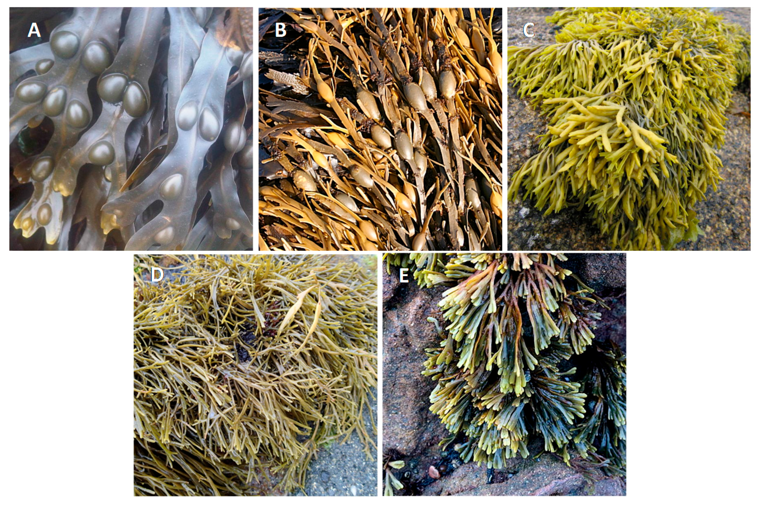
Table 1.
Selected studies of antioxidant activity of phlorotannin extracts of some Fucaceae, as measured by in vitro and in vivo biological models.
Table 1.
Selected studies of antioxidant activity of phlorotannin extracts of some Fucaceae, as measured by in vitro and in vivo biological models.
| Extraction Method | Model | Treatment Conditions | Effect | References |
|---|---|---|---|---|
| F. vesiculosus | ||||
| EtOH 80% → fractionation with n-Hex and EtOAc → subfractionation of EtOAc in Sephadex LH-20 | PMA-treated mononuclear cells from human blood | 10 μM PMA + 1.5 μg/mL of 6 different EtOAc sub-fractions | All sub-fractions (except the 4th) ↓ ROS levels below 65% | [91] |
| MeOH 60% | Caco-2 cells | 100 μg/mL of extract for 24 h | ↑ GSH levels by 31.9% | [88] |
| MeOH 60% | H2O2-induced Caco-2 cells | 24 h pre-treatment with 100 μg/mL of extract for 24 h + 200 μM H2O2 | Restored SOD levels from 64.9 to 89% and ↓ 9.5% of the DNA damage | [39,88] |
| MeOH 60% | t-BHP-induced Caco-2 cells | 100 μg/mL of extract for 24 h + 200 μM t-BHP | ↓ apx. 12% DNA damage in t-BHP-induced cells | [98] |
| Ext1: EtOH 35% Ext2: EtOH 70% | In vitro: PMA or LPS-induced Raw 264.7 cells In vivo: Sprague–Dawley rats | In vitro: 100 ng/mL PMA or LPS + different concentrations of extracts In vivo: oral treatment with 200 mg/kg/day during 4 weeks | In vitro: Ext2: ↓ of O2●− in PMA-induced cells (IC50 = 31 μg/mL), Ext1: ↓ of O2●− in both cell models (IC50 = 38 and 39 μg/mL, respectively); In vivo: Ext2: ↑ reducing power, PON-1 activity and O2•− scavenging activity in the blood plasma (29%, 33% and 25%, respectively) | [97] |
| F. serratus | ||||
| MeOH 60% | Caco-2 cells | 100 μg/mL of extract for 24 h | ↑ GSH levels by 37.4% | [88] |
| MeOH 60% | t-BHP or H2O2-induced Caco-2 cells | 100 μg/mL of extract for 24 h + 1 mM t-BHP or 200 μM H2O2 | Restored SOD levels in both t-BHP and H2O2-induced cells from 73.9–108% and 64.9–89.5%, respectively, and ↓ 13.2% of the H2O2-induced DNA damage | [39,88] |
| Ext1: H2O Ext2: EtOH 80% | t-BHP-induced Caco-2 cells | 100 μg/mL of extracts for 24 h + 1 mM t-BHP | Both extracts ↓ apx. 13% DNA damage in t-BHP-induced cells | [98] |
| A. nodosum | ||||
| Extract with 18% phlorotannins | t-BHP-induced ARPE-19 and WKD cells | 0.1–0.5% extract for 20 min + 500 μM t-BHP | ↓ ROS production close to the negative control on cells treated with 0.2% extract | [38] |
| MeOH 60% → digestion with pepsin at 37 °C and pH 2 → digestion with pancreatin/bile extract at 37 °C pH 6.9 → dialysis with cutoff at 1 kDa | t-BHP-induced HepG-2 cells | 0.5–50 μg/mL of extract for 20 h + 400 μM t-BHP | ↓ ROS and lipid, restored GSH levels to apx. 75% and regulated the activity of GSH-px, GSH-red GSH-tr | [99] |
| MeOH 60% | Caco-2 cells | 100 μg/mL of extract for 24 h | ↑ GSH levels by 35.5% | [88] |
| MeOH 60% | H2O2-induced Caco-2 cells | 100 μg/mL of extract for 24 h + 200 μM H2O2 | Restored SOD levels from 64.9–89.5% | [88] |
| Ext1: H2O Ext2: EtOH 60% Ext3: EtOH 80% | t-BHP or H2O2-induced Caco-2 cells | 100 μg/mL of extracts for 24 h + 1 mM t-BHP or 200 μM H2O2 | Ext1: ↓ 20% H2O2-induced DNA damage; Ext2: ↓ apx. 15% t-BHP -induced DNA damage, Ext3: ↓ apx. 13% DNA damage in both models | [98] |
| P. canaliculata | ||||
| MeOH 60% | Caco-2 cells | 100 μg/mL of extract for 24 h | ↑ GSH levels by 38.7% | [88] |
| MeOH 60% | t-BHP or H2O2-induced Caco-2 cells | 100 μg/mL of extract for 24 h + 1 mM t-BHP or 200 μM H2O2 | Restored SOD levels from 73.9–97% and 64.9–97.4%, respectively | [39,88] |
apx., approximately; EtOAc, ethyl acetate; EtOH, ethanol; Ext, extraction; GSH, glutathione; GSH-px, glutathione peroxidase; GSH-red, glutathione reductase; GSH-tr, glutathione transferase; LPS, lipopolysaccharide; MeOH, methanol; n-Hex, n-hexane; PON-1, paraoxonase 1; SOD, superoxide dismutase; PMA, phorbol-12-myristate-13-acetate; ROS, reactive oxygen species; t-BHP, and tert-butyl hydroperoxide. Cell lines: ARPE-19, human retinal pigment epithelium; Caco-2, human epithelial colorectal adenocarcinoma; HepG-2, liver hepatocellular carcinoma; Raw 264.7, murine macrophages; and WKD, human conjunctival cells.
Table 2.
Selected studies of the anti-diabetic activity of phlorotannin extracts of some Fucaceae, as measured in vitro and in vivo.
Table 2.
Selected studies of the anti-diabetic activity of phlorotannin extracts of some Fucaceae, as measured in vitro and in vivo.
| Extraction Method | Model | Test Conditions | Effect | References |
|---|---|---|---|---|
| F. vesiculosus | ||||
| Sequential extraction with CHCl3 → EtOH 96% → Ac 70% | Measurement of α-glucosidase activity | Crescent concentrations of extracts | EtOH and Ac extracts had the highest inhibitory activity (IC50 = 4.4 and 0.34 μg/mL, respectively) | [111] |
| Ext1: H2O Ext2: EtOH | Measurement of α-glucosidase and α-amylase activities | 0.1–1000 μg/mL of extracts | ↓ enzymatic activity (α-glucosidase: IC50 = 0.32 and 0.49 μg/mL, respectively; α-amylase: IC50 = 59.1 and 63.5 μg/mL, respectively) | [47] |
| Ac 70% → fractionation with DCM, EtOAc and But → subfractionation of EtOAc in Sephadex LH-20 (F1–F4) | BSA-methylglyoxal and BSA-glucose assay | Crescent concentrations of fractions or sub-fractions | Strong ↓ BSA glycation by subfractions, (EC50 apx. 0.16 mg/mL for F1–F4 in BSA-methylglyoxal and 0.05 mg/mL for F1 and F2 in BSA-glucose) | [116] |
| F. distichus | ||||
| EtOH 80% → Fractionation with n-hex, EtOAc, 1-But → subfractionation of EtOAc in Sephadex LH-20 | Measurement of α-glucosidase and α-amylase activities | 1.5–200 μg/mL of subfractions | Subfraction 22 showed ↑ inhibitory activity (IC50 = 0.89 and 13.98 μg/mL, respectively) | [115] |
| A. nodosum | ||||
| EtOH 50% at 80 °C → Fractionation with EtOAc and 1-But → purification in C18 column | Measurement of α-glucosidase activity | Crescent concentrations of fractions | Purified fraction showed ↑ inhibitory activity (IC50 = 24 μg/mL) | [117] |
| Sequential extraction with CHCl3 → EtOH 96% → Ac 70% | Measurement of α-glucosidase activity | Crescent concentrations of extracts | Ac extracts showed ↑ inhibitory activity (IC50 = 0.72 μg/mL) | [111] |
| H2O at 80 °C from algae collected at different seasons | Measurement of α-glucosidase activity | 0.05–0.5 μg/mL of extract | Summer extracts have ↑ inhibitory activity (IC70 = 2.23 μg/mL) | [113] |
| Ext1: H2O Ext2: EtOH | Measurement of α-glucosidase and α-amylase activities | 0.1–1000 μg/mL of extracts | ↓ enzymatic activity (α-glucosidase: IC50 = n.d.; α-amylase: IC50 = 44.7 and 53.6 μg/mL, respectively) | [47] |
| EtOH 50% | 2-deoxyglucose-cultured 3T3-L1 cells | 50–400 μg/mL of extract for 20 min + 1 μCi/mL 2-deoxyglucose | ↑ basal glucose uptake by 3-fold at 400 μg/mL | [117] |
| ACN:0.2% CH2O2 (1:1) → purification in SPE column → fractionation in Sephadex LH-20 | Measurement of α-glucosidase and α-amylase activities in absence or presence of acarbose | Phlorotannin fraction: 2.5–100 μg GAE/mL for α-glucosidase and 50–400 μg GAE/mL for α-amylase; acarbose + phlorotannin fraction: 1 μg/mL + 0.1 μg/GAE –0.25 μg/mL + 0.025 μg/GAE | ↓ enzymatic activity (α-glucosidase: IC50 = 10 μg GAE/mL; α-amylase: IC50 = 0.15 μg GAE/mL). ↓ acarbose concentration needed for an effective enzymatic inhibition (from 1–0.5 μg/mL) | [118] |
| P. canaliculata | ||||
| MeOH 70% | In vitro: measurement of sucrase and maltase activities In vivo: sucrose-fed Wistar rats | In vitro: 0–16.7 mg/mL extract In vivo: oral administration of 1 mg/kg of extract + 0.5 mg/kg of sucrose | In vitro: ↓ enzymatic activity (IC50 = 2.24 and 2.84 mg/mL, respectively) In vivo: ↓ postprandial blood glucose levels | [112] |
| A. nodosum combined with F. vesiculosus | ||||
| Commercial hot water extract InSea2TM (10% polyphenol content in CAE) | In vitro: measurement of α-glucosidase and α-amylase activities In vivo: Wistar rats fed with corn starch + safflower oil | In vitro: 1.25–25 μg/mL of InSea2TM In vivo: oral administration of 7.5 mg/kg of InSea2TM + 2 mL/kg of starch and oil (1:1) | In vitro: ↓ enzymatic activity (IC50 = 2.8 and 5 μg/mL, respectively) In vivo: ↓ 90% postprandial blood glucose and ↓ 40% insulin peak | [119] |
| Commercial hot water extract InSea2TM (10% polyphenol content in CAE) | Human trial | Oral administration of two capsules (500 mg) 30 min prior to carbohydrate ingestion | ↓ insulin incremental area of the curve by 12.1% and ↑ insulin sensitivity by 7.9% | [120] |
Ac, acetone; apx., approximately; BSA, bovine albumin serum; But, butanol; CAE, chlorogenic acid equivalents; DCM, dichloromethane; EtOAc, ethyl acetate; EtOH, ethanol; Ext, extraction; HCl, chloridric acid; GAE, gallic acid equivalents; SPE column, solid-phase extraction column; and ROS, reactive oxygen species; Cell lines: INS-1, rat pancreatic β-cells; and 3T3-L1, preadipocytes.
Table 3.
Selected studies of the anti-inflammatory activity of phlorotannin extracts of some Fucaceae, as measured in in vitro and ex vivo biological models.
Table 3.
Selected studies of the anti-inflammatory activity of phlorotannin extracts of some Fucaceae, as measured in in vitro and ex vivo biological models.
| Extraction method | Model | Test Conditions | Effect | References |
|---|---|---|---|---|
| F. vesiculosus | ||||
| H2O | LPS-induced porcine colonic tissue ex vivo | 1 mg/mL extract + 10 µg/mL LPS | ↓ expression of the genes IL17A, IL8, CCL2, CXCL2, CXCL10, CXCL11, ICAM1, VCAM1, TLR4, TLR7, NFKB1, RELB, MAP3K8, CJUN, PTGS2, C5 and LYZ >2× compared to the control | [128] |
| EtOH 35% | PMA-stimulated RAW 264.7 | 100 ng/mL PMA + different concentrations of extracts | ↓ production of NO● (IC50 = 37 µg/mL) | [97] |
| F. serratus | ||||
| H2O | LPS-induced porcine colonic tissue ex vivo | 1 mg/mL extract + 10 µg/mL LPS | ↓ expression of the genes IL8, IL6 and TNFA below 0.70, 0.69 and 1.15× compared to LPS control, respectively | [42] |
| F. distichus | ||||
| MeOH 80% → fractionation with n-hex, EtOAc and 1-But → subfractionation of EtOAc in flash chromatography | LPS-induced RAW 264.7 cells | 12.5–50 µg/mL a subfraction rich in fucophlorethols for 1 h + 1 µg/mL LPS | ↓ expression of IL-1β, IL-6, IL-17, TNF-α, MCP-1, iNOS, COX-2, ICAM-1, TLR-4 and TLR-9 in a dose-dependent manner | [131] |
| A. nodosum | ||||
| Extract with 18% phlorotannins | LPS-induced U937 cells | 0.05–0.2% of extract for 2 h + 0.5 µg/mL LPS | ↓ levels of TNF-α and IL-6 close to control | [38] |
| H2O → alginate precipitation → ultrafiltration | LPS-induced U937 cells | 0.1 µg extract for 2 h + 0.5 µg/mL LPS | ↓ levels of TNF-α by 94% and IL-6 by 84% | [126] |
| EtOH 80% | LPS-induced porcine colonic tissue ex vivo | 1 mg/mL extract + 10 µg/mL LPS | ↓ expression of the genes IL8, IL6 and TNFA below 0.99, 0.75 and 1.01× compared to LPS control, respectively | [42] |
| EtOH 80% | TNF-α-induced Caco-2 cells | 0.1–1 mg/mL extract + 10 ng/mL TNF-α | ↓ expression of the genes IL8, TNFA, IL1B, IL18, CSF1, CXCL10, CCL5, NFKB2, IKBKB, PTGS2 and MIF by >2× | [127] |
| EtOH 80% → dialysis fractionation into three Mw fractions (<3.5 kDa, 3.5–100 kDa, >100 kDa) | LPS-induced porcine colonic tissue ex vivo | 1 mg/mL extract or Mw fractions + 10 µg/mL LPS | ↓ expression of the genes LYZ, IL8, PTGS2, TLR6, CXCL10, IL6, CXCL11, ICAM, NFKB1 and CXCL2 by >2× either by the crude extract or the three Mw fractions | [127] |
COX-2, cyclooxygenase-2; EtOAc, ethyl acetate; IL, interleukin; iNOS, inducible nitric oxide synthase; LPS, lipopolysaccharide; Mw, molecular weight; NO●, nitric oxide; PMA, phorbol-12-myristate-13-acetate; and TNF-α, tumor necrosis factor-α. Cell lines: RAW 264.7, murine macrophages; U937, human leukemic monocytes; and Caco-2, human colon epithelium.
Table 4.
Selected studies of the antitumor activity of phlorotannin extracts of some Fucaceae, as measured in in vitro biological models.
Table 4.
Selected studies of the antitumor activity of phlorotannin extracts of some Fucaceae, as measured in in vitro biological models.
| Extraction Method | Model | Test Conditions | Effect | References |
|---|---|---|---|---|
| F. vesiculosus | ||||
| Acetone 99.5% → purification by HPLC | PancTu1, Panc89, Panc1 and Colo357 cells | 12.5–100 µg/mL of purified extract | ↓ cell proliferation, ↑ cell cycle inhibitors (IC50 = 17.35 µg/mL, 17.5 µg/mL, 19.23 µg/mL and 28.9 µg/mL, for each cell line, respectively) | [133] |
| H2O → precipitation → normal phase HPLC → reversed phase HPLC→ F15 + F16 | Panc89 and PancTu1 cells | 0.2–200 µg/mL of fractions | ↓ cell proliferation (F15: IC50 = 15.2 and 18.3 μg/mL, respectively; F16: IC50 = 16.4 and 16.2 μg/mL) | [134] |
| F. spiralis | ||||
| Ext1: DCM Ext2: MeOH 100% Ext3: n-hex fraction of Ext2 | HeLa cells | Crescent concentrations of dichloromethane extract | ↑ apoptosis, with Ext1 showing highest activity (IC50 = 10.7 μg/mL) | [135] |
| Ac 70% → purification with cellulose | Hyaluronidase activity measurement | 0.5–2.25 mg/mL of extract | ↓ enzymatic activity (IC50 = 0.73 mg/mL dry weight) | [136] |
| A. nodosum | ||||
| ACN:0.2% CH2O2 (1:1) → purification in SPE columns | Caco-2 cells | 15–42.5 µg/mL of extract | ↓ cell proliferation (IC50 = 33 μg/mL) | [114] |
Ac, acetone; ACN, acetonitrile; DCM, dichloromethane; Ext, extraction; F15, fraction 15; F16, fraction 16; HPLC, high performance liquid chromatography; MeOH, methanol; SPE column, and solid-phase extraction column. Cell lines: Caco-2, human colon cancer; HeLa, human cervix carcinoma; PancTu1, human pancreatic cancer; Colo357, human pancreatic adenosquamous carcinoma; Panc89, human pancreatic cancer; and Panc1, pancreatic carcinoma.
© 2017 by the authors. Licensee MDPI, Basel, Switzerland. This article is an open access article distributed under the terms and conditions of the Creative Commons Attribution (CC BY) license (http://creativecommons.org/licenses/by/4.0/).
Share and Cite
MDPI and ACS Style
Catarino, M.D.; Silva, A.M.S.; Cardoso, S.M. Fucaceae: A Source of Bioactive Phlorotannins. Int. J. Mol. Sci. 2017, 18, 1327. https://doi.org/10.3390/ijms18061327
AMA Style
Catarino MD, Silva AMS, Cardoso SM. Fucaceae: A Source of Bioactive Phlorotannins. International Journal of Molecular Sciences. 2017; 18(6):1327. https://doi.org/10.3390/ijms18061327
Chicago/Turabian StyleCatarino, Marcelo D., Artur M. S. Silva, and Susana M. Cardoso. 2017. "Fucaceae: A Source of Bioactive Phlorotannins" International Journal of Molecular Sciences 18, no. 6: 1327. https://doi.org/10.3390/ijms18061327
Note that from the first issue of 2016, this journal uses article numbers instead of page numbers. See further details here.