Lipidomics by Supercritical Fluid Chromatography


Abstract
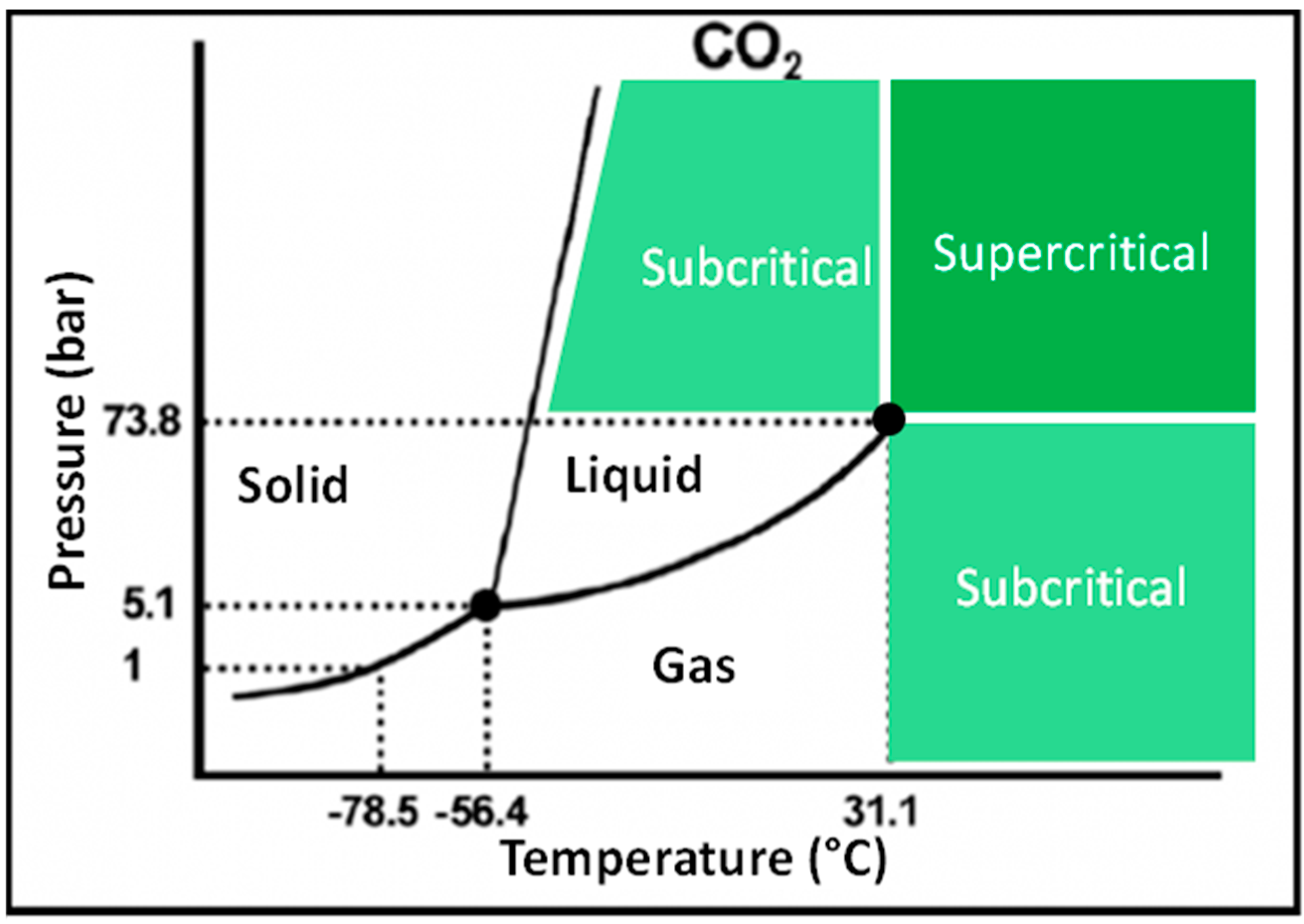
:1. Supercritical Fluids
Physical Chemical Properties


| Type of Fluid | Volumetric Mass Density (g∙cm−3) | Viscosity (cP) | Diffusivity (cm−2∙s−1) |
|---|---|---|---|
| Gas | 10−3 | 10−2 | 0.2 |
| Supercritical state | 0.5 | 5 × 10⁻² | 5 × 10⁻⁴ |
| Liquid | 1 | 1 | 10−5 |
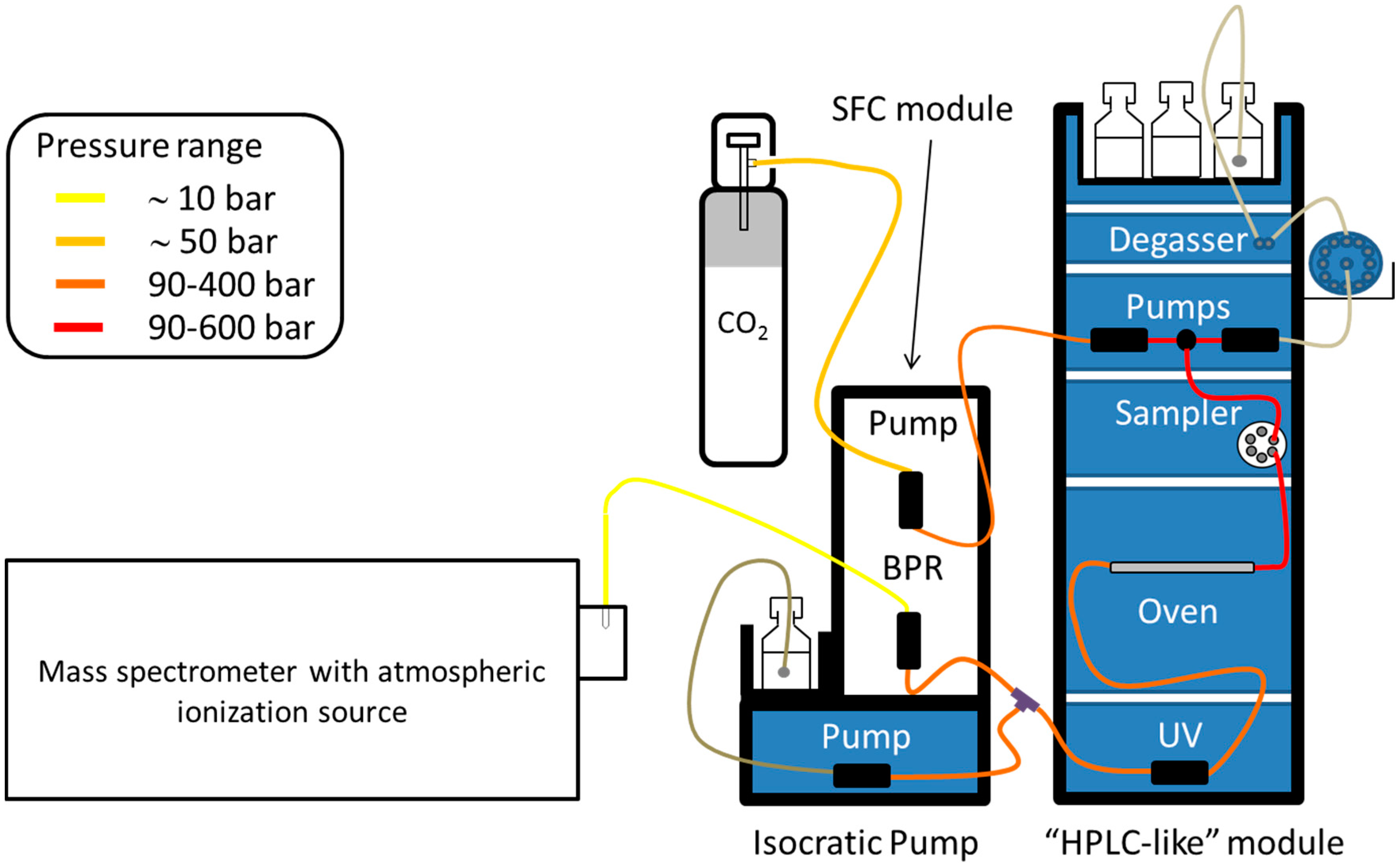
2. Supercritical Fluid Chromatography (SFC)
2.1. Capillary SFC
2.2. Packed Column SFC
2.3. Detection Methods

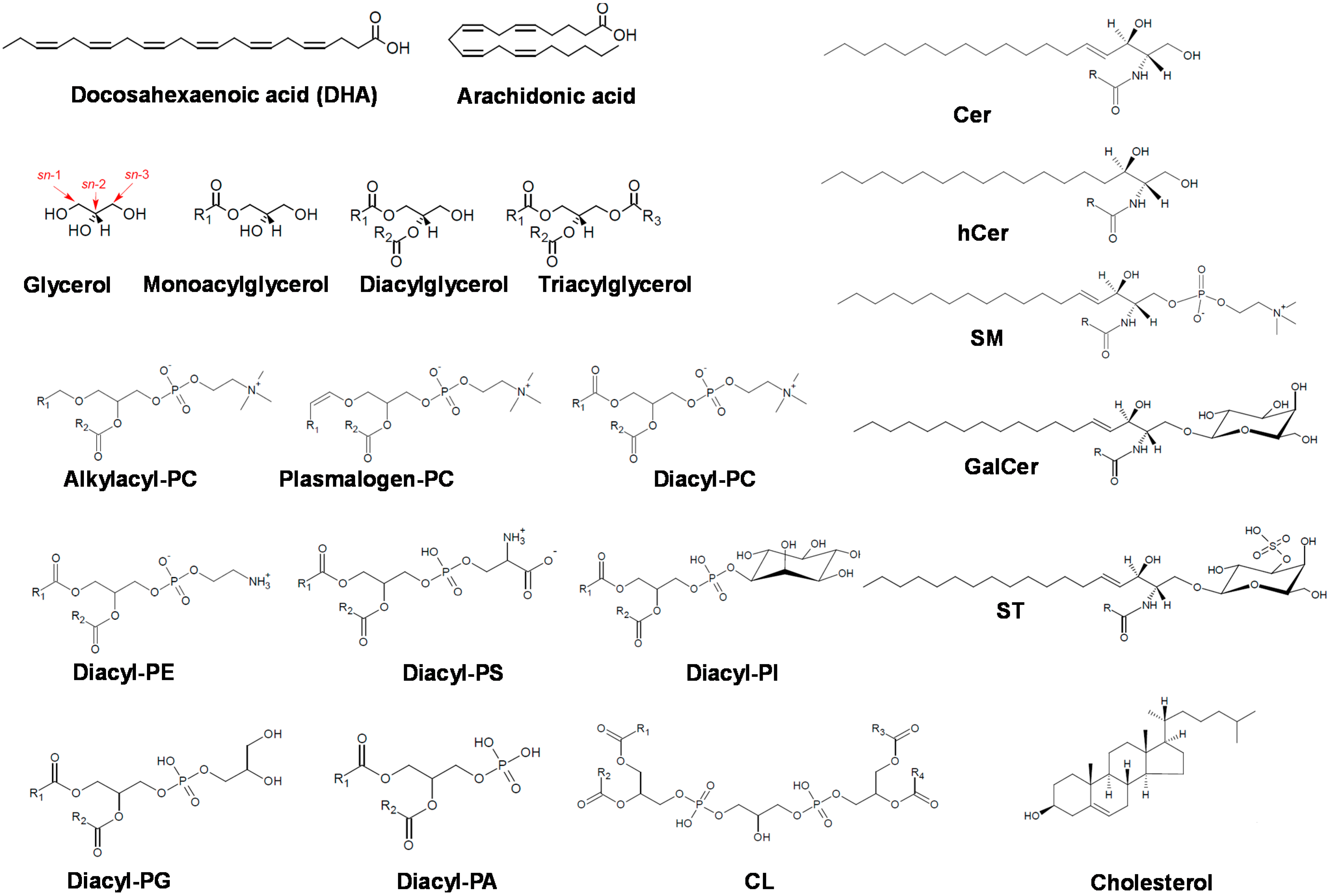
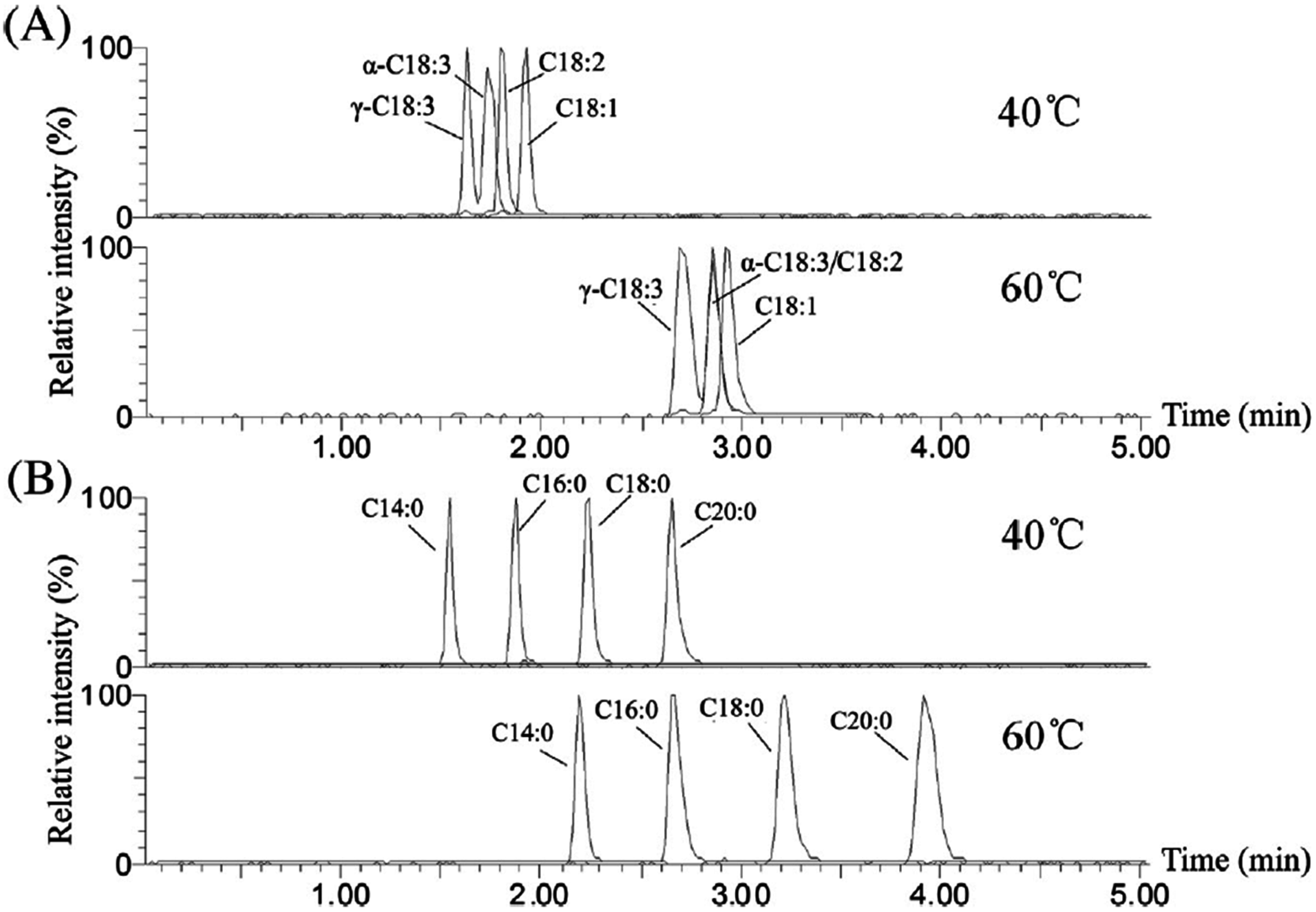
3. Applications to Lipid Analysis

3.1. Fatty Acyls

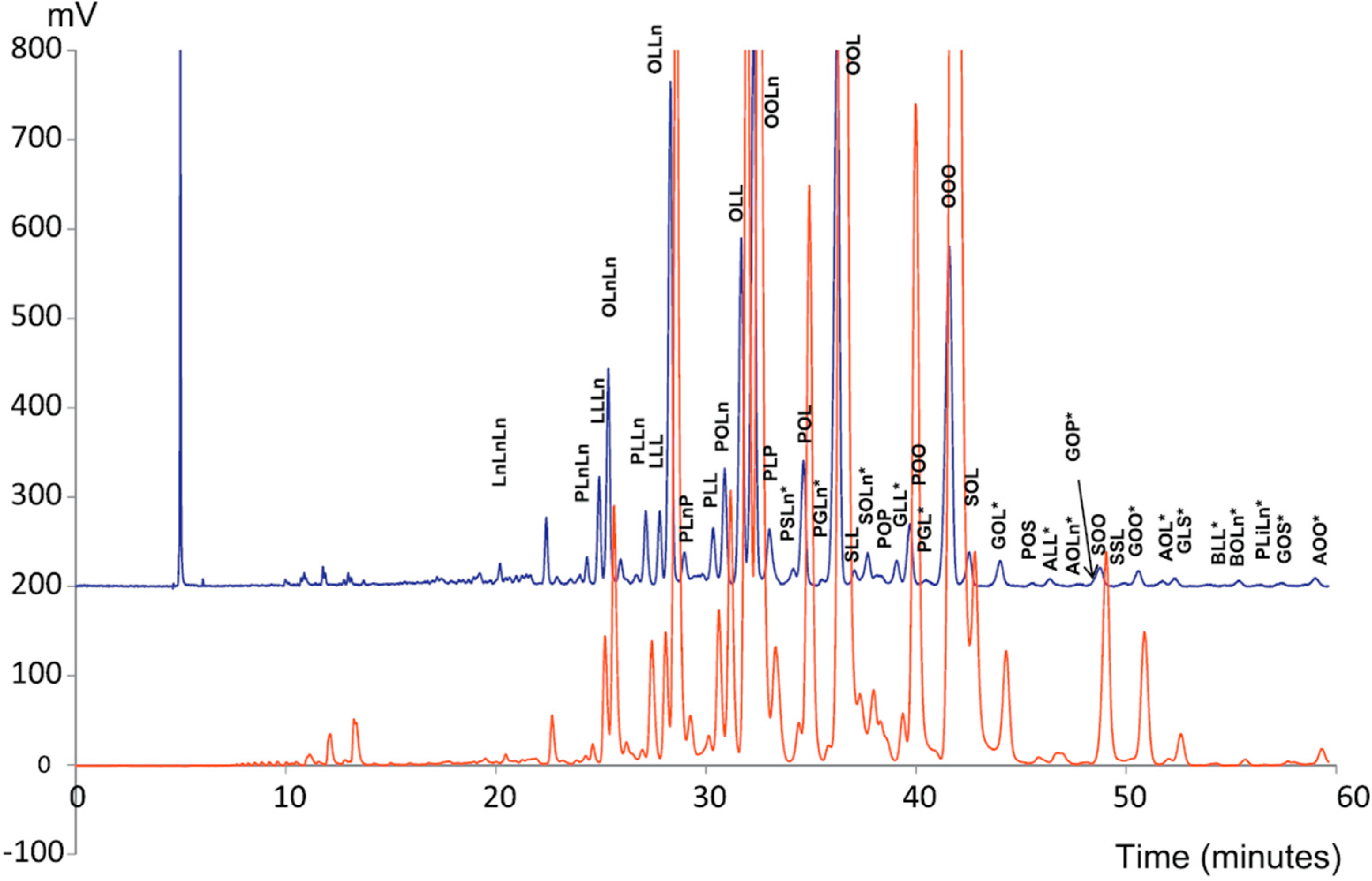
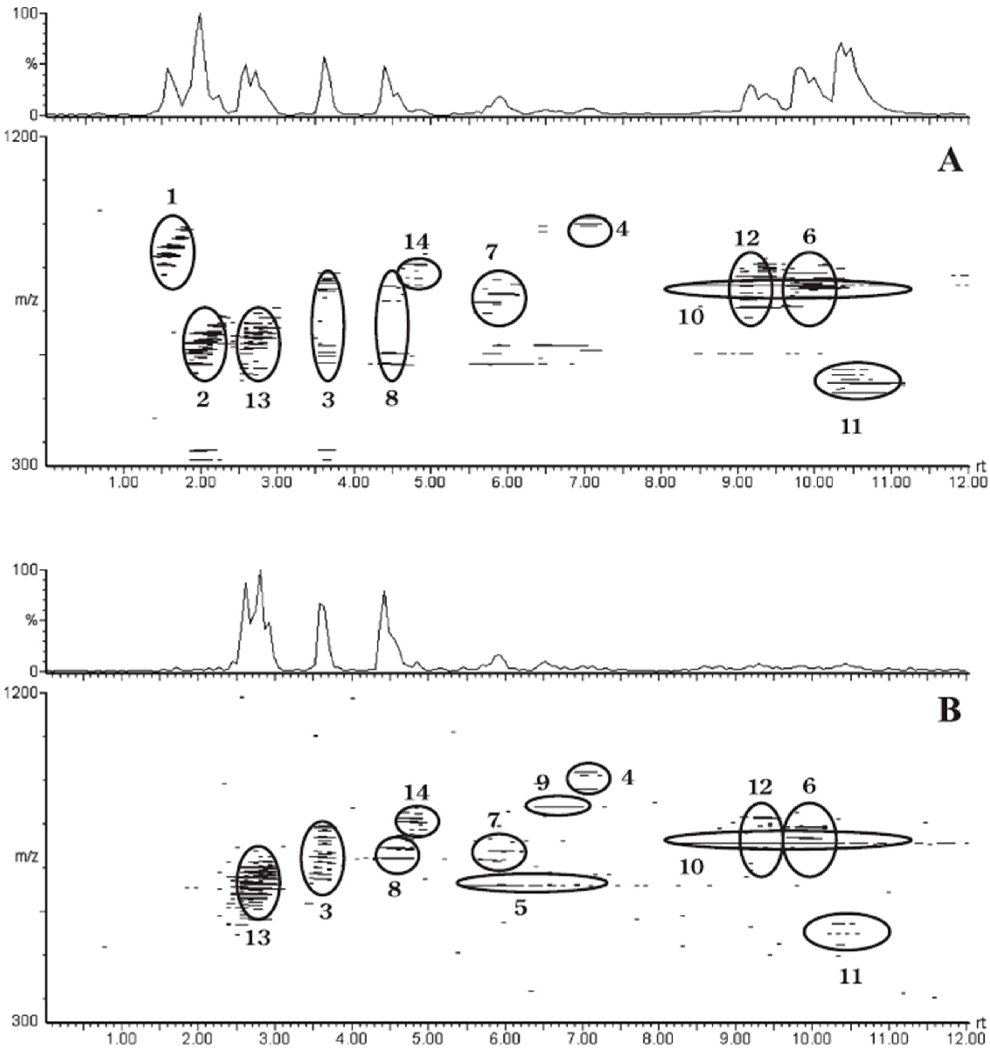
3.2. Glycerolipids

3.3. Glycerophospholipids/Sphingolipids

3.4. Sterol Lipids
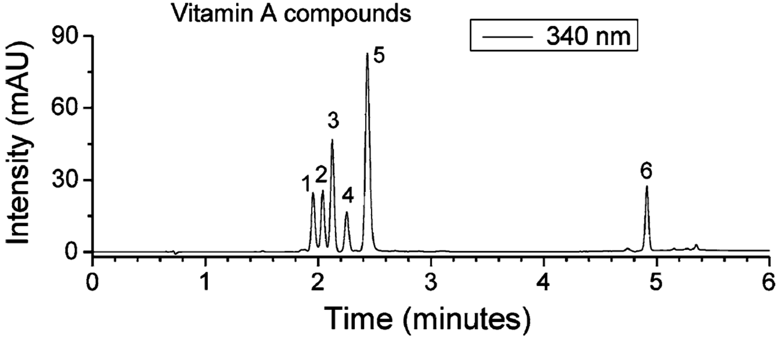
3.5. Prenol Lipids

3.6. Polyketides
4. Perspectives
Acknowledgments
Author Contributions
Conflicts of Interest
References
- Lesellier, E.; West, C. The many faces of packed column supercritical fluid chromatography—A critical review. J. Chromatogr. A 2015, 1382C, 2–46. [Google Scholar] [CrossRef] [PubMed]
- Nováková, L.; Perrenoud, A.G.; Francois, I.; West, C.; Lesellier, E.; Guillarme, D. Modern analytical supercritical fluid chromatography using columns packed with sub-2 μm particles: A tutorial. Anal. Chim. Acta 2014, 824, 18–35. [Google Scholar] [CrossRef] [PubMed]
- Saito, M. History of supercritical fluid chromatography: Instrumental development. J. Biosci. Bioeng. 2013, 115, 590–599. [Google Scholar] [CrossRef] [PubMed]
- Klesper, K.; Corwin, A.H.; Turner, D.A. High pressure gas chromatography above critical temperature. J. Org. Chem. 1962, 27, 700–706. [Google Scholar]
- Novotny, M.V. Recent developments in analytical chromatography. Science 1989, 246, 51–57. [Google Scholar] [CrossRef] [PubMed]
- Lee, M.L.; Markides, K.E. Chromatography with supercritical fluids. Science 1987, 235, 1342–1347. [Google Scholar] [CrossRef] [PubMed]
- Karayannis, N.K.; Corwin, A.H.; Baker, E.W.; Klesper, E.; Walter, J.A. Apparatus and materials for hyperpressure gas chromatography of non-volatile compounds. Anal. Chem. 1968, 40, 1736–1739. [Google Scholar] [CrossRef]
- Giddings, C.G.; Myers, M.N.; King, J.W. Dense gas chromatography at pressures to 2000 atmospheres. J. Chromatogr. Sci. 1969, 7, 276–283. [Google Scholar] [CrossRef]
- Speybrouck, D.; Corens, D.; Argoullon, J.M. Screening strategy for chiral and achiral separations in supercritical fluid chromatography mode. Curr. Top. Med. Chem. 2012, 12, 1250–1263. [Google Scholar] [CrossRef] [PubMed]
- Lesellier, E.; Valarché, A.; West, C.; Dreux, M. Effects of selected parameters on the response of the evaporative light scattering detector in supercritical fluid chromatography. J. Chromatogr. A 2012, 1250, 220–226. [Google Scholar] [CrossRef] [PubMed]
- Xia, Z.; Thurbide, K.B. Universal acoustic flame detection for modified supercritial fluid chromatography. J. Chromatogr. A 2006, 1105, 180–185. [Google Scholar] [CrossRef] [PubMed]
- Ventura, M.C.; Farrell, W.P.; Aurigemma, C.M.; Greig, M.J. Packed column supercritical fluid chromatography/mass spectrometry for high-throughput analysis. Part 2. Anal. Chem. 1999, 71, 4223–4231. [Google Scholar] [CrossRef] [PubMed]
- Méjean, M.; Brunelle, A.; Touboul, D. Quantification of tocopherols and tocotrienols in soybean oil by supercritical-fluid chromatography coupled to high-resolution mass spectrometry. Anal. Bioanal. Chem. 2015. [Google Scholar] [CrossRef] [PubMed]
- Bamba, T.; Lee, J.W.; Matsubara, A.; Fukusaki, E. Metabolic profiling of lipids by supercritical fluid chromatography/mass spectrometry. J. Chromatogr. A 2012, 1250, 212–219. [Google Scholar] [CrossRef] [PubMed]
- Bernal, J.L.; Martin, M.T.; Toribio, L. Supercritical fluid chromatography in food analysis. J. Chromatogr. A 2013, 1313, 24–36. [Google Scholar] [CrossRef] [PubMed]
- Bamba, T.; Shimonishi, N.; Matsubara, A.; Hirata, K.; Nakazawa, Y.; Kobayashi, A.; Fukusaki, E. High throughput and exhaustive analysis of diverse lipids by using supercritical fluid chromatography-mass spectrometry for metabolomics. J. Biosci. Bioeng. 2008, 105, 460–469. [Google Scholar] [CrossRef] [PubMed]
- West, C.; Lesellier, E. A unified classification of stationary phases for packed column supercritical fluid chromatography. J. Chromatogr. A 2008, 1191, 21–39. [Google Scholar] [CrossRef] [PubMed]
- Fahy, E.; Subramaniam, S.; Brown, H.A.; Glass, C.K.; Merrill, A.H., Jr.; Murphy, R.C.; Raetz, C.R.; Russell, D.W.; Seyama, Y.; Shaw, W.; et al. A comprehensive classification system for lipids. J. Lipid Res. 2005, 46, 839–861. [Google Scholar] [CrossRef] [PubMed]
- Fahy, E.; Subramaniam, S.; Murphy, R.C.; Nishijima, M.; Raetz, C.R.; Shimizu, T.; Spener, F.; van Meer, G.; Wakelam, M.J.; Dennis, E.A. Update of the LIPID MAPS comprehensive classification system for lipids. J. Lipid Res. 2009, 50, S9–S14. [Google Scholar] [CrossRef] [PubMed]
- King, J.W.; List, G.R. Supercritical Fluid Technology in Oil and Lipid Chemistry; AOCS Press: Champaign, IL, USA, 1996. [Google Scholar]
- Qu, S.; Du, Z.; Zhang, Y. Direct detection of free fatty acids in edible oils using supercritical fluid chromatography coupled with mass spectrometry. Food Chem. 2015, 170, 463–469. [Google Scholar] [CrossRef] [PubMed]
- Francois, I.; Sandra, P. Comprehensive supercritical fluid chromatography X reversed phase liquid chromatography for the analysis of the fatty acids in fish oil. J. Chromatogr. A 2009, 1216, 4005–4012. [Google Scholar] [CrossRef] [PubMed]
- Hirata, Y.; Sogabe, I. Separation of fatty acid methyl esters by comprehensive two-dimensional supercritical fluid chromatography with packed columns and programming of sampling duration. Anal. Bioanal. Chem. 2014, 378, 1999–2003. [Google Scholar] [CrossRef] [PubMed]
- Hori, K.; Matsubara, A.; Uchikata, T.; Tsumura, K.; Fukusaki, E.; Bamba, T. High-throughput and sensitive analysis of 3-monochloropropane-1,2-diol fatty acid esters in edible oils by supercritical fluid chromatography/tandem mass spectrometry. J. Chromatogr. A 2012, 1250, 99–104. [Google Scholar] [CrossRef] [PubMed]
- Koski, I.J.; Jansson, B.A.; Markides, K.E.; Lee, M.L. Analysis of prostaglandins in aqueous solutions by supercritical fluid extraction and chromatography. J. Pharm. Biomed. Anal. 1991, 9, 281–290. [Google Scholar] [CrossRef]
- Giron, D.; Link, R.; Bouissel, S. Analysis of mono-, di- and triglycerides in pharmaceutical excipients by capillary supercritical fluid chromatography. J. Pharm. Biomed. Anal. 1992, 10, 821–830. [Google Scholar] [CrossRef]
- Kallio, H.; Laakso, P.; Huopalahti, R.; Linko, R.R.; Oksman, P. Analysis of butter fat triacylglycerols by supercritical fluid chromatography/electron impact mass spectrometry. Anal. Chem. 1989, 61, 698–700. [Google Scholar] [CrossRef] [PubMed]
- Laakso, P.; Manninen, P. Identification of milk fat triacylglycerols by capillary supercritical fluid chromatography-atmospheric pressure chemical ionization mass spectrometry. Lipids 1997, 32, 1285–1295. [Google Scholar] [CrossRef] [PubMed]
- Manninen, P.; Laakso, P. Capillary supercritical fluid chromatography—Atmospheric pressure chemical ionization mass spectrometry of gamma- and alpha-linolenic acid containing triacylglycerols in berry oils. Lipids 1997, 32, 825–831. [Google Scholar] [CrossRef] [PubMed]
- Lesellier, E.; Tchapla, A. Retention behavior of triglycerides in octadecylpacked subcritical fluid chromatography with CO2/modifier mobile phases. Anal. Chem. 1999, 71, 5372–5378. [Google Scholar] [CrossRef] [PubMed]
- Lesellier, E.; Bleton, J.; Tchapla, A. Use of relationships between retention behaviors and chemical structures in subcritical fluid chromatography with CO2/modifier mixtures for the identification of triglycerides. Anal. Chem. 2000, 72, 2573–2580. [Google Scholar] [CrossRef] [PubMed]
- Lee, J.W.; Uchikata, T.; Matsubara, A.; Nakamura, T.; Fukusaki, E.; Bamba, T. Application of supercritical fluid chromatography/mass spectrometry to lipid profiling of soybean. J. Biosci. Bioeng. 2012, 113, 262–268. [Google Scholar] [CrossRef] [PubMed]
- Lesellier, E.; Latos, A.; de Oliveira, A.L. Ultra high efficiency/low pressure supercritical fluid chromatography with superficially porous particles for triglyceride separation. J. Chromatogr. A 2014, 1327, 141–148. [Google Scholar] [CrossRef] [PubMed]
- Sandra, P.; Medvedovici, A.; Zhao, Y.; David, F. Characterization of triglycerides in vegetable oils by silver-ion packed-column supercritical fluid chromatography coupled to mass spectroscopy with atmospheric pressure chemical ionization and coordination ion spray. J. Chromatogr. A 2002, 974, 231–241. [Google Scholar] [CrossRef]
- Hirata, Y.; Hashiguchi, T.; Kawata, E. Development of comprehensive two-dimensional packed column supercritical fluid chromatography. J. Sep. Sci. 2003, 26, 531–535. [Google Scholar] [CrossRef]
- Yamada, T.; Uchikata, T.; Sakamoto, S.; Yokoi, Y.; Nishiumi, S.; Yoshida, M.; Fukusaki, E.; Bamba, T. Supercritical fluid chromatography/Orbitrap mass spectrometry based lipidomics platform coupled with automated lipid identification software for accurate lipid profiling. J. Chromatogr. A 2013, 1301, 237–242. [Google Scholar] [CrossRef] [PubMed]
- Lafosse, M.; Elfakir, C.; Morin-Allory, L.; Dreux, M. The advantages of evaporative light scattering detection in pharmaceutical analysis by high performance liquid chromatography and supercritical fluid chromatography. J. High Res. Chromatogr. 1992, 15, 312–318. [Google Scholar] [CrossRef]
- Yip, H.S.H.; Ahraf-Khorassani, M.; Taylor, L.T. Feasibility of phospholipids separation by packedcolumn SFC with mass spectrometric and light scattering detection. Chromatographia 2007, 65, 655–665. [Google Scholar] [CrossRef]
- Lee, J.W.; Yamamoto, T.; Uchikata, T.; Matsubara, A.; Fukusaki, E.; Bamba, T. Development of a polar lipid profiling method by supercritical fluid chromatography/mass spectrometry. J. Sep. Sci. 2011, 34, 3553–3560. [Google Scholar] [CrossRef] [PubMed]
- Uchikata, T.; Matsubara, A.; Nishiumi, S.; Yoshida, M.; Fukusaki, E.; Bamba, T. Development of oxidized phosphatidylcholine isomer profiling method using supercritical fluid chromatography/tandem mass spectrometry. J. Chromatogr. A 2012, 1250, 205–211. [Google Scholar] [CrossRef] [PubMed]
- Kim, D.H.; Lee, K.J.; Heo, G.S. Analysis of cholesterol and cholesteryl esters in human serum using capillary supercritical fluid chromatography. J. Chromatogr. B Biomed. Appl. 1994, 655, 1–8. [Google Scholar] [CrossRef]
- Ong, C.P.; Lee, H.K.; Li, S.F. Supercritical fluid extraction and chromatography of cholesterol in food samples. J. Chromatogr. 1990, 515, 509–513. [Google Scholar] [CrossRef]
- McAllister, H.; Wu, J. Chromatographic separation of bioactive oxycholesterols by GC, HPLC and SFC. Curr. Top. Med. Chem. 2012, 12, 1264–1270. [Google Scholar] [CrossRef] [PubMed]
- Loran, J.S.; Cromie, K.D. An evaluation of the use of supercritical fluid chromatography with light scattering detection for the analysis of steroids. J. Pharm. Biomed. Anal. 1990, 8, 607–611. [Google Scholar] [CrossRef]
- David, P.A.; Novotny, M. Analysis of steroids by capillary supercritical fluid chromatography with phosphorus-selective detection. J. Chromatogr. 1989, 461, 111–120. [Google Scholar] [CrossRef]
- Tuomola, M.; Hakala, M.; Manninen, P. Determination of androstenone in pig fat using packed column supercritical fluid chromatography-mass spectrometry. J. Chromatogr. B Biomed. Sci. Appl. 1998, 719, 25–30. [Google Scholar] [CrossRef]
- Scalia, S.; Games, D.E. Determination of free bile acids in pharmaceutical preparations by packed column supercritical fluid chromatography. J. Pharm. Sci. 1993, 82, 44–47. [Google Scholar] [CrossRef] [PubMed]
- Scalia, S.; Games, D.E. Analysis of conjugated bile acids by packed-column supercritical fluid chromatography. J. Chromatogr. 1992, 574, 197–203. [Google Scholar] [CrossRef]
- Schmitz, H.H.; Artz, W.E.; Poor, C.L.; Dietz, J.M.; Erdman, J.W., Jr. High-performance liquid chromatography and capillary supercritical-fluid chromatography separation of vegetable carotenoids and carotenoid isomers. J. Chromatogr. 1989, 479, 261–268. [Google Scholar] [CrossRef]
- Lesellier, E. Analysis of non-saponifiable lipids by super-/subcritical-fluid chromatography. J. Chromatogr. A 2011, 936, 201–214. [Google Scholar] [CrossRef]
- Bamba, T.; Fukasaki, W.; Kajiyama, S.; Ute, K.; Kitayama, T.; Kobayashi, A. High-resolution analysis of polyprenols by supercritical fluid chromatography. J. Chromatogr. A 2001, 911, 113–117. [Google Scholar] [CrossRef]
- Matsubara, A.; Uchikata, T.; Shinohara, M.; Nishiumi, S.; Yoshida, M.; Fukusaki, E.; Bamba, T. Highly sensitive and rapid profiling method for carotenoids and their epoxidized products using supercritical fluid chromatography coupled with electrospray ionization-triple quadrupole mass spectrometry. J. Biosci. Bioeng. 2012, 113, 782–787. [Google Scholar] [CrossRef] [PubMed]
- Abrahamsson, V.; Rodriguez-Meizoso, I.; Turner, C. Determination of carotenoids in microalgae using supercritical fluid extraction and chromatography. J. Chromatogr. A 2012, 1250, 63–68. [Google Scholar] [CrossRef] [PubMed]
- Wada, Y.; Matsubara, A.; Uchikata, T.; Iwasaki, Y.; Morimoto, S.; Kan, K.; Okura, T.; Fukusaki, E.; Bamba, T. Metabolic profiling of β-cryptoxanthin and its fatty acid esters by supercritical fluid chromatography coupled with triple quadrupole mass spectrometry. J. Sep. Sci. 2011, 34, 3546–3552. [Google Scholar] [CrossRef] [PubMed]
- Mount, D.L.; Todd, G.D.; Navaratnam, V. Packed-column supercritical fluid chromatography of artemisinin (qinghaosu) with electron-capture detection. J. Chromatogr. B Biomed. Appl. 1995, 666, 183–187. [Google Scholar] [CrossRef]
- Kohler, M.; Haerdi, W.; Christen, P.; Veuthey, J.L. Extraction of artemisinin and artemisinic acid from Artemisia annua L. using supercritical carbon dioxide. J. Chromatogr. A 1997, 785, 353–360. [Google Scholar] [CrossRef]
- Smith, R.D.; Udseth, H.R.; Wright, B.W. Rapid and high resolution capillary supercritical fluid chromatography (SFC) and SFC/MS of trichothecenemycotoxins. J. Chromatogr. Sci. 1985, 23, 192–199. [Google Scholar] [CrossRef] [PubMed]
- Schaffrath, M.; Weidmann, V.; Maison, W. Enantioselective high performance liquid chromatography and supercritical fluid chromatography separation of spirocyclicterpenoid flavor compounds. J. Chromatogr. A 2014, 1363, 270–277. [Google Scholar] [CrossRef] [PubMed]
- Tavares, M.C.; Vilegas, J.H.; Lancas, F.M. Separation of underivatised triterpene acids by capillary supercritical fluid chromatography. Phytochem. Anal. 2011, 12, 134–137. [Google Scholar] [CrossRef] [PubMed]
- Lesellier, E.; Destandau, E.; Grigoras, C.; Fougere, L.; Elfakir, C. Fast separation of triterpenoids by supercritical fluid chromatography/evaporative light scattering detector. J. Chromatogr. A 2012, 1268, 157–165. [Google Scholar] [CrossRef] [PubMed]
- Méjean, M.; Vollmer, A.; Brunelle, A.; Touboul, D. Quantification of retinoid compounds by supercritical fluid chromatography coupled to ultraviolet diode array detection. Chromatographia 2013, 76, 1097–1105. [Google Scholar] [CrossRef]
- Matsubara, A.; Harada, K.; Hirata, K.; Fukusaki, E.; Bamba, T. High-accuracy analysis system for the redox status of coenzyme Q10 by online supercritical fluid extraction-supercritical fluid chromatography/mass spectrometry. J. Chromatogr. A 2012, 1250, 76–79. [Google Scholar] [CrossRef] [PubMed]
- Hadj-Mahammed, M.; Badjah-Hadj-Ahmed, Y.; Meklati, B.Y. Behaviour of polymethoxylated and polyhydroxylated flavones by carbon dioxide supercritical fluid chromatography with flame ionization and fourier transform infrared detectors. Phytochem. Anal. 1993, 4, 275–278. [Google Scholar] [CrossRef]
© 2015 by the authors; licensee MDPI, Basel, Switzerland. This article is an open access article distributed under the terms and conditions of the Creative Commons Attribution license (http://creativecommons.org/licenses/by/4.0/).
Share and Cite
Laboureur, L.; Ollero, M.; Touboul, D. Lipidomics by Supercritical Fluid Chromatography. Int. J. Mol. Sci. 2015, 16, 13868-13884. https://doi.org/10.3390/ijms160613868
Laboureur L, Ollero M, Touboul D. Lipidomics by Supercritical Fluid Chromatography. International Journal of Molecular Sciences. 2015; 16(6):13868-13884. https://doi.org/10.3390/ijms160613868
Chicago/Turabian StyleLaboureur, Laurent, Mario Ollero, and David Touboul. 2015. "Lipidomics by Supercritical Fluid Chromatography" International Journal of Molecular Sciences 16, no. 6: 13868-13884. https://doi.org/10.3390/ijms160613868