Article

Diversity of Toxigenic Fungi in Livestock and Poultry Feedstuffs

by
Eman Khalifa
1,†,
Marwa T. Mohesien
2,
Monga I. Mossa
3,
Magdalena Piekutowska
4,
Amnah Mohammed Alsuhaibani
5,
Basel A. Abdel-Wahab
6,7,
Sotohy Ahmed Sotohy
8,
Soumya Ghosh
9,
Yosra A. Helmy
10,11,
Mohamed Hussein
12,† and
Ahmed M. Abdel-Azeem
13,*,†
1
Department of Microbiology, Faculty of Veterinary Medicine, Matrouh University, Matrouh 51511, Egypt
2
Botany and Microbiology Department, Faculty of Science, Damietta University, New Damietta 34511, Egypt
3
Botany and Microbiology Department, Faculty of Science, University of Al Arish, North Sinai 45516, Egypt
4
Department of Geoecology and Geoinformation, Institute of Biology and Earth Sciences, Pomeranian University in Słupsk, Partyzantów 27, 76-200 Słupsk, Poland
5
Department of Physical Sport Science, College of Education, Princess Nourah bint Abdulrahman University, P.O. Box 84428, Riyadh 11671, Saudi Arabia
6
Department of Medical Pharmacology, College of Medicine, Assiut University, Assiut 7111, Egypt
7
Department of Pharmacology, College of Pharmacy, Najran University, Najran P.O. Box 1988, Saudi Arabia
8
Department of Zoonosis and Animal Health, Faculty of Veterinary Medicine, Assiut University, Assiut 71515, Egypt
9
Department of Genetics, Faculty of Natural and Agricultural Sciences, University of the Free State, Bloemfontein 9301, South Africa
10
Department of Animal Hygiene, Zoonoses and Animal Ethology, Faculty of Veterinary Medicine, Suez Canal University, Ismailia 41522, Egypt
11
Department of Veterinary Science, College of Agriculture, Food, and Environment, University of Kentucky, Lexington, KY 40503, USA
12
Plant Pathology, Faculty of Agriculture and Natural Resources, Aswan University, Aswan 81528, Egypt
13
Botany and Microbiology Department, Faculty of Science, University of Suez Canal, Ismailia 41522, Egypt
*
Author to whom correspondence should be addressed.
These authors contributed equally to this work.
Int. J. Environ. Res. Public Health 2022, 19(12), 7250; https://doi.org/10.3390/ijerph19127250
Submission received: 15 May 2022 / Revised: 6 June 2022 / Accepted: 11 June 2022 / Published: 13 June 2022
(This article belongs to the Special Issue Aflatoxins: Food Safety, Human Health and Their Management)

Abstract

:
The purpose of this study was to discover how abundant toxigenic fungi and mycotoxins are in animal feedstuff samples. A total of ninety samples representing various types of animal feedstuff samples were collected from ninety sites in Egypt. Isolation, identification, and determination of mycotoxins (aflatoxins B1, B2, G1, G2, and ochratoxin A) were performed. The results revealed that 79 (87.77%) of the samples were contaminated with fungi, and 1.1 × 10⁵ CFU/g were recovered, including 41 fungal species belonging to 18 genera, such as Zygomycota, which was represented by three species (7.31% of the total species number), teleomorphic Ascomycota (10 species, 24.39%), and anamorphic Ascomycota (28 species, 69.29%). When taxonomically investigated, these species were categorized into 2 phyla, 4 classes, 6 orders, and 12 families (one of them with an uncertain position). Moreover, the genus Aspergillus exhibited 16 species (39.02%). Notably, site no. 6 showed the highest Margalef species richness index at 10.87 followed by site no. 4, while the Shannon diversity index (H) of the recovered taxa was 2.20. Based on the frequency of occurrence, Aspergillus flavus recorded the highest percentage (65.56%) followed by A. niger (50%) and Penicillium chrysogenum (40%). Genus Aspergillus was recorded in 75 samples (88.33%), while Penicillium appeared only in 43 samples, accounting for 47.77% out of 90 samples. The High-performance liquid chromatography (HPLC) analysis showed that aflatoxin B1 (AFB1) was recorded in two animal feedstuff samples at a ratio of 0.851 and 1.363 µg/kg, While AFB2 was discovered in only one animal feedstuff sample at a ratio of 0.479 g/kg. The aflatoxins levels in the positive samples (AFB1 and AFB2) Beef cattle sample components were below the permissible limit for animal feedstuff which is (20 g/kg). Although aflatoxins were found in certain samples, the amounts were much below the maximum residue limits (MRLs) defined by the international authorities or Egyptian guidelines. toxigenic fungi found in contaminated animal feed samples pose a major threat to animal and poultry health, productivity, and even human health. Therefore, periodic monitoring is an excellent way to keep track of their existence and mitigate their hazards.

1. Introduction

The variety of fungi in terms of biology and ecology is astounding. Their nutrient intake comes from externally via three primary strategies: saprobic, parasitic, and mutualistic [1]. Species among the kingdom Fungi (including fossil fungi) are categorized into nineteen phyla of fungi. These are Ascomycota, Aphelidiomycota, Basidiobolomyco-ta, Basidiomycota, Caulochytriomycota, Calcarisporiellomycota, Chytridiomycota, Entorrhizomycota, Entomophthoromycota, Glomeromycota, Kickxellomycota, Mortierellomycota, Monoblepharomycota, Mucoromycota, Neocal-limastigomycota, Rozellomycota, Olpidiomycota, and Zoopagomycota [2]. The enzymatic breakdown of organic materials like cellulose, sugars, chitin, lignin, and keratin is how saprobic patterns get their own energy. The parasitic patterns obtain the same material directly from their residing hosts, regardless of whether animals or plants and the latter are eventually killed or destroyed [3].
For better or for worse, fungi have an impact on every aspect of life. These microorganisms have a wide range of beneficial and harmful effects. A serious plant disease is caused by them, which is detrimental to agriculture. Mycotoxins are also deleterious to food, timber, textiles, seeds, grains, and a wide range of many other materials that are either preserved or produced. As a final ramification, they can cause both superficial and deep mycotic infections in humans and animals, which are risky to both [4,5].
Abdel-Azeem documented 2281 fungi taxa from 755 taxonomic groups in 2010, including 57 myxomycete species, by filtering available sources of information [1]. Traditionally, the toxigenic fungi that contaminate grains have been commonly classified to two sets: “field” fungi that attack seed crops (e.g., Fusarium, Cladosporium, Alternaria spp.), which apparently acquire access to seeds in the course of plant development, and there are “storage” fungi (e.g., Penicillium spp., Aspergillus), that proliferate through storage [6]. Presently, this classification is not fully in accordance to Miller (1995) [7], four toxigenic fungi can be illustrious as follows: (1) plant pathogens, such as Alternaria alternata and Fusarium graminearum; (2) fungi that thrive and generate mycotoxins on cellular senescence or stressed plants, including A. flavus; (3) Fungi that invade the crop early and make the feedstock more vulnerable to contamination after harvesting, e.g., A. flavus; and (4) Fungi existed in decaying plant or soil debris that arise on emerging kernels in the field and thrive in storage if conditions allow, e.g., A. ochraceus and P. verrucosum.
Aspergillus spp. have been identified as plant diseases, and aflatoxin-infected crops have been reintroduced to agricultural soils on occasion. This practice might be problematic as both A. parasiticus and A. flavus can infect crops before harvesting [8].
Aflatoxin has been found in apparently healthy, undamaged seeds, suggesting that the toxin may be delivered from contaminated soil to the fruit [9]. Aflatoxin B1 (AFB1) is capable of moving from the roots to the leaves and stems. When the soil microbiota does not swiftly decompose the aflatoxin present in the plowed under grains and stover, the roots of the next year’s crop seedlings may absorb the aflatoxins to both the stems and leaves [10]. This might be harmful to both the plant’s survival and prosperity and the consumer’s health [11].
The quantity of ochratoxin A (OTA) that aggregates in feed and its dispersion in various farm animals were assessed and compared, with a focus on the oral rout administration of OTA-contaminated meals and the subsequent tendency in an animal’s vital organs. The research stated that biocontrol, physical, and chemical strategies to mycotoxin purification in feed are employed all over the globe, but OTA constituted a severe health threat to farm animals [12]. OTA is thought to be an active carcinogen that reduces animal productivity and may be found in meat and meat products [13].
Contamination of animal feedstuff samples and the raw ingredients with filamentous fungi and mycotoxins can happen both before and after harvesting crops [5]. Aspergillus, Penicillium, Rhizopus, and Cladosporium are considered the chief contaminants of storage fungi [14]. The main toxigenic species that produce aflatoxins and other toxins are Aspergillus species, such as Aspergillus flavus, A. parasiticus, A. nomius, A. tamarii, and A. pseudotamarii. Penicillium and Fusarium are also toxin producers [10,15].
Veterinary diets may comprise cereal grains, primarily maize and/or wheat, and are intended for sick animals. These are frequently infected by Aspergillus [10] and Fusarium pathogens, which can create a variety of toxic mycotoxins [16].
Corn silage has been demonstrated to alter the degree of contamination of feed delivered to livestock in investigations on the fungal diseases of dairy cattle feed. An increased understanding in this area will aid in elucidating the role of this microbiota in the synthesis and/or degradation of mycotoxins found in silage. Although they are opportunist pathogens, some of these fungi are epidemiologically important and provide a high danger of exposure to field workers who handle them carelessly [17].
The carcinogenic effect of many mycotoxins is well documented for different animal species and humans. Several features in animals, such as age, sex, stress, species, and health status, and various disorders and diseases have been observed, including feed rejection, vomiting, decreased egg and milk production, weakened propagative function, neurotoxicosis, nephrotoxicosis, hepatotoxicosis, cancer, abortions, embryotoxicity, and death [18]. Because various types of cattle foods provide a significant source of various mycotoxins, when animals are fed a diet contaminated with a certain mycotoxin, this may cause a “carry-over” of mycotoxin into their products, which are for consumer consumption [19]. Among the recognized mycotoxins, aflatoxins are considered the most harmful and have been surveyed in several countries worldwide [15,20].
The present study aimed to assess the nutritional and hygienic quality and the incidence of selected fungi groups and mycotoxins in animal feeds in different products not previously studied in Egypt. Ninety samples were collected from five governorates and many subregions, particularly from Cairo (the largest producing area in Egypt). The study involved the enumeration, identification, and biodiversity of mold genera and species, the assessment of the mycotoxins natural levels, such as OTA and aflatoxin, and determination of the suitability of using feeders, and some solutions for deactivating mycotoxins in the feed of animals in Egypt.

2. Materials and Methods

2.1. Sampling

Ninety samples of various types of animal feedstuff samples that included different components were collected across five governorates in Egypt, as shown in Table 1. The samples were collected in sterile polyethene bags, closed with rubber bands, and transferred directly to the laboratory for further processing.

2.2. Isolation and Identification

The technique of dilution plate [21] was adopted to obtain a reasonable amount of fungal diversity. Dichloran Rose Bengal Chloramphenicol Agar (DRBC) supplemented with Rose Bengal (1/15,000) and chloramphenicol (50 ppm), for the suppression of bacterial growth [22], was used as the isolation medium. The taxonomic identification of the isolated fungi utilized a phenotypic method down to the species level on standard media mainly according to the subsequent identification keys: Penicillium [23]; Aspergillus [24,25,26]; dematiaceous hyphomycetes [27,28]; Fusarium [28], miscellaneous fungi [29,30]; and ascomycetes [30]. Recovered taxa were deposited in Suez Canal University Fungarium (http://ccinfo.wdcm.org/collection/by_id/1180, accessed on 1 February 2022). The names of the fungal taxa were abbreviated according to Kirk and Ansell [31]. The presented systematic arrangement is according to the recently reported classification system presented in the 10th edition of Ainsworth and Bisby’s Dictionary of Fungi [32]. The authorities, name corrections, and taxonomic assignments of all the taxa reported in the present study were assessed toward the Index Fungorum database (www.indexfungorum.org, accessed on 1 December 2021).

2.3. Equipment and Chemicals

In the present study, the standard and blank aflatoxins B1 and B2 were procured from Sigma Aldrich, Steinheim, Germany, while the Easi-Extract aflatoxin immune-affinity columns (IAC) (OchraTest) were from VICAM, Watertown, MA, USA. The HPLC- solvents grade, including acetonitrile, methanol, and acetone, were obtained from Merck, Darmstadt, Germany. Deionized water and the chemicals and reagents were of the analytical grade. High-performance liquid chromatography (HPLC), Agilent 1100 HPLC system, Agilent Technologies, Waldbronn, Germany, equipped with a quaternary pump model G 1311A, UV detector (Model G 1314A, Agilent, Schönwalde-Glien, Germany) set at a wavelength of 254 nm, with an autosampler (model G1329A VP-ODS, Agilent, Schönwalde-Glien, Germany) and Shim pack (150 × 4.6 mm) column (Shimadzu, Kyoto, Japan) was implemented for aflatoxin determination. The data were integrated and recorded using the Chemstation Software program.
The reagents used are all HPLC standards. Sigma (St. Louis, MO, USA) provided the ochratoxin A and aflatoxin standards, anhydrous sodium sulphate, acetic acid, tetrahydrofuran and diatomaceous earth, whereas hexane, chloroform, acetone, methanol, toluene, formic acid, ethyl acetate, Tris-hydrochloric acid, orthophosphoric acid, and HPLC grade water were acquired from Merck Inc.(Merck, Darmstadt, Germany).
The solid standard of AFG1, AFG2, AFB1, AFB2, and OCA were dissolved in benzene:acetonitrile mixture (98:2, v/v). The actual concentration was determined in accordance with AOAC utilizing the Shimadzu UV-1601 PC spectrophotometer (Shimadzu Scientific Instruments, Japan) [33]. Standard solutions of 9.855 ng mL−1 concentration were also prepared in benzene:acetonitrile (98:2, v/v) and were utilized for plotting the calibration curve in the range 0.1–9.8 ng/mL. The standard solutions were kept at −18 °C in amber-colored vials.

2.4. Quantitative Determination of Aflatoxins

Aflatoxins were recovered by treating 50 g of the samples for 10 minutes with acetone (100 mL) and water (100 mL). 10 g of diatomaceous earth were added to the reaction mixture and gently stirred for 5 minutes prior to filtration through swift filtering filter paper (Whatman No. 1). Precisely, An Erlenmeyer volumetric flask (500 mL) was filled with 100 L of the filtrate and mixed with 5% NaCl (50 mL), and Hexane (50 mL), and the mixture was agitated at 2400 rpm for 5 min by a mechanical shaker. (IKA, GmbH, Breisgau, Germany). The hexane layer was removed. Following that, 5% NaCl (50 mL) and chloroform (150 mL) were dissolved in aqueous layer and gently agitated for 5 min. The chloroform layer was extracted three times, dehydrated over Na2SO4, and concentrated under reduced pressure. 1 mL chloroform was used to re-dissolve the leftovers [34]. An aqueous acetic acid (2 mL, 0.5%) was used to precondition the column, followed by the extracts (1 mL) and acetic acid (4 mL, 0.5%) injected over C18 column. The column was then treated with aqueous acetic acid (0.5 mL, 0.5%) and 20% tetrahydrofuran (THF) to be cleaned. The column was then run through with 2 mL of hexane and dried with nitrogen. The column was rinsed with THF in hexane (3 mL, 25%) and dehydrated for 1 minute using nitrogen. The aflatoxins were eluted using THF in methylene chloride (2 mL, 1%), followed by dehydration over a nitrogen pressure. Prior HPLC injection, the powdered aflatoxins were recovered in methanol (0.5 mL). Each aflatoxin was assessed using HPLC at wavelengths of 365 and 440 nm for excitation and emission, respectively. The mobile phase, composed of toluene, ethyl acetate, methanol and formic acid (90:5:2.5:2.5, v/v), was injected with a 1.0 mL/min steady flow. The sample was then put into the HPLC system at 24 °C in order to acquire the best possible resolution of the aflatoxins. Many blanks (methanol alone) and aflatoxin standard solutions were already injected tandemly. The evaluation of the samples was carried out in triplicates, and the sample was considered to be positive for aflatoxin if the retention duration and peak of the sample corresponded to that of the standard. The Agilent ChemStation Software System was used to determine the amount of each aflatoxin that was present in the samples that were analyzed.

2.5. Quantitative Determination of Ochratoxin A

The sample extraction was performed according to Toscani et al. [35], where an aliquot of the prepared sample (10 g) and a mixture of chloroform in orthophosphoric acid (100 mL, 85%, 100:4, v/v) were mixed for 2 min in a blender. After being mixed, the ingredients were filtered using Whatman No. 3 filter paper, and then the resulting filtrate (60 mL) were transferred to a separating funnel and extracted (2× 5 mL) with a buffer comprising 0.2 M Tris-HCl:acetonitrile 90:10 (v/v). The aqueous layer was carefully combined and probably mixed. Precisely, the aliquot (50 mL) was injected for cleanup to Agilent ZORBAX C18 column (3 μm, 2.1 × 250 mm). The column was rinsed with H2O and drying with air. Using a vacuum manifold, ochratoxin A was eluted with methanol (2 mL). The methanol was removed with nitrogen stream and the residue was redissolved in the mobile phase (acetonitrile:H2O:glacial acetic acid 49.5:49.5:1.0), and injected into the HPLC system. The ochratoxin A was assessed at wavelengths of 380 and 440 nm for excitation and emission, accordingly. The mobile phase applied consist of H2O:acetonitrile:glacial acetic acid 49.5:49.5:1.0; which was injected with a 1 mL/min steady flow. Typically, the same techniques and steps were used for the determination of the aflatoxins and applied to estimate ochratoxin A with the Agilent ChemStation Software System.

2.6. Detection of Mycotoxins in Fungal Fermentation

The fungal cultures were grown in 50 mL potato dextrose broth (PDB) and were incubated for 10 days at 28 °C [36] followed by the extraction of mycotoxins according to a previous study [37] where the broth cultures were homogenized and extracted with 100 mL of chloroform by shaking at 150 rpm for 24 h. The extraction was repeated two times with the same volume of chloroform. Following the extraction, the mixture was filtered, and the chloroform part was separated from the aqueous part using separating funnels. Furthermore, the chloroform extracts were dried over anhydrous sodium sulfate in a steam bath. Thin-layer chromatography (TLC) was used for the detection of the mycotoxins [38] using chloroform:methanol (96:4 v/v) as a solvent system. The TLC plates were visualized under a short and long UV light. The identified mycotoxins were compared with the appropriate reference standards, as previously described [36,39,40].

2.7. Statistical Analyses

The data were collected, checked, revised, and presented in the tables and figures using Microsoft Excel 2016 (16.0 version, Microsoft, Redmond, WA, USA). Normality was checked using Kolmogorov–Smirnov to detect whether the data were parametric or nonparametric at the 0.05 level. Differences between taxonomic groups were checked using the chi-square and Kruskal–Wallis test statistics. The mycotoxins assays from different fungal isolations from various feeding sources were evaluated using a two-way analysis of variance. Fungal diversity was studied using the Margalef and Shannon diversity indices according to Muthukrishnan (2012) [41]. The statistical analyses were carried out using IBM-SPSS version 28.0 for Mac OS [42,43,44].

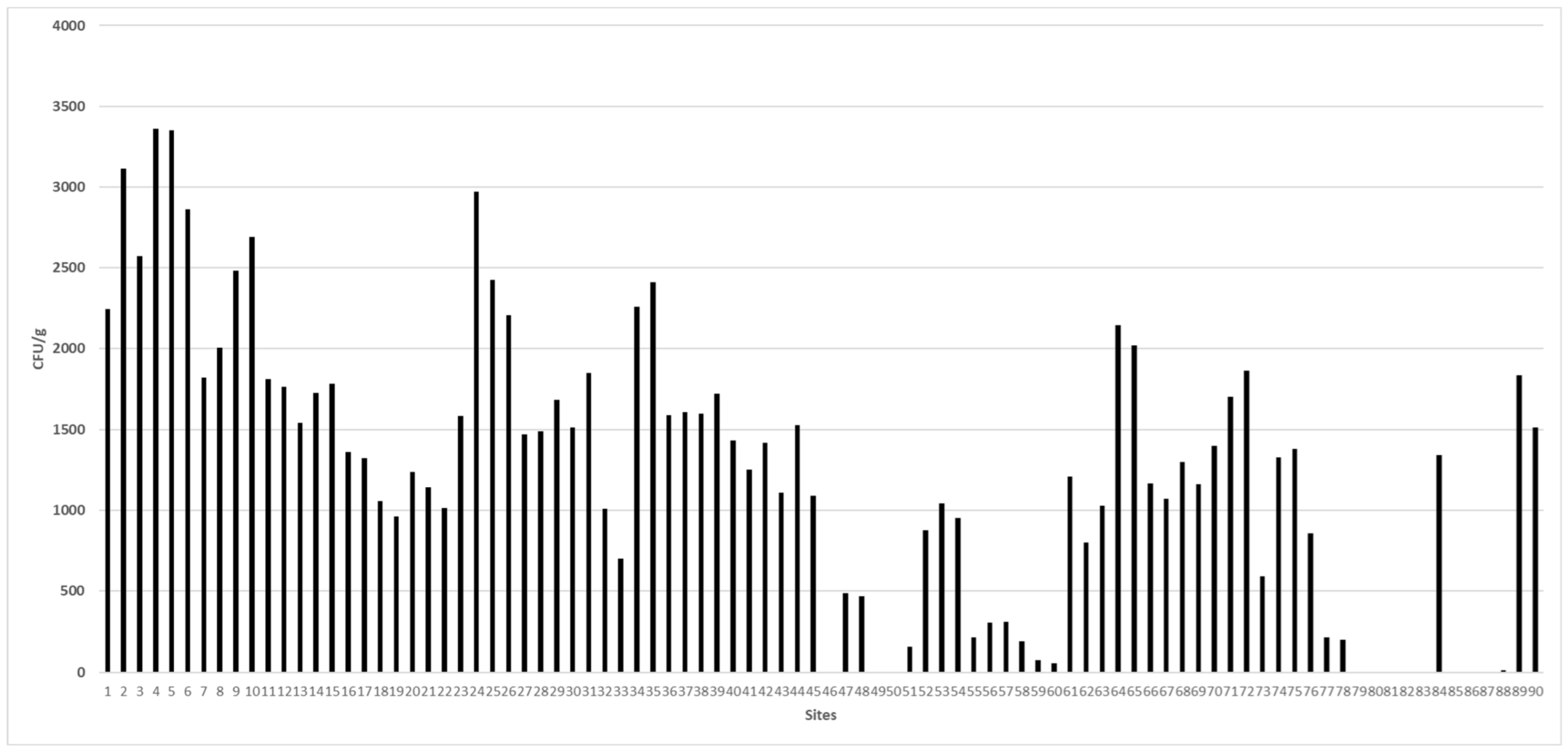
3. Results and Discussion

Mycological examination of 90 animal feedstuff samples revealed that 79 (87.77%) samples were contaminated with different fungal species (1.1 × 10⁵ CFU/g) (Table 2). The fungal load among the samples varied from 17 to 3.3 × 10³ CFU/g, with sample no. 4 being heavily contaminated, which was obtained from rice hulls collected from Alexandria. Other samples of corn bran, yellow corn, broad bean hulls, wheat bran, sorghum grains, soybean hulls, beef cattle feed, and date waste from Alexandria, Beheira, New Valley, and Cairo were also contaminated with relatively high numbers of fungal units.
Taxonomically, the identified taxa were classified to two phyla with four classes, six orders, and eleven families. The order Eurotiales showed the highest species range (28 species) followed by Hypocreales (5 species), Sordariales, and Mucorales (3 species each), while the residual showed the lowest range (1 species). The family Aspergillaceae contributed maximally (28 fungal species out of 41) followed by Chaetomiaceae (3 species), and the remaining families were characterized only by one or two species. On the higher taxa level, Zygomycota was characterized by three species (7.31% of the total species number), teleomorphic Ascomycota had 8 species (19.51%), and anamorphic Ascomycota had 30 species (73.17%). The species identified belonged to 18 genera (Table 2).
Aspergillus was the main genus from the fungal contaminated samples, with a total count of about 39.02% of the total fungal population. Sixteen species of Aspergillus were identified, of which A. flavus and A. niger were the most commonly colonizing fungi at 65.56% and 50%, respectively (Table 2).
The genus Aspergillus was recorded in 75 samples (88.33%), while Penicillium appeared in only 43 samples, accounting for 47.77% of the total samples. The remaining species belonged to the genera Acremonium, Fusarium, Botryotrichum, Chaetomium, Eupenicillium, Trichocladium, Lichtheimia, Monascus, Mucor, Paecilomyces, and Talaromyces (Table 2).
A statistically significant difference among species (p < 0.001 ***), TC (p < 0.001 ***), TC% (p < 0.001 ***), F% (p < 0.001 ***), phyla (p < 0.001 ***), classes (p < 0.001 ***), orders (p < 0.001 ***), and families (p < 0.001 ***) was revealed by the chi-square test (Table 2 and Table 3).
Concerning the biodiversity among the sites, site 6 had the highest Margalef species richness index at 10.87, followed by site 4, and the Shannon diversity index (H) of the identified taxa was 2.20. In terms of the frequency of occurrence, Aspergillus flavus had the highest percentage (65.56 percent), followed by Aspergillus niger (50 percent), and Penicillium chrysogenum (50 percent) (40 percent). The genus Aspergillus was found in 75 samples (88.33 percent), whereas Penicillium was found in only 43 (47.77 percent) of the 90 samples.
The present study analyzed 13 animal feedstuff samples for mycotoxin contamination due to fungal contamination. The lowest and highest fungal yields observed were 2.2 × 10³ CFU/g and 2.9 × 10³ CFU/g, respectively, as depicted (Table 4, Figure 1).
Aspergillus flavus and A. niger were the main species in the studied samples. Samples no. 24 and 26 were contaminated with aflatoxin B1 (0.851 and 1.363 µg/kg, respectively) (Figure S1). Aflatoxin B2, at a ratio of 0.479 µg/kg, was recorded in sample 26. Notably, the current investigation found reduced amounts of aflatoxins. (AFB1 and AFB2) in the positive samples (Figures S2 and S3) in contrast to the permissible ranges for beef cattle animal feedstuff ingredients (20 µg/kg) as reported by good manufacturing practices (GMP) [45].
According to Table 5, various mycotoxins were recorded from the different feeding sources. The mycotoxins assayed included aflatoxins (B1, B2) and ochratoxin A, which were present at a statistically significant level (p = 0.004 **), as revealed by the two-way analysis of variance. The recorded mycotoxins were assayed from different sources, and fungi were statistically significant (p = 0.007 ***), as revealed by the two-way analysis of variance. The highest average level of aflatoxins was 100 in the poultry feed, broad bean hulls, and yellow corn from the fungal isolate A. flavus. In addition, a bean hull revealed aflatoxin B2 at 75 (SD = 50.0) from A. flavus.
Only six A. flavus isolates were capable of producing aflatoxins B1 and B2 at estimated levels of 100 to 200 g/L when 30 fungal isolates representing A. flavus [17], A. niger [7], A. ochraceus [2], and A. parasiticus [4] were tested for their abilities to produce mycotoxins. These isolates were obtained from poultry feeds, bean hulls, rice hulls, and yellow corn (Table 4). Mycotoxins were not produced in detectable amounts by the remaining Aspergillus isolates.
According to various studies conducted by numerous investigators [33,34], the quality of animal feed is a critical requirement for achieving the best animal product yield as well as the preservation of the animals’ health conditions, particularly in intensive industrial production. A periodic analysis was required for the fungal assessment and evaluation of mycotoxin contamination levels, as well as the 41 fungal species from the 18 fungal genera obtained from the various animal feedstuff samples (Table 2). The final genus isolated from mycologically pure materials was Aspergillus, it had such a high total count that it equaled the whole fungal biomass (39.02 percent).
Consistent with the current study, a study in Serbia [46] showed the incidence of fungi in 297 samples of animal feedstuff (maize, ground maize, sunflower, soybean meal, and finished mixture). The total fungal count was 1–9 × 10⁴ CFU/g in most of the examined animal feed samples, with the most prevalent fungi being Fusarium (100% of samples) followed by Aspergillus (87.10%), Rhizopus (83.87%), Penicillium (70.32%), and Mucor (33.55%). Similarly, in Argentina, [47], mycobiota was assessed from corn silage, corn grains, peanut shells, soybean, wheat bran, and alfalfa hay and exhibited a fungal load from 0–2.10 × 10⁸ CFU/g with the presence of aflatoxin B1 (AFB1), fumonisin B1 (FB1), deoxynivalenol (DON), and zearalenone (ZEA) levels in total mixed rations (TMRs) of cattle feed. The dominant genera revealed were Aspergillus spp. (60%) and Fusarium spp. (66.7%), among which Aspergillus fumigatus was predominantly present along with other fungal species, such as A. niger, A. flavus, Fusarium proliferatum, F. subglutinans, F. verticillioides, Penicillium brevicompactum, P. griseofulvin, P. roqueforti, and P. crustosum, as well as representative species of Alternaria, Cladosporium, and Geotrichum.
The current investigation evaluated 30 fungal strains representing A. flavus (17 strains), A. niger [7], A. ochraceus [2], and A. parasiticus [4] for their ability to create mycotoxins. Only six strains of A. flavus were capable of producing aflatoxins B1 and B2 at estimated concentrations of 100–200 g/L. These isolates were from poultry feeds, bean hulls, rice hulls, and yellow corn (Table 5). The remaining Aspergillus strains produced no detectable mycotoxins.
Similar to this investigation, 186 cow feedstuff samples were tested in Iran from traditional and industrial dairy farms in the Hamadan district [45]. The most prevalent fungi were Mucor species (3.4%), Rhizopus (3.9%), Alternaria (4.3%), Cladosporium (9.1%), Fusarium (17.5%), Penicillium (23.7%), and Aspergillus species (37.3%). A. clavatus, A. fumigatus, A. niger, A. flavus, A. parasiticus, A. nidulans, A. terreus, A. ochraceus, and A. ustus were all Aspergillus species. Additionally, the authors observed that concentrate feed and wheat bran were the most contaminated feedstuffs, with the mean colony counts for A. flavus and A. parasiticus at 7.25 102 and 7.50 102 CFU/g, respectively. These fungi were likewise isolated from animal feedstuff samples evaluated in our investigation, except for Alternaria and Cladosporium. Other data from Iran [36,46] indicated fungal contamination in 31.3% of chicken feed samples. Similarly, 51 samples of animal feed were examined in India to determine the presence of toxigenic fungus [47]. This analysis discovered that the Aspergillus species was the most prevalent fungi found in 83% of samples. A. flavus was the most prevalent Aspergillus species, followed by A. niger and A. fumigatus. Penicilium, followed by Fusarium, colonized the feed samples after Aspergillus. In Saudi Arabia [48], 20 samples of soybean meal used in animal feed were evaluated. Al-Seeni stated that the overall fungal counts varied between 3.9 × 10⁴ and 105 CFU/g. The frequently isolated fungi included Aspergillus, Fusarium, and Penicillium. Mycotoxins identified in animal feedstuff samples, namely, soybean, ranged between 1 and 14.8 parts per billion (ppb) for aflatoxins and 1 to 14 parts per billion (ppb) for ochratoxin A. Although the majority of the mycobiota recovered by [38] are identical to those obtained in this study, the current analysis showed that the number of mycotoxins in the Saudi Arabian samples was significantly greater. Mycotoxins’ potential risks can be rapidly mitigated by inspecting plant material for fungal contamination, harvesting, enhancing cultivation, and storage methods, abolishing or diluting toxins from contaminated feeds or foods, and utilizing adsorbents to diminish the toxins’ bioavailability in animal digestive tracts [48].

4. Conclusions

This study showed that animal feed collected from various locations in Egypt, as well as their ingredients, are frequently contaminated with mycotoxigenic fungi and mycotoxins where the main reservoirs for mycotoxins in animal feed are ingredients, particularly rice hulls, corn bran, yellow corn, and broad bean hulls. Many health studies have been performed in the last three decades to explore the hazardous effects of the presence of mycotoxins in animal feeds and human food. The general teratogenicity, toxicity, and carcinogenicity of these molecules constitute a risk to human and animal health. Based on our results, this situation demands immediate necessary control measures. Adequate postharvest drying should be performed, and proper storage conditions should be maintained. Egyptian farmers should be educated and sensitized about proper storage facilities, duration, and conditions for feed and ingredients. To reduce mycotoxin contamination in feed ingredients and finished animal feed, antifungal agents should be used. More research is needed to investigate the synergistic effects of mycotoxins when diets contain more than one mycotoxin. Animal feed is such a critical component of the food chain and must require the quality assurance of rigorous food safety systems. Finally, To promote consumer health, strong restrictions and monitoring systems for testing food and feed for aflatoxin and ochratoxin contamination are strongly recommended. We assume that periodic assessing and the utilize of critical control points (HACCP) and hazard analysis for the control and prevention of mycotoxins in the animal feed industry are required to ensure the feed hygienic quality, productivity and animal health, and prevent human foodborne diseases.

Supplementary Materials

The following supporting information can be downloaded at: https://www.mdpi.com/article/10.3390/ijerph19127250/s1, Figure S1. HPLC chromatogram of aflatoxin B1 in the examined feed samples; Figure S2. HPLC chromatogram of aflatoxin B1 in the examined feed samples; Figure S3. HPLC chromatogram of aflatoxin B2 in the examined feed samples.

Author Contributions

Conceptualization, E.K., M.T.M., M.I.M., S.G., A.M.A.-A. and M.H.; methodology, E.K., M.T.M., M.I.M., M.P., Y.A.H., S.A.S., A.M.A.-A. and S.G.; software, M.I.M., Y.A.H., M.P., A.M.A., B.A.A.-W., S.A.S. and S.G.; validation, E.K., M.T.M., M.I.M., Y.A.H., A.M.A., B.A.A.-W. and S.A.S.; formal analysis, M.T.M., M.I.M., Y.A.H., M.P., A.M.A., B.A.A.-W., S.A.S. and S.G.; investigation, E.K., Y.A.H., M.P., B.A.A.-W., S.A.S., S.G., M.H. and A.M.A.-A.; resources, A.M.A.; data curation, E.K., Y.A.H., A.M.A., B.A.A.-W., M.T.M., M.I.M. and A.M.A.-A.; writing—original draft preparation, E.K., M.T.M., M.I.M., S.G., M.H. and A.M.A.-A.; writing—review and editing, E.K., M.T.M., M.I.M., M.P., Y.A.H., A.M.A., B.A.A.-W., S.A.S., S.G., M.H. and A.M.A.-A.; visualization, E.K., M.T.M., M.I.M., M.P., Y.A.H., A.M.A., B.A.A.-W., S.A.S., S.G., M.H. and A.M.A.-A.; supervision, E.K., M.H. and A.M.A.-A.; project administration, E.K., M.H. and A.M.A.-A.; funding acquisition, A.M.A. All authors have read and agreed to the published version of the manuscript.

Funding

This research was funded by Veterinary Medicine at Matrouh University, Faculty of Veterinary Medicine at Matrouh University, Faculty of Science at University of Al Arish, and Faculty of Science at University of Suez Canal. This research was also funded by Princess Nourah bint Abdulrahman University Researchers Supporting Project number (PNURSP2022R65), Princess Nourah bint Abdulrahman University, Riyadh, Saudi Arabia.

Institutional Review Board Statement

Not applicable.

Informed Consent Statement

Not applicable.

Data Availability Statement

Not applicable.

Acknowledgments

The authors extend their appreciation to Princess Nourah bint Abdulrahman University for funding this work through Researchers Supporting Project number (PNURSP2022R65), Princess Nourah bint Abdulrahman University, Riyadh, Saudi Arabia.

Conflicts of Interest

The authors declare no conflict of interest.

References

  1. Abdel-Azeem, A.M. The history, fungal biodiversity, conservation, and future perspectives for mycology in Egypt. IMA Fungus 2010, 1, 123–142. [Google Scholar] [CrossRef]
  2. Wijayawardene, N.; Hyde, K.D.; Al-Ani, L.K.T.; Tedersoo, L.; Haelewaters, D.; Rajeshkumar, K.C.; Goto, B.T.; Saxena, R.K.; Aptroot, A.; da Silva, G.A.; et al. Outline of Fungi and fungus-like taxa. Mycosphere 2020, 11, 1060–1456. [Google Scholar] [CrossRef]
  3. Moore, D.; Robson, G.D.; Trinci, A.P.J. 21st Century Guidebook to Fungi; Cambridge University Press: Cambridge, UK, 2011. [Google Scholar]
  4. Abdel-Azeem, A.M.; Abo Nahas, H.H.; Abdel-Azeem, M.A.; Tariq, F.J.; Yadav, A.N. Biodiversity and Ecological Perspective of Industrially Important Fungi An Introduction. In Industrially Important Fungi for Sustainable Development. Fungal Biology; Abdel-Azeem, A.M., Yadav, A.N., Yadav, N., Usmani, Z., Eds.; Springer International Publishing: Cham, Switzerland, 2021; pp. 1–34. [Google Scholar]
  5. Abdelmotilib, N.M.; Darwish, A.G.; Abdel-Azeem, A.M.; Sheir, D.H. Fungal Mycotoxins. In Fungi in Sustainable Food Production. Fungal Biology; Dai, X., Sharma, M., Chen, J., Eds.; Springer: Cham, Switzerland, 2021. [Google Scholar]
  6. Legan, J.D. Cereals and cereal products. In The Microbiological Safety and Quality of Food. Gaithersburg; Lund, B.M., Baird-Parker, T.C., Gould, G.W., Eds.; Aspen Publishers Inc.: Boston, MA, USA, 2000; pp. 759–783. [Google Scholar]
  7. Miller, J.D. Fungi and Mycotoxins in Grain—Implications for Stored-Product Research. J. Stored. Prod. Res. 1995, 31, 1–16. [Google Scholar] [CrossRef]
  8. Lillehoj, E.B.; Zuber, M.S. (Eds.) Aflatoxin problem in corn and possible solutions. In Proceedings of the 30th Annual Corn and Sorghum Research Conference, Chicago, IL, USA, 4–6 December 1973. [Google Scholar]
  9. Anderson, H.W.; Nehring, E.W.; Wichser, W.R. Aflatoxin contamination of corn in the field. J. Agric. Food Chem. 1975, 23, 775–782. [Google Scholar] [CrossRef]
  10. Abo Nouh, F.A.; Gezaf, S.A.; Abdel-Azeem, A.M. Aspergillus Mycotoxins: Potential as Biocontrol Agents. In Agriculturally Important Fungi for Sustainable Agriculture. Fungal Biology; Yadav, A., Mishra, S., Kour, D., Yadav, N., Kumar, A., Eds.; Springer: Cham, Switzerland, 2020. [Google Scholar]
  11. Khalifa, S.A.M.; Shedid, E.S.; Saied, E.M.; Jassbi, A.R.; Jamebozorgi, F.H.; Rateb, M.E.; Du, M.; Abdel-Daim, M.M.; Kai, G.-Y.; Al-Hammady, M.A.M.; et al. Cyanobacteria—From the Oceans to the Potential Biotechnological and Biomedical Applications. Mar. Drugs 2021, 19, 241. [Google Scholar] [CrossRef]
  12. Ganesan, A.R.; Balasubramanian, B.; Park, S.; Jha, R.; Andretta, I.; Bakare, A.G.; Kim, I.H. Ochratoxin A: Carryover from animal feed into livestock and the mitigation strategies. Anim. Nutr. 2021, 7, 56–63. [Google Scholar] [CrossRef]
  13. Nazieh, I.; Al Khalaileh, N.I. Prevalence of Ochratoxin A in Poultry Feed and Meat from Jordan. Pak. J. Biol. Sci. 2018, 21, 239–244. [Google Scholar]
  14. Maciorowski, K.G.; Herrera, P.; Jones, F.T.; Pillai, S.D.; Ricke, S.C. Effects on poultry and livestock of feed contamination with bacteria and fungi. Anim. Feed. Sci. Technol. 2007, 133, 109–136. [Google Scholar] [CrossRef]
  15. Murphy, P.A.; Hendrich, S.; Landgren, C.; Bryant, C.M. Food mycotoxins: An update. J. Food Sci. 2006, 71, R51–R65. [Google Scholar] [CrossRef]
  16. Witaszak, N.; Stepien, L.; Bocianowski, J.; Waskiewicz, A. Fusarium Species and Mycotoxins Contaminating Veterinary Diets for Dogs and Cats. Microorganisms 2019, 7, 26. [Google Scholar] [CrossRef] [Green Version]
  17. Alonso, V.A.; Pereyra, C.M.; Keller LA, M.; Dalcero, A.M.; Rosa CA, R.; Chiacchiera, S.M.; Cavaglieri, L.R. Fungi and mycotoxins in silage: An overview. J. Appl. Microbiol. 2013, 115, 637–643. [Google Scholar] [CrossRef]
  18. (Cancer) IIAfRo. Some Traditional Herbal Medicines, Some Mycotoxins, Naphthalene and Styrene; IARC Press: Lyon, France, 2002. [Google Scholar]
  19. Fink-Gremmels, J. The role of mycotoxins in the health and performance of dairy cows. Vet. J. 2008, 176, 84–92. [Google Scholar] [CrossRef]
  20. Magan, N.; Olsen, M. Mycotoxins in Food, 1st ed.; Woodhead Publishing: Cambridge, UK, 2004. [Google Scholar]
  21. Pitt, J.I.; Hocking, A.D. Fungi and Food Spoilage; Springer: Dordrecht, The Netherland; Heidelberg, Germany; London, UK; New York, NY, USA; Cambridge, UK, 2009. [Google Scholar]
  22. Bin-Jumah, M.; Abdel-Fattah, A.-F.M.; Saied, E.M.; El-Seedi, H.R.; Abdel-Daim, M.M. Acrylamide-Induced Peripheral Neuropathy: Manifestations, Mechanisms, and Potential Treatment Modalities. Environ. Sci. Pollut. Res. 2021, 28, 13031–13046. [Google Scholar] [CrossRef]
  23. Pitt, J.I. The Genus Penicillium and Its Teleomorphic States Eupenicillium and Talaromyces; Academic Press: London, UK; New York, NY, USA, 1979; 634p. [Google Scholar]
  24. Raper, K.B.; Fennell, D.I. The Genus Aspergillus; The Williams & Wilkins Company: Baltimore, MD, USA, 1965. [Google Scholar]
  25. Abdel-Azeem, A.M.; Abu-Elsaoud, A.; Darwish, A.M.G.; Balbool, B.A.; Abo Nouh, F.; Abo Nahas, H.H.; El-Azeem, M.A.A.; Ali, N.H.; Kirk, P. The Egyptian Ascomycota 1: Genus Aspergillus. Microb. Biosyst. 2020, 5, 61–99. [Google Scholar] [CrossRef]
  26. Ellis, M.B. Dematiaceous Hyphomycetes; Commonwealth Mycological Institute: London, UK, 1971. [Google Scholar]
  27. Ellis, M.B. More Dematiaceous Hyphomycetes; Commonwealth Mycological Institute: London, UK, 1976. [Google Scholar]
  28. Leslie, J.F.; Summerell, B.A. The Fusarium Laboratory Manual, 1st ed.; Blackwell Publishing Ltd.: Ames, IA, USA, 2006. [Google Scholar]
  29. Domsch, K.H.; Gams, W.; Anderson, T.H. Compendium of Soil Fungi; IHW-Verlag: Eching, Germany, 2007. [Google Scholar]
  30. Guarro, J.; Gene, J.; Stchigel, A.M.; Figueras, M.J. Atlas of Soil Ascomycetes. Holland; APS Press: Utrecht, The Netherlands, 2012. [Google Scholar]
  31. Kirk, P.M.; Ansell, A.E. Authors of Fungal Names; CAB International: London, UK, 1992. [Google Scholar]
  32. Kirk, P.M.; Cannon, P.F.; Minter, D.W.; Stalpers, J.A. Ainsworth & Bisby’s Dictionary of the Fungi, 10th ed.; CAB International: Wallingford, UK, 2008. [Google Scholar]
  33. Horwitz, W. Official Methods of Analysis of AOAC (Association of Official Analytical Chemists) International; AOAC International: Gaithersburg, MD, USA; Academic Press: Washington, DC, USA, 2000. [Google Scholar]
  34. Council, E.E. Commission Regulation No. 401/2006 of 23 February 2006 laying down the methods of sampling and analysis for the official control of the levels of mycotoxins in foodstuffs. Off. J. Eur. Union 2006, L70, 12–34. [Google Scholar]
  35. Toscani, T.; Moseriti, A.; Dossena, A.; Asta, C.D.; Simoncini, N.; Virgili, R. Determination of ochratoxin A in dry-cured meat products by a HPLC-FLD quantitative method. J. Chromatogr. B-Anal. Technol. Biomed. Life Sci. 2007, 855, 242–248. [Google Scholar] [CrossRef]
  36. Zohri, A.A. Mycoflora and Mycotoxins of Some Meat Products; Assiut University: Assiut, Egypt, 1990. [Google Scholar]
  37. Bullerman, L.B.; Hartman, P.A.; Ayres, J.C. Aflatoxin Production in Meats. I. Stored Meats. Appl. Microbiol. 1969, 18, 714. [Google Scholar] [CrossRef]
  38. Elkady, I.A.; Moubasher, M.H. Toxigenicity and Toxins of Stachybotrys Isolates from Wheat Straw Samples in Egypt. Exp. Mycol. 1982, 6, 25–30. [Google Scholar] [CrossRef]
  39. Josefsson, B.G.; Moller, T.E. Screening method for the detection of aflatoxins, ochratoxin, patulin, sterigmatocystin, and zearalenone in cereals. J. Assoc. Off. Anal. Chem. 1977, 60, 1369–1371. [Google Scholar] [CrossRef]
  40. Roberts, B.A.; Patterson, D.S. Detection of twelve mycotoxins in mixed animal feedstuffs, using a novel membrane cleanup procedure. J. Assoc. Off. Anal. Chem. 1975, 58, 1178–1181. [Google Scholar] [CrossRef] [Green Version]
  41. Muthukrishnan, S.; Sanjayan, K.P.; Jahir, H.K. Species composition, seasonal changes and community ordination of alkalotolerant micro fungal diversity in a natural scrub jungle ecosystem of Tamil Nadu, India. Mycosphere 2012, 3, 92–109. [Google Scholar] [CrossRef]
  42. Knapp, H. Introductory Statistics Using SPSS, 2nd ed.; SAGE: Los Angeles, CA, USA, 2017. [Google Scholar]
  43. Mohamed, D.I.; Abou-Bakr, D.A.; Ezzat, S.F.; El-Kareem, H.F.A.; Nahas, H.H.A.; Saad, H.A.; Mehana, A.E.; Saied, E.M. Vitamin D3 Prevents the Deleterious Effects of Testicular Torsion on Testis by Targeting miRNA-145 and ADAM17: In Silico and In Vivo Study. Pharmaceuticals 2021, 14, 1222. [Google Scholar] [CrossRef]
  44. Mohamed, D.I.; Alaa El-Din Aly El-Waseef, D.; Nabih, E.S.; El-Kharashi, O.A.; Abd El-Kareem, H.F.; Abo Nahas, H.H.; Abdel-Wahab, B.A.; Helmy, Y.A.; Alshawwa, S.Z.; Saied, E.M. Acetylsalicylic Acid Suppresses Alcoholism-Induced Cognitive Impairment Associated with Atorvastatin Intake by Targeting Cerebral miRNA155 and NLRP3: In Vivo, and In Silico Study. Pharmaceutics 2022, 14, 529. [Google Scholar] [CrossRef]
  45. Good Manufacturing Practices (GMP). GMP Certification Scheme Animal Feed, Sector 2006, Appendix 1: Product standards; Regulations on Product Standards in the Animal Feed Sector. 2008; pp. 1–39.
  46. Krnjaja, V.; Jelena, L.; Slavica, S. Pathogenic fungi on wheat grain in Serbia. J. Plant Pathol. 2008, 90, 84. [Google Scholar]
  47. Gonzalez-Pereyra, M.L.; Chiacchiera, S.M.; Rosa, C.A.R.; Dalcero, A.M.; Cavaglieri, L.R. Fungal and mycotoxin contamination in mixed feed: Evaluating risk in cattle intensive rearing operations (feedlots). Rev. Bio Cienc. 2012, 2, 68–80. [Google Scholar]
  48. Saied, E.M.; El-Maradny, Y.A.; Osman, A.A.; Darwish, A.M.G.; Abo Nahas, H.H.; Niedbała, G.; Piekutowska, M.; Abdel-Rahman, M.A.; Balbool, B.A.; Abdel-Azeem, A.M. A Comprehensive Review about the Molecular Structure of Severe Acute Respiratory Syndrome Coronavirus 2 (SARS-CoV-2): Insights into Natural Products against COVID-19. Pharmaceutics 2021, 13, 1759. [Google Scholar] [CrossRef]
Figure 1. The fungal load in all the collected samples across various sites in Egypt.
Ijerph 19 07250 g001
Table 1. Types of animal feedstuff samples ingredients collected from different localities in Egypt.
Sites No.Ingredient of Animal Feedstuff SamplesLocalityGovernorate
1–6Rice hullsAmereyahAlexandria
7–11Corn branAmereyahAlexandria
12–17Yellow CornAmereyahAlexandria
18–33Broad bean hullsRasheedBeheira
34–39Beef cattle feedKhargaNew Valley
40Alfalfa hayKhargaNew Valley
41, 42, 45Date wasteKhargaNew Valley
43Soy beanKhargaNew Valley
44Wheat branKhargaNew Valley
46Broiler concentrateKhargaNew Valley
47Yellow cornKhargaNew Valley
48Date wasteKhargaNew Valley
49Broiler poultry feed (19% protein)SemouhaAlexandria
50Broiler poultry feed (23% protein)SemouhaAlexandria
51, 52Rabbit feedCairoCairo
53Rice hulls CairoCairo
54Yellow cornCairoCairo
55Broiler feedCairoCairo
56Wheat branCairoCairo
57Yellow cornCairoCairo
58Wild barleyCairoCairo
59Yellow CornCairoCairo
60SoybeanCairoCairo
61Complete feedstuffCairoCairo
62Bean hullsCairoCairo
63, 64Sorghum grainsCairoCairo
65Soybean hullsCairoCairo
66Wheat grainsCairoCairo
67, 68Broiler feedCairoCairo
69Fine ground cornAssiutAssiut
70Coarse ground cornAssiutAssiut
71White cornAssiutAssiut
72Bean hullsAssiutAssiut
73Wheat branAssiutAssiut
74–78Layer strain poultry feed (17% protein)SemouhaAlexandria
79Magnesium sulphate AssiutAssiut
80Dicalcium phosphateAssiutAssiut
81MethionineAssiutAssiut
82LimeAssiutAssiut
83Premix for layer strainsAssiutAssiut
84Salts for poultryAssiutAssiut
85Vitamins A, DAssiutAssiut
86LysineAssiutAssiut
87Salts for cattleAssiutAssiut
88Premix for beef cattle feedAssiutAssiut
89Beef cattle feedAssiutAssiut
90Poultry feed (17% protein)AssiutAssiut
Table 2. Species list recorded during the study.
Fungal Genera and SpeciesTC% TCFreq.% FPhylumClassOrderFamily
Acremonium roseolum (G. Smith) W. Gams400.03611.1AscomycotaSordariomycetesHypocrealesIncertae sedis
Aspergillus (Total) 0.000 0.0AscomycotaEurotiomycetesEurotialesAspergillaceae
A. egyptiacus Moubasher & Moustafa400.03611.1AscomycotaEurotiomycetesEurotialesAspergillaceae
A. amstelodami (L. Mangin) Thom & Church63205.6241516.7AscomycotaEurotiomycetesEurotialesAspergillaceae
A. candidus Link10,3809.2382325.6AscomycotaEurotiomycetesEurotialesAspergillaceae
A. clavatus Desmazieres400.03611.1AscomycotaEurotiomycetesEurotialesAspergillaceae
A. flavus Link18,92016.8385965.6AscomycotaEurotiomycetesEurotialesAspergillaceae
A. flavipes (Bain. & Sart.) Thom & Church9200.8191112.2AscomycotaEurotiomycetesEurotialesAspergillaceae
A. fumigatus Fresenius16001.424910.0AscomycotaEurotiomycetesEurotialesAspergillaceae
A. niger van Tieghem41,25136.7114550.0AscomycotaEurotiomycetesEurotialesAspergillaceae
A. nidulans (Eidam) G. Winter2000.17855.6AscomycotaEurotiomycetesEurotialesAspergillaceae
A. ochraceus Wilhelm4400.39255.6AscomycotaEurotiomycetesEurotialesAspergillaceae
A. parasiticus Speare9200.8191011.1AscomycotaEurotiomycetesEurotialesAspergillaceae
A. ruber (Jos. König, Spieck. & W. Bremer) Thom & Church40003.5602527.8AscomycotaEurotiomycetesEurotialesAspergillaceae
A. sydowii (Bainier & Sartory) Thom & Church45204.0232022.2AscomycotaEurotiomycetesEurotialesAspergillaceae
A. terreus Thom36803.2752022.2AscomycotaEurotiomycetesEurotialesAspergillaceae
A. versicolor (Vuillemin) Tiraboschi1200.10711.1AscomycotaEurotiomycetesEurotialesAspergillaceae
A. ustus (Bainier) Thom & Church400.03611.1AscomycotaEurotiomycetesEurotialesAspergillaceae
Botryotrichum atrogriseum J.F.H. Beyma400.03611.1AscomycotaSordariomycetesSordarialesChaetomiaceae
Chaetomium globosum Kunze1200.10722.2AscomycotaSordariomycetesSordarialesChaetomiaceae
Eupenicillium inusitatum D.B. Scott8000.71244.4AscomycotaEurotiomycetesEurotialesAspergillaceae
Fusarium (Total)00.00022.2AscomycotaSordariomycetesHypocrealesNectriaceae
F. incarnatum (Desm.) Sacc.400.03611.1AscomycotaSordariomycetesHypocrealesNectriaceae
F. solani (Martius) Saccardo400.03611.1AscomycotaSordariomycetesHypocrealesNectriaceae
Trichocladium griseum (Traaen) X. Wei Wang & Houbraken2000.17833.3AscomycotaSordariomycetesSordarialesChaetomiaceae
Lichtheimia corymbifera (Cohn) Vuill.3600.32055.6ZygomycotaMucoromycetesMucoralesCunningham-ellaceae
Monascus purpureus Went3200.28555.6AscomycotaEurotiomycetesEurotialesMonascaceae
Mucor hiemalis Wehmer400.03611.1ZygomycotaMucoromycetesMucoralesMucoraceae
Paecilomyces variotii Bainier4800.42755.6AscomycotaEurotiomycetesEurotialesAspergillaceae
Penicillium (Total)00.0004550.0AscomycotaEurotiomycetesEurotialesAspergillaceae
P. aurantiogriseum Dierckx2000.17844.4AscomycotaEurotiomycetesEurotialesAspergillaceae
P. chrysogenum Thom10,4209.2733640.0AscomycotaEurotiomycetesEurotialesAspergillaceae
P. citrinum Thom5600.49866.7AscomycotaEurotiomycetesEurotialesAspergillaceae
P. solitum Westling800.07122.2AscomycotaEurotiomycetesEurotialesAspergillaceae
Talaromyces duclauxii (Delacr.) Samson, N. Yilmaz, Frisvad & Seifert6800.6051011.1AscomycotaEurotiomycetesEurotialesAspergillaceae
P. glabrum (Wehmer) Westling1600.14222.2AscomycotaEurotiomycetesEurotialesAspergillaceae
T. islandicus (Sopp) Samson, N. Yilmaz, Frisvad & Seifert1600.14222.2AscomycotaEurotiomycetesEurotialesAspergillaceae
T. pinophilus (Hedgc.) Samson, N. Yilmaz, Frisvad & Seifert31602.8123033.3AscomycotaEurotiomycetesEurotialesAspergillaceae
T. samson N. Yilmaz, Houbraken, Spierenb., Seifert, Peterson, Varga & Frisvad [as ‘purpurogenus’]3200.28555.6AscomycotaEurotiomycetesEurotialesAspergillaceae
Didymella glomerata (Corda) Qian Chen & L. Cai400.03611.1AscomycotaDothideomycetesPleosporomycetidaeDidymellaceae
Rhizopus arrhizus A. Fisch.400.03611.1ZygomycotaMucoromycetesMucoralesRhizopodaceae
Microascus brevicaulis S.P. Abbott800.07122.2AscomycotaSordariomycetesMicroascalesMicroascaceae
Stachybotrys chartarum (Ehrenberg) Hughes3960.35255.6AscomycotaSordariomycetesHypocrealesStachybotryaceae
Trichoderma harzianum Rifai2000.17822.2AscomycotaSordariomycetesHypocrealesHypocreaceae
Gross Total Count112,367100-
Chi-square test/Kruskal–Wallis<0.001 ***<0.001 ***<0.001 ***<0.001 ***<0.001 ***<0.001 ***<0.001 ***<0.001 ***
*** indicates p < 0.001.
Table 3. Chi-square test for assessing the difference by species, TC, frequency, and taxonomic groups. (*** indicates p < 0.001).
Chi-SquaredfSign.
Species743.440<0.001 ***
TC544.751<0.001 ***
TC %310.123<0.001 ***
F %153.215<0.001 ***
Phylum352.51<0.001 ***
Class973.33<0.001 ***
Order1868.96<0.001 ***
Family2049.36<0.001 ***
Table 4. Mycotoxins detected in animal feedstuff samples.
Sample No.Fungal Load
(CFU/g)
Aflatoxins (µg/kg)Ochratoxin A
B1B2G1G2
12.2 × 10³-----
23.1 × 10³-----
32.5 × 10³-----
43.3 × 10³-----
62.8 × 10³-----
102.6 × 10³-----
171.3 × 10³-----
181.0 × 10³-----
242.9 × 1030.851----
252.4 × 10³-----
262.2 × 1031.3630.479---
301.5 × 10³-----
321.0 × 10³-----
Table 5. Assay of mycotoxins from different fungal isolated from various feeding sources.
All(ug/Liter) Using TLC
NameSourceMycotoxinsSourceFungiMycotoxinNMeanSDSECI 95%MinMax
LowerUpper
1A. flavusRice hullsAflatoxins B1, B211160000000
2Corn bran21110000000
3Yellow corn31120000000
4Broad bean hulls41110000000
5Poultry feed51110000000
6Poultry feedAflatoxin B151211000000100100
7Broad bean hullsAflatoxin B251311000000100100
8Bean hullsAflatoxin B1412410081.640.8−302300200
9Bean hullsAflatoxin B24134755025−4.61550100
10Yellow cornAflatoxin B131211000000100100
11A. nigerBeef cattle feedOchratoxin A62450000000
12Alfalfa hay72410000000
13Date wastes12410000000
14A. ochraceusRice hullsOchratoxin A13420000000
15A. parasiticusLayer strain poultry feedAflatoxins54140000000
Source of variationdfFSign.
Corrected model143.3210.007 **
Intercept112.8540.002 **
Source60.017>0.05 ns
Fungi20>0.05 ns
Mycotoxin27.3610.004 **
Source * Mycotoxin30.076>0.05 ns
** indicates p < 0.004, * indicates p < 0.007.
Publisher’s Note: MDPI stays neutral with regard to jurisdictional claims in published maps and institutional affiliations.

