
Root Canal Morphology and Configuration of the Mandibular Canine: A Systematic Review


Abstract
:1. Introduction
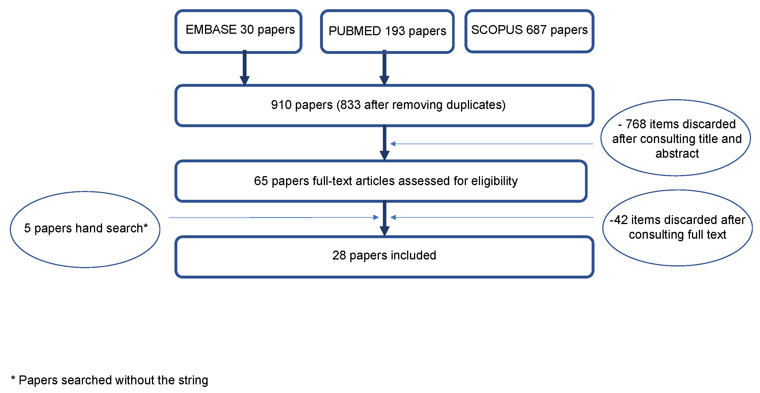
2. Materials and Methods
2.1. Eligibility Criteria
2.2. Information Sources and Search Strategy
2.3. Study Selection
2.4. Data Collection, Summary Measures and Synthesis of Results
2.5. Assessment of Bias across Studies
3. Results
4. Discussion
5. Conclusions
- Mandibular canines are most frequently single-rooted (87.9–100%).
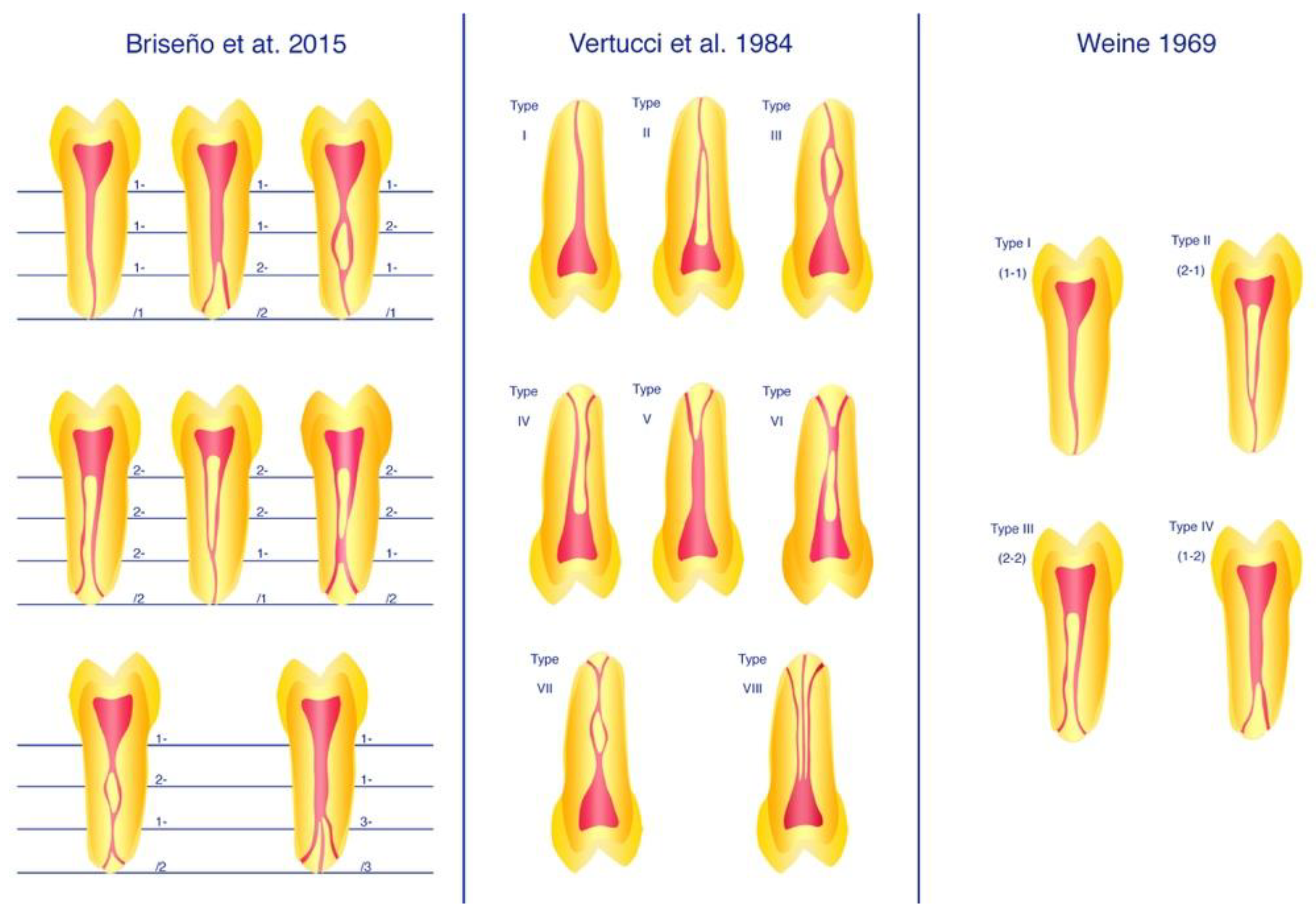
- The most observed RCC is the 1-1-1/1 (Vertucci’s and Weine’s et al. type I), followed by a 2-2-1/1 (Vertucci’s and Weine’s II) and 1-2-1/1 (Vertucci’s III).
- CBCT is widely and, in recent years, most frequently used for in vivo research on the root canal morphology of mandibular canines.
Supplementary Materials
Author Contributions
Funding
Institutional Review Board Statement
Informed Consent Statement
Data Availability Statement
Conflicts of Interest
References
- Vertucci, F.J. Root canal anatomy of the human permanent teeth. Oral Surg. Oral Med. Oral Pathol. 1984, 58, 589–599. [Google Scholar] [CrossRef]
- Weine, F.S.; Healey, H.J.; Gerstein, H.; Evanson, L. Canal configuration in the mesiobuccal root of the maxillary first molar and its endodontic significance. Oral Surg. Oral Med. Oral Pathol. 1969, 28, 419–425. [Google Scholar] [CrossRef]
- Briseno-Marroquin, B.; Paque, F.; Maier, K.; Willershausen, B.; Wolf, T.G. Root Canal Morphology and Configuration of 179 Maxillary First Molars by Means of Micro-computed Tomography: An Ex Vivo Study. J. Endod. 2015, 41, 2008–2013. [Google Scholar] [CrossRef] [Green Version]
- Gulabivala, K.; Aung, T.H.; Alavi, A.; Ng, Y.L. Root and canal morphology of Burmese mandibular molars. Int. Endod. J. 2001, 34, 359–370. [Google Scholar] [CrossRef] [PubMed]
- Çalişkan, M.K.; Pehlivan, Y.; Sepetçioǧlu, F.; Türkün, M.; Tuncer, S.Ş. Root canal morphology of human permanent teeth in a Turkish population. J. Endod. 1995, 21, 200–204. [Google Scholar] [CrossRef]
- Naseri, M.; Ahangari, Z.; Bagheri, N.; Jabbari, S.; Gohari, A. Comparative Accuracy of Cone-Beam Computed Tomography and Clearing Technique in Studying Root Canal and Apical Morphology of Mandibular Canines. Iran. Endod. J. 2019, 14, 271–277. [Google Scholar] [CrossRef]
- Pécora, J.D.; Sousa Neto, M.D.; Saquy, P.C. Internal anatomy, direction and number of roots and size of human mandibular canines. Braz. Dent. J. 1993, 4, 53–57. [Google Scholar] [PubMed]
- Rahimi, S.; Milani, A.S.; Shahi, S.; Sergiz, Y.; Nezafati, S.; Lotfi, M. Prevalence of two root canals in human mandibular anterior teeth in an Iranian population. Indian J. Dent. Res. 2013, 24, 234–236. [Google Scholar] [CrossRef]
- Sert, S.; Bayirli, G.S. Evaluation of the root canal configurations of the mandibular and maxillary permanent teeth by gender in the Turkish population. J. Endod. 2004, 30, 391–398. [Google Scholar] [CrossRef]
- Vertucci, F.J. Root canal anatomy of the mandibular anterior teeth. J. Am. Dent. Assoc. 1974, 89, 369–371. [Google Scholar] [CrossRef]
- Green, D. Double canals in single roots. Oral Surg. Oral Med. Oral Pathol. 1973, 35, 689–696. [Google Scholar] [CrossRef]
- Bakianian Vaziri, P.; Kasraee, S.; Abdolsamadi, H.R.; Abdollahzadeh, S.; Esmaeili, F.; Nazari, S.; Vahedi, M. Root Canal Configuration of One-rooted Mandibular Canine in an Iranian Population: An In Vitro Study. J. Dent. Res. Dent. Clin. Dent. Prospect. 2008, 2, 28–32. [Google Scholar] [CrossRef]
- Altunsoy, M.; Ok, E.; Nur, B.G.; Aglarci, O.S.; Gungor, E.; Colak, M. A cone-beam computed tomography study of the root canal morphology of anterior teeth in a Turkish population. Eur. J. Dent. 2014, 8, 302–306. [Google Scholar] [CrossRef]
- Aminsobhani, M.; Sadegh, M.; Meraji, N.; Razmi, H.; Kharazifard, M.J. Evaluation of the root and canal morphology of mandibular permanent anterior teeth in an Iranian population by cone-beam computed tomography. J. Dent. 2013, 10, 358–366. [Google Scholar]
- Pineda, F.; Kuttler, Y. Mesiodistal and buccolingual roentgenographic investigation of 7,275 root canals. Oral Surg. Oral Med. Oral Pathol. 1972, 33, 101–110. [Google Scholar] [CrossRef]
- Al-Dahman, Y.; Alqedairi, A.; Alfawaz, H.; Alnassar, F.; Al-Jebaly, A. Cone-beam computed tomographic evaluation of root canal morphology of mandibular canines in a Saudi subpopulation. Saudi Endod. J. 2019, 9, 113–118. [Google Scholar] [CrossRef]
- Candeiro, G.T.M.; Monteiro Dodt Teixeira, I.M.; Olimpio Barbosa, D.A.; Vivacqua-Gomes, N.; Alves, F.R.F. Vertucci’s Root Canal Configuration of 14,413 Mandibular Anterior Teeth in a Brazilian Population: A Prevalence Study Using Cone-beam Computed Tomography. J. Endod. 2021, 47, 404–408. [Google Scholar] [CrossRef]
- Doumani, M.; Habib, A.; Alhalak, A.B.; Al-Nahlawi, T.F.; Al Hussain, F.; Alanazi, S.M. Root canal morphology of mandibular canines in the Syrian population: A CBCT Assessment. J. Fam. Med. Prim. Care 2020, 9, 552–555. [Google Scholar] [CrossRef]
- Haghanifar, S.; Moudi, E.; Bijani, A.; Ghanbarabadi, M.K. Morphologic assessment of mandibular anterior teeth root canal using CBCT. Acta Med. Acad. 2017, 46, 85–93. [Google Scholar] [CrossRef]
- Han, T.; Ma, Y.; Yang, L.; Chen, X.; Zhang, X.; Wang, Y. A study of the root canal morphology of mandibular anterior teeth using cone-beam computed tomography in a Chinese subpopulation. J. Endod. 2014, 40, 1309–1314. [Google Scholar] [CrossRef]
- Karobari, M.I.; Noorani, T.Y.; Halim, M.S.; Ahmed, H.M.A. Root and canal morphology of the anterior permanent dentition in Malaysian population using two classification systems: A CBCT clinical study. Aust. Endod. J. 2021, 47, 202–216. [Google Scholar] [CrossRef] [PubMed]
- Kulkarni, V.; Duruel, O.; Ataman-Duruel, E.T.; Tozum, M.D.; Nares, S.; Tozum, T.F. In-depth morphological evaluation of tooth anatomic lengths with root canal configurations using cone beam computed tomography in North American population. J. Appl. Oral Sci. 2020, 28, e20190103. [Google Scholar] [CrossRef] [Green Version]
- Martins, J.N.R.; Marques, D.; Mata, A.; Caramês, J. Root and root canal morphology of the permanent dentition in a Caucasian population: A cone-beam computed tomography study. Int. Endod. J. 2017, 50, 1013–1026. [Google Scholar] [CrossRef] [PubMed] [Green Version]
- Mashyakhy, M. Prevalence of a Second Root and Canal in Mandibular and Maxillary Canines in a Saudi Arabian Population: A Cone-beam Computed Tomography Study. J. Contemp. Dent. Pr. 2019, 20, 773–777. [Google Scholar] [CrossRef]
- Pan, J.Y.Y.; Parolia, A.; Chuah, S.R.; Bhatia, S.; Mutalik, S.; Pau, A. Root canal morphology of permanent teeth in a Malaysian subpopulation using cone-beam computed tomography. BMC Oral Health 2019, 19, 14. [Google Scholar] [CrossRef]
- Raman, S.; Kumar, V.J. A cone-beam computed tomography study of the prevalence of two or more canals in mandibular anteriors in the Chennai population. J. Adv. Pharm. Educ. Res. 2017, 7, 92–95. [Google Scholar]
- Soleymani, A.; Namaryan, N.; Moudi, E.; Gholinia, A. Root Canal Morphology of Mandibular Canine in an Iranian Population: A CBCT Assessment. Iran. Endod. J. 2017, 12, 78–82. [Google Scholar] [CrossRef]
- Somalinga Amardeep, N.; Raghu, S.; Natanasabapathy, V. Root canal morphology of permanent maxillary and mandibular canines in Indian population using cone beam computed tomography. Anat. Res. Int. 2014, 2014, 731859. [Google Scholar] [CrossRef] [Green Version]
- Sroczyk-Jaszczyńska, M.; Kołecki, J.; Lipski, M.; Puciło, M.; Wilk, G.; Falkowski, A.; Kot, K.; Nowicka, A. A study of the symmetry of roots and root canal morphology in mandibular anterior teeth using cone-beam computed tomographic imaging in a Polish population. Folia Morphol. 2020, 79, 835–844. [Google Scholar] [CrossRef] [Green Version]
- Zhengyan, Y.; Keke, L.; Fei, W.; Yueheng, L.; Zhi, Z. Cone-beam computed tomography study of the root and canal morphology of mandibular permanent anterior teeth in a Chongqing population. Ther. Clin. Risk Manag. 2016, 12, 19–25. [Google Scholar] [CrossRef] [Green Version]
- Da Silva, E.J.; de Castro, R.W.; Nejaim, Y.; Silva, A.I.; Haiter-Neto, F.; Silberman, A.; Cohenca, N. Evaluation of root canal configuration of maxillary and mandibular anterior teeth using cone beam computed tomography: An in-vivo study. Quintessence Int. 2016, 47, 19–24. [Google Scholar] [CrossRef]
- Lee, K.W.; Kim, Y.; Perinpanayagam, H.; Lee, J.K.; Yoo, Y.J.; Lim, S.M.; Chang, S.W.; Ha, B.H.; Zhu, Q.; Kum, K.Y. Comparison of alternative image reformatting techniques in micro-computed tomography and tooth clearing for detailed canal morphology. J. Endod. 2014, 40, 417–422. [Google Scholar] [CrossRef]
- Marceliano-Alves, M.F.; de Lima, C.O.; Augusto, C.M.; Almeida Barbosa, A.F.; Vieira Bruno, A.M.; Rosa, A.M.; Lopes, R.T. The internal root canal morphology of single-rooted mandibular canines revealed by micro-computed tomography. J. Conserv. Dent. 2018, 21, 588–591. [Google Scholar] [CrossRef] [PubMed]
- Mazzi-Chaves, J.F.; Silva-Sousa, Y.T.C.; Leoni, G.B.; Silva-Sousa, A.C.; Estrela, L.; Estrela, C.; Jacobs, R.; Sousa-Neto, M.D. Micro-computed tomographic assessment of the variability and morphological features of root canal system and their ramifications. J. Appl. Oral Sci. 2020, 28, e20190393. [Google Scholar] [CrossRef] [Green Version]
- Plotino, G.; Grande, N.M.; Pecci, R.; Bedini, R.; Pameijer, C.H.; Somma, F. Three-dimensional imaging using microcomputed tomography for studying tooth macromorphology. J. Am. Dent. Assoc. 2006, 137, 1555–1561. [Google Scholar] [CrossRef] [PubMed]
- Rhodes, J.S.; Ford, T.R.; Lynch, J.A.; Liepins, P.J.; Curtis, R.V. Micro-computed tomography: A new tool for experimental endodontology. Int. Endod. J. 1999, 32, 165–170. [Google Scholar] [CrossRef] [PubMed]
- Versiani, M.A.; Pécora, J.D.; Sousa-Neto, M.D. Microcomputed tomography analysis of the root canal morphology of single-rooted mandibular canines. Int. Endod. J. 2013, 46, 800–807. [Google Scholar] [CrossRef]
- Wang, M.; Ren, X.; Pan, Y. Micro-computed tomography-based anatomical study of the branch canals in mandibular anterior teeth in a Chinese population. Clin. Oral Investig. 2019, 23, 81–86. [Google Scholar] [CrossRef] [PubMed] [Green Version]
- Da Silva Ramos Fernandes, L.M.P.; Rice, D.; Ordinola-Zapata, R.; Alvares Capelozza, A.L.; Bramante, C.M.; Jaramillo, D.; Christensen, H. Detection of various anatomic patterns of root canals in mandibular incisors using digital periapical radiography, 3 cone-beam computed tomographic scanners, and micro-computed tomographic imaging. J. Endod. 2014, 40, 42–45. [Google Scholar] [CrossRef]
- Liberati, A.; Altman, D.G.; Tetzlaff, J.; Mulrow, C.; Gøtzsche, P.C.; Ioannidis, J.P.; Clarke, M.; Devereaux, P.J.; Kleijnen, J.; Moher, D. The PRISMA statement for reporting systematic reviews and meta-analyses of studies that evaluate healthcare interventions: Explanation and elaboration. BMJ 2009, 339, b2700. [Google Scholar] [CrossRef] [PubMed] [Green Version]
- Henry, B.M.; Tomaszewski, K.A.; Ramakrishnan, P.K.; Roy, J.; Vikse, J.; Loukas, M.; Tubbs, R.S.; Walocha, J.A. Development of the anatomical quality assessment (AQUA) tool for the quality assessment of anatomical studies included in meta-analyses and systematic reviews. Clin. Anat. 2017, 30, 6–13. [Google Scholar] [CrossRef] [PubMed] [Green Version]
- Sert, S.; Aslanalp, V.; Tanalp, J. Investigation of the root canal configurations of mandibular permanent teeth in the Turkish population. Int. Endod. J. 2004, 37, 494–499. [Google Scholar] [CrossRef] [PubMed]
- Wolf, T.G.; Stiebritz, M.; Boemke, N.; Elsayed, I.; Paqué, F.; Wierichs, R.J.; Briseño-Marroquín, B. 3-dimensional Analysis and Literature Review of the Root Canal Morphology and Physiological Foramen Geometry of 125 Mandibular Incisors by Means of Micro-Computed Tomography in a German Population. J. Endod. 2020, 46, 184–191. [Google Scholar] [CrossRef] [PubMed]


| Report | PP | n | Met | Root Canal Configuration Frequency (%) | Number of Roots (%) | |||||||||
|---|---|---|---|---|---|---|---|---|---|---|---|---|---|---|
| Root Canal Configuration | Ve | I | II | III | IV | V | VI | VII | VIII | * | 1 | 2 | ||
| We | I | II | - | III | - | - | - | - | * | |||||
| Br | 1-1-1/1 | 2-2-1/1 | 1-2-1/1 | 2-2-2/2 | 1-1-2/2 | 2-1-2/2 | 1-2-1/2 | 1-1-3/3 | * | |||||
| Pineda and Kuttler, 1972 [15] | MEX | 187 | Rx | 81.5 | 13.5 | - | 5.0 | - | - | - | - | - | - | - |
| Green et al., 1973 [11] | USA | 100 | GR | 87.0 | - | - | 13 | - | - | - | - | - | - | - |
| Vertucci, 1974 [10] | USA | 100 | SC | 78.0 | 14.0 | 2.0 | 6.0 | - | - | - | - | - | - | - |
| Pécora et al., 1993 [7] | BRA | 830 | SC | 92.2 | 4.9 | - | 1.2 | - | - | - | - | - | 98.3 | 1.7 |
| Caliskan et al., 1995 [5] | TUR | 100 | SC | 80.4 | 3.92 | 13.7 | - | 2.0 | - | - | - | - | 100 | - |
| Sert et al., 2004 [42] | TUR | 200 | SC | 76.0 | 16.0 | 6.5 | 1.5 | - | - | - | - | - | - | - |
| Sert and Bayirli, 2004 [9] | TUR | 200 | M | 90.0 | 9.0 | - | - | - | - | - | - | - | - | - |
| SC; Mic | - | - | - | - | - | - | - | - | 1.0 | - | - | |||
| F | 62.0 | 22.0 | 13.0 | 3.0 | - | - | - | - | - | - | - | |||
| Bakianian Vaziri et al., 2008 [12] | IRN | 100 | CR | 88.0 | 5.0 | 7.0 | - | - | - | - | - | - | - | - |
| Aminsobhani et al., 2013 [14] | IRN | 608 | M | 36.0 ± 0.3 | 5.1 ± 0.2 | 1.4 ± 0.1 | 6.4 ± 0.2 | 1.3 ± 0.1 | - | - | - | - | - | - |
| CBCT | - | - | - | - | - | - | - | - | - | 96.3 | 4.7 | |||
| F | 35.8 ± 0.1 | 5.2 ± 0.3 | 1.4 ± 0.1 | 6.4 ± 0.1 | 1.0 ± 0.2 | - | - | - | - | - | - | |||
| Rahimi et al., 2013 [8] | IRN | 149 | SC | 91.6 | 6.11 | 2.29 | - | - | - | - | - | - | 87.9 | 12.1 |
| Altunsoy et al., 2014 [13] | TUR | 1604 | M | 91.0 | 2.6 | 1.5 | 0.9 | 3.5 | - | - | - | - | - | - |
| CBCT | - | - | - | - | - | - | - | - | - | - | - | |||
| F | 94.0 | 1.6 | 0.9 | 1.8 | 1.8 | - | - | - | - | - | - | |||
| Han et al., 2014 [20] | CHN* | 1291 | CBCT | 93.7 | 0.62 | 3.25 | - | 0.54 | - | - | - | - | 98.7 | 1.3 |
| Somalinga Amardeep et al., 2014 [28] | IND | 250 | CBCT | 79.6 | 3.2 | 13.6 | - | 2.0 | - | - | - | 1.6 | 100 | - |
| Zhengyan et al., 2015 [30] | CHN | 1452 | CBCT/T-33 | 96.4 | 0.7 | 1.7 | - | 0.4 | - | - | - | - | 99.2 | 0.8 |
| 1435 | CBCT/T-44 | 95.2 | 0.7 | 2.5 | 0.3 | 0.4 | - | - | - | - | ||||
| da Silva et al., 2016 [31] | BRA | 200 | CBCT | 90.5 | 1.0 | 4.0 | 2.5 | 2.0 | - | - | - | - | - | - |
| Haghanifar et al., 2017 [19] | IRN | 365 | CBCT | 88.2 | 3.3 | 8.1 | - | 0.3 | - | 0.1 | - | - | 99.7 | 0.3 |
| Martins et al., 2017 [23] | PRT | 1200 | CBCT | 90.2 | 3.3 | 2.7 | 1.4 | 2.3 | - | - | - | 0.1 | 97.2 | 2.8 |
| Raman et al., 2017 [26] | IND | 100 | CBCT/T-33 | 78.0 | - | 20.0 | - | - | - | - | - | - | - | - |
| 100 | CBCT/T-43 | 84.0 | - | 14.0 | - | - | - | - | - | - | - | - | ||
| Soleymani et al., 2017 [27] | IRN | 300 | CBCT | 89.7 | 3.7 | 5.7 | - | 1.0 | - | - | - | - | 98.7 | 1.3 |
| Al-Dahman et al., 2019 [16] | SAU | 454 | CBCT | 95.4 | 2.6 | 1.8 | 0.2 | - | - | - | - | - | 99.8 | 0.2 |
| Mashyakhy, 2019 [24] | SAU | 410 | CBCT | 90.7 | - | 6.1 | - | 3.2 | - | - | - | - | 97.3 | 2.7 |
| Naseri et al., 2019 [6] | IRN | 30 | CBCT | 93.9 | - | 6.1 | - | - | - | - | - | - | - | - |
| SC | 90.9 | - | 9.1 | - | - | - | - | - | - | - | - | |||
| Pan et al., 2019 [25] | MYS | 411 | CBCT | 95.1 | 4.9 | - | - | - | - | - | - | - | 98.8 | 1.2 |
| Doumani et al., 2020 [18] | SYR | 418 | CBCT | 95.9 | 0.73 | 3.18 | - | 0.24 | - | - | - | - | 97.9 | 2.2 |
| Karobari et al., 2020 [21] | MYS | 1702 | CBCT | 90.7 | 0.2 | 8.2 | - | 0.7 | - | 0.1 | - | 0.4 | 99.7 | 0.3 |
| Kulkarni et al., 2020 [22] | USA | 259 | CBCT | 85.0 | 14.0 | 1.0 | - | - | - | - | - | - | - | - |
| Sroczyk-Jaszczyńska et al., 2020 [29] | POL | 100 | CBCT/T-33 | 82.0 | 4.0 | 4.0 | 1.0 | 8.0 | - | - | - | - | 92.0 | 8.0 |
| 104 | CBCT/T-43 | 88.2 | - | 3.85 | - | 5.88 | 0.98 | - | - | 0.98 | 96.2 | 3.9 | ||
| Candeiro et al., 2021 [17] | BRA | 4805 | CBCT | 89.1 | 1.58 | 6.66 | 0.10 | 2.41 | - | 0.13 | - | - | 97.6 | 2.4 |
Publisher’s Note: MDPI stays neutral with regard to jurisdictional claims in published maps and institutional affiliations. |
© 2021 by the authors. Licensee MDPI, Basel, Switzerland. This article is an open access article distributed under the terms and conditions of the Creative Commons Attribution (CC BY) license (https://creativecommons.org/licenses/by/4.0/).
Share and Cite
Wolf, T.G.; Anderegg, A.L.; Yilmaz, B.; Campus, G. Root Canal Morphology and Configuration of the Mandibular Canine: A Systematic Review. Int. J. Environ. Res. Public Health 2021, 18, 10197. https://doi.org/10.3390/ijerph181910197
Wolf TG, Anderegg AL, Yilmaz B, Campus G. Root Canal Morphology and Configuration of the Mandibular Canine: A Systematic Review. International Journal of Environmental Research and Public Health. 2021; 18(19):10197. https://doi.org/10.3390/ijerph181910197
Chicago/Turabian StyleWolf, Thomas Gerhard, Andrea Lisa Anderegg, Burak Yilmaz, and Guglielmo Campus. 2021. "Root Canal Morphology and Configuration of the Mandibular Canine: A Systematic Review" International Journal of Environmental Research and Public Health 18, no. 19: 10197. https://doi.org/10.3390/ijerph181910197