
Effectiveness of a Three-Week Inpatient Pulmonary Rehabilitation Program for Patients after COVID-19: A Prospective Observational Study

 , and
, and
Abstract
:1. Introduction
2. Materials and Methods
2.1. Setting and Patients
2.2. Intervention
- Physical training (O) consisted of two obligatory main components: (a) endurance training was scheduled as 3–5 supervised units per week for 30–60 min each time if this was tolerated by the patient; otherwise, the duration and frequency were adjusted individually. The initial training intensity was based on the individual physical performance orienting on the 6MWD performance for cycle training and was controlled by a modified 0–10 BORG scale [17] (target range 4–6). Accordingly, the training intensities were gradually adjusted by the therapist over the course of the rehabilitation in each session. Exercise intensity was controlled by a pulse oximeter (SpO2 target range ≥90%; oxygen administration if necessary) and heart rate. Endurance-oriented exercises in the terrain (e.g., Nordic walking) and indoor sports were also included. (b) Strength training was scheduled for 2–3 supervised sessions per week of 45–60 min each if tolerated; otherwise, the duration and frequency were adjusted individually. Strength training was performed on strength training machines and focused on the major muscle groups (leg press, rowing pull, latissimus pull, butterfly reverse, cable pull, and abdominal exercises). Twelve repetitions in 3 sets were performed until reaching individual local muscular exhaustion at the end of each set. According to this principle, the training intensity was progressively increased. Whole-body vibration training (F) was performed 7 times per week and was used only if there was no clinical evidence of thromboembolic complications and if no elevated D-dimer levels were identified. The training consisted of three sets of 1–2-min sessions. Intensities (16–26 Hz, 1.5–4 amplitude) were determined according to the individual perception on the part of the patients. The goal was muscular exhaustion at the end of the set and the unit. The exercise was performed statically in a squat position. Inspiratory muscle training (F) was provided for patients with inspiratory muscle weakness (PI max < 7 kPa) and was scheduled for 7 sessions per week for 21 min each, of which 1 session per week was supervised.
- Respiratory physiotherapy (O): 2 units of group respiratory physiotherapy per week for 45 min each session. If necessary, supplemented by (F): (a) individual breathing training by physiotherapists, (b) physiotherapy seminar on coughing techniques, (c) mucolytic inhalation therapies (e.g., NaCl inhalation), and (d) Buteyko breathing exercises.
- General physiotherapy (F), e.g., physiotherapeutic pain management, mobility training or gait training to restore and train everyday functions, respectively, to manage everyday tasks (e.g., climbing stairs) or fascia training.
- Patient information about COVID-19 (O) 45-min presentation by a physician.
- Routine medical diagnostics (O): Medical admission and discharge examination, comprehensive lung function and laboratory diagnostics, blood gas analysis, 6-min walk test [18] with measurement of oxygen saturation, and cardiological function diagnostics (electrocardiogram obligatory, echocardiography if indicated). If necessary, further imaging examinations, psychological, psychiatric, or orthopedic examinations, or other specialist consultations were carried out.
- Close medical supervision (O): All patients received regular medical visits, during which all therapy components, including drug therapy, were reviewed. Oxygen therapy at rest and especially during exertion, e.g., as part of exercise therapy, was available. Noninvasive ventilation could be continued during PR and monitored accordingly.
- Psychosocial support: Psychologically guided self-help group on COVID-19 (O). Social counseling (F) and/or individual psychological counseling (F) and/or neuropsychological diagnostics and training (F) were offered if necessary. Furthermore, relaxation techniques (F), such as progressive muscle relaxation or autogenic training, were offered.
- Nutritional counseling (F) was offered for patients with overweight, diabetes, or other comorbid disorders.
- Occupational therapy (F), e.g., prescription and consultation regarding necessary aids, memory training, or training regarding activities of daily life.
2.3. Outcomes and Measures
2.3.1. Primary Outcome
2.3.2. Assessment of Secondary Outcomes and Further Variables
2.3.3. Statistical Analyses
3. Results
3.1. Sample
3.2. Subsamples
3.3. Cardinal Symptoms at the Beginning of PR
3.4. Primary Outcome: Dyspnea
3.5. Secondary Outcomes: Objective Parameters
3.5.1. Physical Capacity
3.5.2. Lung Function
3.5.3. Laboratory Test Results
3.6. Secondary Outcomes: Patient-Reported Outcomes
3.6.1. NRS Cough, Sputum, and Pain
3.6.2. Quality of Life
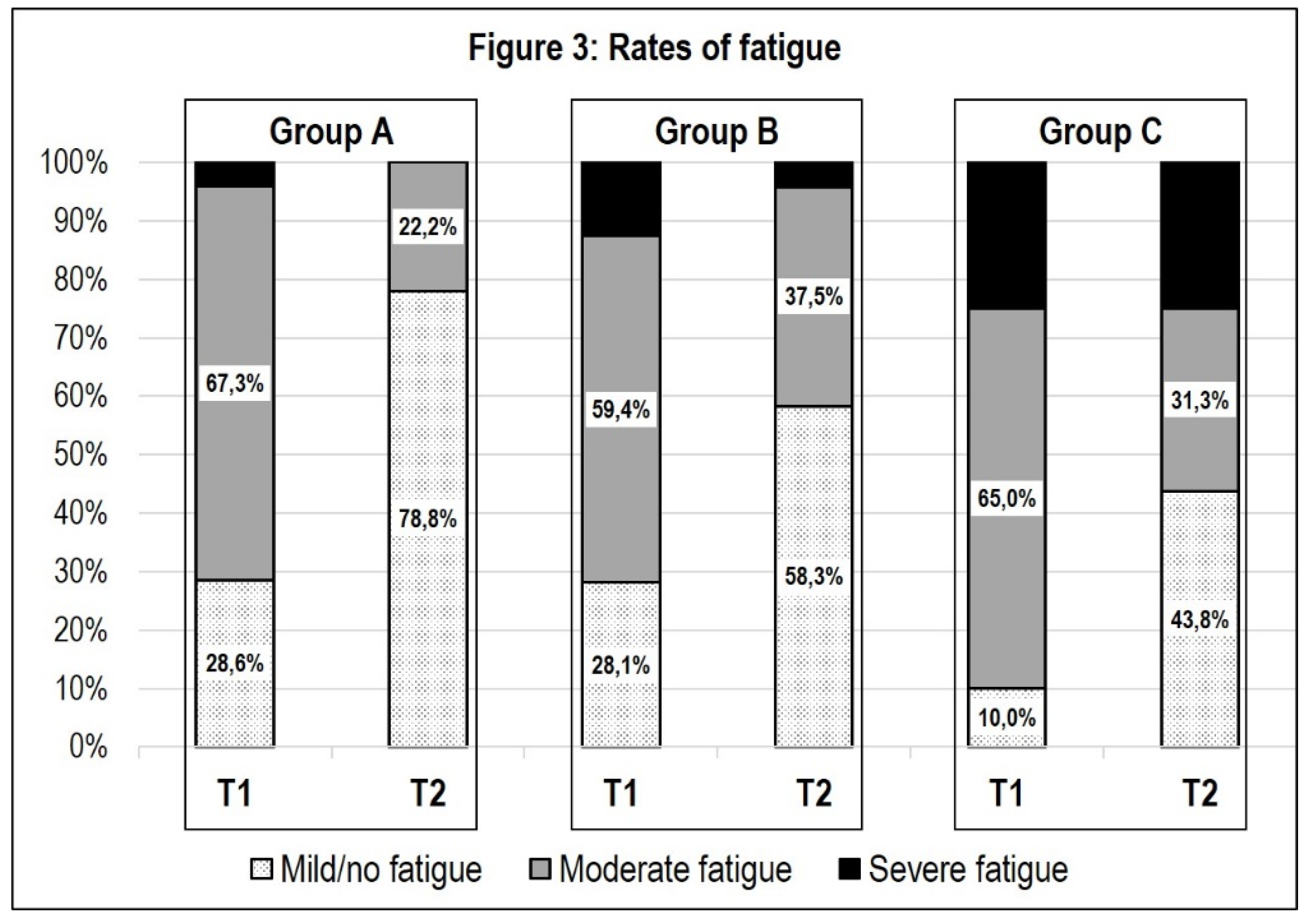
3.6.3. Fatigue
3.6.4. Depression and Anxiety
3.6.5. Rating of the Overall Effectiveness of PR from the Patient’s Perspective and Global Rating of Change in Subjective Health
3.6.6. Safety and Feasibility of PR in Patients after COVID-19
3.6.7. Prediction of Successful PR Concerning the Primary Outcome of Dyspnea
4. Discussion
4.1. Effectiveness of PR
4.2. Primary Outcome: Dyspnea
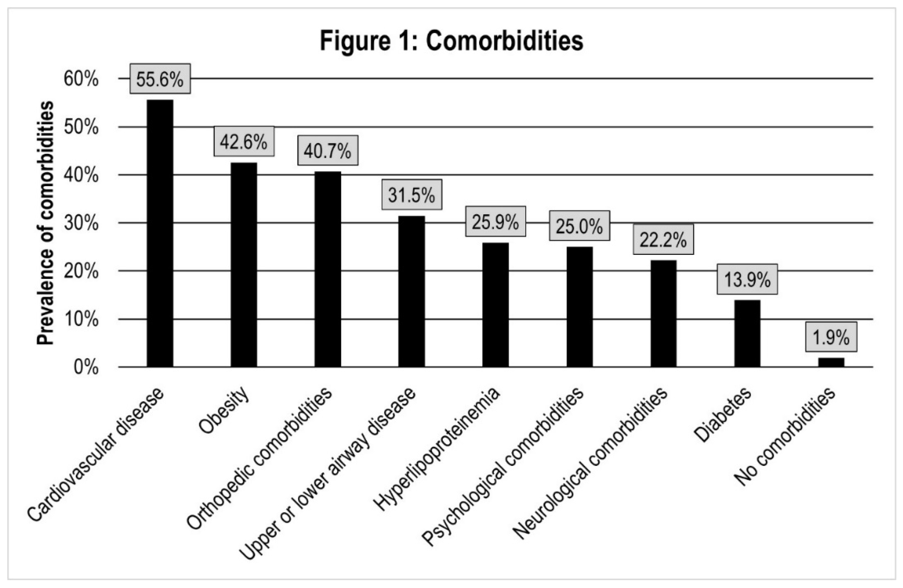
4.3. Comorbidities
4.4. Physical Capacity, Other Clinical Symptoms, and Other Objectively Measured Parameters
4.5. Lung Function Tests
4.6. Quality of Life, Fatigue, Depression, and Anxiety
4.7. Safety and Feasibility of PR in Patients after COVID-19
4.8. Limitations
5. Conclusions
Supplementary Materials
Author Contributions
Funding
Institutional Review Board Statement
Informed Consent Statement
Data Availability Statement
Acknowledgments
Conflicts of Interest
List of Abbreviations (in Alphabetical Order)
| BFI | Brief Fatigue Inventory |
| BNP | Brain Natriuretic Peptide |
| COVID-19 | Coronavirus Disease 2019 |
| CRP | c-reactive Protein |
| EQ-5D-5L | 5-level EuroQol Questionnaire |
| FEV1 | Forced Expiratory Volume in One Second |
| GAD-7 | Generalized Anxiety Disorder-7 Questionnaire |
| GROC | Global Rating of Chance |
| LDH | Lactate Dehydrogenase |
| MCID | Minimal Clinically Important Difference |
| mMRC | Modified Medical Research Council |
| NICE | National Institute for Health and Care Excellence |
| NRS | Numerical Rating Scale |
| PaCO2 | Partial Carbon Dioxide Pressure |
| PaO2 | Partial Oxygen Pressure |
| PCR | Polymerase Chain Reaction |
| PHQ-9 | Patient Health Questionnaire 9 |
| PImax | Maximal Inspiratory Pressure |
| PR | Pulmonary Rehabilitation |
| QoL | Quality of Life |
| RV | Residual Volume |
| SARS-CoV 2 | Severe Acute Respiratory Syndrome Coronavirus 2 |
| sRtot | Total Specific Airway Resistance |
| T1 | Beginning of Pulmonary Rehabilitation |
| T2 | End of Pulmonary Rehabilitation |
| TLC | Total Lung Capacity |
| TLCO | Transfer Factor of the Lung for Carbon Monoxide |
| VAS | Visual Analogue Scale |
| VC | Vital Capacity |
References
- Buitrago-Garcia, D.; Egli-Gany, D.; Counotte, M.J.; Hossmann, S.; Imeri, H.; Ipekci, A.M.; Salanti, G.; Low, N. Occurrence and transmission potential of asymptomatic and presymptomatic SARS-CoV-2 infections: A living systematic review and meta-analysis. PLoS Med. 2020, 17, e1003346. [Google Scholar] [CrossRef] [PubMed]
- Schilling, J.; Lehfeld, A.-S.; Schumacher, D.; Diercke, M.; Buda, S.; Haas, W.; Group, R.C.-S. Krankheitsschwere der ersten COVID-19-Welle in Deutschland basierend auf den Meldungen gemäß Infektionsschutzgesetz. J. Health Monit. 2020, 1–20. [Google Scholar] [CrossRef]
- Huang, C.; Huang, L.; Wang, Y.; Li, X.; Ren, L.; Gu, X.; Kang, L.; Guo, L.; Liu, M.; Zhou, X.; et al. 6-month consequences of COVID-19 in patients discharged from hospital: A cohort study. Lancet 2021, 397, 220–232. [Google Scholar] [CrossRef]
- Halpin, S.J.; McIvor, C.; Whyatt, G.; Adams, A.; Harvey, O.; McLean, L.; Walshaw, C.; Kemp, S.; Corrado, J.; Singh, R.; et al. Postdischarge symptoms and rehabilitation needs in survivors of COVID-19 infection: A cross-sectional evaluation. J. Med. Virol. 2020, 93, 1013–1022. [Google Scholar] [CrossRef]
- Carfi, A.; Bernabei, R.; Landi, F.; Gemelli Against, C.-P.-A.C.S.G. Persistent Symptoms in Patients After Acute COVID-19. JAMA 2020, 324, 603–605. [Google Scholar] [CrossRef] [PubMed]
- Mandal, S.; Barnett, J.; Brill, S.E.; Brown, J.S.; Denneny, E.K.; Hare, S.S.; Heightman, M.; Hillman, T.E.; Jacob, J.; Jarvis, H.C.; et al. ‘Long-COVID’: A cross-sectional study of persisting symptoms, biomarker and imaging abnormalities following hospitalisation for COVID-19. Thorax 2020, 76, 396–398. [Google Scholar] [CrossRef] [PubMed]
- Nalbandian, A.; Sehgal, K.; Gupta, A.; Madhavan, M.V.; McGroder, C.; Stevens, J.S.; Cook, J.R.; Nordvig, A.S.; Shalev, D.; Sehrawat, T.S.; et al. Post-acute COVID-19 syndrome. Nat. Med. 2021, 27, 601–615. [Google Scholar] [CrossRef]
- Armange, L.; Bénézit, F.; Picard, L.; Pronier, C.; Guillot, S.; Lentz, P.-A.; Carré, F.; Tattevin, P.; Revest, M. Prevalence and characteristics of persistent symptoms after non-severe COVID-19: A prospective cohort study. Eur. J. Clin. Microbiol. Infect. Dis. 2021. [Google Scholar] [CrossRef]
- Ordinola Navarro, A.; Cervantes-Bojalil, J.; Cobos Quevedo, O.J.; Avila Martinez, A.; Hernandez-Jimenez, C.A.; Perez Alvarez, E.; Gonzalez Gil, A.; Peralta Amaro, A.L.; Vera-Lastra, O.; Lopez Luis, B.A. Decreased quality of life and spirometric alterations even after mild-moderate COVID-19. Respir. Med. 2021, 181, 106391. [Google Scholar] [CrossRef]
- Stavem, K.; Ghanima, W.; Olsen, M.K.; Gilboe, H.M.; Einvik, G. Prevalence and Determinants of Fatigue after COVID-19 in Non-Hospitalized Subjects: A Population-Based Study. Int. J. Environ. Res. Public Health 2021, 18, 2030. [Google Scholar] [CrossRef]
- Nehme, M.; Braillard, O.; Alcoba, G.; Aebischer Perone, S.; Courvoisier, D.; Chappuis, F.; Guessous, I. COVID-19 Symptoms: Longitudinal Evolution and Persistence in Outpatient Settings. Ann. Intern. Med. 2020, 174, 723–725. [Google Scholar] [CrossRef]
- Spruit, M.A.; Holland, A.E.; Singh, S.J.; Tonia, T.; Wilson, K.C.; Troosters, T. COVID-19: Interim Guidance on Rehabilitation in the Hospital and Post-Hospital Phase from a European Respiratory Society and American Thoracic Society-coordinated International Task Force. Eur. Respir. J. 2020, 56, 2002197. [Google Scholar] [CrossRef] [PubMed]
- Glockl, R.; Buhr-Schinner, H.; Koczulla, A.R.; Schipmann, R.; Schultz, K.; Spielmanns, M.; Stenzel, N.; Dewey, S. [Recommendations from the German Respiratory Society for Pulmonary Rehabilitation in Patients with COVID-19]. Pneumologie 2020, 74, 496–504. [Google Scholar] [CrossRef] [PubMed]
- Vitacca, M.; Lazzeri, M.; Guffanti, E.; Frigerio, P.; D’Abrosca, F.; Gianola, S.; Carone, M.; Paneroni, M.; Ceriana, P.; Pasqua, F.; et al. Italian suggestions for pulmonary rehabilitation in COVID-19 patients recovering from acute respiratory failure: Results of a Delphi process. Monaldi Arch. Chest Dis. 2020, 90. [Google Scholar] [CrossRef]
- DE Sire, A.; Andrenelli, E.; Negrini, F.; Patrini, M.; Lazzarini, S.G.; Ceravolo, M.G. Rehabilitation and COVID-19: A rapid living systematic review by Cochrane Rehabilitation Field updated as of December 31st, 2020 and synthesis of the scientific literature of 2020. Eur. J. Phys. Rehabil. Med. 2021, 57, 181–188. [Google Scholar] [CrossRef]
- Andrenelli, E.; Negrini, F.; de Sire, A.; Patrini, M.; Lazzarini, S.G.; Ceravolo, M.G.; Cochrane, I.M.S.C. Rehabilitation and COVID-19: Update of the rapid living systematic review by Cochrane Rehabilitation Field as of February 28th, 2021. Eur. J. Phys. Rehabil. Med. 2021, 57, 481–484. [Google Scholar] [CrossRef]
- Borg, G.A. Psychophysical bases of perceived exertion. Med. Sci. Sports Exerc. 1982, 14, 377–381. [Google Scholar] [CrossRef] [PubMed]
- Holland, A.E.; Spruit, M.A.; Troosters, T.; Puhan, M.A.; Pepin, V.; Saey, D.; McCormack, M.C.; Carlin, B.W.; Sciurba, F.C.; Pitta, F.; et al. An official European Respiratory Society/American Thoracic Society technical standard: Field walking tests in chronic respiratory disease. Eur. Respir. J 2014, 44, 1428–1446. [Google Scholar] [CrossRef]
- Gift, A.G.; Narsavage, G. Validity of the numeric rating scale as a measure of dyspnea. Am. J. Crit. Care 1998, 7, 200–204. [Google Scholar] [CrossRef]
- Oxberry, S.G.; Bland, J.M.; Clark, A.L.; Cleland, J.G.; Johnson, M.J. Minimally clinically important difference in chronic breathlessness: Every little helps. Am. Heart J. 2012, 164, 229–235. [Google Scholar] [CrossRef]
- Salaffi, F.; Stancati, A.; Silvestri, C.A.; Ciapetti, A.; Grassi, W. Minimal clinically important changes in chronic musculoskeletal pain intensity measured on a numerical rating scale. Eur. J. Pain 2004, 8, 283–291. [Google Scholar] [CrossRef] [PubMed]
- Bestall, J.C.; Paul, E.A.; Garrod, R.; Garnham, R.; Jones, P.W.; Wedzicha, J.A. Usefulness of the Medical Research Council (MRC) dyspnoea scale as a measure of disability in patients with chronic obstructive pulmonary disease. Thorax 1999, 54, 581–586. [Google Scholar] [CrossRef] [Green Version]
- Santus, P.; Tursi, F.; Croce, G.; Di Simone, C.; Frassanito, F.; Gaboardi, P.; Airoldi, A.; Pecis, M.; Negretto, G.; Radovanovic, D. Changes in quality of life and dyspnoea after hospitalization in COVID-19 patients discharged at home. Multidiscip. Respir. Med. 2020, 15, 713. [Google Scholar] [CrossRef] [PubMed]
- Mahler, D.A.; Wells, C.K. Evaluation of clinical methods for rating dyspnea. Chest 1988, 93, 580–586. [Google Scholar] [CrossRef] [PubMed] [Green Version]
- Crisafulli, E.; Clini, E.M. Measures of dyspnea in pulmonary rehabilitation. Multidiscip. Respir. Med. 2010, 5, 202–210. [Google Scholar] [CrossRef] [Green Version]
- Enright, P.L.; Sherrill, D.L. Reference equations for the six-minute walk in healthy adults. Am. J. Respir. Crit. Care Med. 1998, 158, 1384–1387. [Google Scholar] [CrossRef] [PubMed] [Green Version]
- Criee, C.P.; Baur, X.; Berdel, D.; Bosch, D.; Gappa, M.; Haidl, P.; Husemann, K.; Jorres, R.A.; Kabitz, H.J.; Kardos, P.; et al. [Standardization of spirometry: 2015 update. Published by German Atemwegsliga, German Respiratory Society and German Society of Occupational and Environmental Medicine]. Pneumologie 2015, 69, 147–164. [Google Scholar] [CrossRef] [PubMed]
- Criee, C.P.; Sorichter, S.; Smith, H.J.; Kardos, P.; Merget, R.; Heise, D.; Berdel, D.; Kohler, D.; Magnussen, H.; Marek, W.; et al. Body plethysmography--its principles and clinical use. Respir. Med. 2011, 105, 959–971. [Google Scholar] [CrossRef] [PubMed]
- Radbruch, L.; Sabatowski, R.; Elsner, F.; Everts, J.; Mendoza, T.; Cleeland, C. Validation of the German version of the brief fatigue inventory. J. Pain Symptom Manag. 2003, 25, 449–458. [Google Scholar] [CrossRef]
- Mendoza, T.R.; Wang, X.S.; Cleeland, C.S.; Morrissey, M.; Johnson, B.A.; Wendt, J.K.; Huber, S.L. The rapid assessment of fatigue severity in cancer patients: Use of the Brief Fatigue Inventory. Cancer 1999, 85, 1186–1196. [Google Scholar] [CrossRef]
- Oemar, M.; Janssen, B. EQ-5D-5L User Guide2013. (Version 2.0, October 2013).
- Zanini, A.; Aiello, M.; Adamo, D.; Casale, S.; Cherubino, F.; Della Patrona, S.; Raimondi, E.; Zampogna, E.; Chetta, A.; Spanevello, A. Estimation of minimal clinically important difference in EQ-5D visual analog scale score after pulmonary rehabilitation in subjects with COPD. Respir. Care 2015, 60, 88–95. [Google Scholar] [CrossRef] [Green Version]
- Kroenke, K.; Spitzer, R.L.; Williams, J.B. The PHQ-9: Validity of a brief depression severity measure. J. Gen. Intern. Med. 2001, 16, 606–613. [Google Scholar] [CrossRef] [PubMed]
- Spitzer, R.L.; Kroenke, K.; Williams, J.B.; Lowe, B. A brief measure for assessing generalized anxiety disorder: The GAD-7. Arch. Intern. Med. 2006, 166, 1092–1097. [Google Scholar] [CrossRef] [PubMed] [Green Version]
- Gräfe, K.; Zipfel, S.; Herzog, W.; Löwe, B. Screening psychischer Störungen mit dem Gesundheitsfragebogen für Patienten (PHQ-D). Diagnostica 2004, 50, 171–181. [Google Scholar] [CrossRef]
- Kamper, S.J.; Maher, C.G.; Mackay, G. Global rating of change scales: A review of strengths and weaknesses and considerations for design. J. Man. Manip. Ther. 2009, 17, 163–170. [Google Scholar] [CrossRef] [Green Version]
- Alma, H.; de Jong, C.; Jelusic, D.; Wittmann, M.; Schuler, M.; Flokstra-de Blok, B.; Kocks, J.; Schultz, K.; van der Molen, T. Health status instruments for patients with COPD in pulmonary rehabilitation: Defining a minimal clinically important difference. NPJ Prim. Care Respir. Med. 2016, 26, 16041. [Google Scholar] [CrossRef] [Green Version]
- Cohen, J. Statistical Power Analysis for the Behavioral Sciences, 2nd ed.; L. Erlbaum Associates: Hillsdale, NJ, USA, 1988; p. xxi. 567p. [Google Scholar]
- Gloeckl, R.; Leitl, D.; Jarosch, I.; Schneeberger, T.; Nell, C.; Stenzel, N.; Vogelmeier, C.F.; Kenn, K.; Koczulla, A.R. Benefits of pulmonary rehabilitation in COVID-19: A prospective observational cohort study. ERJ Open Res. 2021, 7, 00108-2021. [Google Scholar] [CrossRef]
- Al Chikhanie, Y.; Veale, D.; Schoeffler, M.; Pepin, J.L.; Verges, S.; Herengt, F. Effectiveness of pulmonary rehabilitation in COVID-19 respiratory failure patients post-ICU. Respir. Physiol. Neurobiol. 2021, 287, 103639. [Google Scholar] [CrossRef] [PubMed]
- Cortes-Telles, A.; Lopez-Romero, S.; Figueroa-Hurtado, E.; Pou-Aguilar, Y.N.; Wong, A.W.; Milne, K.M.; Ryerson, C.J.; Guenette, J.A. Pulmonary function and functional capacity in COVID-19 survivors with persistent dyspnoea. Respir. Physiol. Neurobiol. 2021, 288, 103644. [Google Scholar] [CrossRef]
- Carod-Artal, F.J. Post-COVID-19 syndrome: Epidemiology, diagnostic criteria and pathogenic mechanisms involved. Rev. Neurol. 2021, 72, 384–396. [Google Scholar] [CrossRef]
- van der Sar-van der Brugge, S.; Talman, S.; Boonman-de Winter, L.; de Mol, M.; Hoefman, E.; van Etten, R.W.; De Backer, I.C. Pulmonary function and health-related quality of life after COVID-19 pneumonia. Respir. Med. 2021, 176, 106272. [Google Scholar] [CrossRef]
- Blanco, J.R.; Cobos-Ceballos, M.J.; Navarro, F.; Sanjoaquin, I.; Arnaiz de Las Revillas, F.; Bernal, E.; Buzon-Martin, L.; Viribay, M.; Romero, L.; Espejo-Perez, S.; et al. Pulmonary long-term consequences of COVID-19 infections after hospital discharge. Clin. Microbiol. Infect. 2021, 27, 892–896. [Google Scholar] [CrossRef]
- Wu, X.; Liu, X.; Zhou, Y.; Yu, H.; Li, R.; Zhan, Q.; Ni, F.; Fang, S.; Lu, Y.; Ding, X.; et al. 3-month, 6-month, 9-month, and 12-month respiratory outcomes in patients following COVID-19-related hospitalisation: A prospective study. Lancet Respir. Med. 2021, 9, 747–754. [Google Scholar] [CrossRef]
- Fayol, A.; Livrozet, M.; Boutouyrie, P.; Khettab, H.; Betton, M.; Tea, V.; Blanchard, A.; Bruno, R.M.; Hulot, J.S.; French, C.c.s.g. Cardiac performance in patients hospitalized with COVID-19: A 6 month follow-up study. ESC Heart Fail. 2021, 8, 2232–2239. [Google Scholar] [CrossRef] [PubMed]
- Ramadan, M.S.; Bertolino, L.; Marrazzo, T.; Florio, M.T.; Durante-Mangoni, E.; The Monaldi Hospital Cardiovascular Infection Study, G. Cardiac complications during the active phase of COVID-19: Review of the current evidence. Intern. Emerg. Med. 2021. [Google Scholar] [CrossRef] [PubMed]
- Mohr, A.; Dannerbeck, L.; Lange, T.J.; Pfeifer, M.; Blaas, S.; Salzberger, B.; Hitzenbichler, F.; Koch, M. Cardiopulmonary exercise pattern in patients with persistent dyspnoea after recovery from COVID-19. Multidiscip. Respir. Med. 2021, 16, 732. [Google Scholar] [CrossRef]
- Schultz, K.; Jelusic, D.; Wittmann, M.; Kramer, B.; Huber, V.; Fuchs, S.; Lehbert, N.; Wingart, S.; Stojanovic, D.; Gohl, O.; et al. Inspiratory muscle training does not improve clinical outcomes in 3-week COPD rehabilitation: Results from a randomised controlled trial. Eur. Respir. J. 2018, 51, 1702000. [Google Scholar] [CrossRef] [Green Version]
- Curci, C.; Negrini, F.; Ferrillo, M.; Bergonzi, R.; Bonacci, E.; Camozzi, D.M.; Ceravolo, C.; De Franceschi, S.; Guarnieri, R.; Moro, P.; et al. Functional outcome after inpatient rehabilitation in post-intensive care unit COVID-19 patients: Findings and clinical implications from a real-practice retrospective study. Eur. J. Phys. Rehabil. Med. 2021, 57, 443–450. [Google Scholar] [CrossRef]
- Bajgain, K.T.; Badal, S.; Bajgain, B.B.; Santana, M.J. Prevalence of comorbidities among individuals with COVID-19: A rapid review of current literature. Am. J. Infect. Control 2021, 49, 238–246. [Google Scholar] [CrossRef] [PubMed]
- Ng, W.H.; Tipih, T.; Makoah, N.A.; Vermeulen, J.G.; Goedhals, D.; Sempa, J.B.; Burt, F.J.; Taylor, A.; Mahalingam, S. Comorbidities in SARS-CoV-2 Patients: A Systematic Review and Meta-Analysis. mBio 2021, 12. [Google Scholar] [CrossRef]
- Puchner, B.; Sahanic, S.; Kirchmair, R.; Pizzini, A.; Sonnweber, B.; Garimorth, K.; Dareb, B.; Ehling, R. Beneficial effects of multi-disciplinary rehabilitation in post-acute COVID-19 an observational cohort study. Eur. J. Phys. Rehabil. Med. 2021, 57, 189–198. [Google Scholar] [CrossRef] [PubMed]
- Spielmanns, M.; Pekacka-Egli, A.M.; Schoendorf, S.; Windisch, W.; Hermann, M. Effects of a Comprehensive Pulmonary Rehabilitation in Severe Post-COVID-19 Patients. Int. J. Environ. Res. Public Health 2021, 18, 2695. [Google Scholar] [CrossRef] [PubMed]
- Bertolucci, F.; Sagliocco, L.; Tolaini, M.; Posteraro, F. Comprehensive rehabilitation treatment for sub acute COVID-19 patients: An observational study. Eur. J. Phys. Rehabil. Med. 2021, 57, 208–215. [Google Scholar] [CrossRef]
- Wiertz, C.M.H.; Vints, W.A.J.; Maas, G.; Rasquin, S.M.C.; van Horn, Y.Y.; Dremmen, M.P.M.; Hemmen, B.; Verbunt, J.A. COVID-19: Patient characteristics in the first phase of post-intensive care rehabilitation. Arch. Rehabil. Res. Clin. Transl. 2021, 100108. [Google Scholar] [CrossRef]
- Hermann, M.; Pekacka-Egli, A.M.; Witassek, F.; Baumgaertner, R.; Schoendorf, S.; Spielmanns, M. Feasibility and Efficacy of Cardiopulmonary Rehabilitation After COVID-19. Am. J. Phy.s Med. Rehabil. 2020, 99, 865–869. [Google Scholar] [CrossRef]
- Greulich, T.; Koczulla, A.R.; Nell, C.; Kehr, K.; Vogelmeier, C.F.; Stojanovic, D.; Wittmann, M.; Schultz, K. Effect of a Three-Week Inpatient Rehabilitation Program on 544 Consecutive Patients with Very Severe COPD: A Retrospective Analysis. Respiration 2015, 90, 287–292. [Google Scholar] [CrossRef]
- Huppmann, P.; Sczepanski, B.; Boensch, M.; Winterkamp, S.; Schonheit-Kenn, U.; Neurohr, C.; Behr, J.; Kenn, K. Effects of inpatient pulmonary rehabilitation in patients with interstitial lung disease. Eur. Respir. J. 2013, 42, 444–453. [Google Scholar] [CrossRef] [Green Version]
- Schultz, K.; Wittmann, M.; Wagner, R.; Lehbert, N.; Schwarzkopf, L.; Szentes, B.; Nowak, D.; Faller, H.; Schuler, M. In-patient pulmonary rehabilitation to improve asthma control—a randomized controlled study (EPRA, Effectiveness of Pulmonary Rehabilitation for Patients with Asthma). Dtsch. Arztebl. Int. 2021, 118, 23–30. [Google Scholar] [CrossRef]
- Puhan, M.A.; Mador, M.J.; Held, U.; Goldstein, R.; Guyatt, G.H.; Schunemann, H.J. Interpretation of treatment changes in 6-minute walk distance in patients with COPD. Eur. Respir. J. 2008, 32, 637–643. [Google Scholar] [CrossRef] [Green Version]
- Puhan, M.A.; Chandra, D.; Mosenifar, Z.; Ries, A.; Make, B.; Hansel, N.N.; Wise, R.A.; Sciurba, F.; National Emphysema Treatment Trial (NETT) Research Group. The minimal important difference of exercise tests in severe COPD. Eur. Respir. J. 2011, 37, 784–790. [Google Scholar] [CrossRef] [PubMed]
- Geidl, W.; Carl, J.; Schuler, M.; Mino, E.; Lehbert, N.; Wittmann, M.; Pfeifer, K.; Schultz, K. Long-term benefits of adding a pedometer to pulmonary rehabilitation for COPD. The randomized controlled STAR trial. Int. J. Chron. Obstruct. Pulmon. Dis. 2021, (in press). [Google Scholar] [CrossRef] [PubMed]
- Schuler, M.; Strohmayer, M.; Muhlig, S.; Schwaighofer, B.; Wittmann, M.; Faller, H.; Schultz, K. Assessment of depression before and after inpatient rehabilitation in COPD patients: Psychometric properties of the German version of the Patient Health Questionnaire (PHQ-9/PHQ-2). J. Affect. Disord. 2018, 232, 268–275. [Google Scholar] [CrossRef] [PubMed]
- Demeco, A.; Marotta, N.; Barletta, M.; Pino, I.; Marinaro, C.; Petraroli, A.; Moggio, L.; Ammendolia, A. Rehabilitation of patients post-COVID-19 infection: A literature review. J. Int. Med. Res. 2020, 48, 300060520948382. [Google Scholar] [CrossRef] [PubMed]


| Group A “Acute Severe” | Group B “Severe after Interval” | Group C “Mild after Interval” | All Patients | |
|---|---|---|---|---|
| Referral to rehab as | PR after direct transfer from hospital (n = 24) or within one month at the latest after discharge (n = 31) | PR at the earliest after more than 1 month after discharge from hospital | PR after outpatient treatment (n = 16) or monitored in the hospital for a maximum of one night (n = 5) | |
| Number of patients | N = 55 (50.9%) | N = 32 (29.6%) | N = 21 (19.4%) | N = 108 (100%) |
| Age [mean ± SD] (range) | 57.9 ± 10.8 (33–85) | 54.0 ± 9.9 (32–80) | 52.1 ± 6.8 (39–61) | 55.6 ± 10.1 (32–85) |
| Sex [% female] | 38.2% (34 M/21 F) | 34.4% (21 M/11 F) | 81.0% (4 M/17 F) | 45.4% (59 M/49 F) |
| BMI [mean ± SD] (range) | 29.9 ± 5.7 (19.3–53.3) | 31.5 ± 6.7 (21.0–48.3) | 28.9 ± 6.1 (19.9–46.0) | 30.17 ± 6.12 (19.3–53.3) |
| Length of hospitalization [mean ± SD in days] (range) | 31.5 ± 18.7 (3–101) | 19.4 ± 11.0 (3–49) | Monitored in the hospital for a maximum of one night (n = 5) | 25.7 ± 17.8 (1–101) |
| % Oxygen therapy during acute COVID-19 phase | 87.3% | 68.8% | 14.3% | 67.6% |
| % ICU-treatment [mean ± SD in days] (range) | 70.9% 21.7 ± 18.1 (4–97) | 56.3% 14.5 ± 8.8 (5–40) | - | 52.8% 23.6 ± 17.4 (5–97) |
| % Invasive ventilation [mean ± SD in days] (range) | 49.1% 20.0 ± 17.7 (8–87) | 37.5% 11.6 ± 4.9 (5–23) | - | 36.1% 17.6 ± 15.5 (5–87) |
| Duration between discharge from the clinic or the acute COVID-19 phase undergone in an outpatient setting and beginning of PR [mean ± SD in days] (range) | 10.8 ± 11.2 (0–31) | 120.6 ± 70.2 (32–270) | 142.9 ± 55.1 (35–270) | 69.0 ± 75.3 (0–270) |
| T1 | T2 | ||||||
|---|---|---|---|---|---|---|---|
| Group | M (Median) | SD (Range) | M (Median) | SD (Range) | Delta [95% CI] | d [95% CI] | RM-ANOVA F(df), p |
| Dyspnea | |||||||
| NRS “How intense is your dyspnea at rest?” (☺ 0–10 ☹) | |||||||
| All patients N = 82 | 1.70 (1.00) | 1.82 (0.00–8.00) | 1.02 (0.00) | 1.49 (0.00–7.00) | −0.67 [−1.03; −0.32] | −0.41 [−0.64; −0.19] | FT = 13.3(1), p < 0.001 FG = 2.4(2), p = 0.100 FG*T = 0.24(2), p = 0.788 |
| A N = 43 | 1.44 (1.00) | 1.68 (0.00–8.00) | 0.84 (0.00) | 1.34 (0.00–7.00) | −0.60 [−1.06; −0.15] | − 0.41 [−0.72; −0.10] | |
| B N = 24 | 1.63 (1.00) | 1.41 (0.00–5.00) | 1.00 (1.00) | 1.10 (0.00–3.00) | −0.63 [−1.22; −0.03] | −0.49 [−0.86; −0.02] | |
| C N = 15 | 2.53 (2.00) | 2.53 (0.00–7.00) | 1.60 (0.50) | 2.23 (0.00–7.00) | −0.93 [−2.21; −0.35] | −0.39 [−0.92; 0.13] | |
| NRS “How intense is your dyspnea on exertion?” (☺ 0–10 ☹) | |||||||
| All patients N = 82 | 5.56 (6.00) | 2.50 (0.00–10.00) | 3.41 (3.00) | 2.73 (0.00–10.00) | −2.15 [−2.73; −1.57] | −0.82 [−1.01; −0.56] | FT = 39.0(1), p < 0.001 FG = 0.25(2), p = 0.774 FG*T = 65(2), p = 0.524 |
| A N = 43 | 5.60 (5.50) | 2.56 (0.00–10.00) | 3.23 (2.00) | 2.58 (0.00–10.00) | −2.37 [−3.16; −1.58] | −0.92 [−1.28; −0.56] | |
| B N = 24 | 5.46 (6.00) | 2.47 (1.00–9.00) | 3.29 (2.00) | 2.66 (0.00–8.00) | −2.17 [−3.37; −0.96] | −0.85 [−1.21; −0.30] | |
| C N = 15 | 5.60 (6.00) | 2.53 (0.00–9.00) | 4.13 (4.00) | 3.29 (0.00–10.00) | −1.47 [−2.87; −0.06] | −0.58 [−1.12; −0.02] | |
| mMRC (☺ 0–4 ☹) | |||||||
| All patients N = 90 | 2.26 (2.00) | 1.19 (0.00 – 4.00) | 1.51 (2.00) | 1.12 (0.00–4.00) | −0.74 [−0.99; −0.50] | −0.64 [−0.87; −0.42] | FT = 25.1(1), p < 0.001 FG = 1.81(2), p = 0.168 FG*T = 1.14(2), p = 0.323 |
| A N = 48 | 2.52 (2.00) | 1.29 (0.00–4.00) | 1.60 (2.00) | 1.09 (0.00–4.00) | −0.92 [−1.27; −0.57] | −0.76 [−1.08; −0.44] | |
| B N = 26 | 1.88 (2.00) | 0.82 (0.00–4.00) | 1.35 (2.00) | 1.23 (0.00–4.00) | −0.53 [−1.04; −0.04] | −0.43 [−0.83; −0.03] | |
| C N = 16 | 2.06 (2.00) | 1.24 (0.00–4.00) | 1.50 (1.50) | 1.10 (0.00–4.00) | −0.56 [−1.00; −0.13] | −0.69 [−1.23; −0.12] | |
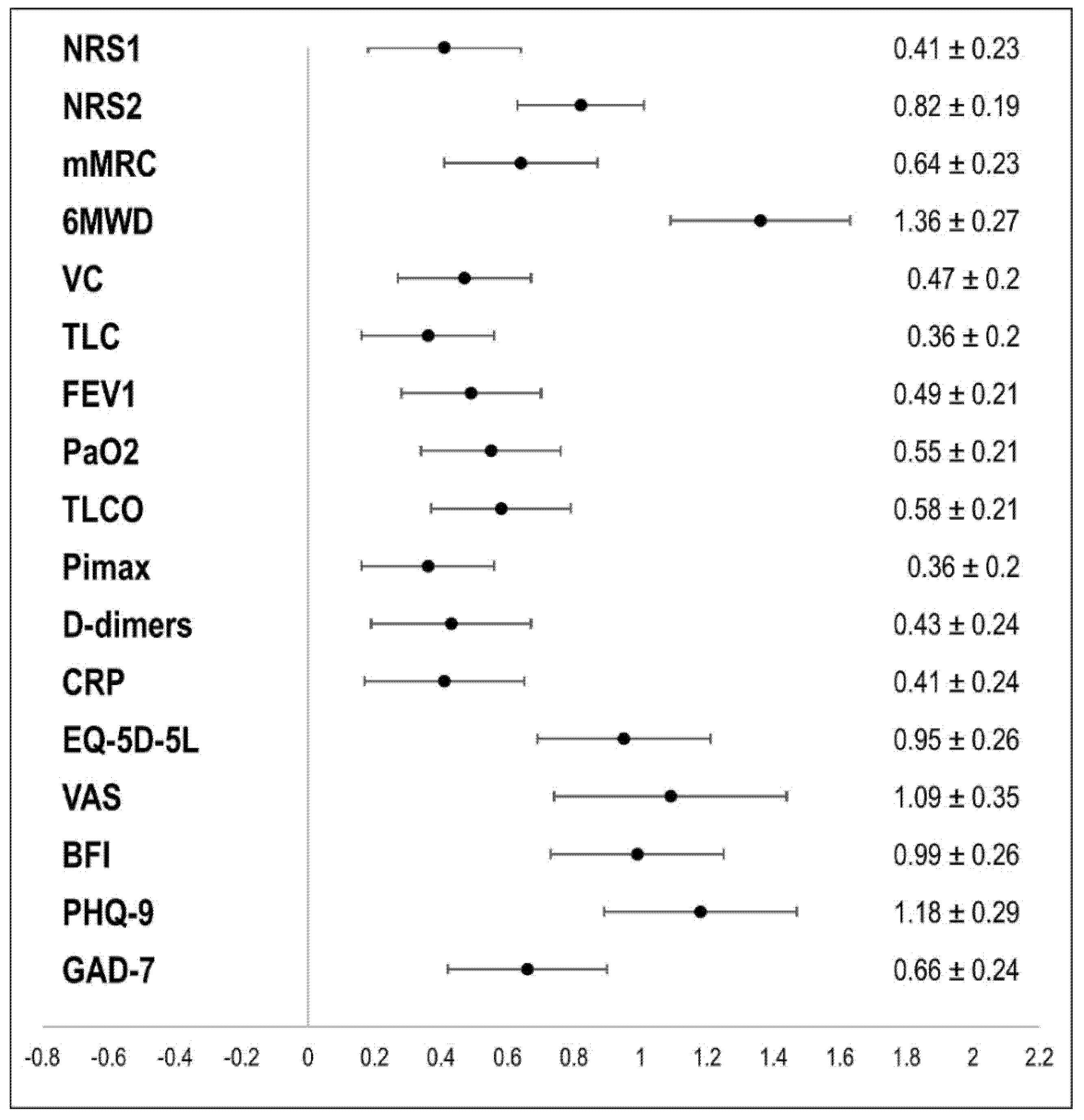
| NRS1 | NRS2 | mMRC | 6MWD | VC | TLC | FEV1 | PaO2 | TLCO | PImax | D-dimers | CRP | EQ-5D-5L | VAS | BFI | PHQ-9 | GAD-7 | |
|---|---|---|---|---|---|---|---|---|---|---|---|---|---|---|---|---|---|
| NRS1 | 1 | 0.341 ** | 0.159 | −0.223 * | 0.010 | 0.058 | 0.143 | −0.004 | 0.141 | 0.153 | -0.014 | −0.058 | 0.134 | 0.077 | 0.193 | 0.196 | 0.261 * |
| NRS2 | 1 | 0.220 | 0.187 | 0.159 | 0.187 | 0.192 | −0.120 | 0.075 | 0.073 | -0.077 | −0.164 | 0.363 ** | 0.273 * | 0.242 * | 0.204 | 0.348 ** | |
| mMRC | 1 | 0.074 | 0.099 | −0.127 | 0.092 | −0.038 | −0.012 | 0.030 | -0.058 | −0.050 | 0.164 | 0.405 ** | 0.313 ** | 0.158 | 0.265 * | ||
| 6MWD | 1 | 0.065 | 0.058 | 0.027 | 0.281 ** | 0.191 | −0.280 ** | 0.195 | 0.273 * | 0.279 * | 0.244 * | 0.203 | 0.124 | 0.045 | |||
| VC | 1 | 0.480 ** | 0.842 ** | 0.249 * | 0.126 | 0.338 ** | 0.295 * | 0.146 | 0.127 | 0.118 | 0.179 | 0.233* | −0.076 | ||||
| TLC | 1 | 0.305 ** | 0.205 * | 0.227 * | 0.222 * | 0.305 * | 0.108 | 0.199 | −0.020 | 0.090 | 0.061 | 0.014 | |||||
| FEV1 | 1 | 0.286 ** | 0.222 * | 0.248 * | 0.229 | 0.197 | 0.124 | 0.145 | 0.145 | 0.290 * | 0.077 | ||||||
| PaO2 | 1 | 0.316 ** | −0.068 | 0.165 | 0.281 | 0.006 | −0.128 | −0.061 | 0.055 | 0.017 | |||||||
| TLCO | 1 | −0.003 | 0.306 * | 0.200 | 0.047 | 0.010 | 0.068 | 0.054 | 0.066 | ||||||||
| PImax | 1 | 0.173 | 0.097 | −0.021 | 0.157 | −0.064 | −0.197 | −0.106 | |||||||||
| D-dimers | 1 | 0.600 ** | 0.162 | 0.155 | 0.062 | −0.041 | −0.171 | ||||||||||
| CRP | 1 | −0.092 | 0.102 | −0.129 | −0.158 | −0.118 | |||||||||||
| EQ-5D-5L | 1 | 0.296 ** | 0.419 ** | 0.059 | 0.106 | ||||||||||||
| VAS | 1 | 0.279 * | 0.077 | 0.318 ** | |||||||||||||
| BFI | 1 | 0.0555 ** | 0.498 ** | ||||||||||||||
| PHQ-9 | 1 | 0.473 ** | |||||||||||||||
| GAD-7 | 1 |
| T1 | T2 | ||||||
|---|---|---|---|---|---|---|---|
| Group | M (Median) | SD (Range) | M (Median) | SD (Range) | Delta [95% CI] | d [95% CI] | RM-ANOVA F(df), p |
| Physical Capacity | |||||||
| 6-MWD [m] | |||||||
| All patients N = 97 | 419 (450) | 127 (13–663) | 530 (530) | 100 (175–740) | 111 [94; 127] | 1.36 [1.08; 1.63] | FT = 130.3(1), p = 0.001 FG = 5.4(2), p = 0.006 FG*T = 4.1(2), p = 0.019 |
| A N = 52 | 377 (392) | 142 (13–603) | 508 (522) | 111 (175–720) | 131 [107; 155] | 1.52 [1.12; 1.92] | |
| B N = 27 | 459 (470) | 79 (270–590) | 555 (564) | 79.5 (406–675) | 96 [72; 120] | 1.61 [1.02; 2.17] | |
| C N = 18 | 480 (479) | 96.8 (275–663) | 554 (544) | 85.9 (380–740) | 74 [33; 114] | 0.91 [0.35; 1.45] | |
| Lung Function | |||||||
| Vital Capacity (VC) [%pred.] | |||||||
| All patients N = 103 | 89.6 (90.9) | 20.7 (33.7–130.1) | 98.0 (100.3) | 16.2 (63.4–129.3) | 8.3 [4.9; 11.8] | 0.47 [0.27; 0.67] | FT = 9.49(1), p = 0.003 FG = 12.0(2), p < 0.001 FG*T = 4.77(2), p = 0.011 |
| A N = 54 | 81.1 (79.7) | 21.7 (33.7–130.1) | 93.6 (96.6) | 16.5 (63.4–129.3) | 12.5 [7.3; 17.7] | 0.66 [0.36; 0.95] | |
| B N = 32 | 95.3 (97.5) | 14.4 (63.0–115.5) | 102.0 (103.6) | 14.5 (64.1–122.7) | 6.7 [0.5; 12.8] | 0.39 [0.03; 0.75] | |
| C N = 17 | 106.3 (104.2) | 12.9 (85.4–129.6) | 104.5 (106.0) | 14.9 (68.4–125.5) | −1.8 [−6.27; 2.64] | −0.21 [−0.69; 0.27] | |
| Total lung capacity (TLC) [%pred.] | |||||||
| All patients N = 103 | 92.8 (95.6) | 17.3 (46.8–134.1) | 97.7 (99.1) | 14.9 (63.7–128.1) | 4.9 [2.3; 7.6] | 0.36 [0.16; 0.56] | FT = 5.61(1), p = 0.020 FG = 14.8(2), p < 0.001 FG*T = 2.47(2), p = 0.089 |
| A N = 54 | 85.4 (86.3) | 17.0 (46.8–121.4) | 92.0 (91.1) | 13.8 (63.7–119.6) | 6.5 [2.7; 10.4] | 0.47 [0.18; 0.74] | |
| B N = 32 | 98.3 (98.1) | 13.9 (70.8–125.7) | 104.1 (106.0) | 14.8 (72.4–128.1) | 5.8 [0.6; 10.9] | 0.41 [0.04; 0.76] | |
| C N = 17 | 105.5 (104.1) | 12.0 (81.1–134.1) | 103.8 (101.6) | 11.1 (81.6–126.9) | −1.7 [−6.9; 3.4] | −0.17 [−0.65; 0.31] | |
| Forced expiratory volume in in one second (FEV1) [%pred.] | |||||||
| All patients N = 103 | 92.3 (92.0) | 20.8 (23.2–135.7) | 100.9 (103.3) | 16.5 (36.7–138.1) | 8.6 [5.2; 12.0] | 0.49 [0.29; 0.70] | FT = 12.6(1), p < 0.001 FG = 7.9(2), p < 0.001 FG*T = 2.88(2), p = 0.061 |
| A N = 54 | 85.0 (85.3) | 23.1 (23.2–135.7) | 97.2 (97.0) | 18.2 (36.7–138.1) | 12.16 [6.5; 17.8] | 0.59 [0.29; 0.87] | |
| B N = 32 | 97.3 (96.8) | 14.0 (69.1–126.2) | 103.7 (106.4) | 13.4 (72.5–131.3) | 6.4 [1.4; 11.5] | 0.46 [0.09; 0.82] | |
| C N = 17 | 105.8 (106.0) | 13.4 (86.7–129.7) | 107.3 (108.7) | 13.4 (76.6–131.2) | 1.5 [−1.6; 4.5] | 0.25 [−0.23; 0.72] | |
| Partial pressure of O2 (PaO2) [mm Hg] | |||||||
| All patients N = 100 | 74.4 (74.5) | 9.0 (43.0–98.0) | 78.4 (78.0) | 7.1 (58.0–95.0) | 4.0 [2.6; 5.5] | 0.55 [0.34; 0.76] | FT = 1571.9(1), p < 0.001 FG = 4.7(2), p = 0.02 FG*T = 6.1(2), p = 0.003 |
| A N = 51 | 71.7 (72.0) | 9.6 (43.0–88.0) | 77.3 (78.0) | 7.4 (58.0–94.0) | 5.6 [3.7; 7.5] | −0.63 [0.50; 1.14] | |
| B N = 31 | 75.6 (73.0) | 7.2 (66.0–92.0) | 79.4 (78.0) | 6.6 (70.0–95.0) | 3.7 [0.9; 6.5] | 0.49 [0.12; 0.86] | |
| C N = 18 | 79.9 (79.5) | 7.3 (69.0–98.0) | 80.0 (79.5) | 6.7 (71.0–91.0) | 0.06 [3.4; −3.5] | 0.01 [−0.45; 0.47] | |
| Diffusion capacity of the lungs for carbon monoxide (TLCO SB) [%pred.] | |||||||
| All patients N = 93 | 73.8 (77.1) | 22.0 (11.7–124.0) | 80.0 (78.4) | 20.3 (35.1–120.2 | 6.2 [4.0; 8.4] | 0.58 [0.36; 0.79] | FT = 13.4(1), p < 0.001 FG = 11.1(2), p < 0.001 FG*T = 8.0(2), p < 0.001 |
| A N = 48 | 63.6 (61.1) | 21.1 (11.7–105.2) | 73.4 (73.9) | 19.7 (38.0–114.8) | 9.9 [6.7; 13.1] | 0.91 [0.57; 1.24] | |
| B N = 30 | 81.4 (84.6) | 18.3 (32.7–107.6) | 85.2 (82.9) | 20.6 (35.1–120.2) | 3.8 [0.48; 7.1] | −0.43 [0.05; 0.80] | |
| C N = 15 | 91.6 (90.7) | 12.9 (70.2–124.0) | 90.6 (92.4) | 14.0 (65.9–118.2) | −1.0 [−5.9; 3.9] | −0.11 [−0.62; 0.40] | |
| Maximal inspiratory pressure (PImax) [%pred.] | |||||||
| All patients N = 98 | 62.4 (59.7) | 25.9 (6.6–133.3) | 70.1 (67.4) | 24.7 (19.9–133.3) | 7.7 [3.4; 12.0] | 0.36 [0.15; 0.56] | FT = 8.85(1), p = 0.004 FG = 2.6(2), p = 0.083 FG*T = 0.98(2), p = 0.38 |
| A N = 52 | 64.5 (63.0) | 27.6 (6.6–133.3) | 70.9 (68.9) | 23.4 (19.9–131.0) | 6.3 [1.1; 11.5] | 0.34 [0.06; 0.61] | |
| B N = 31 | 63.5 (61.9) | 26.5 (20.1–116.3) | 75.5 (69.6) | 28.6 (21.5–133.3) | 12.0 [1.7; 22.3] | 0.43 [0.05; 0.79] | |
| C N = 15 | 52.4 (54.0) | 15.3 (27.7–88.2) | 56.1 (53.3) | 14.7 (25.8–88.2) | 3.7 [−3.2; 10.6] | 0.30 [−0.23; 0.81] | |
| Laboratory blood tests | |||||||
| D-dimers [ng/mL] (Normal value < 500 ng/mL) | |||||||
| All patients N = 69 | 1082.2 (517.0) | 1329.5 (173.0–6824.0) | 614.0 (414.0) | 578.3 (182.0–2931.0) | −468.2 [-734.9; −201.5] | −0.43 [−0.67; −0.17] | FT = 3.48(1), p = 0.07 FG = 3.93(2), p = 0.024 FG*T = 5.32(2), p = 0.007 |
| A N = 41 | 1490.9 (866.0) | 1571.4 (197.0–6824.0) | 725.8 (539.0) | 611.6 (182.0–2931.0) | −765.1 [−1194.9; −335.2] | -0.56 [−0.89; −0.23] | |
| B N = 18 | 536.9 (383.0) | 502.6 (173.0–2308.0) | 516.7 (358.0) | 601.5 (213.0–2826.0) | −20.3 [−111.5; 71.0] | -0.11 [−0.35; 0.57] | |
| C N = 10 | 387.9 (385.5) | 108.0 (265.0–583.0) | 330.8 (283.5) | 127.3 (226.0–571.0) | −57.1 [−146.6; 32.4] | −0.46 [−1.10; 0.21] | |
| C-reactive protein (CRP) [mg/L] (Normal value < 5.0 mg/L) | |||||||
| All patients N = 71 | 5.86 (3.70) | 5.62 (0.50–27.70) | 4.10 (2.80) | 4.08 (0.60–20.80) | −1.77 [−2.78; 0.75] | −0.41 [−0.65; −0.16] | FT = 4.8(1), p = 0.032 FG = 1.4(2), p = 0.251 FG*T = 1.8(2), p = 0.178 |
| A N = 39 | 7.01 (4.20) | 6.60 (0.80–27.70) | 4.47 (2.70) | 4.69 (0.60–20.80) | −2.54 [−4.19; 0.90] | 0.50 [−0.83; −0.17] | |
| B N = 21 | 4.93 (3.50) | 4.15 (0.90–13.70) | 3.69 (3.10) | 3.08 (0.60–11.00) | −1.25 [−2.78; 0.29] | −0.37 [−0.81; 0.08] | |
| C N = 11 | 3.55 (3.10) | 2.89 (0.50–8.90) | 3.56 (3.00) | 3.57 (0.60–12.50) | 0.02 [−1.15; 1.18] | 0.01 [−0.58; 0.60] | |
| T1 | T2 | ||||||
|---|---|---|---|---|---|---|---|
| Group | M (Median) | SD (Range) | M (Median) | SD (Range) | Delta [95% CI] | d [95% CI] | RM-ANOVA F(df), p |
| Quality of life | |||||||
| EQ-5D-5L (☺ 5–25 ☹) | |||||||
| All patients N = 82 | 11.65 (12.00) | 3.16 (5.00–20.00) | 9.23 (8.50) | 3.02 (5.00–16.00) | −2.41 [−2.98; −1.85] | −0.95 [−1.21; −0.68] | FT = 52.6(1), p < 0.001 FG = 0.803(2), p = 0.452 FG*T = 1.81(2), p = 0.169 |
| A N = 42 | 11.60 (12.00) | 2.93 (5.00–18.00) | 8.67 (8.00) | 2.82 (5.00–15.00) | −2.93 [−3.71; −2.15] | −1.17 [−1.56; −0.77] | |
| B N = 25 | 11.48 (12.00) | 3.04 (5.00–17.00) | 9.52 (9.00) | 2.84 (5.00–15.00) | −1.96 [−2.86; −1.06] | −0.90 [−1.36; −0.42] | |
| C N = 15 | 12.07 (11.50) | 4.06 (6.00–20.00) | 10.33 (11.00) | 3.62 (5.00–16.00) | −1.73 [−3.44; −0.03] | −0.56 [−1.10; −0.01] | |
| EQ-5D-5L-VAS (☺ 100–0 ☹) | |||||||
| All patients N = 83 | 50.01 (50.00) | 17.03 (0.00–90.00) | 68.05 (70.00) | 16.07 (25.00–95.00) | 18.04 [14.6; 21.4] | 1.09 [0.88; 1.44]. | FT = 86.4(1), p < 0.001 FG = 5.64(2), p = 0.005 FG*T = 1.23(2), p = 0.296 |
| A N = 43 | 49.53 (50.00) | 17.28 (0.00–90.00) | 70.12 (70.00) | 15.59 (30.00–95.00) | 20.58 [15.7; 25.5] | 1.30 [0.89; 1.70] | |
| B N = 24 | 56.71 (57.50) | 12.07 (24.00–80.00) | 71.50 (72.50) | 12.15 (50.00–90.00) | 14.79 [8.45; 21.13] | 0.99 [0.49; 1.47] | |
| C N = 16 | 41.25 (35.00) | 19.28 (15.00–80.00) | 57.31 (60.00) | 18.75 (25.00–85.00) | 16.06 [7.92; 24.20] | 1.05 [0.43; 1.66] | |
| Fatigue | |||||||
| BFI (☺ 0–10 ☹) | |||||||
| All patients N = 81 | 4.39 (4.33) | 2.05 (0.00–8.56) | 2.69 (2.11) | 2.08 (0.00–8.33) | −1.70 [−2.08; −1.32] | −0.99 [−1.25; −0.72] | FT = 54.4(1), p < 0.001 FG = 5.76(2), p = 0.005 FG*T = 3.41(2), p = 0.041 |
| A N = 82 | 4.05 (4.13) | 1.88 (0.00–8.00) | 1.92 (1.40) | 1.43 (0.00–5.56) | −2.13 [−2.59; −1.66] | −1.24 [−1.87; −0.10] | |
| B N = 24 | 4.47 (4.78) | 2.19 (0.22–8.56) | 3.03 (2.61) | 2.16 (0.11–7.56) | −1.44 [−2.16; −0.72] | −0.85 [−1.31; −0.37] | |
| C N = 15 | 5.21 (4.56) | 2.15 (2.11–8.44) | 4.30 (3.78) | 2.50 (0.67–8.33) | −0.91 [−2.07; 0.25] | −0.43 [−0.96; 0.10] | |
| Depression and anxiety | |||||||
| PHQ-9 (☺ 0–27 ☹) | |||||||
| All patients N = 78 | 9.95 (10.00) | 5.28 (0.00–22.00) | 5.68 (4.00) | 4.20 (0.00–18.00) | −4.27 [−5.09; −3.45] | −1.18 [−1.47; −0.89] | FT = 75.2(1), p < 0.001 FG = 1.26(2), p = 0.289 FG*T = 1.22(2), p = 0.299 |
| A N = 42 | 9.67 (9.00) | 5.44 (0.00–22.00) | 4.83 (4.00) | 3.58 (0.00–14.00) | −4.83 [−5.96; −3.71] | −1.34 [−1.75; −0.92] | |
| B N = 23 | 9.83 (10.00) | 5.11 (1.00–22.00) | 6.00 (5.00) | 4.34 (0.00–16.00) | −3.83 [−5.39; −2.26] | −1.06 [−1.56; −0.54] | |
| C N = 13 | 11.08 (11.00) | 5.31 (1.00–20.00) | 7.85 (6.00) | 5.21 (1.00–18.00) | −3.23 [−5.40; −1.06} | −0.90 [0.24; 1.54] | |
| GAD-7 (☺ 0–21 ☹) | |||||||
| All patients N = 77 | 6.39 (5.00) | 4.92 (0.00–21.00) | 4.00 (3.00) | 3.83 (0.00–15.00) | −2.39 [−3.22; −1.56] | −0.66 [−0.90; −0.41] | FT = 22.1(1), p < 0.001 FG = 0.939(2), p = 0.396 FG*T = 1.62(2), p = 0.205 |
| A N = 40 | 6.30 (5.00) | 5.44 (0.00–21.00) | 3.40 (3.00) | 3.17 (0.00–12.00) | −2.90 [−4.05; −1.75] | −0.80 [−1.16; −0.44] | |
| B N = 22 | 6.18 (6.00) | 3.80 (0.00–13.00) | 3.73 (3.00) | 3.97 (0.00–15.00) | −2.45 [−1.07: −0.66] | −0.79 [−1.26; −0.30] | |
| C N = 15 | 6.93 (7.00) | 5.19 (0.00–16.00) | 6.00 (7.00) | 4.75 (0.00–15.00) | −0.93 [−3.30; 1.43] | −0.22 [−0.73; 0.30] | |
| Group | “How Effective Was the Rehabilitation Program for You?” ☺ 0–10 ☹ | “Compared to the Time Just Before I Started Rehabilitation, My Health is Now:” Very Much Worse (−7)–Unchanged (0)–Very Much Better (+7) | “Compared to the Time before My Corona Infection, My Health Is Now:” Very Much Worse (−7)–Unchanged (0)–Very Much Better (+7) | |||
|---|---|---|---|---|---|---|
| M (Median) | SD (Range) | M (Median) | SD (Range) | M (Median) | SD (Range) | |
| All patients | 8.28 (9.00) | 1.96 0.00–10.00 | 4.34 (5.00) | 2.08 −6.00–7.00 | −1.56 −3.00 | 3.59 −7.00–7.00 |
| A | 8.93 (9.50) | 1.42 4.00–10.00 | 5.16 (5.00) | 1.48 0.00–7.00 | −0.53 −1.00 | 3.83 −7.00–7.00 |
| B | 7.78 (8.00) | 1.64 4.00–10.00 | 4.04 (4.00) | 1.37 −1.00–7.00 | −1.88 −3.00 | 2.52 −5.00–5.00 |
| C | 7.07 (8.00) | 2.87 0.00–10.00 | 2.81 (3.50) | 2.21 −6.00–6.00 | −4.25 −5.00 | 3.01 −7.00–4.00 |
Publisher’s Note: MDPI stays neutral with regard to jurisdictional claims in published maps and institutional affiliations. |
© 2021 by the authors. Licensee MDPI, Basel, Switzerland. This article is an open access article distributed under the terms and conditions of the Creative Commons Attribution (CC BY) license (https://creativecommons.org/licenses/by/4.0/).
Share and Cite
Hayden, M.C.; Limbach, M.; Schuler, M.; Merkl, S.; Schwarzl, G.; Jakab, K.; Nowak, D.; Schultz, K. Effectiveness of a Three-Week Inpatient Pulmonary Rehabilitation Program for Patients after COVID-19: A Prospective Observational Study. Int. J. Environ. Res. Public Health 2021, 18, 9001. https://doi.org/10.3390/ijerph18179001
Hayden MC, Limbach M, Schuler M, Merkl S, Schwarzl G, Jakab K, Nowak D, Schultz K. Effectiveness of a Three-Week Inpatient Pulmonary Rehabilitation Program for Patients after COVID-19: A Prospective Observational Study. International Journal of Environmental Research and Public Health. 2021; 18(17):9001. https://doi.org/10.3390/ijerph18179001
Chicago/Turabian StyleHayden, Markus C., Matthias Limbach, Michael Schuler, Steffen Merkl, Gabriele Schwarzl, Katalin Jakab, Dennis Nowak, and Konrad Schultz. 2021. "Effectiveness of a Three-Week Inpatient Pulmonary Rehabilitation Program for Patients after COVID-19: A Prospective Observational Study" International Journal of Environmental Research and Public Health 18, no. 17: 9001. https://doi.org/10.3390/ijerph18179001