Distribution of Non-Persistent Endocrine Disruptors in Two Different Regions of the Human Brain


 ,
,
Abstract
:1. Introduction
2. Methods
3. Results
4. Discussion
5. Conclusions
Supplementary Materials
Acknowledgments
Author Contributions
Conflicts of Interest
References
- Zoeller, R.T.; Brown, T.R.; Doan, L.L.; Gore, A.C.; Skakkebaek, N.E.; Soto, A.M.; Woodruff, T.J.; Vom Saal, F.S. Endocrine-disrupting chemicals and public health protection: A statement of principles from The Endocrine Society. Endocrinology 2012, 153, 4097–4110. [Google Scholar] [CrossRef] [PubMed]
- Calafat, A.M.; Valentin-Blasini, L.; Ye, X. Trends in exposure to chemicals in personal care and consumer products. Curr. Environ. Health Rep. 2015, 2, 348–355. [Google Scholar] [CrossRef] [PubMed]
- Heffernan, A.L.; Baduel, C.; Toms, L.M.; Calafat, A.M.; Ye, X.; Hobson, P.; Broomhall, S.; Mueller, J.F. Use of pooled samples to assess human exposure to parabens, benzophenone-3 and triclosan in Queensland, Australia. Environ. Int. 2015, 85, 77–83. [Google Scholar] [CrossRef] [PubMed]
- North, E.J.; Halden, R.U. Plastics and environmental health: The road ahead. Rev. Environ. Health 2013, 28, 1–8. [Google Scholar] [CrossRef] [PubMed]
- Vandenberg, L.N.; Hauser, R.; Marcus, M.; Olea, N.; Welshons, W.V. Human exposure to bisphenol A (BPA). Reprod. Toxicol. 2007, 24, 139–177. [Google Scholar] [CrossRef] [PubMed]
- CDC. Fourth National Report on Human Exposure to Environmental Chemicals, Updated Tables; Centers for Disease Control and Prevention: Atlanta, GA, USA, 2015; Volume 1, p. 1075.
- Frederiksen, H.; Jensen, T.K.; Jorgensen, N.; Kyhl, H.B.; Husby, S.; Skakkebaek, N.E.; Main, K.M.; Juul, A.; Andersson, A.M. Human urinary excretion of non-persistent environmental chemicals: An overview of Danish data collected between 2006 and 2012. Reproduction 2014, 147, 555–565. [Google Scholar] [CrossRef] [PubMed]
- Dewalque, L.; Pirard, C.; Charlier, C. Measurement of urinary biomarkers of parabens, benzophenone-3, and phthalates in a Belgian population. Biomed. Res. Int. 2014, 2014, 649314. [Google Scholar] [CrossRef] [PubMed]
- Tefre de Renzy-Martin, K.; Frederiksen, H.; Christensen, J.S.; Boye Kyhl, H.; Andersson, A.M.; Husby, S.; Barington, T.; Main, K.M.; Jensen, T.K. Current exposure of 200 pregnant Danish women to phthalates, parabens and phenols. Reproduction 2014, 147, 443–453. [Google Scholar] [CrossRef] [PubMed]
- Boberg, J.; Taxvig, C.; Christiansen, S.; Hass, U. Possible endocrine disrupting effects of parabens and their metabolites. Reprod. Toxicol. 2010, 30, 301–312. [Google Scholar] [CrossRef] [PubMed]
- Lang, I.A.; Galloway, T.S.; Scarlett, A.; Henley, W.E.; Depledge, M.; Wallace, R.B.; Melzer, D. Association of urinary bisphenol A concentration with medical disorders and laboratory abnormalities in adults. JAMA 2008, 300, 1303–1310. [Google Scholar] [CrossRef] [PubMed]
- Artacho-Cordon, F.; Arrebola, J.P.; Nielsen, O.; Hernandez, P.; Skakkebaek, N.E.; Fernandez, M.F.; Andersson, A.M.; Olea, N.; Frederiksen, H. Assumed non-persistent environmental chemicals in human adipose tissue; matrix stability and correlation with levels measured in urine and serum. Environ. Res. 2017, 156, 120–127. [Google Scholar] [CrossRef] [PubMed]
- Geens, T.; Neels, H.; Covaci, A. Distribution of bisphenol-A, triclosan and n-nonylphenol in human adipose tissue, liver and brain. Chemosphere 2012, 87, 796–802. [Google Scholar] [CrossRef] [PubMed]
- Fernandez, M.F.; Arrebola, J.P.; Taoufiki, J.; Navalón, A.; Ballesteros, O.; Pulgar, R.; Vilchez, J.L.; Olea, N. Bisphenol-A and chlorinated derivatives in adipose tissue of women. Reprod. Toxicol. 2007, 24, 259–264. [Google Scholar] [CrossRef] [PubMed]
- Wang, L.; Asimakopoulos, A.G.; Kannan, K. Accumulation of 19 environmental phenolic and xenobiotic heterocyclic aromatic compounds in human adipose tissue. Environ. Int. 2015, 78, 45–50. [Google Scholar] [CrossRef] [PubMed]
- NCD Risk Factor Collaboration (NCD-RisC). Trends in adult body-mass index in 200 countries from 1975 to 2014: A pooled analysis of 1698 population-based measurement studies with 19.2 million participants. Lancet 2016, 387, 1377–1396. [Google Scholar]
- Heindel, J.J.; vom Saal, F.S.; Blumberg, B.; Bovolin, P.; Calamandrei, G.; Ceresini, G.; Cohn, B.A.; Fabbri, E.; Gioiosa, L.; Kassotis, C.; et al. Parma consensus statement on metabolic disruptors. Environ. Health 2015, 14, 54. [Google Scholar] [CrossRef] [PubMed]
- Darbre, P.D. Endocrine disruptors and obesity. Curr. Obes. Rep. 2017, 6, 18–27. [Google Scholar] [CrossRef] [PubMed]
- Heindel, J.J.; Blumberg, B.; Cave, M.; Machtinger, R.; Mantovani, A.; Mendez, M.A.; Nadal, A.; Palanza, P.; Panzica, G.; Sargis, R.; et al. Metabolism disrupting chemicals and metabolic disorders. Reprod. Toxicol. 2017, 68, 3–33. [Google Scholar] [CrossRef] [PubMed]
- Heindel, J.J.; Newbold, R.; Schug, T.T. Endocrine disruptors and obesity. Nat. Rev. Endocrinol. 2015, 11, 653–661. [Google Scholar] [CrossRef] [PubMed]
- Janesick, A.S.; Blumberg, B. Obesogens: An emerging threat to public health. Am. J. Obstet. Gynecol. 2016, 214, 559–565. [Google Scholar] [CrossRef] [PubMed]
- Trasande, L.; Attina, T.M.; Blustein, J. Association between urinary bisphenol A concentration and obesity prevalence in children and adolescents. JAMA 2012, 308, 1113–1121. [Google Scholar] [CrossRef] [PubMed]
- Spiegelman, B.M.; Flier, J.S. Obesity and the regulation of energy balance. Cell 2001, 104, 531–543. [Google Scholar] [CrossRef]
- Williams, G.; Harrold, J.A.; Cutler, D.J. The hypothalamus and the regulation of energy homeostasis: Lifting the lid on a black box. Proc. Nutr. Soc. 2000, 59, 385–396. [Google Scholar] [CrossRef] [PubMed]
- Kinch, C.D.; Ibhazehiebo, K.; Jeong, J.H.; Habibi, H.R.; Kurrasch, D.M. Low-dose exposure to bisphenol A and replacement bisphenol S induces precocious hypothalamic neurogenesis in embryonic zebrafish. Proc. Natl. Acad. Sci. USA 2015, 112, 1475–1480. [Google Scholar] [CrossRef] [PubMed]
- Angle, B.M.; Do, R.P.; Ponzi, D.; Stahlhut, R.W.; Drury, B.E.; Nagel, S.C.; Welshons, W.V.; Besch-Williford, C.L.; Palanza, P.; Parmigiani, S.; et al. Metabolic disruption in male mice due to fetal exposure to low but not high doses of bisphenol A (BPA): Evidence for effects on body weight, food intake, adipocytes, leptin, adiponectin, insulin and glucose regulation. Reprod. Toxicol. 2013, 42, 256–268. [Google Scholar] [CrossRef] [PubMed]
- MacKay, H.; Patterson, Z.R.; Abizaid, A. Perinatal exposure to low-dose bisphenol-A disrupts the structural and functional development of the hypothalamic feeding circuitry. Endocrinology 2017, 158, 768–777. [Google Scholar] [CrossRef] [PubMed]
- Swaab, D.F. The Human Hypothalamus. Basic and Clinical Aspects. Part I: Nuclei of the Hypothalamus. In Handbook of Clinical Neurology; Elsevier: Amsterdam, The Netherlands, 2003; Chapter 11. [Google Scholar]
- Klioueva, N.M.; Rademaker, M.C.; Dexter, D.T.; Al-Sarraj, S.; Seilhean, D.; Streichenberger, N.; Schmitz, P.; Bell, J.E.; Ironside, J.W.; Arzberger, T.; et al. BrainNet Europe’s Code of Conduct for brain banking. J. Neur. Transm. 2015, 122, 937–940. [Google Scholar] [CrossRef] [PubMed]
- Braak, H.; Braak, E. Neuropathological stageing of Alzheimer-related changes. Acta Neuropathol. 1991, 82, 239–259. [Google Scholar] [CrossRef] [PubMed]
- Machin, D.; Campbell, M.; Fayers, P.; Pinol, A. Sample Size Tables for Clinical Studies, 2nd ed.; Blackwell Science: Malden, MA, USA, 1997. [Google Scholar]
- Zar, J.H. Biostatistical Analysis, 2nd ed.; Prentice-Hall: Englewood Cliffs, NJ, USA, 1984. [Google Scholar]
- Hu, P.; Chen, X.; Whitener, R.J.; Boder, E.T.; Jones, J.O.; Porollo, A.; Chen, J.; Zhao, L. Effects of parabens on adipocyte differentiation. Toxicol. Sci. 2013, 131, 56–70. [Google Scholar] [CrossRef] [PubMed]
- Hu, P.; Kennedy, R.C.; Chen, X.; Zhang, J.; Shen, C.L.; Chen, J.; Zhao, L. Differential effects on adiposity and serum marker of bone formation by post-weaning exposure to methylparaben and butylparaben. Environ. Sci. Pollut. Res. Int. 2016, 23, 21957–21968. [Google Scholar] [CrossRef] [PubMed]
- Marques, F.; Sousa, J.C.; Sousa, N.; Palha, J.A. Blood-brain-barriers in aging and in Alzheimer’s disease. Mol. Neurodegener. 2013, 8, 38. [Google Scholar] [CrossRef] [PubMed] [Green Version]
- Ronn, M.; Lind, L.; Orberg, J.; Kullberg, J.; Soderberg, S.; Larsson, A.; Johansson, L.; Ahlstrom, H.; Lind, P.M. Bisphenol A is related to circulating levels of adiponectin, leptin and ghrelin, but not to fat mass or fat distribution in humans. Chemosphere 2014, 112, 42–48. [Google Scholar] [CrossRef] [PubMed]


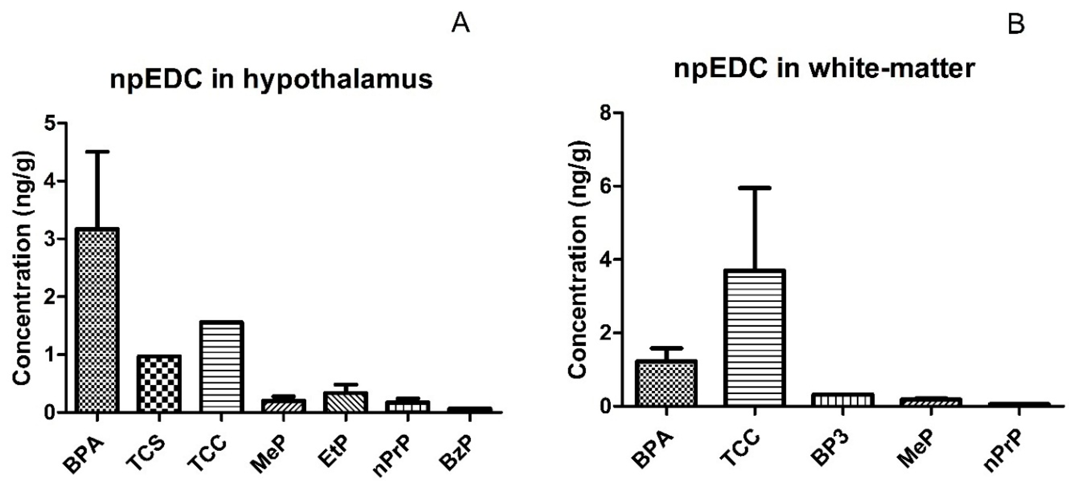
| Hypothalamus (n = 24) | White-Matter Brain (n = 10) | |||||||||||||||||
|---|---|---|---|---|---|---|---|---|---|---|---|---|---|---|---|---|---|---|
| Compound | LOD (ng/g) | N (%) > LOD | Mean | SD | Median | Min | P25 | P75 | Max | N (%) > LOD | Mean | SD | Median | Min | P25 | P75 | Max | |
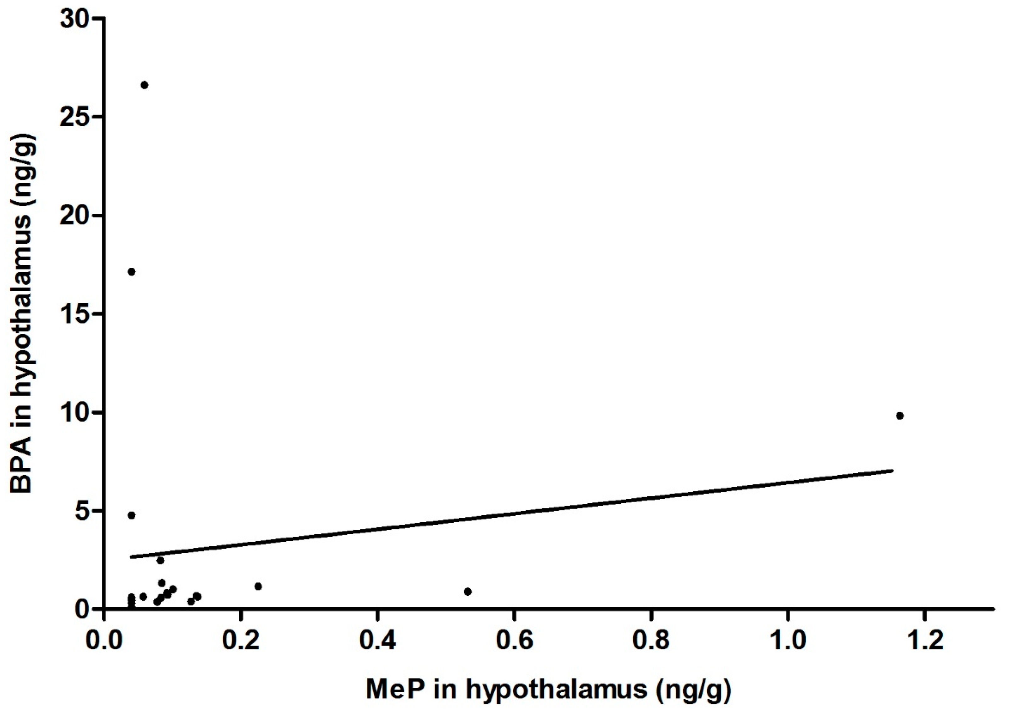
| Phenols | BPA | 0.14 | 23 (96) | 3.17 | 6.42 | 0.68 | 0.32 | 0.53 | 1.23 | 26.62 | 9 (90) | 1.23 | 1.07 | 0.82 | 0.30 | 0.38 | 1.90 | 3.32 |
| TCS | 0.73 | 1 (4) | 0.97 | - | 0.97 | 0.97 | - | - | 0.97 | 0 | <LOD | - | <LOD | <LOD | <LOD | <LOD | <LOD | |
| TCC | 0.92 | 1 (4) | 1.56 | - | 1.56 | 1.56 | - | - | 1.56 | 2 (20) | 3.70 | 3.18 | 3.70 | 1.45 | 1.45 | 5.95 | 5.95 | |
| BP3 | 0.18 | 0 | <LOD | - | <LOD | <LOD | <LOD | <LOD | <LOD | 1 (10) | 0.32 | - | 0.32 | 0.32 | - | - | 0.32 | |
| 2.4-DCP | 0.10 | 0 | <LOD | - | <LOD | <LOD | <LOD | <LOD | <LOD | 0 | <LOD | - | <LOD | <LOD | <LOD | <LOD | <LOD | |
| 2.5-DCP | 1.83 | 0 | <LOD | - | <LOD | <LOD | <LOD | <LOD | <LOD | 0 | <LOD | - | <LOD | <LOD | <LOD | <LOD | <LOD | |
| 2.4.5-TCP | 0.49 | 0 | <LOD | - | <LOD | <LOD | <LOD | <LOD | <LOD | 0 | <LOD | - | <LOD | <LOD | <LOD | <LOD | <LOD | |
| 2-PP | 0.10 | 0 | <LOD | - | <LOD | <LOD | <LOD | <LOD | <LOD | 0 | <LOD | - | <LOD | <LOD | <LOD | <LOD | <LOD | |
| 4-PP | 1.31 | 0 | <LOD | - | <LOD | <LOD | <LOD | <LOD | <LOD | 0 | <LOD | - | <LOD | <LOD | <LOD | <LOD | <LOD | |
| Parabens | MeP | 0.06 | 15 (63) | 0.20 | 0.29 | 0.09 | 0.06 | 0.08 | 0.14 | 1.16 | 3 (30) | 0.18 | 0.06 | 0.15 | 0.14 | 0.14 | 0.20 | 0.26 |
| EtP | 0.06 | 3 (13) | 0.34 | 0.26 | 0.36 | 0.07 | 0.21 | 0.47 | 0.58 | 0 | <LOD | - | <LOD | <LOD | <LOD | <LOD | <LOD | |
| nPrP | 0.05 | 5 (21) | 0.17 | 0.15 | 0.12 | 0.05 | 0.08 | 0.20 | 0.41 | 1 (10) | 0.06 | - | 0.06 | 0.06 | - | - | 0.06 | |
| BzP | 0.05 | 1 (4) | 0.06 | - | 0.06 | 0.06 | - | - | 0.06 | 0 | <LOD | - | <LOD | <LOD | <LOD | <LOD | <LOD | |
| i-PrP | 0.05 | 0 | <LOD | - | <LOD | <LOD | <LOD | <LOD | <LOD | 0 | <LOD | - | <LOD | <LOD | <LOD | <LOD | <LOD | |
| i-BuP | 0.06 | 0 | <LOD | - | <LOD | <LOD | <LOD | <LOD | <LOD | 0 | <LOD | - | <LOD | <LOD | <LOD | <LOD | <LOD | |
| n-BuP | 0.08 | 0 | <LOD | - | <LOD | <LOD | <LOD | <LOD | <LOD | 0 | <LOD | - | <LOD | <LOD | <LOD | <LOD | <LOD | |
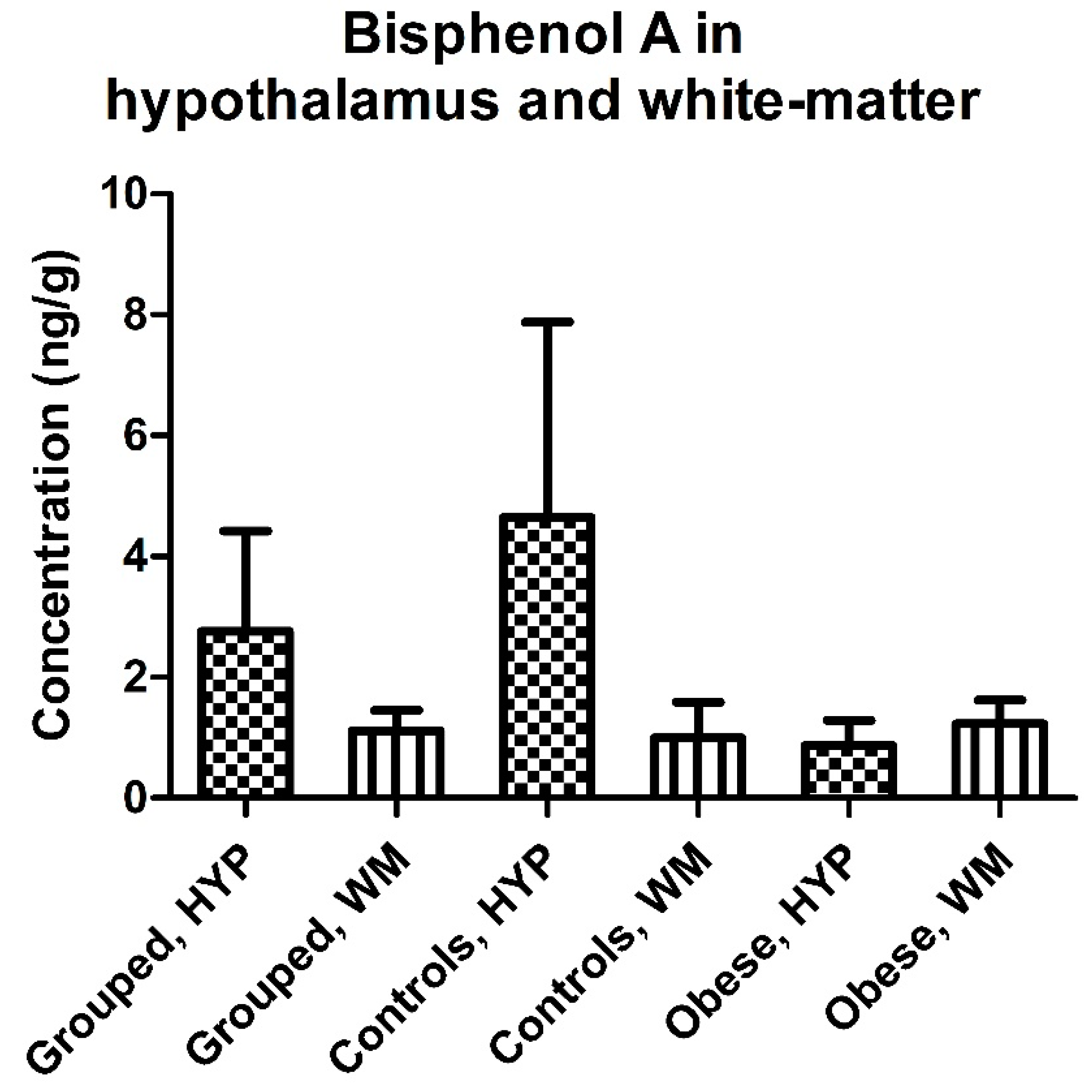
| Hypothalamus (n = 12 Controls, 12 Obese) | White-Matter Brain (n = 5 Controls, 5 Obese) | |||||||||||||
|---|---|---|---|---|---|---|---|---|---|---|---|---|---|---|
| Compound | Status | N (%) > LOD | Mean | Median | P25 | P75 | Max | N (%) > LOD | Mean | Median | P25 | P75 | Max | |
| Phenols | BPA | controls | 12 (100) | 4.49 | 0.63 | 0.48 | 2.89 | 26.62 | 4 (80) | 1.22 | 0.60 | 0.38 | 2.07 | 3.32 |
| obese | 11 (92) | 1.73 | 0.73 | 0.56 | 1.23 | 9.82 | 5 (100) | 1.23 | 1.01 | 0.53 | 1.90 | 2.39 | ||
| TCS | controls | 0 | <LOD | <LOD | <LOD | <LOD | <LOD | 0 | <LOD | <LOD | <LOD | <LOD | <LOD | |
| obese | 1 (8) | 0.97 | 0.97 | - | - | 0.97 | 0 | <LOD | <LOD | <LOD | <LOD | <LOD | ||
| TCC | controls | 0 | <LOD | <LOD | <LOD | <LOD | <LOD | 1 (20) | 5.95 | 5.95 | - | - | 5.95 | |
| obese | 1 (8) | 1.56 | 1.56 | - | - | 1.56 | 1 (20) | 1.45 | 1.45 | - | - | 1.45 | ||
| BP3 | controls | 0 | <LOD | <LOD | <LOD | <LOD | <LOD | 1 (20) | 0.32 | 0.32 | - | - | 0.32 | |
| obese | 0 | <LOD | <LOD | <LOD | <LOD | <LOD | <LOD | <LOD | <LOD | <LOD | <LOD | <LOD | ||
| Parabens | MeP | controls | 6 (50) | 0.17 | 0.11 | 0.06 | 0.14 | 0.53 | 1 (20) | 0.26 | 0.26 | - | - | 0.26 |
| obese | 9 (75) | 0.23 | 0.09 | 0.08 | 0.14 | 1.16 | 2 (40) | 0.15 | 0.15 | 0.14 | 0.15 | 0.15 | ||
| EtP | controls | 2 (17) | 0.32 | 0.32 | 0.07 | 0.58 | 0.58 | 0 | <LOD | <LOD | <LOD | <LOD | <LOD | |
| obese | 1 (8) | 0.36 | 0.36 | - | - | 0.36 | 0 | <LOD | <LOD | <LOD | <LOD | <LOD | ||
| nPrP | controls | 3 (25) | 0.13 | 0.12 | 0.10 | 0.16 | 0.20 | 0 | <LOD | <LOD | <LOD | <LOD | <LOD | |
| obese | 2 (17) | 0.23 | 0.23 | 0.05 | 0.41 | 0.41 | 1 (20) | 0.06 | 0.06 | - | - | 0.06 | ||
| BzP | controls | 0 | <LOD | <LOD | <LOD | <LOD | <LOD | 0 | <LOD | <LOD | <LOD | <LOD | <LOD | |
| obese | 1 (8) | 0.06 | 0.06 | - | - | 0.06 | 0 | <LOD | <LOD | <LOD | <LOD | <LOD | ||
© 2017 by the authors. Licensee MDPI, Basel, Switzerland. This article is an open access article distributed under the terms and conditions of the Creative Commons Attribution (CC BY) license (http://creativecommons.org/licenses/by/4.0/).
Share and Cite
Van der Meer, T.P.; Artacho-Cordón, F.; Swaab, D.F.; Struik, D.; Makris, K.C.; Wolffenbuttel, B.H.R.; Frederiksen, H.; Van Vliet-Ostaptchouk, J.V. Distribution of Non-Persistent Endocrine Disruptors in Two Different Regions of the Human Brain. Int. J. Environ. Res. Public Health 2017, 14, 1059. https://doi.org/10.3390/ijerph14091059
Van der Meer TP, Artacho-Cordón F, Swaab DF, Struik D, Makris KC, Wolffenbuttel BHR, Frederiksen H, Van Vliet-Ostaptchouk JV. Distribution of Non-Persistent Endocrine Disruptors in Two Different Regions of the Human Brain. International Journal of Environmental Research and Public Health. 2017; 14(9):1059. https://doi.org/10.3390/ijerph14091059
Chicago/Turabian StyleVan der Meer, Thomas P., Francisco Artacho-Cordón, Dick F. Swaab, Dicky Struik, Konstantinos C. Makris, Bruce H. R. Wolffenbuttel, Hanne Frederiksen, and Jana V. Van Vliet-Ostaptchouk. 2017. "Distribution of Non-Persistent Endocrine Disruptors in Two Different Regions of the Human Brain" International Journal of Environmental Research and Public Health 14, no. 9: 1059. https://doi.org/10.3390/ijerph14091059