Article

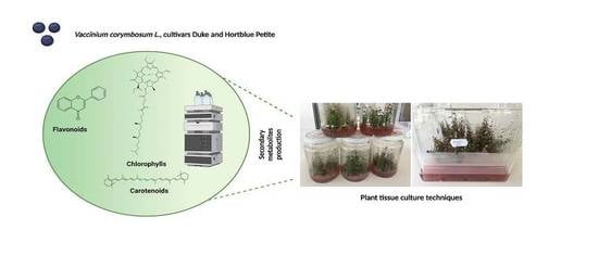
Micropropagation of Vaccinium corymbosum L.: An Alternative Procedure for the Production of Secondary Metabolites

by
Doina Clapa
1,2,
Silvia-Amalia Nemeș
2,
Floricuța Ranga
2,
Monica Hârța
2,3,
Dan-Cristian Vodnar
2,* and
Lavinia-Florina Călinoiu
4,*
1
Institute of Advanced Horticulture Research of Transylvania, University of Agricultural Sciences and Veterinary Medicine Cluj-Napoca, Calea Mănăştur 3-5, 400372 Cluj-Napoca, Romania
2
Institute of Life Sciences, University of Agricultural Sciences and Veterinary Medicine Cluj-Napoca, Calea Mănăştur 3-5, 400372 Cluj-Napoca, Romania
3
Faculty of Horticulture, University of Agricultural Sciences and Veterinary Medicine Cluj-Napoca, Calea Mănăştur 3-5, 400372 Cluj-Napoca, Romania
4
Faculty of Food Science and Technology, University of Agricultural Sciences and Veterinary Medicine Cluj-Napoca, Calea Mănăştur 3-5, 400372 Cluj-Napoca, Romania
*
Authors to whom correspondence should be addressed.
Horticulturae 2022, 8(6), 480; https://doi.org/10.3390/horticulturae8060480
Submission received: 18 April 2022 / Revised: 12 May 2022 / Accepted: 26 May 2022 / Published: 28 May 2022
(This article belongs to the Special Issue Plant Tissue and Organ Cultures for Crop Improvement in Omics Era)

Abstract

:
In vitro culture has become a dependable approach for the mass production of plant material as the market for innovative plant-derived medicinal approaches has grown significantly. Furthermore, because it permits manipulation of biosynthetic routes to boost the production and accumulation of certain compounds, this technology has enormous potential for the manufacture of natural bioactive chemicals. As a result, the goal of this study was to develop an efficient micropropagation system for biomass production and to investigate the accumulation of bioactive compounds from Vaccinium corymbosum L., Duke and Hortblue Petite cultivars. Two in vitro plant tissue culture systems were used for shoots production: a solid medium (5 g/L Plant agar) and liquid medium (Plantform bioreactor). The culture medium used was Woddy Plant Medium (WPM) supplemented with two growth regulators: 0.5 mg/L and 1 mg/L zeatina (Z) and 5 mg/L N6-(2-Isopentenyl) adenine (2iP). The content of phenolic compounds, carotenoids, and chlorophylls of the in vitro shoot extracts were examined via the HPLC-DAD-MS/MS technique. The results showed that cv. Hortblue Petite produced a higher amount of biomass compared with cv. Duke, on all variants of culture media in both systems (solid and liquid), while the shoots extract of the Duke variety in the liquid culture system (under all concentrations of growth regulators) had the highest content of total phenolic compounds (16,665.61 ± 424.93 μg/g). In the case of the lipophilic compounds analysed (chlorophylls and carotenoids), the solid medium reported the highest values, whereas media supplemented with 0.5 mg/L Z was proved to have the richest total content for both cultivars.

Graphical Abstract

1. Introduction

In the last decade, the production of secondary metabolites from medicinal plants, also known as Plant-Derived Medicinal Compounds (PDMC), has gained attention. Horticultural plants can be considered as important, sustainable, and low-cost biomass sources for obtaining high-complex PDMCs for use as medicines as information about their pharmacological qualities increases [1,2,3]. Biotechnological techniques, including plant cell and tissue culture and plant genetic transformation, are increasingly being used in vitro to manufacture high-quality and uncommon PDMC [4,5]. Vaccinium corymbosum (blueberry) is a deciduous shrub plant belonging to the Ericaceae family. Blueberry fruits and plant tissue have long been recognised for their many health benefits due to their chemical composition, which includes compounds such as polyphenols, vitamin C, dietary fibres and minerals. In traditional medicine, blueberry plants were used to treat diseases such as the common cold, inflammation, diabetes, and ocular dysfunction [6] or premature aging and anaemia [7]. Nowadays, both the fruits and tissues of several Vaccinum sp., such as V. corymbosum, V. angustifolium and V. myrtillus, are consumed as food supplements, pharmaceuticals, food natural additives and cosmetics [1,2,8]. Due to the significant amount of phenolics, flavonols and anthocyanins in blueberries, plant tissue extracts showed excellent biological potential in terms of antibacterial [9], anticancer [10] and antioxidant activity [4,5,6]. The antioxidant properties of the supplements were shown to significantly reduce the severity of viral infections [11]. Furthermore, phenolic and flavonoid compounds are not equally spread in all the plant tissues or organs, and compared with fruits, leaves of different Vaccinium sp. contain higher levels of phenolic compounds and lower levels of anthocyanins [12]. Studies have shown that the main phenolic compounds found in the plant tissues and that are responsible for the biologically significant potential of Vaccinium sp. are chlorogenic acid, quercetin, caffeic acid, proanthocyanidins, catechin and epicatechin [13,14,15,16,17].
Plant metabolites can be isolated from naturally grown plants, however, due to environmental limits, as seasonal restrictions, particular soil selection or growth conditions, the commercial production is limited [18]. Traditional methods take a long time since the plant needs several years to mature and attain the necessary level of metabolite production. Therefore, plant tissue cultures were used as an important tool for the continuous production of active compounds in addition to traditional methods [19]. Plant tissue cultures offer a number of advantages over conventional plants cultivation methods: independence from geographical and seasonal variations, year-round production, relatively short growth cycles, mass propagation of a uniform, high quality plant material and lack of use of pesticides and herbicides [20,21].
Different groups of metabolites have been examined in the biomass of shoots, roots or calluses grown in vitro in several plant species such as: shoot cultures of Ruta graveolens [22], a biomass of shoots and differentiating callus culture of Schisandra chinensis [23], the shoots and roots of Eryngium planum [24], biomass from Aronia melanocarpa shoots and callus cultures [25], the leaves and roots of Rehmannia glutinosa [26], leaves of micropropagated plants of Lycium schweinfurthii [27], leaves of Kaempferia parviflora from in vitro cultured plantlets [28], tissue cultures of six genotypes of Deschampsia Antarctica [29], biomass of in vitro shoots and roots of Eryngium species (E. campestre, E. maritimum, E. planum) [30] or leaves from micropropagated plantlets of Passiflora setacea cv BRS Pérola do Cerrado [31].
Therefore, numerous studies showed that micropropagation protocols have been developed for hundreds of plant species, including highbush blueberries [12]. Some of them reveal that V. corimbossum has proven to be an exceptionally suitable species for in vitro cultures [12,32,33,34,35,36]. Previous findings [32,37,38,39] show that the most commonly used culture media for blueberries are Woody Plant Medium (WPM) [40], Murashige and Skoog medium (MS) [41] and Anderson’s Rhododendron medium (AN) [42]. Several studies have been performed investigating the effect of different cytokines on the efficacy of shoot propagation, and the most effective in the micropropagation of highbush blueberry proved to be zeatin (Z) and 2-isopentenyladenine (2iP) [32,37,43,44,45]. Usually, the culture media for highbush blueberry micropropagation are solidified with agar, but the most recent progress in blueberry micropropagation has been the use of bioreactor systems with liquid media for shoot proliferation. Explants can be cultivated in liquid media in a temporary immersion system (TIS), where the gaseous exchange is not hampered and explants can efficiently utilise nutrients. However, because in vitro shoots are often particularly sensitive to liquid media, vitrification can be detected. This problem can be prevented by exposing the explant to liquid and air in the vessel alternately. Furthermore, liquid culture techniques allow for media replenishment without the need to change containers, which might shorten the culture duration. As liquid culture systems are less labour intensive and require less time for sub-culturing, they can help overcome this cost barrier [35].
In vitro culture has become a dependable approach for the mass production of plant material as the market for innovative plant-derived medicinal approaches has grown significantly. Therefore, the present study was aimed to develop an efficient micropropagation system to produce biomass used for the production of important bioactive compounds from V. corymbosum L., Duke and Hortblue Petite cultivars, as alternative bioactive resources. Two in vitro plant tissue culture systems (solid and liquid media) and also two cytokines (Z and 2iP) in different concentrations (0.5 mg/L, 1 mg/L, 5 mg/L) were used to obtain shoots for biomass production rich in secondary metabolites. Furthermore, using the HPLC-DAD-MS/MS technique, the total contents of phenolic compounds, carotenoids and chlorophylls of in vitro shoot extracts were examined. The comparisons among the highbush blueberry cultivars under different in vitro culture systems aimed to evaluate the changes in the polyphenolic, carotenoid and chlorophyll composition and content. The significance of the present study supports the finding of alternative sources of health-related compounds for the medical and pharmaceutical sectors by the production of a rich secondary metabolites profile of in vitro-propagated Vaccinium corymbosum plants. Since it permits the manipulation of biosynthetic routes to boost the production and accumulation of certain compounds, this technology has enormous potential for the manufacture of natural bioactive chemicals.

2. Materials and Methods

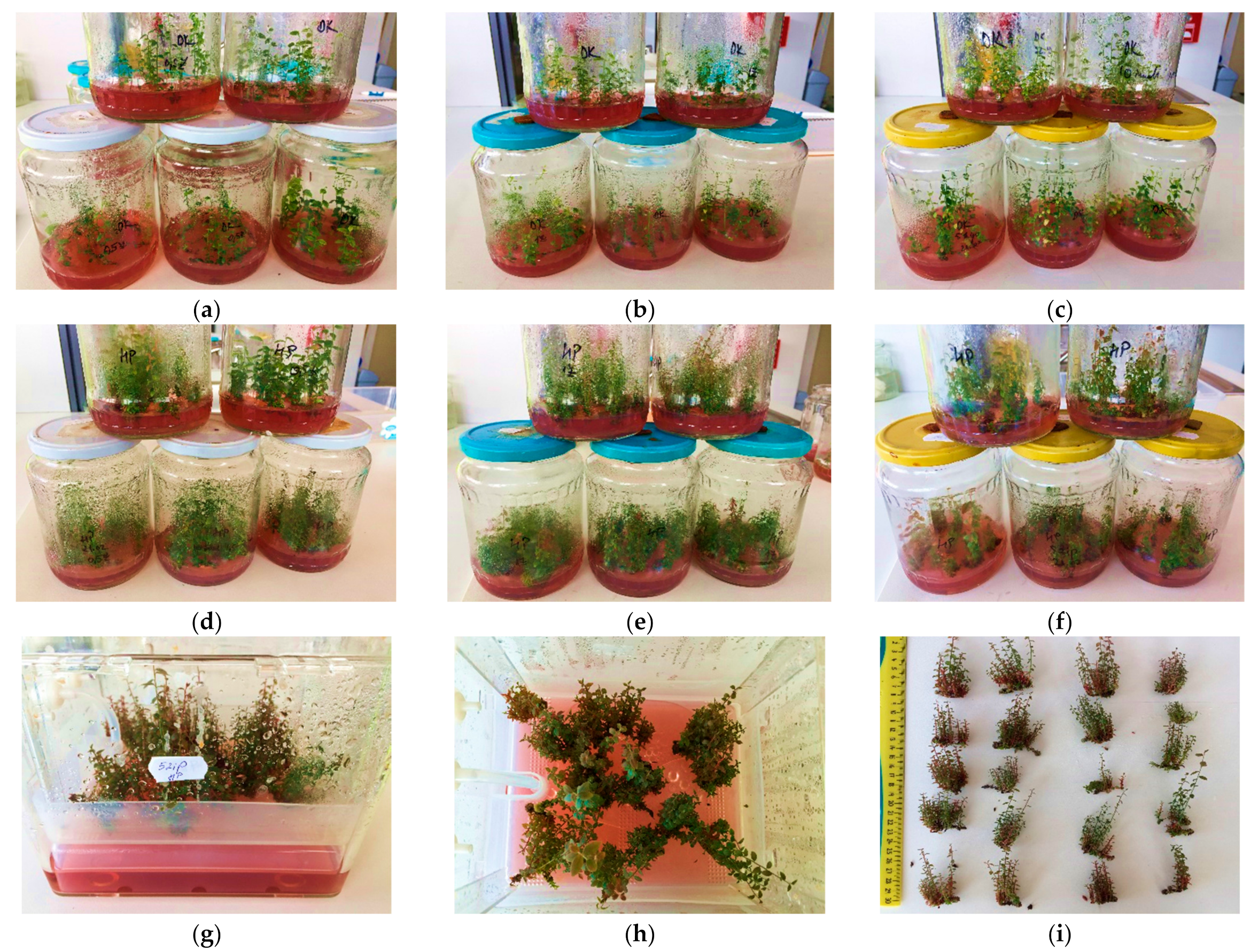
2.1. Shoot Proliferation and Biomass Production in Solid and TIS Culture Systems

In this study, two commercial varieties of highbush blueberry (V. corymbosum L.), Duke and Hortblue Petite, were used. The explants used for this experiment were excised from the in vitro culture of highbush blueberry (12 weeks old) which had been cultured on WPM with 100 mg/L Sequestrene 138, 1 mg/L zeatina (Z), 3% (w/v) sugar, 4 g/L Plant agar, pH 5, according to the protocol of Clapa et al. [46].
For the proliferation of shoots and the production of biomass, two in vitro culture systems were used: solid and liquid (TIS). For solid media, 720 mL jars were used as culture vessels, which were 9 cm in diameter and 13.5 cm high with screw caps, and 100 mL media/jar. TIS culture systems used Plantform bioreactors [23], with a volume of 500 mL media/bioreactor. The immersion time was one at four hours lasting for 1 min and aeration was once/hour lasting for 4 min, according to our previous optimised study [46]. The basal culture medium used for both types of culture systems (solid and TIS) had the same composition as that described above, but with three variations of growth regulators, 0.5 mg/L Z, 1 mg/L Z and 5 mg/L N6-(2-Isopentenyl) adenine (2iP), respectively. The culture medium was solidified with 5 g/L Plant agar. All the components were purchased from Duchefa Biochemie BV (Haarlem, Netherlands)
In the culture, vessels with solid media were inoculated with 10 fragments of shoots, and in the bioreactors, were inoculated with 20 fragments of shoots, each one of 1.5–2 cm. In vitro cultures were incubated in the growth room at 16-h photoperiod with 32.4 μmol m2s1 light intensity (Philips Core Pro LED tube 1200 mm 16 W 865 CG, 1600 lm Cool Daylight) at 23 ± 3 °C and 50 ± 2% humidity. Both data for solid media and liquid media were collected after 12 weeks of culture for analysis. The analysed data refer to the proliferation rate (PR), the average length of shoots (SL) and the fresh weight (FW). The proliferation rate represents the number of shoots obtained/initial explant and FW was calculated shoots from 50 initial explant/cultivar/experimental treatments. Fresh shoots obtained in vitro were subsequently used for the extraction of phenolic compounds, carotenoids and chlorophylls.

2.2. Ultrasound-Assisted Extraction of Total Phenolic Compounds

The in vitro shoots obtained for all the variants of both cultivars and both culture systems were dried and fine grounded. A quantity of 5 g fine powder was mixed with 5 mL 70% v/v ethanol in water. The mixture was exposed for 1 min at the vortex (Heidolph Reax top) and 30 min in an ultrasonic bath (Elmasonic E 15 H, Elma, Singen, Germany) at 20 °C. Then, the samples were centrifuged (Eppendorf Centrifuge 5804) for 10 min at 10,000 rotations/min. The supernatant was filtered using a nylon filter (Chromafil Xtra PA-45/13, Macherey-Nagel, Düren, Germany) and stored at freezing temperature (−18 °C) until future analysis.

2.3. Quantitative and Qualitative Analysis of Total Phenolic Compounds by HPLC-DAD-ESI-MS

Quantification and qualitative characterisation of phenolic compounds in the shoots extract were conducted by an HPLC-DAD-ESI-MS system containing an Agilent 1200 HPLC with DAD detector, attached to an MS-detector single-quadrupole Agilent 6110 (Agilent Technologies, Santa Clara, CA, USA). For separation, the Eclipse column, XDB C18 (4.6 × 150 mm, particle size 5 µm) (Agilent Technologies, Santa Clara, CA, USA), was used at 25 °C. Two gradients composed of 0.1% acetic acid in distilled water (v/v) (solvent A) and 0.1% acetic acid in acetonitrile (v/v) (solvent B) were used at a flow rate of 0.5 mL/minute, succeeding the elution programme detailed by Dulf et al. [47]. For MS fragmentation, the ESI (+) module was used, with the following working parameters: scanning range between 100 and 1200 m/z, capillary voltage 3000 V, the temperature at 350 °C and with a nitrogen debit of 8 L/min. The eluent was monitored by DAD, and the absorbance spectra, recorded at 200–600 nm, were measured and collected constantly in the course of each run. For analysing the data, Agilent ChemStation Software (Rev B.04.02 SP1, Palo Alto, CA, USA) was used. Chromatograms were recorded at wavelengths of 280 and 340 nm. For the quantitative determination of phenolic compounds, a calibration curve was performed for each subclass: catechin calibration curve (r2 = 0.9985) for flavanols, chlorogenic acid calibration curve (r2 = 0.9937) for hydroxycinnamic acids, and quercetin curve (r2 = 0.9951) for the flavonol subclass.

2.4. Extraction of Carotenoid and Chlorophyll Compounds

Carotenoids and chlorophylls were extracted from the fine powder (10 g) obtained from the shoots according to the previously described protocol [48,49]. Total carotenoids and chlorophylls were extracted using methanol:ethyl acetate:petroleum ether (1:1:1, v/v/v). The extraction was made under continuous stirring for 4 h in dark conditions. The extract was washed successively with water, diethyl ether and NaCl saturated solution. The ether phase was evaporated using a rotavapor at 35 °C. The obtained oleoresin was dissolved in a known volume of ethyl acetate.

2.5. Quantitative and Qualitative Analysis of Carotenoids and Chlorophylls by HPLC-DAD-ESI-MS

HPLC analyses for separable carotenoids and chlorophylls compounds were carried out on an Agilent 1200 system with a DAD and MS detectors (Agilent Technologies, Santa Clara, CA, USA) using a reversed-phase EC 250/4.6 Nucleodur 300-5 C-18 with the column of 250 × 4.6 mm, 5 µm (Macherey-Nagel, Düren, Germany) at 25 °C, according to a previous validated method [50]. The mobile phase consisted of mixtures of acetonitrile: water (9:1, v/v) with 0.25% triethylamine (A) and ethyl acetate with 0.25% triethylamine (B). The gradient started with 90% A at 0 min to 50% A at 10 min. The percentage of A decreased from 50% at 10 min to 10% A at 20 min. The flow rate was 1 mL/min and the chromatograms were monitored at 450 nm. The HPLC peaks were identified using carotenoid standards from Sigma Company, St. Louis, MO, USA.

2.6. Data Analysis

One-way analysis of variance (ANOVA) was performed for treatments within each cultivar and for all the cultivars within all treatments to investigate whether the differences in physiological and biochemical parameters of the in vitro plants were affected by the presence of different in vitro culture systems and different concentrations of PGRs added to the culture media. Post hoc testing for the ANOVAs was performed using Tukey’s honestly significant difference test (Tukey test) using a p < 0.05 significance level to determine the statistically significant differences between the means. Values shown (in text and figures) are means ± SE (standard error). In addition, the parameters recorded in plants were correlated by heat map analysis, using the OriginPro 9.8.0.0. software (Northampton, MA, USA).

3. Results and Discussions

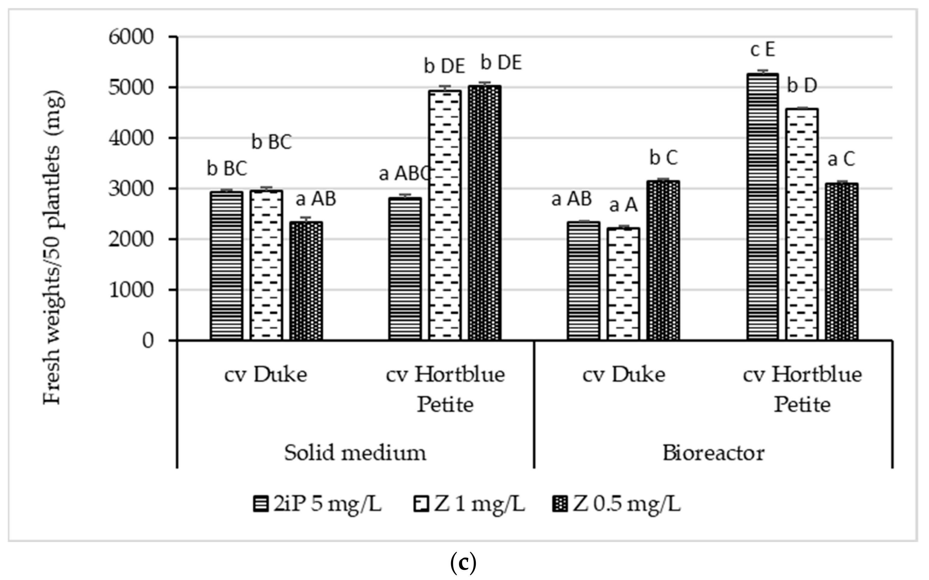
3.1. Shoot Proliferation and Biomass Production (Shoots) in Solid and TIS Culture Systems

Our preliminary research [28,29,30,31,32,33,34,35,36] has shown that micropropagation is influenced by the type of plant growth regulators (PGRs), culture media and plant species. The results of the present study showed that the two in vitro culture systems (solid and liquid [Plantform bioreactor via a TIS]), as well the two PGRs, 2iP (5 mg/L) and Z (1 mg/L Z; 0.5 mg/L), differently influenced the proliferation rate, length of shoots and fresh weights of both V. corymbosum cultivars (Duke and Hortblue Petite) (Figure 1).
The greatest proliferation rates (PR) were recorded in the TIS as compared with the solid media, respectively, la Hortblue Petite compared with Duke. Among PGRs, the Z generated the highest PR values (Figure 2a). In the TIS, the Hortblue Petite variety showed a PR up to 2.5–6 times higher for all three concentrations of growth regulators, in comparison with the Duke variety. Thus, the 1 mg/L Z treatment stimulated the higher PR (12.65 ± 0.97), however, this is not significantly different from the PR obtained in the culture media supplemented with 5 mg/L 2iP (12.58 ± 1.04). Contrary, Duke had the highest PR (3.70 ± 0.32) in the culture media supplemented with 0.5 mg/L Z. In solid media, Hortblue Petite had again the highest PR (7.94 ± 0.25) in the medium supplemented with 1 mg/L Z while Duke presented a PR of just 2.98 ± 0.25 (Figure 2a).
Similar results were obtained in our previous studies where the Duke variety had the lowest proliferation rate (2.11 ± 0.15) compared to Hortblue Petite (8.50 ± 0.51) on agar gelled with WPM culture medium and supplemented with 1 mg/L Z [51]. Another study showed [44] that Duke cultivar cultured on Anderson (1980) culture medium supplemented with 0.5 mg/L Z or 2iP generated a similar number of shoots/explant, their numbers ranging from 2 to 4. Similar results have been reported by [52], who micropropagated Biloxi, Sharp Blue and Brillita blueberry cultivars in TIBs, and found that the plant number proved to be superior in blueberries propagated in TIBs compared to those propagated in solidified media with 8 g/L agar.
Regarding shoot lengths (Figure 2b), they recorded maximum values at Hortblue Petite on solid culture medium supplemented with 0.5 mg/L Z (3.40 ± 0.20 cm), while Duke had the longest shoots in the TIS in the culture medium supplemented with 5 mg/L 2iP (3.07 ± 0.28 cm), in agreement with our previously reported results [34,46,51].
The fresh weight (FW) is a very important parameter in biomass research where secondary chemicals are sought. The maximum FW production in solid media was achieved by Hortblue Petite at a concentration of 0.5 mg/L Z (5017.80 ± 8.49 mg), and the lowest FW in the culture medium supplemented with 5 mg/L 2iP (2825.25 ± 67.71 mg). For the Duke variety, the highest FW was obtained in the culture media supplemented with 1 mg/L Z (2957.70 ± 65.50 mg) and with 5 mg/L 2iP (2924.30 ± 54.32 mg) (insignificant differences, p < 0.05), and the lowest FW in the culture medium supplemented with 0.5 mg/L Z. In both blueberry varieties, the highest FW was obtained in the TIS, respectively 5270.65 ± 63.21 mg in Hortblue Petite in a liquid culture medium supplemented with 5 mg/L 2iP and 3144 ± 39.69 mg in Duke on a culture medium supplemented with 0.5 mg/L Z (Figure 2c).

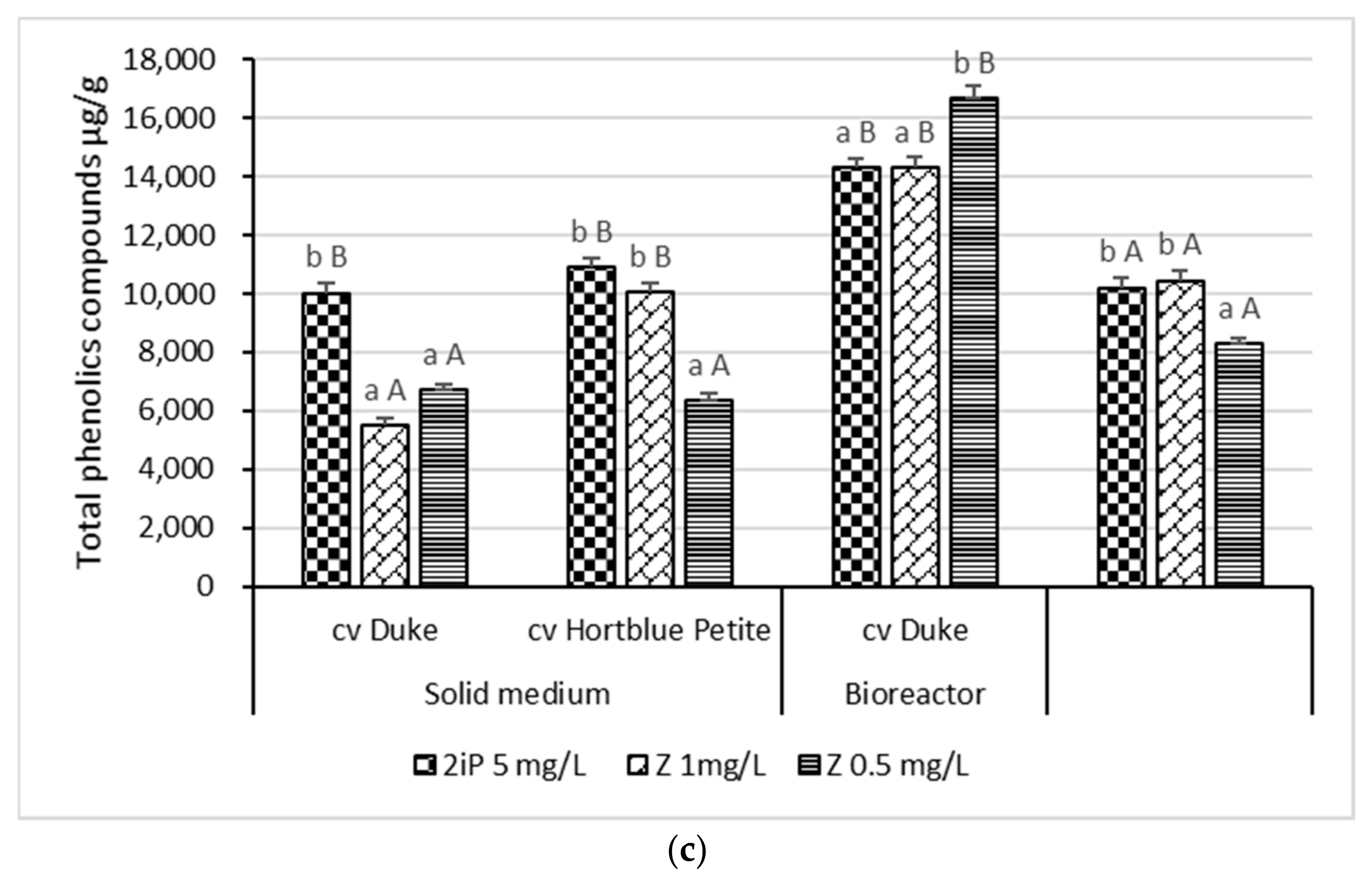
3.2. Content of Phenolic Compounds

From our knowledge, the phenolic, carotenoid and chlorophyll content of Vaccinium shoots generated in vitro via two different culture systems (solid and liquid) and three growth regulators has never been investigated before. Therefore, the present study provides important optimised conditions for the production of in vitro secondary metabolites for the commercial V. corymbosum cultivars, Duke and Hortblue Petite.
In this study, in the shoots of the highbush blueberry (V. corymbosum L.) cvs Duke and Hortblue Petite, after 12 weeks of in vitro culture in solid and liquid media, 11 phenolic compounds were identified, originating from three phenolic groups: hydroxycinnamic acids, flavonols and flavanols, whereas 10 were found in Duke shoots extracts and 11 in Hortblue Petite leaf extracts (Table S1).
The five hydroxycinnamic acids identified were: quinic acid, neoclorogenic acid (3-caffeoylquinic acid), chlorogenic acid (5-caffeoylquinic acid), caffeic acid and feruloylquinic acid. Among the flavanols group, three compounds were detected: catechin, epicatechin and epicatechin-gallate (ECG). Furthermore, in the flavonols group, three compounds were detected also: quercetin-glucoside, kaempferol-glucoside and quercetin. These results are not entirely similar to our previous research [2] where the findings, after analysing the leaves from six commercial highbush blueberry varieties (Elliot, Toro, Duke, Bluecrop, Spartan, Nelson), have shown four hydroxycinnamic acids (among which was also dicaffeoylquinic acid (present in the Duke cultivar) which was not identified in the present study), six flavanol compounds (gallocatechin, catechin, epicatechin, procyanidin dimer I, procyanidin dimer II and procyanidin trimer—only two common flavanols), six flavonols (quercetin-rutinoside (Rutin), quercetin-glucoside, quercetin-acetyl-rhamnoside, quercetin-arabinoside, quercetin-diglucoside and quercetin—only two common flavonols), and the anthocyanins class represented by cyanidin-glucoside, cyanidin-arabinoside and cyanidin-acetyl-glucoside (not identified in the leaves of the Duke variety and being in line with present findings). The differences in the phenolic compounds identified among the extracts coming from the (i) in vitro-derived Duke cultivar shoots and (ii) the leaves of the Duke cultivar (naturally grown in culture with a controlled drip irrigation system and acid brown soil) might be explained by the different plant parts analysed (shoots vs. leaves), the growing conditions (light, temperature, etc.) and the culture media, adjusting the hormonal conditions (which are proved to stimulate cell expansion and division) in temporary immersion bioreactors was shown to be productive for the accumulation of in vitro phenolics in blueberries (Vaccinum corymbosum) [52] (which was proved to re-direct the biosynthesis of phenolic compounds toward the phenylpropanoids pathway) [52,53].
The content of individual phenolic compounds in the shoot extracts of the two highbush blueberry varieties, for both types of culture systems and three variations of growth regulators, expressed as μg/g sample, was detailed in Table 1.
These findings are in line with our previous studies on Vaccinium sp. [1,2,3]. In the case of both cultivars, the most abundant compound for both types of culture systems and all PGRs, was represented by the Epicatechin-gallate (ECG) belonging to the flavanols class. The second most abundant compound was catechin within the same class of flavanols. In the case of the Duke shoots extracts, the kaempferol-glucoside compound was not detected, whereas quercetin registered the lowest levels for both cultivars and all media-derived conditions (Table 1). Therefore, it can be stated that the flavonols were the poorest represented class in the in vitro-derived highbush blueberry shoot extracts (cvs Duke and Hortblue Petite) while the flavanol class was the best represented one. Moreover, the anthocyanins group was not detected among any of the cultivar’s shoot extract types.
Concerning the Duke cultivar grown in solid media, among all of the phenolic compounds identified, the ECG, from the flavanol group, registered the highest amount, ranging from 1920.981 ± 1.73 μg/g (as the lowest level, in media adjusted with 1 mg/L Z) to 3866.141 ± 3.86 μg/g (as the highest level, registered for the 2iP in a concentration of 5 mg/L). Interestingly, in the existing literature [3,7,54,55], chlorogenic acid was reported to be the most abundant phenolic compound in different blueberry cultivars (fruits and leaves, as well). Possible explanations could be derived from the differences among natural habitats and in vitro cultures (time of harvest, geographical area) and the extraction method. The second most abundant compound was catechin (from the same flavanol class) registering the highest concentration (2246.689 ± 8.12 μg/g) in the same condition (5 mg/L 2iP). Epicatechin was the minor compound identified within this group, among all of the three-growth variations of regulators, however, the maximum amount was registered again by the 2iP supplemented media. Therefore, it can be stated that on a solid medium, the flavanol class is stimulated the most to be produced in cells of in vitro blueberry shoots by supplementation with 5 mg/L 2iP (in comparison to Z). Regarding the hydroxycinnamic acids, this class was the best represented in terms of the numbers of compounds (5) and the second as concentrations found, whereas quinic acid registered the highest values of all three types of PGRs, with the maximum amount being in the media supplemented with 5 mg/L 2iP. However, in the case of the other four hydroxycinnamic acids, the maximum value was registered by the media with 0.5 mg/L Z, followed by the media with 5 mg/L 2iP. Moreover, there were statistically significant differences (p < 0.05) among all the three variations of PGRs for this phenolics class. The flavonol class registered the lowest concentrations for all the three compounds, on all growth conditions, whereas kaempferol-glucoside was totally absent on the solid media for this specific blueberry cultivar (Duke). Quercetin registered values lower than 1.5 μg/g, whereas quercetin-glucoside was best accumulated in media supplemented with 0.5 mg/L Z (such as in the case of most hydroxycinnamic acids).
The same Duke cultivar grown in the TIS reached between a two- and ten-times higher phenolics concentration than on the solid media, for all three types of PGRs. For example, ECG, the most abundant compound identified for both the solid and liquid media, doubled its concentration on the bioreactor with zeatin, whereas on 2iP supplementation, it had a 6.23% relative percentage increase. In the case of feruloylquinic acid, an nine-fold increase was registered on the liquid media vs. the solid media supplemented with 1 mg/L Z (from 187.467 ± 1.63 μg/g to 1652.177 ± 1.98 μg/g). Quinic acid doubled or tripled its amounts on zeatin growth conditions (both concentrations tested), while on 2iP-enriched media had a 43% increase vs. the solid media. Bioreactor cultures allowed for the enhanced manipulation of both physical and chemical conditions, such as air exchange, photosynthetic photon flux and CO2 concentration, resulting in a more efficient plant performance during in vitro micropropagation [56,57,58]. In a previous study, the efficiency of the micropropagation of blueberries in TIBs under photomixotrophic conditions was demonstrated, whereas an increase in both the plant multiplication rate and total number of internodes has been achieved in three commercial cultivars. According to another recent study on micropropagated blueberries [52], the temporary immersion biorector (TIBs) culture was explored for production of phenylpropanoids, whereas the links between the oxidative burst (H2O2) and the phenylpropanoid pathways during plant micropropagation in TIBs was demonstrated.
In the case of the flavanol and hydroxycinnamic (except for feruloylquinic acid) classes, the media supplemented with 0.5 mg/L Z stimulated the highest accumulation of compounds, whereas for the flavonol class and feruloylquinic acid, the 2iP (5 mg/L) had the most favourable influence on their production (accumulation, synthesis).
For the Duke variety, the liquid media via the TIS represented the most suitable culture system, whereas, together with 0.5 mg/L Z, the maximum phenolics production was stimulated, which is in accordance with the above-mentioned studies that demonstrated the efficacy of the TISs’ system for V. corymbosum.
Concerning the Hortblue Petite cultivar grown in solid media, among all of the phenolic compounds identified, the ECG, from the flavanol group, registered the highest amount with 1 mg/L Z (3185.247 ± 1.91 μg/g) and 5 mg/L 2iP (3168.711 ± 2.01 μg/g). The second most abundant compound was catechin (from the same flavanol class), registering the highest concentration (2383.38 ± 1.39 μg/g) in the same condition (1 mg/L Z), followed closely by epicatechin with 5 mg/L 2iP (2207.68 ± 1.29 μg/g). For all these three phenolics, the 0.5 mg/L Z condition succeeded the least to support a high production of secondary metabolites. These results are similar with those obtained for Duke cultivar, with the flavanol class being the best stimulated to be produced in cells of in vitro blueberry shoots by supplementation with 5 mg/L 2iP and 1 mg/L Z. As for the Duke variety, the hydroxycinnamic acids class was the best represented in terms of the number of compounds (five) and the second as concentrations found, whereas quinic acid registered the highest values on 1 mg/L Z and 5 mg/L 2iP PGRs, with the maximum amount in the media supplemented with 5 mg/L 2iP (1300.584 ± 3.31 μg/g). In the media supplemented with 0.5 mg/L Z, the feruloylquinic acid reported the highest value (1051.008 ± 2.16 μg/g). However, also in the case of the other three hydroxycinnamic acids, the maximum value was registered by the media with 0.5 mg/L Z, followed by the media with 5 mg/L 2iP hormone, as for the Duke cultivar. Moreover, there were statistically significant differences (p < 0.05) among all the three variations of PGRs for this phenolics class. Interestingly, the neoclorogenic acid and clorogenic acid presented the lowest concentrations among phenolic acids. When compared with the Duke cultivar, the flavonol class was better stimulated by the in vitro culture system supported by PGRs, with concentrations for quercetin-glucoside being two times higher than in the Duke cultivar, and in higher amounts than those of clorogenic and neoclorogenic acids, on all growth conditions. Quercetin registered values up to 100 times higher than its counterpart, the Duke cultivar, whereas kaemferol glucoside, absent for Duke, reported values higher than 100 μg/g on all the three PGRs. Therefore, it can be stated that for the Hortblue Petite variety, the flavonol class was highly stimulated by in vitro conditions registering important concentrations when compared to Duke.
The same Hortblue Petite cultivar grown in the TIS reached a phenolics concentration between two and three times higher than on the solid media for all three types of PGRs, with the exception of the flavonol class (where all the three compounds registered lower concentrations for all the three PGRs) and two phenolic acids (caffeic acid and feruloylquinic acid—which halved the amounts in the case of 0.5 mg/L Z). Additionally, the Duke cultivar registered higher concentrations in the TIS for all PGRs.
For example, ECG, the most abundant compound identified for both solid and liquid media, increased its concentration in the TIS 0.5 mg/L Z with a 68.95% relative percentage increase, on 1 mg/L Z with a 8.48% relative percentage increase and on 5 mg/L 2iP with a 1.42% relative percentage increase. In the case of the catechin and epicatechin compounds, the TIS supported a two-times higher concentration in the media supplemented with 0.5 mg/L Z. In the case of neoclorogenic and clorogenic acids, the media supplemented with 1 mg/L Z had a double amount of the TIS culture when compared to solid media, and quinic acid increased its amounts of zeatin growth conditions by 31.15% and 16.83%, respectively, with 1 mg/L Z. In contrast to Duke, the increases were much lower for TIS vs. the solid medium.
For the Hortblue Petite variety, the liquid media via the TIS represented the most suitable culture system, whereas being together with 1 mg/L Z stimulated the maximum phenolics production, which is in accordance with the results for the Duke variety where 0.5 mg/L Z supported the highest production of phenolics.

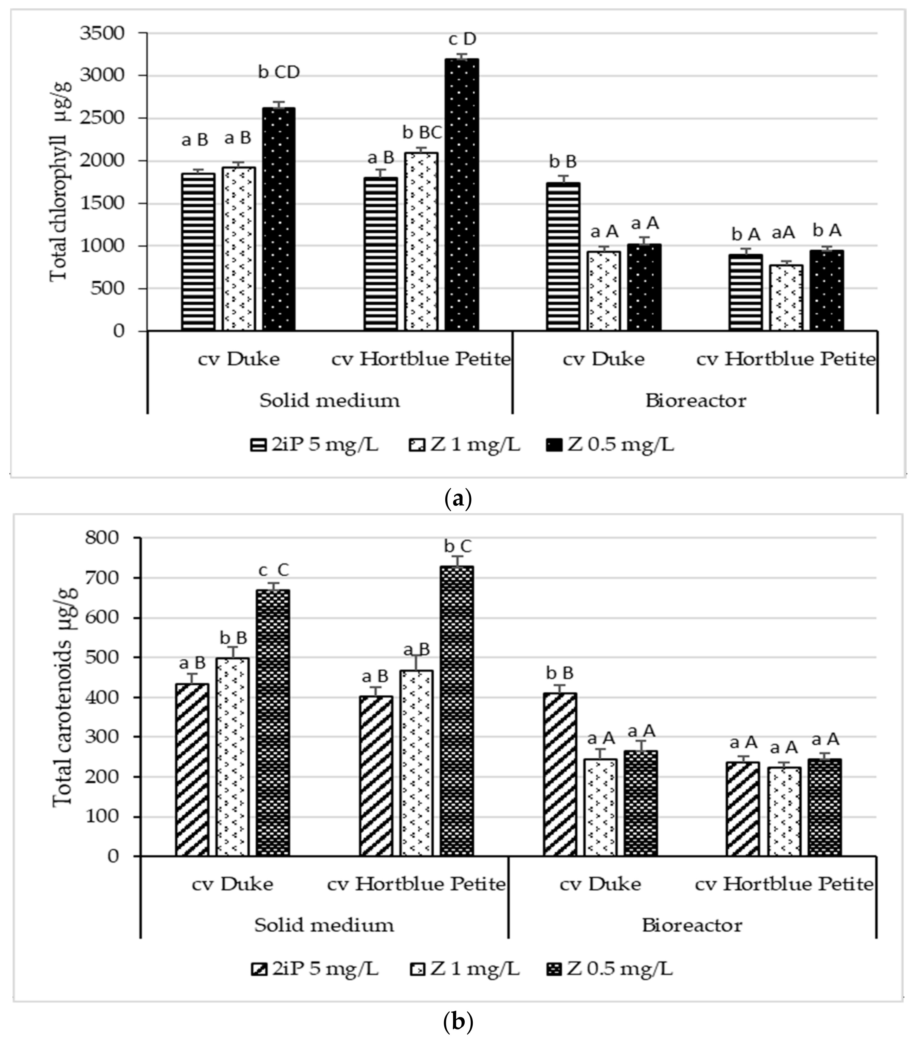
3.3. Content of Carotenoids and Chlorophylls

In Table 2 and Table S2, the identification of the carotenoid and chlorophyll compounds in the shoot extracts of the in vitro highbush blueberry varieties, and the content of individual carotenoid and chlorophyll compounds, for both types of culture systems and three variations of PGRs, were represented.
In the case of both cultivars, the solid media (agar) supported the highest carotenoid and chlorophyll contents, with 0.5 mg/L Z supplementation reaching the highest values. As in accordance with the literature, chlorophyll a was identified in the highest amount, ranging from 1477.052 ± 4.46 μg/g (maximum value) to 451.440 ± 1.48 μg/g (lowest value) for the Duke cultivar, and from 1325.398 ± 5.32 μg/g to 479.281 ± 2.87 μg/g for Hortblue Petite. Chlorophyll b was found in approximately half amounts when compared to chlorophyll a for both cultivars and all PGRs. Moreover, pheophytin b registered the lowest quantities, precisely under 22 μg/g, for both cultivars and both types of culture media (the lowest being approximately 5 μg/g in 1 mg/L Z TIS for both cultivars). It is interesting to observe that pheophytin a and b were found in high amounts compared to the plants; in general, amounts which are supported by an acidic culture media, the same as the ones used in the present study (pH = 5). Hortblue Petite registered the highest concentration of pheophytin a (1097.240 ± 1.74 μg/g in solid media with 0.5 mg/L Z).
Among the carotenoid class, the βCarotene compound was best represented in a concentration of 260.840 ± 1.43 μg/g, in the Duke cultivar in solid media, being 2.5 times higher than its counterpart in the TIS. The same trendline was observed in the Hortblue Petite variety for this compound. Lutein was reported in the lowest quantities on all the three PGRs, for both types of media and cultivars, with values approximately 2.5 times lower in the TIS culture. Duke, again, reported the highest values when compared to Hortblue Petite.
The total chlorophylls content in the highbush blueberry samples, expressed as a μg/g sample, is illustrated in Figure 3a.
The solid medium reported the highest contents for both varieties of all PGRs, whereas the media supplemented with 0.5 mg/L Z showed the best chlorophylls biosynthesis (2624.13 ± 63.96 μg/g for Duke and 3191.78 ± 57.71 μg/g for Hortblue Petite). In the TIS, Duke presented the highest chlorophylls content (1743.24 ± 80.07 μg/g) in the media supplemented with 5 mg/L 2iP, whereas for Hortblue Petite, there were no significant differences for the chlorophylls content among the PGRs’ variations.
The total carotenoids content was represented in Figure 3b, expressed as a μg/g sample. Again, the solid medium reported the highest values in terms of lipophilic compounds, where the media supplemented with 0.5 mg/L Z was proved the richest content for both cultivars (668.96 ± 18.96 μg/g—Duke; and 727.71 ± 27.71 μg/g—Hortblue Petite). The TIS conditions proved having a similar trendline as for the chlorophyll compounds, namely with a 2iP growth regulator enhancing the most carotenoid content in Duke (410.06 ± 20.07 μg/g), while for Hortblue Petite, a linearity was maintained.
According to the existing literature, the chlorophylls and carotenoid content for in vitro plant cultures seems to be more influenced by the specificity of the plant rather than by the culture system. For example, the highest contents of chlorophyll and carotenoid in Cnidium officinale were obtained in a semi-solid culture (0.8% (w/v) of agar), being in accordance with our results, whereas for Fragaria × ananassa Duch, the photosynthetic pigments, Chl a (18.2 mg∙g−1 FW), Chl b (6.7 mg∙g−1 FW), and the carotenoids values (4.5 mg∙g−1 FW) were the highest in the TIS culture, which is opposite to our results, while for Chrysanthemum morifolium Ramat, there were no differences between treatments (solid vs. TIS). [Hwang]. Within the same study, it was interesting that strawberries had the highest chlorophyll content per unit weight in the TIS system, which had the largest leaf area, while C. officinale had the highest chlorophyll content in the plants grown in the semi-solid culture, which had the smallest leaf area.
Regarding the phenolics values, as illustrated in Figure 3c, the TIS conditions supported the richest contents, for both varieties, on all three PGRs variations, whereas Duke presented the highest concentration in 0.5 mg/L Z (16,665.62 ± 424.63 μg/g).
A heat map was applied to visualise the variation in the content of the 18 phenolic, carotenoid and chlorophyll compounds in the 12-shoot samples variants (Figure 4). The phenolic, carotenoid and chlorophyll compounds were clustered according to the Pearson correlation coefficient. The column denotes highbush blueberry samples of two types of culture systems, and the row denotes the individual phenolic, carotenoid and chlorophyll compounds. A yellow box indicates that the chemical compounds’ content was lower than the average level and a blue box indicates the chemical compounds’ content was higher than the average level.
As can be seen in Figure 4, the Hortblue Petite shoots obtained in the solid culture system supplemented with 5 mg/L 2iP have the lowest content of chemical compounds forming a single group, while the group with the highest content of compounds is represented by the Duke variety shoots obtained on the three environmental variants in the liquid system. The phenolic, carotenoid and chlorophyll compounds were clustered according to the Pearson correlation coefficient into two major groups: the first group comprises carotenoid and chlorophyll compounds (Pheophytin a, Pheophytin b, βCarotene, Chlorophyll a, Zeaxanthin, Lutein, Chlorophyll b) along with three phenolic compounds (Quercetin, Kaemferol glucoside Quercetin glucoside) and the second group, namely the phenolic group, is composed of Quinic acid, Neochlorogenic acid, Chlorogenic acid, Catechin, Epicatechin, Caffeic acid, ECG and Feruroylquinic acid.

4. Conclusions

The results of this study show that the chemical profiles of micropropagated highbush blueberries are influenced by the genotype, culture system and growth regulators added in the culture media. For the Duke variety, the liquid media via the TIS with 0.5 mg/L Z represented the most suitable culture system for the maximum production of phenolics, while for the Hortblue Petite variety, the same liquid media via the TIS, but with 1 mg/L Z stimulated the maximum biosynthesis of the phenolics. In the case of the lipophilic compounds analysed (chlorophylls and carotenoids), the solid medium reported the highest values, where the media supplemented with 0.5 mg/L Z was proved the richest total content for both cultivars. According to the heat map, the group with the highest content of compounds was represented by the Duke variety shoots obtained on the three environmental variants in the liquid system. Based on the present studies, we can propose an in vitro plant tissue culture system in the liquid media in a bioreactor and also a WPM medium supplemented with Z or 2iP (according to variety) to obtain shoots for biomass production as a source of phenolic compounds. Shoot in vitro cultures of V. corymbosum may be considered a valuable alternative source of biomass that is rich in valuable bioactive compounds. The rich secondary metabolites profile of in vitro-propagated Vaccinium corymbosum plants offers significant alternative sources of health-related compounds for the medical and pharmaceutical sectors.

Supplementary Materials

The following supporting information can be downloaded at: www.mdpi.com/article/10.3390/horticulturae8060480/s1, Table S1: Identification of the phenolic compounds from the flavonoid class (flavanols, flavonols) and hydroxycinnamic acids class in the shoot extracts of the in vitro highbush blueberry varieties; Table S2: Identification of the carotenoid and chlorophyll compounds in the shoot extracts of the in vitro highbush blueberry varieties.

Author Contributions

Conceptualisation, D.C. and L.-F.C.; methodology, D.C., S.-A.N., F.R. and L.-F.C.; software, D.C.; validation, D.C., L.-F.C., M.H. and D.-C.V.; formal analysis, D.C.; investigation, F.R. and D.C.; resources, D.C. and D.-C.V.; writing—original draft preparation, S.-A.N., D.C. and L.-F.C.; writing—review and editing, D.C. and L.-F.C.; supervision, D.C., D.-C.V. and M.H.; project administration, D.C. and D.-C.V.; funding acquisition, D.C. and D.-C.V. All authors have read and agreed to the published version of the manuscript.

Funding

This research was funded by a grant of the Romanian National Authority for Scientific Research and Innovation, CNCS/CCCDI—UEFISCDI, project number PN-III-P2-2.1-PTE-2019-0670 within PNCDI III, and the National Research Development Projects to finance excellence (PFE)—14/2022-2024 granted by the Romanian Ministry of Research and Innovation and by the European Union’s Horizon 2020 research and innovation programme under the Marie Skłodowska-Curie grant agreement No 101007783—FRIETS.

Institutional Review Board Statement

Not applicable.

Informed Consent Statement

Not applicable.

Data Availability Statement

Not applicable.

Conflicts of Interest

The authors declare no conflict of interest.

References

  1. Ștefănescu, B.-E.; Călinoiu, L.F.; Ranga, F.; Fetea, F.; Mocan, A.; Vodnar, D.C.; Crișan, G. Chemical Composition and Biological Activities of the Nord-West Romanian Wild Bilberry (Vaccinium myrtillus L.) and Lingonberry (Vaccinium vitis-idaea L.) Leaves. Antioxidants 2020, 9, 495. [Google Scholar] [CrossRef] [PubMed]
  2. Ștefănescu, B.-E.; Călinoiu, L.F.; Ranga, F.; Fetea, F.; Mocan, A.; Vodnar, D.C.; Crișan, G. The Chemical and Biological Profiles of Leaves from Commercial Blueberry Varieties. Plants 2020, 9, 1193. [Google Scholar] [CrossRef] [PubMed]
  3. Ștefănescu, B.E.; Szabo, K.; Mocan, A.; Crişan, G. Phenolic Compounds from Five Ericaceae Species Leaves and Their Related Bioavailability and Health Benefits. Molecules 2019, 24, 2046. [Google Scholar] [CrossRef] [PubMed] [Green Version]
  4. Cardoso, J.C.; de Oliveira, M.E.B.; Cardoso, F.D.C. Advances and Challenges on the in vitro Production of Secondary Metabolites from Medicinal Plants. Hortic. Bras. 2019, 37, 124–132. [Google Scholar] [CrossRef] [Green Version]
  5. Martău, G.A.; Călinoiu, L.-F.; Vodnar, D.C. Bio-Vanillin: Towards a Sustainable Industrial Production. Trends Food Sci. Technol. 2021, 109, 579–592. [Google Scholar] [CrossRef]
  6. Ferlemi, A.-V.; Lamari, F.N. Berry Leaves: An Alternative Source of Bioactive Natural Products of Nutritional and Medicinal Value. Antioxidants 2016, 5, 17. [Google Scholar] [CrossRef]
  7. Wang, T.; Guo, N.; Wang, S.-X.; Kou, P.; Zhao, C.-J.; Fu, Y.-J. Ultrasound-Negative Pressure Cavitation Extraction of Phenolic Compounds from Blueberry Leaves and Evaluation of Its DPPH Radical Scavenging Activity. Food Bioprod. Process. 2018, 108, 69–80. [Google Scholar] [CrossRef]
  8. Ferlemi, A.-V.; Makri, O.E.; Mermigki, P.G.; Lamari, F.N.; Georgakopoulos, C.D. Quercetin Glycosides and Chlorogenic Acid in Highbush Blueberry Leaf Decoction Prevent Cataractogenesis in vivo and in vitro: Investigation of the Effect on Calpains, Antioxidant and Metal Chelating Properties. Exp. Eye Res. 2016, 145, 258–268. [Google Scholar] [CrossRef]
  9. Pervin, M.; Hasnat, M.A.; Lim, B.O. Antibacterial and Antioxidant Activities of Vaccinium corymbosum L. Leaf Extract. Asian Pac. J. Trop. Dis. 2013, 3, 444–453. [Google Scholar] [CrossRef]
  10. Ribera-Fonseca, A.; Jiménez, D.; Leal, P.; Riquelme, I.; Roa, J.C.; Alberdi, M.; Peek, R.M.; Reyes-Díaz, M. The Anti-Proliferative and Anti-Invasive Effect of Leaf Extracts of Blueberry Plants Treated with Methyl Jasmonate on Human Gastric Cancer In Vitro Is Related to Their Antioxidant Properties. Antioxidants 2020, 9, 45. [Google Scholar] [CrossRef] [Green Version]
  11. Vodnar, D.-C.; Mitrea, L.; Teleky, B.-E.; Szabo, K.; Călinoiu, L.-F.; Nemeş, S.-A.; Martău, G.-A. Coronavirus Disease (COVID-19) Caused by (SARS-CoV-2) Infections: A Real Challenge for Human Gut Microbiota. Front. Cell. Infect. Microbiol. 2020, 10, 786. [Google Scholar] [CrossRef] [PubMed]
  12. Debnath, S.C. Propagation of Vaccinium in Vitro. Int. J. Fruit Sci. 2007, 6, 47–71. [Google Scholar] [CrossRef]
  13. Harris, C.S.; Burt, A.J.; Saleem, A.; Le, P.M.; Martineau, L.C.; Haddad, P.S.; Bennett, S.A.L.; Arnason, J.T. A Single HPLC-PAD-APCI/MS Method for the Quantitative Comparison of Phenolic Compounds Found in Leaf, Stem, Root and Fruit Extracts of Vaccinium Angustifolium. Phytochem. Anal. 2007, 18, 161–169. [Google Scholar] [CrossRef] [PubMed]
  14. Maya-Cano, D.A.; Arango-Varela, S.; Santa-Gonzalez, G.A. Phenolic Compounds of Blueberries (Vaccinium Spp) as a Protective Strategy against Skin Cell Damage Induced by ROS: A Review of Antioxidant Potential and Antiproliferative Capacity. Heliyon 2021, 7, e06297. [Google Scholar] [CrossRef] [PubMed]
  15. Ramata-Stunda, A.; Valkovska, V.; Borodušķis, M.; Livkiša, D.; Kaktiņa, E.; Silamiķele, B.; Borodušķe, A.; Pentjušs, A.; Rostoks, N. Development of Metabolic Engineering Approaches to Regulate the Content of Total Phenolics, Antiradical Activity and Organic Acids in Callus Cultures of the Highbush Blueberry (Vaccinium corymbosum L.). Agron. Res. 2020, 18, 1860–1872. [Google Scholar] [CrossRef]
  16. Ștefănescu, B.E.; Nemes, S.-A.; Teleky, B.-E.; Călinoiu, L.F.; Mitrea, L.; Martău, G.A.; Szabo, K.; Mihai, M.; Vodnar, D.C.; Crișan, G. Microencapsulation and Bioaccessibility of Phenolic Compounds of Vaccinium Leaf Extracts. Antioxidants 2022, 11, 674. [Google Scholar] [CrossRef]
  17. Vodnar, D.C.; Călinoiu, L.F.; Mitrea, L.; Precup, G.; Bindea, M.; Păcurar, A.M.; Szabo, K.; Ştefănescu, B.E. 15-A New Generation of Probiotic Functional Beverages Using Bioactive Compounds From Agro-Industrial Waste. In Functional and Medicinal Beverages; Grumezescu, A.M., Holban, A.M., Eds.; Academic Press: Cambridge, MA, USA, 2019; pp. 483–528. ISBN 978-0-12-816397-9. [Google Scholar]
  18. Bakhshipour, M.; Mafakheri, M.; Kordrostami, M.; Zakir, A.; Rahimi, N.; Feizi, F.; Mohseni, M. In Vitro Multiplication, Genetic Fidelity and Phytochemical Potentials of Vaccinium arctostaphylos L.: An Endangered Medicinal Plant. Ind. Crops Prod. 2019, 141, 111812. [Google Scholar] [CrossRef]
  19. Yue, W.; Ming, Q.; Lin, B.; Rahman, K.; Zheng, C.-J.; Han, T.; Qin, L. Medicinal Plant Cell Suspension Cultures: Pharmaceutical Applications and High-Yielding Strategies for the Desired Secondary Metabolites. Crit. Rev. Biotechnol. 2016, 36, 215–232. [Google Scholar] [CrossRef]
  20. Rao, S.R.; Ravishankar, G.A. Plant Cell Cultures: Chemical Factories of Secondary Metabolites. Biotechnol. Adv. 2002, 20, 101–153. [Google Scholar] [CrossRef]
  21. Debnath, M.; Malik, C.P.; Bisen, P.S. Micropropagation: A Tool for the Production of High Quality Plant-Based Medicines. Curr. Pharm. Biotechnol. 2006, 7, 33–49. [Google Scholar] [CrossRef] [Green Version]
  22. Ekiert, H.; Szewczyk, A.; Kuś, A. Free Phenolic Acids in Ruta graveolens L. in Vitro Culture. Die Pharm.-Int. J. Pharm. Sci. 2009, 64, 692–694. [Google Scholar] [CrossRef]
  23. Szopa, A.; Ekiert, H. In Vitro Cultures of Schisandra Chinensis (Turcz.) Baill. (Chinese Magnolia Vine)—A Potential Biotechnological Rich Source of Therapeutically Important Phenolic Acids. Appl. Biochem. Biotechnol. 2012, 166, 1941–1948. [Google Scholar] [CrossRef] [PubMed] [Green Version]
  24. Thiem, B.; Kikowska, M.; Krawczyk, A.; Więckowska, B.; Sliwinska, E. Phenolic Acid and DNA Contents of Micropropagated Eryngium planum L. Plant Cell Tiss Organ Cult. 2013, 114, 197–206. [Google Scholar] [CrossRef] [Green Version]
  25. Szopa, A.; Ekiert, H. Production of Biologically Active Phenolic Acids in Aronia Melanocarpa (Michx.) Elliott in Vitro Cultures Cultivated on Different Variants of the Murashige and Skoog Medium. Plant Growth Regul. 2014, 72, 51–58. [Google Scholar] [CrossRef] [Green Version]
  26. Piątczak, E.; Grzegorczyk-Karolak, I.; Wysokińska, H. Micropropagation of Rehmannia Glutinosa Libosch.: Production of Phenolics and Flavonoids and Evaluation of Antioxidant Activity. Acta Physiol. Plant. 2014, 36, 1693–1702. [Google Scholar] [CrossRef] [Green Version]
  27. Mamdouh, D.; Mahgoub, H.A.M.; Gabr, A.M.M.; Ewais, E.A.; Smetanska, I. Genetic Stability, Phenolic, Flavonoid, Ferulic Acid Contents, and Antioxidant Activity of Micropropagated Lycium Schweinfurthii Plants. Plants 2021, 10, 2089. [Google Scholar] [CrossRef]
  28. Park, H.-Y.; Kim, K.-S.; Ak, G.; Zengin, G.; Cziáky, Z.; Jekő, J.; Adaikalam, K.; Song, K.; Kim, D.-H.; Sivanesan, I. Establishment of a Rapid Micropropagation System for Kaempferia Parviflora Wall. Ex Baker: Phytochemical Analysis of Leaf Extracts and Evaluation of Biological Activities. Plants 2021, 10, 698. [Google Scholar] [CrossRef]
  29. Twardovska, M.O.; Konvalyuk, I.I.; Lystvan, K.V.; Andreev, I.O.; Kunakh, V.A. THE CONTENT OF PHENOLIC COMPOUNDS AND FLAVONOIDS IN Deschampsia Antarctica TISSUE CULTURE. Biotechnol. Acta 2021, 14, 59–66. [Google Scholar] [CrossRef]
  30. Kikowska, M.; Chanaj-Kaczmarek, J.; Derda, M.; Budzianowska, A.; Thiem, B.; Ekiert, H.; Szopa, A. The Evaluation of Phenolic Acids and Flavonoids Content and Antiprotozoal Activity of Eryngium Species Biomass Produced by Biotechnological Methods. Molecules 2022, 27, 363. [Google Scholar] [CrossRef]
  31. Santos-Tierno, R.; Garcia, R.; Fonseca, E.; Faleiro, F.; Moreira, D.; Pacheco, G.; Mansur, E. Flavonoid Content and Antioxidant Potential of Leaf Extracts of Passiflora Setacea Cv BRS Prola Do Cerrado, a New Wild Passion Fruit Cultivar. JMPR 2022, 16, 26–34. [Google Scholar] [CrossRef]
  32. Ružić, D.; Vujović, T.; Libiakova, G.; Cerović, R.; Gajdošova, A. Micropropagation in Vitro of Highbush Blueberry (Vaccinium corymbosum L.). J. Berry Res. 2012, 2, 97–103. [Google Scholar] [CrossRef] [Green Version]
  33. Vescan, L.; Pamfil, D.; Clapa, D.; Fira, A.; Sisea, C.; Pop, I.; Petricele, I.; Ciuzan, O.; Pop, R. Efficient Micropropagation Protocol for Highbush Blueberry (Vaccinium corymbosum L.) Cv. “Elliot.”. Rom. Biotechnol. Lett. 2012, 17, 6893–6902. [Google Scholar]
  34. Borsai, O.; Hârța, M.; Pamfil, D.; Clapa, D. The Effect of Cytokinins on Micropropagation Success of Highbush Blueberry (Vaccinium corymbosum L.). Agricultura 2019, 111, 93–100. [Google Scholar] [CrossRef]
  35. Debnath, S.C.; Arigundam, U. In Vitro Propagation Strategies of Medicinally Important Berry Crop, Lingonberry (Vaccinium Vitis-Idaea L.). Agronomy 2020, 10, 744. [Google Scholar] [CrossRef]
  36. Georgieva, L.M.; Kondakova, V. In Vitro Propagation of Vaccinium corymbosum L. Available online: https://www.semanticscholar.org/paper/In-vitro-propagation-of-Vaccinium-corymbosum-L-L.-eorgieva/72d1a184be09813f1b35f44915a165f29ed8ee79 (accessed on 14 April 2022).
  37. Gajdosova, A.; Ostrolucka, M.G.; Libiakova, G.; Ondruskova, E.; Simala, D. Microclonal Propagation of Vaccinium Sp. and Rubus Sp. and Detection of Genetic Variability in Culture in Vitro. J. Fruit Ornam. Plant Res. 2006, 14, 1–5. [Google Scholar]
  38. Tetsumura, T.; Matsumoto, Y.; Sato, M.; Honsho, C.; Yamashita, K.; Komatsu, H.; Sugimoto, Y.; Kunitake, H. Evaluation of Basal Media for Micropropagation of Four Highbush Blueberry Cultivars. Sci. Hortic. 2008, 119, 72–74. [Google Scholar] [CrossRef]
  39. Sedlak, J.; Paprstein, F. In vitro multiplication of highbush blueberry (Vaccinium corymbosum l.) Cultivars. Acta Hortic. 2009, 810, 575–580. [Google Scholar] [CrossRef]
  40. Lloyd, G.; McCown, B. Commercially-Feasible Micropropagation of Mountain Laurel, Kalmia Latifolia, by Use of Shoot-Tip Culture. Comb. Proc. Int. Plant Propagators Soc. 1980, 30, 421–427. [Google Scholar]
  41. A Revised Medium for Rapid Growth and Bio Assays with Tobacco Tissue Cultures-Murashige-1962-Physiologia Plantarum-Wiley Online Library. Available online: https://onlinelibrary.wiley.com/doi/10.1111/j.1399-3054.1962.tb08052.x (accessed on 29 March 2022).
  42. Anderson, W.C. Tissue culture propagation of red and black raspberries, rubus idaeus and r. Occidentalis. Acta Hortic. 1980, 112, 13–20. [Google Scholar] [CrossRef]
  43. Reed, B.M.; Abdelnour-Esquivel, A. The Use of Zeatin to Initiate in Vitro Cultures of Vaccinium Species and Cultivars. HortScience 1991, 26, 3. Available online: https://journals.ashs.org/hortsci/view/journals/hortsci/26/10/article-p1320.xml (accessed on 14 April 2022). [CrossRef] [Green Version]
  44. Ostrolucka, M.G.; Libiakova, G.; Ondruskova, E.; Gajdosova, A. In Vitro Propagation of Vaccinium Species. Acta Univ. Latv. Biol. 2005, 676, 207–212. [Google Scholar]
  45. Wang, Y.; Dong, X.; Huang, H.-Y.; Wang, Y.-Z.; Wang, Y.; Dong, X.; Huang, H.-Y.; Wang, Y.-Z. Establishment of Efficient Adventitious Shoots Induction System and Ex Vitro Rooting in Vaccinium Corymbosum (Ericaceae). Bot. Sci. 2019, 97, 180–191. [Google Scholar] [CrossRef] [Green Version]
  46. Clapa, D.; Hârța, M.; Borsai, O.; Pamfil, D. Micropropagation of Vaccinium corymbosum L. and Corylus avellana L. Using a Temporary Immersion Bioreactor System. Agric.-Rev. Știință Pract. Agric. 2019, 28, 101–108. [Google Scholar]
  47. Dulf, F.V.; Vodnar, D.C.; Dulf, E.-H.; Toşa, M.I. Total Phenolic Contents, Antioxidant Activities, and Lipid Fractions from Berry Pomaces Obtained by Solid-State Fermentation of Two Sambucus Species with Aspergillus Niger. J. Agric. Food Chem. 2015, 63, 3489–3500. [Google Scholar] [CrossRef]
  48. Bunea, A.; Rugină, D.; Sconţa, Z.; Pop, R.M.; Pintea, A.; Socaciu, C.; Tăbăran, F.; Grootaert, C.; Struijs, K.; VanCamp, J. Anthocyanin Determination in Blueberry Extracts from Various Cultivars and Their Antiproliferative and Apoptotic Properties in B16-F10 Metastatic Murine Melanoma Cells. Phytochemistry 2013, 95, 436–444. [Google Scholar] [CrossRef]
  49. Parlog, R.; Vodnar, D.; Francisc, D.; Leopold, L.; Socaciu, C. HPLC-PDA and UV-VIS Spectrometry Analysis Used to Fingerprint Sea Buckthorn (Hippophae rhamnoides L.) Berries Comparatively with Leaves and Seeds Extracts. Bull. UASVM Agric. 2009, 66, 409–414. [Google Scholar] [CrossRef]
  50. Pintea, A.; Varga, A.; Stepnowski, P.; Socaciu, C.; Culea, M.; Diehl, H.A. Chromatographic Analysis of Carotenol Fatty Acid Esters in Physalis Alkekengi and Hippophae Rhamnoides. Phytochem. Anal. 2005, 16, 188–195. [Google Scholar] [CrossRef]
  51. Molnar, S.; Clapa, D.; Mitre, V. Response of the Five Highbush Blueberry Cultivars to In Vitro Induced Drought Stress by Polyethylene Glycol. Agronomy 2022, 12, 732. [Google Scholar] [CrossRef]
  52. Arencibia, A.D.; Gómez, A.; Mora, P.; Orellana, F.; Alarcón, J.E. Photomixotrophic Cultures of Blueberries (Vaccinium corymbosum) Accumulate or Release Phenylpropanoids via Inductive Treatments. Acta Physiol. Plant. 2018, 40, 1–5. [Google Scholar] [CrossRef]
  53. Nielsen, E.; Temporiti, M.E.E.; Cella, R. Improvement of Phytochemical Production by Plant Cells and Organ Culture and by Genetic Engineering. Plant Cell Rep. 2019, 38, 1199–1215. [Google Scholar] [CrossRef]
  54. Wang, L.-J.; Wu, J.; Wang, H.-X.; Li, S.-S.; Zheng, X.-C.; Du, H.; Xu, Y.-J.; Wang, L.-S. Composition of Phenolic Compounds and Antioxidant Activity in the Leaves of Blueberry Cultivars. J. Funct. Foods 2015, 16, 295–304. [Google Scholar] [CrossRef]
  55. Wang, H.; Guo, X.; Hu, X.; Li, T.; Fu, X.; Liu, R.H. Comparison of Phytochemical Profiles, Antioxidant and Cellular Antioxidant Activities of Different Varieties of Blueberry (Vaccinium Spp.). Food Chem. 2017, 217, 773–781. [Google Scholar] [CrossRef] [PubMed]
  56. Arencibia, A.D.; Vergara, C.; Quiroz, K.; Carrasco, B.; Bravo, C.; Garcia-Gonzales, R. An Approach for Micropropagation of Blueberry (Vaccinium corymbosum L.) Plants Mediated by Temporary Immersion Bioreactors (TIBs). Am. J. Plant Sci. 2013, 4, 1022–1028. [Google Scholar] [CrossRef] [Green Version]
  57. Arencibia, A.D.; Bernal, A.; Yang, L.; Cortegaza, L.; Carmona, E.R.; Pérez, A.; Hu, C.-J.; Li, Y.-R.; Zayas, C.M.; Santana, I. New Role of Phenylpropanoid Compounds during Sugarcane Micropropagation in Temporary Immersion Bioreactors (TIBs). Plant Sci. 2008, 175, 487–496. [Google Scholar] [CrossRef]
  58. Bernal, A.; Machado, P.; Cortegaza, L.; Carmona, E.R.; Rivero, O.; Zayas, C.M.; Nodarse, O.; Perez, A.; Santana, I.; Arencibia, A.D. Priming and Biopriming Integrated into the Sugarcane Micropropagation Technology by Temporary Immersion Bioreactors (TIBS). Sugar Tech 2008, 10, 42–47. [Google Scholar] [CrossRef]
Figure 1. In vitro propagation of V. corymbosum L. cvs Duke and Hortblue Petite: (a) Duke after 12 weeks of in vitro culture in WPM medium (McCown’s Woody Plant medium) supplied with 0.5 mg/L zeatin (Z) and gelled with 5 g L Plant agar; (b) Duke after 12 weeks of in vitro culture in WPM medium supplied with 1 mg/L Z and gelled with 5 g/L Plant agar; (c) Duke after 12 weeks of in vitro culture in WPM medium supplied with 5 mg/L N6-(2-Isopentenyl) adenine (2iP) and gelled with 5 g/L Plant agar; (d) Hortblue Petite after 12 weeks of in vitro culture in WPM medium supplied with 0.5 mg/L Z and gelled with 5 g/L Plant agar; (e) Hortblue Petite after 12 weeks of in vitro culture in WPM medium supplied with 1 mg/L Z and gelled with 5 g/L Plant agar; (f) Hortblue Petite after 12 weeks of in vitro culture in WPM medium supplied with 5 mg/L 2iP and gelled with 5 g/L Plant agar; (gi) Hortblue Petite after 12 weeks of in vitro culture in WPM medium supplied with 5 mg/L 2iP in bioreactor.
Horticulturae 08 00480 g001
Figure 2. Measured growth parameters in the highbush blueberry (V. corymbosum L.) cvs Duke and Hortblue Petite after 12 weeks of in vitro culture in solid and liquid media (McCown’s Woody Plant (WPM) medium supplied with 0.5 mg/L zeatin (Z), 1 mg/L Z and 5 mg/L N6-(2-Isopentenyl) adenine (2iP)). (a) Proliferation rate; (b) Average length of shoots (cm); (c) Fresh weights/shoots from 50 initial explants (mg). The values shown are means ± S.E. Different lowercase letters indicate significant differences between the means of the same cultivar considering the three types of growth regulators, individually for both types of medium (solid and bioreactor/liquid). Capital letters indicate significant differences between the means of all analysed samples, according to Tukey’s HSD test (p < 0.05).
Horticulturae 08 00480 g002aHorticulturae 08 00480 g002b
Figure 3. Total chlorophylls content in highbush blueberry samples, expressed as μg/g sample (a). Total carotenoids content in blueberry samples, expressed as μg/g sample (b). Total phenolics content in blueberry samples, expressed as μg/g sample (c). Different lowercase letters indicate significant differences between the means of the same cultivar variations of growth regulators. Capital letters indicate significant differences between the means of all analysed samples according to Tukey’s HSD test (p < 0.05).
Horticulturae 08 00480 g003aHorticulturae 08 00480 g003b
Figure 4. Heat map showing the content of individual phenolic, carotenoid and chlorophyll compounds in the shoot extracts of the two highbush blueberry varieties (DK—Duke, and HP—Hortblue Petite), on two types of culture systems (solid medium/5 g/L Plant agar and liquid medium/Plantform bioreactor) and three variation of growth regulators (0.5 mg/L and 1 mg/L zeatin and 5 mg/L N6-(2-Isopentenyl) adenine). A yellow box indicates the chemical compounds’ content was lower than the average level, and a blue box indicates the chemical compounds’ content was higher than the average level.
Horticulturae 08 00480 g004
Table 1. The content of individual phenolic compounds in the shoot extracts of the two highbush blueberry (V. corymbosum) varieties cultivated in vitro, using HPLC-MS and expressed as μg/g sample.
SampleQuinic AcidNeochlorogenic AcidChloroge
Nic Acid
CatechinEpicate
Chin
Caffeic AcidECGFeruroylquinic AcidQuercetin
Glucoside
Kaemferol
Glucoside
Quercetin
DK 0.5 mg/L Z agar1382.466 ± 2.63 b35.528 ± 0.34 a300.148 ± 1.54 a1005.697 ± 8.54 c869.839 ± 2.01 b371.429 ± 2.15 a2237.107 ± 1.78 b331.029 ± 2.88 a176.430 ± 1.58 a0.0001.404 ± 0.11 a
DK 1 mg/L Z agar978.196 ± 3.42 c27.499 ± 0.27 b255.332 ± 1.38 c1098.291 ± 8.39 b625.820 ± 1.69 c312.996 ± 1.51 c1920.981 ± 1.73 c187.467 ± 1.63 c98.994 ±1.14 b0.0000.151 ± 0.01 c
DK 5 mg/L 2iP agar1543.237 ± 4.01 a34.533 ± 0.31 a281.310 ± 1.41 b2246.689 ± 8.12 a1308.753 ± 0.52 a332.997 ± 2.26 b3866.141 ± 3.86 a319.550 ± 1.88 b93.055 ± 1.09 b0.0001.081 ± 0.09 b
DK 0.5 mg/L Z TIS2640.141 ± 1.53 a149.138 ± 1.29 a1020.510 ± 1.09 a3347.069 ± 3.41 a1721.375 ± 2.27 a752.859 ± 3.98 a5324.222 ± 0.53 a1553.827 ± 2.19 c152.952 ± 0.92 b0.0003.523 ± 0.29 b
DK 1 mg/L Z TIS2330.046 ± 1.21 b132.169 ± 1.33 a918.432 ± 7.81 c2808.755 ± 3.65 b1694.765 ± 2.13 a438.434 ± 2.12 c4196.936 ± 0.88 b1652.177 ± 1.98 b142.669 ± 0.86 b0.0003.028 ± 0.25 b
DK 5 mg/L 2iP TIS2213.671 ± 1.63 c112.085 ± 1.24 b952.229 ± 1.14 b2734.314 ± 3.52 b1431.193 ± 1.87 b524.608 ± 2.71 b4107.055 ± 0.71 b2050.486 ± 3.08 a164.153 ±1.01 a0.0004.509 ± 0.37 a
HP 0.5 mg/L Z agar642.250 ± 1.99 c93.707 ± 0.38 a545.274 ± 1.68 a881.729 ± 3.13 c600.083 ± 1.51 c413.909 ± 2.62 a1678.137 ± 2.28 b1051.008 ± 2.16 a249.367 ± 0.22 b158.447 ± 1.28 a56.438 ± 0.31 b
HP 1 mg/L Z agar998.923 ± 2.45 b47.324 ± 0.34 c212.932 ± 0.51 b2383.382 ± 1.39 a1682.349 ± 3.16 b311.495 ± 0.63 b3185.247 ± 1.91 a737.267 ± 1.83 c302.407 ± 0.34 a122.569± 1.12 b56.768 ± 0.24 b
HP 5 mg/L 2iP agar1300.584 ± 3.31 a75.218 ± 0.26 b 183.185 ± 0.65 c2188.658 ± 1.11 b2207.689 ± 1.29 a292.779 ± 0.49 b3168.711 ± 2.01 a968.351 ± 2.31 b294.522 ± 0.31 a103.902 ± 1.33 c106.941 ± 0.22 a
HP 0.5 mg/L Z TIS842.677 ± 2.09 c111.915 ± 1.27 a386.031 ± 0.95 b1655.987 ± 2.12 c1512.319 ± 2.04 c261.306 ± 1.01 b2835.774 ± 1.96 c399.163 ± 0.82 b196.743 ± 1.14 b41.275 ± 0.33 c27.170 ± 0.25 a
HP 1 mg/L Z TIS1166.982 ± 1.39 b119.642 ± 0.71 a432.270 ± 0.74 a2157.515 ± 1.85 b1955.351 ± 1.26 a309.360 ± 0.63 a3455.840 ± 2.68 a565.243 ± 1.06 a228.391 ± 1.16 a53.644 ± 0.32 a4.412 ± 0.35 b
HP 5 mg/L 2iP TIS1369.291 ± 1.21 a75.055 ± 0.46 b285.923 ± 1.52 c2231.319 ± 1.36 a1896.649 ± 1.49 b277.908 ± 1.53 b3213.485 ± 2.41 b580.403 ± 1.22 a227.659 ± 1.14 a45.010 ± 0.31 b4.513 ± 0.38 b
Values (expressed as mean values ± SE, μg/g, n = 3) in the same column followed by different letters (a–c) indicate significant differences (p < 0.05) between the three variations of growth regulators, individual for each type of culture system (agar; TIS) and for each cultivar. DK—Duke; HP—Hortblue Petite; Z—zeatin; 2iP—N6-(2-Isopentenyl) adenine.
Table 2. The content of individual carotenoid and chlorophyll compounds in the shoot extracts of the two highbush blueberry varieties, for both types of culture systems and three variations of growth regulators, using HPLC-MS and expressed as μg/g sample.
SampleChlorophyll bChlorophyll aPheophytin bPheophytin aLuteinZeaxanthinβCarotene
DK 0.5 mg/L Z agar644.062 ± 1.86 a1477.052 ± 4.46 a11.911 ± 0.34 a491.115 ± 1.09 a188.620 ± 0.68 a219.501 ± 1.45 a260.840 ± 1.43 a
DK 1 mg/L Z agar469.433 ± 1.68 b1067.194 ± 3.04 b7.356 ± 0.32 c377.915 ± 1.4 b139.483 ± 1.25 b163.189 ± 0.72 b195.700 ± 0.84 b
DK 5 mg/L 2iP agar459.969 ± 1.24 b1001.148 ± 3.02 c7.684 ± 0.35 b376.419 ± 1.17 b121.156 ± 0.91 c143.927 ± 0.88 c168.881 ± 1.13 c
DK 0.5 mg/L Z TIS259.848 ± 1.77 b451.440 ± 1.48 b6.376 ± 0.3 b305.701 ± 1.84 b71.805 ± 0.51 b92.652 ± 0.63 b100.477 ± 0.71 b
DK 1 mg/L Z TIS238.211 ± 1.36 c458.142 ± 1.36 b5.319 ± 0.25 c231.642 ± 1.23 c63.867 ± 0.37 c85.171 ± 0.56 c96.221 ± 0.62 b
DK 5 mg/L 2iP TIS411.889 ± 1.22 a775.069 ± 1.74 a12.515 ± 0.44 a543.771 ± 1.64 a111.326 ± 1.14 a138.635 ± 0.92 a160.107 ± 1.1 a
HP 0.5 mg/L Z agar746.370 ± 1.91 a1325.398 ± 5.32 a22.775 ± 0.19 a1097.240 ± 1.74 a224.295 ± 1.68 a256.210 ± 1.44 a247.204 ± 1.42 a
HP 1 mg/L Z agar508.559 ± 1.63 b983.638 ± 5.12 b11.835 ± 0.46 b584.039 ± 2.14 b127.022 ± 1.05 b157.510± 1.09 b183.069 ± 1.46 b
HP 5 mg/L 2iP agar416.239 ± 1.37 c810.501 ± 4.59 c10.707 ± 0.39 b563.313 ± 3.49 c103.812 ± 0.73 c131.853 ± 0.81 c166.514± 1.31 c
HP 0.5 mg/L Z TIS229.551 ± 1.71 a526.567 ± 2.36 b6.565 ± 0.23 a131.904 ± 0.68 b71.208 ± 0.12 a83.581 ± 0.11 a89.815 ± 0.17 b
HP 1 mg/L Z TIS205.921 ± 1.78 c479.281 ± 2.87 c5.776 ± 0.19 b81.926 ± 0.17 c64.666 ± 0.13 b77.565 ± 0.1 b80.958 ± 0.25 c
HP 5 mg/L 2iP TIS255.609 ± 1.54 b536.385 ± 2.2 a6.960 ± 0.21 a140.627 ± 0.4 a65.755 ± 0.07 b78.300 ± 0.16 b91.844 ± 0.28 a
Values (expressed as mean values ± SD, μg/g, n = 3) in the same column followed by different letters (a–c) indicate significant differences (p < 0.05) between the three variations of growth regulators, individual for each type of culture system (agar; TIS—bioreactor) and for each type of cultivar (one-way analysis of variance (ANOVA); multiple comparison test; Tukey multiple range test (p = 0.05); GraphPad Prism Version 8.0.1, Graph Pad Software, Inc., San Diego, CA, USA). DK—Duke; HP—Hortblue Petite; Z—zeatin; 2iP—N6-(2-Isopentenyl) adenine.
Publisher’s Note: MDPI stays neutral with regard to jurisdictional claims in published maps and institutional affiliations.

