Article

Platanus hybrida’s Phenolic Profile, Antioxidant Power, and Antibacterial Activity against Methicillin-Resistant Staphylococcus aureus (MRSA)

1
Microbiology and Antibiotic Resistance Team (MicroART), Department of Veterinary Sciences, University of Trás-os-Montes and Alto Douro (UTAD), 5000-801 Vila Real, Portugal
2
Associated Laboratory for Green Chemistry (LAQV-REQUIMTE), University NOVA of Lisbon, 2829-516 Caparica, Portugal
3
Department of Genetics and Biotechnology, University of Trás-os-Montes and Alto Douro (UTAD), 5000-801 Vila Real, Portugal
4
Functional Genomics and Proteomics Unit, University of Trás-os-Montes and Alto Douro (UTAD), 5000-801 Vila Real, Portugal
5
Centre for the Research and Technology of Agro-Environmental and Biological Sciences (CITAB), University of Trás-os-Montes and Alto Douro (UTAD), 5000-801 Vila Real, Portugal
6
Department of Agronomy, School of Agrarian and Veterinary Sciences, University of Trás-os-Montes e Alto Douro (UTAD), 5000-801 Vila Real, Portugal
7
Centro de Investigação de Montanha (CIMO), Instituto Politécnico de Bragança (IPB), 5300-253 Bragança, Portugal
8
Veterinary and Animal Research Centre (CECAV), University of Trás-os-Montes and Alto Douro (UTAD), 5000-801 Vila Real, Portugal
9
Veterinary and Animal Research Centre, Associate Laboratory for Animal and Veterinary Science (AL4AnimalS), University of Trás-os-Montes and Alto Douro (UTAD), 5000-801 Vila Real, Portugal
*
Authors to whom correspondence should be addressed.
Horticulturae 2022, 8(3), 243; https://doi.org/10.3390/horticulturae8030243
Submission received: 8 February 2022 / Revised: 3 March 2022 / Accepted: 9 March 2022 / Published: 11 March 2022

Abstract

:
Methicillin-resistant S. aureus (MRSA) are a threat to public health as they frequently reveal a multidrug-resistant pattern. Researchers all over the world are on an urgent hunt for new treatments to help fight infections before antibiotics become obsolete, and some natural alternatives, such as polyphenols, have already exhibited therapeutic properties. Therefore, this study aimed to determine the phenolic profile, antioxidant capacity, and antimicrobial activity against MRSA of the leaf, fruit, and stem bark extracts of Platanus hybrida. The polyphenols were extracted with a water/ethanol (20:80) mixture and the methodology included HPLC-DAD, DPPH, FRAP, and CuPRAC. To address this issue from a One Health perspective, the Kirby–Bauer disc diffusion method was performed against nine MRSA strains from three different sources (livestock, wild animals, and humans). Fourteen phenolics were identified and the leaf extract showed the highest phenolic content, followed by the fruit extract. The leaf extract also showed the highest antioxidant capacity while the fruit extract had the lowest antioxidant capacity. Both leaf and fruit extracts inhibited the growth of strains from all sources, while the stem bark extract did not inhibit the growth of human strains. This work highlights the complex chemical composition and the antioxidative and antimicrobial potential of extracts derived from P. hydrida.

Graphical Abstract

1. Introduction

The commensal flora of many animal species and humans often includes Staphylococcus aureus. These bacteria have a high ability to easily acquire antibiotic resistance, starting with the penicillin resistance in 1940, followed by the methicillin resistance in 1961 [1]. Methicillin-resistant S. aureus (MRSA) is a worldwide threat to public health since they frequently show a multidrug-resistant pattern which includes several antimicrobial classes, such as macrolides, fluoroquinolones, aminoglycosides, tetracyclines, and lincosamides [2]. Moreover, MRSA can form biofilms which allows them to resist to adverse conditions, such as sunlight and desiccation, and favor their persistence and dissemination in the environment [3]. These bacteria are responsible for multiple diseases ranging from superficial skin infections to life-threatening conditions [4]. MRSA infections were mostly associated with hospitals and health care institutions (HA-MRSA) but, currently, it is known that these bacteria also emerge in the community (CA-MRSA). Furthermore, they colonize not only humans but also pets and livestock including poultry, cows, and pigs [1]. CA-MRSA are genetically different from HA-MRSA and the emergence of LA-MRSA implies that the infected animal can be a MRSA reservoir that may lead to human infections [2]. Since these bacteria affect the health of humans, animals, and the environment, it makes sense to address this problem with a coordinated and multi-sectoral approach, such as One Health [5]. The One Health approach combines human, animal, and environmental components to address global health challenges that have an ecological interconnection and plays a part in promoting public health and health education [6].
The number of infections caused by antibiotic-resistant bacteria is rapidly growing and is consequently causing serious public health problems and economic challenges. According to the World Health Organization, more than two million people are infected by drug-resistant bacteria [7]. Moreover, recent projections indicate that infections caused by these bacteria will result in 10 million annual deaths by 2050, becoming a more frequent cause of death than cancer [8]. Although antibiotic-resistant bacteria are a threat to public health, antibiotic-resistant genes are accountable for the growing number of antibiotic-tolerant microbials. The overprescription of antibiotics, their misuse by patients, and their use as growth promotors and feed additives in livestock are the main causes of the high prevalence of antibiotic-resistant bacteria and antibiotic-resistant genes [9]. Soon, many bacteria will develop resistance mechanisms to all or most of the antibiotics used nowadays. Standard antibiotics are already becoming inefficient, which makes the discovery and development of new effective antibiotics an urgent need and it is important that the spread of resistant bacteria is contained not only from person to person but also between human, agriculture sectors, wildlife, and the environment [5,10].
In recent years, an increase in the scientific community’s interest in studying the biological properties of plants has been observed [11,12,13]. Some of the plants’ mechanisms of action remain unknown but it has been shown that several natural phenolic compounds are responsible for their bioactive properties [14]. Plants are an invaluable source of these compounds and their antimicrobial activity has been considered a possible therapeutic tool [15]. Besides antimicrobial activity, phenolic compounds have also shown antioxidant, anti-inflammatory, anti-hepatoxic, and several other properties which are mostly related to their antioxidant power, their ability to modulate or inhibit topoisomerase-like enzymes, and their effect as chelating agents [16]. Antioxidants are vital in food systems and in the human body because they reduce oxidative processes and the harmful effects of reactive oxygen species [17]. The identification of natural antioxidants has also drawn attention in the livestock industry since oxidative stress has been identified as one of the main threats to animal welfare [18]. Therefore, phytochemical studies of plant materials are important to determine the concentration of bioactive elements and to clarify many interesting aspects related to their presence.
Platanus, also known as plane tree, is a small genus of trees that belongs to the Platanaceae family [19]. These trees are commonly used in large cities of Mediterranean countries as a setting for outdoor recreational and sporting activities or to provide air-quality services, such as air pollution removal by intercepting particulate matter and pollutants on plant surfaces [20]. Some studies have mentioned that this tree is also used to treat some dermatological, gastrointestinal, rheumatic, and inflammatory diseases. Moreover, several pharmacological properties such as cytotoxic, cytostatic, antimicrobial, and antiseptic effects have been credited to Platanus species [19,21,22]. Platanus hybrida Brot. (syn. Platanus × acerifolia (Ait.) Willd, Platanus × hispanica Mill. Ex Münchh) is a crossbreed between Platanus orientalis L. (Oriental origin) and Platanus occidentalis L. (American origin) [23]. It is possible that the original cross occurred in 1640, after which this tree became extensively planted in London and other main European cities [24]. This plane tree has several valuable traits, such as fast growth, wide adaptability, resistance to environmental pollution, and ability to absorb most harmful gases and heavy metals [25]. Besides being commonly cultivated and highly valued as an ornamental tree, it has been reported that P. hybrida’s stems, leaves, buds, barks, and blossoms can be used medicinally for the treatment of different diseases, such as dysentery, ophthalmia, and toothache [26]. Although there is some information about the bioactive properties of various Platanus spp., few studies have been conducted with P. hybrida. This tree was selected to be investigated because it is available in a great quantity, easy to obtain, cheap, and because of its promising phytochemical and biological profile [27].
Due to the increasing prevalence of MRSA-related infections in both humans and animals, and the emergent need for efficient antibiotic therapies, the phenolic compounds of the components of P. hybrida were extracted, their phenolic profile was characterized, their antioxidant power was determined, and their antimicrobial activity against MRSA isolated from wild animals, livestock, and humans was evaluated to identify possible therapeutic tools or, at least, therapeutic adjuvants.

2. Materials and Methods
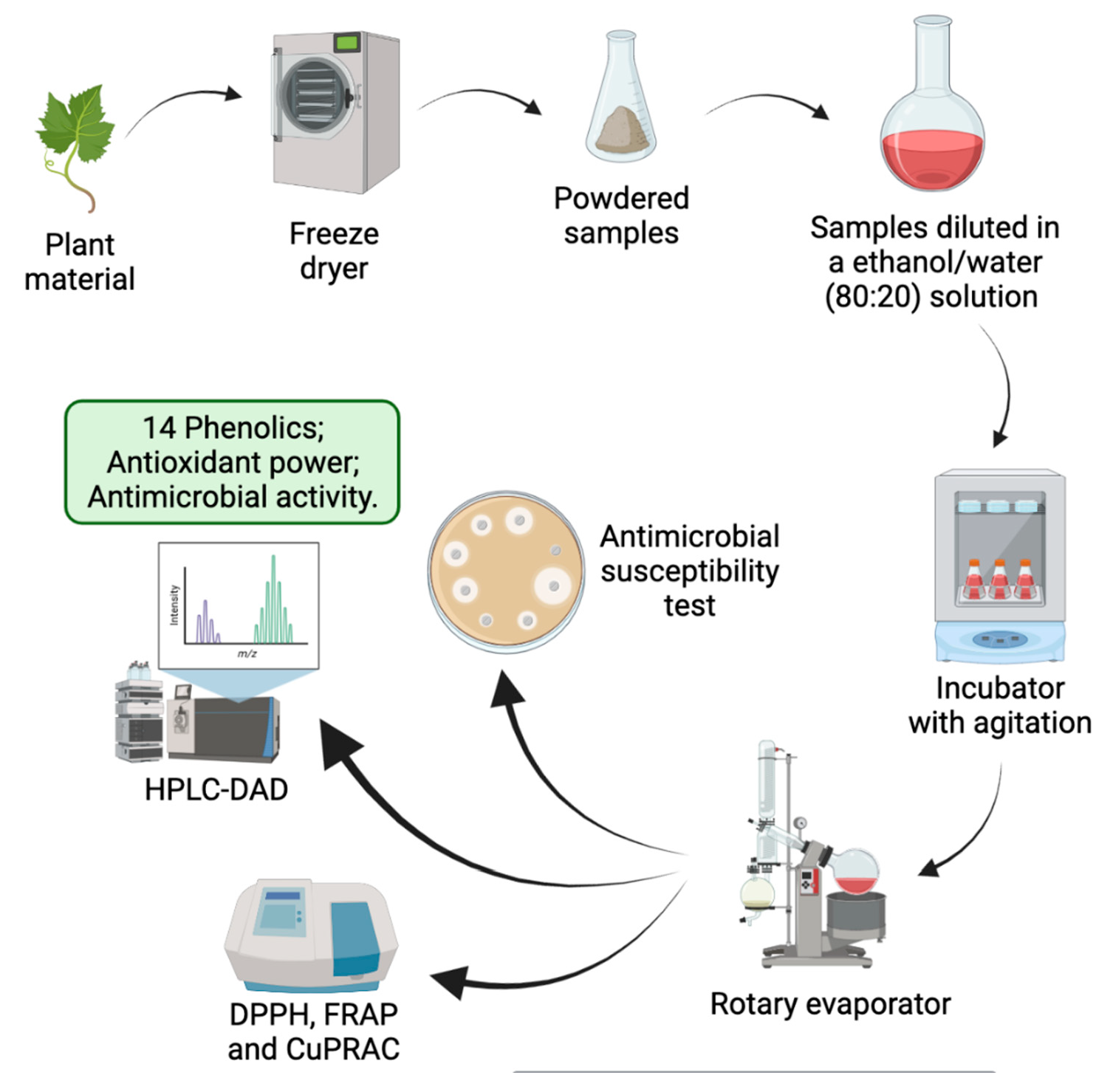
2.1. Plant Material

The plant material was collected in July 2020 in Trás-os-Montes and Alto Douro, Portugal (41°16′35” N 7°28′26” W), and included leaf, fruit, and stem bark of P. hybrida. These were separated, lyophilized, mill powdered with a commercial blender, and stored in a desiccator for future extraction [28].

2.2. Phenolic Extracts

The extraction of phenolic compounds was performed as described by Silva et al. (2020) with minor modifications [16]. Two grams of each powdered sample was added to 100 mL of an ethanol/water (80:20) solution and stirred for two hours at room temperature in a dark place. Afterward, all samples were centrifuged at 10.000 RPM for 15 min. Each supernatant was filtered and saved, and the pellet was rejected. Then, the solvent was evaporated on a rotatory evaporator at 40 °C under reduced pressure. Lastly, the achieved dry residues were weighed and redissolved in dimethyl sulfoxide (DMSO) or methanol to a final concentration of 100 and 50 mg/mL, respectively, and stored at −20 °C, until further analysis.

2.3. Phenolic Profile

To determine the profile and content of phenolic compounds, the samples were submitted to High-Performance Liquid Chromatography–Diode Array Detector (HPLC-DAD), as described by Aires and Carvalho (2020) [29]. An eluent formed by water with 0.1% trifluoroacetic acid (TFA) (solvent A) and acetonitrile with 0.1% TFA (solvent B), was injected in 10 µL of each extract. The flow rate of the elution was 1 mL/min, and the gradient was: 0% solvent B at 0 min, 0% solvent B at 5 min, 20% solvent B at 15 min, 50% solvent B at 30 min, 100% solvent B at 45 min, 100% solvent B at 50 min, 0% solvent B at 55 min, and 0% solvent B at 60 min. Chromatograms were recorded at 254 and 280 nm for benzoic acids and flavan-3-ols the, 320 nm for cinnamic acids, and 370 nm for flavonoids, with a C18 column (250 × 46 mm, 5 mm, ACE HPLC Columns, Advanced Chromatography Technologies Ltd., Abeerden, Scotland, UK). Peak retention time, UV spectra, UV max absorbance bands, and comparison with external commercial standards (Extrasynthese, Cedex, France) allow the identification of individual polyphenols. To prepare external standards, we used a methanol/water (70:30) solution at a concentration of 1.0 mg/mL and ran it in HPLC-DAD simultaneously to the samples. The internal standard method was used to determine the quantity of each polyphenol and the results were expressed as mg per gram of dry weight. The analysis was performed in duplicate.

2.4. Antioxidant Power

Powdered lyophilized samples were extracted with 1 mL of methanol and the antioxidant activity of each sample was assessed by three different methods: DPPH (2,2-diphenyl-1-picrylhydrazyl), FRAP (Ferric Reducing Antioxidant Power), and CuPRAC (Cupric Reducing Antioxidant Capacity). All analyses were performed in triplicate.

2.4.1. DPPH Assay

The DPPH assay was used to determine the DPPH radical scavenging activity of each extract. A DPPH solution (4 mg of 2.2-diphenyl-1- picrylhydrazyl radical in 100 mL of 95% of ethanol) was prepared and 15 µL of the plane tree extracts and 285 µL of the DPPH solution were added to each well of a 96 well microplate. A blank sample (extraction solvent instead of a sample with all the reagents) was added to one well. Then, the microplates were incubated at room temperature, in the dark, for 30 min. The absorbance values were measured at 517 nm in a microplate reader (MultiskanTM GO Microplate Photometer, Waltham, MA, USA). The DPPH radical scavenging capacity was calculated according to the following formula: % DPPH scavenging capacity = [(Absblank − Abssample)/Absblank] × 100 [30]. The results of the antioxidant power were expressed as the ability to reduce the DPPH radical, that is, the amount of extract required to reduce the initial DPPH concentration by 50% (EC50).

2.4.2. FRAP Assay

The FRAP assay was conducted as described in Benzie and Strain (1996) with minor alterations [31]. We prepared a FRAP reagent solution by mixing sodium acetate buffer (300 mM, pH 3.6) with 10 nM TPTZ solution (40 mM HCl as solvent) and 20 mM iron (III) chloride solution in a volume proportion of 10:1:1, respectively. Subsequently, in each well of a 96-well microplate, we added 25 µL of the plane tree extracts and 275 µL of the FRAP solution reagent. Then, the microplates were incubated at room temperature, in the dark, for 5 min. The absorbance values were measured at 593 nm in a microplate reader (MultiskanTM FC Microplate Photometer, Waltham, MA, USA). As a control standard, we used FeSO4 (Sigma-Aldrich, Tauferkichen, Germany) and the results were expressed as µmol of FeSO4 equivalents per gram of sample (µmol/FeSO4E/g/dw). Furthermore, EC50 values were calculated based on a dose–response curve.

2.4.3. CuPRAC Assay

The CuPRAC assay was performed colorimetrically according to Aires et al. (2017) [30]. We used a 96-well microplate and in each well, we added sequentially 50 µL of CuCl2 (10 mM in water), 50 µL of neocuproine (Sigma-Aldrich, Tauferkichen, Germany) at 7.5 mM in 96% ethanol, 50 µL of NH4Ac buffer (1 mM in water, pH 7.0), 25 µL of each sample, and lastly 25 µL of bidistilled H2O. Then, the microplates were incubated at room temperature, in the dark, for 30 min. The absorbance values were measured at 450 nm in a microplate reader (MultiskanTM GO Microplate Photometer, Waltham, MA, USA) against a blank sample (all reagents except CuCl2). As a control for a calibration curve, we used the commercial compound Trolox and the results were expressed as µM of Trolox equivalents per gram of sample (µM/TE/g/dw). As in FRAP assay, EC50 values were also calculated based on a dose–response curve.

2.5. Antimicrobial Activity

2.5.1. Bacterial Strains

The antimicrobial susceptibility was tested against nine MRSA strains isolated from different sources: three from hares (wild animals), three from rabbits (livestock), and three from humans. The wild animals’ MRSA strains were isolated from the buccal mucosa and perianal skin of wild Iberian hares (Lepus granatensis), the livestock MRSA strains were isolated from purulent lesions of rabbits from a slaughterhouse, and the human MRSA strains were isolated from infected diabetic foot ulcers. The strains’ resistance phenotype and genotype have been previously described (Table 1) [1,32,33,34]. For further analysis, all bacterial strains were seeded in Brain Heart Infusion (BHI) agar (Oxoid, Basingstoke, UK) and incubated overnight at 37 °C.

2.5.2. Antimicrobial Susceptibility Test

The antimicrobial susceptibility was tested by the Kirby–Bauer disc diffusion method. Before the inoculation on Müller–Hinton (MH) agar (Oxoid, Basingstoke, UK), the colonies obtained from BHI agar were suspended in a physiological solution to a turbidity corresponding to 0.5 McFarland standard. The dry residues that were redissolved in DMSO to a final concentration of 100 mg/mL were diluted to 75, 50, 25, and 10 mg/mL, and all five concentrations were tested against the MRSA strains. Sterile blank discs (6 mm) were loaded with 20 µL of every extract concentration, placed on the inoculated MH plates, and incubated for 24 h at 37 °C. The measurement of the inhibition zones allowed the assessment of the antimicrobial activity of the extracts against the MRSA strains [28].

2.6. Statistical Analysis

The results are expressed as mean values and standard deviation (SD). The antioxidant measurements were analyzed using the one-way analysis of variance (ANOVA). Different values of antioxidant activities were compared using the Pearson correlation test. A p-value of <0.05 was considered significant and the statistical analysis was carried out with IBM SPSS Statistics v 28.0.1.0.

3. Results

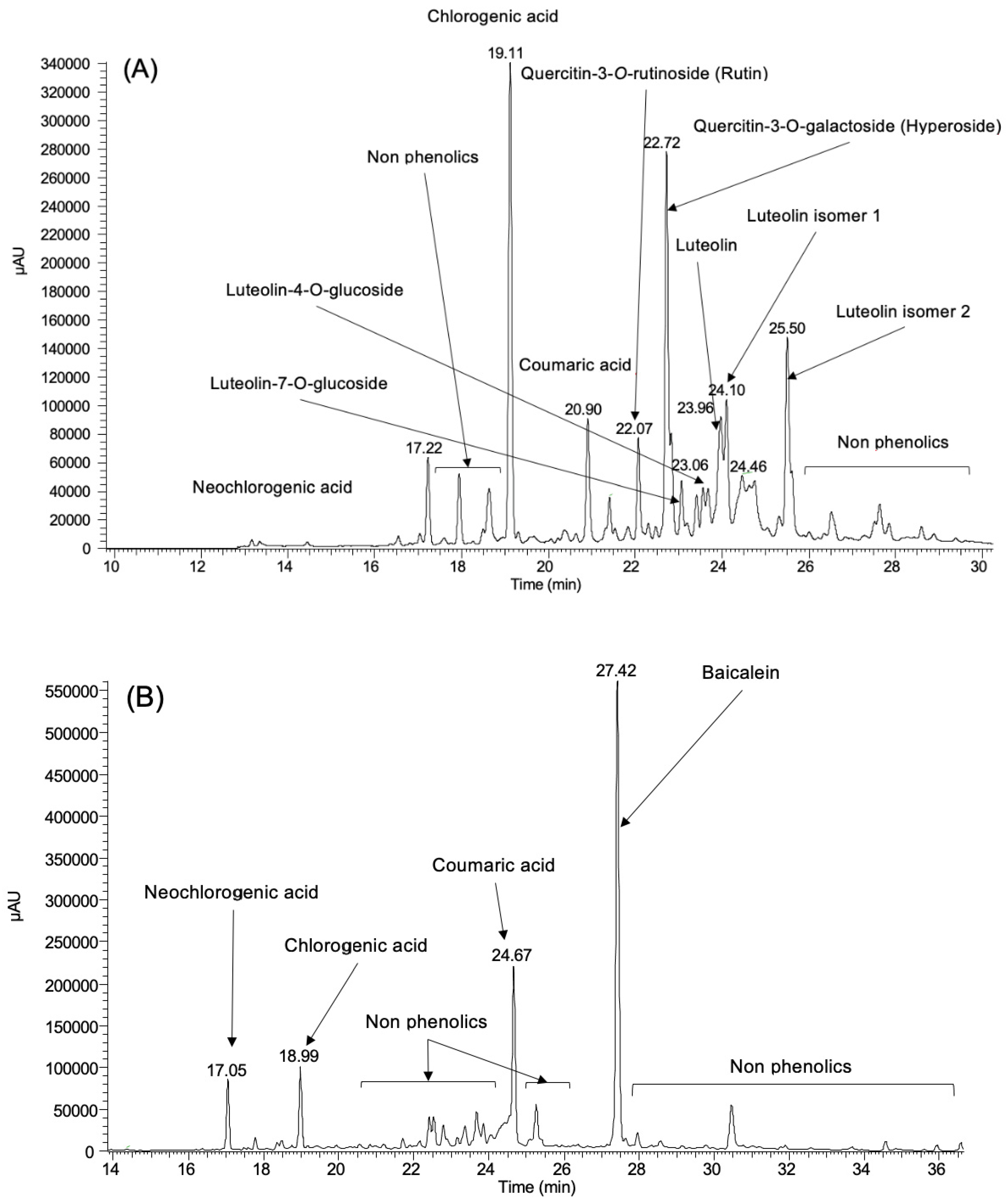
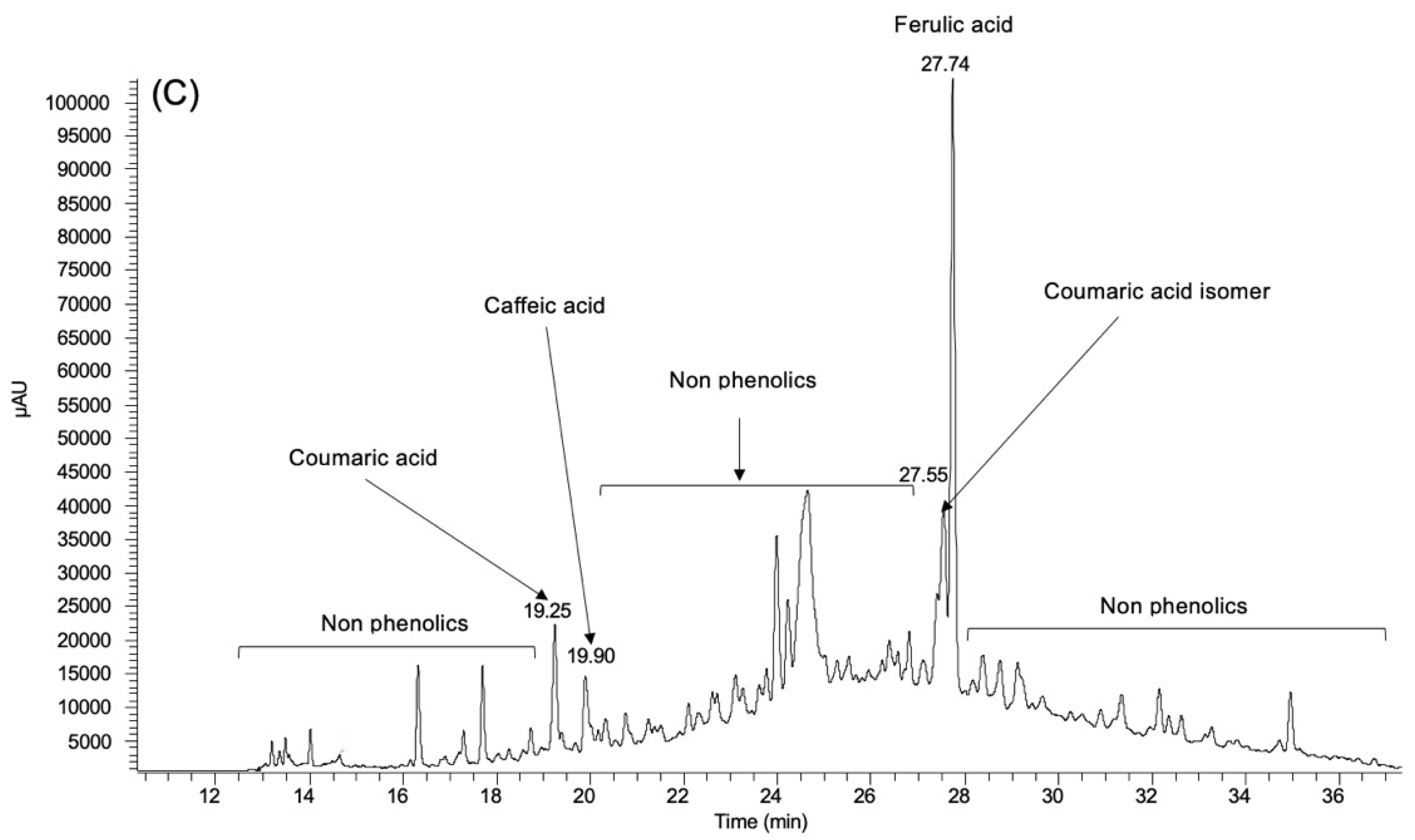
3.1. Phenolic Profile

The extraction of phenolic compounds is a critical step since it can be influenced by numerous parameters, such as extraction time, temperature, solvent-to-sample ratio, number of repeat extractions of the sample as well as solvent type [35]. Two major parameters that affect the phenolics extracted from plants are time and temperature. Extended extraction times and high temperatures usually degrade or lead to unwanted reactions to plant phenolics [36]. Moreover, studies have revealed that high amounts of phenolics are obtained in the first minute of extraction and that a longer time did not affect the number of phenolics extracted [35]. In addition to time and temperature, the selection of the extraction solvent also influences the extraction of polyphenols [36]. Several studies have shown that water–ethanol mixtures are better solvents for phenolic extractions [37,38].
The phenolic profile of P. hybrida’s components was measured by HPLC-DAD and the individual phenolics identified were neochlorogenic acid, chlorogenic acid, coumaric acid, coumaric acid isomer, caffeic acid, ferulic acid, quercetin-3-O-rutinoside, quercetin-3-O-galactoside, baicalein, luteolin-7-O-glucoside, luteolin-4-O-glucoside, luteolin, luteolin isomer 1, and luteolin isomer 2 (Figure 1).
The phenolic profile of each extract is presented in Table 2. The only phenolic compound common to all extracts was coumaric acid. This is a plant-derived secondary metabolite that is generally found in fruits, vegetables, mushrooms, and cereals [39]. Several studies have shown that this phenol acts on the reduction of oxidative stress and inflammatory reactions through scavenging and antioxidant properties. Besides that, it possesses other bioactivities such as anti-cancer, anti-melanogenic, and anti-diabetic [40].
The leaf extract was the plant material that showed the highest content of phenolics and quercetin-3-O-galactoside and chlorogenic acid represent, respectively, 39% and 19% of the total individual phenolics identified in the extract. These findings are important because both phenolics are responsible for many important and therapeutic functions, such as antioxidant, antibacterial, anti-inflammatory, and free-radical-scavenging properties. Quercetin-3-O-galactoside is a flavonoid that is commonly found in fruits, vegetables, wines, herbs, and beverages while chlorogenic acid is a phenolic acid that can be typically found in food, particularly, coffee and tea [41,42]. Nevertheless, chlorogenic acid has been identified in Platanus occidentalis’ leaves [43]. This acid is a very significant and biologically active dietary polyphenol and it has been described as a suitable preservative and food additive because of its antimicrobial effect and inhibitory outcome on multidrug efflux mechanisms and biofilm-producing ability of multidrug-resistant bacteria [41].
The phenolic profile of P. hydrida’s fruit extract revealed that baicalein was the main phenolic with a 70% prevalence. Baicalein is a flavone that was originally isolated from the roots of Scutellaria baicalensis, and it is accountable for most of the health benefits attributed to this plant, namely, antioxidant, anti-viral, anti-inflammatory, and anti-cancer effects [44]. Comparing our results with other studies performed with S. baicalensis, it is possible to notice that the obtained level of baicalein is high [45,46]. However, these differences could be explained by external factors such as the type of solvent used in the extraction process [14]. Neochlorogenic acid, chlorogenic acid, and coumaric acid have also been identified in fruits of P. occidentalis [43].
The stem bark extract was the element that exhibited the lowest content of phenolics and the majority (59%) of the total individual phenolics identified in this extract was represented by ferulic acid. Phenolic acids, particularly ferulic acid, are normally found in stems since they constitute lignin which is a significant element of stems [47]. This phenolic acid has an active role in many biological processes, such as oxidative stress, inflammation, vascular endothelial injury, fibrosis, apoptosis, and platelet aggregation [48].

3.2. Antioxidant Power

The assessment of the antioxidant power using different assays is important to obtain the overall antioxidant potential of any matrix [49]. Therefore, in this study, to determine the antioxidant power of all phenolic extracts we used three spectrophotometric methods: DPPH, FRAP, and CuPRAC. Spectrophotometric methods have many advantages such as cost and speed. Moreover, they do not require a high level of specialization from the technician performing the analysis, being suitable for a screening approach that can be performed even in small laboratories [16]. The results, which are presented in Table 3, are expressed in effective concentration (EC50), and lower values are related to a higher antioxidant power.
Although the results were all very similar, the antioxidant power of each extract was slightly higher with the in vitro FRAP assay. This assay is a widely used method that is reasonably simple, quick, and economical. Different types of samples can be tested with the FRAP assay and it measures accurately and with high sensitivity and duplicability the combined antioxidant activity of redox-active components [50].
The DPPH assay results for the fruit extract were not coherent with the other results. DPPH is a stable free radical that receives an electron or hydrogen radical to become a stable diamagnetic molecule. Antioxidants can affect the DPPH radicals because of their ability to donate hydrogen [51]. The second most prevalent phenolic identified in the fruit extract profile was coumaric acid which has an alkene group adjacent to the carboxylic group. This structural characteristic could explain the obtained results since it interferes with the transfer of H atoms from the carboxylic group [52].
To study the relationships between the sum of individual phenolics obtained by HPLC-DAD and the antioxidant assays, and between all antioxidant assays, we used a regression analysis. All Pearson correlation coefficients are shown in Table 4.
The relationship between all antioxidant assays showed a significant positive correlation and the leaf extracts showed the highest antioxidant power, followed by the stem bark extract and fruit extract. Since the stem bark extract was the one that exhibited the lowest phenolic content, it was expected that they also demonstrated the lowest antioxidant power [53]. However, in our work, the sum of individual phenolics and the three antioxidant assays exhibited an insignificant correlation. This suggests that the antioxidant power is not associated with the phenolic compounds in this tree. The sum of all individual phenolics does not reveal the total antioxidant potential of the mixture due to synergistic, additive, or antagonistic effects that might occur [54]. It is not easy to predict the antioxidant power of merged phytochemicals, so it is necessary to perform specific experimental studies of the combinatorial system and to consider the assessment of mechanisms of interaction [55].
A study conducted by Dogan and Anuk (2019) demonstrated that the use of P. orientalis leaf at certain quantities revealed a potential antioxidant activity [21]. As previously described, the main phenolic identified in the leaf’s extract was quercetin-3-O-galactoside which could explain the leaf’s antioxidant power since quercetin-3-O-galactoside possesses a significant antioxidant activity [56,57,58]. According to Chatzigeorgiou et al. (2017), the fruits of P. orientalis have also shown antioxidant effects [22]. Baicalein, the most prevalent phenolic identified in the fruit of P. hybrida, has exhibited antioxidant properties, probably by chelating metal transition ions or by scavenging free radicals through the donation of hydrogen atoms or electrons [59]. Both main phenolics identified in the leaf and fruit extracts of P. hybrida are flavonoids: quercetin-3-O-galactoside is a flavonol and baicalein is a flavone [60]. These two subclasses of flavonoids have highly similar structures but flavonols are typically more active than flavones as antioxidants which is in agreement with our results [61,62].
Due to the lack of studies performed with Platanus spp., it was not possible to compare the antioxidant power of the stem bark extract with results from other researchers. However, the antioxidant activity of the stem bark extract could be justified by the presence of ferulic acid since it has previously shown effective antioxidant properties [63].

3.3. Antimicrobial Activity

The plane tree extracts were tested against nine MRSA strains isolated from three different sources (livestock, wild animals, and humans) and the results are presented in Table 5. The Kirby–Bauer disc diffusion method evaluates the antimicrobial susceptibility through the inhibition zone diameter and all extracts exhibited inhibition zones against at least two different strains. The leaf extract inhibited the growth of all bacterial strains, while the fruit extract inhibited the growth of eight bacterial strains and the stem bark extract inhibited the growth of two bacterial strains (none isolated from humans). The strains included in this study have been previously described and all demonstrated resistant patterns [1,32,33,34]. The extracts’ ability to suppress the growth of these MRSA strains while other classical antimicrobial agents could not, highlights that these extracts are potent anti-MRSA agents. Ibrahim et al. (2009) described several metabolites from P. occidentalis as viable growth inhibitors of MRSA [64]. Moreover, many natural sources of phenolic compounds have also demonstrated good anti-MRSA activity [65,66,67].
In general, the fruit extracts inhibited microbial growth at lower concentrations and presented slightly higher inhibition zones. The efficacy of the fruit extracts from Platanus spp. against drug-resistant bacteria was already demonstrated by Chatzigeorgiou et al. (2017) [22]. The main phenolic compound found in the fruit extract, as well as in the leaf extract, was a flavonoid (baicalein). Flavonoids are frequently synthesized by plants as a defense mechanism against microorganisms [68]. Baicalein, in particular, has shown an excellent antibacterial activity [69]. Therefore, their antimicrobial outcome is no surprise and is possibly caused by their complex formation with both extracellular and soluble proteins, as well as the bacterial cell wall and cell membranes [70]. Moreover, it has been shown that the combination of baicalein with oxacillin against MRSA inhibits their cell wall synthesis, causing bacterial death, which could be an advantage for the drug-resistant bacteria fight [71].
The antibacterial activity of the leaf extracts could be related to their phenolic components, particularly quercetin-3-O-galactoside. Macêdo et al. (2021) and Upadhyay et al. (2010) evaluated the antimicrobial effect of quercetin and, in both studies, this flavonoid inhibited the growth of several bacteria [72,73]. Previous studies have considered the leaf extract to be nontoxic, highly active, and selective against MRSA [64].
Changes in the cell membrane, inactivation of essential enzymes, and/or modification of the functions of genetic material are some mechanisms that allow phenolic compounds to suppress microbial growth [16]. Since the stem bark extract was the extract that demonstrated the lowest phenolic content, it was expected that it demonstrated a lower antimicrobial activity when compared to the leaf or fruit extracts. However, while the stem bark extract was not very effective against the MRSA strains tested in this study, they are a source of ferulic acid and, according to Ibitoye and Ajiboye (2019), this phenolic acid potentiates the antibacterial activity of quinolone-based antibiotics against Acinetobacter baumannii which is a pathogenic Gram-negative bacteria [74]. Due to their distinctive structure, Gram-negative bacteria are typically more resistant than Gram-positive bacteria [75]. To better understand these results and confirm that the stem bark extracts could be efficient against pathogenic bacteria, further studies should be carried out.

4. Conclusions

P. hybrida is a biological sample with a substantial value of phytochemicals that have been showing several potential positive effects on human health. The results obtained with this work highlight the complex chemical composition and the antioxidative and antimicrobial potential of extracts derived from P. hydrida. Since resistant bacteria can affect humans, animals, and the environment, the extracts’ ability to suppress microbial growth of strains from all sources suggests that these extracts could be used as a food supplement or a pharmaceutical additive to help fight drug-resistant bacteria. However, we must consider that the structural characteristics of the phenolic compounds and external factors such as the solvent, time, and temperature can affect the bacterial response to natural compounds. Therefore, to figure out how these extracts act on pathogenic bacteria, to explore their toxicity, and to guarantee that there would be no side effects, more studies, such as in vivo trials, should be carried out. Moreover, the extraction of polyphenols from the components of P. hybrida and, consequently, their use as a pharmaceutical tool, supports the production and consumption of natural resources which is a major contribution to the circular economy.

Author Contributions

Conceptualization, V.S. and P.P.; formal analysis, J.R. and I.G.; funding acquisition, P.P.; investigation, J.R. and V.S.; methodology, J.R., V.S., A.A. and R.C.; supervision, A.A., L.B. and P.P.; validation, A.A., G.I. and P.P.; writing—original draft, J.R.; writing—review and editing. All authors have read and agreed to the published version of the manuscript.

Funding

This work was supported by the projects UIDB/CVT/00772/2020 and LA/P/0059/2020 funded by the Portuguese Foundation for Science and Technology (FCT). This work was also supported by the Associate Laboratory for Green Chemistry-LAQV, which is financed by national funds from FCT/MCTES (UIDB/50006/2020 and UIDP/50006/2020) funded by FCT. The authors acknowledge the financial support of FCT by national funds under the project UIDB/04033/2020. Vanessa Silva is grateful to FCT for financial support through PhD grant SFRH/BD/137947/2018. To FCT, Portugal for financial support through national funds FCT/MCTES to CIMO (UIDB/00690/2020) and the contract of L. Barros through the institutional scientific employment program-contract. This work was supported.

Institutional Review Board Statement

Not applicable.

Informed Consent Statement

Not applicable.

Data Availability Statement

Not applicable.

Conflicts of Interest

The authors declare no conflict of interest.

References

  1. Silva, V.; Pereira, J.E.; Maltez, L.; Ferreira, E.; Manageiro, V.; Caniça, M.; Capelo, J.L.; Igrejas, G.; Poeta, P. Diversity of methicillin-resistant staphylococci among wild Lepus granatensis: First detection of mecA-MRSA in hares. FEMS Microbiol. Ecol. 2020, 96, 163–172. [Google Scholar] [CrossRef] [PubMed]
  2. Algammal, A.M.; Hetta, H.F.; Elkelish, A.; Alkhalifah, D.H.H.; Hozzein, W.N.; Batiha, G.E.S.; El Nahhas, N.; Mabrok, M.A. Methicillin-resistant Staphylococcus aureus (MRSA): One health perspective approach to the bacterium epidemiology, virulence factors, antibiotic-resistance, and zoonotic impact. Infect. Drug Resist. 2020, 13, 3255–3265. [Google Scholar] [CrossRef] [PubMed]
  3. Amaro, F.; Martín-González, A. Microbial warfare in the wild—The impact of protists on the evolution and virulence of bacterial pathogens. Int. Microbiol. 2021, 24, 559–571. [Google Scholar] [CrossRef] [PubMed]
  4. Bernier-Lachance, J.; Arsenault, J.; Usongo, V.; Parent, E.; Labrie, J.; Jacques, M.; Malouin, F.; Archambault, M. Prevalence and characteristics of livestock- associated methicillin-resistant Staphylococcus aureus (LA-MRSA) isolated from chicken meat in the province of Quebec, Canada. PLoS ONE 2020, 15, e0227183. [Google Scholar] [CrossRef] [PubMed]
  5. Collignon, P.J.; McEwen, S.A. One health-its importance in helping to better control antimicrobial resistance. Trop. Med. Infect. Dis. 2019, 4, 22. [Google Scholar] [CrossRef] [Green Version]
  6. Redican, K.J.; Akpinar-Elci, M. Integrating One Health into Professional Preparation Education for Public Health and Health Education Specialists. Am. J. Heal. Educ. 2021, 52, 11–17. [Google Scholar] [CrossRef]
  7. Tan, L.; Zhou, Z.; Liu, X.; Li, J.; Zheng, Y.; Cui, Z.; Yang, X.; Liang, Y.; Li, Z.; Feng, X.; et al. Overcoming Multidrug-Resistant MRSA Using Conventional Aminoglycoside Antibiotics. Adv. Sci. 2020, 7, 1902070. [Google Scholar] [CrossRef] [Green Version]
  8. Gupta, A.; Mumtaz, S.; Li, C.H.; Hussain, I.; Rotello, V.M. Combatting antibiotic-resistant bacteria using nanomaterials. Chem. Soc. Rev. 2019, 48, 415–427. [Google Scholar] [CrossRef]
  9. García, J.; García-Galán, M.J.; Day, J.W.; Boopathy, R.; White, J.R.; Wallace, S.; Hunter, R.G. A review of emerging organic contaminants (EOCs), antibiotic resistant bacteria (ARB), and antibiotic resistance genes (ARGs) in the environment: Increasing removal with wetlands and reducing environmental impacts. Bioresour. Technol. 2020, 307, 123228. [Google Scholar] [CrossRef]
  10. Zipperer, A.; Konnerth, M.C.; Laux, C.; Berscheid, A.; Janek, D.; Weidenmaier, C.; Burian, M.; Schilling, N.A.; Slavetinsky, C.; Marschal, M.; et al. Human commensals producing a novel antibiotic impair pathogen colonization. Nature 2016, 535, 511–516. [Google Scholar] [CrossRef]
  11. Martins, N.; Barros, L.; Santos-Buelga, C.; Henriques, M.; Silva, S.; Ferreira, I.C.F.R. Evaluation of bioactive properties and phenolic compounds in different extracts prepared from Salvia officinalis L. Food Chem. 2014, 170, 378–385. [Google Scholar] [CrossRef] [PubMed] [Green Version]
  12. Uysal, A.; Zengin, G.; Mahomoodally, M.F.; Picot-Allain, C.; Jekő, J.; Cziáky, Z.; Rodrigues, M.J.; Ak, G.; Polat, R.; Urusan, Z.; et al. A comparative study on biological properties and chemical profiles of different solvent extracts from Centaurea bingoelensis, an endemic plant of Turkey. Process Biochem. 2021, 102, 315–324. [Google Scholar] [CrossRef]
  13. Kowalczyk, T.; Sitarek, P.; Toma, M.; Rijo, P.; Domínguez-Martín, E.; Falcó, I.; Sánchez, G.; Śliwiński, T. Enhanced Accumulation of Betulinic Acid in Transgenic Hairy Roots of Senna obtusifolia Growing in the Sprinkle Bioreactor and Evaluation of Their Biological Properties in Various Biological Models. Chem. Biodivers. 2021, 18, e2100455. [Google Scholar] [CrossRef] [PubMed]
  14. Barral-Martinez, M.; Garcia-Oliveira, P.; Nuñez-Estevez, B.; Silva, A.; Finimundy, T.C.; Calhelha, R.; Nenadic, M.; Sokovic, M.; Barroso, F.; Simal-Gandara, J.; et al. Plants of the Family Asteraceae: Evaluation of Biological Properties and Identification of Phenolic Compounds. Chem. Proc. 2021, 5, 51. [Google Scholar] [CrossRef]
  15. Álvarez-Martínez, F.J.; Barrajón-Catalán, E.; Micol, V. Tackling antibiotic resistance with compounds of natural origin: A comprehensive review. Biomedicines 2020, 8, 405. [Google Scholar] [CrossRef]
  16. Silva, V.; Falco, V.; Dias, M.I.; Barros, L.; Silva, A.; Capita, R.; Alonso-Calleja, C.; Amaral, J.S.; Igrejas, G.; Ferreira, I.C.F.R.; et al. Evaluation of the phenolic profile of Castanea sativa mill. By-products and their antioxidant and antimicrobial activity against multiresistant bacteria. Antioxidants 2020, 9, 87. [Google Scholar] [CrossRef] [Green Version]
  17. Gulcin, İ. Antioxidants and antioxidant methods: An updated overview. Arch. Toxicol. 2020, 94, 651–715. [Google Scholar] [CrossRef] [Green Version]
  18. Wu, Z.; Tan, B.; Liu, Y.; Dunn, J.; Martorell Guerola, P.; Tortajada, M.; Cao, Z.; Ji, P. Chemical Composition and Antioxidant Properties of Essential Oils from Peppermint, Native Spearmint and Scotch Spearmint. Molecules 2019, 24, 2825. [Google Scholar] [CrossRef] [Green Version]
  19. Hajhashemi, V.; Ghannadi, A.; Mousavi, S. Antinociceptive study of extracts of Platanus orientalis leaves in mice. Res. Pharm. Sci. 2011, 6, 123–128. [Google Scholar]
  20. Lara, B.; Rojo, J.; Fernández-González, F.; Pérez-Badia, R. Prediction of airborne pollen concentrations for the plane tree as a tool for evaluating allergy risk in urban green areas. Landsc. Urban Plan. 2019, 189, 285–295. [Google Scholar] [CrossRef]
  21. Dogan, A.; Anuk, O.O. Investigation of the phytochemical composition and antioxidant properties of chinar (Platanus orientalis L.) leaf infusion against ethanol-induced oxidative stress in rats. Mol. Biol. Rep. 2019, 46, 3049–3061. [Google Scholar] [CrossRef] [PubMed]
  22. Chatzigeorgiou, S.; Thai, Q.D.; Tchoumtchoua, J.; Tallas, K.; Tsakiri, E.N.; Papassideri, I.; Halabalaki, M.; Skaltsounis, A.L.; Trougakos, I.P. Isolation of natural products with anti-ageing activity from the fruits of Platanus orientalis. Phytomedicine 2017, 33, 53–61. [Google Scholar] [CrossRef]
  23. Ribeiro, J.; Silva, V.; Aires, A.; Carvalho, R.; Igrejas, G.; Poeta, P. Antimicrobial Activity of Phenolic Compounds Extracted from Platanus hybrida: Exploring Alternative Therapies for a Post-Antibiotic Era. Proceedings 2021, 66, 18. [Google Scholar] [CrossRef]
  24. Vrinceanu, D.; Berghi, O.; Cergan, R.; Dumitru, M.; Ciuluvica, R.; Giurcaneanu, C.; Neagos, A. Urban allergy review: Allergic rhinitis and asthma with plane tree sensitization (Review). Exp. Ther. Med. 2021, 21, 275. [Google Scholar] [CrossRef] [PubMed]
  25. Liu, G.; Huang, J.; Chen, L.; Bao, M. Plant regeneration from excised hypocotyl explants of Platanus acerifolia willd. Vitr. Cell. Dev. Biol.—Plant 2002, 38, 558–563. [Google Scholar] [CrossRef]
  26. Kutbay, I.; Akfırat, F.Ş. Mapping of Biochemical Constituents in Platanus acerifolia Leaves By Analytical Techniques. Procedia—Soc. Behav. Sci. 2015, 195, 1719–1727. [Google Scholar] [CrossRef] [Green Version]
  27. Thai, Q.D.; Tchoumtchoua, J.; Makropoulou, M.; Boulaka, A.; Meligova, A.K.; Mitsiou, D.J.; Mitakou, S.; Michel, S.; Halabalaki, M.; Alexis, M.N.; et al. Phytochemical study and biological evaluation of chemical constituents of Platanus orientalis and Platanus × acerifolia buds. Phytochemistry 2016, 130, 170–181. [Google Scholar] [CrossRef]
  28. Silva, V.; Igrejas, G.; Falco, V.; Santos, T.P.; Torres, C.; Oliveira, A.M.P.; Pereira, J.E.; Amaral, J.S.; Poeta, P. Chemical composition, antioxidant and antimicrobial activity of phenolic compounds extracted from wine industry by-products. Food Control 2018, 92, 516–522. [Google Scholar] [CrossRef] [Green Version]
  29. Aires, A.; Carvalho, R. Kiwi fruit residues from industry processing: Study for a maximum phenolic recovery yield. J. Food Sci. Technol. Technol 2020, 57, 4265–4276. [Google Scholar] [CrossRef]
  30. Aires, A.; Carvalho, R.; Matos, M.; Carnide, V.; Silva, A.P.; Gonçalves, B. Variation of chemical constituents, antioxidant activity, and endogenous plant hormones throughout different ripening stages of highbush blueberry (Vaccinium corymbosum L.) cultivars produced in centre of Portugal. J. Food Biochem. 2017, 41, e12414. [Google Scholar] [CrossRef]
  31. Benzie, I.F.F.; Strain, J.J. The Ferric Reducing Ability of Plasma (FRAP) as a Measure of “Antioxidant Power”: The FRAP Assay. Anal. Biochem. 1996, 239, 70–76. [Google Scholar] [CrossRef] [PubMed] [Green Version]
  32. Silva, V.; Almeida, F.; Carvalho, J.A.; Castro, A.P.; Ferreira, E.; Manageiro, V.; Tejedor-Junco, M.T.; Caniça, M.; Igrejas, G.; Poeta, P. Emergence of community-acquired methicillin-resistant Staphylococcus aureus EMRSA-15 clone as the predominant cause of diabetic foot ulcer infections in Portugal. Eur. J. Clin. Microbiol. Infect. Dis. 2020, 39, 179–186. [Google Scholar] [CrossRef] [PubMed]
  33. Silva, V.; Almeida, F.; Silva, A.; Correia, S.; Carvalho, J.A.; Castro, A.P.; Ferreira, E.; Manageiro, V.; Caniça, M.; Igrejas, G.; et al. First report of linezolid-resistant cfr-positive methicillin-resistant Staphylococcus aureus in humans in Portugal. J. Glob. Antimicrob. Resist. 2019, 17, 323–325. [Google Scholar] [CrossRef] [PubMed]
  34. Silva, V.; De Sousa, T.; Gómez, P.; Sabença, C.; Vieira-Pinto, M.; Capita, R.; Alonso-Calleja, C.; Torres, C.; Capelo, J.L.; Igrejas, G.; et al. Livestock-associated methicillin-resistant Staphylococcus aureus (MRSA) in purulent subcutaneous lesions of farm rabbits. Foods 2020, 9, 439. [Google Scholar] [CrossRef] [PubMed] [Green Version]
  35. Jahromi, S.G. Extraction Techniques of Phenolic Compounds from Plants; Woodhead Publishing: Thorston, UK, 2019. [Google Scholar]
  36. Khoddami, A.; Wilkes, M.A.; Roberts, T.H. Techniques for analysis of plant phenolic compounds. Molecules 2013, 18, 2328–2375. [Google Scholar] [CrossRef] [PubMed]
  37. Sik, B.; Hanczné, E.L.; Kapcsándi, V.; Ajtony, Z. Conventional and nonconventional extraction techniques for optimal extraction processes of rosmarinic acid from six Lamiaceae plants as determined by HPLC-DAD measurement. J. Pharm. Biomed. Anal. 2020, 184, 113173. [Google Scholar] [CrossRef] [PubMed]
  38. Kaderides, K.; Papaoikonomou, L.; Serafim, M.; Goula, A.M. Microwave-assisted extraction of phenolics from pomegranate peels: Optimization, kinetics, and comparison with ultrasounds extraction. Chem. Eng. Process.—Process Intensif. 2019, 137, 1–11. [Google Scholar] [CrossRef]
  39. Zabad, O.M.; Samra, Y.A.; Eissa, L.A. P-Coumaric acid alleviates experimental diabetic nephropathy through modulation of Toll like receptor-4 in rats. Life Sci. 2019, 238, 116965. [Google Scholar] [CrossRef]
  40. Roychoudhury, S.; Sinha, B.; Choudhury, B.P.; Jha, N.K.; Palit, P.; Kundu, S.; Mandal, S.C.; Kolesarova, A.; Yousef, M.I.; Ruokolainen, J.; et al. Scavenging properties of plant-derived natural biomolecule para-coumaric acid in the prevention of oxidative stress-induced diseases. Antioxidants 2021, 10, 1205. [Google Scholar] [CrossRef]
  41. Naveed, M.; Hejazi, V.; Abbas, M.; Kamboh, A.A.; Khan, G.J.; Shumzaid, M.; Ahmad, F.; Babazadeh, D.; FangFang, X.; Modarresi-Ghazani, F.; et al. Chlorogenic acid (CGA): A pharmacological review and call for further research. Biomed. Pharmacother. 2018, 97, 67–74. [Google Scholar] [CrossRef]
  42. Hu, C.; Chen, Y.; Cao, Y.; Jia, Y.; Zhang, J. Metabolomics analysis reveals the protective effect of quercetin-3-O-galactoside (Hyperoside) on liver injury in mice induced by acetaminophen. J. Food Biochem. 2020, 44, e13420. [Google Scholar] [CrossRef] [PubMed]
  43. Al-Naib, F.A.-G.; Rice, E.L. Allelopathic Effects of Platanus occidentalis. Torrey Bot. Soc. 1971, 98, 75–82. [Google Scholar] [CrossRef]
  44. Tuli, H.S.; Aggarwal, V.; Kaur, J.; Aggarwal, D.; Parashar, G.; Parashar, N.C.; Tuorkey, M.; Kaur, G.; Savla, R.; Sak, K.; et al. Baicalein: A metabolite with promising antineoplastic activity. Life Sci. 2020, 259, 118183. [Google Scholar] [CrossRef]
  45. Lin, M.C.; Tsai, M.J.; Wen, K.C. Supercritical fluid extraction of flavonoids from Scutellariae Radix. J. Chromatogr. A 1999, 830, 387–395. [Google Scholar] [CrossRef]
  46. Georgieva, Y.; Katsarova, M.; Stoyanov, P.; Mladenov, R.; Denev, P.; Teneva, D.; Plotnikov, E.; Bozov, P.; Dimitrova, S. Metabolite profile and antioxidant activity of some species of genus Scutellaria growing in bulgaria. Plants 2021, 10, 45. [Google Scholar] [CrossRef] [PubMed]
  47. Silva, V.; Singh, R.K.; Gomes, N.; Soares, B.G.; Silva, A.; Falco, V.; Capita, R.; Alonso-Calleja, C.; Pereira, J.E.; Amaral, J.S.; et al. Comparative insight upon chitosan solution and chitosan nanoparticles application on the phenolic content, antioxidant and antimicrobial activities of individual grape components of Sousão variety. Antioxidants 2020, 9, 178. [Google Scholar] [CrossRef] [Green Version]
  48. Li, D.; Rui, Y.X.; Guo, S.D.; Luan, F.; Liu, R.; Zeng, N. Ferulic acid: A review of its pharmacology, pharmacokinetics and derivatives. Life Sci. 2021, 284, 119921. [Google Scholar] [CrossRef]
  49. López-Froilán, R.; Hernández-Ledesma, B.; Cámara, M.; Pérez-Rodríguez, M.L. Evaluation of the Antioxidant Potential of Mixed Fruit-Based Beverages: A New Insight on the Folin-Ciocalteu Method. Food Anal. Methods 2018, 11, 2897–2906. [Google Scholar] [CrossRef]
  50. Benzie, I.F.F.; Devaki, M. The ferric reducing/antioxidant power (FRAP) assay for non-enzymatic antioxidant capacity: Concepts, procedures, limitations and applications. Meas. Antioxid. Act. Capacit. Recent Trends Appl. 2018, 5, 77–106. [Google Scholar] [CrossRef]
  51. Aires, A.; Carvalho, R.; Rosa, E.A.S.; Saavedra, M.J. Phytochemical characterization and antioxidant properties of baby-leaf watercress produced under organic production system. CYTA—J. Food 2013, 11, 343–351. [Google Scholar] [CrossRef]
  52. Mercado-Mercado, G.; de la Rosa, L.A.; Alvarez-Parrilla, E. Effect of pectin on the interactions among phenolic compounds determined by antioxidant capacity. J. Mol. Struct. 2020, 1199, 126967. [Google Scholar] [CrossRef]
  53. Parikh, B.; Patel, V.H. Total phenolic content and total antioxidant capacity of common Indian pulses and split pulses. J. Food Sci. Technol. 2018, 55, 1499–1507. [Google Scholar] [CrossRef] [PubMed]
  54. Olszowy-Tomczyk, M. Synergistic, antagonistic and additive antioxidant effects in the binary mixtures. Phytochem. Rev. 2020, 19, 63–103. [Google Scholar] [CrossRef]
  55. Velásquez, P.; Giordano, A.; Valenzuela, L.M.; Montenegro, G. Combined antioxidant capacity of Chilean bee hive products using mixture design methodology. Lwt 2022, 155, 112982. [Google Scholar] [CrossRef]
  56. Yamazaki, E.; Inagaki, M.; Kurita, O.; Inoue, T. Antioxidant activity of Japanese pepper (Zanthoxylum piperitum DC.) fruit. Food Chem. 2007, 100, 171–177. [Google Scholar] [CrossRef]
  57. Conforti, F.; Statti, G.A.; Tundis, R.; Menichini, F.; Houghton, P. Antioxidant activity of methanolic extract of Hypericum triquetrifolium Turra aerial part. Fitoterapia 2002, 73, 479–483. [Google Scholar] [CrossRef]
  58. Dutta, S.; Kundu, A. Macroporous resin-assisted enrichment, characterizations, antioxidant and anticandidal potential of phytochemicals from Trachyspermum ammi. J. Food Biochem. 2021, e13847. [Google Scholar] [CrossRef]
  59. Wang, X.; Li, X.; Li, H. Reassessment of Antioxidant Activity of Baicalein in vitro. Asian J. Pharm. Biol. Res. 2011, 1, 186–194. [Google Scholar]
  60. Cai, Y.; Luo, Q.; Sun, M.; Corke, H. Antioxidant activity and phenolic compounds of 112 traditional Chinese medicinal plants associated with anticancer. Life Sci. 2004, 74, 2157–2184. [Google Scholar] [CrossRef]
  61. Yang, G.; Hong, S.; Yang, P.; Sun, Y.; Wang, Y.; Zhang, P.; Jiang, W.; Gu, Y. Discovery of an ene-reductase for initiating flavone and flavonol catabolism in gut bacteria. Nat. Commun. 2021, 12, 790. [Google Scholar] [CrossRef] [PubMed]
  62. Woodman, O.L.; Meeker, W.F.; Boujaoude, M. Vasorelaxant and antioxidant activity of flavonols and flavones: Structure-activity relationships. J. Cardiovasc. Pharmacol. 2005, 46, 302–309. [Google Scholar] [CrossRef] [PubMed]
  63. Kikuzaki, H.; Hisamoto, M.; Hirose, K.; Akiyama, K.; Taniguchi, H. Antioxidant properties of ferulic acid and its related compounds. J. Agric. Food Chem. 2002, 50, 2161–2168. [Google Scholar] [CrossRef] [PubMed]
  64. Ibrahim, M.A.; Mansoor, A.A.; Gross, A.; Ashfaq, M.K.; Jacob, M.; Khan, S.I.; Hamann, M.T. Methicillin-resistant Staphylococcus aureus (MRSA)-active metabolites from Platanus occidentalis (American sycamore). J. Nat. Prod. 2009, 72, 2141–2144. [Google Scholar] [CrossRef] [Green Version]
  65. Jalil, M.T.M.; Zakaria, N.A.; Ibrahim, D. Effect of culture medium incorporated with ocimum sanctum extract in enhancing anti-mrsa activity of endophytic fungus, Lasiodiplodia pseudotheobromae. J. Pure Appl. Microbiol. 2021, 15, 1398–1408. [Google Scholar] [CrossRef]
  66. Tayel, A.A.; Shaban, S.M.; Moussa, S.H.; Elguindy, N.M.; Diab, A.M.; Mazrou, K.E.; Ghanem, R.A.; El-Sabbagh, S.M. Bioactivity and application of plant seeds’ extracts to fight resistant strains of Staphylococcus aureus. Ann. Agric. Sci. 2018, 63, 47–53. [Google Scholar] [CrossRef]
  67. Oliveira, D.M.; Oliveira, D.B.C.; Nunes, Y.R.F.; Almeida Alves, T.M.; Kohlhoff, M.; Andrade, A.A.; Cota, B.B. Natural Occurring Phenolic Derivatives from Mauritia flexuosa (Buriti) Stems and Their Potential Antibacterial Activity against Methicillin-Resistant Staphylococcus aureus (MRSA). Chem. Biodivers. 2022, 19, e202100788. [Google Scholar] [CrossRef] [PubMed]
  68. Górniak, I.; Bartoszewski, R.; Króliczewski, J. Comprehensive review of antimicrobial activities of plant flavonoids. Phytochem. Rev. 2019, 18, 241–272. [Google Scholar] [CrossRef] [Green Version]
  69. Hu, X.; Wang, X.; Han, L.; Li, S.; Zhou, W. Antioxidant and antimicrobial polyvinyl alcohol electrospun nanofibers containing baicalein-hydroxypropyl-β-cyclodextrin inclusion complex. Colloids Surfaces A Physicochem. Eng. Asp. 2021, 614, 126135. [Google Scholar] [CrossRef]
  70. Othman, L.; Sleiman, A.; Abdel-Massih, R.M. Antimicrobial activity of polyphenols and alkaloids in middle eastern plants. Front. Microbiol. 2019, 10, 911. [Google Scholar] [CrossRef] [PubMed]
  71. Jubair, N.; Rajagopal, M.; Chinnappan, S.; Abdullah, N.B.; Fatima, A. Review on the Antibacterial Mechanism of Plant-Derived Compounds against Multidrug-Resistant Bacteria (MDR). Evidence-based Complement. Altern. Med. 2021, 2021, 3663315. [Google Scholar] [CrossRef] [PubMed]
  72. Macêdo, S.K.S.; Almeida, T.S.; Alencar Filho, J.M.T.; Lima, K.S.B.; Libório, R.C.; Costa, M.M.; Rolim Neto, P.J.; Rolim, L.A.; Nunes, X.P. Phytochemical identification and quantification of quercetin in Triplaris gardneriana wedd. leaves by HPLC-DAD with evaluation of antibacterial activity. Nat. Prod. Res. 2021, 35, 3083–3088. [Google Scholar] [CrossRef] [PubMed]
  73. Upadhyay, N.K.; Yogendra Kumar, M.S.; Gupta, A. Antioxidant, cytoprotective and antibacterial effects of Sea buckthorn (Hippophae rhamnoides L.) leaves. Food Chem. Toxicol. 2010, 48, 3443–3448. [Google Scholar] [CrossRef] [PubMed]
  74. Ibitoye, O.B.; Ajiboye, T.O. Ferulic acid potentiates the antibacterial activity of quinolone-based antibiotics against Acinetobacter baumannii. Microb. Pathog. 2019, 126, 393–398. [Google Scholar] [CrossRef] [PubMed]
  75. Breijyeh, Z.; Jubeh, B.; Karaman, R. Resistance of gram-negative bacteria to current antibacterial agents and approaches to resolve it. Molecules 2020, 25, 1340. [Google Scholar] [CrossRef] [Green Version]
Figure 1. HPLC-DAD chromatograms of polyphenols standards used for identification and quantification in P. hybrida extracts ((A)—leaf extract; (B)—fruit extract; (C)—stem bark extract).
Horticulturae 08 00243 g001aHorticulturae 08 00243 g001b
Table 1. Characteristics of the MRSA strains isolated from three different sources.
SourceBacterial StrainResistanceReference
PhenotypeGenotype
RabbitsVS2746PEN-FOX-ERY-DA-TETmecA, tetK, ermC[34]
VS2747PEN-FOX-CIP-ERY-DAmecA, ermC[34]
VS2752CIP-CN-TOB-ERY-DA-TETermC, aac(6′)-Ie-aph(2″)-Ia[34]
HaresVS2730PEN-FOX-ERYblaZ, ermC, ermB[1]
VS2731PEN-FOX-ERY-DAermC, mphC[1]
VS2732PEN-FOX-CN-ERY-DAermC, mphC, aac(6′)-le-aph(2″)-la[1]
HumansVS2704PEN-FOX-CIP-LNZ-CN-ERY-DA-TET-STXblaZ, aac(6′)-Ie-aph(2″)-Ia, tetK-teto-tetL, cfr, dfrA, dfrG, dfrK[33]
VS2705PEN-FOX-CIP-CN-TOB-KAN-ERYblaZ, ermA, aac(6′)-Ie-aph(2′′)-Ia, ant(4′)-Ia, aph(3′)-IIIa[32]
VS2713PEN-FOX-CN-TOB-ERY-DAblaZ, ermC, aac(6′)-Ie-aph(2″)-Ia, msr(A/B), ant(4′)-Ia, mphC, linA[32]
Antimicrobial abbreviations: PEN—penicillin; FOX—cefoxitin; CIP—ciprofloxacin; LNZ—linezolid; CN—gentamicin; TOB—tobramycin; KAN—kanamycin; ERY—erythromycin; DA—clindamycin; TET—tetracycline; STX—trimethoprim-sulfamethoxazole.
Table 2. The average levels and standard deviation (SD) of the individual phenolics identified in P. hybrida’s extracts expressed as mg/g of dry weight.
PhenolicsLeafFruitStem Bark
Neochlorogenic acid0.57 ± 0.0370.72 ± 0.106nd
Chlorogenic acid3.02 ± 0.0010.92 ± 0.105nd
Coumaric acid0.92 ± 0.0252.17 ± 0.0080.21 ± 0.018
Coumaric acid isomerndnd0.30 ± 0.040
Caffeic acidndnd0.16 ± 0.029
Ferulic acidndnd0.99 ± 0.053
Quercetin-3-O-rutinoside1.08 ± 0.022ndnd
Quercetin-3-O-galactoside6.06 ± 0.143ndnd
Baicaleinnd8.87 ± 0.198nd
Luteolin-7-O-glucoside0.43 ± 0.072ndnd
Luteolin-4-O-glucoside0.32 ± 0.054ndnd
Luteolin1.23 ± 0.162ndnd
Luteolin isomer 10.85 ± 0.055ndnd
Luteolin isomer 21.17 ± 0.044ndnd
Total15.65 ± 1.63112.68 ± 2.3741.66 ± 0.269
“nd” means not detected. Values are expressed as mean ± SD (standard deviation) of two replicates. “Total” represents the sum of all individual phenolics identified by HPLC-DAD.
Table 3. The antioxidant power of P. hybrida’s extracts according to three different methods (DPPH, FRAP, and CuPRAC), expressed in EC50 (mg/mL).
AssayLeafFruitStem Bark
DPPH1.13 ± 0.004 a1.65 ± 0.112 b1.17 ± 0.021 a
FRAP1.03 ± 0.003 a1.11 ± 0.013 a1.05 ± 0.004 a
CuPRAC1.05 ± 0.004 a1.10 ± 0.012 a1.07 ± 0.008 a
Values are expressed as mean ± SD (standard deviation) of three replicates. Different superscript letters (a and b) indicate significant differences at p-value < 0.05.
Table 4. Pearson correlations coefficients (R) between the sum of individual phenolics obtained by HPLC-DAD and the antioxidant values obtained by DPPH, FRAP, and CuPRAC assays.
DPPHFRAPCuPRAC
HPLC-DAD0.25 b0.25 b−0.27 b
DPPH 0.98 a0.94 a
FRAP 0.99 a
Different superscript letters (a and b) indicate significant differences at p-value < 0.05.
Table 5. The diameter of the inhibition zone (mm) of each extract concentration tested against the nine MRSA strains, obtained by the Kirby–Bauer disc diffusion method.
SourceBacterial StrainExtract
Concentration (mg/mL)
Diameter of Inhibition Zone (mm)
LeafFruitStem Bark
RabbitsVS27461001414-
751313-
501112-
25810-
10---
VS2747100131412
75131411
50111411
2581010
10-99
VS27521001114-
751114-
501113-
25810-
10---
HaresVS27301001212-
751211-
501110-
2599-
10---
VS27311001214-
751214-
501113-
25811-
10---
VS2732100141410
751314-
501213-
251211-
10109-
HumansVS27041001015-
751013-
501012-
25-11-
10-9-
VS270510013--
7512--
509--
25---
10---
VS27131001314-
751313-
501013-
25-12-
10---
“-” means no inhibition activity.
Publisher’s Note: MDPI stays neutral with regard to jurisdictional claims in published maps and institutional affiliations.

