
High Co-Infection Rate of Trichomonas vaginalis and Candidatus Mycoplasma Girerdii in Gansu Province, China


1
Department of Medical Microbiology and Parasitology, School of Basic Medical Sciences, Fudan University, Shanghai 200032, China
2
Department of Medical Microbiology and Parasitology, Medical College of He Xi University, Zhangye 734000, China
*
Authors to whom correspondence should be addressed.
Healthcare 2021, 9(6), 706; https://doi.org/10.3390/healthcare9060706
Submission received: 9 May 2021
/
Revised: 3 June 2021
/
Accepted: 7 June 2021
/
Published: 10 June 2021
(This article belongs to the Special Issue Diagnosis and Treatment for Women's Health)
Abstract
:Trichomonas vaginalis (Tv) is a flagellated protist parasite that infects the human urogenital tract. The symbiotic relationship between Tv and Mycoplasma hominis has been reported. Recent studies have identified a new Mycoplasma strain, Candidatus Mycoplasma girerdii (Ca. M. girerdii), present in the vaginal secretions of women and have shown that this strain may be related to trichomoniasis. Here, we evaluated the presence of Tv, M. hominis and Ca. M. girerdii in 312 clinical samples from adult women diagnosed with vaginitis in Zhangye, Gansu province, China. Among these samples, 94, 153, and 48 were Tv, M. hominis and Ca. M. girerdii positive, respectively. Moreover, Tv was highly frequent in 17–30-year-old women in this region. Forty samples (83.3%) positive for Ca. M. girerdii were also positive for Tv. Six Tv isolates were successfully cultured, including five isolates that showed symbiotic relationships with Mycoplasma. This is the first report to evaluate the genetic characteristics of Ca. M. girerdii in China and may therefore provide insights into the effects of Ca. M. girerdii on the reproductive health of women.
1. Introduction
Trichomonas vaginalis (Tv) is the causative agent of trichomoniasis. This organism adapts to the surroundings of the human urogenital tract and causes trichomoniasis and urethritis. Tv is considered a nonviral sexually transmitted infection (STI) and is responsible for almost 2.5 billion annual cases of STIs worldwide [1]. The World Health Organization estimated that the global prevalence of Tv in 2016 was approximately 156 million cases, accounting for almost half of the global burden of STIs during that year [2]. Trichomoniasis is a sex-related infection that mainly affects women. The infection is often asymptomatic, and when present, its symptoms can range widely from itching to burning, dyspareunia, congestion of the vaginal mucosa or cervix, and malodorous discharge [1].
In recent years, various studies have shown that Tv has developed a symbiosis with several sexually transmitted pathogens. In particular, the symbiotic relationship between Mycoplasma and Tv has attracted much interest among researchers [1]. Indeed, the symbiosis between Tv and Mycoplasma hominnis was the first described between two obligated pathogens responsible for human parasitic diseases [3]. Tv has been shown to undergo symbiosis with M. hominnis, which can be present in up to 53% of asymptomatic females [4]. The co-infection rate of M. hominnis with Tv has been shown to range from 20% to over 90% [5,6,7,8]. The presence of M. hominnis synergistically upregulates the pro-inflammatory response in human macrophages exposed to Tv [9]. Moreover, a recent study identified a new Mycoplasma species, which shares an association with Tv. Initially, this species was termed “Mnola” and was found to be 85% identical to the closest human-associated species (M. genitalium and M. pneumoniae) and 94% identical to the closest cloned sequences [10]. Fettweis et al. used a metagenomic strategy to identify and expand Mnola and proposed the genome sequences of four independently identified strains [11]. As a result, the genome was named Candidatus Mycoplasma girerdii (Ca. M. girerdii) [11]. A considerable number of Tv-infected women have a unique vaginal microbiota, including a predominance of Ca. M. girerdii [10]. Moreover, a previous study reported that 54% of Tv isolates harbored Mycoplasma spp., and phylogenetic analysis revealed the newly identified species to be Ca. M. girerdii (59.3%) [12,13].
In this study, we report the detection and genetic characteristics of Ca. M. girerdii in China for the first time. These findings provide a theoretical basis for future research on the pathogenicity of Tv and Ca. M. girerdii anda may facilitate analyses of Ca. M. girerdii as an essential symbiotic organism compared with other Mycoplasma spp.
2. Materials and Methods
2.1. Collection of Clinical Specimens
We collected 312 clinical samples from adult women (17–75 years of age) with vaginitis between July and October 2018. Women who attended the Affiliated Zhangye People’s Hospital of the Medical College of He Xi University and were diagnosed with vulva pruritus accompanied by gray-white foamy leucorrhea, which was suspected to be caused by Tv infection, were enrolled in this study. Vaginal swabs were collected during routine pelvic examinations; one sample was obtained from each patient. No written informed consent was required from the patients because the study was deemed exempt. All samples were anonymized. The Institutional Ethics Committees of Affiliated Zhangye People’s Hospital of Medical College of He Xi University approved the study protocol (Ethics approval no.: 20180703).
2.2. Extraction of Genomic DNA
Vaginal swab specimens were collected from women with pruritus vulvae and gray-white foamy leucorrhoea, which was suspected to be caused by Tv infection. Each vaginal swab was divided into two parts and each was placed in a 2-mL saline tube. One tube was used for wet mount and culture, the other tube was immediately stored at −20 °C for PCR diagnosis. Total genomic DNA was extracted using a DNeasy Blood and Tissue kit (Tiangen, Beijing, China) according to the manufacturer’s experimental procedures and methods [14].
2.3. Polymerase Chain Reaction (PCR) Amplification
Primers were designed to target the conserved regions of the 18S ribosomal gene of Tv and the 16S rRNA gene of Mycoplasma. Table 1 shows the primer sequences used in this study. Tv18S and Tv18AS primers were designed according to the full-length sequence of the Tv 18S rRNA gene (accession no.: U17510). Mh16S2S and Mh16S2AS primers were designed according to the full-length sequence of the M. hominis 16S rRNA gene (accession no.: M96660). Furthermore, Mg16S-423F/Mg16S-736R and Mg207F/Mg1408R primers were also designed according to the Ca. M. girerdii 16S rRNA gene (accession no.: CP007711). After specific identification and sequencing, the designed Ca. M. girerdii primers were confirmed as targeting the Ca. M. girerdii gene by BLAST search. PCR was performed using a thermal cycler Gene Amp PCR system (Bio-Rad Laboratories, Hercules, CA, USA).
Standard PCR was conducted in a total volume of 50 μL. The amplification procedure included 3 min of denaturation at 94 °C, followed by 35 cycles of denaturation for 15 s at 94 °C, 30 s of annealing (temperatures are shown in Table 1), and 30 s of extension (or 90 s for primers Mg207F and Mg1048R) at 72 °C. A final extension step at 72 °C for 7 min was also included in each cycle [14].
2.4. Parasites and Culture Conditions
Clinical samples of Tv were collected and examined by wet mount; samples positive for Tv trophozoites were cultured in Diamond’s TYM medium supplemented with 10% horse serum (Gibco, Grand Island, NY, USA), penicillin (250 U/mL), streptomycin (250 μg/mL), and amphotericin B (50 μg/mL) (Thermo Fisher Scientific, Waltham, MA, USA). After subculturing 5 times, the antibiotics were successively decreased to penicillin (50 U/mL), and streptomycin (50 μg/mL).
2.5. Confocal Microscopy
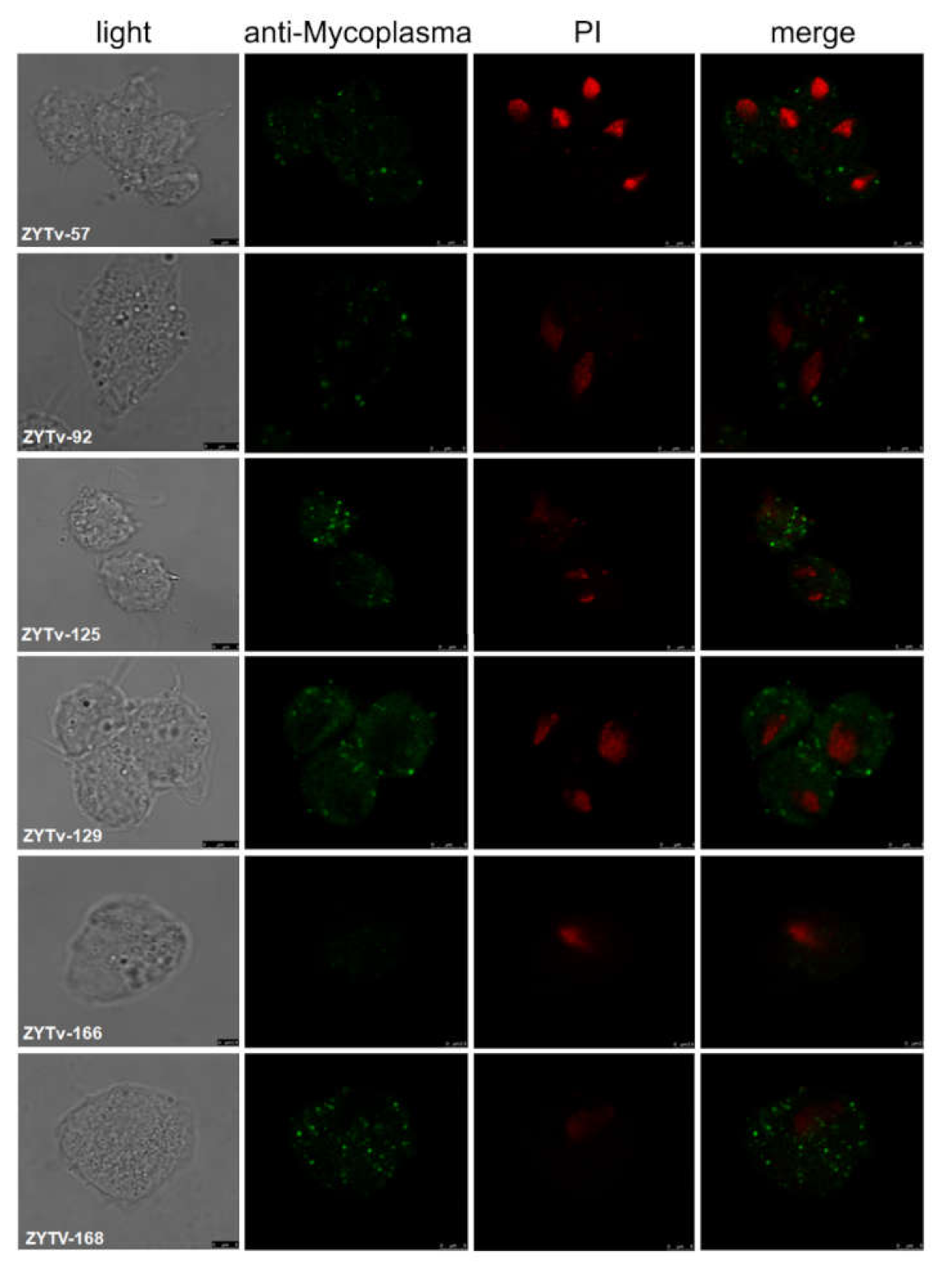
Indirect immunofluorescence assays were used to assess the location of Mycoplasma in Tv cells. Tv trophozoites were gently washed with phosphate-buffered saline (PBS), fixed with 4% paraformaldehyde in PBS, and permeabilized in 0.1% Triton X-100 (Amresco, Solon, OH, USA). Mycoplasma cells were detected by incubating with anti-M. hominis p120 monoclonal antibodies (H37S) (Thermo Fisher Scientific), followed by incubation with Alexa Fluor 488 goat anti-mouse IgG1 (H + L) antibodies (Abcam, Cambridge, UK). Confocal images were acquired using a confocal microscope (Leica TCS SP8; Leica, Wetzlar, Germany).
2.6. Data Analysis and Statistics
SPSS 21 statistical software was used to analyze epidemiological data. Associations of the positive rate of Tv with patient age and living area were analyzed using regression analysis. Tv infection and co-infection rates were analyzed using χ2 tests.
A BLAST analysis was performed against a local database. The database contained 60 Ca. M. girerdii 16S rRNA gene sequences, which were previously detected in vaginal specimens by us or by other researchers, as reported in the literature. All sequences were initially aligned using ClustalW multiple alignments in BioEdit software. A phylogenetic tree was established using the Test Neighbor-Joining Tree settings in MEGA-X software [15].
3. Results
3.1. Epidemiological Characteristics of Trichomonas Vaginalis
We collected vaginal swab specimens from women with pruritus vulvae and gray-white foamy leucorrhoea, which was suspected to be caused by Tv infection. In total, 94 Tv-positive samples were obtained from 312 patients with vaginitis, yielding a positive rate of 30.1% for Tv infection. Notably, patient’s age was found to play a significant role in the onset of Tv. Additionally, there were 105 patients with vaginitis and 44 (41.9%) patients positive for Tv infection between the ages of 17 and 30 years (p = 0.011; Table 2).
3.2. Co-Infection Rate of Tv and Various Mycoplasma
Sequence analysis of all Mycoplasma PCR products isolated from 312 samples revealed that 48 samples (15.4%) harbored Ca. M. girerdii and 153 (49%) samples harbored M. hominis. Notably, 40 samples (83.3%) that were positive for Ca. M. girerdii were also positive for Tv. Co-infections of M. hominis/Tv was detected in 52.3% (80/153) of cases. In all 218 Tv-negative samples, eight and 73 were positive for Ca. M. girerdii and M. hominis, respectively. This observation indicated that Ca. M. girerdii (p < 0.001) and M. hominis (p < 0.001) were significantly associated with Tv infection (Table 3).
3.3. Tv Gene Analysis
In this study, 18S rDNA gene sequences of six Zhangye Tv samples were sequenced and used in multiple sequences alignments. The Zhangye isolates of the sequenced Tv 18S rDNA gene were identical to those of the Henan isolates (KM603328, KM603336), the Philippines isolates (KM282385, JX943583), the Iran isolates (KX061402, KX061408), the USA isolate (EU215370), and the Vienna isolate (AY338476). These findings indicated that the Zhangye population of Tv parasites showed the same level of genetic diversity.
3.4. Ca. M. Girerdii Gene Analyses
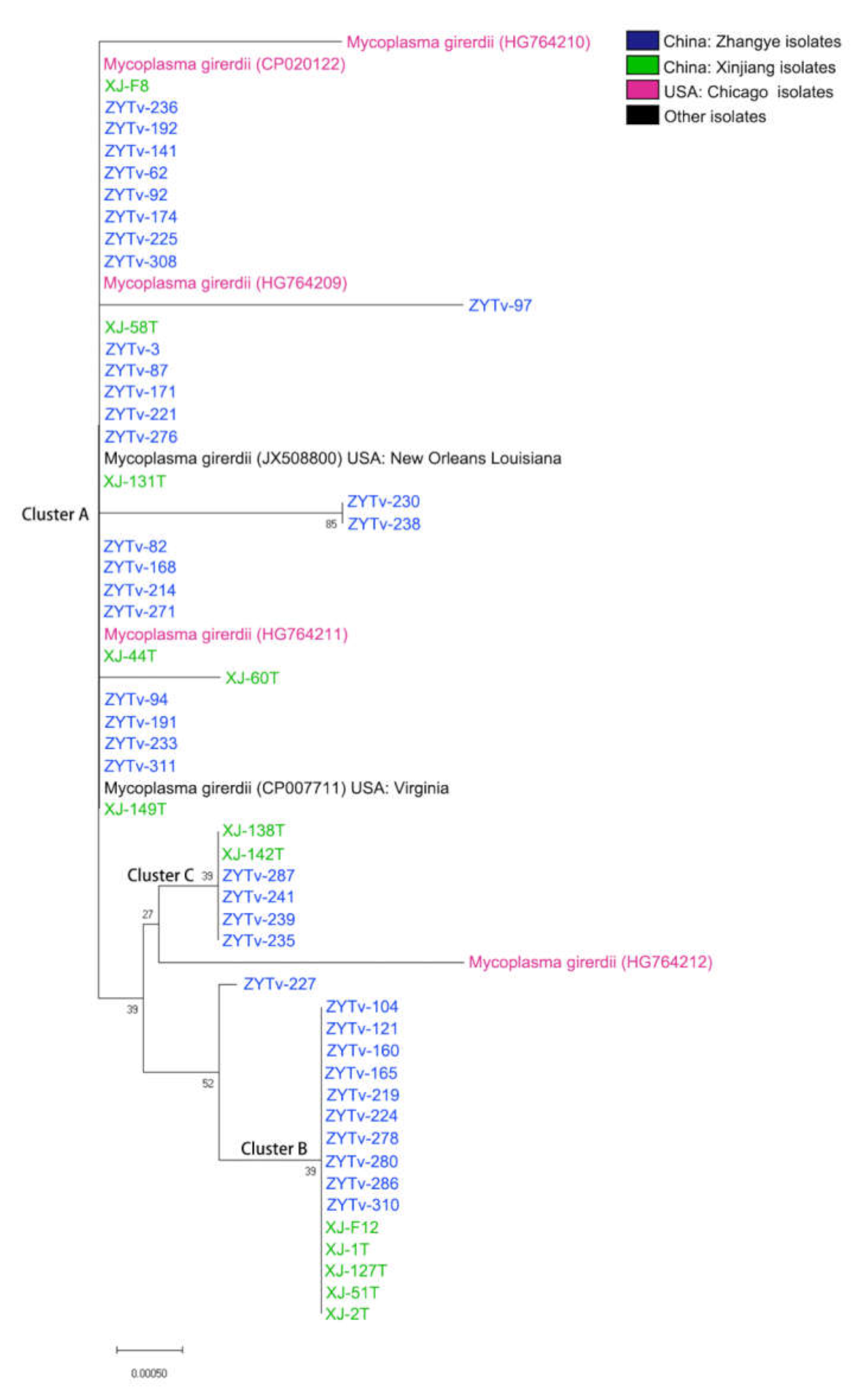
In this study, we obtained 48 Ca. M. girerdii 16S rDNA gene sequences. In total, 59 Ca. M. girerdii 16S rDNA gene sequences were used to generate a phylogenetic tree (Figure 1). The Ca. M. girerdii gene sequences included those from 39 Zhangye isolates and 13 Xinjiang isolates. The same gene sequence of seven isolates obtained from different sources could be divided into three clusters: A, B, and C. The Zhangye Ca. M. girerdii isolates are most closely associated with all genes that have been reported with similar genetic distances among isolates from Xinjiang (Figure 1). The Zhangye Ca. M. girerdii 16S rDNA gene sequences were submitted to the Genebank database (access number: LC554417 to LC554421).
3.5. Mycoplasma Localized in Tv Cells
In this study, six Tv isolates were successfully cultured. In addition, Mycoplasma symbiosis was detected by PCR (primers in Table 1) in these isolates at first and at every month for 6 months of continuous culture. One isolate, ZYTv-92, had both M. hominis and Ca. M. girerdii symbiosis. The classifications of Mycoplasma symbiosis for six isolates are listed in Table 4. The cellular localization of M. hominis in the trophozoites of Tv after 6 months of continuous culture was evaluated, and the results suggested that the clinical samples ZYTv-57, ZYTv-92, ZYTv-125, ZYTv-129, and ZYTv-168 exhibited M. hominis symbiosis (Figure 2).
4. Discussion
The estimated global prevalence of new trichomoniasis cases was 143 million among women ages 15–49 years in 2012 [2]. Moreover, Tv infection rates are relatively high (8.5%) in community samples from young women of reproductive age in India [16], and Tv infection prevalence has been estimated to be 0.5% among men and 1.8% among women aged 18–59 years in the USA, with the highest rates among black men (4.2%) and women (8.9%) [17]. Additionally, the prevalence of Tv infection is approximately 9.9% in rural China [18], and in Iran, the highest and lowest percentages of Tv infection in women were observed in patients 45–50 and 20–30 years old, respectively [19]. In this study, we found that the positive rate of Tv in patients with vaginitis was 30.1% in Zhangye, Gansu province, China; the high positive rate of Tv in this region may be related to the presence of vaginitis in the patients included in this study.
In our study, we found that Tv was highly prevalent in women between the ages of 17 and 30 years in our study area; this may be related to the increased sexual activity of women during this period [20]. A recent study found Tv to be significantly associated with having two or more sexual partners in the past year. The concurrency of sexual relationships and the number of sexual partners may differ by occupation, residence, and education level [21].
Multiple sequence comparison of the Tv 18S rDNA gene demonstrated that there was low genetic diversity among the Tv isolates. However, moderate-to-high genetic differentiation between isolates from different sites was observed in Tv isolates from Northwest China, which should be considered a single population. Moreover, the Tv gene from Zhangye isolates was mainly identical, and homology was similar to that in samples from the Philippines and Henan. These findings further confirmed that all Tv isolates originated from a common ancestor [14,22].
Studies of the symbiosis of pathogenic bacteria with protozoan vectors have provided insights into the symbiosis of organisms, although the exact mechanisms remain unknown. Indeed, the symbiotic relationship between Mycoplasma and Tv has attracted much attention [1], and researchers have been particularly interested in evaluating the symbiosis of M. hominis with Tv [12]. Symbiosis between Tv and M. hominis has been extensively studied and is observed in up to 90% of Tv clinical isolates in Europe [23]. Several groups used PCR to demonstrate the presence of M. hominis in Tv isolates of different geographic origins, with infection rates ranging from 5% to over 89% [1]. Moreover, Ca. M. girerdii has recently been shown to be associated with trichomonads identified in the vagina by researchers using next-generation sequencing [10]. In this study, M. hominis and Ca. M. girerdii were strongly associated with Tv in the examined population. Thus, Ca. M. girerdii may be another specific Mycoplasma species that can cause co-infections with Tv. Our results indicate that Ca. M. girerdii exhibits strong and unique associations with Tv. To the best of our knowledge, this is the first study to report the occurrence of Ca. M. girerdii in China.
Our findings were consistent with two previous reports on the prevalence of Ca. M. girerdii [10,11]. Accordingly, Tv may be the driving force behind infection with Ca. M. girerdii, and this transformation of the vaginal environment may enhance its causative force [12,24]. Notably, Ca. M. girerdii genome sequencing highlighted the presence of putative virulence genes, such as hemolysin, endopeptidase, and collagenase [25]. Ca. M. girerdii seems to lack gluconeogenesis and the tricarboxylic acid cycle, similar to other Mycoplasma species. Moreover, the arginine dihydrolase pathway and urease genes are absent from Ca. M. girerdii, implying that Ca. M. girerdii cannot utilize arginine and urea. This may be the reason for Ca. M. girerdii dependence on Tv [11,25].
The phylogenetic tree of Mycoplasma 16S rDNA gene sequences showing the phylogenetic position of the Zhangye Ca. M. girerdii gene sequence within all Mycoplasma gene sequences reported in GenBank demonstrated that the Ca. M. girerdii gene of Zhangye isolates was mainly concentrated in the same cluster. This species-specific relationship is most likely related to the genetic characteristics of M. genitalium and uncultured Mycoplasma spp. These results were consistent with studies of this uncultivated species, and the organism showed 85% identity with the closest human-associated species, M. genitalium [24].
5. Conclusions
Overall, our findings demonstrated a high co-infection rate between Tv and Mycoplasma in Zhangye, Gansu Provence, China. Ca. M. girerdii and M. hominis were the most closely related Mycoplasma species showing co-infections with Tv in Zhangye. These findings provide important insights and further our understanding of the impact of Ca. M. girerdii on the reproductive health of women. Additionally, Zhangye Ca. M. girerdiiisolates were most closely associated with strains from the USA and Xinjiang. Clarifying the reasons for the strong and unique association between Ca. M. girerdii and Tv is likely to have wider implications for the epidemiology and disease control of trichomoniasis.
Author Contributions
Conceptualization, X.C. and M.F.; methodology, S.X., Z.W., H.Z. and Y.F.; writing—original draft preparation, S.X. and M.F.; writing—review and editing, X.C. and M.F.; visualization, S.X. and M.F.; funding acquisition, X.C. All authors have read and agreed to the published version of the manuscript.
Funding
This research was funded by NSFC, grant number 81630057.
Institutional Review Board Statement
The Institutional Ethics Committees of Affiliated Zhangye People’s Hospital of Medical College of He Xi University approved the study protocol (Ethics approval no.: 20180703).
Informed Consent Statement
Informed consent was obtained from all subjects involved in the study.
Data Availability Statement
The data presented in this study are available on request from the corresponding author.
Conflicts of Interest
The authors declare no conflict of interest.
References
- Fichorova, R.; Fraga, J.; Rappelli, P.; Fiori, P.L. Trichomonas vaginalis infection in symbiosis with Trichomonasvirus and Mycoplasma. Res. Microbiol. 2017, 168, 882–891. [Google Scholar] [CrossRef] [PubMed]
- Rowley, J.; Hoorn, S.V.; Korenromp, E.; Low, N.; Unemo, M.; Abu-Raddad, L.J.; Chico, R.M.; Smolak, A.; Newman, L.; Gottlieb, S.; et al. Chlamydia, gonorrhoea, trichomoniasis and syphilis: Global prevalence and incidence estimates. Bull. World Health Organ. 2019, 97, 548–562P. [Google Scholar] [CrossRef] [PubMed]
- Ladefoged, A.S. Molecular dissection of Mycoplasma hominis. APMIS. Suppl. 2000, 97, 1–45. [Google Scholar] [CrossRef] [PubMed]
- Waites, K.B.; Katz, B.; Schelonka, R.L. Mycoplasmas and Ureaplasmas as Neonatal Pathogens. Clin. Microbiol. Rev. 2005, 18, 687–702. [Google Scholar] [CrossRef] [PubMed] [Green Version]
- Rappelli, P.; Addis, M.F.; Carta, F.; Fiori, P.L. Mycoplasma hominis parasitism of Trichomonas vaginalis. Lancet 1998, 352, 1286. [Google Scholar] [CrossRef]
- Hampl, V.; Vaňáčová, Š.; Kulda, J.; Flegr, J. Concordance between genetic relatedness and phenotypic similarities of Trichomonas vaginalis strains. BMC Evol. Biol. 2001, 1, 11. [Google Scholar] [CrossRef] [PubMed]
- Xiao, J.C.; Xie, L.F.; Fang, S.L.; Gao, M.Y.; Zhu, Y.; Song, L.Y.; Zhong, H.M.; Lun, Z.R. Symbiosis of Mycoplasma hominis in Trichomonas vaginalis may link metronidazole resistance in vitro. Parasitol. Res. 2006, 100, 123–130. [Google Scholar] [CrossRef] [PubMed]
- Butler, S.E.; Augostini, P.; Secor, W.E. Mycoplasma hominis infection of Trichomonas vaginalis is not associated with metronidazole-resistant trichomoniasis in clinical isolates from the United States. Parasitol. Res. 2010, 107, 1023–1027. [Google Scholar] [CrossRef] [PubMed]
- Fiori, P.L.; Diaz, N.; Cocco, A.R.; Rappelli, P.; Dessi, D. Association of Trichomonas vaginalis with its symbiont Mycoplasma hominis synergistically upregulates the in vitro proinflammatory response of human monocytes. Sex. Transm. Infect. 2013, 89, 449–454. [Google Scholar] [CrossRef] [PubMed]
- Martin, D.H.; Zozaya, M.; Lillis, R.A.; Myers, L.; Nsuami, M.J.; Ferris, M.J. Unique Vaginal Microbiota That Includes an Unknown Mycoplasma-Like Organism Is Associated with Trichomonas vaginalis Infection. J. Infect. Dis. 2013, 207, 1922–1931. [Google Scholar] [CrossRef]
- Fettweis, J.M.; Serrano, M.G.; Huang, B.; Brooks, J.P.; Glascock, A.L.; Sheth, N.U.; Strauss, J.F.; Jefferson, K.K.; Buck, G.A. Vaginal Microbiome Consortium an Emerging Mycoplasma Associated with Trichomoniasis, Vaginal Infection and Disease. PLoS ONE 2014, 9, e110943. [Google Scholar] [CrossRef] [Green Version]
- Ioannidis, A.; Papaioannou, P.; Magiorkinis, E.; Magana, M.; Ioannidou, V.; Tzanetou, K.; Burriel, A.R.; Tsironi, M.; Chatzipanagiotou, S. Detecting the Diversity of Mycoplasma and Ureaplasma Endosymbionts Hosted by Trichomonas vaginalis Isolates. Front. Microbiol. 2017, 8, 1188. [Google Scholar] [CrossRef]
- Conrad, M.D.; Gorman, A.W.; Schillinger, J.A.; Fiori, P.L.; Arroyo, R.; Malla, N.; Dubey, M.L.; Gonzalez, J.; Blank, S.; Secor, W.E.; et al. Extensive Genetic Diversity, Unique Population Structure and Evidence of Genetic Exchange in the Sexually Transmitted Parasite Trichomonas vaginalis. PLoS Negl. Trop. Dis. 2012, 6, e1573. [Google Scholar] [CrossRef] [Green Version]
- Liu, J.; Feng, M.; Wang, X.; Fu, Y.; Ma, C.; Cheng, X. Unique Trichomonas vaginalis gene sequences identified in multinational regions of Northwest China. Biosci. Trends 2017, 11, 303–307. [Google Scholar] [CrossRef] [Green Version]
- Megarani, D.; Nugroho, H.A.; Andarini, Z.P.; Surbakti, Y.D.; Widayanti, R. Genetic characterization and phylogenetic study of Indonesian indigenous catfish based on mitochondrial cytochrome B gene. Vet. World 2020, 13, 96–103. [Google Scholar] [CrossRef] [Green Version]
- Madhivanan, P.; Bartman, M.T.; Pasutti, L.; Krupp, K.; Arun, A.; Reingold, A.L.; Klausner, J.D. Prevalence of Trichomonas vaginalis infection among young reproductive age women in India: Implications for treatment and prevention. Sex. Health 2009, 6, 339–344. [Google Scholar] [CrossRef] [Green Version]
- Patel, E.U.; Gaydos, A.C.; Packman, Z.R.; Quinn, T.C.; Tobian, A.A.R. Prevalence and Correlates of Trichomonas vaginalis Infection Among Men and Women in the United States. Clin. Infect. Dis. 2018, 67, 211–217. [Google Scholar] [CrossRef] [Green Version]
- Feng, R.-M.; Wang, M.Z.; Smith, J.S.; Dong, L.; Chen, F.; Pan, Q.-J.; Zhang, X.; Qiao, Y.-L.; Zhao, F.-H. Risk of high-risk human papillomavirus infection and cervical precancerous lesions with past or current trichomonas infection: A pooled analysis of 25,054 women in rural China. J. Clin. Virol. 2018, 99–100, 84–90. [Google Scholar] [CrossRef]
- Nikpay, S.; Otaghi, M.; Azami, M.; Karimi, M.; Abdi, J. Trichomonas Vaginalis Infection Among Women Attending Laboratory Centers in Ilam, Iran. Infect. Disord. Drug Targets 2020, 20, 98–101. [Google Scholar] [CrossRef]
- Buvé, A.; Weiss, H.A.; Laga, M.; Van Dyck, E.; Musonda, R.; Zekeng, L.; Kahindo, M.; Anagonou, S.; Morison, L.; Robinson, N.J.; et al. The epidemiology of trichomoniasis in women in four African cities. AIDS 2001, 15, S89–S96. [Google Scholar] [CrossRef]
- Field, N.; Clifton, S.; Alexander, S.; Ison, A.C.; Khanom, R.; Saunders, P.; Hughes, G.; Heath, L.; Beddows, S.; Mercer, C.; et al. Trichomonas vaginalis infection is uncommon in the British general population: Implications for clinical testing and public health screening. Sex. Transm. Infect. 2016, 94, 226–229. [Google Scholar] [CrossRef] [PubMed] [Green Version]
- Ibáñez-Escribano, A.; Nogal-Ruiz, J.J.; Arán, V.J.; Escario, J.A.; Gómez-Barrio, A.; Alderete, J. Determination of internal transcribed spacer regions (ITS) in Trichomonas vaginalis isolates and differentiation among Trichomonas species. Parasitol. Int. 2014, 63, 427–431. [Google Scholar] [CrossRef] [PubMed] [Green Version]
- Dessi, D.; Delogu, G.; Emonte, E.; Catania, M.R.; Fiori, P.L.; Rappelli, P. Long-Term Survival and Intracellular Replication of Mycoplasma hominis in Trichomonas vaginalis Cells: Potential Role of the Protozoon in Transmitting Bacterial Infection. Infect. Immun. 2005, 73, 1180–1186. [Google Scholar] [CrossRef] [PubMed] [Green Version]
- Costello, E.K.; Sun, C.L.; Carlisle, E.M.; Morowitz, M.J.; Banfield, J.F.; Relman, D.A. Candidatus Mycoplasma girerdii replicates, diversifies, and co-occurs with Trichomonas vaginalis in the oral cavity of a premature infant. Sci. Rep. 2017, 7, 1–11. [Google Scholar] [CrossRef]
- Dessi, D.; Margarita, V.; Cocco, A.R.; Marongiu, A.; Fiori, P.L.; Rappelli, P. Trichomonas vaginalis and Mycoplasma hominis: New tales of two old friends. Parasitology 2019, 146, 1150–1155. [Google Scholar] [CrossRef]
Figure 1.
NJ tree analysis of 16S rDNA gene sequences showing the phylogenetic position of the Zhangye Ca. M. girerdii gene se-quence within all Ca. M. girerdii gene sequences reported in GenBank (Xinjiang samples were collected by Liu Jun in our laboratory). The tree was derived using 16S rDNA gene sequences (approximately 1200 nucleotides) obtained from GenBank. Original values are shown at the branch points. Gene accession numbers and sample source locations are shown after the organism names included in the tree.

Figure 2.
The cellular localization of M. hominis in Tv isolates. Four panels represent the same area of a protozoan monolayer immunostained as described in Materials and Methods. Light: surface image of Tv; anti-Mycoplasma: FITC fluorescence showing intracellular M. hominis (green); PI: images of the nucleus of Tv after PI staining (red); merge: superimposed panels of anti-M. hominis and PI.


Table 1.
Primers used in this study.
| Gene or Locus | Sequence (5′–3′) | Product (bp) | AT (°C) |
|---|---|---|---|
| Tv18S | ATCAGAGGCACGCCATTC | 580 | 60 |
| Tv18AS | CGCCCTTGATCGACAGAA | ||
| Mh16S2S | GGCTAATGCCGGATACGC | 330 | 60 |
| Mh16S2S | GGTACCGTCAGTCTGCAAT | ||
| Mg16S-423F | TTTATTAGGGACGAACGGCACT | 313 | 55 |
| Mg16S-736R | CAGTTGTGACCTAAGTTCTCGC | ||
| Mg207F | TTTAGGATGAGGGTGCGGTTTA | 1201 | 55 |
| Mg1408R | TAGCAGGACGGTTTTAGGTATT |
Table 2.
Age distribution of Trichomonas vaginalis infection among all patients with vaginitis.
| Age (years) | Total Patients (n = 312) | Patients with Tv Infection (n = 94) (30.1%) | p-Value | OR (95% CI) |
|---|---|---|---|---|
| 17–30 | 105 | 44 (41.9%) | 0.011 | 3.275 (1.319–8.134) |
| 31–40 | 62 | 12 (19.4%) | 0.781 | 1.159 (0.410–3.274) |
| 41–50 | 106 | 31 (29.2%) | 0.128 | 2.054 (0.813–5.189) |
| 51–75 | 39 | 7 (17.9%) | 1 |
Abbreviations: CI, confidence interval; OR, odds ratio.
Table 3.
Characteristics of Tv+ women (cases) and Tv—women (controls).
| Total Samples (n = 312) | ||||
|---|---|---|---|---|
| Mycoplasma Positive | χ2 p-Value | |||
| Mycoplasma positive rate | Tv positive (n = 94) | Tv negative (n = 218) | ||
| Ca. M. girerdii | 48 (15.4%) * | 40 (83.3%) ** | 8 | <0.001 |
| M. hominis | 153 (49%) * | 80 (52.3) ** | 73 | <0.001 |
*: Percentage in total samples; **: Percentage in Mycoplasma-positive samples.
Table 4.
Mycoplasma-symbiotic (+) and Mycoplasma-free (−) in Tv isolates.
| Tv Isolates | Type of Mycoplasma Symbiosis | |
|---|---|---|
| M. hominis | Ca. M. Girerdii | |
| ZYTv-166 | − | − |
| ZYTv-57 | + | − |
| ZYTv-92 | + | + |
| ZYTv-125 | + | − |
| ZYTv-129 | + | − |
| ZYTv-168 | + | − |
Publisher’s Note: MDPI stays neutral with regard to jurisdictional claims in published maps and institutional affiliations. |
© 2021 by the authors. Licensee MDPI, Basel, Switzerland. This article is an open access article distributed under the terms and conditions of the Creative Commons Attribution (CC BY) license (https://creativecommons.org/licenses/by/4.0/).
Share and Cite
MDPI and ACS Style
Xu, S.; Wang, Z.; Zhou, H.; Fu, Y.; Feng, M.; Cheng, X. High Co-Infection Rate of Trichomonas vaginalis and Candidatus Mycoplasma Girerdii in Gansu Province, China. Healthcare 2021, 9, 706. https://doi.org/10.3390/healthcare9060706
AMA Style
Xu S, Wang Z, Zhou H, Fu Y, Feng M, Cheng X. High Co-Infection Rate of Trichomonas vaginalis and Candidatus Mycoplasma Girerdii in Gansu Province, China. Healthcare. 2021; 9(6):706. https://doi.org/10.3390/healthcare9060706
Chicago/Turabian StyleXu, Shuhui, Zhixin Wang, Hang Zhou, Yongfeng Fu, Meng Feng, and Xunjia Cheng. 2021. "High Co-Infection Rate of Trichomonas vaginalis and Candidatus Mycoplasma Girerdii in Gansu Province, China" Healthcare 9, no. 6: 706. https://doi.org/10.3390/healthcare9060706
Note that from the first issue of 2016, this journal uses article numbers instead of page numbers. See further details here.