Brief Report

The Use of the Shikani Video-Assisted Intubating Stylet Technique in Patients with Restricted Neck Mobility

1
Department of Anesthesia, Hualien Tzuchi Hospital, Hualien 97002, Taiwan
2
Bio-Math Laboratory, Department of Financial Engineering, Providence University, Taichung 43301, Taiwan
3
Department of Anesthesia, Critical Care and Pain Medicine, Massachusetts General Hospital, Harvard Medical School, Boston, MA 02114, USA
4
Division of Otolaryngology-Head and Neck Surgery, LifeBridge Sinai Hospital, Baltimore, MD 21218, USA
5
Division of Otolaryngology-Head and Neck Surgery, MedStar Union Memorial Hospital, Baltimore, MD 21218, USA
*
Author to whom correspondence should be addressed.
Healthcare 2022, 10(9), 1688; https://doi.org/10.3390/healthcare10091688
Submission received: 8 June 2022 / Revised: 1 September 2022 / Accepted: 1 September 2022 / Published: 4 September 2022
(This article belongs to the Special Issue All-Time Paradigm Shift of Anesthesia Management and Pain Medicine)

Abstract

:
Among all the proposed predictors of difficult intubation defined by the intubation difficulty scale, head and neck movement (motility) stands out and plays as a crucial factor in determining the success rate and the degree of ease on endotracheal intubation. Aside from other airway tools (e.g., supraglottic airway devices), optical devices have been developed and applied for more than two decades and have shown their superiority to conventional direct laryngoscopes in many clinical scenarios and settings. Although awake/asleep flexible fiberoptic bronchoscopy is still the gold standard in patients with unstable cervical spines immobilized with a rigid cervical collar or a halo neck brace, videolaryngoscopy has been repeatedly demonstrated to be advantageous. In this brief report, for the first time, we present our clinical experience on the routine use of the Shikani video-assisted intubating stylet technique in patients with traumatic cervical spine injuries immobilized with a cervical stabilizer and in a patient with a stereotactic headframe for neurosurgery. Some trouble-shooting strategies for this technique are discussed. This paper demonstrates that the video-assisted intubating stylet technique is an acceptable alternative airway management method in patients with restricted or confined neck motility.

1. Introduction

Direct laryngoscopy, the most commonly employed method of tracheal intubation, creates some degree of cervical spine extension while aligning the oropharyngeal and laryngeal axes. The sniffing position, traditionally used during tracheal intubation, involves the near-full extension of the atlanto-occipital and atlanto-axial joints and the flexion of the lower cervical spine [1]. This may present an issue during the endotracheal intubation of trauma patients with cervical spine (C-spine) injuries and an unstable cervical spine, even more so if the situation is also complicated by a difficult airway. A variety of intubation devices have been used to assist in the direct endotracheal tube placement with minimal manipulation of the cervical spine. Videolaryngoscopy (VL) using a GlideScope has been proposed to be safer and more effective than direct laryngoscopy (DL) using a Macintosh blade in patients with limited C-spine mobility [2,3,4,5,6,7,8,9,10,11,12]. The GlideScope has some innate shortcomings, however; it still often involves some degree of C-spine movement, and the intubation time when using the GlideScope is longer than that when using DL, i.e., 53 s vs. 24 s [13]. Other airway devices have also been successfully used in patients with restricted or confined C-spine injuries, including supraglottic airway devices, gum elastic bougies, and rigid endoscopes [14,15,16]. Flexible fiberoptic laryngoscopes and/or bronchoscopes remained as the gold standard for suspected or confirmed C-spine injuries for many years [17,18,19,20,21]. The pros and cons of the available airway management tools for patients with a potential cervical spine injury have been reviewed [1]. It was not until the introduction of optical stylets in the 1980s and 1990s that newer and safer methods of intubation were introduced, including the Shikani optical stylet and the Bonfils stylet, which are associated with a significantly lesser extension at C1–C2 and C2–C3, as compared to intubation by direct laryngoscopy [16,22,23,24,25,26].
Our institutions have extensive experience with the Shikani optical stylet, which has been widely studied in the management of a variety of difficult airways [27,28,29,30] and which has also proved effective in clinical conditions related to C-spine motility [31,32,33,34,35,36,37,38,39,40,41]. In this report, we present our experience with the Shikani video-assisted intubating stylet technique (Figure 1) as the routine airway management modality for patients with restricted head/neck mobility.

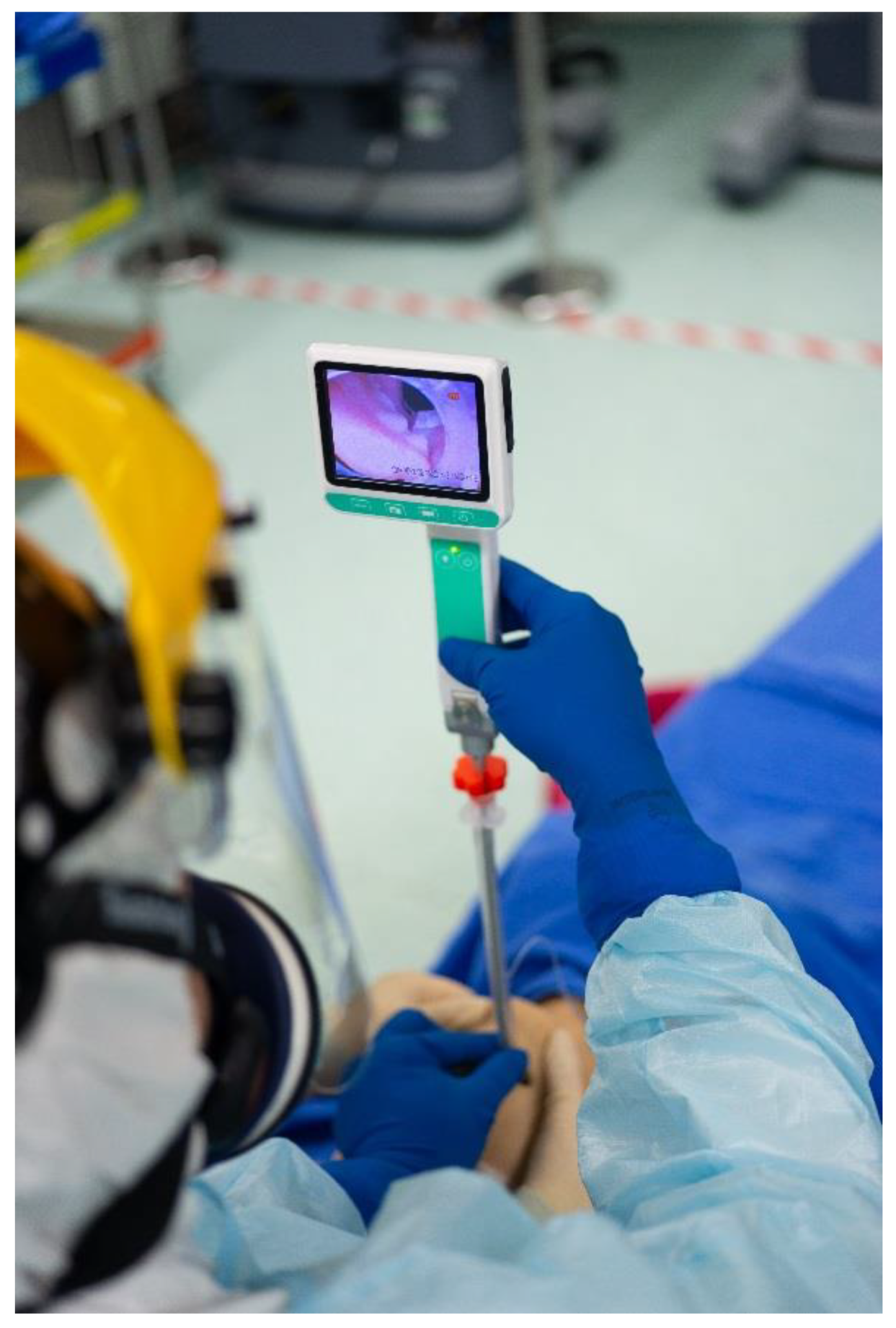
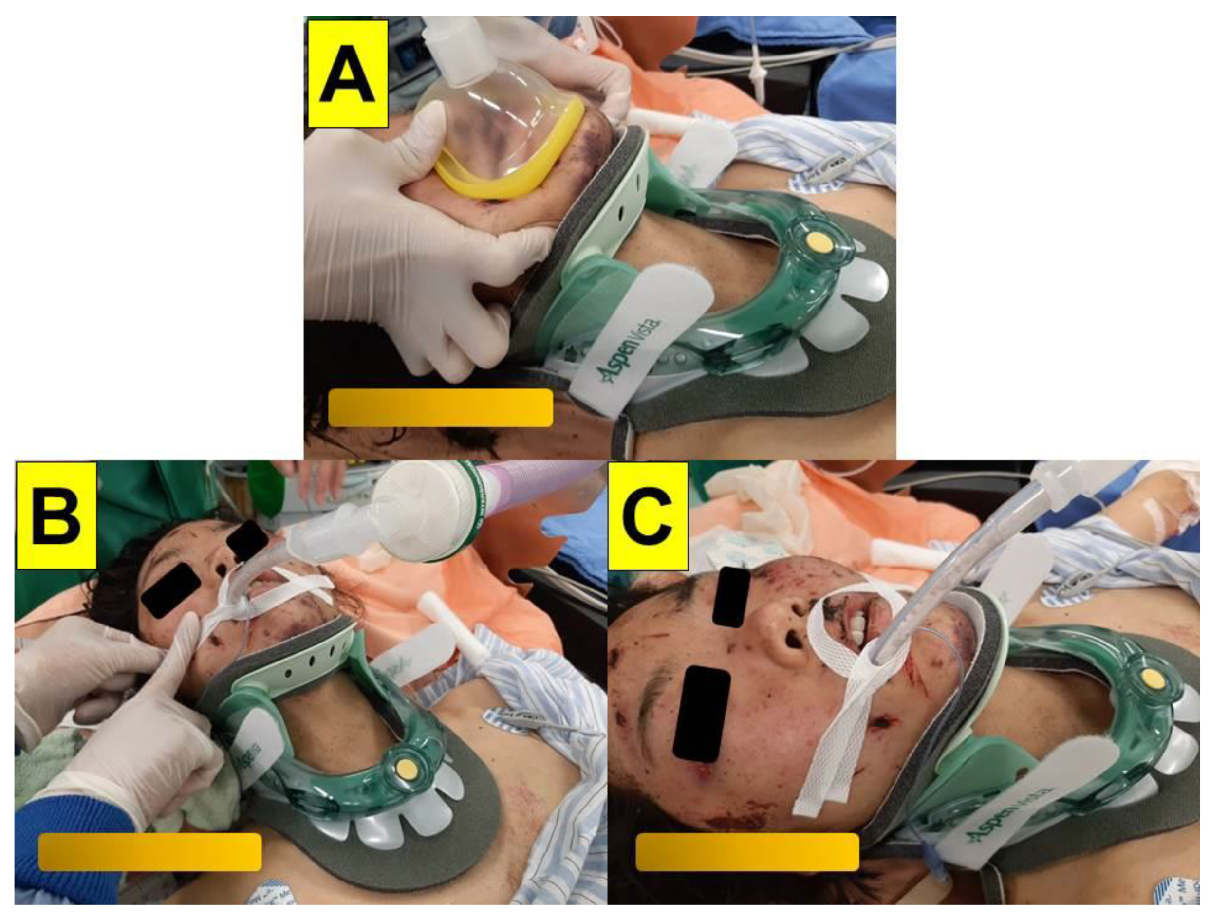
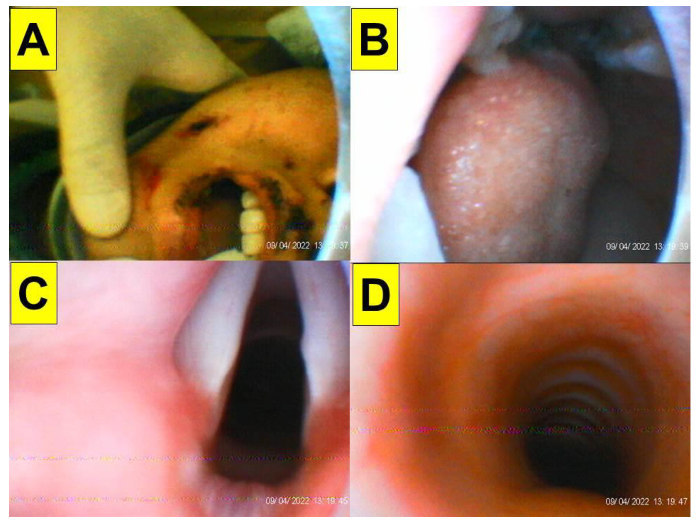
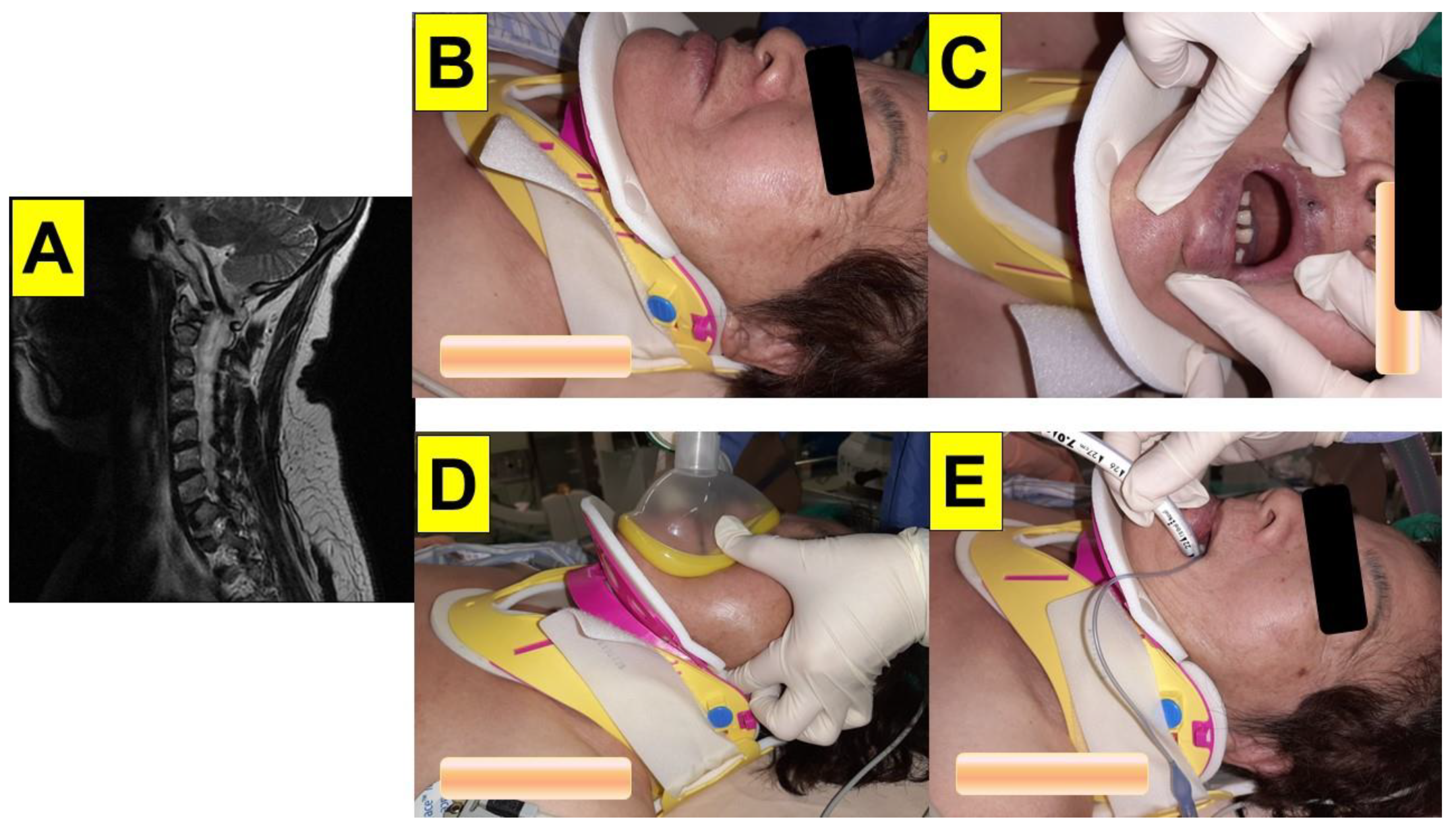
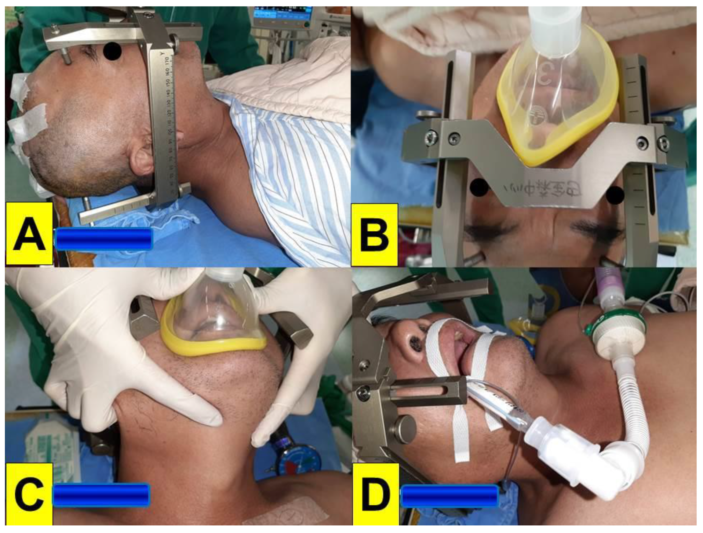
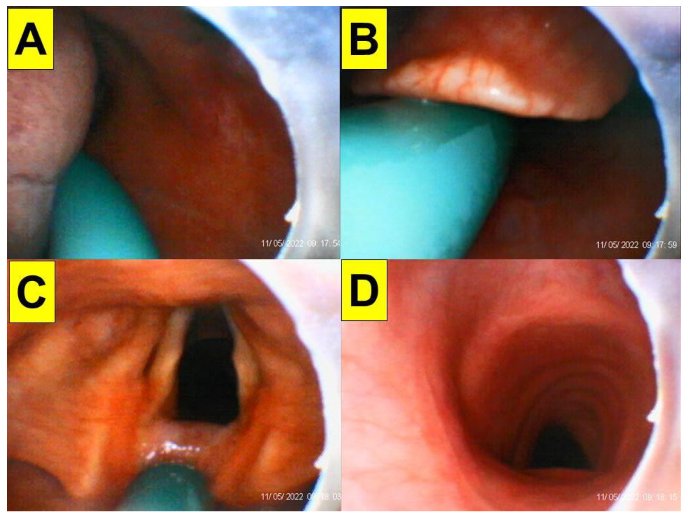
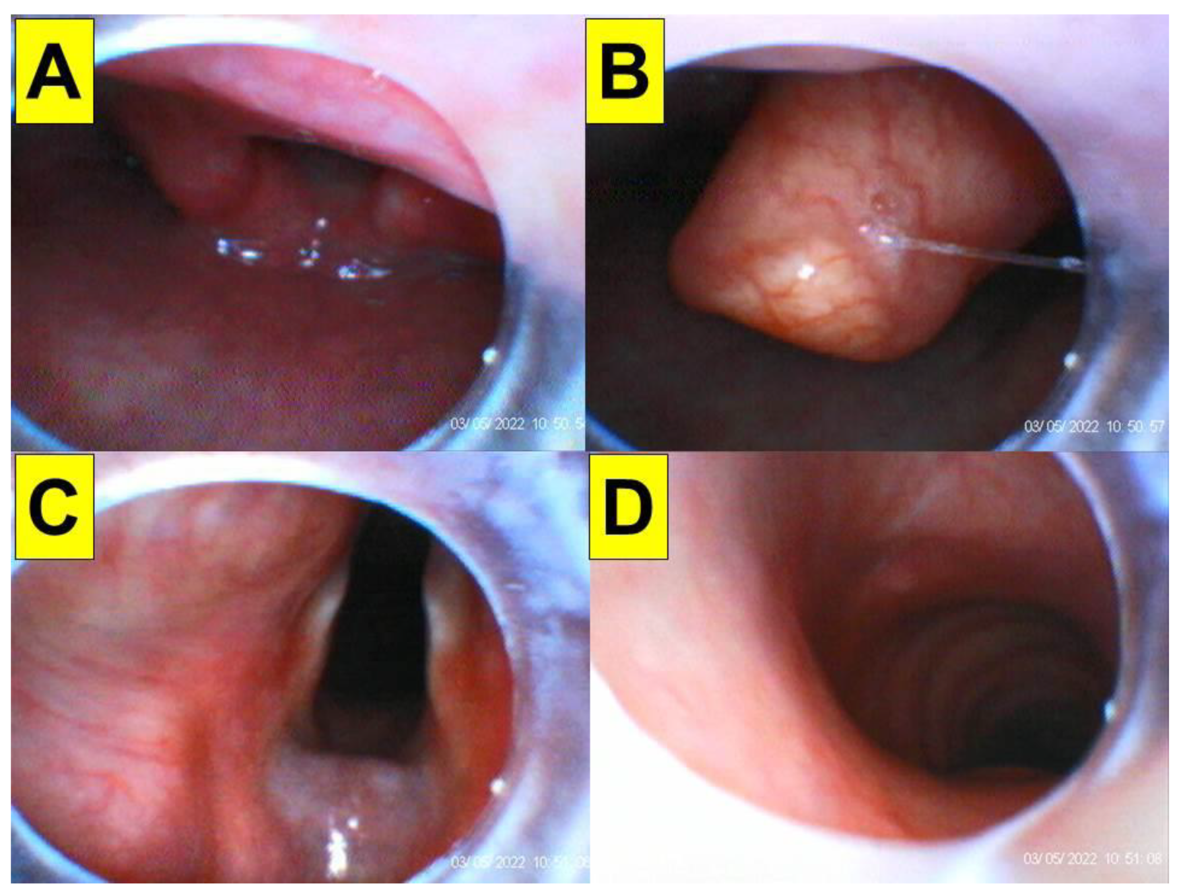
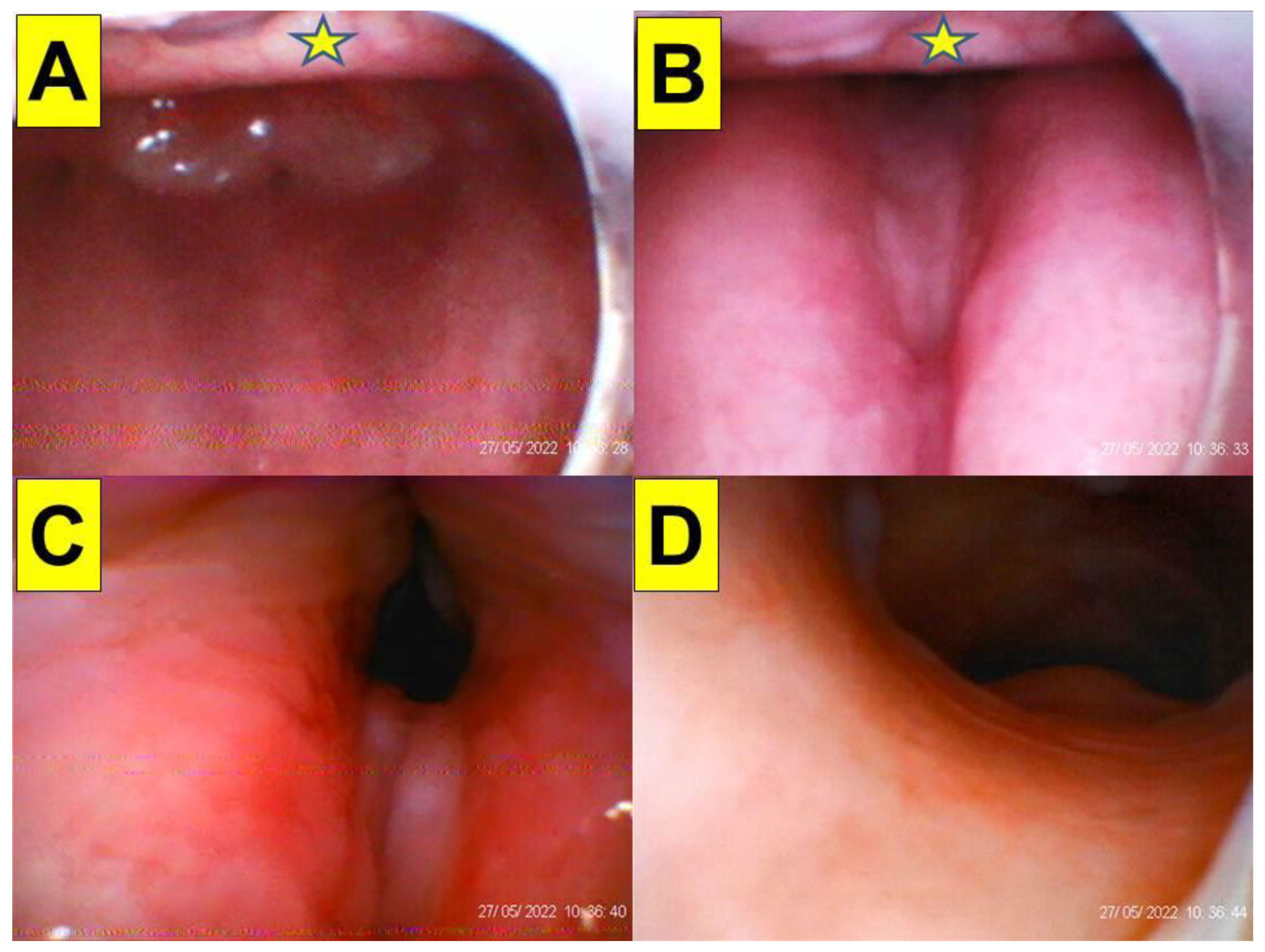
2. Case Presentations

Case 1 (multiple-trauma case with rigid cervical collar immobilization): A 30-year-old female (height 170 cm, weight 68 kg, BMI 23.5 kg/m2) with no underlying disease was admitted to the emergency department due to multiple trauma. She fell from a 70 m-high cliff while climbing a mountain two days before. She sustained a right ankle open fracture with dislocation, a 5th cervical spine lamina fracture, a 3rd lumbar spine fracture, a left distal radius fracture, and a right ulnar shaft fracture. She was taken to the operating room for emergency external skeletal fixation (ESF) with debridement of the right ankle open fracture. The patient had a cervical collar that limited the neck mobility, and mouth opening was thee fingers wide (Figure 2). Minimal movement of the cervical spine during airway intervention and positioning of the patient to avoid further spine injury were made with caution. Standard vital-sign monitoring with electrocardiography (ECG), arterial blood pressure, pulse oximeter (SpO2), peripheral nerve stimulator (ToF), and end-tidal CO2 (EtCO2) were applied. The modified rapid sequence induction technique was adopted with adequate pre-oxygenation, glycopyrrolate (0.2 mg), lidocaine (20 mg), propofol (2.5 mg/kg), and high-dose rocuronium (1.0 mg/kg). Cricoid pressure was not applied during apneic face mask oxygenation. Endotracheal intubation was performed using the video-assisted intubating stylet technique. The airway was then secured swiftly and accurately (from the lip to the trachea, 7 s, Figure 3). Anesthesia was maintained with inhalational anesthetic and rocuronium. The whole anesthesia was uneventful, and the patient was extubated post-operatively.
Case 2 (multiple trauma with rigid cervical collar immobilization): A 71-year-old female patient (height 160 cm, weight 70 kg, BMI 27.3 kg/m2, ASA class IIE) presented to the emergency department due to a traffic accident. Her past medical history included severe osteoporosis, neck pain, and left arm paresthesia. A radiographic work-up revealed fractures of the left distal tibial and fibular shafts. No apparent fracture of the cervical spine was noted, but severe degenerative joint disease with neuroforaminal stenosis and spinal nerve root impingement at the 5th/6th cervical vertebrae were present. The patient was scheduled for left ankle open reduction and internal fixation. She presented to the operating room with a cervical collar, and the range of motion of the neck was limited (Figure 4). The patient opened her mouth with limited vertical opening (two fingers wide). Minimal movement of the cervical spine during airway intervention and positioning of the patient was made with caution to avoid further spine injury. Standard vital-sign monitoring with electrocardiography (ECG), arterial blood pressure, pulse oximeter (SpO2), peripheral nerve stimulator (ToF), and end-tidal CO2 (EtCO2) were applied. General anesthesia was induced with glycopyrrolate, lidocaine, fentanyl, propofol, and rocuronium. She was intubated using a video-assisted intubating stylet (Figure 5). It took 40 s (from the lip to the trachea) to complete the intubating process without the jaw-thrust maneuver.
Case 3 (stereotactic neurosurgery with deep-brain stimulation): A 57-year-old male (height 169.9 cm, weight 92.7 kg, BMI 32.0 kg/m2) with a history of hypertension, type 2 diabetes mellitus, hyperlipidemia, gout, and Parkinson’s disease was scheduled to undergo a stereotactic implantation of deep-brain stimulation electrodes under general anesthesia. A Leksell G-frame (Elekta Instrument Inc., Norcross, GA, USA) was applied for the stereotactic procedure. The head frame was secured on a Mayfield adaptor, and the target coordinates were applied to the stereotactic frame and the working stage. The head frame for stereotactic neurosurgery allows the precise localization of the desired target in the central nervous system. Such frame mounting is performed under local anesthesia. After mounting the frame in several potential clinical scenarios, magnetic resonance imaging (MRI) and stereotactical computed tomography (CT) were carried out. The patient was then sent to the operating room with a stereotactic frame on the head (Figure 6). Standard monitoring with electrocardiography (ECG), arterial blood pressure (NIBP), pulse oximeter (SpO2), peripheral nerve stimulator (ToF), and end-tidal CO2 (ETCO2) were applied. General anesthesia was induced with fentanyl, propofol, and rocuronium. Endotracheal intubation using DL would not have been possible due to the head frame. Intubation was hence performed using a video-assisted intubating stylet (Figure 6 and Figure 7). An assistant facilitated the intubating procedure with the jaw thrust maneuver. It took 20 s (from the lip to the trachea) to complete the intubating process. The anesthesia was maintained with remifentanil or alfentanil and sevoflurane. The entire deep-brain stimulation (DBS) procedure lasted for 5 h. The patient was extubated at the end of the procedure. The details of the endotracheal intubation in the presence of a stereotactic headframe are shown in Figure 6 and Figure 7 and in the Supplementary Materials, Video S4.

3. Discussion

In this case series, we demonstrated the use of the Shikani video-assisted intubating stylet technique in patients with restricted neck extension. The Shikani Optical Stylet (SOS; Clarus Medical, Minneapolis, MN, USA) was firstly evaluated in 1999 in a cohort of 120 adults and children scheduled for ENT procedures [24]. It has been extensively used since and proved to be invaluable in the management of a variety of difficult airways. It has also been reported that it is associated with less cervical spine motion than direct laryngoscope (DL) using a Macintosh blade [26]. The Shikani stylet was found to be safe and effective in patients undergoing surgery for cervical spondylosis, when compared with Macintosh laryngoscopy, especially beneficial in the setting of a difficult airway [36]. Awake tracheal intubation with the Shikani stylet was found as equally successful and faster as awake intubation with a flexible fiberscope in patients with unstable cervical spine [36]. To our knowledge, this is the first case report in the literature describing the use of optical stylets during stereotactic brain surgery (Figure 6 and Figure 7). The stereotactic headframe used to stabilize the head and neck during stereotactic brain surgery makes accessing the airway a challenge, and conventional DL is of limited use in such scenarios [42,43]. Few studies proposed alternative airway management modalities, including using a flexible FOB, an intubating laryngeal mask airway (ILMA), a gum elastic bougie, a lightwand (e.g., Trachlight™), and videolaryngoscopy (VL) in some difficult airway scenarios [44,45,46,47,48]. The airway operators should know in advance that the in-place headframe may make it difficult to access the upper airway and thus have a contingency plan to handle the airway, should the need unexpectedly arise. An Allen wrench should always be available to immediately remove the crossbars of the stereotactic headframe, if needed.
A mannequin study showed that intubating with VL under an ideal situation for the Leksell front bar in situ is possible [48]. To remove just the front bar is a simpler and more streamlined approach to provide a quicker airway control in case of emergency. A mannequin study was also conducted to determine the ease of emergency airway management with a stereotactic headframe in situ [47]. Using an LMA to rescue the airway was a little quicker than using DL and VL. More than 300 DBS operations were performed by neurosurgeons at our institution in the last 10 years. The video-assisted intubating stylet (VS) technique was our preferred method for tracheal intubation in these general anesthesia cases. In our experience, the presence of the stereotactic headframe in situ has never become a barrier or a problem when using such intubation technique (Figure 7 and Supplementary Materials, Video S4).
In patients with cervical spine injuries, airway manipulation (e.g., chin lift, jaw thrust, head extension, DL) may cause segmental spine motion (rotation or displacement) and worsen the cervical instability. Flexible fiberoptic-guided nasal tracheal intubation has been reported to minimize the need of moving the cervical spine [49,50]. When compared with DL using the Macintosh blade, VL was shown to cause less upper cervical spine motion [7]. Various VL products have been compared in such restricted C-spine motion scenario, evaluating success rate, glottis visualization, intubation time, and complications [9]. VL was also found to be advantageous in facilitating nasotracheal intubation in patients with simulated C-spine injuries [51,52], and VL produced a better glottic visualization during intubation under general anesthesia with neuromuscular blockade and manual in-line stabilization. On the other hand, when dealing with a non-pathologic C-spine, the difference between the two as regards spine movement was not significant [53].
The Shikani video-assisted intubating stylet (VS) technique provides an advantage when intubating such patients with limited head/neck motion [26]. Several studies have been conducted to compare this technique with VL (using the Airway Scope®) in patients with cervical spine immobilization (restricted by a hard cervical collar or manual in-line stabilization) [41]. When first-attempt success, overall success, intubation time, and tissue damage were used as outcome indicators, VS and VL were comparable [41]. Inconsistent study results exist, however. Although both VL (Airway Scope®) and VS (StyletScope™) have high success rates in a simulated difficult airway achieved by a rigid collar, VL is faster and less likely to cause esophageal intubation [54]. VL has a higher first-attempt success rate for tracheal intubation and a shorter intubation time than VS under the condition of cervical spine manual inline stabilization [34]. On the contrary, VS produces less cervical spine motion than VL during tracheal intubation in patients with simulated cervical immobilization [33]. This finding is consistent with an earlier report that VS (Shikani stylet, Clarus Video System-Trachway®) was comparable to VL (Airway Scope®) in the success rate for tracheal intubation but provided a faster and easier intubation in patients with cervical collars [40]. Similarly, VS has been demonstrated to be a viable alternative to VL in patients with rigid cervical collars simulating a difficult airway [37]. Numerous studies have been conducted to compare various airway tools under different scenarios (Table 1). In our institution, we conduct about 600 C-spines surgeries annually, and the VS technique is used in the great majority of cases.
The cases in this report indicate that VS has a high first-attempt success rate, is smooth, and allows for a short intubating time, with no significant soft tissue injury in patients with a stereotactic headframe (Figure 6 and Figure 7). Similarly, this is also true when VS is applied in patients with a rigid cervical collar, since we observed little if any, risky movement of the cervical spine (Figure 2, Figure 3, Figure 4 and Figure 5). This finding is consistent with experiences described in our earlier clinical reports when we did not have any concerns of restricted neck motility [27,28,29,30]. Although external laryngeal manipulation might be helpful for VL in patients with C-spine immobilization [62], such maneuver is not necessary when using VS. Usually, sizable mouth opening and optimal jaw thrust are enough for maneuvering the intubating stylet into the oropharyngeal and laryngeal inlets and then obtaining sound glottis visualization. It should be noted that in some patients in which neck manipulation (e.g., extension/flexion, jaw-thrust, or laryngoscopy) cannot be performed, the epiglottis may just lie down against the posterior pharyngeal wall (an example is shown in Figure 5). In such condition, there are several strategies can be tried. Strategy 1: Pushing the intubating stylets underneath the epiglottis before directing it anteriorly. This would be easier if the patient has an omega-shaped epiglottis (e.g., Figure 8) or if there is minimal space underneath the epiglottis (e.g., Figure 9). In that case, the stylet could pass directly into the glottis under the epiglottis. Strategy 2: Maneuvering the tip of the stylet first to the right or to the left edge of the epiglottis and then sweeping the stylet backward to the midline and subsequently anteriorly into the glottis area. Strategy 3: Directing the stylet into the right or left piriform fossa and then retracting it back in the midline position. Strategy 4: Directing the tip of the stylet towards the mid post-cricoid area and then withdrawing it until the posterior glottis comes into view. Similar trouble-shooting suggestions and tips have also been previously reported for the Bonfils stylet [22].
The advantages and disadvantages of various airway management options for a patient with restricted cervical spine mobility are summarized in the Table 2. The airway operators should always remember to follow the clinical airway management guidelines when a difficult airway is anticipated. If the patient’s head/neck motility is severely restricted, and difficult tracheal intubation is expected, awake/asleep FOB under adequate topical local anesthesia can always be considered. However, this paper demonstrates that the video-assisted intubating stylet technique is an acceptable alternative airway management method in patients with restricted or confined neck motility.

Supplementary Materials

The following supporting information can be downloaded at: https://www.mdpi.com/article/10.3390/healthcare10091688/s1, Video S1: Intubation process in a mannequin; Video S2: Intubation process in Case 1; Video S3: Intubation process in Case 2; Video S4: Intubation process in Case 3; Video S5: Intubation process in the case of Figure 8; Video S6: Intubation process in the case of Figure 9.

Author Contributions

Conceptualization, H.-N.L., J.Z.Q. and A.S.; methodology, H.-N.L., T.-L.S., K.-P.K. and C.-Y.H.; validation, T.-L.S., K.-P.K. and C.-Y.H.; formal analysis, H.-N.L.; investigation, H.-N.L.; resources, H.-N.L.; data curation, H.-N.L., T.-L.S., K.-P.K. and C.-Y.H.; writing—original draft preparation, H.-N.L.; writing—review and editing, J.Z.Q. and A.S.; visualization, H.-N.L., T.-L.S., K.-P.K. and C.-Y.H.; supervision, H.-N.L.; project administration, H.-N.L.; funding acquisition, H.-N.L. All authors have read and agreed to the published version of the manuscript.

Funding

This research received no external funding.

Institutional Review Board Statement

The study was conducted according to the guidelines of the Declaration of Helsinki and approved by REC, Hualien Tzuchi Hospital (approved letter number: CR111-06).

Informed Consent Statement

Written informed consent was obtained from the patients or their legally authorized representatives.

Data Availability Statement

Not applicable.

Acknowledgments

Supported by Hualien Tzuchi Hospital.

Conflicts of Interest

The authors declare no conflict of interest.

References

  1. Austin, N.; Krishnamoorthy, V.; Dagal, A. Airway management in cervical spine injury. Int. J. Crit. Illn. Inj. Sci. 2014, 4, 50–56. [Google Scholar] [CrossRef]
  2. Law, J.A.; Duggan, L.V.; Asselin, M.; Baker, P.; Crosby, E.; Downey, A.; Hung, O.R.; Kovacs, G.; Lemay, F.; Noppens, R.; et al. Canadian Airway Focus Group updated consensus-based recommendations for management of the difficult airway: Part 2. Planning and implementing safe management of the patient with an anticipated difficult airway. Can. J. Anaesth. 2021, 68, 1405–1436. [Google Scholar] [CrossRef] [PubMed]
  3. Apfelbaum, J.L.; Hagberg, C.A.; Connis, R.T.; Abdelmalak, B.B.; Agarkar, M.; Dutton, R.P.; Fiadjoe, J.E.; Greif, R.; Klock, P.A.; Mercier, D.; et al. 2022 American Society of Anesthesiologists Practice Guidelines for Management of the Difficult Airway. Anesthesiology 2022, 136, 31–81. [Google Scholar] [CrossRef] [PubMed]
  4. van Zundert, A.; Stessel, B.; De Ruiter, F.; Giebelen, D.; Weber, E. Video-assisted laryngoscopy: A useful adjunct in endotracheal intubation. Acta Anaesthesiol. Belg. 2007, 58, 129–131. [Google Scholar] [PubMed]
  5. Vermelis, A.M.; Mateijsen, N.; Giebelen, D.; Meeusen, V.; Wong, D.T.; van Zundert, A.A. Successful use of videolaryngoscopy in an adult patient with acute epiglottitis: A case report. Acta Anaesthesiol. Belg. 2010, 61, 67–70. [Google Scholar] [PubMed]
  6. Turkstra, T.P.; Pelz, D.M.; Jones, P.M. Cervical spine motion: A fluoroscopic comparison of the AirTraq laryngoscope vs. the Macintosh laryngoscope. Anesthesiology 2009, 111, 97–101. [Google Scholar] [CrossRef]
  7. Paik, H.; Park, H.P. Randomized crossover trial comparing cervical spine motion during tracheal intubation with a Macintosh laryngoscope vs. a C-MAC D-blade videolaryngoscope in a simulated immobilized cervical spine. BMC Anesthesiol. 2020, 20, 201. [Google Scholar] [CrossRef]
  8. Saricicek, V.; Mizrak, A.; Gul, R.; Goksu, S.; Cesur, M. GlideScope video laryngoscopy use tracheal intubation in patients with ankylosing spondylitis: A series of four cases and literature review. J. Clin. Monit. Comput. 2014, 28, 169–172. [Google Scholar] [CrossRef]
  9. Suppan, L.; Tramèr, M.R.; Niquille, M.; Grosgurin, O.; Marti, C. Alternative intubation techniques vs Macintosh laryngoscopy in patients with cervical spine immobilization: Systematic review and meta-analysis of randomized controlled trials. Br. J. Anaesth. 2016, 116, 27–36. [Google Scholar] [CrossRef]
  10. Singleton, B.N.; Morris, F.K.; Yet, B.; Buggy, D.J.; Perkins, Z.B. Effectiveness of intubation devices in patients with cervical spine immobilisation: A systematic review and network meta-analysis. Br. J. Anaesth. 2021, 126, 1055–1066. [Google Scholar] [CrossRef]
  11. Kleine-Brueggeney, M.; Greif, R.; Schoettker, P.; Savoldelli, G.L.; Nabecker, S.; Theiler, L.G. Evaluation of six videolaryngoscopes in 720 patients with a simulated difficult airway: A multicentre randomized controlled trial. Br. J. Anaesth. 2016, 116, 670–679. [Google Scholar] [CrossRef]
  12. Kleine-Brueggeney, M.; Buttenberg, M.; Greif, R.; Nabecker, S.; Theiler, L. Evaluation of three unchannelled videolaryngoscopes and the Macintosh laryngoscope in patients with a simulated difficult airway: A randomised, controlled trial. Anaesthesia 2017, 72, 370–378. [Google Scholar] [CrossRef] [PubMed]
  13. Kill, C.; Risse, J.; Wallot, P.; Seidl, P.; Steinfeldt, T.; Wulf, H. Videolaryngoscopy with GlideScope reduces cervical spine movement in patients with unsecured cervical spine. J. Emerg. Med. 2013, 44, 750–756. [Google Scholar] [CrossRef] [PubMed]
  14. Waltl, B.; Melischek, M.; Schuschnig, C.; Kabon, B.; Erlacher, W.; Nasel, C.; Fuchs, M.; Kapral, S. Tracheal intubation and cervical spine excursion: Direct laryngoscopy vs. intubating laryngeal mask. Anaesthesia 2001, 56, 221–226. [Google Scholar] [CrossRef] [PubMed]
  15. Abramson, S.I.; Holmes, A.A.; Hagberg, C.A. Awake insertion of the Bonfils Retromolar Intubation FiberscopeTM in five patients with anticipated difficult airways. Anesth. Analg. 2008, 106, 1215–1217. [Google Scholar] [CrossRef]
  16. Rudolph, C.; Schneider, J.P.; Wallenborn, J.; Schaffranietz, L. Movement of the upper cervical spine during laryngoscopy: A comparison of the Bonfils intubation fibrescope and the Macintosh laryngoscope. Anaesthesia 2005, 60, 668–672. [Google Scholar] [CrossRef]
  17. Wong, D.M.; Prabhu, A.; Chakraborty, S.; Tan, G.; Massicotte, E.M.; Cooper, R. Cervical spine motion during flexible bronchoscopy compared with the Lo-Pro GlideScope®. Br. J. Anaesth. 2009, 102, 424–430. [Google Scholar] [CrossRef]
  18. Swain, A.; Bhagat, H.; Gupta, V.; Salunke, P.; Panda, N.B.; Sahu, S. Intubating laryngeal mask airway-assisted flexible bronchoscopic intubation is associated with reduced cervical spine motion when compared with C-MAC video laryngoscopy-guided intubation: A prospective randomized cross over Trial. J. Neurosurg. Anesthesiol. 2020, 32, 242–248. [Google Scholar] [CrossRef]
  19. Agrawal, S.; Salunke, P.; Gupta, S.; Swain, A.; Jangra, K.; Panda, N.; Sahu, S.; Gupta, V.; Bloria, S.; Kataria, K.K.; et al. Fiberoptic bronchoscopy vs. video laryngoscopy guided intubation in patients with craniovertebral junction instability: A cinefluroscopic comparison. Surg. Neurol. Int. 2021, 12, 92. [Google Scholar] [CrossRef]
  20. Moore, A.; Schricker, T. Awake videolaryngoscopy vs. fiberoptic bronchoscopy. Curr. Opin. Anaesthesiol. 2019, 32, 764–768. [Google Scholar] [CrossRef]
  21. Alhomary, M.; Ramadan, E.; Curran, E.; Walsh, S.R. Videolaryngoscopy vs. fibreoptic bronchoscopy for awake tracheal intubation: A systematic review and meta-analysis. Anaesthesia 2018, 73, 1151–1161. [Google Scholar] [CrossRef] [PubMed]
  22. Thong, S.Y.; Wong, T.G. Clinical uses of the Bonfils Retromolar Intubation Fiberscope: A review. Anesth. Analg. 2012, 115, 855–866. [Google Scholar] [CrossRef] [PubMed]
  23. Hung, O.R.; Pytka, S.; Morris, I.; Murphy, M.; Launcelott, G.; Stevens, S.; MacKay, W.; Stewart, R.D. Clinical trial of a new lightwand device (Trachlight) to intubate the trachea. Anesthesiology 1995, 83, 509–514. [Google Scholar] [CrossRef] [PubMed]
  24. Shikani, A.H. New “seeing” stylet-scope and method for the management of the difficult airway. Otolaryngol. Head Neck Surg. 1999, 120, 113–116. [Google Scholar] [CrossRef]
  25. Levitan, R.M. Design rationale and intended use of a short optical stylet for routine fiberoptic augmentation of emergency laryngoscopy. Am. J. Emerg. Med. 2006, 24, 490–495. [Google Scholar] [CrossRef]
  26. Turkstra, T.P.; Pelz, D.M.; Shaikh, A.A.; Craen, R.A. Cervical spine motion: A fluoroscopic comparison of Shikani Optical Stylet vs. Macintosh laryngoscope. Can. J. Anaesth. 2007, 54, 441–447. [Google Scholar] [CrossRef]
  27. Luk, H.N.; Yang, Y.L.; Huang, C.H.; Su, I.M.; Tsai, P.B. Application of plastic sheet barrier and video intubating stylet to protect tracheal intubators during Coronavirus Disease 2019 pandemic: A Taiwan experience. Cell Transpl. 2021, 30, 963689720987527. [Google Scholar] [CrossRef]
  28. Tsai, P.B.; Luk, H.N. Sheet barrier and intubating stylet. Encyclopedia 2021, 1, 1058–1075. [Google Scholar] [CrossRef]
  29. Yang, C.P.; Luk, H.N.; Qu, J.Z.; Shikani, A. The use of a video-assisted intubating stylet technique in a critically ill and contagious COVID-19 patient. Healthcare 2022, 10, 388. [Google Scholar] [CrossRef]
  30. Tsay, P.J.; Yang, C.P.; Luk, H.N.; Qu, J.Z.; Shikani, A. Video-assisted intubating stylet technique for difficult intubation: A case series report. Healthcare 2022, 10, 741. [Google Scholar] [CrossRef]
  31. Houde, B.J.; Williams, S.R.; Cadrin-Chênevert, A.; Guilbert, F.; Drolet, P. A comparison of cervical spine motion during orotracheal intubation with the Trachlight® or the flexible fiberoptic bronchoscope. Anesth. Analg. 2009, 108, 1638–1643. [Google Scholar] [CrossRef] [PubMed]
  32. Turkstra, T.P.; Craen, R.A.; Pelz, D.M.; Gelb, A.W. Cervical spine motion: A fluoroscopic comparison during intubation with lighted stylet, GlideScope, and Macintosh laryngoscope. Anesth. Analg. 2005, 101, 910–915. [Google Scholar] [CrossRef] [PubMed]
  33. Nam, K.; Lee, Y.; Park, H.P.; Chung, J.; Yoon, H.K.; Kim, T.K. Cervical spine motion during tracheal intubation using an Optiscope vs. the McGrath videolaryngoscope in patients with simulated cervical immobilization: A prospective randomized crossover study. Anesth. Analg. 2019, 129, 1666–1672. [Google Scholar] [CrossRef] [PubMed]
  34. Yoon, H.K.; Lee, H.C.; Park, J.B.; Oh, H.; Park, H.P. McGrath MAC videolaryngoscope vs. Optiscope video stylet for tracheal intubation in patients with manual inline cervical stabilization: A randomized trial. Anesth. Analg. 2020, 130, 870–878. [Google Scholar] [CrossRef]
  35. Xu, M.; Li, X.X.; Guo, X.Y.; Wang, J. Shikani Optical Stylet vs. Macintosh laryngoscope for intubation in patients undergoing surgery for cervical spondylosis: A randomized controlled trial. Chin. Med. J. 2017, 130, 297–302. [Google Scholar] [CrossRef]
  36. Mahrous, R.S.S.; Ahmed, A.M.M. The Shikani Optical Stylet as an alternative to awake fiberoptic intubation in patients at risk of secondary cervical spine injury: A randomized controlled trial. J. Neurosurg. Anesthesiol. 2018, 30, 354–358. [Google Scholar] [CrossRef]
  37. Phua, D.S.; Mah, C.L.; Wang, C.F. The Shikani Optical Stylet as an alternative to the GlideScope® videolaryngoscope in simulated difficult intubations-a randomised controlled trial. Anaesthesia 2012, 67, 402–406. [Google Scholar] [CrossRef]
  38. Park, J.W.; An, S.; Park, S.; Nahm, F.S.; Han, S.H.; Kim, J.H. Comparison of a new video intubation stylet and McGrath® MAC video laryngoscope for intubation in an airway manikin with normal airway and cervical spine immobilization scenarios by novice personnel: A randomized crossover study. Biomed. Res. Int. 2021, 2021, 4288367. [Google Scholar] [CrossRef]
  39. Wang, M.; Ding, R.; Li, Z.; He, M.; Liu, C. Comparison of intubation of Shikani optical stylet and Clarus Video Stylet in patients with cervical spine immobilization. J. Pract. Med. 2017, 24, 3933–3936. [Google Scholar]
  40. Kim, J.K.; Kim, J.A.; Kim, C.S.; Ahn, H.J.; Yang, M.K.; Choi, S.J. Comparison of tracheal intubation with the Airway Scope or Clarus Video System in patients with cervical collars. Anaesthesia 2011, 66, 694–698. [Google Scholar] [CrossRef]
  41. Hung, K.C.; Chang, Y.J.; Chen, I.W.; Lin, C.M.; Liao, S.W.; Chin, J.C.; Chen, J.Y.; Yew, M.; Sun, C.K. Comparison of video-stylet and video-laryngoscope for endotracheal intubation in adults with cervical neck immobilisation: A meta-analysis of randomised controlled trials. Anaesth. Crit. Care Pain Med. 2021, 40, 100965. [Google Scholar] [CrossRef] [PubMed]
  42. Osborn, I.P.; Naruse, R.; Ferrario, L. Airway management in neurosurgical patients. In Hagberg and Benumof’s Airway Management, 4th ed.; Chapter 40; Elsevier: Philadelphia, PA, USA, 2018; pp. 717–731. [Google Scholar]
  43. Spiekermann, B.F.; Stone, D.J.; Bogdonoff, D.L.; Yemen, T.A. Airway management in neuroanaesthesia. Can. J. Anaesth. 1996, 43, 820–834. [Google Scholar] [CrossRef] [PubMed]
  44. Langeron, O.; Semjen, F.; Bourgain, J.L.; Marsac, A.; Cros, A.M. Comparison of the intubating laryngeal mask airway with the fiberoptic intubation in anticipated difficult airway management. Anesthesiology 2001, 94, 968–972. [Google Scholar] [CrossRef] [PubMed]
  45. Ferson, D.Z.; Rosenblatt, W.H.; Johansen, M.J.; Osborn, I.; Ovassapian, A. Use of the intubating LMA-FastrachTM in 254 patients with difficult-to-manage airways. Anesthesiology 2001, 95, 1175–1181. [Google Scholar] [CrossRef]
  46. Tang, W.; Wei, P.; Huang, J.; Zhang, N.; Zhou, H.; Zhou, J.; Zheng, Q.; Li, J.; Wang, Z. Nasotracheal intubation-extubation-intubation and asleep-awake-asleep anesthesia technique for deep brain stimulation. BMC Anesthesiol. 2019, 19, 14. [Google Scholar] [CrossRef]
  47. Brockerville, M.; Unger, Z.; Rowland, N.C.; Sammartino, F.; Manninen, P.H.; Venkatraghavan, L. Airway management with a stereotactic headframe in situ-A mannequin study. J. Neurosurg. Anesthesiol. 2018, 30, 44–48. [Google Scholar] [CrossRef]
  48. Dalby, T.; Iorio-Morin, C.; Kalia, S.; Venkatraghavan, L.; Dinsmore, M. Airway management with Leksell frame in situ with or without frontal bar: A mannequin study. Can. J. Neurol. Sci. 2022, 49, 579–582. [Google Scholar] [CrossRef]
  49. Brimacombe, J.; Keller, C.; Künzel, K.H.; Gaber, O.; Boehler, M.; Pühringer, F. Cervical spine motion during airway management: A cinefluoroscopic study of the posteriorly destabilized third cervical vertebrae in human cadavers. Anesth. Analg. 2000, 91, 1274–1278. [Google Scholar] [CrossRef]
  50. Holmes, M.G.; Dagal, A.; Feinstein, B.A.; Joffe, A.M. Airway management practice in adults with an unstable cervical spine: The Harborview Medical Center experience. Anesth. Analg. 2018, 127, 450–454. [Google Scholar] [CrossRef]
  51. Seo, K.H.; Kim, K.M.; John, H.; Jun, J.H.; Han, M.; Kim, S. Comparison of C-MAC D-blade videolaryngoscope and McCoy laryngoscope efficacy for nasotracheal intubation in simulated cervical spinal injury: A prospective randomized comparative study. BMC Anesthesiol. 2020, 20, 114. [Google Scholar] [CrossRef]
  52. Kuo, Y.M.; Lai, H.Y.; Tan, E.C.; Li, Y.S.; Chiang, T.Y.; Huang, S.S.; Huang, W.C.; Chu, Y.C. Cervical spine immobilization does not interfere with nasotracheal intubation performed using GlideScope videolaryngoscopy: A randomized equivalence trial. Sci. Rep. 2022, 12, 4041. [Google Scholar] [CrossRef] [PubMed]
  53. Robitaille, A.; Williams, S.R.; Tremblay, M.H.; Guilbert, F.; Thériault, M.; Drolet, P. Cervical spine motion during tracheal intubation with manual in-line stabilization: Direct laryngoscopy vs. GlideScope® videolaryngoscopy. Anesth. Analg. 2008, 106, 935–941. [Google Scholar] [CrossRef] [PubMed]
  54. Komatsu, R.; Kamata, K.; Hamada, K.; Sessler, D.I.; Ozaki, M. Airway scope and StyletScope for tracheal intubation in a simulated difficult airway. Anesth. Analg. 2009, 108, 273–279. [Google Scholar] [CrossRef] [PubMed]
  55. Koh, J.C.; Lee, J.S.; Lee, Y.W.; Chang, C.H. Comparison of the laryngeal view during intubation using Airtraq and Macintosh laryngoscopes in patients with cervical spine immobilization and mouth opening limitation. Korean J. Anesthesiol. 2010, 59, 314–318. [Google Scholar] [CrossRef]
  56. Gill, N.; Purohit, S.; Kalra, P.; Lall, T.; Khare, A. Comparison of hemodynamic responses to intubation: Flexible fiberoptic bronchoscope vs. McCoy laryngoscope in presence of rigid cervical collar simulating cervical immobilization for traumatic cervical spine. Anesth. Essays Res. 2015, 9, 337–342. [Google Scholar] [CrossRef]
  57. Bathory, I.; Frascarolo, P.; Kern, C.; Schoettker, P. Evaluation of the GlideScope® for tracheal intubation in patients with cervical spine immobilisation by a semi-rigid collar. Anaesthesia 2009, 64, 1337–1341. [Google Scholar] [CrossRef]
  58. Kim, D.H.; Yoo, J.Y.; Ha, S.Y.; Chae, Y.J. Comparison of the paediatric blade of the Pentax-AWS and Ovassapian airway in fibreoptic tracheal intubation in patients with limited mouth opening and cervical spine immobilization by a semi-rigid neck collar: A randomized controlled trial. Br. J. Anaesth. 2017, 119, 993–999. [Google Scholar] [CrossRef]
  59. Maharaj, C.H.; Buckley, E.; Harte, B.H.; Laffey, J.G. Endotracheal intubation in patients with cervical spine immobilization: A comparison of Macintosh and Airtraq laryngoscopes. Anesthesiology 2007, 107, 53–59. [Google Scholar] [CrossRef]
  60. McElwain, J.; Laffey, J.G. Comparison of the C-MAC®, Airtraq®, and Macintosh laryngoscopes in patients undergoing tracheal intubation with cervical spine immobilization. Br. J. Anaesth. 2011, 107, 258–264. [Google Scholar] [CrossRef]
  61. Joseph, J.; Sequeira, T.; Upadya, M. Comparison of the use of McCoy and TruView EVO2 laryngoscopes in patients with cervical spine immobilization. Saudi J. Anaesth. 2012, 6, 248–253. [Google Scholar] [CrossRef]
  62. Kim, Y.J.; Hur, C.; Yoon, H.K.; Lee, H.C.; Park, H.P.; Oh, H. Effects of external laryngeal manipulation on cervical spine motion during videolaryngoscopic intubation under manual in-line stabilization: A randomized crossover trial. J. Clin. Med. 2021, 10, 2931. [Google Scholar] [CrossRef] [PubMed]
  63. Vijayakumar, V.; Rao, S.; Shetty, N. A Comparison of Macintosh and Airtraq laryngoscopes for endotracheal intubation in adult patients with cervical spine immobilization using manual in line axial stabilization: A prospective randomized study. J. Neurosurg. Anesthesiol. 2016, 28, 296–302. [Google Scholar] [CrossRef] [PubMed]
  64. Varshney, P.; Ahmed, S.M.; Siddiqui, O.A.; Nadeem, A.; Ajmal, P.M. A comparative evaluation of Airtraq and Hansraj video laryngoscopes in patients undergoing tracheal intubation with cervical spine immobilization-A randomized prospective study. J. Anaesthesiol. Clin. Pharmacol. 2021, 37, 336–341. [Google Scholar] [CrossRef] [PubMed]
  65. Liu, E.H.; Goy, R.W.; Tan, B.H.; Asai, T. Tracheal intubation with videolaryngoscopes in patients with cervical spine immobilization: A randomized trial of the Airway Scope® and the GlideScope®. Br. J. Anaesth. 2009, 103, 446–451. [Google Scholar] [CrossRef]
  66. Sinha, R.; Ray, B.R.; Sharma, A.; Pandey, R.K.; Punj, J.; Darlong, V.; Trikha, A. Comparison of the C-MAC video laryngoscope size 2 Macintosh blade with size 2 C-MAC D-blade for laryngoscopy and endotracheal intubation in children with simulated cervical spine injury: A prospective randomized crossover study. J. Anaesthesiol. Clin. Pharmacol. 2019, 35, 509–514. [Google Scholar] [CrossRef] [PubMed]
  67. Chandy, J.; Pillai, R.; Mathew, A.; Philip, A.V.; George, S.P.; Sahajanandan, R. A randomized clinical trial comparing the King Vision (channeled blade) and the CMAC (D blade) videolaryngoscopes in patients with cervical spine immobilization. J. Anaesthesiol. Clin. Pharmacol. 2021, 37, 604–609. [Google Scholar] [CrossRef]
  68. Malik, M.A.; Maharaj, C.H.; Harte, B.H.; Laffey, J.G. Comparison of Macintosh, Truview EVO2®, Glidescope®, and Airwayscope® laryngoscope use in patients with cervical spine immobilization. Br. J. Anaesth. 2008, 101, 723–730. [Google Scholar] [CrossRef]
  69. Malik, M.A.; Subramaniam, R.; Churasia, S.; Maharaj, C.H.; Harte, B.H.; Laffey, J.G. Tracheal intubation in patients with cervical spine immobilization: A comparison of the Airwayscope®, LMA CTrach®, and the Macintosh laryngoscopes. Br. J. Anaesth. 2009, 102, 654–661. [Google Scholar] [CrossRef]
  70. Enomoto, Y.; Asai, T.; Arai, T.; Kamishima, K.; Okuda, Y. Pentax-AWS, a new videolaryngoscope, is more effective than the Macintosh laryngoscope for tracheal intubation in patients with restricted neck movements: A randomized comparative study. Br. J. Anaesth. 2008, 100, 544–548. [Google Scholar] [CrossRef]
  71. Aoi, Y.; Inagawa, G.; Hashimoto, K.; Tashima, H.; Tsuboi, S.; Takahata, T.; Nakamura, K.; Goto, T. Airway scope laryngoscopy under manual inline stabilization and cervical collar immobilization: A crossover in vivo cinefluoroscopic study. J. Trauma 2011, 71, 32–36. [Google Scholar] [CrossRef]
  72. Yoo, J.Y.; Park, S.Y.; Kim, J.Y.; Kim, M.; Haam, S.J.; Kim, D.H. Comparison of the McGrath videolaryngoscope and the Macintosh laryngoscope for double lumen endobronchial tube intubation in patients with manual in-line stabilization: A randomized controlled trial. Medicine 2018, 97, e0081. [Google Scholar] [CrossRef] [PubMed]
  73. Roh, G.U.; Kwak, H.J.; Lee, K.C.; Lee, S.Y.; Kim, J.Y. Randomized comparison of McGrath MAC videolaryngoscope, Pentax Airway Scope, and Macintosh direct laryngoscope for nasotracheal intubation in patients with manual in-line stabilization. Can. J. Anaesth. 2019, 66, 1213–1220. [Google Scholar] [CrossRef] [PubMed]
  74. Chan, W.H.; Cheng, C.P.; Chiu, Y.L.; Hsu, Y.C.; Hu, M.H.; Huang, G.S. Two head positions for orotracheal intubation with the trachway videolight intubating stylet with manual in-line stabilization: A randomized controlled trial. Medicine 2020, 99, e19645. [Google Scholar] [CrossRef] [PubMed]
  75. Madziala, M.; Smereka, J.; Dabrowski, M.; Leung, S.; Ruetzler, K.; Szarpak, L. A comparison of McGrath MAC® and standard direct laryngoscopy in simulated immobilized cervical spine pediatric intubation: A manikin study. Eur. J. Pediatr. 2017, 176, 779–786. [Google Scholar] [CrossRef] [PubMed]
  76. Choi, J.W.; Kim, J.A.; Jung, H.J.; Kim, W.H. Tracheal intubation with a McGrath® Series 5 video laryngoscope by novice personnel in a cervical-immobilized manikin. J. Emerg. Med. 2016, 50, 61–66. [Google Scholar] [CrossRef] [PubMed]
  77. Smereka, J.; Ladny, J.R.; Naylor, A.; Ruetzler, K.; Szarpak, L. C-MAC compared with direct laryngoscopy for intubation in patients with cervical spine immobilization: A manikin trial. Am. J. Emerg. Med. 2017, 35, 1142–1146. [Google Scholar] [CrossRef] [PubMed]
  78. Jain, D.; Dhankar, M.; Wig, J.; Jain, A. Comparison of the conventional CMAC and the D-blade CMAC with the direct laryngoscopes in simulated cervical spine injury-a manikin study. Braz. J. Anesthesiol. 2014, 64, 269–274. [Google Scholar] [CrossRef]
  79. Karczewska, K.; Szarpak, L.; Smereka, J.; Dabrowski, M.; Ladny, J.R.; Wieczorek, W.; Robak, O.; Frass, M.; Ahuja, S.; Ruetzler, K. ET-View compared to direct laryngoscopy in patients with immobilized cervical spine by unexperienced physicians: A randomized crossover manikin trial. Anaesthesiol. Intensive Ther. 2017, 49, 274–282. [Google Scholar] [CrossRef]
  80. Gawlowski, P.; Smereka, J.; Madziala, M.; Cohen, B.; Ruetzler, K.; Szarpak, L. Comparison of the ETView Single Lumen and Macintosh laryngoscopes for endotracheal intubation in an airway manikin with immobilized cervical spine by novice paramedics: A randomized crossover manikin trial. Medicine 2017, 96, e5873. [Google Scholar] [CrossRef]
  81. Pius, J.; Noppens, R.R. Learning curve and performance in simulated difficult airway for the novel C-MAC® video-stylet and C-MAC® Macintosh video laryngoscope: A prospective randomized manikin trial. PLoS ONE 2020, 15, e0242154. [Google Scholar] [CrossRef]
  82. Nair, S.M.; Menon, G.D.; George, M.; Issac, E.; Bhaskaran, R. Comparison of performance characteristics of C-MAC video, McCoy, and Macintosh laryngoscopes in elective cervical spine surgery. J. Anaesthesiol. Clin. Pharmacol. 2021, 37, 569–573. [Google Scholar] [CrossRef] [PubMed]
  83. Bharti, N.; Arora, S.; Panda, N.B. A comparison of McCoy, TruView, and Macintosh laryngoscopes for tracheal intubation in patients with immobilized cervical spine. Saudi J. Anaesth. 2014, 8, 188–192. [Google Scholar] [CrossRef] [PubMed]
  84. Patel, N.; Desai, D.J. Tracheal intubation with King Vision video laryngoscope in patients with cervical spine instability-Comparison of straight vs. curved reinforced endotracheal tubes. Indian J. Anaesth. 2021, 65, 650–655. [Google Scholar] [CrossRef]
  85. Shravanalakshmi, D.; Bidkar, P.U.; Narmadalakshmi, K.; Lata, S.; Mishra, S.K.; Adinarayanan, S. Comparison of intubation success and glottic visualization using King Vision and C-MAC videolaryngoscopes in patients with cervical spine injuries with cervical immobilization: A randomized clinical trial. Surg. Neurol. Int. 2017, 8, 19. [Google Scholar] [CrossRef] [PubMed]
  86. Jakhar, R.; Saigal, D.; Kale, S.; Aggarwal, S. Comparison of videolaryngoscope and intubating laryngeal mask airway for tracheal intubation with manual-in-line stabilization in patients undergoing cervical spine surgery. Anesth. Essays Res. 2020, 14, 485–491. [Google Scholar] [CrossRef]
Figure 1. A representative demonstration of applying the video-assisted intubating stylet technique in a mannequin model. With proper mouth opening and jaw thrust assisted by a second airway operator, it is easy and smooth to complete the tracheal intubation process (also see Supplementary Materials, Video S1).
Healthcare 10 01688 g001
Figure 2. (Case 1) A 30-year-old woman with multiple trauma for emergency surgery. A rigid cervical collar was applied before the induction of anesthesia. (A) Face mask ventilation, (B,C) airway secured after endotracheal intubation using the video-assisted intubating stylet technique.
Healthcare 10 01688 g002
Figure 3. (Case 1) Close-up views during endotracheal intubation using the video-assisted intubating stylet technique. (A) Mouth opening, (B) oral space, (C) full glottic visualization, (D) entry into the trachea. The intubation process was smooth and took 7 s (from lip to trachea) to complete. A suction tube was used to remove a copious secretion before the intubation (also see Supplementary Materials, Video S2).
Healthcare 10 01688 g003
Figure 4. (Case 2) A 71-year-old woman with multiple trauma for emergency surgery. (A) Severe degenerative joint disease with neuroforaminal stenosis and spinal nerve root impingement at the 5th/6th cervical vertebrae, (B) a rigid cervical collar was applied before the induction of anesthesia, (C) relatively small mouth opening, (D) face mask ventilation during the induction of anesthesia, (E) endotracheal tube secured after the intubation using the video-assisted intubating stylet technique.
Healthcare 10 01688 g004
Figure 5. (Case 2) Close-up views during the endotracheal intubation using the video-assisted intubating stylet technique. (A) Before reaching the laryngeal inlet, (B) jaw-thrust maneuver was not applied, and the dropped epiglottis (denoted by the star) was attached to the posterior pharyngeal wall, (C) approaching of the intubating stylet to the glottis was facilitated by maneuvering it through the right side of the space (denoted by the white arrow), (D) reverting the stylet to the midline under the dropped epiglottis and directing it to the glottis, (E) full glottic visualization, (F) entry into the trachea. The intubation process was smooth and took 40 s to complete without the jaw-thrust maneuver. A suction tube was used to remove a copious secretion before the intubation (also see Supplementary Materials, Video S3).
Healthcare 10 01688 g005
Figure 6. (Case 3) A 57-year-old man underwent stereotactic neurosurgery with deep-brain stimulation for Parkinson’s disease. (A) A stereotactic headframe was mounted before the induction of anesthesia, (B,C) face mask ventilation during the induction of anesthesia and (D) after endotracheal intubation. The video-assisted intubating stylet technique was used (also see Figure 7).
Healthcare 10 01688 g006
Figure 7. (Case 3) Close-up views during the endotracheal intubation using the video-assisted intubating stylet technique. (A) Oral space, (B) epiglottis, (C) full glottic visualization, (D) entry into the trachea. The intubation process was smooth and took 20 s to complete. A suction tube was used to remove a copious secretion before the intubation (also see Supplementary Materials, Video S4).
Healthcare 10 01688 g007
Figure 8. An omega-shaped epiglottis facilitates the intubating stylet technique. (A) Quick approach to the glottis region from the left side of the epiglottis, (B) view of the omega-shaped epiglottis, (C) full glottic visualization, (D) entry into the trachea. The patient was a 73-year-old man (154 cm, 65 kg, BMI 27.4 kg/m2, neck circumference 35 cm) who received orthopedic surgery (also see the Supplementary Materials, Video S5). It took 20 s (from the lip to the trachea) to complete the intubating process.
Healthcare 10 01688 g008
Figure 9. Minimal space beneath the epiglottis, enough for a midline passage of the intubating stylet. (A) Lifting up the flat epiglottis by the jaw-thrust maneuver, (B) drop of the epiglottis against the posterior pharyngeal wall without jaw-thrust effort, (C) full glottis visualization, (D) entry into the trachea. The star denoted the epiglottis in (A,B). The patient was a 74-year-old woman (152 cm, 78 kg, BMI 33.7 kg/m2, neck circumference 52 cm) who received orthopedic surgery (also see the Supplementary Materials, Video S6). It took 28 s (from the lip to the trachea) to complete the intubating process.
Healthcare 10 01688 g009
Table 1. Comparison among various airway tools in restricted C-spine scenarios.
DLVLFOBVS
Using a rigid cervical collar to simulate a difficult airway in patients
Koh [55]+
Gill [56] +
Paik [7]+
Kleine-Brueggeney [11] +
Kleine-Brueggeney [12]+
Bathory [57]+
Seo [51]+
Kim [58] +
Nam [33] +
Phua [37] +
Kim [40] +
Using manual in-line axial cervical spine stabilization to simulate a difficult airway in patients
Turkstra [6]+
Maharaj [59]+
Houde [31] ++
McElwain [60]+
Joseph [61]+
Kim [62] +
Robitaille [53]+
Vijayakumar [63]+
Varshney [64] +
Liu [65] +
Sinha [66] +
Chandy [67] +
Malik [68]+
Malik [69]+
Enomoto [70]+
Aoi [71] +
Yoo [72]+
Roh [73]+
Chan [74] +
Turkstra [32] +
Using a rigid cervical collar/manual in-line stabilization to simulate a difficult airway in mannequins
Park [38] +
Madziala [75]+
Choi [76]+
Smereka [77]+
Jain [78]+
Karczewska [79]+
Gawlowski [80]+
Pius [81] +
Cervical spines injuries patients in the real world
Yoon [34] +
Xu [35] +
Mahrous [36] ++
Wang [39] +
Nair [82]+
Bharti [83]+
Patel [84] +
Shravanalakshmi [85] +
Jakhar [86] +
Kuo [52] +
Shih [this paper] +
DL: conventional direct laryngoscopy; VL: all kinds of video-laryngoscopy; FOB: flexible fiberoptic bronchoscopy (used either alone or combined with other adjunctive airway tools); VS: video-assisted intubating stylet (including all optical stylets and endoscopy). +/−: + indicates better, and − indicates worse when traditional parameters were used for comparison (first-attempt and overall success rates, time to intubate, glottis visualization, subjective easiness for intubation, need for additional external maneuvers, autonomic stimulation, less complications, cervical spine motion).
Table 2. Comparison of airway management modalities for patients with restricted cervical spine mobility.
DLVLSGDFOB (Awake)VS
Need patient’s cooperation+
Allow neurologic exam before/after intubation+
Require wide mouth opening++
Possible difficult laryngoscopy++
Always better laryngeal visualization+++
Operator’s skill/experience demanded++
High first-attempt success rate++++
High overall success rate++++
Fast intubation/easiness+++
Blood/secretion may obscure the camera view, and suction is required+++
Often require an adjunctive tool (e.g., GEB)++
Jaw-thrust is a helpful maneuver+
Dental/soft tissue injuries++
Autonomic stimulation++
Risk of pulmonary aspiration under RSI++++
Availability/affordability++
For routine use++++
Speedy preparedness, easy maintenance++++
DL: direct laryngoscope. VL: videolaryngoscope. SGD: supra-glottic device. FOB: fiberoptic bronchoscope. VS: video-assisted intubating stylet. GEB: gum elastic bougie. RSI: rapid sequence induction/intubation.
Publisher’s Note: MDPI stays neutral with regard to jurisdictional claims in published maps and institutional affiliations.

