Article

Independent COL5A1 Variants in Cats with Ehlers-Danlos Syndrome

1
Institute of Genetics, Vetsuisse Faculty, University of Bern, 3001 Bern, Switzerland
2
Dermfocus, University of Bern, 3001 Bern, Switzerland
3
Anicura Kleintierspezialisten Augsburg, 86157 Augsburg, Germany
4
Department of Medical Sciences, School of Veterinary Medicine, University of Wisconsin, 2015 Linden Drive, Madison, WI 53706, USA
5
Department of Clinical Sciences, James L. Voss Veterinary Teaching Hospital, Fort Collins, CO 80525, USA
6
Division of Veterinary Anatomy, Vetsuisse Faculty, University of Bern, 3001 Bern, Switzerland
7
VET Zentrum AG, 8422 Pfungen, Switzerland
8
Institute of Animal Pathology, Vetsuisse Faculty, University of Bern, 3001 Bern, Switzerland
9
Dermatology Unit, Clinic for Small Animal Internal Medicine, Vetsuisse Faculty, University of Zurich, 8057 Zurich, Switzerland
*
Author to whom correspondence should be addressed.
These authors contributed equally to this work.
These authors are joint senior authors.
Genes 2022, 13(5), 797; https://doi.org/10.3390/genes13050797
Submission received: 5 April 2022 / Revised: 26 April 2022 / Accepted: 27 April 2022 / Published: 29 April 2022
(This article belongs to the Section Animal Genetics and Genomics)

Abstract

:
We investigated four cats with similar clinical skin-related signs strongly suggestive of Ehlers-Danlos syndrome (EDS). Cases no. 1 and 4 were unrelated and the remaining two cases, no. 2 and 3, were reportedly siblings. Histopathological changes were characterized by severely altered dermal collagen fibers. Transmission electron microscopy in one case demonstrated abnormalities in the collagen fibril organization and structure. The genomes of the two unrelated affected cats and one of the affected siblings were sequenced and individually compared to 54 feline control genomes. We searched for private protein changing variants in known human EDS candidate genes and identified three independent heterozygous COL5A1 variants. COL5A1 is a well-characterized candidate gene for classical EDS. It encodes the proα1 chain of type V collagen, which is needed for correct collagen fibril formation and the integrity of the skin. The identified variants in COL5A1 are c.112_118+15del or r.spl?, c.3514A>T or p.(Lys1172*), and c.3066del or p.(Gly1023Valfs*50) for cases no. 1, 2&3, and 4, respectively. They presumably all lead to nonsense-mediated mRNA decay, which results in haploinsufficiency of COL5A1 and causes the alterations of the connective tissue. The whole genome sequencing approach used in this study enables a refinement of the diagnosis for the affected cats as classical EDS. It further illustrates the potential of such experiments as a precision medicine approach in animals with inherited diseases.

1. Introduction

Ehlers-Danlos syndromes (EDS) are heritable disorders of connective tissue that share several clinical features, such as skin hyperextensibility, joint hypermobility, abnormal wound healing, and easy bruising [1]. Based on clinical findings and the mode of inheritance, different subtypes are recognized in humans, caused by alterations in 20 different genes [1,2,3] (Supplementary Table S1). One subtype is classical EDS, which is caused by defects in type V collagen and results from pathogenic variants in COL5A1 or COL5A2 [2,4,5,6,7,8,9]. Type V collagen is a major regulator of collagen fibril diameter and has a critical role during the early process of collagen fibril nucleation [1]. Targeted and spontaneous animal models for EDS have recently been reviewed [10].
Spontaneous cases of EDS have been previously described in cats as cutaneous asthenia or dermatosparaxis (OMIA 000327-9685; [10,11,12,13,14,15,16,17,18,19,20,21,22,23]). A candidate causative variant in COL5A1 has been identified in one cat. The affected cat carried a heterozygous single base pair deletion, c.3420del, resulting in a frameshift and premature stop codon [24].
In this study, we characterized the clinical and histopathological phenotype of four cats with clinical signs of classical EDS and performed genetic analyses to search for the causative genetic variants.

2. Materials and Methods

2.1. Animal Selection

This study included four EDS-affected cats, one Bengal cat (case no. 1), two presumably related shelter cats reported to be Bombay with unknown lineage (cases no. 2 and 3), and one domestic shorthair cat (case no. 4). For case no. 4, we additionally had samples of two unaffected siblings and both parents. Samples from 54 additional genetically diverse control cats of the Vetsuisse Biobank were used for the whole genome sequencing data analysis (Supplementary Table S2).

2.2. Clinical and Histopathological Examinations

All cats underwent clinical examination. For case no. 4, hematological and biochemical parameters were measured, and ELISA tests for Feline Leukemia Virus (antigens), Feline Immuno-deficiency Virus (antibodies), and Feline Coronavirus (antibodies) were performed (IDEXX Laboratories, Kornwestheim, Germany). Furthermore, a skin intradermal allergy test (Artuvetrin Haut Test, Nextmune, Leipzig, Germany) and a serum allergy test (IDEXX Laboratories, Kornwestheim, Germany) were carried out.
Skin biopsies were obtained from cases no. 1, 2, and 4. The biopsies were fixed in 10% buffered formalin and processed routinely. The slides were stained with hematoxylin and eosin (H&E), periodic acid–Schiff (PAS), and Masson’s Trichrome (MT), prior to histopathological examination.

2.3. Transmission Electron Microscopy (TEM)

Skin punch biopsies from case no. 4 and the skin punch biopsy of an age-matched control cat were fixed in a 2.5% glutaraldehyde/0.1 M cacodylate buffer solution, then washed in 0.1 M cacodylate buffer (cacodylic acid sodium salt trihydrate; Merck KGaA, Darmstadt, Germany), and afterwards contrasted for 4 h in 1% osmium tetroxide (Polysciences Europe GmbH, Hirschberg, Germany). Samples were then dehydrated in an ascending series of ethanol and embedded in a mixture of Epon 812 (epoxy resin), dodecenylsuccinic anhydride (plasticizer), methylnadic anhydride (hardener), DMP 30 (catalyst) (all: Merck KGaA, Darmstadt, Germany), and polymerized at 60 °C for 5 days. Semi- and ultrathin sections were cut with an ultra-microtome Reichert Ultracut S (Leica, Wetzlar, Germany). Semithin sections (0.5 µm) were stained with 1% Toluidine blue solution (Merck KGaA, Darmstadt, Germany). From the semi-thin sections, representative areas for ultrastructural analysis were selected by light microscopy (Axioimager, Zeiss, Oberkochen, Germany). Ultrathin (80 nm) sections were mounted on copper-grids (Agar Scientific Ltd., Stansted, Essex, UK), contrasted with lead citrate and UranyLess (Electron Microscopy Sciences, Hatfield, PA, USA), and examined with a transmission electron microscope (CM12; Zeiss, Oberkochen, Germany).

2.4. DNA Extraction

Genomic DNA was isolated from EDTA blood using the Maxwell® RSC Whole Blood DNA Kit with the Maxwell® RSC instrument (Promega, Dübendorf, Switzerland).

2.5. Whole-Genome Sequencing and Variant Calling

Illumina TruSeq PCR-free DNA libraries were prepared for cases no. 1, 2, and 4 and sequenced on a NovaSeq 6000 instrument. The sequence data were submitted to the European Nucleotide Archive with the study accession PRJEB7401 and sample accessions SAMEA5885921 (case no. 1), SAMEA5885927 (case no. 2), and SAMEA7853381 (case no. 4). Mapping and alignment were performed as described [25]. Variant calling was performed using GATK HaplotypeCaller [26] in gVCF mode as described [25]. To predict the functional effects of the called variants, SnpEff version 4.3t software [27] together with NCBI annotation release 104 for the felCat9.0 genome reference assembly was used. For variant filtering, we used 54 control genomes (Supplementary Table S2).

2.6. Gene Analysis

We used the felCat9.0 reference genome assembly and NCBI annotation release 104. Numbering within the feline COL5A1 gene corresponds to the NCBI RefSeq accession numbers XM_023242950.1 (mRNA) and XP_023098718.1 (protein).

2.7. PCR and Sanger Sequencing

Candidate variants were confirmed and genotyped either by direct Sanger sequencing of PCR amplicons or fragment length analysis of PCR products on a 5200 Fragment Analyzer (Agilent, Basel, Switzerland). PCR products were amplified from genomic DNA using AmpliTaq Gold 360 Mastermix (Thermo Fisher Scientific, Reinach, Switzerland) together with a forward and reverse primer (Supplementary Table S3). After treatment with exonuclease I and alkaline phosphatase, amplicons were sequenced on an ABI 3730 DNA Analyzer (Thermo Fisher Scientific). Sanger sequences were analyzed using the Sequencher 5.1 software (GeneCodes, Ann Arbor, MI, USA).

3. Results

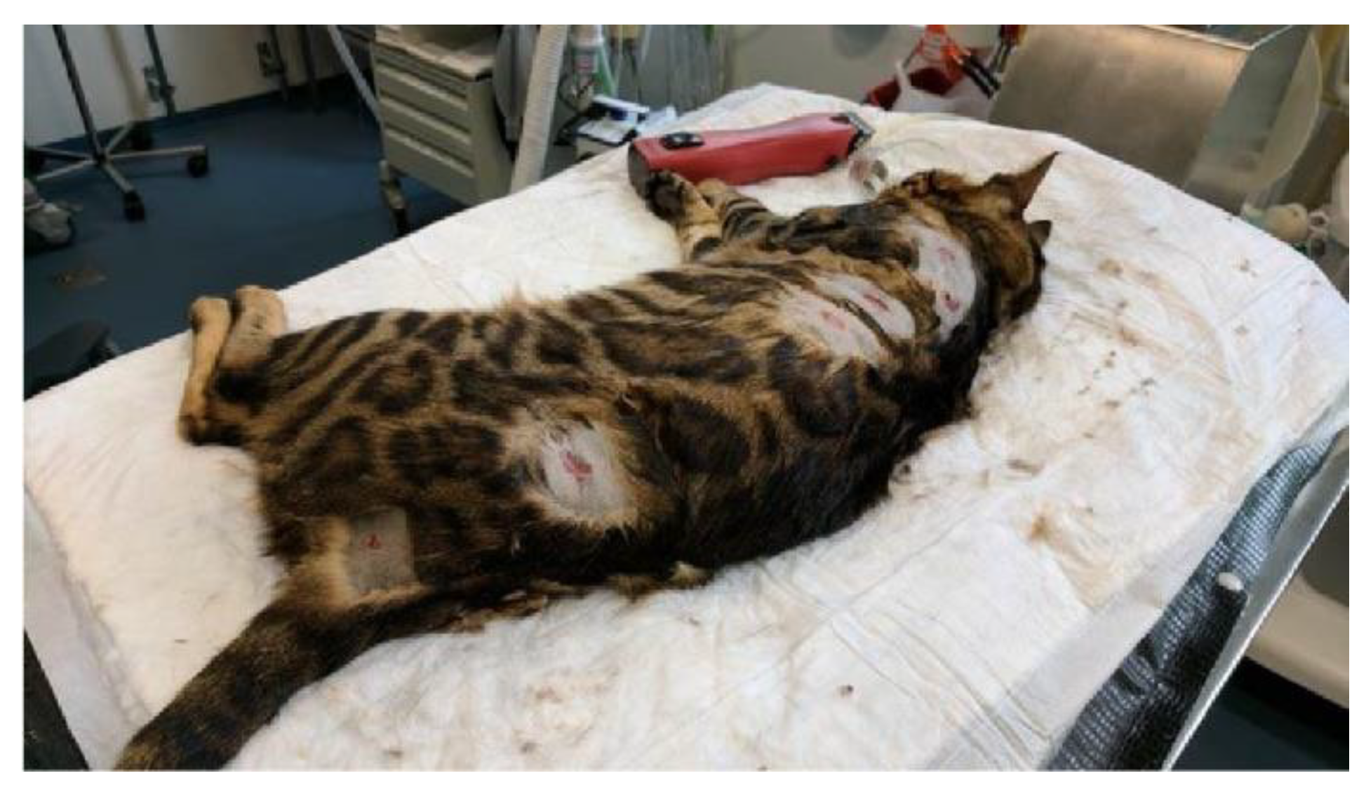
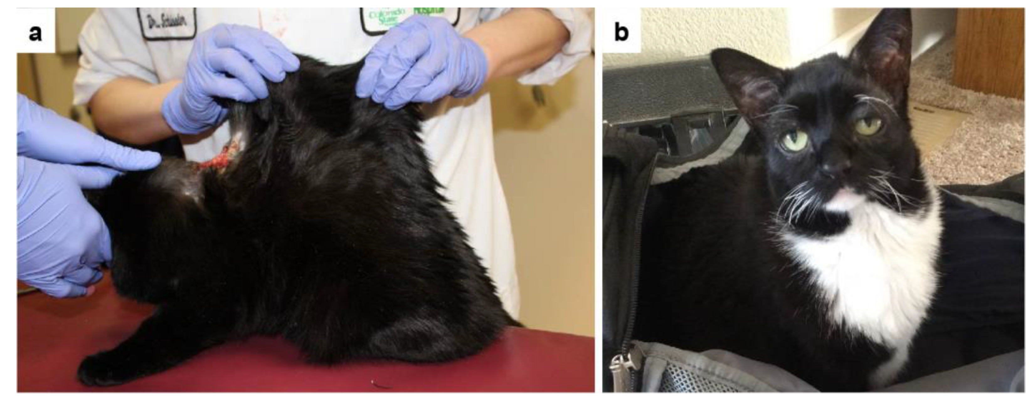
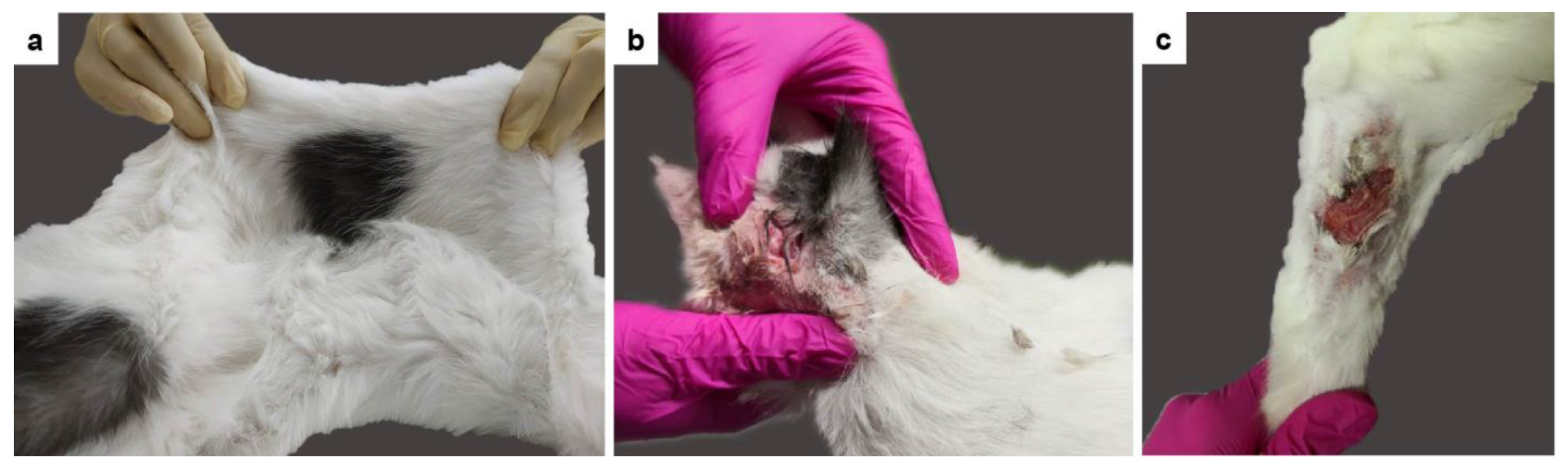
3.1. Clinical History and Examination

Case no. 1 was a 1-year-old purebred Bengal cat with a history of multiple repetitive skin lesions after minimal trauma occurring since the age of 6 months. At the time of presentation, a scarring alopecic lesion of about 5 × 5 cm was visible on the dorsal neck, which resulted from a previous trauma. Furthermore, the skin was diffusely thinner and much more elastic compared to the skin of a healthy young Bengal cat. In addition, the patient presented with multiple small skin tears distributed over the entire trunk/back area (Figure 1). The cat had no pruritus and there were no signs of active inflammation. Skin cytology, skin scrapings, and the trichogram were unremarkable.
Cases no. 2 and 3, presumably related female spayed Bombay cats, were presented for multiple wounds in the hyperextensible skin predominantly at the neck, base of ear, and shoulders (Figure 2a). The wounds occurred after self-trauma or exuberant play. Case no. 2 additionally showed spasms of the cutaneous trunci followed by frantic self-mutilation since adoption at 8 weeks of age. At 11 months of age, case no. 2 also experienced multiple partial and grand mal seizures necessitating phenobarbital therapy. Case no. 3 experienced a grade 3 patellar luxation at 18 months of age presumed to be related to joint hypermobility due to EDS. Excessive facial skin folds were apparent (Figure 2b).
Case no. 4 was a 2-year-old male castrated indoor DSH cat that was examined for recurrent laceration wounds on the neck and shoulders since adoption (one year before), and moderate head and neck pruritus (observed all year-round with exacerbation in June and August). Hematology and biochemistry (including basal cortisol) were normal, and Feline Leukemia Virus, Feline Immunodeficiency Virus, and Feline Coronavirus were negative one year before referral. On physical examination, a 1 cm scar on the ventral neck and a 0.5 cm crust above the right eye were noted. The skin extensibility index was 22% (Figure 3a). No joint laxity, hernias, ocular, or heart abnormalities were found. Neither ecto- and endoparasite treatment of both cats, nor an eight-week elimination diet improved the pruritus. Over the course of the disease, the cat developed new lesions due to pruritus when treated only with oral antihistamines (Fenistil, GlaxoSmithKline Consumer Healthcare GmbH & Co. KG, Munich, Germany) (Figure 3b,c). With short courses of systemic prednisolone (injections of 2 mg/kg, or orally 0.6 mg/kg once daily, as needed), the scratching stopped, and the lesions healed. Systemic medication was withdrawn as indicated prior to biopsy sampling. A welfare score (8; 0–21) was not suggestive of a psychogenic induced pruritus [28]. Based on all the above and since the patient fulfilled Favrot’s criteria [29], which help to establish the diagnosis of atopic dermatitis, a feline atopic skin syndrome (FASS) was diagnosed and a concomitant collagen disorder leading to lacerations due to pruritus (traumatic splitting) was suspected [30]. A skin intradermal allergy test as well as a serum allergy test were negative and could not identify the triggering agents of FASS. The owner declined antiallergic therapies other than antihistamines, and the cat was euthanized 2.5 years after the first presentation due to a very large laceration wound and deterioration, despite surgical and emergency treatment.
Two unaffected siblings and both parents of the patient were also clinically evaluated. They did not have any history compatible with allergy and no abnormality was noted. Their skin extensibility indices were normal (father: 15.6%, mother: 6.6%, brother 17.6%, sister: 10.7%).

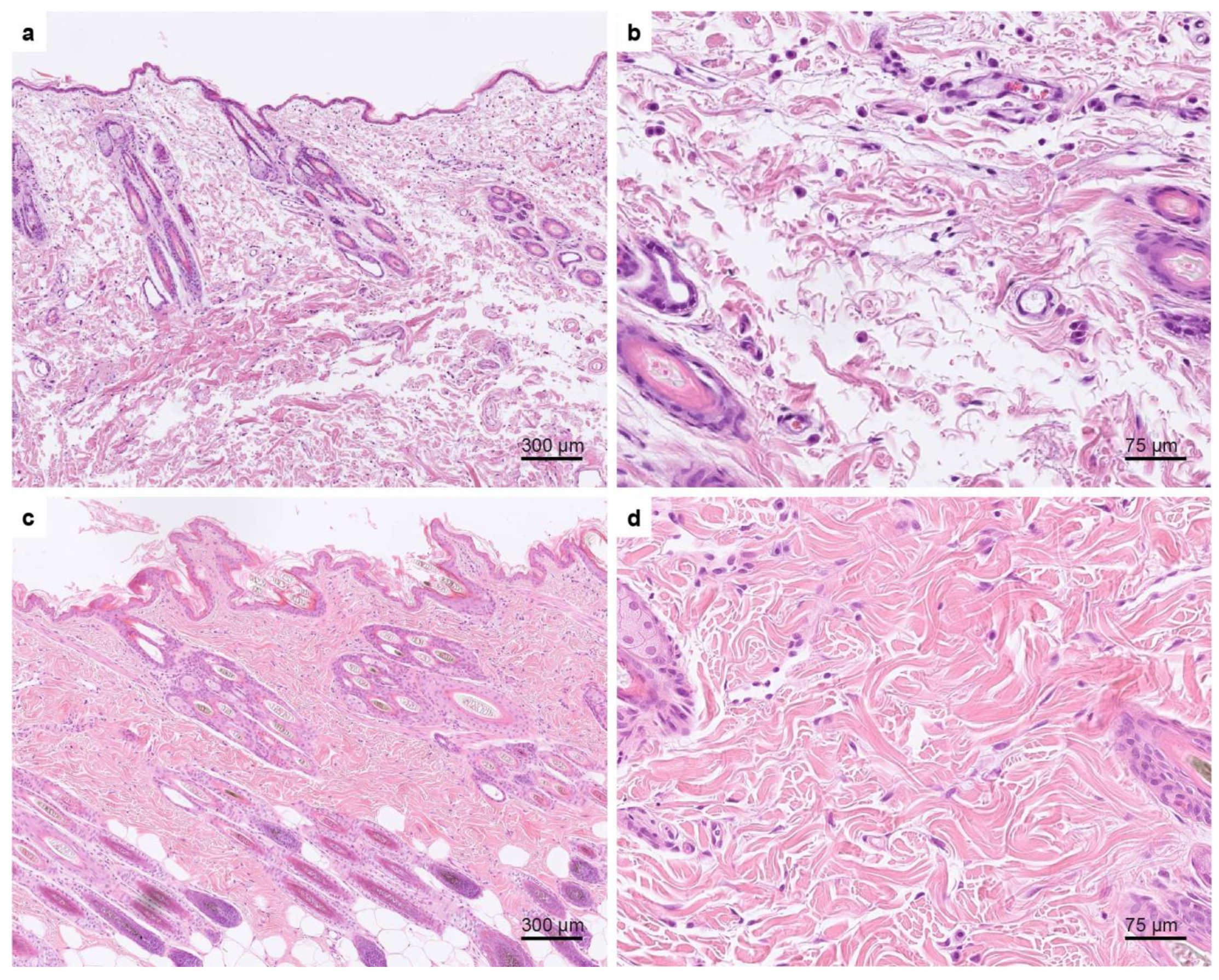
3.2. Histopathological Examination

The pathological changes in the skin biopsies were characterized by severely altered dermal collagen fibers and a slightly thinner epidermis. Changes were more severe in the superficial and mid dermis but were also present in the deep dermis. The abnormal collagen fibers were haphazardly arranged, shortened, and sometimes curled. They were uneven in length and width. Many fibers were very wispy and the interfibrillar spaces were widened. Multifocally small areas of hemorrhage were seen (Figure 4).

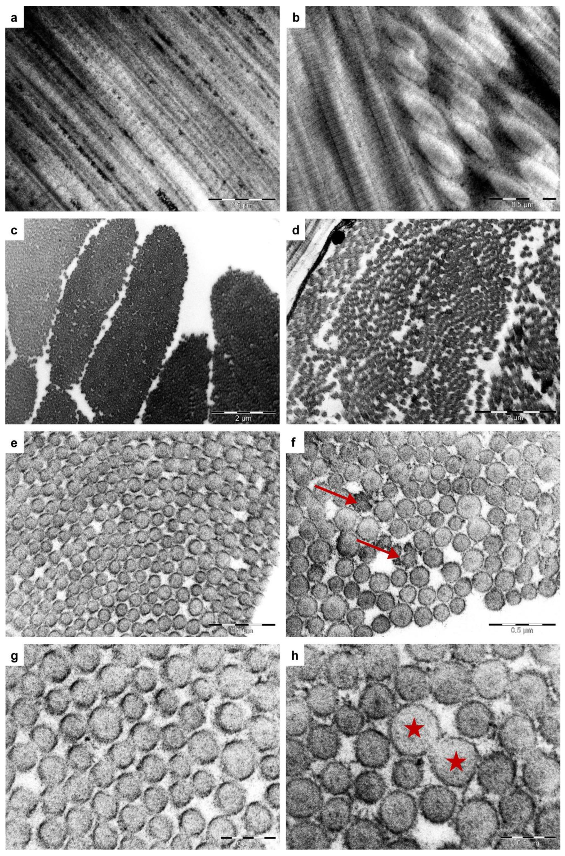
3.3. Ultrastructural Examination

TEM analysis of case no. 4 skin connective tissue revealed abnormalities in the collagen fibril organization and structure. Abnormal findings from the reticular part of the dermis were loosely packed and disorganized collagen fibers (Figure 5b,d). Individual collagen fibers contained curled fibrils (Figure 5b,d). Higher magnifications showed cross-sections of collagen fibrils with different diameters and irregularly outlined fibrils within a fiber (Figure 5f,h). In contrast, the connective tissue of an age-matched control cat contained densely packed collagen fibers with parallel-aligned fibril organization (Figure 5a,c). Cross-sections of fibrils showed uniform diameters and regularly shaped, almost round outlines (Figure 5e,g).

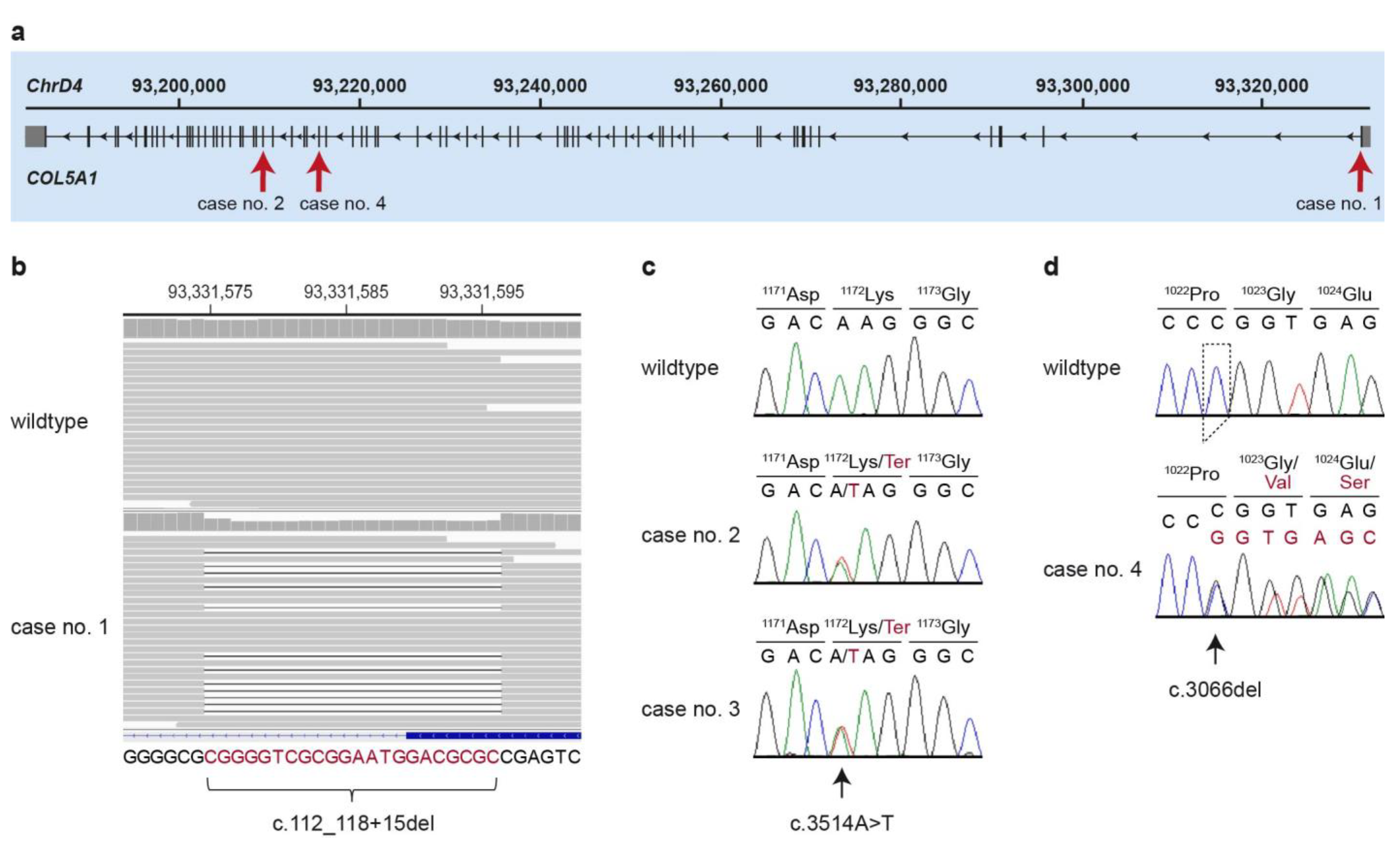
3.4. Genetic Analysis

To identify the causative genetic variants, the genomes of three affected cats (cases no. 1, 2, and 4) were sequenced. Case no. 3 was presumably related to case no. 2 and assumed to share the same pathogenic variant. Since the other cats were not known to be related, we hypothesized that their phenotypes were due to independent pathogenic variants. We therefore performed an individual variant filtering in each of the affected cats by comparing them to the genomes of 54 control cats. We focused our search on private protein-changing variants in the 20 known EDS candidate genes (Table 1 and Tables S1, S2 and S4).
In case no. 1, we identified one heterozygous private protein-changing variant within an EDS candidate gene (Figure 6a). The variant, COL5A1:c.112_118+15del, is a 22 bp deletion that removes the exon1/intron1 boundary (Table 2, Figure 6b). The presence of the variant was confirmed by PCR and fragment length analysis.
In case no. 2, we identified another single candidate variant, a heterozygous nonsense variant in COL5A1, predicted to truncate 36% of the coding sequence (Table 2). We confirmed identical heterozygous genotypes in cases no. 2 and 3 by Sanger sequencing (Figure 6a,c).
In case no. 4, we identified three heterozygous variants in known EDS candidate genes, a frameshift deletion in COL5A1 and two missense variants in TNXB. The targeted genotyping of all three variants in the complete family comprising both parents and two additional unaffected littermates excluded the TNXB variants (Supplementary Figure S1). The remaining variant, COL5A1:c.3066del, is a one base pair deletion, resulting in a frameshift and predicted to truncate 44% of the open reading frame (Table 2, Figure 6a,d). The genotyping results in the family further revealed that COL5A1:c.3066del represented a de novo variant as the mutant allele was absent from the leukocyte DNA of both parents.

4. Discussion

In this study, we describe four cats with classical EDS. All cats were characterized by similar clinical skin-related symptoms strongly suggestive of EDS [2]. Histopathology of the examined skin biopsies was also suggestive of EDS. In addition, TEM on case no. 4 identified abnormal morphological features indicative of EDS, such as variable diameters of collagen fibrils, disorganized, distorted collagen fibers, and irregularly outlined fibrils, which have been reported in the literature to be present in most patients with a genetically confirmed diagnosis of EDS [31,32,33].
Whole genome sequencing was performed to confirm the suspected clinical and histological diagnosis and to identify the underlying causative genetic variants in all four cats. We identified three independent heterozygous private protein-changing variants in COL5A1 in the four investigated affected cats. A de novo mutation event was experimentally confirmed for case no. 4. We assume that the pathogenic COL5A1 variants in the other affected cats were also due to recent de novo mutation events. For cases no. 2 and 3, the most likely scenario was a germline mutation in one of their parents. These results resemble previously described findings in a cat and three dogs affected by EDS due to de novo mutations in COL5A1 [24,34].
COL5A1 encodes the proα1 chain of type V collagen, which together with type I collagen forms heterotypical fibrils. Type V collagen is a major regulator of collagen fibrillogenesis, and the reduction in collagen V expression results in fewer collagen I fibrils with increased diameters [35,36]. Correct fibril formation is required for the integrity of the skin [1].
In humans, the functional loss of one COL5A1 allele is the most commonly reported molecular mechanism in classical EDS [36,37]. As type V procollagen molecules cannot accommodate more than a single proα2(V) chain, the reduction in available proα1(V) chains results in the production of about half the normal amount of type V collagen [38]. We assume that all three newly reported feline variants lead to nonsense-mediated mRNA decay, which results in a haploinsufficiency of COL5A1 and causes the alterations of the connective tissue.
Case no. 2 experienced epilepsy and presumptive hyperesthesia signs. Notably, an EDS-affected Burmese cat was described as experiencing prolonged episodes of disturbing vocalizations that were considered to be partial seizures. Upon necropsy, this cat was found to have flattened cerebral gyri [14]. Epilepsy has been noted in human patients with EDS and is associated with many structural brain defects, including periventricular heterotopia in patients with COL5A1 variants [39,40]. EDS-associated small fiber polyneuropathy in human patients manifests as acute, unpredictable burning pain, muscle cramping, and also compulsive scratching due to severe episodic itch [41]. Given comparable observations in human EDS patients and the earlier reported EDS-affected Burmese cat, it is reasonable to suspect that epilepsy and hyperesthesia were likely part of the EDS phenotype in case no 2.
Case no. 4 was diagnosed with EDS and concurrent FASS. Diagnosis of FASS can be difficult since it is a clinical diagnosis, and no diagnostic tests exist to diagnose or exclude FASS. Allergy tests such as serology and intradermal testing only help to identify the triggering agents to perform allergen specific immunotherapy. However, FASS cases have sometimes been described to have negative allergy testing results [30]. Furthermore, the cat was presented with head and neck pruritus. This is a skin reaction pattern that is consistent with the diagnosis of FASS after excluding other possible causes, as performed in our case [30]. Therefore, based on the clinical presentation and the fact that the cat showed a seasonal exacerbation, as well as the response to histamines (partial) and steroids (complete), we concluded that the itchiness has an underlying allergic cause. A neurological component of the itch therefore seemed unlikely, but it could not be completely excluded without further diagnostic imaging or necropsy, which was declined by the owner.
EDS-affected pets with other concurrent diseases inducing self-trauma (such as itchiness in case no. 4 due to FASS) have exacerbated symptoms that might lead to euthanasia. A faster whole genome sequencing-based diagnosis of the primary collagen genetic disease might lead to a better overall management of the patients, with the potential of decreasing the euthanasia rates of such complicated cases. A precise classification of EDS relies on molecular confirmation with identification of a causative variant [2]. This study illustrates the potential of whole genome sequencing as a precision medicine approach in animals with inherited diseases.
Consensus criteria for the interpretation of genetic variants in domestic animals have not yet been defined. However, such criteria have been established for human patients and they have proven highly useful to provide a standardized classification reflecting the strength of evidence for pathogenicity [42]. When we apply the human guidelines to the investigated cats, all three reported COL5A1 variants would be classified as pathogenic according to the ACMG/AMP criteria [42]. All three reported variants represent null variants (very strong evidence of pathogenicity, PVS1), are absent from controls (moderate evidence of pathogenicity, PM2), and the patients’ phenotypes are highly specific for a disease with a single genetic etiology (supporting evidence of pathogenicity, PP4). For case no. 4 we have the confirmed de novo mutation event and family segregation data as additional criteria for pathogenicity.

5. Conclusions

We characterized four cats with EDS and identified three independent causal variants in the COL5A1 gene. De novo mutation events leading to haploinsufficiency of COL5A1 appear to represent an important molecular etiology in feline EDS.

Supplementary Materials

The following are available online at https://www.mdpi.com/article/10.3390/genes13050797/s1, Figure S1: Pedigree with genotypes from the family of case no. 4. Table S1: Candidate Genes; Table S2: Public Genomes; Table S3: Primer Sequences; Table S4: Private Variants.

Author Contributions

Conceptualization, T.L., N.A., N.M.F. and S.K. (Sabine Kaessmeyer); investigation, S.K. (Sarah Kiener), N.A., J.S., F.L., S.S., M.W., N.M.F. and S.K. (Sabine Kaessmeyer); data curation, V.J.; visualization, S.K. (Sarah Kiener), N.A., J.S., M.W., N.M.F. and S.K. (Sabine Kaessmeyer); writing—original draft preparation, S.K. (Sarah Kiener), N.A., M.W., N.M.F. and S.K. (Sabine Kaessmeyer); writing—review and editing, S.K. (Sarah Kiener), N.A., J.S., P.-K.H., F.L., V.J., C.S., S.S., M.W., U.M., T.L., N.M.F. and S.K. (Sabine Kaessmeyer). All authors have read and agreed to the published version of the manuscript.

Funding

This research was funded by the Swiss National Science Foundation, grant number 310030_200354.

Institutional Review Board Statement

All animal procedures were performed according to local regulations. The “Cantonal Committee for Animal Experiments” approved the collection of blood samples from cats (Canton of Bern; permit 71/19).

Informed Consent Statement

The owners of the cats in this study gave informed consent to use samples and data for research.

Data Availability Statement

The accessions for the sequence data reported in this study are listed in Table S2.

Acknowledgments

The authors are grateful to the cat owners who donated samples and participated in the study. We are grateful to Patricia A. Cole who performed the histopathology on case no. 2, but sadly passed away during the course of this study. We thank the Next Generation Sequencing Platform of the University of Bern for performing the high-throughput experiments and the Interfaculty Bioinformatics Unit of the University of Bern for providing high-performance computing infrastructure. We gratefully acknowledge the technical assistance of Helga Mogel in the TEM lab. The TEM study was performed with the support of the interfaculty Microscopy Imaging Center (MIC) of the University of Bern.

Conflicts of Interest

The authors declare no conflict of interest.

References

  1. Malfait, F.; Castori, M.; Francomano, C.A.; Giunta, C.; Kosho, T.; Byers, P.H. The Ehlers–Danlos syndromes. Nat. Rev. Dis. Prim. 2020, 6, 64. [Google Scholar] [CrossRef] [PubMed]
  2. Malfait, F.; Francomano, C.; Byers, P.; Belmont, J.; Berglund, B.; Black, J.; Bloom, L.; Bowen, J.M.; Brady, A.F.; Burrows, N.P.; et al. The 2017 international classification of the Ehlers-Danlos syndromes. Am. J. Med. Genet. Part C Semin. Med. Genet. 2017, 175, 8–26. [Google Scholar] [CrossRef] [PubMed] [Green Version]
  3. Blackburn, P.R.; Xu, Z.; Tumelty, K.E.; Zhao, R.W.; Monis, W.J.; Harris, K.G.; Gass, J.M.; Cousin, M.A.; Boczek, N.J.; Mitkov, M.V.; et al. Bi-allelic Alterations in AEBP1 Lead to Defective Collagen Assembly and Connective Tissue Structure Resulting in a Variant of Ehlers-Danlos Syndrome. Am. J. Hum. Genet. 2018, 102, 696–705. [Google Scholar] [CrossRef] [PubMed] [Green Version]
  4. Nicholls, A.C.; Oliver, J.E.; McCarron, S.; Harrison, J.B.; Greenspan, D.S.; Pope, F.M. An exon skipping mutation of a type V collagen gene (COL5A1) in Ehlers-Danlos syndrome. J. Med. Genet. 1996, 33, 940–946. [Google Scholar] [CrossRef] [Green Version]
  5. Wenstrup, R. A splice-junction mutation in the region of COL5A1 that codes for the carboxyl propeptide of pro alpha 1(V) chains results in the gravis form of the Ehlers-Danlos syndrome (type I). Hum. Mol. Genet. 1996, 5, 1733–1736. [Google Scholar] [CrossRef] [Green Version]
  6. De Paepe’, A.; Nuytinck, L.; Hausser, I.; Anton-Lamprecht, I.; Naeyaert2, J.-M. Mutations in the COL5A1 Gene Are Causal in the Ehlers-Danlos Syndromes I and II. Am. J. Hum. Genet 1997, 60, 547–554. [Google Scholar]
  7. Richards, A.J.; Martin, S.; Nicholls, A.C.; Harrison, J.B.; Pope, F.M.; Burrows, N.P. A single base mutation in COL5A2 causes Ehlers-Danlos syndrome type II. J. Med. Genet. 1998, 35, 846–848. [Google Scholar] [CrossRef] [Green Version]
  8. Bouma, P.; Cabral, W.A.; Cole, W.G.; Marini, J.C. COL5A1 Exon 14 Splice Acceptor Mutation Causes a Functional Null Allele, Haploinsufficiency of α1(V) and Abnormal Heterotypic Interstitial Fibrils in Ehlers-Danlos Syndrome II. J. Biol. Chem. 2001, 276, 13356–13364. [Google Scholar] [CrossRef] [Green Version]
  9. Symoens, S.; Syx, D.; Malfait, F.; Callewaert, B.; De Backer, J.; Vanakker, O.; Coucke, P.; De Paepe, A. Comprehensive molecular analysis demonstrates type V collagen mutations in over 90% of patients with classic EDS and allows to refine diagnostic criteria. Hum. Mutat. 2012, 33, 1485–1493. [Google Scholar] [CrossRef]
  10. Vroman, R.; Malfait, A.-M.; Miller, R.E.; Malfait, F.; Syx, D. Animal Models of Ehlers–Danlos Syndromes: Phenotype, Pathogenesis, and Translational Potential. Front. Genet. 2021, 12, 1610. [Google Scholar] [CrossRef]
  11. Scott, D.V. Cutaneous asthenia in a cat, resembling Ehlers-Danlos syndrome in man. Vet. Med. Small Anim. Clin. 1974, 69, 1256–1258. [Google Scholar] [PubMed]
  12. Smids, M.Y.L. Experience with a cat with cutis asthenia (Ehlers-Danlos syndrome). Tijdschr. Diergeneeskd. 2008, 133, 612–614. [Google Scholar] [PubMed]
  13. Halper, J. Connective Tissue Disorders in Domestic Animals. In Progress in Heritable Soft Connective Tissue Diseases; Springer: Dordrecht, The Netherlands, 2014; pp. 231–240. [Google Scholar]
  14. Hansen, N.; Foster, S.F.; Burrows, A.K.; Mackie, J.; Malik, R. Cutaneous asthenia (Ehlers–Danlos-like syndrome) of Burmese cats. J. Feline Med. Surg. 2015, 17, 954–963. [Google Scholar] [CrossRef] [PubMed]
  15. Roberts, J.H.; Halper, J. Connective Tissue Disorders in Domestic Animals. In Advances in Experimental Medicine and Biology; Springer: Dordrecht, The Netherlands, 2021; Volume 1348, pp. 325–335. [Google Scholar]
  16. Patterson, D.F.; Minor, R.R. Hereditary fragility and hyperextensibility of the skin of cats. A defect in collagen fibrillogenesis. Lab. Investig. 1977, 37, 170–179. [Google Scholar]
  17. Counts, D.F.; Byers, P.H.; Holbrook, K.A.; Hegreberg, G.A. Dermatosparaxis in a Himalayan cat: I. Biochemical studies of dermal collagen. J. Investig. Dermatol. 1980, 74, 96–99. [Google Scholar] [CrossRef] [Green Version]
  18. Holbrook, K.A.; Byers, P.H.; Counts, D.F.; Hegreberg, G.A. Dermatosparaxis in a Himalayan cat: II. Ultrastructural studies of dermal collagen. J. Investig. Dermatol. 1980, 74, 100–104. [Google Scholar] [CrossRef] [Green Version]
  19. Verweij, C.G.; van Zuylen, A.L. Cutaneous asthenia, a congenital skin disease in a Burmese cat. Tijdschr. Diergeneeskd. 1986, 111, 244–246. [Google Scholar]
  20. Freeman, L.J.; Hegreberg, G.A.; Robinette, J.D. Ehlers-Danlos syndrome in dogs and cats. Semin. Vet. Med. Surg. (Small Anim.) 1987, 2, 221–227. [Google Scholar]
  21. Plotnick, A.; Brunt, J.E.; Reitz, B. Cutaneous Asthenia in a Cat. Feline Pract. 1992, 20, 9–12. [Google Scholar]
  22. Sequeira, J.L.; Rocha, N.S.; Bandarra, E.P.; Figueiredo, L.M.; Eugenio, F.R. Collagen dysplasia (cutaneous asthenia) in a cat. Vet. Pathol. 1999, 36, 603–606. [Google Scholar] [CrossRef] [Green Version]
  23. Benitah, N.; Matousek, J.L.; Barnes, R.F.; Lichtensteiger, C.A.; Campbell, K.L. Diaphragmatic and perineal hernias associated with cutaneous asthenia in a cat. J. Am. Vet. Med. Assoc. 2004, 224, 706–709, 698. [Google Scholar] [CrossRef] [PubMed]
  24. Spycher, M.; Bauer, A.; Jagannathan, V.; Frizzi, M.; De Lucia, M.; Leeb, T. A frameshift variant in the COL5A1 gene in a cat with Ehlers-Danlos syndrome. Anim. Genet. 2018, 49, 641–644. [Google Scholar] [CrossRef] [PubMed]
  25. Jagannathan, V.; Drögemüller, C.; Leeb, T.; Aguirre, G.; André, C.; Bannasch, D.; Becker, D.; Davis, B.; Ekenstedt, K.; Faller, K.; et al. A comprehensive biomedical variant catalogue based on whole genome sequences of 582 dogs and eight wolves. Anim. Genet. 2019, 50, 695–704. [Google Scholar] [CrossRef] [Green Version]
  26. McKenna, A.; Hanna, M.; Banks, E.; Sivachenko, A.; Cibulskis, K.; Kernytsky, A.; Garimella, K.; Altshuler, D.; Gabriel, S.; Daly, M.; et al. The genome analysis toolkit: A MapReduce framework for analyzing next-generation DNA sequencing data. Genome Res. 2010, 20, 1297–1303. [Google Scholar] [CrossRef] [PubMed] [Green Version]
  27. Cingolani, P.; Platts, A.; Wang, L.L.; Coon, M.; Nguyen, T.; Wang, L.; Land, S.J.; Lu, X.; Ruden, D.M. A program for annotating and predicting the effects of single nucleotide polymorphisms, SnpEff: SNPs in the genome of Drosophila melanogaster strain w1118; iso-2; iso-3. Fly 2012, 6, 80–92. [Google Scholar] [CrossRef] [PubMed] [Green Version]
  28. Titeux, E.; Gilbert, C.; Briand, A.; Cochet-Faivre, N. From Feline Idiopathic Ulcerative Dermatitis to Feline Behavioral Ulcerative Dermatitis: Grooming Repetitive Behaviors Indicators of Poor Welfare in Cats. Front. Vet. Sci. 2018, 5, 16. [Google Scholar] [CrossRef] [PubMed] [Green Version]
  29. Favrot, C.; Steffan, J.; Seewald, W.; Hobi, S.; Linek, M.; Marignac, G.; Olivry, T.; Beco, L.; Nett, C.; Fontaine, J.; et al. Establishment of diagnostic criteria for feline nonflea-induced hypersensitivity dermatitis. Vet. Dermatol. 2012, 23, 45-e11. [Google Scholar] [CrossRef]
  30. Santoro, D.; Pucheu-Haston, C.M.; Prost, C.; Mueller, R.S.; Jackson, H. Clinical signs and diagnosis of feline atopic syndrome: Detailed guidelines for a correct diagnosis. Vet. Dermatol. 2021, 32, 26-e6. [Google Scholar] [CrossRef]
  31. Angwin, C.; Ghali, N.; Baker, D.; Brady, A.F.; Pope, F.M.; Vandersteen, A.; Wagner, B.; Ferguson, D.J.P.; Dijk, F. Electron microscopy in the diagnosis of Ehlers–Danlos syndromes: Correlation with clinical and genetic investigations. Br. J. Dermatol. 2020, 182, 698–707. [Google Scholar] [CrossRef]
  32. Uri, M.; Verin, R.; Ressel, L.; Buckley, L.; McEwan, N. Ehlers–Danlos Syndrome Associated with Fatal Spontaneous Vascular Rupture in a Dog. J. Comp. Pathol. 2015, 152, 211–216. [Google Scholar] [CrossRef]
  33. Weingart, C.; Haußer, I.; Kershaw, O.; Kohn, B. Ehlers-Danlos-like-Syndrom bei einer Katze. Schweiz. Arch. Tierheilkd. 2014, 156, 543–548. [Google Scholar] [CrossRef] [PubMed]
  34. Bauer, A.; Bateman, J.F.; Lamandé, S.R.; Hanssen, E.; Kirejczyk, S.G.M.; Yee, M.; Ramiche, A.; Jagannathan, V.; Welle, M.; Leeb, T.; et al. Identification of Two Independent COL5A1 Variants in Dogs with Ehlers–Danlos Syndrome. Genes 2019, 10, 731. [Google Scholar] [CrossRef] [PubMed] [Green Version]
  35. Birk, D.E. Type V collagen: Heterotypic type I/V collagen interactions in the regulation of fibril assembly. Micron 2001, 32, 223–237. [Google Scholar] [CrossRef]
  36. Schwarze, U.; Atkinson, M.; Hoffman, G.G.; Greenspan, D.S.; Byers, P.H. Null Alleles of the COL5A1 Gene of Type V Collagen Are a Cause of the Classical Forms of Ehlers-Danlos Syndrome (Types I and II). Am. J. Hum. Genet. 2000, 66, 1757–1765. [Google Scholar] [CrossRef] [PubMed] [Green Version]
  37. Wenstrup, R.J.; Florer, J.B.; Willing, M.C.; Giunta, C.; Steinmann, B.; Young, F.; Susic, M.; Cole, W.G. COL5A1 Haploinsufficiency Is a Common Molecular Mechanism Underlying the Classical Form of EDS. Am. J. Hum. Genet. 2000, 66, 1766–1776. [Google Scholar] [CrossRef] [Green Version]
  38. Wenstrup, R.J.; Florer, J.B.; Davidson, J.M.; Phillips, C.L.; Pfeiffer, B.J.; Menezes, D.W.; Chervoneva, I.; Birk, D.E. Murine model of the Ehlers-Danlos syndrome. col5a1 haploinsufficiency disrupts collagen fibril assembly at multiple stages. J. Biol. Chem. 2006, 281, 12888–12895. [Google Scholar] [CrossRef] [Green Version]
  39. Savasta, S.; Merli, P.; Ruggieri, M.; Bianchi, L.; Spartà, M.V. Ehlers–Danlos syndrome and neurological features: A review. Child’s Nerv. Syst. 2011, 27, 365–371. [Google Scholar] [CrossRef]
  40. Cortini, F.; Villa, C. Ehlers-Danlos syndromes and epilepsy: An updated review. Seizure 2018, 57, 1–4. [Google Scholar] [CrossRef] [Green Version]
  41. Cazzato, D.; Castori, M.; Lombardi, R.; Caravello, F.; Bella, E.D.; Petrucci, A.; Grammatico, P.; Dordoni, C.; Colombi, M.; Lauria, G. Small fiber neuropathy is a common feature of Ehlers-Danlos syndromes. Neurology 2016, 87, 155–159. [Google Scholar] [CrossRef] [Green Version]
  42. Richards, S.; Aziz, N.; Bale, S.; Bick, D.; Das, S.; Gastier-Foster, J.; Grody, W.W.; Hegde, M.; Lyon, E.; Spector, E.; et al. Standards and guidelines for the interpretation of sequence variants: A joint consensus recommendation of the American College of Medical Genetics and Genomics and the Association for Molecular Pathology. Genet. Med. 2015, 17, 405–423. [Google Scholar] [CrossRef] [Green Version]
Figure 1. Clinical image of the EDS-affected cat no. 1. Multiple small skin tears were distributed over the entire back.
Genes 13 00797 g001
Figure 2. Clinical images of the EDS-affected cats no. 2 and 3. (a) Severe hyperextensibility of the dorsal skin. The skin extensibility index was 19% [20]. Note an extensive laceration on the back. (b) Excessive skin tissue is visible on the face of case no. 3.
Genes 13 00797 g002
Figure 3. Clinical images of the EDS-affected cat no. 4. (a) The abnormal extensibility of the dorsal skin is clearly visible. The skin extensibility index was 22%. (b,c) Lacerations of the skin on the head and the leg.
Genes 13 00797 g003
Figure 4. Hematoxylin and eosin-stained skin biopsies of case no. 4 and an age-matched control. (a,b) Histopathological changes seen in the affected cat. The epidermis appears thinner than in the skin biopsies of the age-matched control cat (c,d). (a) The dermal collagen fibers are haphazardly arranged, shortened, and sometimes curled. They were uneven in length and width. Many fibers are very wispy and the interfibrillar spaces are widened. (b) Higher magnification of the dermal changes seen in the affected cat. (c) Normal skin of an age-matched control cat. The dermal collagen fibers are much thicker and longer and the interfibrillar spaces are smaller than in the affected cat. (d) Higher magnification of normal skin.
Genes 13 00797 g004
Figure 5. Ultrastructural morphology of the skin from one of the affected cats (case no. 4) and an age-matched control cat. (a,c,e,g) Reticular connective tissue of a normal cat’s skin (control cat). (a) Longitudinal section of parallel-aligned collagen fibrils (×53.000). (c) Collagen fibers are composed of densely packed fibrils (cross-section, ×11.500). (e,g) Details of cross-sectioned collagen fibers. Consistent diameters of collagen fibrils with regularly shaped and almost round outlines (×53.000, ×110.000). (b,d,f,h) Representative collagen fibril abnormalities observed in the reticular dermis of case no. 4 cat. (b) Longitudinal sections show disordered, curled fibrils (×53.000). (d) Collagen fibers are loosely packed (cross-section, ×11.500). (f) Collagen fibrils with irregular outlines (arrows, ×53.000) and variable diameters (h). (h) Asterisks indicate fibrils with a diameter almost twice as large as surrounding fibrils with a normal diameter (×110.000).
Genes 13 00797 g005
Figure 6. Details of the COL5A1 variants. (a) Overview of the major transcript isoform (XM_023242950.1) of the COL5A1 gene with the intronic and exonic regions. The positions of all three identified variants are indicated by arrows. (b) Integrative Genomics Viewer (IGV) screenshot showing the short-read alignments of a control and the EDS-affected cat (case no. 1) at the position of the deletion. The heterozygous deletion is visible in the short-read alignments and the reduced coverage at the deleted bases. The sequence at the bottom represents the sequence of the coding strand in 3’ to 5’ orientation. The deleted bases are indicated in red. (c) Representative electropherograms of a control and two EDS-affected cats (case no. 2 and 3) are shown. The variable position is indicated by an arrow and the amino acid translations are given. Mutant alleles are indicated in red. (d) Representative electropherograms of a control and an EDS-affected cat (case no. 4) are shown. The amino acid translations of the wildtype and mutant alleles are indicated.
Genes 13 00797 g006
Table 1. Results of variant filtering in the affected cats against 54 control genomes.
Filtering StepVariants Case No. 1Variants Case No. 2Variants Case No. 4
homhethomhethomhet
all variants5,758,0349,091,6444,612,8798,593,8794,949,2508,310,846
private variants44,007938,0186242194,31511,606168,540
protein-changing private variants9129011588051775
in 20 known EDS candidate genes010103
Table 2. Variant designations of the identified COL5A1 variants according to Human Genome Variation Society (HGVS) nomenclature.
CatsHGVS Variant Designations
Genomic (felCat9.0)mRNA (XM_023242950.1)Protein (XP_023098718.1)
case no. 1ChrD4:93,331,577_93,331,598delc.112_118+15delr.spl?
cases no. 2 and 3ChrD4:93,209,345T>Ac.3514A>Tp.(Lys1172*)
case no. 4ChrD4:93,215,496delc.3066delp.(Gly1023Valfs*50)
Publisher’s Note: MDPI stays neutral with regard to jurisdictional claims in published maps and institutional affiliations.

