Article

Genetic and Clinical Findings in an Ethnically Diverse Cohort with Retinitis Pigmentosa Associated with Pathogenic Variants in CERKL

1
Oxford Eye Hospital, John Radcliffe Hospital, Oxford University Hospitals NHS Foundation Trust, Oxford OX3 9DU, UK
2
Nuffield Laboratory of Ophthalmology, Nuffield Department of Clinical Neuroscience, University of Oxford, Level 6 John Radcliffe Hospital, Headley Way, Oxford OX3 9DU, UK
3
Oxford Medical Genetics Laboratories, Oxford University Hospitals NHS Foundation Trust, Oxford OX3 7LE, UK
*
Authors to whom correspondence should be addressed.
Affiliated to the Oxford Eye Hospital during this study.
Genes 2020, 11(12), 1497; https://doi.org/10.3390/genes11121497
Submission received: 18 November 2020 / Revised: 9 December 2020 / Accepted: 10 December 2020 / Published: 12 December 2020
(This article belongs to the Special Issue Genomics and Therapeutics of Hereditary Eye Disease)

Abstract

:
Autosomal recessive retinitis pigmentosa is caused by mutations in over 40 genes, one of which is the ceramide kinase-like gene (CERKL). We present a case series of six patients from six unrelated families diagnosed with inherited retinal dystrophies (IRD) and with two variants in CERKL recruited from a multi-ethnic British population. A retrospective review of clinical data in these patients was performed and included colour fundus photography, fundus autofluorescence (AF) imaging, spectral domain–optical coherence tomography (SD–OCT), visual fields and electroretinogram (ERG) assessment where available. Three female and three male patients were included. Age at onset ranged from 7 years old to 45 years, with three presenting in their 20s and two presenting in their 40s. All but one had central visual loss as one of their main presenting symptoms. Four patients had features of retinitis pigmentosa with significant variation in severity and extent of disease, and two patients had no pigment deposition with only macular involvement clinically. Seven variants in CERKL were identified, of which three are novel. The inherited retinopathies associated with the CERKL gene vary in age at presentation and in degree of severity, but generally are characterised by a central visual impairment early on.

1. Introduction

Inherited retinal dystrophies (IRD) are a heterogeneous group of disorders associated with the dysfunction or death of photoreceptors, resulting in varying severity of visual loss. IRD has an incidence of 1 in 2000–3000, affecting an estimated two million people worldwide [1]. Retinitis pigmentosa (RP; Mendelian Inheritance in Man (MIM) #268000) is the most common IRD, affecting approximately 1 in 4000 people [2], characterised by symptoms of nyctalopia, peripheral visual field loss with a clinical appearance of intraretinal bone spicule pigmentation, pale discs and attenuated vasculature. RP is associated with significant genotypic and phenotypic heterogeneity, with 41 genes described in association with the autosomal recessive form to date (RetNet: https://sph.uth.edu/retnet/accessed 1 July 2020). In 1998, an RP locus (RP26) was mapped to an 11 cM region on chromosome 2q31–32 in a consanguineous Spanish family by Bayes and colleagues [3]. Subsequently, Tuson and colleagues identified a novel gene from the region that was expressed in the retina, named ceramide kinase-like (CERKL) [4]. The coding exons of CERKL were sequenced in the original RP26 family, and all patients were found to be homozygous for a nonsense mutation c.769C > T, p.Arg257* (now annotated as c.847C > T, p.Arg283*, using the numbering based on transcript NM_001030311, which consists of 14 exons) [4]. A further unrelated Spanish family was also identified with the same homozygous change [4].
CERKL shares 29% identity (50% similarity) with the ceramide kinase (CERK) protein, which phosphorylates ceramide to ceramide 1-phosphate, a sphingolipid metabolite which is involved in proliferation, apoptosis, phagocytosis and inflammation [5]. As yet, no kinase activity is reported for CERKL and its function remains unclear [6,7].
The CERKL gene is composed of 14 exons but alternative splicing produces multiple transcripts (Figure 1) [8,9]. Most of the previous studies examining variants in CERKL used isoform NM_201548, but this only contains 13 exons and is missing exon 5. Table 1 lists all the previously published mutations and the novel changes described in the patients in the present study, using NM_001030311 (which consists of 14 coding exons) as the reference. To date, 39 different mutations in CERKL have been identified (see Figure 1 and Table 1 for summary). The majority of these CERKL-associated IRD studies are case reports and many include the results from next-generation sequencing (NGS) testing with minimal phenotype data. There are, however, four studies reporting on at least four families, but these pertain to one ethnicity only in each study (Yemenite Jewish, Spanish, Tunisian and Finnish) [10,11,12,13]. In these reported populations, CERKL is a significant gene contributing to autosomal recessive RP; presumably due to a founder mutation effect.
The development of next-generation sequencing (NGS) techniques over the past 20 years led to diagnostic laboratories having the capability of screening known RP genes simultaneously [14]. Although this increased the detection rate, enabling a comprehensive genotype/phenotype analysis of cohorts of patients with same gene variants, it also identifies variants in other genes whose relevance may be difficult to determine. We report phenotypic and genotypic characterisation of six patients from six unrelated families with different ethnicities, and report three novel and four previously described variants in CERKL.
The CERKL gene on 2q31–32 consists of 14 exons and spans approximately 12 kb of genomic DNA. Exons are shown as boxes and introns as lines; all are to scale. The position of all the CERKL variants reported to date are marked. The mutations identified in our cohort are marked, those novel to this study are shown below in red and those reported previously are highlighted in bold above the structure (see Table 1 for details).

2. Materials and Methods

The clinical notes of patients seen at the Oxford Eye Hospital, identified to have CERKL variants by the Oxford Medical Genetics Laboratory, were reviewed to obtain full ophthalmic details, history of symptom onset and family pedigrees. The study adhered to the tenets of the Declaration of Helsinki and was approved by the Central Oxford Research Ethics Committee and the Research and Development Department of the Oxford Radcliffe Hospitals NHS Trust (RetGene 08/H0302/96). Informed consent was obtained from all participants.

2.1. Clinical Studies

Clinical data collected included best corrected visual acuity, slit-lamp biomicroscopy, digital colour fundus photography, short-wavelength (488) fundus autofluorescence (Spectralis; Heidelberg Engineering, Heidelberg, Germany), spectral domain–optical coherence tomography (SD–OCT; Spectralis, Heidelberg Engineering, Heidelberg, Germany), Goldmann visual field analysis and electrodiagnostic testing, where possible. The Heidelberg software was used for retinal segmentation of the ganglion cell layer (GC) and the inner plexiform layer (IPL), which were then used to measure the thickness of the GC–IPL layers in the areas temporal and nasal to the area of macular atrophy.

2.2. CERKL Mutation Screening

CERKL variants were identified by targeted sequencing performed by the Oxford Medical Genetics Laboratory using their next generation sequencing (NGS) phenotype panels. Enrichment for the CERKL gene was achieved as part of a customised HaloPlex enrichment system kit (Agilent Technologies, Santa Clara, USA) designed to capture the coding exons and at least 10 bp of the flanking introns of 111 retinal genes in the retinal dystrophy panel [14]. HaloPlex reactions were prepared as per the manufacturer’s instructions. Libraries were pooled into batches of 14 and sequenced on an Illumina MiSeq instrument (Illumina) using a MiSeq v3 kit (Illumina, San Diego, USA), as per the manufacturer’s instructions. Reads were aligned using Burrow-Wheeler Aligner BWA [15] and variants were called using Platypus [49]. All variants identified by NGS were confirmed by Sanger sequencing and investigated according to the Association of Clinical Genetic Science guidelines [50]. This included filtering of candidate variants by minor allele frequency against the ExAC dataset [51].
Chromosome 2 position was based on build GRCh37/hg19 and nucleotide and protein numbering is based on CERKL transcript NM_001030311, comprising 14 exons. The Genome Aggregation Database (gnomAD) (available online: http://gnomad.broadinstitute.org, accessed on 1 July 2020) was used to determine the carrier frequency of the minor allele in the general population. In silico analysis using 3 different prediction methods to determine the deleteriousness of the variants, Polyphen2 [52] Sorting Intolerant from Tolerance (SIFT) [53] and Mutation Taster [54] was carried out on the variants identified. Variants in other genes in these patients identified by this methodology were also reported, and family segregation studies were performed where possible.

3. Results

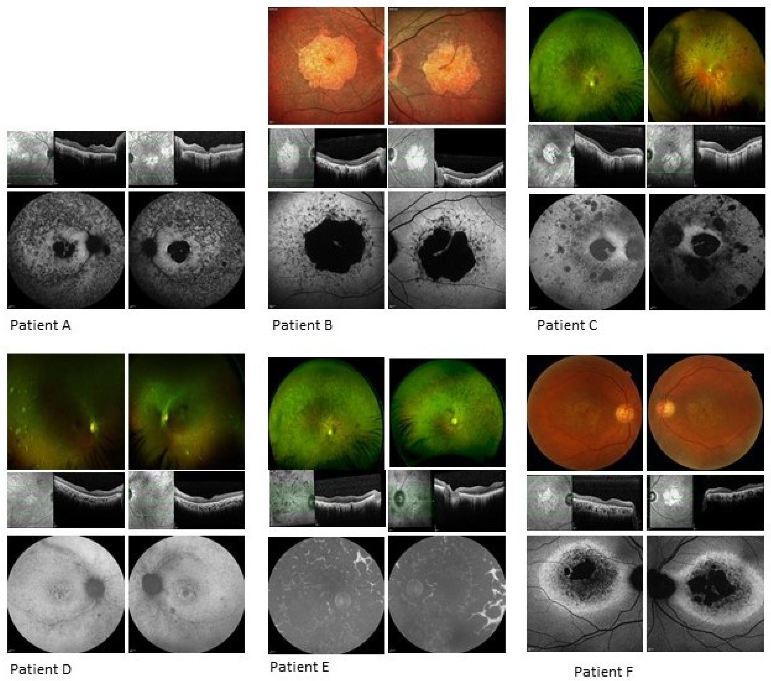
3.1. Clinical Analysis
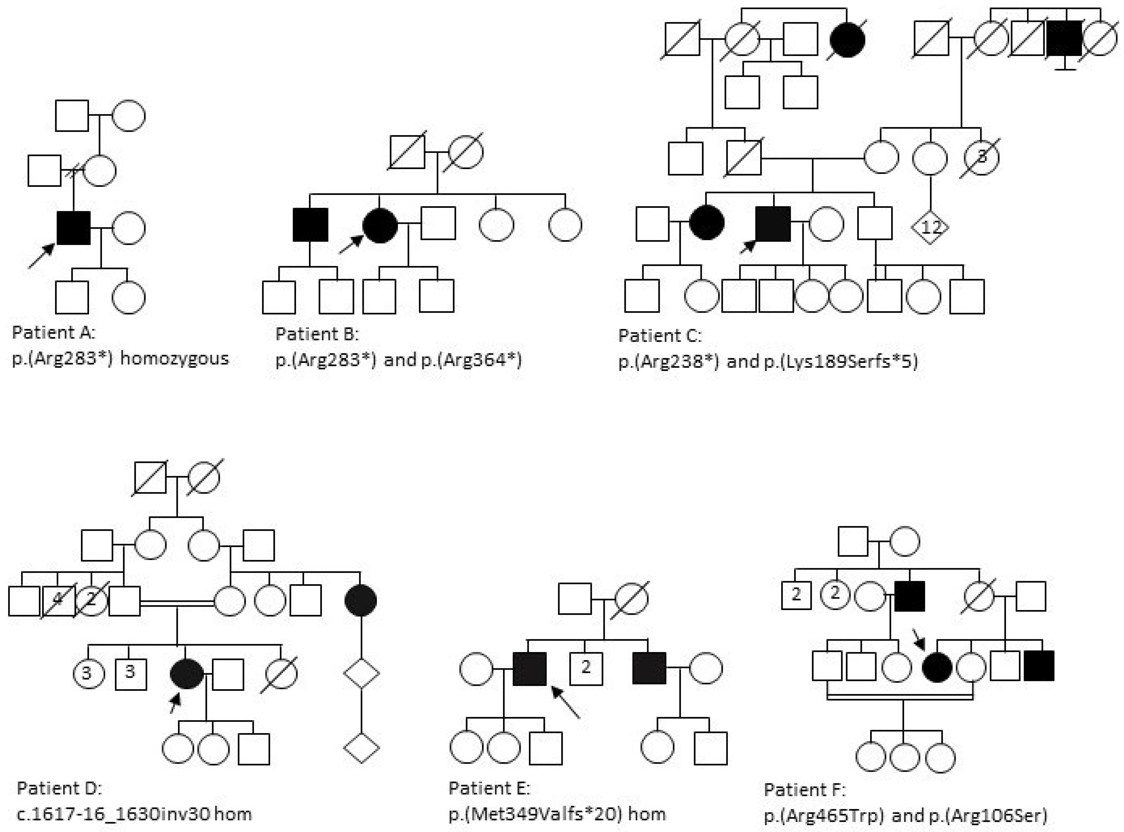
The clinical data of six patients from six unrelated families (Figure 2) with a diagnosis of a retinal dystrophy and with at least two variants in CERKL were reviewed (Figure 3, Table 2 and Table 3). The study comprised three females and three males, with age at presentation ranging from childhood to mid-40s. Symptom type and onset and clinical phenotypes are summarised in Table 2. Autofluorescence (AF) and optical coherence tomography (OCT) imaging are summarised in Table 3 and seen in Figure 3.

3.1.1. Patient A

A 34-year-old white Caucasian male, homozygous for c.847C > T, p.(Arg283*) in CERKL, first noticed visual symptoms at the age of 25, reporting nyctalopia and difficulty with colour vision. He was fit and well other than a diagnosis of Marfan Syndrome. There was no family history of eye disease. Examination revealed visual acuities of 6/24 in the right and hand movements only in the left. Fundoscopy and imaging demonstrated bilateral attenuated vessels, intraretinal bone spicule pigmentation and patches of peripheral atrophy with central macular atrophy. AF imaging revealed significant central atrophy with foveal sparing. Patchy AF signal was noted in the mid- to far-periphery. OCT imaging showed significant central atrophy bilaterally (Figure 3, Table 2 and Table 3). Visual field testing and electrophysiology testing were not available.

3.1.2. Patient B

A 58-year-old Indian female, compound heterozygous for c.847C > T, p.(Arg283*) and c.1090C > T, p.(Arg364*) CERKL mutations, reported reduction of vision in both eyes at the age of 43 years. Her brother had similar symptoms at age 16, with poor central and peripheral vision. Her general health was significant for a history of iron-deficiency anaemia. Examination revealed visual acuities of 2/60 in the right and 6/36 in the left. Fundoscopy and imaging showed bilateral macular atrophy with preserved central islands. AF imaging demonstrated a central atrophic area in both maculae with central sparing and patchy mixed increased and loss of AF signal extending from atrophy to arcades. OCT imaging revealed loss of outer retina bilaterally (Figure 3, Table 2 and Table 3). Goldmann visual fields showed central reduction in sensitivity extending to 20 degrees on the right and left bilaterally (see Figure S1). Electrophysiology showed extinguished PERG, reduction in a- and b-wave amplitudes on the rod ERG and reduction in b-wave amplitude for the standard bright white flash. Cone responses showed reduction in amplitudes and the 30 Hz flicker was delayed and reduced.

3.1.3. Patient C

A 56-year-old white Caucasian male, compound heterozygous for c.847C > T, p.(Arg283*) and c.566_569delinsGTG, p.(Lys189Serfs*5) CERKL mutations, first noticed symptoms of nyctalopia and visual loss aged 25 years. Regarding family history, his maternal great uncle was said to be visually impaired but the aetiology of this is unknown. Examination revealed visual acuities of no perception of light in both eyes. Fundoscopy and imaging revealed pale discs, grossly attenuated vasculature and central macular atrophy. Widespread intraretinal bone spicule pigmentation was noted primarily in the mid-periphery, and patchy nummular atrophic and hyper pigmented patches were observed in the mid-periphery to periphery mainly in the temporal retina. AF imaging demonstrated loss-of-signal consistent with central atrophy with a strip of minimal central sparing. There were also peripheral areas of loss-of-signal consistent with the chorioretinal atrophic patches. Fundus fluorescein angiography clearly demonstrated the central and inferior retinal patches of total retinal loss (see Figure S2). OCT imaging revealed loss of outer retina bilaterally (Figure 3, Table 2 and Table 3). Visual field testing was not performed due to poor visual acuity and electrophysiology was not available.

3.1.4. Patient D

A 30-year-old Pakistani female, homozygous for a c.1617-16_1630inv30 CERKL mutation, had symptoms of photopsia, photophobia, nyctalopia, abnormalities in her colour vision and central and peripheral visual field loss in her mid-twenties. Her maternal aunt was also reported to have visual impairment but of unknown aetiology. Examination revealed visual acuities of 6/18 on the right and 6/12 on the left with refraction of R: −0.50/−1.50 × 30 and L: +0.50/−1.50 × 15. Fundoscopy and imaging showed pale discs, attenuated vasculature and distinct nummular RPE depigmentation in the mid-periphery with chorioretinal atrophic patches temporally. Sparse intraretinal bone spicule pigmentation was present. AF imaging demonstrated increased signal in the maculae around the foveal loss-of-signal, with patchy, punctate loss extending from and including the arcades. OCT imaging showed focal foveal disruption of ellipsoid layer and loss of the outer retinal layers in both eyes (Figure 3, Table 2 and Table 3). Goldmann visual fields showed bilateral constriction to less than 10 degrees (see Figure S1). Electrophysiology testing showed no measurable components for PERG, cone and 30 Hz ERG, rod and standard flash and maximal ERG.

3.1.5. Patient E

A 58-year-old Indian male, homozygous for a c.1045_1046delAT, p.(Met349Valfs*20) mutation in CERKL, noted symptoms of nyctalopia and a slow gradual deterioration in vision in his teens, with a sharp deterioration in the last five years. His brother is also affected by the same condition (see Figure 2). Examination revealed visual acuities of perception of light bilaterally. Fundoscopy and imaging demonstrated significantly attenuated vessels, pale discs, and dense intraretinal bone spicule pigmentation scattered throughout the fundus, with central atrophy. AF imaging showed complete loss-of-signal in patches throughout the fundi, indicating severe coalescing atrophy. OCT imaging revealed loss of outer retina bilaterally (Figure 3, Table 2 and Table 3). Visual field testing was not possible due to poor visual acuity. Electrophysiology testing was not available.

3.1.6. Patient F

A 57-year-old Kashmiri female, compound heterozygous for c.1393C > T, p.(Arg465Trp) and c.316C > A, p.(Arg106Ser) mutations in CERKL, noticed deterioration of her central vision at age 40. Her brother and uncle were noted to have visual impairment. Examination revealed visual acuities of 6/60 bilaterally. Fundoscopy and imaging demonstrated central atrophy bilaterally. AF imaging showed significant central loss of signal denoting atrophy with a thick band of increased AF extending to the disc and around the circumference of the atrophy. OCT imaging revealed significant outer retinal loss in both eyes (Figure 3, Table 2 and Table 3). Neither visual fields nor electrophysiology were available.

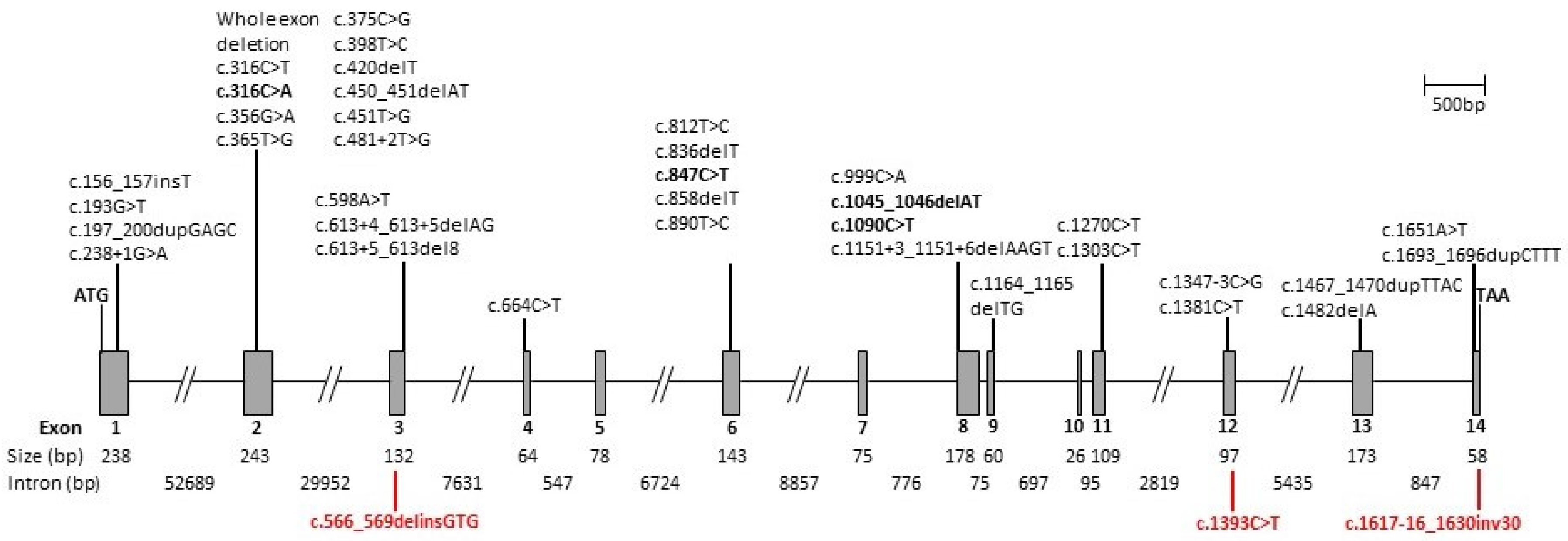
3.2. Genetic Analysis

The six patients described here all had two disease-causing variants in CERKL (Figure 1). Three were homozygotes and three were compound heterozygotes (Table 2). Seven variants in CERKL were identified in total, three of which were novel (Figure 1, and Table 1). The most common mutation in our cohort was the nonsense variant c.847C > T, p.(Arg283*) in exon 6, which was identified in three of our patients (Patient A is homozygous and Patients B and C are both heterozygous). This is also the most common CERKL variant reported to date (Table 1). Another previously described nonsense mutation (c.1090C > T p.(Arg364*)) was found in Patient B [22,46]. We also found two missense mutations, one of which was novel (Figure 1, Table 1). In silico analysis was performed using three different prediction methods to determine the deleteriousness of the novel variant. Two of them, SIFT [53], and Mutation Taster [54], predicted the c.1393C > T variant to be damaging and disease-causing, respectively. However, Polyphen2 [52] predicted it to be benign. The remaining variants were a previously reported deletion, a novel indel and a novel inversion. Patient C was a compound heterozygote with c.847C > T p.(Arg283*) and the novel c.566_569delinsGTG, resulting in a frameshift and premature termination of the protein (p.(Lys189Serfs*5)). Patient D and Patient E were homozygous for a novel inversion and a novel indel respectively, the inversion (c. 1617-16_1630inv30) resulting in a 30bp inversion in exon 14, causing the splice acceptor site to be lost. The novel indel in Patient E causes a frameshift in exon 8, resulting in a premature termination of the protein. The mutations published to date, including the novel variants reported in this study, are illustrated schematically in Figure 1, demonstrating no significant clustering and they are located throughout the entire gene.
As the patients were screened using either a panel of 55 or 111 IRD genes, variants in other genes were also described in three of them (Table S2). Patient D was heterozygous for a PDE6A variant (c. 769C > T p.(Arg257*)) of known pathogenicity and patient E was heterozygous for an IMPG2 variant (c. 789C > G p.(Ser263Arg)) of uncertain pathogenicity. Patient F was heterozygous for USH2A c.3812-3_3837dup p.(Met1280*) of presumed pathogenicity, as well as for NRL c.11c > T p(Pro4Leu) of uncertain pathogenicity; these are discussed in more detail below (Table S2).

4. Discussion

This study of six patients from different families with pathogenic variants in CERKL is one of the largest multi-ethnic studies describing CERKL-associated IRD to date. Of the seven different pathogenic variants, three are novel. The phenotype, although variable and with different ages of onset, shows significant similarity in symptom presentation, with five of the six reporting a deterioration in central vision. Three patients were affected in their twenties and two in their forties, similar to the literature. Patient E presented at the age of seven and demonstrated the most severe phenotype. However, in the Finnish cohort described by Avela et al. the early-onset patients did not always have severe phenotypes [10]. Patients A, B, C and F showed clear central atrophy with varying degrees of a residual foveal sparing. These features were seen but less clearly demarcated in patient D, where the infrared imaging showed the central atrophic changes more distinctly and could just be an earlier stage of the same phenotype. Indeed, the phenotype of rod cone dystrophy with initially preserved central vision despite earlier macular involvement was described clearly by Khan and Abu-Safieh, who reported an almost identical phenotype [44]. Also, the peripheral nummular chorioretinal atrophic areas seen in patient D were described by Fernandez et al. in their seven patients with the c.769C > T p.(Arg257*) variant [13]. However, for patient E the changes were so advanced that, although there was a macular atrophic area, the same phenotype was not distinguishable.
For patient F, the ring of AF surrounding the atrophic change was different in its extent compared to the rest of this cohort. However, a similar phenotype was described in CERKL IRD before in a patient compound heterozygous for c.375C > G, and c.193G > T in Avela et al.’s Finnish CERKL cohort (patient 4 in Avela et al.) [10].
As described in other reports, peripheral involvement was variable, ranging from mild pigment clumping to severe widespread atrophy, as seen in patient E. Two of our cohort (B and F) had no visible peripheral changes at all.
Patients D, E and F also possess variants in other known IRD genes (Table S2), but none of these variants were considered to be disease-causing, either because they were seen in a heterozygous state in genes causing autosomal recessive disease or because the phenotype previously described for the gene was not consistent. For example, Patient D was heterozygous for a PDE6A variant (c.769C > T p.(Arg257*)) of known pathogenicity and patient E was heterozygous for an IMPG2 variant (c.789C > G p.(Ser263Arg)) of uncertain pathogenicity, but in both cases a second variant was not found (Table S2). Additionally, segregation studies for patient E identified the same CERKL variant (p.(Met349Valfs*20)) in the heterozygous state in his unaffected father. His unaffected mother is deceased and so was unavailable for analysis. Patient F was heterozygous for variants in USH2A and NRL (Table S2). The USH2A, c.3812-3_3837dup p.(Met1280*) is of presumed pathogenicity, however, the phenotype is not typical of an USH2A-related retinopathy and patient F is not deaf (although not all patients with USH2A variants have hearing impairment). The NRL c.11C > T p(Pro4Leu) variant is of uncertain pathogenicity and NRL-associated RP recessive or dominant is rare, and usually associated with a severe early-onset phenotype, which is not present in patient F, who was 40 years old at the age of diagnosis [55]. In addition, although only the segregation of the CERKL variants were investigated, her affected brother possesses the same two CERKL variants, whereas her unaffected brother has neither.
Herein, we describe a cohort of six ethnically diverse patients, including white Caucasian, Indian, Pakistani, and Kashmiri origins. Previous reports in Finnish [10], Yemeni Jewish [11] and Spanish populations [13] reported the CERKL variants (c.375C > G, p.(Cys125Trp); c.238 + 1G > A and c.847C > T, p.(Arg283*) respectively) as a significant cause of arRP due to founder mutations. Three of our cohort, which did not include any patients from these populations, did have the c.847C > T variant found in the Spanish population. However, in other populations, CERKL variants are a rare cause of arRP, so there are limited phenotypic data in the literature [10,43]. The CERKL phenotype reported was variable in severity, degree of field loss and presence or lack of abnormal pigmentation. However there appeared to be a typical phenotype of early macular involvement with progression over time with concurrent varying degrees of progressive photoreceptor degeneration. The macular atrophy, seen particularly clearly on autofluorescence imaging, may be associated with a preserved small residual island of tissue [36]. As yet, there is no clear explanation for its persistence. It is also observed in some other IRDs, such as ABCA4 retinopathies; Bax et al. made the observation that these patients were in their fifth decade and that foveal sparing is observed in patients with a low rate of progression [56]. The Finnish cohort described by Avela et al. demonstrates a wide range of severity, as also seen in our cohort. Recently, Yu et al. demonstrated progressive degeneration of rod and cone outer segments in a Cerkl-knockout zebrafish model, with rod degeneration preceding cone degeneration [57]. There was no clearly observed genotype phenotype correlation, either in our cohort or in the literature, in patients with pathogenic variants in CERKL. The variants described to date (Table 1) are distributed across the entire gene (Figure 1), and the exact function of CERKL remains unclear. Although it shows 50% similarity to the ceramide kinase (CERK) protein, which phosphorylates ceramide to ceramide 1-phosphate and encodes a potential diacylglycerol kinase (DAG) domain, there is no evidence of any kinase activity in CERKL [6]. Indeed, studies examining the transcriptional complexity of CERKL in the retina demonstrate that there are isoforms that do not contain the DAG domain [8,9]. There is some evidence for CERKL having a role in protecting retinal cells from injury caused by oxidative stress [9,58]. A recent study describing the generation of a mouse model using CRISPR-Cas9 editing to delete the Cerkl locus showed that total ablation of the locus was embryonically lethal [7]. The authors therefore generated a model where the knockout allele (CerklKO) was in trans with a knockdown allele (CerklKD). These animals showed defects in the photoreceptor outer segments but did not initially show any alteration in the electrophysiological response, although older mice did show some changes [7]. This model may prove to be useful in gaining some understanding of the function of CERKL/Cerkl in the mammalian retina.
To date, there are no therapeutic strategies to correct or treat the retinal degeneration caused by mutations in CERKL. However, gene therapy for CERKL using adeno-associated viral (AAV) vectors is a possibility due to a combination of the recessive nature of the disease and small coding sequence at 1599 base pairs, which is easily encodable by adeno-associated viral (AAV) vectors similar to Luxturna, which is approved for Leber’s Congenital Amaurosis (LCA) type II. One problem, however, is that CERKL may also have a vital role in the inner retina [4], but in this series we did not identify any significant inner retinal changes. Identifying the correct mRNA sequence will be essential in developing future gene therapy treatments, but CERKL has many known splice isoforms [8]. Hence, our observation of novel mutations in exons 2, 3, 6 and 8 confirms the likely critical role of these exons in the CERKL isoform required in photoreceptors.

5. Conclusions

In conclusion, CERKL mutations are an uncommon cause of arRP, but they are a significant cause of disease in populations with founder mutations [10,11,13]. The importance of seeking further information from family segregation studies is highlighted by the families described in this study. Identifying the correct genetic variant will be of significant importance when potential therapies become available. It is highly likely that, in some cases, revisiting the genotype, particularly in cases where more than one IRD gene variant is identified, will be required. This study adds to the phenotype spectrum of pathogenic variants in CERKL and highlights specific difficulties in diagnosis when more than one gene variant is present.

Supplementary Materials

The following are available online at https://www.mdpi.com/2073-4425/11/12/1497/s1, Figure S1: Goldmann visual fields showing two timepoints in patients B and D; Figure S2: Fundus fluorescein angiography; Table S1: The thickness of the ganglion cell layer–inner plexiform layer in the nasal and temporal retina; Table S2: Variants identified in IRD genes other than CERKL.

Author Contributions

Conceptualization, S.M.D. and S.H.; collection of patient data, S.M.D., T.N., V.T., M.S. (Mital Shah), R.E.M.; investigation, S.M.D., T.N., V.T., S.B., M.S. (Mital Shah), S.A.-K., R.E.M., M.S. (Morag Shanks), P.C. and S.H.; writing—original draft preparation, S.M.D., S.B., S.H.; writing—review and editing, S.M.D., S.B., S.A-K., S.H.; supervision, S.M.D and S.H. All authors have read and agreed to the published version of the manuscript.

Funding

The Thames Valley and South Midlands Clinical Research Network provided funding support for the clinical aspects of this study. Saoud Al-Khuzaei is supported by a scholarship from the Qatar National Research Fund (GSRA6-1-0329-19010).

Acknowledgments

The authors would like to thank the Eye Research Group Oxford (ERGO) for their support of this study, in particular Alexina Fantato, Clare Arnison-Newgass, Sophie Marlowe, and Lidia Milla, as well as the patients who gave their consent to participate in this study.

Conflicts of Interest

The authors declare no conflict of interest.

References

  1. Berger, W.; Kloeckener-Gruissem, B.; Neidhardt, J. The molecular basis of human retinal and vitreoretinal diseases. Prog. Retin. Eye Res. 2010, 29, 335–375. [Google Scholar] [CrossRef] [PubMed] [Green Version]
  2. Hartong, D.T.; Berson, E.L.; Dryja, T.P. Retinitis pigmentosa. Lancet 2006, 368, 1795–1809. [Google Scholar] [CrossRef]
  3. Bayes, M.; Goldaracena, B.; Martinez-Mir, A.; Iragui-Madoz, M.I.; Solans, T.; Chivelet, P.; Bussaglia, E.; Ramos-Arroyo, M.A.; Baiget, M.; Vilageliu, L.; et al. A new autosomal recessive retinitis pigmentosa locus maps on chromosome 2q31-q33. J. Med. Genet. 1998, 35, 141–145. [Google Scholar] [CrossRef] [PubMed] [Green Version]
  4. Tuson, M.; Marfany, G.; Gonzalez-Duarte, R. Mutation of CERKL, a novel human ceramide kinase gene, causes autosomal recessive retinitis pigmentosa (RP26). Am. J. Hum. Genet. 2004, 74, 128–138. [Google Scholar] [CrossRef] [Green Version]
  5. Gomez-Munoz, A.; Presa, N.; Gomez-Larrauri, A.; Rivera, I.G.; Trueba, M.; Ordonez, M. Control of inflammatory responses by ceramide, sphingosine 1-phosphate and ceramide 1-phosphate. Prog. Lipid Res. 2016, 61, 51–62. [Google Scholar] [CrossRef]
  6. Bornancin, F.; Mechtcheriakova, D.; Stora, S.; Graf, C.; Wlachos, A.; Devay, P.; Urtz, N.; Baumruker, T.; Billich, A. Characterization of a ceramide kinase-like protein. Biochim. Biophys. Acta 2005, 1687, 31–43. [Google Scholar] [CrossRef]
  7. Domenech, E.B.; Andres, R.; Lopez-Iniesta, M.J.; Mirra, S.; Garcia-Arroyo, R.; Milla, S.; Sava, F.; Andilla, J.; Loza-Alvarez, P.; de la Villa, P.; et al. A New Cerkl Mouse Model Generated by CRISPR-Cas9 Shows Progressive Retinal Degeneration and Altered Morphological and Electrophysiological Phenotype. Investig. Ophthalmol. Vis. Sci. 2020, 61, 14. [Google Scholar] [CrossRef]
  8. Garanto, A.; Riera, M.; Pomares, E.; Permanyer, J.; de Castro-Miro, M.; Sava, F.; Abril, J.F.; Marfany, G.; Gonzalez-Duarte, R. High transcriptional complexity of the retinitis pigmentosa CERKL gene in human and mouse. Investig. Ophthalmol. Vis. Sci. 2011, 52, 5202–5214. [Google Scholar] [CrossRef] [Green Version]
  9. Tuson, M.; Garanto, A.; Gonzalez-Duarte, R.; Marfany, G. Overexpression of CERKL, a gene responsible for retinitis pigmentosa in humans, protects cells from apoptosis induced by oxidative stress. Mol. Vis. 2009, 15, 168–180. [Google Scholar]
  10. Avela, K.; Sankila, E.M.; Seitsonen, S.; Kuuluvainen, L.; Barton, S.; Gillies, S.; Aittomaki, K. A founder mutation in CERKL is a major cause of retinal dystrophy in Finland. Acta Ophthalmol. 2017. [Google Scholar] [CrossRef] [Green Version]
  11. Auslender, N.; Sharon, D.; Abbasi, A.H.; Garzozi, H.J.; Banin, E.; Ben-Yosef, T. A common founder mutation of CERKL underlies autosomal recessive retinal degeneration with early macular involvement among Yemenite Jews. Investig. Ophthalmol. Vis. Sci. 2007, 48, 5431–5438. [Google Scholar] [CrossRef] [PubMed] [Green Version]
  12. Habibi, I.; Chebil, A.; Falfoul, Y.; Allaman-Pillet, N.; Kort, F.; Schorderet, D.F.; El Matri, L. Identifying mutations in Tunisian families with retinal dystrophy. Sci. Rep. 2016, 6, 37455. [Google Scholar] [CrossRef] [PubMed] [Green Version]
  13. Avila-Fernandez, A.; Riveiro-Alvarez, R.; Vallespin, E.; Wilke, R.; Tapias, I.; Cantalapiedra, D.; Aguirre-Lamban, J.; Gimenez, A.; Trujillo-Tiebas, M.J.; Ayuso, C. CERKL mutations and associated phenotypes in seven Spanish families with autosomal recessive retinitis pigmentosa. Investig. Ophthalmol. Vis. Sci. 2008, 49, 2709–2713. [Google Scholar] [CrossRef] [PubMed] [Green Version]
  14. Shah, M.; Shanks, M.; Packham, E.; Williams, J.; Haysmoore, J.; MacLaren, R.E.; Nemeth, A.H.; Clouston, P.; Downes, S.M. Next generation sequencing using phenotype-based panels for genetic testing in inherited retinal diseases. Ophthalmic Genet. 2020, 41, 331–337. [Google Scholar] [CrossRef]
  15. Tang, Z.; Wang, Z.; Wang, Z.; Ke, T.; Wang, Q.K.; Liu, M. Novel compound heterozygous mutations in CERKL cause autosomal recessive retinitis pigmentosa in a nonconsanguineous Chinese family. Arch. Ophthalmol. 2009, 127, 1077–1078. [Google Scholar] [CrossRef] [Green Version]
  16. Ellingford, J.M.; Barton, S.; Bhaskar, S.; O’Sullivan, J.; Williams, S.G.; Lamb, J.A.; Panda, B.; Sergouniotis, P.I.; Gillespie, R.L.; Daiger, S.P.; et al. Molecular findings from 537 individuals with inherited retinal disease. J. Med. Genet. 2016. [Google Scholar] [CrossRef] [Green Version]
  17. Birtel, J.; Eisenberger, T.; Gliem, M.; Muller, P.L.; Herrmann, P.; Betz, C.; Zahnleiter, D.; Neuhaus, C.; Lenzner, S.; Holz, F.G.; et al. Clinical and genetic characteristics of 251 consecutive patients with macular and cone/cone-rod dystrophy. Sci. Rep. 2018, 8, 4824. [Google Scholar] [CrossRef]
  18. Carss, K.J.; Arno, G.; Erwood, M.; Stephens, J.; Sanchis-Juan, A.; Hull, S.; Megy, K.; Grozeva, D.; Dewhurst, E.; Malka, S.; et al. Comprehensive Rare Variant Analysis via Whole-Genome Sequencing to Determine the Molecular Pathology of Inherited Retinal Disease. Am. J. Hum. Genet. 2017, 100, 75–90. [Google Scholar] [CrossRef] [Green Version]
  19. Patel, N.; Aldahmesh, M.A.; Alkuraya, H.; Anazi, S.; Alsharif, H.; Khan, A.O.; Sunker, A.; Al-Mohsen, S.; Abboud, E.B.; Nowilaty, S.R.; et al. Expanding the clinical, allelic, and locus heterogeneity of retinal dystrophies. Genet. Med. 2016, 18, 554–562. [Google Scholar] [CrossRef] [Green Version]
  20. Ellingford, J.M.; Campbell, C.; Barton, S.; Bhaskar, S.; Gupta, S.; Taylor, R.L.; Sergouniotis, P.I.; Horn, B.; Lamb, J.A.; Michaelides, M.; et al. Validation of copy number variation analysis for next-generation sequencing diagnostics. Eur. J. Hum. Genet. 2017, 25, 719–724. [Google Scholar] [CrossRef] [Green Version]
  21. Ali, M.; Ramprasad, V.L.; Soumittra, N.; Mohamed, M.D.; Jafri, H.; Rashid, Y.; Danciger, M.; McKibbin, M.; Kumaramanickavel, G.; Inglehearn, C.F. A missense mutation in the nuclear localization signal sequence of CERKL (p.R106S) causes autosomal recessive retinal degeneration. Mol. Vis. 2008, 14, 1960–1964. [Google Scholar] [PubMed]
  22. Weisschuh, N.; Mayer, A.K.; Strom, T.M.; Kohl, S.; Glockle, N.; Schubach, M.; Andreasson, S.; Bernd, A.; Birch, D.G.; Hamel, C.P.; et al. Mutation Detection in Patients with Retinal Dystrophies Using Targeted Next Generation Sequencing. PLoS ONE 2016, 11, e0145951. [Google Scholar] [CrossRef] [PubMed]
  23. Roberts, L.; Ratnapriya, R.; du Plessis, M.; Chaitankar, V.; Ramesar, R.S.; Swaroop, A. Molecular Diagnosis of Inherited Retinal Diseases in Indigenous African Populations by Whole-Exome Sequencing. Investig. Ophthalmol. Vis. Sci. 2016, 57, 6374–6381. [Google Scholar] [CrossRef] [PubMed] [Green Version]
  24. Littink, K.W.; Koenekoop, R.K.; van den Born, L.I.; Collin, R.W.; Moruz, L.; Veltman, J.A.; Roosing, S.; Zonneveld, M.N.; Omar, A.; Darvish, M.; et al. Homozygosity mapping in patients with cone-rod dystrophy: Novel mutations and clinical characterizations. Investig. Ophthalmol. Vis. Sci. 2010, 51, 5943–5951. [Google Scholar] [CrossRef] [PubMed] [Green Version]
  25. Xu, Y.; Guan, L.; Shen, T.; Zhang, J.; Xiao, X.; Jiang, H.; Li, S.; Yang, J.; Jia, X.; Yin, Y.; et al. Mutations of 60 known causative genes in 157 families with retinitis pigmentosa based on exome sequencing. Hum. Genet. 2014, 133, 1255–1271. [Google Scholar] [CrossRef] [PubMed]
  26. Nishiguchi, K.M.; Tearle, R.G.; Liu, Y.P.; Oh, E.C.; Miyake, N.; Benaglio, P.; Harper, S.; Koskiniemi-Kuendig, H.; Venturini, G.; Sharon, D.; et al. Whole genome sequencing in patients with retinitis pigmentosa reveals pathogenic DNA structural changes and NEK2 as a new disease gene. Proc. Natl. Acad. Sci. USA 2013, 110, 16139–16144. [Google Scholar] [CrossRef] [Green Version]
  27. Azab, B.; Barham, R.; Ali, D.; Dardas, Z.; Rashdan, L.; Bijawi, M.; Maswadi, R.; Awidi, A.; Jafar, H.; Abu-Ameerh, M.; et al. Novel CERKL variant in consanguineous Jordanian pedigrees with inherited retinal dystrophies. Can. J. Ophthalmol. 2019, 54, 51–59. [Google Scholar] [CrossRef]
  28. Huang, X.F.; Huang, F.; Wu, K.C.; Wu, J.; Chen, J.; Pang, C.P.; Lu, F.; Qu, J.; Jin, Z.B. Genotype-phenotype correlation and mutation spectrum in a large cohort of patients with inherited retinal dystrophy revealed by next-generation sequencing. Genet. Med. 2015, 17, 271–278. [Google Scholar] [CrossRef] [Green Version]
  29. Glockle, N.; Kohl, S.; Mohr, J.; Scheurenbrand, T.; Sprecher, A.; Weisschuh, N.; Bernd, A.; Rudolph, G.; Schubach, M.; Poloschek, C.; et al. Panel-based next generation sequencing as a reliable and efficient technique to detect mutations in unselected patients with retinal dystrophies. Eur. J. Hum. Genet. 2014, 22, 99–104. [Google Scholar] [CrossRef]
  30. Wang, J.; Zhang, V.W.; Feng, Y.; Tian, X.; Li, F.Y.; Truong, C.; Wang, G.; Chiang, P.W.; Lewis, R.A.; Wong, L.J. Dependable and efficient clinical utility of target capture-based deep sequencing in molecular diagnosis of retinitis pigmentosa. Investig. Ophthalmol. Vis. Sci. 2014, 55, 6213–6223. [Google Scholar] [CrossRef]
  31. de Castro-Miro, M.; Tonda, R.; Escudero-Ferruz, P.; Andres, R.; Mayor-Lorenzo, A.; Castro, J.; Ciccioli, M.; Hidalgo, D.A.; Rodriguez-Ezcurra, J.J.; Farrando, J.; et al. Novel Candidate Genes and a Wide Spectrum of Structural and Point Mutations Responsible for Inherited Retinal Dystrophies Revealed by Exome Sequencing. PLoS ONE 2016, 11, e0168966. [Google Scholar] [CrossRef] [PubMed] [Green Version]
  32. Perez-Carro, R.; Corton, M.; Sanchez-Navarro, I.; Zurita, O.; Sanchez-Bolivar, N.; Sanchez-Alcudia, R.; Lelieveld, S.H.; Aller, E.; Lopez-Martinez, M.A.; Lopez-Molina, M.I.; et al. Panel-based NGS Reveals Novel Pathogenic Mutations in Autosomal Recessive Retinitis Pigmentosa. Sci. Rep. 2016, 6, 19531. [Google Scholar] [CrossRef] [PubMed]
  33. Abu-Safieh, L.; Alrashed, M.; Anazi, S.; Alkuraya, H.; Khan, A.O.; Al-Owain, M.; Al-Zahrani, J.; Al-Abdi, L.; Hashem, M.; Al-Tarimi, S.; et al. Autozygome-guided exome sequencing in retinal dystrophy patients reveals pathogenetic mutations and novel candidate disease genes. Genome Res. 2013, 23, 236–247. [Google Scholar] [CrossRef] [PubMed] [Green Version]
  34. Coppieters, F.; Van Schil, K.; Bauwens, M.; Verdin, H.; De Jaegher, A.; Syx, D.; Sante, T.; Lefever, S.; Abdelmoula, N.B.; Depasse, F.; et al. Identity-by-descent-guided mutation analysis and exome sequencing in consanguineous families reveals unusual clinical and molecular findings in retinal dystrophy. Genet. Med. 2014, 16, 671–680. [Google Scholar] [CrossRef] [Green Version]
  35. Pomares, E.; Marfany, G.; Brion, M.J.; Carracedo, A.; Gonzalez-Duarte, R. Novel high-throughput SNP genotyping cosegregation analysis for genetic diagnosis of autosomal recessive retinitis pigmentosa and Leber congenital amaurosis. Hum. Mutat. 2007, 28, 511–516. [Google Scholar] [CrossRef]
  36. Aleman, T.S.; Soumittra, N.; Cideciyan, A.V.; Sumaroka, A.M.; Ramprasad, V.L.; Herrera, W.; Windsor, E.A.; Schwartz, S.B.; Russell, R.C.; Roman, A.J.; et al. CERKL mutations cause an autosomal recessive cone-rod dystrophy with inner retinopathy. Investig. Ophthalmol. Vis. Sci. 2009, 50, 5944–5954. [Google Scholar] [CrossRef] [Green Version]
  37. Clark, G.R.; Crowe, P.; Muszynska, D.; O’Prey, D.; O’Neill, J.; Alexander, S.; Willoughby, C.E.; McKay, G.J.; Silvestri, G.; Simpson, D.A. Development of a diagnostic genetic test for simplex and autosomal recessive retinitis pigmentosa. Ophthalmology 2010, 117, 2169–2177. [Google Scholar] [CrossRef]
  38. Stone, E.M.; Andorf, J.L.; Whitmore, S.S.; DeLuca, A.P.; Giacalone, J.C.; Streb, L.M.; Braun, T.A.; Mullins, R.F.; Scheetz, T.E.; Sheffield, V.C.; et al. Clinically Focused Molecular Investigation of 1000 Consecutive Families with Inherited Retinal Disease. Ophthalmology 2017, 124, 1314–1331. [Google Scholar] [CrossRef]
  39. Sengillo, J.D.; Cho, G.Y.; Paavo, M.; Lee, W.; White, E.; Jauregui, R.; Sparrow, J.R.; Allikmets, R.; Tsang, S.H. Hyperautofluorescent Dots are Characteristic in Ceramide Kinase Like-associated Retinal Degeneration. Sci. Rep. 2019, 9, 876. [Google Scholar] [CrossRef] [Green Version]
  40. Wolock, C.J.; Stong, N.; Ma, C.J.; Nagasaki, T.; Lee, W.; Tsang, S.H.; Kamalakaran, S.; Goldstein, D.B.; Allikmets, R. A case-control collapsing analysis identifies retinal dystrophy genes associated with ophthalmic disease in patients with no pathogenic ABCA4 variants. Genet. Med. 2019, 21, 2336–2344. [Google Scholar] [CrossRef]
  41. Nadeem, R.; Kabir, F.; Li, J.; Gradstein, L.; Jiao, X.; Rauf, B.; Naeem, M.A.; Assir, M.Z.; Riazuddin, S.; Ayyagari, R.; et al. Mutations in CERKL and RP1 cause retinitis pigmentosa in Pakistani families. Hum. Genome Var. 2020, 7, 14. [Google Scholar] [CrossRef] [PubMed]
  42. Rodriguez-Munoz, A.; Aller, E.; Jaijo, T.; Gonzalez-Garcia, E.; Cabrera-Peset, A.; Gallego-Pinazo, R.; Udaondo, P.; Salom, D.; Garcia-Garcia, G.; Millan, J.M. Expanding the Clinical and Molecular Heterogeneity of Nonsyndromic Inherited Retinal Dystrophies. J. Mol. Diagn. 2020, 22, 532–543. [Google Scholar] [CrossRef] [PubMed]
  43. Sen, P.; Maitra, P.; Natarajan, S.; Sripriya, S.; Mathavan, S.; Bhende, M.; Manchegowda, P.T. CERKL mutation causing retinitis pigmentosa(RP) in Indian population—A genotype and phenotype correlation study. Ophthalmic Genet. 2020, 41, 570–578. [Google Scholar] [CrossRef] [PubMed]
  44. Khan, A.O.; Abu-Safieh, L. Rod-Cone Dystrophy with Initially Preserved Visual Acuity Despite Early Macular Involvement Suggests Recessive CERKL Mutations. Ophthalmic Genet. 2015, 36, 369–372. [Google Scholar] [CrossRef] [PubMed]
  45. Biswas, P.; Duncan, J.L.; Maranhao, B.; Kozak, I.; Branham, K.; Gabriel, L.; Lin, J.H.; Barteselli, G.; Navani, M.; Suk, J.; et al. Genetic analysis of 10 pedigrees with inherited retinal degeneration by exome sequencing and phenotype-genotype association. Physiol. Genom. 2017, 49, 216–229. [Google Scholar] [CrossRef] [PubMed]
  46. Boulanger-Scemama, E.; El Shamieh, S.; Demontant, V.; Condroyer, C.; Antonio, A.; Michiels, C.; Boyard, F.; Saraiva, J.P.; Letexier, M.; Souied, E.; et al. Next-generation sequencing applied to a large French cone and cone-rod dystrophy cohort: Mutation spectrum and new genotype-phenotype correlation. Orphanet J. Rare Dis. 2015, 10, 85. [Google Scholar] [CrossRef] [Green Version]
  47. Wang, L.; Zhang, J.; Chen, N.; Wang, L.; Zhang, F.; Ma, Z.; Li, G.; Yang, L. Application of Whole Exome and Targeted Panel Sequencing in the Clinical Molecular Diagnosis of 319 Chinese Families with Inherited Retinal Dystrophy and Comparison Study. Genes 2018, 9, 360. [Google Scholar] [CrossRef] [Green Version]
  48. Fu, Q.; Wang, F.; Wang, H.; Xu, F.; Zaneveld, J.E.; Ren, H.; Keser, V.; Lopez, I.; Tuan, H.F.; Salvo, J.S.; et al. Next-generation sequencing-based molecular diagnosis of a Chinese patient cohort with autosomal recessive retinitis pigmentosa. Investig. Ophthalmol. Vis. Sci. 2013, 54, 4158–4166. [Google Scholar] [CrossRef] [Green Version]
  49. Rimmer, A.; Phan, H.; Mathieson, I.; Iqbal, Z.; Twigg, S.R.F.; Consortium, W.G.S.; Wilkie, A.O.M.; McVean, G.; Lunter, G. Integrating mapping-, assembly- and haplotype-based approaches for calling variants in clinical sequencing applications. Nat. Genet. 2014, 46, 912–918. [Google Scholar] [CrossRef] [Green Version]
  50. Richards, S.; Aziz, N.; Bale, S.; Bick, D.; Das, S.; Gastier-Foster, J.; Grody, W.W.; Hegde, M.; Lyon, E.; Spector, E.; et al. Standards and guidelines for the interpretation of sequence variants: A joint consensus recommendation of the American College of Medical Genetics and Genomics and the Association for Molecular Pathology. Genet. Med. 2015, 17, 405–424. [Google Scholar] [CrossRef]
  51. Lek, M.; Karczewski, K.J.; Minikel, E.V.; Samocha, K.E.; Banks, E.; Fennell, T.; O’Donnell-Luria, A.H.; Ware, J.S.; Hill, A.J.; Cummings, B.B.; et al. Analysis of protein-coding genetic variation in 60,706 humans. Nature 2016, 536, 285–291. [Google Scholar] [CrossRef] [PubMed] [Green Version]
  52. Adzhubei, I.A.; Schmidt, S.; Peshkin, L.; Ramensky, V.E.; Gerasimova, A.; Bork, P.; Kondrashov, A.S.; Sunyaev, S.R. A method and server for predicting damaging missense mutations. Nat. Methods 2010, 7, 248–249. [Google Scholar] [CrossRef] [PubMed] [Green Version]
  53. Ng, P.C.; Henikoff, S. SIFT: Predicting amino acid changes that affect protein function. Nucleic Acids Res. 2003, 31, 3812–3814. [Google Scholar] [CrossRef] [PubMed] [Green Version]
  54. Schwarz, J.M.; Cooper, D.N.; Schuelke, M.; Seelow, D. MutationTaster2: Mutation prediction for the deep-sequencing age. Nat. Methods 2014, 11, 361–362. [Google Scholar] [CrossRef] [PubMed]
  55. Nishiguchi, K.M.; Friedman, J.S.; Sandberg, M.A.; Swaroop, A.; Berson, E.L.; Dryja, T.P. Recessive NRL mutations in patients with clumped pigmentary retinal degeneration and relative preservation of blue cone function. Proc. Natl. Acad. Sci. USA 2004, 101, 17819–17824. [Google Scholar] [CrossRef] [PubMed] [Green Version]
  56. Bax, N.M.; Valkenburg, D.; Lambertus, S.; Klevering, B.J.; Boon, C.J.F.; Holz, F.G.; Cremers, F.P.M.; Fleckenstein, M.; Hoyng, C.B.; Lindner, M.; et al. Foveal Sparing in Central Retinal Dystrophies. Investig. Ophthalmol. Vis. Sci. 2019, 60, 3456–3467. [Google Scholar] [CrossRef] [Green Version]
  57. Yu, S.; Li, C.; Biswas, L.; Hu, X.; Liu, F.; Reilly, J.; Liu, X.; Liu, Y.; Huang, Y.; Lu, Z.; et al. CERKL gene knockout disturbs photoreceptor outer segment phagocytosis and causes rod-cone dystrophy in zebrafish. Hum. Mol. Genet. 2017, 26, 2335–2345. [Google Scholar] [CrossRef]
  58. Li, C.; Wang, L.; Zhang, J.; Huang, M.; Wong, F.; Liu, X.; Liu, F.; Cui, X.; Yang, G.; Chen, J.; et al. CERKL interacts with mitochondrial TRX2 and protects retinal cells from oxidative stress-induced apoptosis. Biochim. Biophys. Acta 2014, 1842, 1121–1129. [Google Scholar] [CrossRef] [Green Version]
Figure 1. Schematic diagram of the CERKL genomic locus and location of identified mutations.
Genes 11 01497 g001
Figure 2. Pedigrees of families. Arrows indicate the proband and filled circles and squares represent affected females and males respectively.
Genes 11 01497 g002
Figure 3. Retinal imaging of patients A to F. Each panel shows colour imaging, spectral domain–optical coherence tomography (SD–OCT) and fundus autofluorescence (FAF), showing the range of phenotypes. See Table 2 and Table 3 for further details. The thickness of the ganglion cell–inner plexiform layer (GC–IPL) layers in the areas temporal and nasal to the area of macular atrophy were also measured (see Table S1).
Genes 11 01497 g003
Table 1. Variants identified in CERKL to date. Numbering is based on transcript NM_001030311 which contains 14 coding exons. Variants identified in this study are shaded blue.
Variant EffectReferences
c.156_157insTExon 1GAG > TGAp.(Glu53*)[15]
c.193G > TExon 1GAG > TAGp.(Glu65*)[10,16]
c.197_200dupGAGCExon 1CTG > AGCp.(Leu68Serfs*15)[17,18]
c.238 + 1G > AIntron 1 Splicing defect[11,19]
Whole exon deletionExons 1 and 2 Loss of function[20]
Whole exon deletionExon 2 Loss of function[17]
c.316C > TExon 2CGT > TGTp.(Arg106Cys)[10,16]
c.316C > AExon 2CGT > AGTp.(Arg106Ser)[18,21]
This report
c.356G > AExon 2GGT > GATp.(Gly119Asp)[22]
c.365T > GExon 2CTC > CGCp.(Leu122Arg)[23]
c.375C > GExon 2TGC > TGGp.(Cys125Trp)[10,16,24]
c.398T > CExon 2CTA > CCAp.(Leu133Pro)[25]
c.420delTExon 2CTT > CTAp.(Leu140Leufs*2)[26]
c.450_451delATExon 2ATA > ATGp.(Ile150Metfs*3)[27]
c.451T > GExon 2TGG > GGGp.(Trp151Gly)[28]
c.481 + 2T > GIntron 2 Splicing defect[29]
c.566_569delinsGTGExon 3 p.(Lys189Serfs*5)This report
c.598A > TExon 3AAA > TAAp.(Lys200*)[26,30]
c.613 + 4_613 + 5delAGIntron 3 Splicing defect[18]
c.613 + 5_613del8Intron 3 Splicing defect[31]
c.664C > TExon 4CAG > TAGp.(Gln222*)[32]
c.812T > CExon 6CTG > CCGp.(Leu271Pro)[30,33]
c.836delTExon 6ATG > AGGp.(Met279Argfs*6)[15,28]
c.847C > TExon 6CGA > TGAp.(Arg283*)[4,10,13,16,17,18,22,24,26,30,32,34,35,36,37,38,39,40,41,42,43]
This report
c.858delTExon 6ACT > ACCp.(Thr260Thrfs*10)[26]
c.890T > CExon 6ATA > ACAp.(Ile297Thr)[19,30]
c.899-1G > AIntron 6 Splicing defect[43]
c.999C > AExon 8TGC > TGAp.(Cys333*)[44]
c.1045_1046delATExon 8ATG > GTTp.(Met349Valfs*20)[16,43,45]
This report
c.1090C > TExon 8CGA > TGAp.(Arg364*)[22,46]
This report
c.1151 + 3_1151 + 6delAAGTIntron 8 Splicing defect[12,18]
c.1164_1165delTGExon 9TGT > TGAp.(Cys388*)[27,36]
c.1270C > TExon 11CAG > TAGp.(Gln424*)[29]
c.1303C > TExon11CGA > TGAp.(Arg435*)[39]
c.1347 − 3C > GIntron 11 Splicing defect[18]
c.1381C > TExon 12CGA > TGAp.(Arg461*)[38]
c.1393C > T Exon 12CGG > TGGp.(Arg465Trp)This report
c.1467_1470dupTTACExon 13ACT > TTAp.(Thr491Leufs*4)[47]
c.1482delAExon 13GAA > GAGp.(Glu494Glufs*1)[28,48]
c.1617-16_1630inv30Partial intron 13/exon 14 This report
c.1651A > TExon 14AGC > TGCp.(Ser551Cys)[17]
c.1693_1696dupCTTTExon 14TAT > TCTp.(Tyr548Serfs*19)[25]
Table 2. Clinical Information.
PatientGenderEthnicitySymptoms at PresentationAge at First PresentationAge at Last VisitLens StatusVisual Acuity (logMAR) at Last VisitVisual Fields (Horizontal Loss)FundoscopyVariant
ODOSODOSODOS
AMWhite Caucasian (Irish)↓ colour vision nyctalopia2535NN6/24HMNPNPRP phenotype with widespread pigment, disc pallor, attenuated vessels and central atrophy sparing the fovea.p.(Arg283*)
Hom
BFIndian↓ central vision slow dark adaptation4560NN2/606/36Central loss extending to 10–30°Central loss extending to 20–30°No pigment,
central atrophy with foveal sparing.
p.(Arg283*)
p.(Arg364*)
CMWhite Caucasian↓ central vision, photopsia, nyctalopia and field loss2056PseudophakicPseudophakicNPLNPLNPNPRP phenotype with widespread pigment, disc pallor, attenuated vessels and central atrophy with strip of foveal sparing.p.(Arg283*)
p.(Lys189Serfs*5)
DFPakistaniPhotosensitivity ↓ colour vision
↓ central vision field loss, photopsia, nyctalopia
2530NN6/186/12<10°<10°Sparse pigmentation, disc pallor, attenuated vessels; distinct nummular RPE depigmentation in the mid-periphery with chorioretinal atrophic patches temporally. c.1617-16_1630inv30
Hom
EMIndian↓ central vision, nyctalopia714PSCLOPSCLOPOLPOLNPNPDense pigmentation throughout the fundus, disc pallor, significantly attenuated vessels; widespread atrophy throughout fundus.p.(Met349Valfs*20)
Hom
FFKashmiri↓ central vision4057NN6/606/60NPNPNo pigment, central atrophy, normal colour discs.p.(Arg465Trp)
p.(Arg106Ser)
Abbreviations: ↓ = reduced; AF=autofluorescence; HM = hand movements; N = normal; NA = not available; NP = not performed; NPL = no perception of light; POL = perception of light; PSCLO = posterior subcapsular lens opacities.
Table 3. Phenotypic features observed on autofluorescence and optical coherence tomography imaging in patients A–F.
PatientAutofluorescenceCMT OD (μm)CMT OS (μm)OCT
AWell demarcated dense loss of signal centrally, less dense atrophy external to arcades; increased focal AF signal at foveae, surrounded by atrophy within maculae.266237Significant loss of ellipsoid zone in central retina with foveal sparing at site of increased AF signal; thin retinas, epiretinal changes.
BAtrophy primarily confined to macular region with concentric patchy loss of signal and increased signal extending to arcades; foveal preservation.229266Significant loss of outer retinal layers in macular region, with central foveal preservation in remnant corresponding to AF increased signal.
CLoss of signal consistent with central atrophy with a strip of minimal central sparing. There were also peripheral areas of loss-of-signal consistent with the chorioretinal atrophic patches.210193Loss of outer retina bilaterally.
DIncreased signal in the maculae surrounding the foveal atrophy, with patchy, punctate loss extending from and including the arcades.176183Focal foveal disruption of ellipsoid layer and loss of the outer retinal layers in both eyes.
EComplete loss of signal consistent with coalescing patches of atrophy throughout the fundi, including both maculae. 279285Loss of outer retina bilaterally with thickened inner retina.
FAF imaging showed significant central loss of signal denoting atrophy with a thick band of increased AF extending to the disc and around the circumference of the atrophy.126131Significant outer retinal loss in both eyes.
Publisher’s Note: MDPI stays neutral with regard to jurisdictional claims in published maps and institutional affiliations.

