
Application of Hydrogels as Three-Dimensional Bioprinting Ink for Tissue Engineering

1
School of Life Science, Zhengzhou University, Zhengzhou 450001, China
2
School of Materials Science and Engineering, Zhengzhou University, Zhengzhou 450001, China
*
Authors to whom correspondence should be addressed.
Gels 2023, 9(2), 88; https://doi.org/10.3390/gels9020088
Submission received: 14 December 2022
/
Revised: 15 January 2023
/
Accepted: 18 January 2023
/
Published: 19 January 2023
(This article belongs to the Special Issue Hydrogels in Action: Self-Assembly, Responsivity and Sensing)
Abstract
:The use of three-dimensional bioprinting technology combined with the principle of tissue engineering is important for the construction of tissue or organ regeneration microenvironments. As a three-dimensional bioprinting ink, hydrogels need to be highly printable and provide a stiff and cell-friendly microenvironment. At present, hydrogels are used as bioprinting inks in tissue engineering. However, there is still a lack of summary of the latest 3D printing technology and the properties of hydrogel materials. In this paper, the materials commonly used as hydrogel bioinks; the advanced technologies including inkjet bioprinting, extrusion bioprinting, laser-assisted bioprinting, stereolithography bioprinting, suspension bioprinting, and digital 3D bioprinting technologies; printing characterization including printability and fidelity; biological properties, and the application fields of bioprinting hydrogels in bone tissue engineering, skin tissue engineering, cardiovascular tissue engineering are reviewed, and the current problems and future directions are prospected.
1. Introduction
Total organ transplantation is currently one of the best ways to treat end-stage organ diseases. Although the number of organ donations has gradually increased in the past decades, the supply of solid organ transplants is still far from meeting the demand [1]. It is a pity that there were over 150 thousand candidates on the waiting list in Europe in 2018, but only 34,221 organ transplants were achieved that year [2]. During COVID-19‘s global pandemic, organ transplants increased the risk of patients contracting COVID-19 and progressing to serious illness [3]. After transplantation, many patients develop acute rejection within a year, and chronic rejection has a lifelong impact on patients. Creating autologous on-demand organs through biological manufacturing is a better way to solve organ deficiency and rejection [4]. Therefore, personalized artificial organs for disease treatment are urgently needed, but the great challenge lies in their manufacturing, which requires billions of cells to quickly organize into functional structural units [5].
Three-dimensional (3D) printing technology emerged in the 1980s and has been used in the direction of highly specialized demand, such as model making, and has become a multifunctional technology platform for computer-aided design (CAD) and rapid manufacturing [6]. This technology is a method to make 3D object rapid prototyping directly from CAD data, which is segmented according to a certain layer thickness and printed with specific materials through 3D printers with different functions [7]. The materials and products have great advantages in customizing shapes, customizing pore size/porosity, and adjusting mechanical properties [8]. It is more flexible, with less material and processing waste. Three-dimensional bioprinting enables unprecedented accuracy and precision by patterning cells and biological materials in 3D volumes in a highly repeatable manner supported by programmed robotic fabrication mechanisms [9]. It has been used to print tissue structures, organ modules, and organ-on-a-chip devices [10]. Hydrogels are important in 3D bioprinting due to their excellent water absorption, which can encapsulate nutrients and growth factors, mimic the natural tissue microenvironment, and achieve a high degree of biocompatibility [9]. They can be designed in any size, form, or shape [11]. With the progress of technology, many new 3D printing technologies and hydrogel bioinks have emerged, but there is a lack of systematic reviews on the functional materials of modified hydrogel inks and the latest technologies of 3D bioprinting. Therefore, this paper reviews the common polymers and their composite functions, the latest 3D printing techniques, characterization methods, biological properties, and their applications in the field of tissue engineering.
2. Hydrogel Inks and Technologies of 3D Bioprinting
In the process of 3D bioprinting, hydrogel materials, bioprinting technology, and cells are closely related. Bioprinting technology has increased exponentially since it came out because it can print complex structures simply and quickly [12]. The materials of hydrogels using different bioprinting technologies also tend to be diversified as shown in Figure 1.
2.1. Materials as Hydrogel Inks
The sources of hydrogel materials are mainly divided into polymers and modified polymer complexes. The former are natural or artificial polymers which can form hydrogels, while the latter are polymer composite inks constructed by adding functional molecules on the basis of them. Natural hydrogels currently used for bioprinting mainly include collagen, gelatin, alginate, fibrin, silk, chitosan, hyaluronic acid, etc. Collagen, the most abundant extracellular matrix protein and a commonly used bioprinting material, has been widely used in tissue engineering due to its unique biocompatibility and low immunogenicity [13]. Gelatin, the second most commonly used material, is a natural protein with amphiphilic behavior due to basic and acidic amino acid functional groups, originating from collagen hydrolysis [14]. It has great potential in the field of bioprinting due to its water solubility, biocompatibility, biodegradability, handleability, low cost, and immunogenicity [15,16]. Alginate hydrogels, as one of the most popular natural materials, exhibit gelling abilities, low toxicity, high availability, and a low cost [17]. For example, 3D bioprinting technology based on gelatin/alginate hydrogel is used to prepare a multilayer composite scaffold with stratum, corneum, and dermis simulating the hair follicle microenvironment in vivo [18]. Fibrin hydrogels, with their biocompatibility, biodegradability, and tunable mechanical and nanofibrous structural properties, have been widely used in tissue engineering and more recently in 3D bioprinting [19]. Silk fibroin is a natural protein with excellent mechanical properties, biodegradability, biocompatibility, and bio-absorbability in bioprinting hydrogel materials, which has attracted great attention in many fields [20]. A methacrylic light-curable silk fibroin bioink for digital light processing 3D biological printing has been developed to produce a high mechanical stability, biocompatibility, and accurate printing for tissue engineering applications [21]. Chitosan, a polycationic biocompatible natural polymer, whose solution is stable and viscous under physiological conditions, forms a 3D printing scaffold to support cell proliferation and differentiation [22]. For example, a chitosan self-healing hydrogel for injectable and printable inks was prepared using phenol functionalized chitosan and dibenzaldehyde capped by distal chelating polyethylene glycol. The phenol functionalization of chitosan can introduce unique interactions that enable hydrogels to have fast gelation rates, self-repair abilities, and distant critical gel behavior, as well as secondary visible light crosslinking abilities [23]. Hyaluronic acid, a natural linear endogenous polysaccharide, has important biological characteristics such as biocompatibility, biodegradability, and non-immunogenicity, so it is widely used in hydrogel scaffold materials [24]. For example, a new type of composite hydrogel ink was prepared by rapidly forming a dynamic covalent bond between phenylboric acid-grafted hyaluronic acid and polyvinyl alcohol and further stabilizing gelatin by secondary crosslinking between the acrylate part of phenylboric acid-grafted hyaluronic acid and the mercaptan-free group [25]. After the addition of carrageenan, the alginate/carrageenan composite hydrogel has an enhanced rheological behavior and can be used in extrusion-based bioprinting [26]. In short, these polymers come from natural materials that usually have biocompatibility.
Ideal materials usually have excellent mechanical properties, biodegradability, a controllable degradation rate, and nontoxic degradation products [27]. Artificial polymers play a key role in supporting cell and biomolecule activity in 3D printing. Their networks comprise repeatable inert units, and their properties in terms of mechanical properties and immunogenicity are usually superior to those of natural polymers [28]. For example, polyethylene glycol (PEG), an artificial polyether with a linear and branched structure, has non-immunogenicity, hydrophilicity, and biocompatibility. It is a well-known artificial biomaterial, commonly used as a photo-crosslinked bioink for bioprinting. A novel molecularly engineered microcapillary-based PEG-based bioink for bioprinting was prepared that transiently binds low molecular weight gelatin fragments [29]. Poloxamer is a triblock copolymer synthesized from polyoxyethylene-polypropylene oxide-polyethylene oxide; bioinks based on poloxamer 407 and PEG blends provide hydrogels with different yield stresses [30]. Thermal responsive Pluronic/alginate semisynthetic hydrogel was used for bioprinting 3D liver structures, which had the characteristics of high shape fidelity, moderate deposition conditions, and controlling the gelation mechanism. In addition, the dissolution of sacrificial Pluronic templates significantly improved the diffusivity of printing hydrogels [31]. Polysaccharide hydrogel composites of nanocellulose, agarose, and sodium alginate were used to encapsulate cells as biological inks; polyvinyl alcohol was used as a sacrificial material to construct noncollapsing structures such as lips and noses, and nutrient networks gradually formed vascular structures for personalized, complex, and vascularized tissue engineering [32].
Bioprinting is used to facilitate the fabrication of engineered tissue for patient-specific defect repair and to develop promising technologies for in vitro tissue/organ models for ex vivo testing. However, due to the inherent swelling/shrinkage and bioinert properties of most polymer hydrogels, polymer-based ink materials typically result in low scaffold fidelity, insufficient mechanical strength, and loss of osteogenesis induction. Functional molecules are often used to improve the performance of biological inks. For example, human mesenchymal stem cells loaded with graphene oxide/alginate/gelatin composite bioinks were developed to form 3D bone-like scaffolds using 3D bioprinting technology [33]. The main challenges of tissue manufacturing using hydrogels are their lack of regeneration, uncontrolled expansion, degradation, and their inability to maintain 3D structures on their own. Nanomaterials, including nanoparticles, nanofibers, and nanospheres, are commonly added to various bioinks, giving tissue-engineered scaffolds new properties such as magnetism, electrical conductivity, and mechanical strength [34,35]. For example, SiO2 nanoparticles can interact with alginate–gelatin polymer networks and are embedded into alginate/gelatin hydrogels as patient-specific virtual defects to make nano-particle hydrogel composite inks for bone grafts [36].
The materials of 3D printing hydrogel inks mainly come from natural polymer materials with biocompatibility, and artificial polymers or nanomaterials are usually used to improve their mechanical properties, printing fidelity, and other properties to meet application requirements.
2.2. Technologies of 3D Bioprinting
The methods of synthesizing hydrogels using bioprinting technology also tend to be diversified; 3D bioprinting technologies mainly include inkjet bioprinting, extrusion bioprinting, laser-assisted bioprinting, stereolithography bioprinting, suspension bioprinting, and digital 3D bioprinting technologies, etc.
2.2.1. Inkjet Bioprinting
Inkjet technology, capable of producing droplets in the picolitre volume range, jetting thousands of times in seconds, and printing noncontact, has been widely used since its inception and is currently used in medicine for drug development, scaffold construction, and cell deposition, etc., and has also made important breakthroughs in the field of bioprinting [37]. Inkjet printing is capable of being compatible with many biological materials and printing through the same structure with different cell densities by varying the droplet density or size [38]. For example, the advantage of on-demand droplet inkjet printing lies in the use of interfacial fluid forces to guide the self-assembly of cells, peptide amphiphiles, and a variety of extracellular matrix proteins and biomolecules into arranged or disordered nanofibers, hydrogel structures with different geometric shapes and sizes, surface morphologies, and high-order structures constrained by molecular diffusion. The combined NIH-3T3 and adipose-derived stem cells have a high cell viability in the complex structure of bioprinting [39]. It can produce ink droplets of controllable size and transport them to a specific location, with the characteristics of low cost, high cell vitality, and high speed. However, there are few materials and crosslinking agents suitable for hydrogel bioinks at present due to the requirements of inkjet bioprinting on biocompatibility, mechanical properties, fluidity, and viscosity of the bioinks. The design of nozzles, including the size and number, limits the concentration and number of cells [37,40].
2.2.2. Extrusion Bioprinting
Extrusion bioprinting is a successful 3D printing method in tissue engineering and biological manufacturing [41]. The direct writing-based extrusion bioprinting machine works by loading the bioink into a syringe barrel and then extruding it through the tip of a micronozzle. The method is simple, economical, and expansible and is widely used at present [42]. A multi-material extrusion biological printing platform was developed. The platform enables the continuous deposition of multiple coded bioinks, rapid and smooth switching between different reservoirs for the rapid fabrication of complex structures, and digitally controlled extrusion of bioinks from a single print head consisting of bundled capillaries in coordination with the programmed motion of a motorized table [43]. It is beneficial to improve the printability and fidelity to create 3D structures with preferred shapes and forms by improving the viscosity of the bioink. However, the viscosity not only increases the shear stress and weakens the cell compatibility, but it also increases the flow resistance, which may cause nozzle blockage [44]. Additionally, the resolution is limited, and it is especially difficult to achieve submillimeter resolutions [45]. Due to the changes in nozzle geometry, printing time, dispensing pressure, and biological ink concentration, the cell vitality is low and the material is missing [46].
2.2.3. Laser-Assisted Bioprinting
Laser-assisted bioprinting is also named laser-induced forward transfer bioprinting. In contrast to inkjet bioprinting and extrusion-based bioprinting, laser-based bioprinting is nozzleless, which makes it an efficient tool that can adapt to the viscosity of the bioinks [47]. Because of its automation, high cell survival, high resolution, and good accuracy, laser biological printing technology can print biological ink in situ so as to promote tissue regeneration and create 3D-defined precancer and cancer models. For example, different combinations of acinar and/or ductal exocrine pancreatic cells were replicated to form spheroids with laser-assisted bioprinting, which constituted a 3D model suitable for studying the initial stages of cancer development. However, the workstation that requires a laser source to participate in construction is complex [48].
2.2.4. Stereolithography Bioprinting
Stereolithography is another well-established method capable of producing high-resolution structures with a high dimensional accuracy and smooth surface quality of printed parts, enabling the removal of uncured resin from the final product [41,49]. A rapid hydrogel stereolithography method has been proposed, which can accurately control the photopolymerization conditions, establish the low suction drive and high-speed flow of the hydrogel prepolymer, and support the continuous replenishment of the prepolymer solution under the curing part and the uninterrupted growth of the part. Using the stereolithography method to allow rapid printing of centimeter-sized solid hydrogel models in a few minutes can significantly reduce part deformation and cell damage caused by long-term exposure to environmental stress in traditional 3D printing methods. The embedded vascular network manufactured by multiscale printing allows for the medium perfusion needed to maintain high cell viability and metabolic function in the deep core of the large model, which provides the possibility for the construction of large-scale hydrogel engineering tissue models [50]. However, its ability to capture the spatial-heterogeneity that permeates mammalian tissues and determines structural–functional relationships is limited [51].
2.2.5. Suspension Bioprinting
Infiltration-induced suspension bioprinting is a novel printing technology based on the suspension system of hydrogel materials, such as hyaluronic acid. Because of its self-healing rheological and shear thinning properties, HA is simple to prepare, reusable, and it is easy to adjust its osmotic pressure [52,53]. The change in osmotic pressure can guide the expansion or contraction of bioinks based on the 3D-printed gelatin methylacryl (GelMA), which can adjust the physical characteristics of the 3D-printed scaffold including its micromorphology, fiber diameter, water absorption, and mechanical strength. A scaffold containing human umbilical vein endothelial cells with resolution and cell viability was prepared using infiltration-induced suspension bioprinting technology, which is expected to meet the requirements of scaffolds [54]. Suspension baths can take into account the printability and biological activity of biological ink, broaden the range of materials suitable for 3D biological printing, and are not limited by the printing position, thus forming a complex structure. However, the printing temperature is determined by the temperature of the suspension medium [55,56].
2.2.6. Digital 3D Bioprinting
Bioprinting is a powerful tissue engineering application technology that enables the design and simulation of different tissues and organs through digital control, and its high throughput and precise control of scaffolds and cells are valuable among the many advantages reported by bioprinting. Digital-based 3D bioprinting technology has developed rapidly recently, such as digital light projection (DLP), digital assembly of spherical particles (DASP), and digital near-infrared photopolymerization (DNP). The emergence of 3D printing technology based on DLP has greatly promoted the biological manufacture of photopolymerized hydrogel biomaterials. In particular, this technology can achieve higher spatial resolutions in the micron scale (3–5 μm) and faster printing times in the order of seconds [57]. DLP-based 3D bioprinting technology provides a high resolution and spatiotemporal control, and it is used to prepare microscale acellular and cellular GelNB-GelSH constructs [58]. Based on the advances in embedded printing, a 3D bioprinting technology has been developed which allows for DASP. Different from existing 3D printing technology based on droplets, DASP can generate, deposit, and assemble viscoelastic bioink droplets on demand in a cell-compatible environment [59]. At present, the in vivo application strategy of 3D printing macro products is limited to surgical implantations or in situ 3D printing of exposed trauma, both of which need to expose the application site. Therefore, a 3D printing technology based on DNP was developed, which can perform noninvasive 3D biological printing of tissue structures in vivo. (Figure 2) In this technology, the customized CAD model data is sent to the DMD chip. The digital micromirror equipment is modulated into a customized pattern by near infrared and is dynamically projected to induce the polymerization of the monomer solution in space. Through the use of patterned near-infrared external irradiation, the subcutaneous injection of biological ink containing UCNP@LAP nano-initiator can convert near-infrared light into 365 nm light, thus initiating the optical mode-controlled polymerization of monomers and noninvasive in situ printing into customized tissue structures [60]. Fine structures with multicell-laden hydrogels can be manufactured. However, more accurate fine tissues constructed in situ require more complex workstations, high-precision instruments, and composite functional hydrogel biological ink materials [56].
Their advantages and disadvantages are summarized in Table 1. With the advances in technology, 3D biological printing technologies emerge one after another, and some new methods are being invented. The ultimate goal of these methods is to ensure cell activity, printing fidelity, and the long life of the scaffolds to meet the clinical needs of tissues or organs.
3. Printing Characteristics and Biological Properties of 3D Bioprinting Hydrogels
Three-dimensional bioprinting is an additive manufacturing technique for building complex tissues and organs [61]. Hydrogels have porous structures that can encapsulate and support different cells and active substances. Whether the crosslinking mechanism of hydrogels is physical and/or chemical, it is generally required to have stable printing characteristics (such as density, viscosity, fluidity, and deformability) and to be compatible with organisms (nontoxic, degradable, and adhesive and porous) [62,63,64]. Therefore, it is necessary to systematically study the overall characteristics of 3D-printed hydrogels to determine their applicability.
Printing fidelity is the degree to which printing is carried out according to a CAD plan. Printing fidelity typically refers to the characteristics of the printed specimens, and it is usually characterized by the diameter, uniformity, angle, and area of the printed strands. The three-dimensional bioprinting of hydrogels with sufficient structural and shape fidelity has been a challenge due to the inherent flow behavior and weak mechanical properties of hydrogels, especially in the fabrication of large clinical-scale tissue constructs [65]. It is a key parameter used to describe the bioprinting properties of bioinks. Cationic modified silica nanoparticles are added to the anionic polymer mixture composed of gelatin and alginate. Because of the electrostatic interactions between nanoparticles and polymers, nanoparticles can significantly inhibit the shrinkage and swelling of printing structures during crosslinking, which leads to high printing fidelity [66]. The viscosity, shear thinning, and thixotropic behavior of 3D bioprinting hydrogels can be measured by rheology. Viscosity is a basic characteristic of biological ink, which affects printing quality and processing as well as cell viability. Frequency scanning has been used to measure the viscosity of bioinks at different shear rates [67,68]. The effect of electric charge on 3D biological printing can be reflected in the effect on the viscosity and the shear rate of the bioink. At the same shear rate, the viscosity of bioinks is the highest for anionic polymers, the lowest for cationic polymers, and medium for nonionic polymers [44]. Hydrogel precursors usually exhibit non-Newtonian behaviors, so the shear stress–shear rate curve obtained by the shear rate scan is nonlinear and concave, which is characterized by a low viscosity. Low-viscosity materials may have adverse effects on printing pressure control [69]. Based on rheological properties, a high viscosity at low shear rates enables the developed bioinks to maintain a stable structure after leaving the printing nozzle. However, excessive viscosity usually limits cell survival and function. Therefore, a delicate balance should be struck between the printing fidelity and cellular compatibility of bioinks to achieve optimal printing efficiency [70,71]. A generic rheological model can be used to make viscosity data compatible with finite element methods and other simulation programs. Different rheological model results provided by bioinks can be used to further optimize the printing process of 3D bioprinting hydrogels. Ideally, the hydrogel 3D printing process involves three stages: (1) structural evolution of the bioink from a liquid state to a gel state during extrusion; (2) to form a printing layer and adhesion between layers on the substrate to print multilayer structures; and (3) the self-supporting stage of structural recovery [72]. Axel et al. used rheological analysis to evaluate the printing behavior on a wide range of key process parameters of extrusion-based bioprinting to enable the identification of hydrogels in liquid, gelatinous, and intermediate states [73]. The elastic modulus is another important property that determines the deformation of 3D structures under bending. It also affects the printing ability because less-elastic compounds are too hard and break in the nozzle, while the more-elastic compounds are too soft [74]. A series of hydrogels with tunable modulus mechanical properties were developed as bioinks, such as polyurethane–gelatin, dopamine-functionalized GelMA, and acrylate β-cyclodextrin, and are more helpful to obtain shape fidelity [75,76]. In addition, the 3D bioprinting hydrogel structure, pore size, and porosity can also be observed through scanning electron microscope images [77]. In practical applications, hydrogels need to have a high porosity to support oxygen and nutrient delivery. In addition, the communication between pores is also conducive to promoting cell migration and angiogenesis [78]. Therefore, structural factors such as pore size have been used to characterize the printability of hydrogels. In order to evaluate the structure more accurately, the promising imaging techniques of microcomputed tomography, X-ray propagation-based imaging in combination with CT imaging, have been used to reconstruct the entire 3D structure of bioprints and analyze porosity [79].
Hydrogels made by 3D bioprinting generally have biocompatibility, biodegradability, and a relatively low cost. To enhance their biomimicry, 3D printing hydrogels are often combined with cells, growth factors, cytokines, and other molecules to suit specific biological applications, as shown in Table 2.
3D-bioprinted hydrogels provide cells with suitable microenvironments, such as CCD-986Sk cells which showed activity in sodium alginate-xanthan gum@ carboxylated-cellulose nanocrystal (SA-XG@cCNCs) hydrogel which provided living space for the cells and could transport nutrients into the cells [80]. Similarly, methylcellulose/alginate hydrogel can maintain stable activity for 7 days, indicating that the hydrogel can support cell survival [81]. The cells in gelatin–alginate hydrogel also remained active and exhibited an increased cell proliferation compared to the two-dimensional culture group [18]. Cells in alginate, fibrin, and GelMA hydrogels differentiated and increased the microvascular stability of human umbilical vein endothelial cells (HUVECs) and human bone marrow mesenchymal stem cells with different proportions [83]. These biological and physical characteristics indicate that bioinks can print dynamic and personalized biological structures and can be matured in vitro in xenogeneic environments [84]. The results of interleukin-4 coated with GelMA-dex hydrogel and human mesenchymal stem cells indicate that this bioink has antibacterial and potentially anti-inflammatory properties, which provides a new method for suppressing bacterial infections and immune regulation [85]. Since cartilage regeneration typically takes a long time, 3D bioprinting hydrogels with a long-term stability and mechanical integrity would be beneficial for this purpose [21]. By optimizing the fibrinogen concentration, modification, crosslinking method, etc., stable fibrin hydrogels can be prepared, showing cytocompatibility, promoting cell attachment, diffusion, and proliferation on the hydrogel so as to facilitate the embedding of human chondrocytes providing enough time to form new cartilage [88]. Hydrogels can protect cells from shear stress-induced cell membrane damage during 3D printing and can improve cell viability in printed structures [89], which provides a suitable 3D microenvironment for cell adhesion, proliferation, and migration [80,90]. A hydrogel containing aspartic acid was prepared by Motealleh et al. This hydrogel has a strong mechanical structure and allows the tissue structure to have large pores, which can effectively improve cell adhesion, migration, proliferation, growth, tissue, oxygen, and nutrition transport [91]. Therefore, 3D-bioprinted scaffolds can ensure the survival of the vast majority of cells and allow them to proliferate, differentiate, undergo phenotypic changes, and function, and the development of printable, fidelity, and biocompatible 3D printing scaffolds remains the focus of characterization, but it is still far from clinical needs.
4. Application of 3D Bioprinting Hydrogels
3D bioprinting has become a promising technology, which combines cells with hydrogel inks to generate tissue-like structures through layer-by-layer manufacturing methods, simulating the microenvironment of tissues and cells for tissue engineering repair and reconstruction [92]. This part mainly summarizes the application of bone, skin, and cardiovascular tissue engineering.
4.1. Bone Tissue Engineering
Bone is a complex structure that consists of hierarchical tissue and mineralized collagen fibers, a vascular system, etc. [93]. Bones have a strong regenerative capacity and are able to repair small cracks and fractures on their own. However, when there is a bone defect larger than two centimeters, the bone cannot repair itself [94]. Bone defects need to be reconstructed using custom-made grafts to restore structure and function. However, traditional hydrogels lack precise control over the internal structure as well as the distribution of growth factors, etc., and in order to overcome these shortcomings, innovative methods of 3D bioprinting have been introduced [14].
Using 3D bioprinting to process stem cell-bearing biomaterials opens up potential for bone tissue engineering to create living 3D structures [95]. Crosslinked hydrogel inks based on calcium ions are widely used, such as sodium alginate, GelMA, etc. For example, Marcia et al. developed a formulation of a GelMA/MSNCaPDex hydrogel bioink with biocompatibility and potential for 3D bioprinting stem cell-bearing structures [96]. Based on extruded 3D bioprinting technology, alginate, gelatin, and human mesenchymal stem cells were combined to form a simple and low-cost biological ink for the manufacturing of 3D bone-like tissues containing osteoblast tissue [97] (Figure 3). Hernandez-Gonzalez, A.C., et al. reported a nuclear/shell structure scaffold consisting of calcium-deficient hydroxyapatite and a pre-osteoblast MC3T3-E1-loaded alginate hydrogel, and the 3D-printed design includes a metal core nozzle covered by an external nozzle that extrudes the housing under pneumatic and mechanical pressure [98]. Cidonio, G. et al. used Laponite®–alginate–methylcellulose to inoculate human bone marrow stromal cells as bioink for generating bone mineral tissue in vitro and in vivo [99]. Three-dimensional bioprinting can combine biological components (especially collagen) with supporting hydrogels to increase stiffness; bone marrow mesenchymal stem cells show osteogenesis differentiation abilities in agarose collagen hydrogels with low hardness and do not limit the stretching and branching of cells [100]. In addition, Yuan, W. et al. combined silica nanoparticles with surface-modified methacryloyl groups in gelatin host–guest hydrogels in light crosslinking; this hydrogel can accelerate the diffusion of stem cells wrapped in it and enhance the mechanical sensing ability of the stem cells, thereby enhancing the bone-forming ability of the stem cells [101]. In addition, based on silica, Monavari, M. et al. added mesoporous silica–calcium oxide nanoparticles into alginate dialdehyde–gelatin hydrogels that not only enhance the mechanical strength of the hydrogel structure, but also promote cell adhesion and proliferation, and at the same time, the hydrogel can release icariin to promote osteogenesis [102]. However, in the process of drug release, the pores inside traditional hydrogels have a great influence on the drug release carried by them. Therefore, Gupta, D. et al. combined 3D printing and freeze-drying technology to prepare a gelatin–gellan gum composite scaffold, which has a complex shape with multiscale porosity. The performance of loading antibiotics, cells, etc., is enhanced, which can better promote bone formation [103]. In conclusion, many achievements have been made in the application of hydrogels as bioinks for bone tissue engineering. However, there are not enough types of hydrogels to match the mechanical properties of natural bone, and it is still difficult to easily achieve high mineralization of bone tissue and proliferation of cell diversity during application. Many hydrogel-loaded stem cells are used as bioinks to promote bone regeneration by 3D bioprinting, but new materials and methods are needed to meet the requirement of differentiation.
4.2. Skin Tissue Engineering
Skin is the organ that protects the body from the environment, microorganisms, parasites, heat, ultraviolet rays, and water loss [104]. Meanwhile, skin-related diseases are also listed as the fourth most nonfatal disease in the world, affecting about one-third of the world’s population [105]. Normal wound healing includes a series of events, such as hemostasis, inflammation, proliferation, and extracellular matrix remodeling [106]. Traditional wound dressings can protect wounds from contamination, but these dressings need to be changed frequently [107]. In addition, they limit the movement of human joints, thus causing great inconvenience to patients [108].
The application of 3D bioprinting methods in wound healing and skin regeneration began in 2009 [109]. Hydrogels have become the best choice of bioinks for 3D bioprinting due to their biocompatibility and degradability [110]. There are many studies on the use of 3D-printed hydrogels as biological scaffolds for skin injury treatment. Highly ductile and highly elastic biocompatible scaffolds prepared from catechol-modified hyaluronic acid and alginate as materials are reported [111]. Using aminooxy-terminated Pluronic F127 and oxidized dextran as materials toughened by low temperatures (about 16 °C) and high temperatures (37 °C), the 3D-printed hydrogel has a high toughness and biocompatibility and can support the adhesion and proliferation of skin cells [112]. In the study by Zidaric, T. et al., nano-fibrillated cellulose was combined with alginate, carboxymethyl cellulose, and human-derived skin fibroblasts to produce a bioink that made it possible to print complex skin structures similar to the original dermis of the skin [113]. The alginate/gelatin composite hydrogel prepared by Liu, P., et al. also underwent a two-step gelation mechanism and was loaded with human amniotic epithelial cells with superior epithelial cell phenotype and Wharton gum-derived mesenchymal stem cells with excellent angiogenesis potential and fibroblast phenotypes [114]. Three-dimensional-printed hydrogels can not only be used as biological scaffolds, but also can be loaded with antibacterial drugs for the targeted treatment of skin lesions. In a recent study, a carboxymethylcellulose–human keratin hydrogel coated with clindamycin, which has cytocompatibility and a cell survival rate of more than 90%, was prepared by physically and chemically characterizing it to indicate its applicability to the application of skin dressings [115]. Rastin, H. et al. designed a hydrogel based on methylcellulose/alginate, and gallium was used in the formulation of this novel bioink, which can crosslink with Alg to stabilize the hydrogel, while the crosslinked hydrogel exhibited a strong antibacterial activity with a sterilization rate of 99.99% [81]. Si, H. et al. introduced a novel hyaluronic acid-based hydrogel for bioprinting, which is prepared by mixing the indiscriminate methacrylic anhydride and 3,30-dithiobis (propionylhydrazide) and incorporating Naficillin into the hydrogel, which proved to be a prospect in wound repair by comparing the drug release curve and cytocompatibility of the hydrogel [116]. In addition, endothelial cells were derived from induced pluripotent stem cells, fibroblasts, pericytes, and human keratinocytes and were used to produce skin-equivalent tissues with various physiological complexities, including human epidermis and full-thickness skin equivalents for drug screening. (Figure 4) The obtained skin model has layered structure markers of dermis and epidermis and has the physiological function of the skin barrier. Therefore, the physiologically close atopic dermatitis disease model in this study can be used to quickly understand the pathological mechanism and test the efficacy and toxicity of drugs [117]. It can be seen that 3D-printed hydrogels, as the carriers of drugs or cells, have a broad prospect in the application of skin injury treatment. Hyaluronic acid and sodium alginate are widely used as hydrogel inks in the construction of skin tissue engineering, but it is difficult to fully realize the construction of artificial skin because of the complex structure and appendages of skin. It is necessary to explore new biological hydrogel inks and printing technologies to construct orderly tissue-engineered skin from multiple dimensions of time and space.
4.3. Cardiovascular Tissue Engineering
The cardiovascular system consists mainly of the heart, blood vessels, and lymphatic vessels [118]. Cardiovascular diseases currently account for one of the highest numbers of deaths in the world each year due to the high prevalence of coronary artery disease, the sharp rise in population ageing, and the continued rise in obesity [119], with 23.3 million people expected to die annually by 2030 [120]. Current treatments for severe cardiovascular diseases are vascular grafts for bypass surgery to replace damaged blood vessels, but autografts are often unusable and have the potential to cause secondary harm to patients [121]. Thus, many technologies have been developed to mimic the manufacturing of human vascular network systems, and 3D bioprinting technology has become an important tool for the manufacturing of vascular biochemical structures due to its advantages, such as the control of vascular growth, the scalability of the manufacturing process, and the repeatability [122].
The composition of bioinks affects various characteristics of the printed structure, and hydrogels such as alginate and gelatin show great potential when used as biomaterials for 3D bioprinting applications [123]. Liu, Y. et al. fabricated a polycythane–alginate printable dual-network hydrogel loaded with HUVECs, a highly mechanical hydrogel that can be bioprinted as artificial small-diameter blood vessels, and the total height of the model was 12 mm [124]. In the study by Zhang et al., alginate hydrogels containing HUVECs were printed by coaxial 3D printing, forming hollow filaments by crosslinking [125]. The three-dimensional-printed core microstructure of the alginate core with a biocompatibility of different sizes and traits was further embedded in the bio-support material and HUVECs were sowed after optical crosslinking and immersion in EDTA solution and were colonized to form a perivascular network [126]. In addition, 3D-printed hydrogels can also be used as cardiac patches to treat cardiovascular disease using aerosol jet printing two-dimensional titanium carbide MXene-hydrogel composites to design human heart patches [127]. Methacrylate gelatin/polyethylene glycol diacrylate/alginate, through light crosslinking, can be used for adjuvant treatment of heart valve disease [128]. A 3D-bioprinted hydrogel scaffold was prepared by using the two-step crosslinking method, in which alginate components were physically crosslinked by CaCl2 and then GelMA components were chemically crosslinked by ultraviolet light. (Figure 5) It can accurately control the large-scale anisotropic structure of microfibers and promote induced pluripotent stem cells to form an endothelialized human myocardial model. When further combined with a designed microfluidic perfusion bioreactor, the myocardial platform on the endothelial chip is used to screen the cardiovascular toxicity of drug compounds [129]. It can be seen that the use of 3D-printed hydrogels for the treatment of cardiovascular diseases has a broad prospect. However, 3D-bioprinted patient-specific heart patches to treat heart failure are still in the preclinical stage. Given the complex structure of heart muscles with artificial blood vessels, 3D-printed hydrogels still have a lot of room to search for heart-specific bioinks to meet cell heterogeneity and cardiac function requirements. From the perspective of application, processability and printability, high fidelity, and biocompatibility are the key points that need to be paid attention to at present. Due to the inherent instability of hydrogels, the fidelity of printing is affected. It is important to find a balance between the two and to create a 3D hydrogel structure with high fidelity and cytocompatibility. Powerful platforms with modern 3D bioprinting technologies can further expand the application of bioprinting soft tissue structuresw.
5. Conclusions and Respective
This paper mainly reviews the development of hydrogel inks and technologies, printing characterization, and biological properties of 3D-bioprinted hydrogels. As an integration of various innovative technologies, the 3D bioprinting technology has broad prospects in tissue engineering applications. There is hope to alleviate the high demand for organ transplants and the low supply of available organ donors [130]. With the accelerated aging of the population and the increase in human life expectancy, the demand for biosynthetic materials will further increase, and 3D bioprinting will increasingly play an important role in tissue engineering and regenerative medicine [131]. However, the current structural complexity and structural accuracy of 3D bioprinting are far from meeting the needs [132]. As one of the irreplaceable elements of 3D bioprinting, bioink has recently attracted investment. In 2016, the sales of bioink in the global market were approximately USD 70 million [10]. Most of the published works are also limited in the use of hydrogels as bioink, mostly focusing on alginate, gelatin, and hyaluronic acid. The use of hydrogel biological ink can be molded during or immediately after printing. It is easy to create a 3D structure with a preferred shape and form, and its appropriate mechanical properties can control cell behavior. However, hydrogels also have some limitations as a bioinks, such as the poor mechanical strength of gelatin and the need to be exposed to ultraviolet light when methacrylic gelatin is covalently crosslinked, which may be detrimental to cell survival [133]. Currently, there are relatively few types of hydrogels that can be selected as bioinks, and different 3D printing methods also have different requirements for the properties of hydrogels. In 3D bioprinting, soft hydrogels are good for cell survival and differentiation, but it is difficult to maintain larger structures. It is still challenging to develop new biological inks and their additives that have biocompatibility and lead to shear thinning behavior, appropriate mechanical strength, printability, and fidelity. Therefore, it is also important for the design and synthesis of hydrogels as bioinks. With the development of artificial intelligence technology, the increasing demand, and continuous advancement of technology, the further development of hydrogel functions and the application prospects of 3D printing hydrogels in the field of tissue engineering will continue to increase, which is also an opportunity.
Author Contributions
Conceptualization, K.Z. and J.L.; writing—original draft preparation, M.X. and J.S.; writing—review and editing, K.Z. and J.L.; funding acquisition, K.Z. and J.L.; investigation, S.Z. All authors have read and agreed to the published version of the manuscript.
Funding
This research was funded by National Natural Science Foundation of China, grant number U2004164 and U1904145; the Key Scientific and Technological Research Projects in the Henan Province, grant number 222102310234.
Institutional Review Board Statement
Not applicable.
Informed Consent Statement
Not applicable.
Data Availability Statement
Not applicable.
Acknowledgments
Thanks to Y.L. (Yiming Liu) from the Department of Stomatology, the First Affiliated Hospital of Zhengzhou University for the investigation and advice.
Conflicts of Interest
The authors declare no conflict of interest.
References
- Tran, Z.; Hernandez, R.; Madrigal, J.; Kim, S.T.; Verma, A.; Rabkin, D.G.; Benharash, P. Center-Level Variation in Transplant Rates Following the Heart Allocation Policy Change. JAMA Cardiol. 2022, 7, 277–285. [Google Scholar] [CrossRef]
- Weinfurtner, K.; Reddy, K.R. Hepatitis C viraemic organs in solid organ transplantation. J. Hepatol. 2021, 74, 716–733. [Google Scholar] [CrossRef]
- DeFilippis, E.M.; Farr, M.A.; Givertz, M.M. Challenges in Heart Transplantation in the Era of COVID-19. Circulation 2020, 141, 2048–2051. [Google Scholar] [CrossRef] [PubMed] [Green Version]
- Wolf, K.J.; Weiss, J.D.; Uzel, S.G.M.; Skylar-Scott, M.A.; Lewis, J.A. Biomanufacturing human tissues via organ building blocks. Cell Stem Cell 2022, 29, 667–677. [Google Scholar] [CrossRef] [PubMed]
- Skylar-Scott, M.A.; Uzel, S.G.M.; Nam, L.L.; Ahrens, J.H.; Truby, R.L.; Damaraju, S.; Lewis, J.A. Biomanufacturing of organ-specific tissues with high cellular density and embedded vascular channels. Sci. Adv. 2019, 5, eaaw2459. [Google Scholar] [CrossRef] [PubMed] [Green Version]
- Ligon, S.C.; Liska, R.; Stampfl, J.; Gurr, M.; Mülhaupt, R. Polymers for 3D Printing and Customized Additive Manufacturing. Chem. Rev. 2017, 117, 10212–10290. [Google Scholar] [CrossRef] [PubMed] [Green Version]
- Jin, Z.; Li, Y.; Yu, K.; Liu, L.; Fu, J.; Yao, X.; Zhang, A.; He, Y. 3D Printing of Physical Organ Models: Recent Developments and Challenges. Adv. Sci. 2021, 8, e2101394. [Google Scholar] [CrossRef]
- Wang, C.; Huang, W.; Zhou, Y.; He, L.; He, Z.; Chen, Z.; He, X.; Tian, S.; Liao, J.; Lu, B.; et al. 3D printing of bone tissue engineering scaffolds. Bioact. Mater. 2020, 5, 82–91. [Google Scholar] [CrossRef] [PubMed]
- Pan, T.; Song, W.; Xin, H.; Yu, H.; Wang, H.; Ma, D.; Cao, X.; Wang, Y. MicroRNA-activated hydrogel scaffold generated by 3D printing accelerates bone regeneration. Bioact. Mater. 2022, 10, 1–14. [Google Scholar] [CrossRef]
- Wang, Y.; Li, J.; Li, Y.; Yang, B. Biomimetic bioinks of nanofibrillar polymeric hydrogels for 3D bioprinting. Nano Today 2021, 39, 101180. [Google Scholar] [CrossRef]
- Unagolla, J.M.; Jayasuriya, A.C. Hydrogel-based 3D bioprinting: A comprehensive review on cell-laden hydrogels, bioink formulations, and future perspectives. Appl. Mater. Today 2020, 18, 100479. [Google Scholar] [CrossRef]
- Mallakpour, S.; Tukhani, M.; Hussain, C.M. Recent advancements in 3D bioprinting technology of carboxymethyl cellulose-based hydrogels: Utilization in tissue engineering. Adv. Colloid Interface Sci. 2021, 292, 102415. [Google Scholar] [CrossRef] [PubMed]
- Sarrigiannidis, S.O.; Rey, J.M.; Dobre, O.; González-García, C.; Dalby, M.J.; Salmeron-Sanchez, M. A tough act to follow: Collagen hydrogel modifications to improve mechanical and growth factor loading capabilities. Mater. Today Bio 2021, 10, 100098. [Google Scholar] [CrossRef]
- Ojansivu, M.; Rashad, A.; Ahlinder, A.; Massera, J.; Mishra, A.; Syverud, K.; Finne-Wistrand, A.; Miettinen, S.; Mustafa, K. Wood-based nanocellulose and bioactive glass modified gelatin-alginate bioinks for 3D bioprinting of bone cells. Biofabrication 2019, 11, 035010. [Google Scholar] [CrossRef] [PubMed]
- Chen, H.; Wu, D.; Ma, W.; Wu, C.; Tian, Y.; Wang, S.; Du, M. Strong fish gelatin hydrogels enhanced by carrageenan and potassium sulfate. Food Hydrocoll. 2021, 119, 106841. [Google Scholar] [CrossRef]
- Flores-Torres, S.; Peza-Chavez, O.; Kuasne, H.; Munguia-Lopez, J.G.; Kort-Mascort, J.; Ferri, L.; Jiang, T.; Rajadurai, C.V.; Park, M.; Sangwan, V.; et al. Alginate-gelatin-Matrigel hydrogels enable the development and multigenerational passaging of patient-derived 3D bioprinted cancer spheroid models. Biofabrication 2021, 13, 025001. [Google Scholar] [CrossRef] [PubMed]
- Patrick, P.S.; Bear, J.C.; Fitzke, H.E.; Zaw-Thin, M.; Parkin, I.P.; Lythgoe, M.F.; Kalber, T.L.; Stuckey, D.J. Radio-metal cross-linking of alginate hydrogels for non-invasive in vivo imaging. Biomaterials 2020, 243, 119930. [Google Scholar] [CrossRef] [PubMed]
- Kang, D.; Liu, Z.; Qian, C.; Huang, J.; Zhou, Y.; Mao, X.; Qu, Q.; Liu, B.; Wang, J.; Hu, Z.; et al. 3D bioprinting of a gelatin-alginate hydrogel for tissue-engineered hair follicle regeneration. Acta Biomater. 2022; in press. [Google Scholar] [CrossRef]
- de Melo, B.A.G.; Jodat, Y.A.; Cruz, E.M.; Benincasa, J.C.; Shin, S.R.; Porcionatto, M.A. Strategies to use fibrinogen as bioink for 3D bioprinting fibrin-based soft and hard tissues. Acta Biomater. 2020, 117, 60–76. [Google Scholar] [CrossRef] [PubMed]
- Sun, W.; Gregory, D.A.; Tomeh, M.A.; Zhao, X. Silk Fibroin as a Functional Biomaterial for Tissue Engineering. Int. J. Mol. Sci. 2021, 22, 1499. [Google Scholar] [CrossRef]
- Kim, S.H.; Hong, H.; Ajiteru, O.; Sultan, M.T.; Lee, Y.J.; Lee, J.S.; Lee, O.J.; Lee, H.; Park, H.S.; Choi, K.Y.; et al. 3D bioprinted silk fibroin hydrogels for tissue engineering. Nat. Protoc. 2021, 16, 5484–5532. [Google Scholar] [CrossRef] [PubMed]
- Yazdi, M.K.; Taghizadeh, A.; Taghizadeh, M.; Stadler, F.J.; Farokhi, M.; Mottaghitalab, F.; Zarrintaj, P.; Ramsey, J.D.; Seidi, F.; Saeb, M.R.; et al. Agarose-based biomaterials for advanced drug delivery. J. Control. Release 2020, 326, 523–543. [Google Scholar] [CrossRef] [PubMed]
- Liu, Y.; Wong, C.W.; Chang, S.W.; Hsu, S.H. An injectable, self-healing phenol-functionalized chitosan hydrogel with fast gelling property and visible light-crosslinking capability for 3D printing. Acta Biomater. 2021, 122, 211–219. [Google Scholar] [CrossRef] [PubMed]
- Trombino, S.; Servidio, C.; Curcio, F.; Cassano, R. Strategies for Hyaluronic Acid-Based Hydrogel Design in Drug Delivery. Pharmaceutics 2019, 11, 407. [Google Scholar] [CrossRef] [Green Version]
- Shi, W.; Fang, F.; Kong, Y.; Greer, S.E.; Kuss, M.; Liu, B.; Xue, W.; Jiang, X.; Lovell, P.; Mohs, A.M.; et al. Dynamic hyaluronic acid hydrogel with covalent linked gelatin as an anti-oxidative bioink for cartilage tissue engineering. Biofabrication 2021, 14, 014107. [Google Scholar] [CrossRef] [PubMed]
- Kim, M.H.; Lee, Y.W.; Jung, W.K.; Oh, J.; Nam, S.Y. Enhanced rheological behaviors of alginate hydrogels with carrageenan for extrusion-based bioprinting. J. Mech. Behav. Biomed. Mater. 2019, 98, 187–194. [Google Scholar] [CrossRef]
- Liu, F.; Wang, X. Synthetic Polymers for Organ 3D Printing. Polymers 2020, 12, 1765. [Google Scholar] [CrossRef]
- Mancha Sánchez, E.; Gómez-Blanco, J.C.; López Nieto, E.; Casado, J.G.; Macías-García, A.; Díaz Díez, M.A.; Carrasco-Amador, J.P.; Torrejón Martín, D.; Sánchez-Margallo, F.M.; Pagador, J.B. Hydrogels for Bioprinting: A Systematic Review of Hydrogels Synthesis, Bioprinting Parameters, and Bioprinted Structures Behavior. Front. Bioeng. Biotechnol. 2020, 8, 776. [Google Scholar] [CrossRef]
- Piluso, S.; Skvortsov, G.A.; Altunbek, M.; Afghah, F.; Khani, N.; Koç, B.; Patterson, J. 3D bioprinting of molecularly engineered PEG-based hydrogels utilizing gelatin fragments. Biofabrication 2021, 13, 045008. [Google Scholar] [CrossRef]
- Ribeiro, A.; Blokzijl, M.M.; Levato, R.; Visser, C.W.; Castilho, M.; Hennink, W.E.; Vermonden, T.; Malda, J. Assessing bioink shape fidelity to aid material development in 3D bioprinting. Biofabrication 2017, 10, 014102. [Google Scholar] [CrossRef]
- Gori, M.; Giannitelli, S.M.; Torre, M.; Mozetic, P.; Abbruzzese, F.; Trombetta, M.; Traversa, E.; Moroni, L.; Rainer, A. Biofabrication of Hepatic Constructs by 3D Bioprinting of a Cell-Laden Thermogel: An Effective Tool to Assess Drug-Induced Hepatotoxic Response. Adv. Healthc. Mater. 2020, 9, e2001163. [Google Scholar] [CrossRef] [PubMed]
- Zou, Q.; Tian, X.; Luo, S.; Yuan, D.; Xu, S.; Yang, L.; Ma, M.; Ye, C. Agarose composite hydrogel and PVA sacrificial materials for bioprinting large-scale, personalized face-like with nutrient networks. Carbohydr. Polym. 2021, 269, 118222. [Google Scholar] [CrossRef] [PubMed]
- Zhang, J.; Eyisoylu, H.; Qin, X.H.; Rubert, M.; Müller, R. 3D bioprinting of graphene oxide-incorporated cell-laden bone mimicking scaffolds for promoting scaffold fidelity, osteogenic differentiation and mineralization. Acta Biomater. 2021, 121, 637–652. [Google Scholar] [CrossRef] [PubMed]
- Bhattacharyya, A.; Janarthanan, G.; Tran, H.N.; Ham, H.J.; Yoon, J.; Noh, I. Bioink homogeneity control during 3D bioprinting of multicomponent micro/nanocomposite hydrogel for even tissue regeneration using novel twin screw extrusion system. Chem. Eng. J. 2021, 415, 128971. [Google Scholar] [CrossRef]
- Zhu, K.; Shin, S.R.; van Kempen, T.; Li, Y.C.; Ponraj, V.; Nasajpour, A.; Mandla, S.; Hu, N.; Liu, X.; Leijten, J.; et al. Gold Nanocomposite Bioink for Printing 3D Cardiac Constructs. Adv. Funct. Mater. 2017, 27, 1605352. [Google Scholar] [CrossRef]
- Roopavath, U.K.; Soni, R.; Mahanta, U.; Deshpande, A.S.; Rath, S.N. 3D printable SiO(2) nanoparticle ink for patient specific bone regeneration. RSC Adv. 2019, 9, 23832–23842. [Google Scholar] [CrossRef] [Green Version]
- Li, X.; Liu, B.; Pei, B.; Chen, J.; Zhou, D.; Peng, J.; Zhang, X.; Jia, W.; Xu, T. Inkjet Bioprinting of Biomaterials. Chem. Rev. 2020, 120, 10793–10833. [Google Scholar] [CrossRef]
- Angelopoulos, I.; Allenby, M.C.; Lim, M.; Zamorano, M. Engineering inkjet bioprinting processes toward translational therapies. Biotechnol. Bioeng. 2020, 117, 272–284. [Google Scholar] [CrossRef]
- Hedegaard, C.L.; Collin, E.C.; Redondo-Gómez, C.; Nguyen, L.T.H.; Ng, K.W.; Castrejón-Pita, A.A.; Castrejón-Pita, J.R.; Mata, A. Hydrodynamically Guided Hierarchical Self-Assembly of Peptide–Protein Bioinks. Adv. Funct. Mater. 2018, 28, 1703716. [Google Scholar] [CrossRef]
- Zhang, P.; Wang, H.; Wang, P.; Zheng, Y.; Liu, L.; Hu, J.; Liu, Y.; Gao, Q.; He, Y. Lightweight 3D bioprinting with point by point photocuring. Bioact. Mater. 2021, 6, 1402–1412. [Google Scholar] [CrossRef]
- Mobaraki, M.; Ghaffari, M.; Yazdanpanah, A.; Luo, Y.; Mills, D.K. Bioinks and bioprinting: A focused review. Bioprinting 2020, 18, e00080. [Google Scholar] [CrossRef]
- Boularaoui, S.; Al Hussein, G.; Khan, K.A.; Christoforou, N.; Stefanini, C. An overview of extrusion-based bioprinting with a focus on induced shear stress and its effect on cell viability. Bioprinting 2020, 20, e00093. [Google Scholar] [CrossRef]
- Liu, W.; Zhang, Y.S.; Heinrich, M.A.; De Ferrari, F.; Jang, H.L.; Bakht, S.M.; Alvarez, M.M.; Yang, J.; Li, Y.C.; Santiago, G.T.-D.; et al. Rapid Continuous Multimaterial Extrusion Bioprinting. Adv. Mater. 2017, 29, 1604630. [Google Scholar] [CrossRef] [PubMed] [Green Version]
- Sakai, S.; Yoshii, A.; Sakurai, S.; Horii, K.; Nagasuna, O. Silk fibroin nanofibers: A promising ink additive for extrusion three-dimensional bioprinting. Mater. Today Bio 2020, 8, 100078. [Google Scholar] [CrossRef]
- Ataie, Z.; Kheirabadi, S.; Zhang, J.W.; Kedzierski, A.; Petrosky, C.; Jiang, R.; Vollberg, C.; Sheikhi, A. Nanoengineered Granular Hydrogel Bioinks with Preserved Interconnected Microporosity for Extrusion Bioprinting. Small 2022, 18, e2202390. [Google Scholar] [CrossRef]
- Yu, Y.; Zhang, Y.; Martin, J.A.; Ozbolat, I.T. Evaluation of cell viability and functionality in vessel-like bioprintable cell-laden tubular channels. J. Biomech. Eng. 2013, 135, 91011. [Google Scholar] [CrossRef] [Green Version]
- Yang, H.; Yang, K.H.; Narayan, R.J.; Ma, S. Laser-based bioprinting for multilayer cell patterning in tissue engineering and cancer research. Essays Biochem. 2021, 65, 409–416. [Google Scholar] [CrossRef] [PubMed]
- Hakobyan, D.; Médina, C.; Dusserre, N.; Stachowicz, M.L.; Handschin, C.; Fricain, J.C.; Guillermet-Guibert, J.; Oliveira, H. Laser-assisted 3D bioprinting of exocrine pancreas spheroid models for cancer initiation study. Biofabrication 2020, 12, 035001. [Google Scholar] [CrossRef] [PubMed]
- Mukhtarkhanov, M.; Perveen, A.; Talamona, D. Application of Stereolithography Based 3D Printing Technology in Investment Casting. Micromachines 2020, 11, 946. [Google Scholar] [CrossRef]
- Anandakrishnan, N.; Ye, H.; Guo, Z.; Chen, Z.; Mentkowski, K.I.; Lang, J.K.; Rajabian, N.; Andreadis, S.T.; Ma, Z.; Spernyak, J.A.; et al. Fast Stereolithography Printing of Large-Scale Biocompatible Hydrogel Models. Adv. Healthc. Mater. 2021, 10, e2002103. [Google Scholar] [CrossRef]
- Grigoryan, B.; Sazer, D.W.; Avila, A.; Albritton, J.L.; Padhye, A.; Ta, A.H.; Greenfield, P.T.; Gibbons, D.L.; Miller, J.S. Development, characterization, and applications of multi-material stereolithography bioprinting. Sci. Rep. 2021, 11, 3171. [Google Scholar] [CrossRef] [PubMed]
- Delač Marion, I.; Grgičin, D.; Salamon, K.; Bernstorff, S.; Vuletić, T. Polyelectrolyte Composite: Hyaluronic Acid Mixture with DNA. Macromolecules 2015, 48, 2686–2696. [Google Scholar] [CrossRef]
- Fan, C.; Xu, K.; Huang, Y.; Liu, S.; Wang, T.; Wang, W.; Hu, W.; Liu, L.; Xing, M.; Yang, S. Viscosity and degradation controlled injectable hydrogel for esophageal endoscopic submucosal dissection. Bioact. Mater. 2021, 6, 1150–1162. [Google Scholar] [CrossRef] [PubMed]
- Wang, C.; Honiball, J.R.; Lin, J.; Xia, X.; Lau, D.S.A.; Chen, B.; Deng, L.; Lu, W.W. Infiltration from Suspension Systems Enables Effective Modulation of 3D Scaffold Properties in Suspension Bioprinting. ACS Appl. Mater. Interfaces 2022, 14, 27575–27588. [Google Scholar] [CrossRef]
- McCormack, A.; Highley, C.B.; Leslie, N.R.; Melchels, F.P.W. 3D Printing in Suspension Baths: Keeping the Promises of Bioprinting Afloat. Trends Biotechnol. 2020, 38, 584–593. [Google Scholar] [CrossRef] [Green Version]
- Ma, Y.; Wei, W.; Gong, L.; Li, C.; Hong, Y.; Wang, X.; Liang, R.; Shao, Q.; Liang, Q.; Huang, W.; et al. Biomacromolecule-based agent for high-precision light-based 3D hydrogel bioprinting. Cell Rep. Phys. Sci. 2022, 3, 100985. [Google Scholar] [CrossRef]
- Yu, C.; Schimelman, J.; Wang, P.; Miller, K.L.; Ma, X.; You, S.; Guan, J.; Sun, B.; Zhu, W.; Chen, S. Photopolymerizable Biomaterials and Light-Based 3D Printing Strategies for Biomedical Applications. Chem. Rev. 2020, 120, 10695–10743. [Google Scholar] [CrossRef]
- Yu, C.; Miller, K.L.; Schimelman, J.; Wang, P.; Zhu, W.; Ma, X.; Tang, M.; You, S.; Lakshmipathy, D.; He, F.; et al. A sequential 3D bioprinting and orthogonal bioconjugation approach for precision tissue engineering. Biomaterials 2020, 258, 120294. [Google Scholar] [CrossRef]
- Zhu, J.; He, Y.; Kong, L.; He, Z.; Kang, K.Y.; Grady, S.P.; Nguyen, L.Q.; Chen, D.; Wang, Y.; Oberholzer, J.; et al. Digital Assembly of Spherical Viscoelastic Bio-Ink Particles. Adv. Funct. Mater. 2022, 32, 2109004. [Google Scholar] [CrossRef]
- Chen, Y.; Zhang, J.; Liu, X.; Wang, S.; Tao, J.; Huang, Y.; Wu, W.; Li, Y.; Zhou, K.; Wei, X.; et al. Noninvasive in vivo 3D bioprinting. Sci. Adv. 2020, 6, eaba7406. [Google Scholar] [CrossRef]
- Tang, M.; Rich, J.N.; Chen, S. Biomaterials and 3D Bioprinting Strategies to Model Glioblastoma and the Blood-Brain Barrier. Adv. Mater. 2021, 33, e2004776. [Google Scholar] [CrossRef] [PubMed]
- Beh, C.W.; Yew, D.S.; Chai, R.J.; Chin, S.Y.; Seow, Y.; Hoon, S.S. A fluid-supported 3D hydrogel bioprinting method. Biomaterials 2021, 276, 121034. [Google Scholar] [CrossRef] [PubMed]
- Kumar, V.; Vlaskin, M.S.; Grigorenko, A.V. 3D Bioprinting to Fabricate Living Microalgal Materials. Trends Biotechnol. 2021, 39, 1243–1244. [Google Scholar] [CrossRef] [PubMed]
- Shin, J.Y.; Yeo, Y.H.; Jeong, J.E.; Park, S.A.; Park, W.H. Dual-crosslinked methylcellulose hydrogels for 3D bioprinting applications. Carbohydr. Polym. 2020, 238, 116192. [Google Scholar] [CrossRef] [PubMed]
- Ning, L.; Mehta, R.; Cao, C.; Theus, A.; Tomov, M.; Zhu, N.; Weeks, E.R.; Bauser-Heaton, H.; Serpooshan, V. Embedded 3D Bioprinting of Gelatin Methacryloyl-Based Constructs with Highly Tunable Structural Fidelity. ACS Appl. Mater. Interfaces 2020, 12, 44563–44577. [Google Scholar] [CrossRef]
- Lee, M.; Bae, K.; Guillon, P.; Chang, J.; Arlov, Ø.; Zenobi-Wong, M. Exploitation of Cationic Silica Nanoparticles for Bioprinting of Large-Scale Constructs with High Printing Fidelity. ACS Appl. Mater. Interfaces 2018, 10, 37820–37828. [Google Scholar] [CrossRef] [Green Version]
- Gillispie, G.; Prim, P.; Copus, J.; Fisher, J.; Mikos, A.G.; Yoo, J.J.; Atala, A.; Lee, S.J. Assessment methodologies for extrusion-based bioink printability. Biofabrication 2020, 12, 022003. [Google Scholar] [CrossRef]
- Bhusal, A.; Dogan, E.; Nguyen, H.A.; Labutina, O.; Nieto, D.; Khademhosseini, A.; Miri, A.K. Multi-material digital light processing bioprinting of hydrogel-based microfluidic chips. Biofabrication 2022, 14, 014103. [Google Scholar] [CrossRef]
- Ning, L.; Zhu, N.; Mohabatpour, F.; Sarker, M.D.; Schreyer, D.J.; Chen, X. Bioprinting Schwann cell-laden scaffolds from low-viscosity hydrogel compositions. J. Mater. Chem. B 2019, 7, 4538–4551. [Google Scholar] [CrossRef]
- Decante, G.; Costa, J.B.; Silva-Correia, J.; Collins, M.N.; Reis, R.L.; Oliveira, J.M. Engineering bioinks for 3D bioprinting. Biofabrication 2021, 13, 032001. [Google Scholar] [CrossRef]
- Amaral, A.J.R.; Gaspar, V.M.; Lavrador, P.; Mano, J.F. Double network laminarin-boronic/alginate dynamic bioink for 3D bioprinting cell-laden constructs. Biofabrication 2021, 13, 035045. [Google Scholar] [CrossRef] [PubMed]
- Dorishetty, P.; Dutta, N.K.; Choudhury, N.R. Bioprintable tough hydrogels for tissue engineering applications. Adv. Colloid Interface Sci. 2020, 281, 102163. [Google Scholar] [CrossRef] [PubMed]
- Pössl, A.; Hartzke, D.; Schmidts, T.M.; Runkel, F.E.; Schlupp, P. A targeted rheological bioink development guideline and its systematic correlation with printing behavior. Biofabrication 2021, 13, 035021. [Google Scholar] [CrossRef] [PubMed]
- Gao, F.; Xu, Z.; Liang, Q.; Liu, B.; Li, H.; Wu, Y.; Zhang, Y.; Lin, Z.; Wu, M.; Ruan, C.; et al. Direct 3D Printing of High Strength Biohybrid Gradient Hydrogel Scaffolds for Efficient Repair of Osteochondral Defect. Adv. Funct. Mater. 2018, 28, 1706644. [Google Scholar] [CrossRef]
- Dai, W.; Zhang, L.; Yu, Y.; Yan, W.; Zhao, F.; Fan, Y.; Cao, C.; Cai, Q.; Hu, X.; Ao, Y. 3D Bioprinting of Heterogeneous Constructs Providing Tissue-Specific Microenvironment Based on Host–Guest Modulated Dynamic Hydrogel Bioink for Osteochondral Regeneration. Adv. Funct. Mater. 2022, 32, 2200710. [Google Scholar] [CrossRef]
- Hsieh, C.T.; Hsu, S.H. Double-Network Polyurethane-Gelatin Hydrogel with Tunable Modulus for High-Resolution 3D Bioprinting. ACS Appl. Mater. Interfaces 2019, 11, 32746–32757. [Google Scholar] [CrossRef]
- Ying, G.; Jiang, N.; Parra, C.; Tang, G.; Zhang, J.; Wang, H.; Chen, S.; Huang, N.P.; Xie, J.; Zhang, Y.S. Bioprinted Injectable Hierarchically Porous Gelatin Methacryloyl Hydrogel Constructs with Shape-Memory Properties. Adv. Funct. Mater. 2020, 30, 2003740. [Google Scholar] [CrossRef]
- Rastogi, P.; Kandasubramanian, B. Review of alginate-based hydrogel bioprinting for application in tissue engineering. Biofabrication 2019, 11, 042001. [Google Scholar] [CrossRef]
- Lee, A.; Hudson, A.R.; Shiwarski, D.J.; Tashman, J.W.; Hinton, T.J.; Yerneni, S.; Bliley, J.M.; Campbell, P.G.; Feinberg, A.W. 3D bioprinting of collagen to rebuild components of the human heart. Science 2019, 365, 482–487. [Google Scholar] [CrossRef]
- Kumar, A.; IA, I.M.; Han, S.S. 3D printable carboxylated cellulose nanocrystal-reinforced hydrogel inks for tissue engineering. Biofabrication 2020, 12, 025029. [Google Scholar] [CrossRef] [PubMed]
- Rastin, H.; Ramezanpour, M.; Hassan, K.; Mazinani, A.; Tung, T.T.; Vreugde, S.; Losic, D. 3D bioprinting of a cell-laden antibacterial polysaccharide hydrogel composite. Carbohydr. Polym. 2021, 264, 117989. [Google Scholar] [CrossRef] [PubMed]
- Sun, Y.; Yu, K.; Nie, J.; Sun, M.; Fu, J.; Wang, H.; He, Y. Modeling the printability of photocuring and strength adjustable hydrogel bioink during projection-based 3D bioprinting. Biofabrication 2021, 13, 035032. [Google Scholar] [CrossRef]
- Nulty, J.; Freeman, F.E.; Browe, D.C.; Burdis, R.; Ahern, D.P.; Pitacco, P.; Lee, Y.B.; Alsberg, E.; Kelly, D.J. 3D bioprinting of prevascularised implants for the repair of critically-sized bone defects. Acta Biomater. 2021, 126, 154–169. [Google Scholar] [CrossRef] [PubMed]
- Mendes, B.B.; Gómez-Florit, M.; Hamilton, A.G.; Detamore, M.S.; Domingues, R.M.A.; Reis, R.L.; Gomes, M.E. Human platelet lysate-based nanocomposite bioink for bioprinting hierarchical fibrillar structures. Biofabrication 2019, 12, 015012. [Google Scholar] [CrossRef] [PubMed]
- Wang, M.; Li, W.; Luo, Z.; Tang, G.; Mu, X.; Kuang, X.; Guo, J.; Zhao, Z.; Flores, R.S.; Jiang, Z.; et al. A multifunctional micropore-forming bioink with enhanced anti-bacterial and anti-inflammatory properties. Biofabrication 2022, 14, 024105. [Google Scholar] [CrossRef]
- Gao, G.; Schilling, A.F.; Hubbell, K.; Yonezawa, T.; Truong, D.; Hong, Y.; Dai, G.; Cui, X. Improved properties of bone and cartilage tissue from 3D inkjet-bioprinted human mesenchymal stem cells by simultaneous deposition and photocrosslinking in PEG-GelMA. Biotechnol. Lett. 2015, 37, 2349–2355. [Google Scholar] [CrossRef]
- Koch, L.; Deiwick, A.; Franke, A.; Schwanke, K.; Haverich, A.; Zweigerdt, R.; Chichkov, B. Laser bioprinting of human induced pluripotent stem cells-the effect of printing and biomaterials on cell survival, pluripotency, and differentiation. Biofabrication 2018, 10, 035005. [Google Scholar] [CrossRef]
- Rajput, M.; Mondal, P.; Yadav, P.; Chatterjee, K. Light-based 3D bioprinting of bone tissue scaffolds with tunable mechanical properties and architecture from photocurable silk fibroin. Int. J. Biol. Macromol. 2022, 202, 644–656. [Google Scholar] [CrossRef]
- Hull, S.M.; Brunel, L.G.; Heilshorn, S.C. 3D Bioprinting of Cell-Laden Hydrogels for Improved Biological Functionality. Adv. Mater. 2022, 34, e2103691. [Google Scholar] [CrossRef]
- Hasturk, O.; Jordan, K.E.; Choi, J.; Kaplan, D.L. Enzymatically crosslinked silk and silk-gelatin hydrogels with tunable gelation kinetics, mechanical properties and bioactivity for cell culture and encapsulation. Biomaterials 2020, 232, 119720. [Google Scholar] [CrossRef]
- Motealleh, A.; Dorri, P.; Schäfer, A.H.; Kehr, N.S. 3D bioprinting of triphasic nanocomposite hydrogels and scaffolds for cell adhesion and migration. Biofabrication 2019, 11, 035022. [Google Scholar] [CrossRef] [PubMed]
- Zhu, H.; Monavari, M.; Zheng, K.; Distler, T.; Ouyang, L.; Heid, S.; Jin, Z.; He, J.; Li, D.; Boccaccini, A.R. 3D Bioprinting of Multifunctional Dynamic Nanocomposite Bioinks Incorporating Cu-Doped Mesoporous Bioactive Glass Nanoparticles for Bone Tissue Engineering. Small 2022, 18, e2104996. [Google Scholar] [CrossRef] [PubMed]
- Derkus, B.; Okesola, B.O.; Barrett, D.W.; D’Este, M.; Chowdhury, T.T.; Eglin, D.; Mata, A. Multicomponent hydrogels for the formation of vascularized bone-like constructs in vitro. Acta Biomater. 2020, 109, 82–94. [Google Scholar] [CrossRef] [PubMed]
- Elkhoury, K.; Morsink, M.; Sanchez-Gonzalez, L.; Kahn, C.; Tamayol, A.; Arab-Tehrany, E. Biofabrication of natural hydrogels for cardiac, neural, and bone Tissue engineering Applications. Bioact. Mater. 2021, 6, 3904–3923. [Google Scholar] [CrossRef]
- Liu, Y.; Li, Z.; Li, J.; Yang, S.; Zhang, Y.; Yao, B.; Song, W.; Fu, X.; Huang, S. Stiffness-mediated mesenchymal stem cell fate decision in 3D-bioprinted hydrogels. Burn. Trauma 2020, 8, tkaa029. [Google Scholar] [CrossRef]
- Tavares, M.T.; Gaspar, V.M.; Monteiro, M.V.; Farinha, J.P.S.; Baleizão, C.; Mano, J.F. GelMA/bioactive silica nanocomposite bioinks for stem cell osteogenic differentiation. Biofabrication 2021, 13, 035012. [Google Scholar] [CrossRef]
- Zhang, J.; Wehrle, E.; Adamek, P.; Paul, G.R.; Qin, X.H.; Rubert, M.; Müller, R. Optimization of mechanical stiffness and cell density of 3D bioprinted cell-laden scaffolds improves extracellular matrix mineralization and cellular organization for bone tissue engineering. Acta Biomater. 2020, 114, 307–322. [Google Scholar] [CrossRef]
- Hernández-González, A.C.; Téllez-Jurado, L.; Rodríguez-Lorenzo, L.M. Alginate hydrogels for bone tissue engineering, from injectables to bioprinting: A review. Carbohydr. Polym. 2020, 229, 115514. [Google Scholar] [CrossRef]
- Cidonio, G.; Glinka, M.; Kim, Y.H.; Kanczler, J.M.; Lanham, S.A.; Ahlfeld, T.; Lode, A.; Dawson, J.I.; Gelinsky, M.; Oreffo, R.O.C. Nanoclay-based 3D printed scaffolds promote vascular ingrowth ex vivo and generate bone mineral tissue in vitro and in vivo. Biofabrication 2020, 12, 035010. [Google Scholar] [CrossRef] [Green Version]
- Campos, D.F.D.; Blaeser, A.; Buellesbach, K.; Sen, K.S.; Xun, W.; Tillmann, W.; Fischer, H. Bioprinting Organotypic Hydrogels with Improved Mesenchymal Stem Cell Remodeling and Mineralization Properties for Bone Tissue Engineering. Adv. Healthc. Mater. 2016, 5, 1336–1345. [Google Scholar] [CrossRef]
- Yuan, W.; Wang, H.; Fang, C.; Yang, Y.; Xia, X.; Yang, B.; Lin, Y.; Li, G.; Bian, L. Microscopic local stiffening in a supramolecular hydrogel network expedites stem cell mechanosensing in 3D and bone regeneration. Mater. Horiz. 2021, 8, 1722–1734. [Google Scholar] [CrossRef] [PubMed]
- Monavari, M.; Homaeigohar, S.; Fuentes-Chandía, M.; Nawaz, Q.; Monavari, M.; Venkatraman, A.; Boccaccini, A.R. 3D printing of alginate dialdehyde-gelatin (ADA-GEL) hydrogels incorporating phytotherapeutic icariin loaded mesoporous SiO(2)-CaO nanoparticles for bone tissue engineering. Mater. Sci. Eng. C Mater. Biol. Appl. 2021, 131, 112470. [Google Scholar] [CrossRef] [PubMed]
- Gupta, D.; Vashisth, P.; Bellare, J. Multiscale porosity in a 3D printed gellan-gelatin composite for bone tissue engineering. Biomed. Mater. 2021, 16, 034103. [Google Scholar] [CrossRef] [PubMed]
- Derr, K.; Zou, J.; Luo, K.; Song, M.J.; Sittampalam, G.S.; Zhou, C.; Michael, S.; Ferrer, M.; Derr, P. Fully Three-Dimensional Bioprinted Skin Equivalent Constructs with Validated Morphology and Barrier Function. Tissue Eng. Part C Methods 2019, 25, 334–343. [Google Scholar] [CrossRef] [Green Version]
- Hosseini, M.; Shafiee, A. Engineering Bioactive Scaffolds for Skin Regeneration. Small 2021, 17, e2101384. [Google Scholar] [CrossRef]
- Chouhan, D.; Dey, N.; Bhardwaj, N.; Mandal, B.B. Emerging and innovative approaches for wound healing and skin regeneration: Current status and advances. Biomaterials 2019, 216, 119267. [Google Scholar] [CrossRef]
- Smandri, A.; Nordin, A.; Hwei, N.M.; Chin, K.Y.; Abd Aziz, I.; Fauzi, M.B. Natural 3D-Printed Bioinks for Skin Regeneration and Wound Healing: A Systematic Review. Polymers 2020, 12, 1782. [Google Scholar] [CrossRef]
- Qu, J.; Zhao, X.; Liang, Y.; Zhang, T.; Ma, P.X.; Guo, B. Antibacterial adhesive injectable hydrogels with rapid self-healing, extensibility and compressibility as wound dressing for joints skin wound healing. Biomaterials 2018, 183, 185–199. [Google Scholar] [CrossRef]
- Antezana, P.E.; Municoy, S.; Álvarez-Echazú, M.I.; Santo-Orihuela, P.L.; Catalano, P.N.; Al-Tel, T.H.; Kadumudi, F.B.; Dolatshahi-Pirouz, A.; Orive, G.; Desimone, M.F. The 3D Bioprinted Scaffolds for Wound Healing. Pharmaceutics 2022, 14, 464. [Google Scholar] [CrossRef]
- Saroia, J.; Yanen, W.; Wei, Q.; Zhang, K.; Lu, T.; Zhang, B. A review on biocompatibility nature of hydrogels with 3D printing techniques, tissue engineering application and its future prospective. Bio-Des. Manuf. 2018. [Google Scholar] [CrossRef]
- Zhou, Y.; Yue, Z.; Chen, Z.; Wallace, G. 3D Coaxial Printing Tough and Elastic Hydrogels for Tissue Engineering Using a Catechol Functionalized Ink System. Adv. Healthc. Mater. 2020, 9, e2001342. [Google Scholar] [CrossRef] [PubMed]
- Li, Z.; Liu, L.; Chen, Y. Direct 3D printing of thermosensitive AOP127-oxidized dextran hydrogel with dual dynamic crosslinking and high toughness. Carbohydr. Polym. 2022, 291, 119616. [Google Scholar] [CrossRef] [PubMed]
- Zidarič, T.; Milojević, M.; Gradišnik, L.; Stana Kleinschek, K.; Maver, U.; Maver, T. Polysaccharide-Based Bioink Formulation for 3D Bioprinting of an In Vitro Model of the Human Dermis. Nanomaterials 2020, 10, 733. [Google Scholar] [CrossRef] [PubMed] [Green Version]
- Liu, P.; Shen, H.; Zhi, Y.; Si, J.; Shi, J.; Guo, L.; Shen, S.G. 3D bioprinting and in vitro study of bilayered membranous construct with human cells-laden alginate/gelatin composite hydrogels. Colloids Surf. B Biointerfaces 2019, 181, 1026–1034. [Google Scholar] [CrossRef] [PubMed]
- Zennifer, A.; Senthilvelan, P.; Sethuraman, S.; Sundaramurthi, D. Key advances of carboxymethyl cellulose in tissue engineering & 3D bioprinting applications. Carbohydr. Polym. 2021, 256, 117561. [Google Scholar] [CrossRef]
- Si, H.; Xing, T.; Ding, Y.; Zhang, H.; Yin, R.; Zhang, W. 3D Bioprinting of the Sustained Drug Release Wound Dressing with Double-Crosslinked Hyaluronic-Acid-Based Hydrogels. Polymers 2019, 11, 1584. [Google Scholar] [CrossRef] [PubMed] [Green Version]
- Liu, X.; Michael, S.; Bharti, K.; Ferrer, M.; Song, M.J. A biofabricated vascularized skin model of atopic dermatitis for preclinical studies. Biofabrication 2020, 12, 035002. [Google Scholar] [CrossRef] [PubMed]
- Hann, S.Y.; Cui, H.; Esworthy, T.; Miao, S.; Zhou, X.; Lee, S.J.; Fisher, J.P.; Zhang, L.G. Recent advances in 3D printing: Vascular network for tissue and organ regeneration. Transl. Res. J. Lab. Clin. Med. 2019, 211, 46–63. [Google Scholar] [CrossRef]
- Mahmud, Z.; Hassan, M.; Hasan, A.; Gomes, V.G. 3D printed nanocomposites for tailored cardiovascular tissue constructs: A minireview. Materialia 2021, 19, 101184. [Google Scholar] [CrossRef]
- Fazal, F.; Diaz Sanchez, F.J.; Waqas, M.; Koutsos, V.; Callanan, A.; Radacsi, N. A modified 3D printer as a hybrid bioprinting-electrospinning system for use in vascular tissue engineering applications. Med. Eng. Phys. 2021, 94, 52–60. [Google Scholar] [CrossRef]
- Wang, D.; Xu, Y.; Li, Q.; Turng, L.S. Artificial small-diameter blood vessels: Materials, fabrication, surface modification, mechanical properties, and bioactive functionalities. J. Mater. Chem. B 2020, 8, 1801–1822. [Google Scholar] [CrossRef]
- Hauser, P.V.; Chang, H.M.; Nishikawa, M.; Kimura, H.; Yanagawa, N.; Hamon, M. Bioprinting Scaffolds for Vascular Tissues and Tissue Vascularization. Bioengineering 2021, 8, 178. [Google Scholar] [CrossRef] [PubMed]
- Somasekhar, L.; Huynh, N.D.; Vecheck, A.; Kishore, V.; Bashur, C.A.; Mitra, K. Three-dimensional printing of cell-laden microporous constructs using blended bioinks. J. Biomed. Mater. Res. Part A 2022, 110, 535–546. [Google Scholar] [CrossRef] [PubMed]
- Liu, Y.; Zhang, Y.; An, Z.; Zhao, H.; Zhang, L.; Cao, Y.; Mansoorianfar, M.; Liu, X.; Pei, R. Slide-Ring Structure-Based Double-Network Hydrogel with Enhanced Stretchability and Toughness for 3D-Bio-Printing and Its Potential Application as Artificial Small-Diameter Blood Vessels. ACS Appl. Bio Mater. 2021, 4, 8597–8606. [Google Scholar] [CrossRef] [PubMed]
- Askari, M.; Naniz, M.A.; Kouhi, M.; Saberi, A.; Zolfagharian, A.; Bodaghi, M. Recent progress in extrusion 3D bioprinting of hydrogel biomaterials for tissue regeneration: A comprehensive review with focus on advanced fabrication techniques. Biomater. Sci. 2021, 9, 535–573. [Google Scholar] [CrossRef] [PubMed]
- Sousa, C.F.V.; Saraiva, C.A.; Correia, T.R.; Pesqueira, T.; Patrício, S.G.; Rial-Hermida, M.I.; Borges, J.; Mano, J.F. Bioinstructive Layer-by-Layer-Coated Customizable 3D Printed Perfusable Microchannels Embedded in Photocrosslinkable Hydrogels for Vascular Tissue Engineering. Biomolecules 2021, 11, 863. [Google Scholar] [CrossRef] [PubMed]
- Basara, G.; Saeidi-Javash, M.; Ren, X.; Bahcecioglu, G.; Wyatt, B.C.; Anasori, B.; Zhang, Y.; Zorlutuna, P. Electrically conductive 3D printed Ti(3)C(2)T(x) MXene-PEG composite constructs for cardiac tissue engineering. Acta Biomater. 2022, 139, 179–189. [Google Scholar] [CrossRef]
- Kang, L.H.; Armstrong, P.A.; Lee, L.J.; Duan, B.; Kang, K.H.; Butcher, J.T. Optimizing Photo-Encapsulation Viability of Heart Valve Cell Types in 3D Printable Composite Hydrogels. Ann. Biomed. Eng. 2017, 45, 360–377. [Google Scholar] [CrossRef] [Green Version]
- Zhang, Y.S.; Arneri, A.; Bersini, S.; Shin, S.R.; Zhu, K.; Goli-Malekabadi, Z.; Aleman, J.; Colosi, C.; Busignani, F.; Dell’Erba, V.; et al. Bioprinting 3D microfibrous scaffolds for engineering endothelialized myocardium and heart-on-a-chip. Biomaterials 2016, 110, 45–59. [Google Scholar] [CrossRef] [Green Version]
- Li, H.; Tan, C.; Li, L. Review of 3D printable hydrogels and constructs. Mater. Des. 2018, 159, 20–38. [Google Scholar] [CrossRef]
- Li, J.; Wu, C.; Chu, P.K.; Gelinsky, M. 3D printing of hydrogels: Rational design strategies and emerging biomedical applications. Mater. Sci. Eng. R Rep. 2020, 140, 100543. [Google Scholar] [CrossRef]
- Zhang, T.; Zhao, W.; Xiahou, Z.; Wang, X.; Zhang, K.; Yin, J. Bioink design for extrusion-based bioprinting. Appl. Mater. Today 2021, 25, 101227. [Google Scholar] [CrossRef]
- Li, H.; Tan, Y.J.; Li, L. A strategy for strong interface bonding by 3D bioprinting of oppositely charged κ-carrageenan and gelatin hydrogels. Carbohydr. Polym. 2018, 198, 261–269. [Google Scholar] [CrossRef] [PubMed]
Figure 1.
Materials and technologies for 3D-bioprinted hydrogels.

Figure 2.
Schematic diagram of DNP-based noninvasive 3D bioprinting [60].
Figure 2.
Schematic diagram of DNP-based noninvasive 3D bioprinting [60].

Figure 3.
Alginate bioink for 3D bioprinting for bone tissue engineering scaffolds [97].
Figure 3.
Alginate bioink for 3D bioprinting for bone tissue engineering scaffolds [97].

Figure 4.
Bioprinting process of vascularized full-thickness skin [117].
Figure 4.
Bioprinting process of vascularized full-thickness skin [117].

Figure 5.
A schematic diagram of the process of constructing endothelialized myocardium based on 3D bioprinting [129].
Figure 5.
A schematic diagram of the process of constructing endothelialized myocardium based on 3D bioprinting [129].


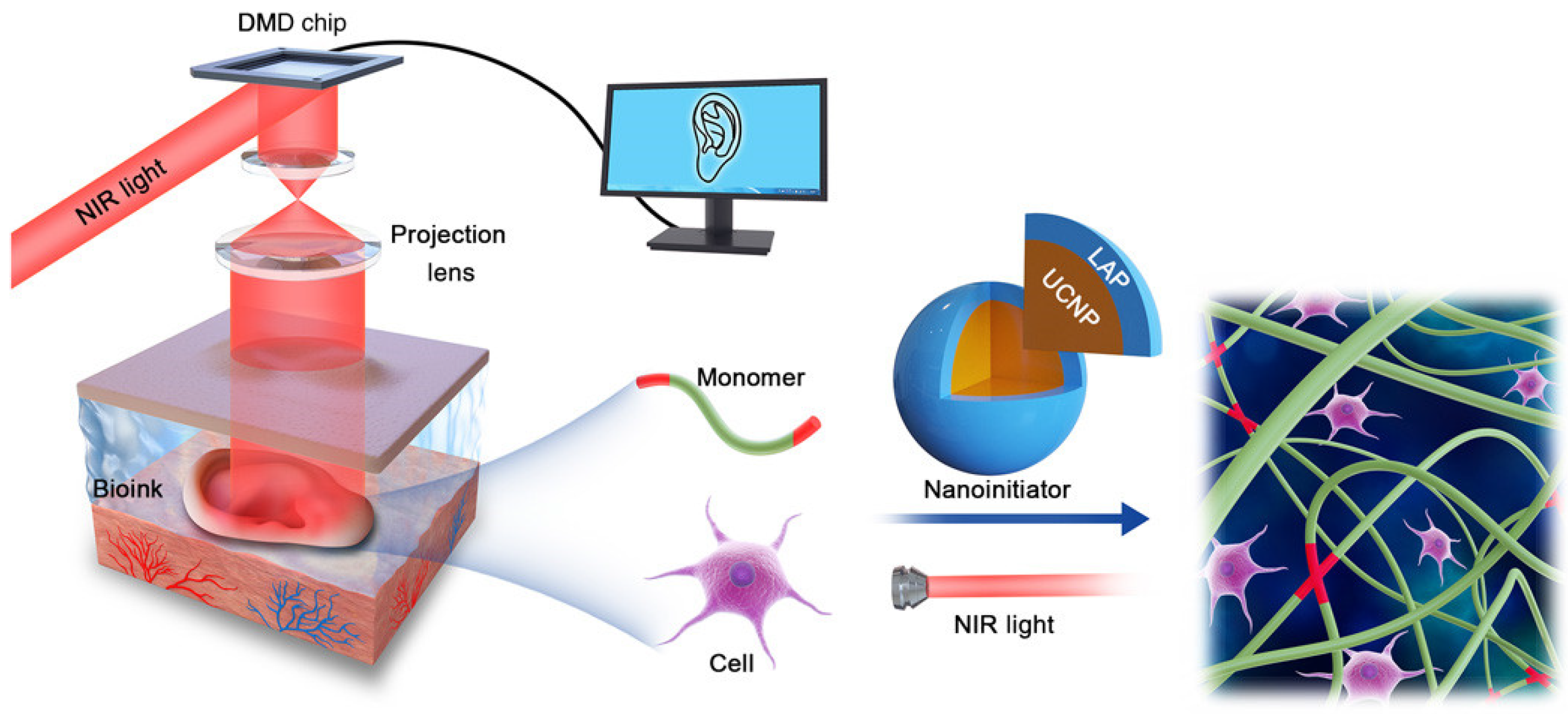
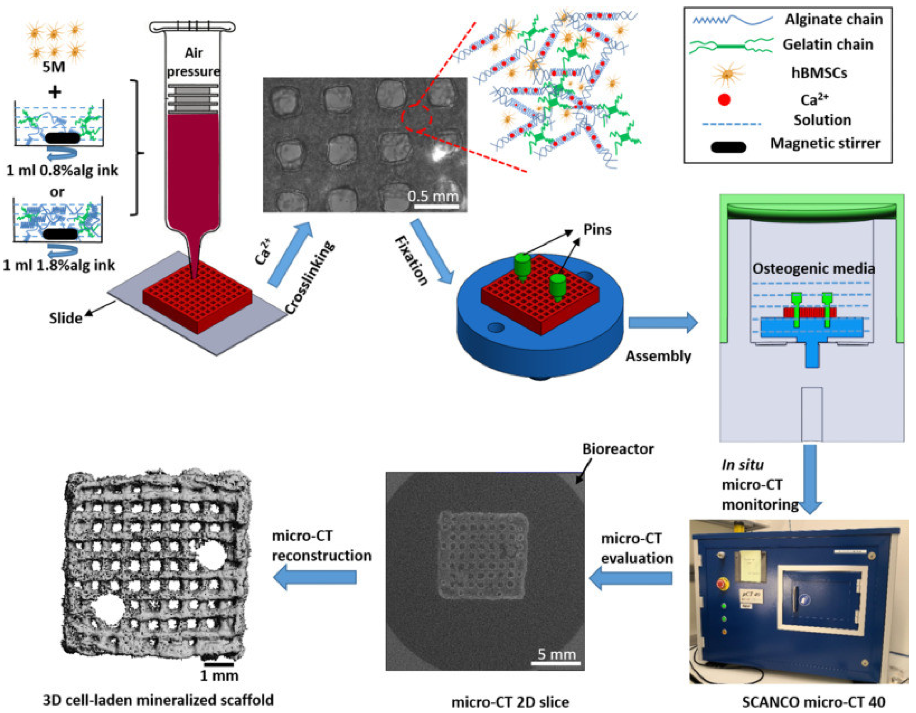
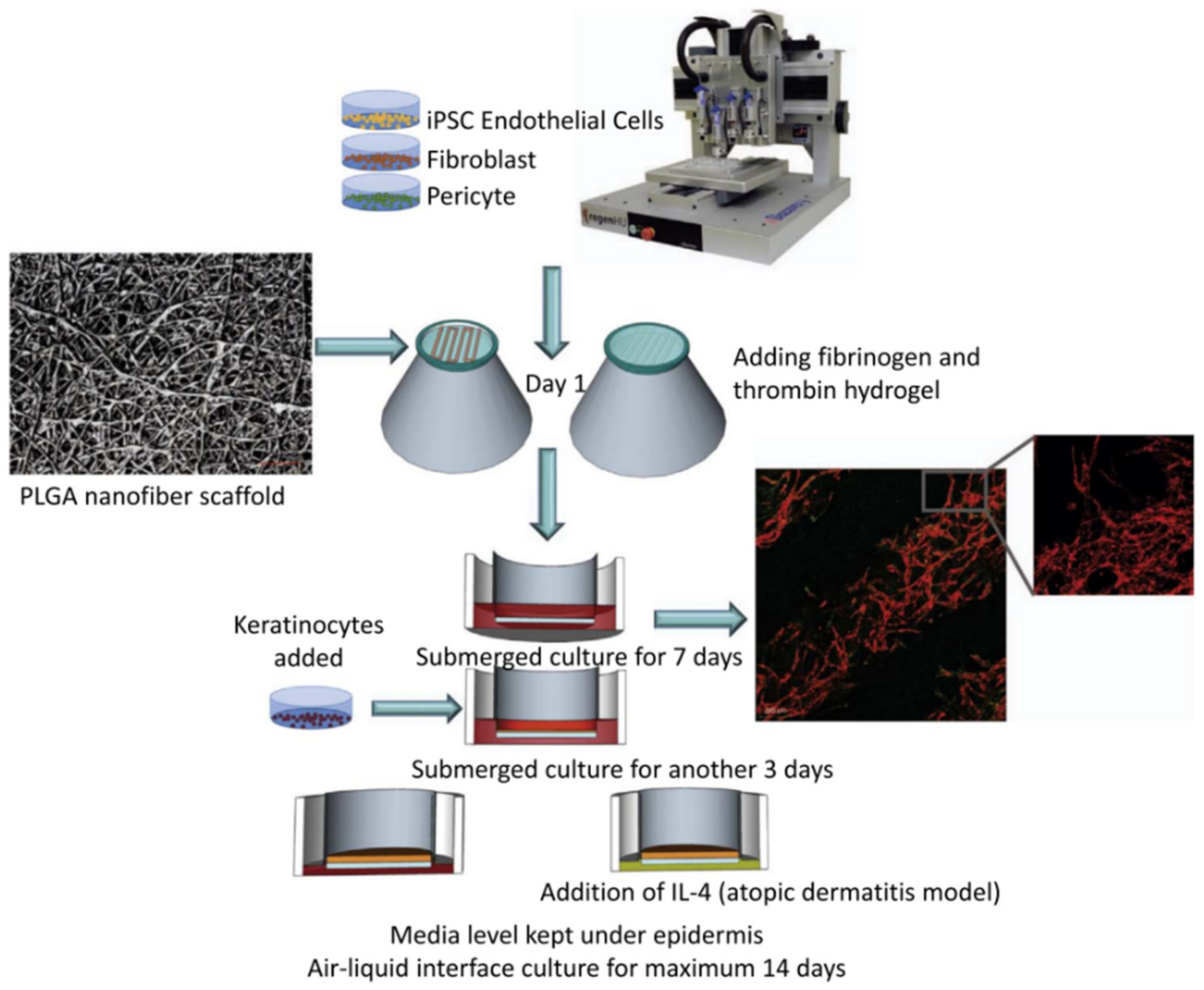
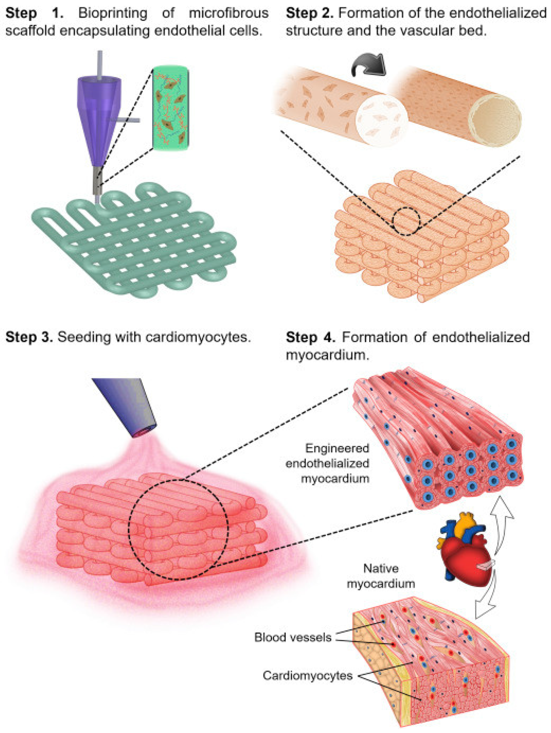
Table 1.
Types of 3D bioprinting technologies and their advantages and disadvantages.
| 3D Bioprinting Technologies | Advantages | Disadvantages | References |
|---|---|---|---|
| Inkjet bioprinting | Noncontact, easy, low cost, high cell vitality, and high speed | Few materials, low driving pressure, low printing accuracy, and small-size structures | [37,40] |
| Extrusion bioprinting | Wide range of materials, low cost, simple process and easy to use, good printability and fidelity, and large-size structures with preferred shapes and forms | Nozzle blockages, longer print times, low cell viability and missing materials, and layer-by-layer deposition limitations | [44,45,46] |
| Laser-assisted bioprinting | No nozzles, high resolution, automation, high cell vitality, and high repeatability and efficiency | The workstation is complex and requires a laser source | [47,48] |
| Stereolithography bioprinting | High print resolution, large-scale tissue, rapid printing, and high cell viability | Limited ability to capture the spatial heterogeneity | [50,51] |
| Suspension bioprinting | Stabilizes the gel form, maintains cell viability, broadens the application range of printing materials, and can encapsulate very-low-viscosity bioinks | The printing temperature is determined by the temperature of the suspension medium | [55,56] |
| Digital 3D bioprinting technologies | Higher spatial resolution, simple, faster print times, better cell viability, and can perform noninvasive 3D biological printing of tissue structures in vivo | More complex workstations, high-precision instruments, composite functional hydrogel biological ink materials | [56,60] |
Table 2.
Biological properties of 3D bioprinting hydrogels loaded with different cells or factors.
| Cells/Factors | Hydrogel Composition | Target Tissue | Cellular Response | Bioprinting Method | Ref. |
|---|---|---|---|---|---|
| CCD-986Sk cells | SA (2.5%)-XG(6%) @cCNCs (55%) | Skin | >80% cell viability, up to day 14 | Extrusion bioprinting | [80] |
| Human foreskin fibroblast cells | Methylcellulose/Alginate(2%) | Skin | >90% cell viability, up to day 7, high metabolic activity | Extrusion bioprinting | [81] |
| FBs, HUVECs, DPCs, EPCs | Gelatin (20%)-alginate (3%) | Skin | >80% cell viability, up to day 7, HF regeneration | Extrusion bioprinting | [18] |
| HUVECs | GelMA (10%) | Vessels | >90% cell viability, high proliferation rate, endothelial cell functionalization | Digital 3D bioprinting technologies | [82] |
| HUVECs, hBMSC | Alginate (1%), fibrin (30 mg/mL) and GelMA (5%) | Vascularisation and bone formation | VEGF, FGF-2, ANG-1 and EGF increased, cell differentiation | Extrusion bioprinting | [83] |
| Platelet lysate | PL (160 mgmL−1)-CNC (2.88%) | Tissues and organ surrogates | High cell viability, showing exceptionally fast spreading, growth, and synthesis of new ECM | Extrusion bioprinting | [84] |
| IL-4, MSCs | GelMA (20%)-dex (10%) | Skin | Excellent cytocompatibility, IL-4 and MSCs can synergistically induce macrophage polarization towards an anti-inflammatory M2 phenotype | Extrusion bioprinting, digital 3D bioprinting technologies | [85] |
| hMSCs | PEG (10%)-GelMA (1.5%) | Bones | >80% cell viability, the printed stem cells exhibit strong osteogenic and chondrogenic differentiation abilities | Inkjet bioprinting | [86] |
| hiPSC-MSCs, hSMCs | PEGDA (3%)-GelMA (7%) | Vessels | >80% cell viability, cell viability and physiological functions can be maintained at a high level | Stereolithography bioprinting | [50] |
| hiPSCs | HA(15%) | Tissuea and organa | Higher cell viability | Laser-assisted bioprinting | [87] |
| HUVECs | HA (3%) | Cell scaffolds | >90% cell viability, up to day 7 | Suspension bioprinting | [54] |
Disclaimer/Publisher’s Note: The statements, opinions and data contained in all publications are solely those of the individual author(s) and contributor(s) and not of MDPI and/or the editor(s). MDPI and/or the editor(s) disclaim responsibility for any injury to people or property resulting from any ideas, methods, instructions or products referred to in the content. |
© 2023 by the authors. Licensee MDPI, Basel, Switzerland. This article is an open access article distributed under the terms and conditions of the Creative Commons Attribution (CC BY) license (https://creativecommons.org/licenses/by/4.0/).
Share and Cite
MDPI and ACS Style
Xie, M.; Su, J.; Zhou, S.; Li, J.; Zhang, K. Application of Hydrogels as Three-Dimensional Bioprinting Ink for Tissue Engineering. Gels 2023, 9, 88. https://doi.org/10.3390/gels9020088
AMA Style
Xie M, Su J, Zhou S, Li J, Zhang K. Application of Hydrogels as Three-Dimensional Bioprinting Ink for Tissue Engineering. Gels. 2023; 9(2):88. https://doi.org/10.3390/gels9020088
Chicago/Turabian StyleXie, Mengbo, Jingjing Su, Shengxi Zhou, Jingan Li, and Kun Zhang. 2023. "Application of Hydrogels as Three-Dimensional Bioprinting Ink for Tissue Engineering" Gels 9, no. 2: 88. https://doi.org/10.3390/gels9020088
Note that from the first issue of 2016, this journal uses article numbers instead of page numbers. See further details here.