
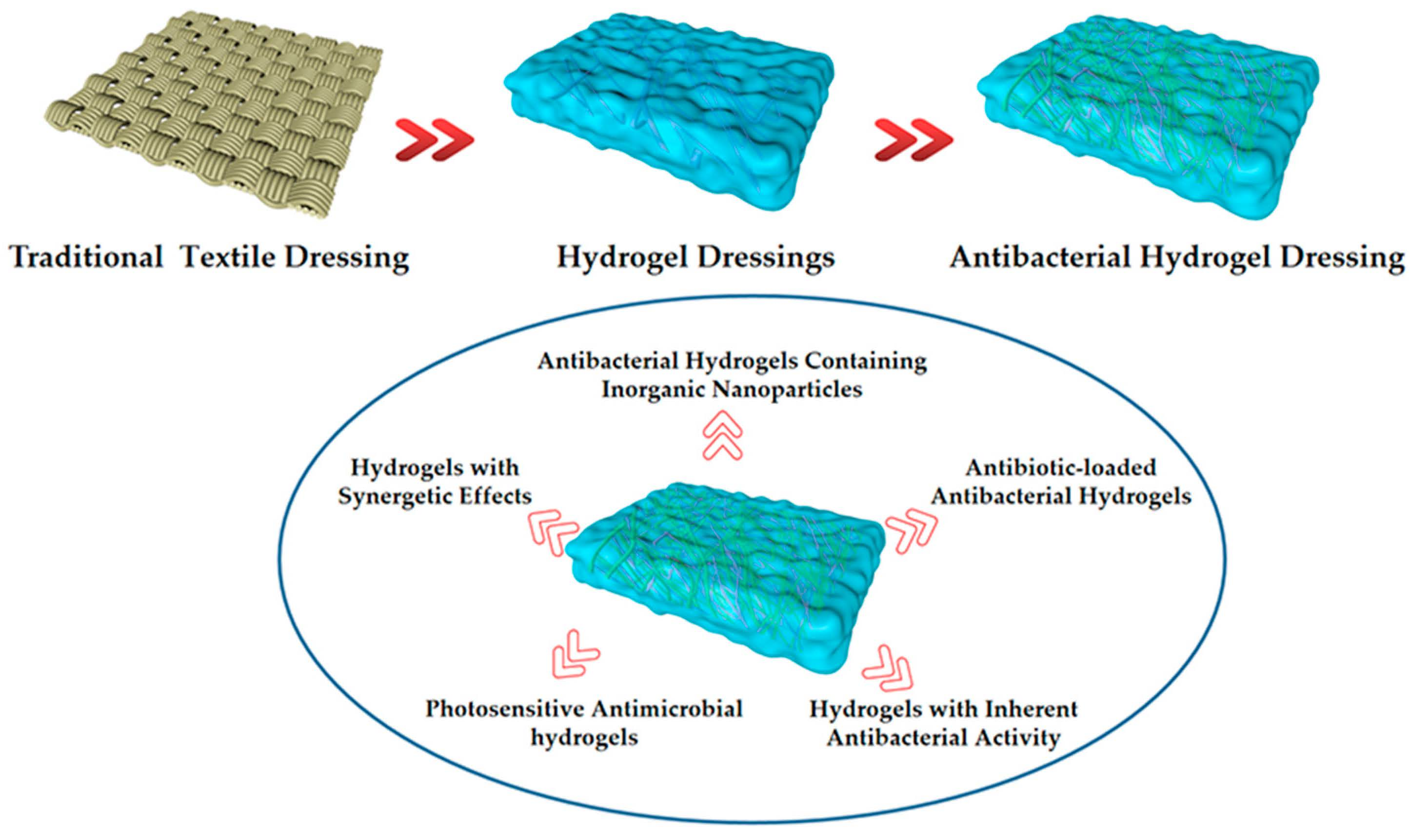
Progress in Antibacterial Hydrogel Dressing

1
College of Light Industry and Textile, Qiqihar University, Qiqihar 161006, China
2
Engineering Research Center for Hemp and Product in Cold Region of Ministry of Education, Qiqihar 161006, China
*
Authors to whom correspondence should be addressed.
Gels 2022, 8(8), 503; https://doi.org/10.3390/gels8080503
Submission received: 14 July 2022
/
Revised: 7 August 2022
/
Accepted: 9 August 2022
/
Published: 12 August 2022
(This article belongs to the Special Issue Antimicrobial Hydrogels)
Abstract
:Antibacterial hydrogel has excellent antibacterial property and good biocompatibility, water absorption and water retention, swelling, high oxygen permeability, etc.; therefore, it widely applied in biomedicine, intelligent textiles, cosmetics, and other fields, especially for medical dressing. As a wound dressing, the antibacterial hydrogel has the characteristics of absorbing wound liquid, controlling drug release, being non-toxic, being without side effects, and not causing secondary injury to the wound. Its preparation method is simple, and can crosslink via covalent or non-covalent bond, such as γ-radiation croFsslinking, free radical polymerization, graft copolymerization, etc. The raw materials are easy to obtain; usually these include chondroitin sulfate, sodium alginate, polyvinyl alcohol, etc., with different raw materials being used for different antibacterial modes. According to the hydrogel matrix and antibacterial mode, the preparation method, performance, antibacterial mechanism, and classification of antibacterial hydrogels are summarized in this paper, and the future development direction of the antibacterial hydrogel as wound dressing is proposed.

1. Introduction
Human skin is an effective barrier to protect the subcutaneous tissue from microbial penetration, and dressing is a temporary substitute skin barrier function of medical textiles. Trauma to the skin can cause many reactions, including increased metabolism, decreased body temperature, excessive loss of water and protein, and imbalance of the endocrine and immune systems. Because of the different causes of wound formation, wound size, shape, exudate volume, and other aspects also vary; therefore, wound dressing requirements are also different [1]. The traditional textile dressing, though its price is lower, has no practical anti-infection function, is dry and easily adheres to the wound, and is not conducive to the anti-infection wound exudate absorption and healing. Most traditional dressings have only a single biological activity, which cannot be applied quickly and efficiently to treat different wounds [2]. Wounds can be acute or chronic. An acute wound is a wound that forms suddenly and will heal soon; a chronic injury does not heal as quickly as expected and usually takes one to three months or longer to heal [3]. The main reason for the failure of chronic wounds to heal quickly is the accumulation of microorganisms in the wound bed. Chronic wound infections can lead to complications such as amputation, sepsis, and even death [4]. Wound stent dressings can treat chronic wounds, such as those caused by dermal injuries or burns, which can help reduce scar formation [5]. There are mainly 3–10 dominant microorganisms in the wound, including Staphylococcus aureus, Streptococcus, Pseudomonas, anaerobic bacteria, etc. In addition, there are hundreds of microorganisms of different species attached and embedded in the extracellular matrix of biofilm [6]. The ideal dressing should be non-toxic, have no side effects, be moisturizing and absorbent, and promote healing without causing secondary injury, rather than merely acting as a protective barrier [7,8,9]. Therefore, the development of new medical dressings has gradually become more popular. Various wound dressings have been studied, such as rubber [10,11,12], polyionic liquid membrane [13,14], electrostatically spun nanofibers [15,16,17], and hydrogel [18]. Among these dressings, a hydrogel is the material that best meets people’s requirements for ideal dressings [19]. In 1960, Wichterle and Lim successfully prepared a hydrogel by polymerizing methyl-2-hydroxyethyl methacrylate and made the first contact lens in history [20], thus starting the application research on hydrogels.
Hydrogel is a kind of polymer material with a three-dimensional network structure, which is composed of polymer chains crosslinked through physical or covalent bonds [21,22]. Since they contain a large number of hydrophilic groups, hydrogels can absorb tens or even hundreds of times their own mass of water; they are of high hydrophilicity, display good swelling in water, and have good water retention [23]. Due to high water content, hydrogels can cool the wound and reduce the warm feeling associated with the inflamed tissue [24]. With good biocompatibility and cell adhesion, hydrogels can directly contact the injury, reduce the loss of body fluid, and prevent secondary infection injury. They can be used locally to help wound healing [25,26,27]. Hydrogels can also be coated on catheters, central venous catheters, joint implants, and dental implants for drug release [28,29,30]. By grafting hydrogel monomer onto fibers, coating hydrogel onto fabrics, intelligent textiles, or textile dressings, and printing and dyeing, wastewater adsorption materials can be made [31,32]. Due to a series of excellent properties, hydrogels have received much attention, especially in biomedical fields [33,34]. However, with the abuse of traditional hydrogels, bacteria become resistant to antibiotics, and conventional hydrogels gradually lose their advantages. To solve these problems, antibacterial hydrogels, which have the dual functions of hydrogel and antibacterial, were found. The preparation process of antibacterial hydrogels is simple and the structure is diverse, which attracts attention, and a variety of advanced antibacterial hydrogels have been developed successively.
Antibacterial hydrogel has been widely used in many fields, such as biomedicine and intelligent textiles, and it is one of the suitable biological materials for drug delivery in the area of antibacterial treatment. Antibacterial hydrogels have also become a new focus of research, and their research and application have also begun to develop rapidly. Various preparation methods of antibacterial hydrogels have been widely used in biomedical, tissue-engineering, and intelligent textiles, such as drug delivery, regenerative medicine, trauma dressing, vitro diagnosis, etc. [35]. As a medical dressing, an antibacterial hydrogel can have multiple properties simultaneously. The most important property is bacteriostatic or antibacterial. The antibacterial spectrum of antibacterial hydrogels is determined by the antibacterial components carried, and various materials have different antibacterial properties against different strains [36]. There are many methods for testing antibacterial properties of antibacterial hydrogels, such as the bacteriostatic circle method, oscillation method, plate colony counting method, OD counting method, scanning electron microscopy method, etc. Among them, the bacteriostatic circle method is commonly used because of its simple operation and intuitive results [37,38]. For example, the silver nanoparticles doped conductive polymer hydrogel system (Ag-NPs/CPH) can be used as a dressing, which has good electrical conductivity, biocompatibility, mechanical properties, and antibacterial properties, and can be used to heal seriously infected injuries [39]. TiO2 combined with nanoparticles also has bacteriostatic properties. Khashan et al. prepared TiO2 nanoparticles by liquid laser ablation, which has excellent bacteriostatic properties, and can be applied in hydrogels to better inhibit bacteria [40]. After the copolymer of methylacryamide (MA Ade) and 3-dimethyl (methylacryl oxyethyl) ammonium propane sulfonate (DMAPS) was composed, and chitosan was introduced and the synthesized PDMAPS-co-PMA-Ade/chitosan hydrogel was used as dressing with antibacterial activity and repeated adhesion ability. Wang et al. synthesized the polyionic liquid antibacterial hydrogel PMAV. They have high protein adsorption properties, good mechanical properties, and biocompatibility. They can be used repeatedly and maintain good antibacterial properties. They are available for reuse as non-releasing antibacterial hydrogel dressings [41,42].
To prevent the infection of pathogenic microorganisms in vitro, the use of antibacterial hydrogel dressing directly in contact with human tissue can effectively isolate microorganisms and maintain oxygen to the wound site in order to aid the wound in healing as soon as possible. This paper summarizes the mechanism, performance, and preparation of antibacterial hydrogels as wound dressing, and finally, prospects for the development of antibacterial hydrogels.
2. Preparation of Antibacterial Hydrogels
The raw material of antibacterial hydrogel for dressing mainly consists of three parts, namely the monomer, initiator, and crosslinking agent. In general, hydrogels are prepared by hydrophilic monomers. In some exceptional cases, hydrophobic monomers can be added to adjust some specific application properties of hydrogels.
There are various methods for preparing antibacterial hydrogels, such as the radiation method, chemical crosslinking, physical crosslinking, polymerization grafting, free radical crosslinking polymerization, chemical–physical crosslinking, and so on. The raw materials used are different, the preparation methods are other, and the strains inhibited are other, but all of them have significant antibacterial effects. The preparation methods, materials, antibacterial ability, and application of various antibacterial hydrogels are shown in Table 1.
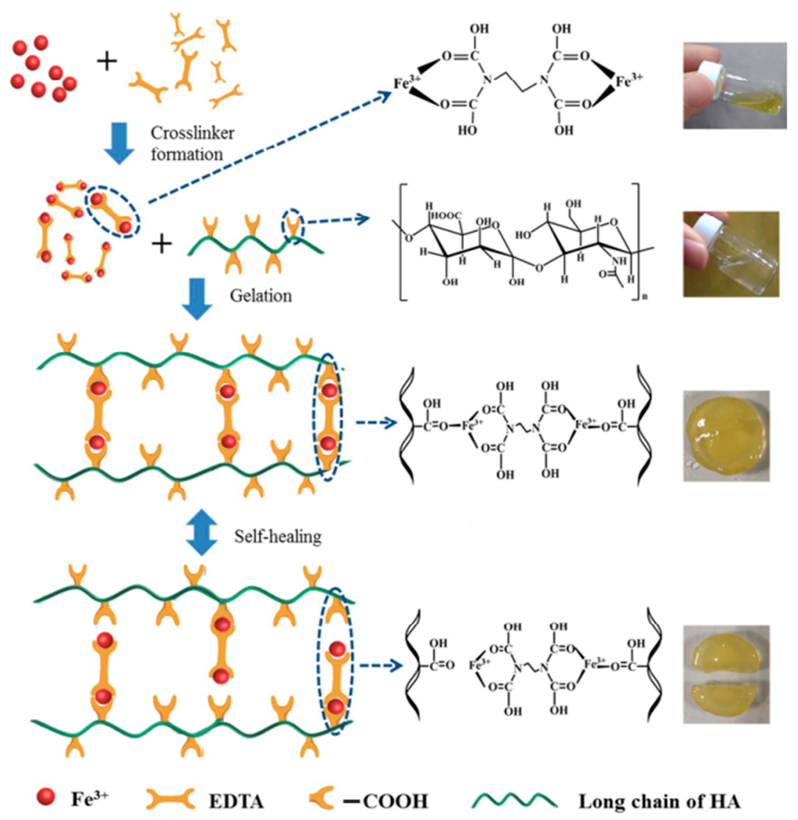
Physical crosslinking is formed by non-covalent crosslinking, such as hydrogen bonds, interactions between hydrophobic groups, ion complexation, electrostatic interactions, etc. Hydrogels formed by physical crosslinking are quickly dissolved when environmental conditions such as temperature, pH value, and the ionic strength of solution change [43]. For example, Tian et al. used sodium ferric diamine tetraacetate (EDTA-Fe3+) as a crosslinking agent, physically crosslinking with hyaluronic acid (HA), and a hydrogel that can be triggered by bacteria at the site of infection and can quickly self-heal was developed. The dynamic ionic bond between EDTA-Fe3+ and HA can rapidly reestablish within minutes. When used as a dressing, HA can degrade at the site of infection, and hydrogels can release a Fe3+ complex locally. The complex is quickly adsorbed by surrounding bacteria and reduced to Fe2+, which produces hydrogen peroxide (H2O2) with bacteria and inflammatory cells. It forms hydroxyl free radicals through the Fenton reaction to destroy the bacterial structure. It can effectively kill Escherichia coli and Staphylococcus aureus, promote angiogenesis, and create new skin within ten days. The formation and self-healing mechanism of the HA-Fe-EDTA hydrogel is shown in Figure 1 [44].
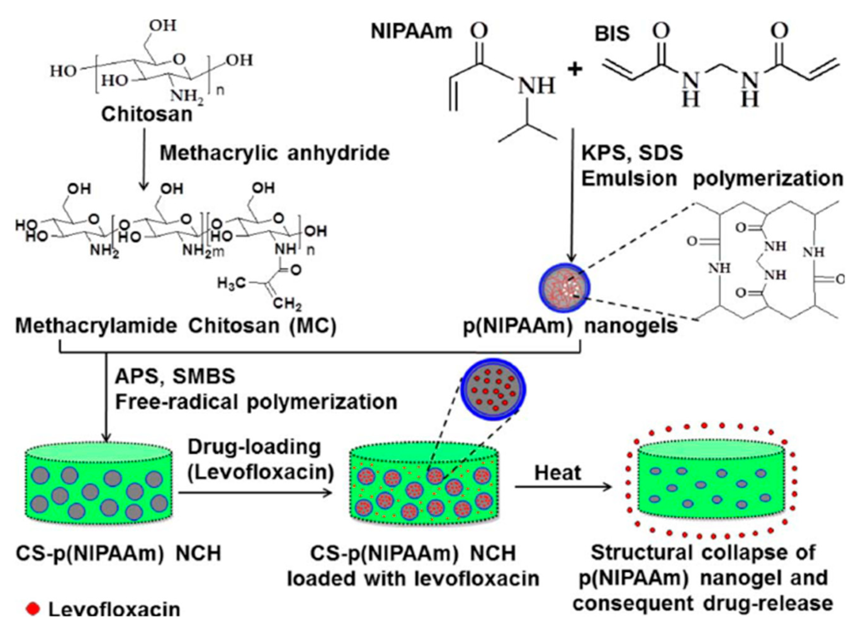
Chemical crosslinking hydrogels are made by covalent crosslinking, and polymer chains are covalently bonded by crosslinking agents [45]. Hydrogels formed by chemical cross-linking are relatively stable. For example, Li et al. studied the preparation of polyurethane/polyvinyl alcohol hydrogels by chemical crosslinking and in situ synthesis. Adding silver particles to the hydrogel can improve the antibacterial activity of the hydrogel; improve Young’s modulus, tensile strength, and elongation; and maintain the water absorption ability good biocompatibility of the hydrogel, which is an excellent choice for wound dressing [46]. Shin et al. prepared thermoresponsive nanocomposite hydrogels by combining nanostructured particles with 3D hydrogels. The preparation of CS-p(NIPAAm) nanocomposite hydrogel is shown in Figure 2. Poly(N-isopropyl acrylamide) [P(NIPAAm)] nanocomposite hydrogels were synthesized by emulsion polymerization and introduced into methyl acrylamide CS (MC) solution. They were then embedded into the hydrogel for free radical-induced crosslinking reaction. Finally, levofloxacin (LFX) was loaded into the nanocomposite hydrogel to enhance the drug loading ability of the hydrogel [47].
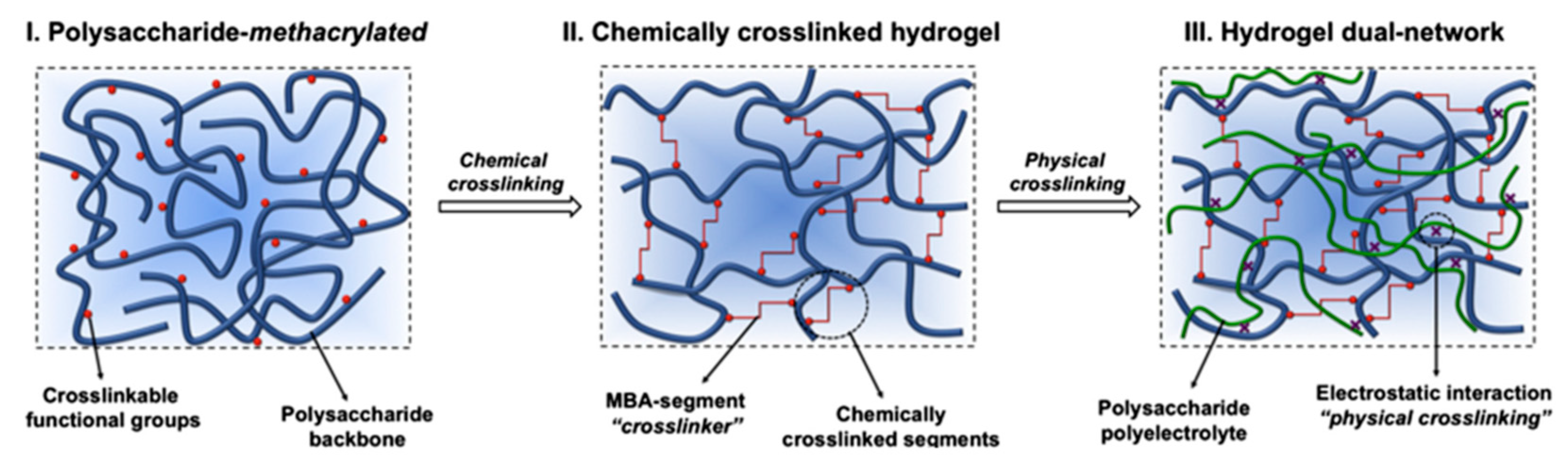
Chemical–physical crosslinking. Fajardo et al. used K2S2O8 as an initiator and glycidyl methacrylate (GMA), chitosan (CHT), or chondroitin sulfate (CS) as raw materials to synthesize polymers. Then, chemical hydrogels (CHT-gel and CS-gel) were prepared by chemical crosslinking using N,N-methylene bisacrylamide (MBA) as a cross-linking agent. The two chemical hydrogels were then immersed into the CS or CHT stock solution for physical crosslinking. Due to the polyelectrolyte complexation between CHT and CS chains, the dual network antibacterial hydrogels (CHT-gel/CS and CS-gel/CHT) were prepared. The formation of a double network hydrogel is shown in Figure 3.
Radiation crosslinking refers to the technical means of a crosslinking reaction between long polymer chains caused by radiation. Alcantara et al. prepared antibacterial hydrogels using poly(N-vinyl-2-pyrrolidone) (PVP) and polyvinyl alcohol (PVA) by the method of radiation crosslinking. The aqueous solution containing AgNO3 was added to PVP and PVA by γ-ray irradiation with a 60 Co source, which could realize the crosslinking of polymers and the synthesis of AgNPs simultaneously. PVP/AgNPs hydrogel has antibacterial properties against Pseudomonas aeruginosa and Staphylococcus aureus. PVA/AgNPs hydrogel has antibacterial properties against Pseudomonas aeruginosa and antibacterial activity against Staphylococcus aureus. Two antibacterial hydrogels can be used as dressings to treat common wounds and burns [49].
In a free radical polymerization reaction, the monomer molecules are activated into active free radicals under the action of light, heat, radiation, and initiator, then linked with the monomer polymerization to form polymers. Wang et al. effectively prepared CMO-loaded composite emulsion hydrogel by acrylamide radical polymerization using oil-in-water concentrated emulsion as a continuous phase. The prepared CMO-Loaded composite hydrogel has good long-term antibacterial activity against Staphylococcus aureus and Escherichia coli [50].
Graft copolymerization is a reaction in which a macromolecular chain binds an appropriate branch or functional side group by chemical bonding. Grafting modification of polymer is an effective method to improve the properties of polymer materials. The supernormal transition metals Ce (IV) and Cu (III), which react with −OH groups, can produce free radicals to initiate vinyl polymerization. Ma et al. prepared antimicrobial hydrogels by grafting polymers on the surface of Heloxite nanotubes (HNT) with dimethyl aminoethyl methacrylate (DMAEMA) and sodium acrylate (AA-NA) using a REDOX system mediated by supernormal transition metals (Ce(IV) and Cu(III)) in aqueous medium at 35 °C. The prepared polycationic grafted nanotubes and polyanionic grafted nanotubes can be mixed in an aqueous medium to form hydrogels, and the prepared hydrogels have an antibacterial effect on Escherichia coli [51].

Table 1.
Preparation methods, properties, and application of various antibacterial hydrogels.
| Synthetic Methods | Species of Hydrogels | Materials | Antimicrobial Capability | Application | Ref. |
|---|---|---|---|---|---|
| Chemical crosslinking | Acacia gum-PVA hydrogel | Acacia gum, PVA, glutaraldehyde, salicylic | Against Bacillus subtilis, P. aeruginosa, E. coli and S. aureus | Wound dressing | [52] |
| Silk fibroin crosslinked glycyrrhizic acid and silver sydrogels | SF, Ag, GA | Against S. aureus, P. aeruginosa | [53] | ||
| PHCI hydrogel | 1,3-dibromo-2-propanol, trans-1,4-cyclohexanediamine | Can adsorb and kill S.aureus and E.coli electrostatically | [54] | ||
| Silk fibroin/chitosan hydrogel | SF, CS, LiBr | Against Bacillus subtilis, methicillin-resistant S. aureus, and E. coli strains with a contact-killing efficacy of 100% | [55] | ||
| Physical crosslinking | Antibacterial chitosan/silver bio-nanocomposite | STPP, chitosan, AgNPs | The antibacterial activity against E. coli and S. aureus lasted for 1 week. | Drug carrier | [56] |
| Polysaccharide based physically crosslinked double-network antibacterial hydrogel | SA, CS, Zn2+ | Against E. coli and S. aureus | Biomedicineureus fields | [57] | |
| AA-Al3+-MGA-[VBIm]Br hydrogel | AA, 1-vinyl-3-butylimidazolium, COOH-modified gum arabic, AlCl3 | Against E. coli, S. aureus, and C. albicans. | Wound dressing | [58] | |
| PVA-TA hydrogel | PVA, TA | Against E. coli and S. aureus | Biomedical fields | [59] | |
| Freezing-thawing cycles | Nano-TiO2/CMCS/PVA composite hydrogel | PVA, CMCS, Nano-TiO2 | Against E. coli and S. aureus | Cosmetics, medical dressings | [60] |
| AgNPs and PVA/CH hydrogel | AgNPs, PVA, CH | Against gram + ve and gram − ve bacteria | Wound dressing | [61] | |
| A polyvinyl alcohol (PVA) hydrogel loaded with guava leaf extract (GLE) | GLE, PVA | Against S. aureus and P. aeruginosa. | [62] | ||
| Uv crosslinking method | PVA-SbQ/MMT composite hydrogel | MMT, PVA-SbQ | against S. aureus was up to 99.95%. | Wound dressing | [63] |
| Solution polymerization | ZnO@GDM hydrogel | ZnO, GelMA, DMAA, MAA | Against E.coli reached more than 98% | Biomedical fields | [64] |
| Poly(DMA-co-AAc) hydrogel | DMA, AAc, ammonium persulfate | Inhibit the growth of S. aureus. | Antibacterial materials | [65] | |
| Photoinduction | Hydrogel containing silver nanoparticles | AgNO3, MDEA, acrylamide, bis-AAm | Fully inhibition of the growth of Acinetobacter johnsonii and E. coli. | Wound dressing | [66] |
| Antibacterial acrylamide hydrogels containing silver | Acrylamide, silver nitrate, trisodium citrate dihydrate,1-[4-(2-hydroxy-ethoxy)phenyl]-2- hydroxy-2-methyl-1-propane-1-one | Against pathogenic E. coli O157:H7, S. aureus, and non-pathogenic E. coli K-12 | Water-based applications | [67] | |
| Chitosan-PEG hydrogels | Chitosan derivatives, PEG | 100% inhibition of the E. coli and S. aureus | Bio-functional materials | [68] | |
| Coacervation | CS@CMC@ZeoliteP@KDF hydrogel | ZeoliteP, KDF, CMC, CS | Inhibit the growth of E.coli and S. aureus. | Bacteriostatic agent | [69] |
| Chemical–physical crosslinking | DR-CC hydrogel | Carboxylated chitosan, diazoresin | Can kill E. coli and S.aureus. | Wound dressing | [70] |
| β-CD/PEI/PVA composite hydrogels | β-CD, PVA, PEI | Against E. coli and S. aureus | [71] | ||
| Lignin hydrogels | SBMA, lignin-MA | Antimicrobial performance of 94.8% reduction of E. coli and 95.7% of S. aureus. | Biomedical fields | [72] | |
| Chitosan/PV A-based hydrogels | CS, PVA | Against S.aureus and K. pneumonia | Oral dressing | [73] | |
| Free radical crosslinking polymerization | P(MMA-co-MAA)/Ag nanocomposite | MMA, MAA, AgNPs | Against S. aureus and B. subtilis. | Smart material | [74] |
| Poly(N-[3-(dimethylaminopropyl)] methacrylamide) hydrogels | Cetyltrimethylammonium bromide, KPS, BIS | Against E. coli | Antimicrobial agent | [75] | |
| Tea Ag nanocomposite hydrogels | Acrylamide, MBA, TEMDA, KPS | Against E. coli and S. aureus. | Wound dressing | [76] | |
| D/SD-g-PAA-based hydrogels | MBA, AA | Can kill S. aureus and E. coli and Klebsiella spp. | [77] | ||
| Gamma-ray irradiation | Metronidazole/poly(acrylic acid) hydrogel | Metronidazole, poly(acrylic acid) | Against E. coli, S. aureus, S. mutans. | Wound dressing | [78] |
| P-PVA hydrogel | phosphorus-containing PVA | Against various fungi and bacteria | Biological fields | [79] | |
| Poly(Agar-co-AAc) hydrogels | AAc, agar | Against E. coli and S. aureus. | Wound dressing | [80] | |
| Polyvinyl pyrrolidone/carboxymethyl cellulose hydrogels | Polyvinyl pyrrolidone, carboxymethyl cellulose, AgNPs | Against bacterial, S. aureus, P. aeruginosa, E. coli and Candida albicans | Therapeutic dressing | [81] | |
| Reverse emulsification- diffusion | Alginate nano hydrogel | Alginate, iron (II)-chloride, sodium lactate | Against E. coli and S. aureus. | Smart textile | [82] |
3. Performance of Antibacterial Hydrogels
As an ideal medical dressing, antibacterial hydrogel should have the following properties: excellent antibacterial performance, high permeability, good biocompatibility, and water absorption capacity.
3.1. Antibacterial Properties
As a medical dressing, antibacterial performance is the primary performance that a hydrogel must have. It should have an antibacterial or bacteriostatic effect, inhibit the breeding of harmful microorganisms, and resist wound infection [83]. The antibacterial effect is mainly tested by the bacteriostatic zone method; that is, the bacteriostatic zone diagram and diameter comparison of hydrogel against Escherichia coli (typical Gram-negative bacteria) and Staphylococcus aureus (specific Gram-positive bacteria) were tested via an in vitro antibacterial experiment. For example, Gharibi et al. prepared an antibacterial hydrogel containing torymic, Si-CAQ (3-glycidyloxypropyl) trimethoxysilane, and poly(vinyl alcohol) for wound dressing. The hydrogel has good antibacterial activity against Bacillus subtilis, METHicillin-resistant Staphylococcus aureus, and Escherichia coli, and the bactericidal effect is 100%. It can prevent the development of infection at the injured site and promote wound healing [84]. Sun et al. prepared sodium alginate double crosslinking hydrogel fibers loaded with sulfadiazine and crosslinked with calcium ion and glutaraldehyde. The sodium alginate hydrogel fibers had good mechanical strength, biocompatibility, and sustainable drug release, and the antibacterial test showed that the antibacterial hydrogel could kill 99.9% of Staphylococcus aureus and Escherichia coli [85].
Withanage et al. prepared (hyaluronic acid)/(spider silk) hydrogels by the method of chemical crosslinking. Antibacterial properties of hyaluronic acid/spider silk-based hydrogels are shown in Figure 4. The water absorption capacity of the hydrogel can reach 15–30 times its weight and has vigorous antibacterial activity against Gram-negative and Gram-positive bacteria. It is suitable for wound dressings, prosthesis implant coatings, and catheter coatings [86].
3.2. Water Absorption and Swelling Capacity
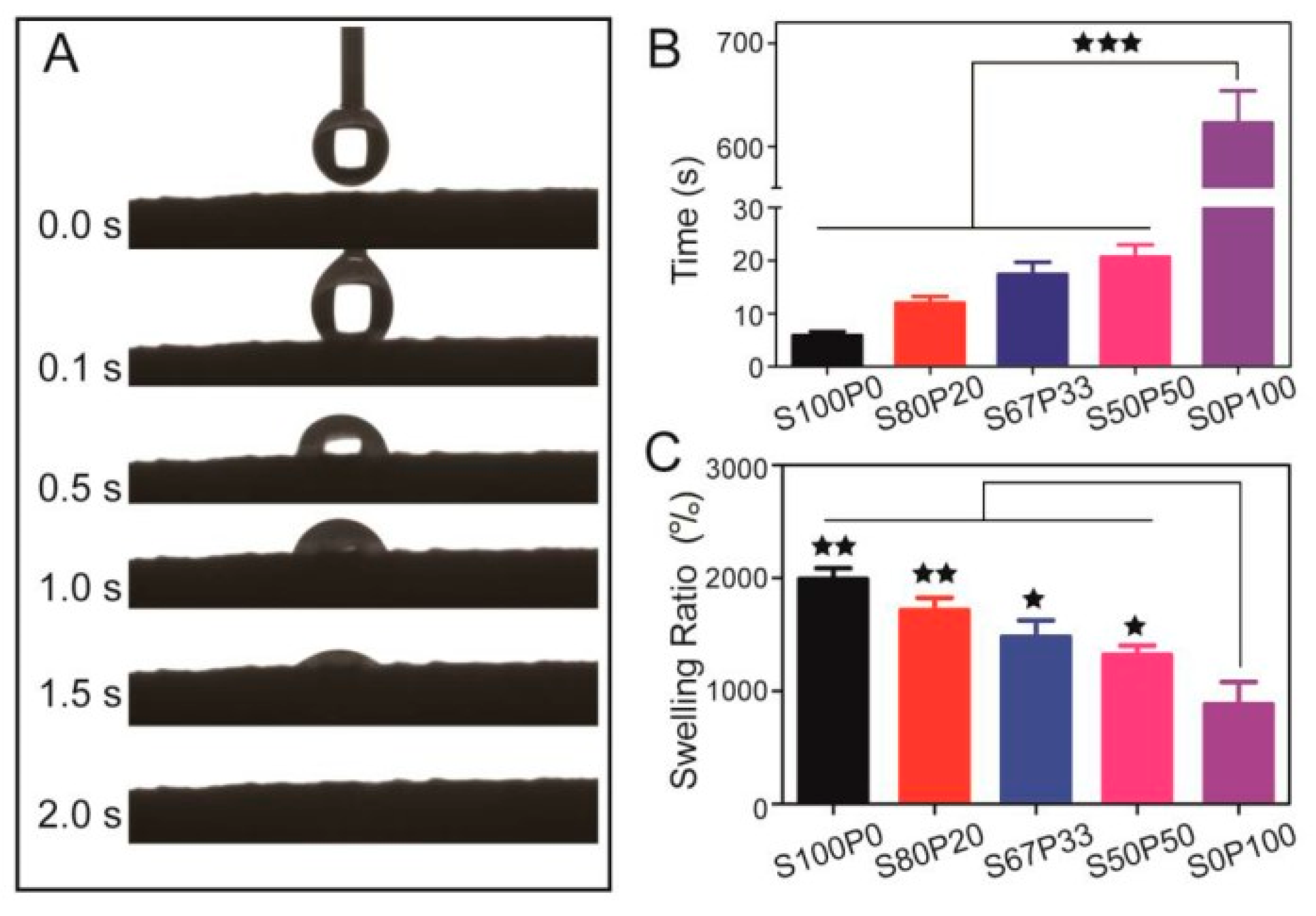
Because they contain a large number of hydrophilic groups, hydrogels have intense swelling and water absorption capacity. Swelling occurs when the gel absorbs water, which is crucial for the gel, because it affects the spread of the bioactive agent and drug that is trapped. When applied in wound dressings, they can absorb tissue fluid or blood from the wound, keep the wound surface moist, and prevent the loss of tissue fluid. Generally, the water absorption properties of treated materials are studied by moisture regain, water retention, wet fabric drying time at room temperature, and vertical core wicking test. For example, Zhang et al. prepared an antibacterial wound dressing by loading antibacterial drugs onto aramid nanofiber (ANFs) hydrogel. The water content of ANFs hydrogels is more than 98 percent, they have high water adsorption properties (>10,000%) and good water retention ability (water retention of more than 5000 percent after incubating at RH 30% for eight h), and they are capable of absorbing and retaining the wound exudate to form a moist environment [87]. Khan et al. developed a chitosan-based antibacterial hydrogel, which has a swelling ratio of 1036%; anda water absorption ratio of more than ten times [88]. Gang et al. designed a sericin/poly(vinyl alcohol) hydrogel as a drug delivery carrier for wound dressing; its water absorption and swelling behavior are shown in Figure 5.
Figure 5A shows that SS/PVA (S50P50) hydrogel absorbed water droplets in 2 s and had super hydrophilicity. Figure 5B shows the time required for different SS/PVA hydrogels to reach swelling equilibrium. The swelling equilibrium time of S50P50 is 20 s, indicating that its hydrophilicity is good. Figure 5C shows sericin and SS/PVA hydrogels had swelling ratios of 1200–1800 percent, whereas that of PVA had 800 percent, indicating SS/PVA hydrogel had the best swelling ability.
3.3. Drug Release Properties
The unique network structure makes antibacterial hydrogel very suitable for drug carriers. When used in wound dressings, it has excellent drug release performance, that is, the ability to continuously release antibacterial drugs, which can be sustained for a long time to promote wound healing, and can avoid frequent dressing changes, thus reducing the risk of wound overexposure to bacteria [90,91]. Drug release properties can generally be analyzed by measuring in vitro release time or cumulative drug release over a certain period. For example, Erdagi studied the neomycin release performance of the crosslinking gelatin/DGN-NC-based hydrogels; the results showed a rapid release within the initial 15 min and then sustained release for 9–12 h. The material is suitable for a polymer antibiotic matrix in wound dressings [92]. Ak. Prepared poly(vinyl alcohol) chitosan-loaded AgNPs hydrogel. They showed that the maximum release amount of the hydrogel was 91.83%, and the release time of the concentration-dependent inhibition zone (ZOI) was over 24 h, respectively [93]. Guan et al. prepared Ag/AgO/carboxymethyl chitosan antibacterial hydrogel, with a cumulative drug release of 75.20% within 12 h and a maximum inhibition rate of 92.32% on E. coli [94]. It is an effective method of drug delivery.
3.4. Permeability
To prevent the growth of anaerobic bacteria, leading to wound infection and inflammation, antibacterial hydrogel dressings need to have specific air permeability. Singer et al. developed a hydrogel wound dressing for drug delivery using polymerization of polysaccharide gum. The prepared hydrogel membrane was permeable to O2 and H2O vapors, but impervious to microorganisms, and can protect the wound surface from bacterial invasion [95]. Wang studied the application of Chitosan-graft-poly(N-isopropyl acrylamide) (CS-g-PNIPAAm) microgels in a water vapor-permeable fabric. It can meet the requirement of high water vapor permeability at high temperatures [96]. Yan prepared methyl acrylamide adenine and 3-(methyl acryloxyethyl)-dimethyl-ammonium propane sulfonate copolymerized hydrogels, and loaded chitosan to prepare semi-interpenetrating polymer network hydrogels (PDM/C). The WVTR of band-aid, gauze, and PDM/C hydrogel were 2399 ± 524, 2045 ± 131, and 781 ± 148 g/m2/day, respectively. The WVTR of PDM/C hydrogel was much smaller than that of the other two wound dressings, indicating that PDM/C hydrogel can effectively reduce water evaporation and keep the wound moist [97].
3.5. Biocompatibility
Biocompatibility is an essential performance of antibacterial hydrogels. As dressings, antibacterial hydrogels need to be non-toxic to cells and not toxic to human tissues. Zhu used several reagents including chitosan, glacial acetic acid, nanosilver, and poloxamer to prepare nanosilver antibacterial hydrogel. The antibacterial hydrogel made was found to have good biocompatibility and can effectively promote cell proliferation and differentiation without releasing any toxic substances through biocompatibility tests [98]. Chen et al. synthesized PaA-Cu-MOP hydrogel. After testing, PaA-Cu-MOP hydrogel can promote cell proliferation and blood vessel generation, up-regulate cytokines, and even outperform recombinant human epidermal growth factors. Therefore, PaA-Cu-MOP hydrogel has excellent biocompatibility and can accelerate wound healing [99].
4. Classification and Mechanism of Antibacterial Hydrogels
Depending on the antibacterial mode, an antibacterial hydrogel can be divided into five categories: (1) antibacterial hydrogel containing inorganic nanoparticles; (2) antibiotic-loaded antibacterial hydrogel that can directly kill cells to achieve sterilization; (3) hydrogel with inherent antimicrobial activity; (4) photosensitive antibacterial hydrogel; and (5) hydrogels with synergetic effects. Currently, most of the research is on antibacterial hydrogels containing silver nanoparticles, chitosan hydrogels, and antibiotic-loaded hydrogels.
4.1. Antibacterial Hydrogels Containing Inorganic Nanoparticles
As antibacterial agents, inorganic nanoparticles have attracted attention due to their broad spectrum of antibacterial activity and non-drug resistance. Metal ions in inorganic nanoparticles and metal nanoparticles show high catalytic activity in the ortho–para proton conversion reaction. Therefore, more attention has been paid to metal ions such as gold (Au), silver (Ag), and copper (Cu). Because the reaction rate of metal particles depends on the properties of the metal: Au > Ag > Cu, among them, silver has a high-cost performance, making silver nanoparticles become a hot research topic [100]. Metal oxides in inorganic nanoparticles can interact with the surface of the material, so metal oxides in the form of nanoparticles, such as titanium dioxide (TiO2), zinc oxide (ZnO), nickel oxide (NiO), etc., have also become the leading research object [101].
4.1.1. Antibacterial Hydrogel Containing Silver Nanoparticles
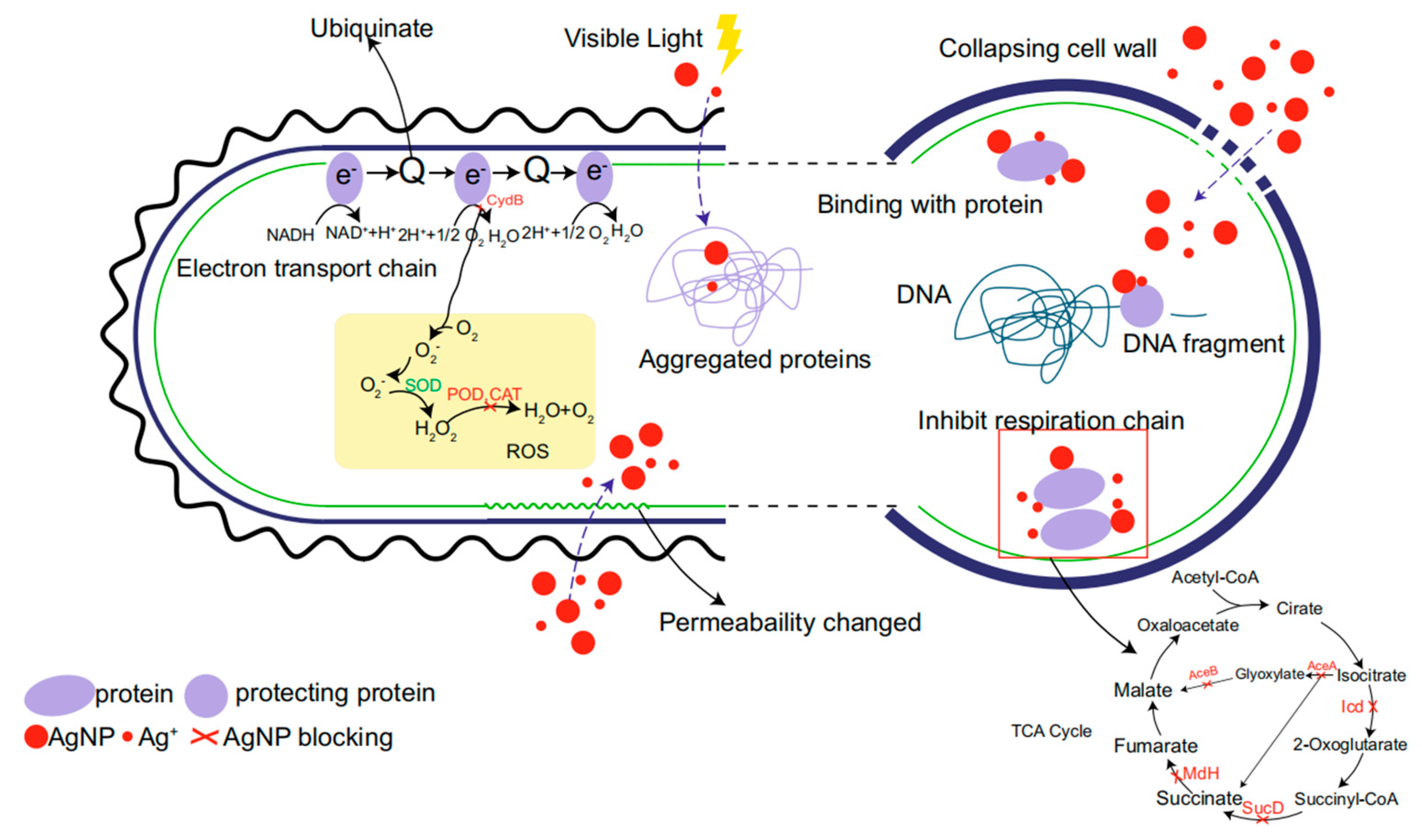
It is well-known that silver nanoparticles (Ag NPs) have potent bactericidal activity against a variety of bacteria. The significant antibacterial effect of Ag NPs is due to their large specific surface area, which increases the contact area between Ag NPs and the bacterial membrane. This induces cytoplasmic leakage to inhibit bacterial growth. In addition, Ag NPs have great binding power and affinity with most macromolecules, and their contact with bacterial membranes will lead to the disintegration and death of bacterial cells. So far, the efficient antibacterial activity of Ag NPs has been confirmed in many studies. Ag NPs have been found to kill many species of bacteria, including Staphylococcus aureus, Pseudomonas aeruginosa, Escherichia coli, Bacillus subtilis, Vibrio cholera, Salmonella typhus, Enterococcus faecalis, Klebsiella sp., Listeria sp., and Acinetobacter sp. [102,103,104]. At present, the antibacterial mechanism of Ag NPs is still controversial. Still, its antibacterial mechanism can be summarized into six types: interference with bacterial protein synthesis and folding inhibition of bacterial respiratory chain, induction of bacterial genetic toxicity, induction of photocatalytic destruction of bacterial protein and rupture of the cell membrane, and induction of bacterial oxidative stress reaction to produce reactive oxygen species [105,106,107]. The antibacterial mechanism of silver nanoparticles is shown in Figure 6. Silver ions can disrupt the respiration of bacteria and destroy the cell membrane of bacteria. To bond with the DNA in the cell membrane and to prevent the replication of genetic information of bacterial cells from achieving the bactericidal effect, through research, silver has a more substantial bactericidal effect in the nanometer state.
4.1.2. Antibacterial Hydrogel Containing Zinc Oxide Nanoparticles
Zinc Oxide nanoparticles (ZnO NPs) can kill microorganisms by a variety of mechanisms: (1) ZnO NPs can bind firmly to bacterial cell membranes and destroy lipids and proteins of cell membranes; the result is an increase in membrane permeability, which causes cytoplasmic content to flow out of the cell, leading to cell death. (2) ZnO NPs can also induce the formation of Zn2+ and reactive oxygen species (ROS), which can destroy bacterial cells. (3) Small particles of ZnO NPs can increase the membrane permeability and then enter the cytoplasm of bacterial cells, exerting oxidative stress on the cytoplasm and producing substantial toxicity to cells [109,110]. (4) Under ultraviolet radiation, many free radicals are generated on the surface of zinc oxide nanoparticles. When the free radicals contact microorganisms, the organic matter of microorganisms is oxidized into carbon dioxide. Zinc oxide nanoparticles can kill microorganisms in a short time. Hydrogels containing zinc oxide nanoparticles have the potential to facilitate rapid wound healing [111,112]. Majumder et al. prepared the biomimetic composite dressing by grafting hydrogel onto silk fabric and being further coated sonochemical with ZnO NPs to impart these dressing antimicrobial properties [113]. Baghaie et al. successfully prepared a novel synthetic hydrogel film by immobilizing ZnO powder on PVA/starch/chitosan polymer. The results showed that the addition of ZnO NPs could improve the tensile strength of the hydrogel and reduce the porosity of the hydrogel membrane, and the cell viability was over 87%. The wound healing rate in the hydrogel containing ZnO NPs was 96%, while the wound healing rate in the control group was 79% [114].
4.1.3. Antibacterial Hydrogel Containing Titanium Dioxide Nanoparticles
The antibacterial mechanism of titanium dioxide nanoparticles (TiO2 NPs) is that TiO2 will generate reactive oxygen species (ROS), including hydrogen peroxide (H2O2) and hydroxyl radical (·OH), after being exposed to near-ultraviolet light in the process of photocatalysis. When TiO2 radiated by UV light is close to bacteria, ROS damages the bacterial cell membrane, damages the semi-permeability of the membrane, interferes with and oxidizes the bacterial cell membrane, and achieves the purpose of sterilization. TiO2 NPs can kill Escherichia coli, Staphylococcus aureus, Pseudomonas aeruginosa, Enterococcus faecalis, Candida albicans, and other bacteria [115,116], and TiO2 NPs also has a specific destructive effect on viruses. TiO2 was used to coordinate with Ag ions to enhance their antibacterial activity. Ghosh et al. synthesized silver-modified titanium dioxide nanoparticles using the green hydrosol-gel method. The product is heat-treated at 450 °C and 600 °C and can achieve a good bactericidal effect. The amount of silver added and the heat treatment process have a significant influence on the phase composition, microstructure, and properties of TiO2. This material is suitable for application in healthcare due to its excellent antimicrobial properties, helping to reduce the spread of Gram-negative bacteria such as E. coli [117,118].
4.1.4. Antibacterial Hydrogel Containing Other Nanoparticles
In addition to the above-mentioned commonly used nanoparticles, there are several other antibacterial hydrogels containing nanoparticles, such as magnetic nanoparticles, copper nanoparticles, etc. Among them, magnetic nanoparticles have been widely used in biomedicine, nuclear magnetic resonance imaging, and environmental protection, due to their unique physical and chemical properties. The antibacterial mechanism of magnetic nanoparticles is mainly interference with the sulfhydryl group of bacterial proteins and then causing damage to bacteria. Xiong found that magnetic particles showed a particular antibacterial effect on Escherichia coli, Staphylococcus aureus, Pseudomonas aeruginosa, Salmonella typhi, Pasteurella multocida, and other drug-resistant bacteria. Compared with standard drugs, magnetic nanoparticles showed more substantial antibacterial effects against all bacteria [119]. Magnetic nanoparticles are mostly combined with other metals or metal compounds to achieve the purpose of antibacterial, such as Ag@Fe3O4, Fe3O4@TiO2, and CuO@Fe3O4 [120,121].
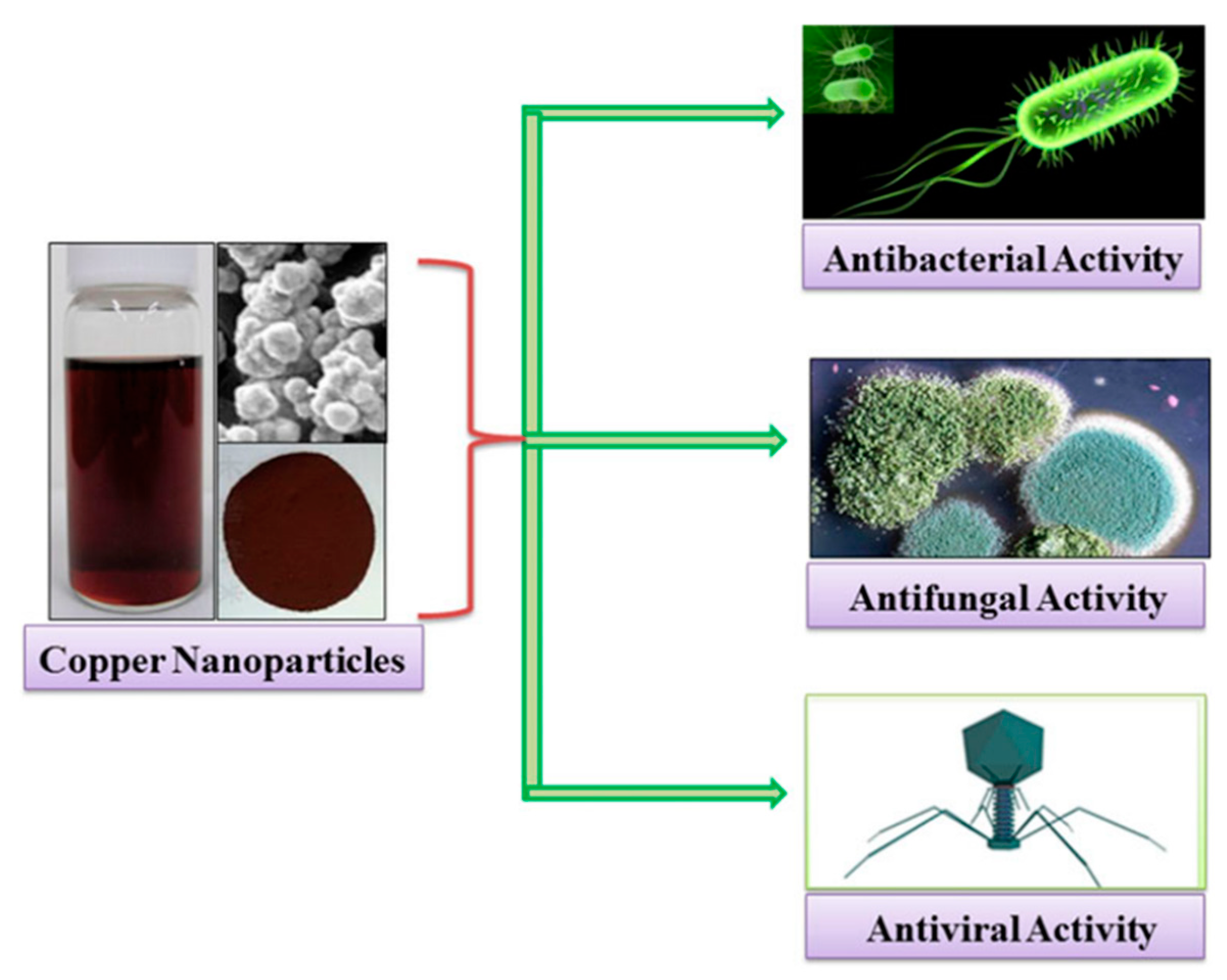
Das et al. found through the Kirby–Bauer diffusion method that copper nanoparticles could effectively inhibit the growth of Staphylococcus aureus, Bacillus subtilis, and Escherichia coli. Gopalakrishnan et al. proposed a possible mechanism for the mode of action of copper oxide nanoparticles against Escherichia coli. The copper oxide nanoparticles adsorbed on the cell surface interact with the cell wall and subsequently cause damage to the cell membrane, increasing its permeability and reducing bacterial viability in copper oxide solution [122]. Chang et al. explained the toxic effects of copper and zinc oxide nanoparticles on eukaryotic cells. Because of their small size, nanoparticles can diffuse directly into cells through pores in the cell membrane or enter cells through ion channels and transporters in the cell membrane. Nanoparticles that enter cells can interact directly with oxidizing organelles such as mitochondria. Subsequently, redox-active proteins stimulate cells to produce reactive oxygen species (ROS), which are induced by ions (Cu2+) produced by nanoparticles through various chemical reactions. ROS can cause DNA strand breaks and affect gene expression. In addition, Cu2+ ions can form chelates with biomolecules or free metal ions from specific metalloproteins, leading to cytotoxicity [123]. The schematic representation of the antimicrobial activity of copper nanoparticles against bacteria, fungi, and viruses is shown in Figure 7.
In addition, there are magnesium oxide nanoparticles (MgONPs). One of the antibacterial mechanisms of MgONPs is that MgONPs induce oxidative stress in bacterial cells and damage membrane integrity [124]. Another antibacterial mechanism that Leung found through three control experiments is that MgONPs could still produce antibacterial activity against Escherichia coli even though MgONPs did not produce reactive oxygen species. After performing characterization tests, Leung et al. suggested that the antibacterial effect was achieved by adsorbing the phosphate groups from the cell surface on the MgO surface [125].
4.2. Antibiotic-Loaded Antibacterial Hydrogels
Antibiotics are commonly used to treat bacterial infections. A three-dimensional network porous structure enables the hydrogels to be used as delivery systems for loading antibacterial drugs [126]. Antibacterial hydrogels can release drugs locally in small quantities, reduce bacterial resistance to antibiotics, and provide sufficient bacteria-killing doses for a long time [127]. The antibiotics commonly loaded in antibacterial hydrogels can be divided into aminoglycosides (gentamicin and streptomycin), glycopeptides (vancomycin), and quinolones (ciprofloxacin and levofloxacin) [128]. The main bactericidal mechanisms of conventional antibiotics are as follows: glycopeptide antibiotics inhibit the synthesis of the bacterial cell walls; aminoglycoside antibiotics block protein synthesis pathways; and quinolone antibiotics inhibit the synthesis of nucleic acids [129].
4.2.1. Aminoglycoside-Loaded Antibacterial Hydrogels
Aminoglycoside antibiotics are widely used and effective broad-spectrum antibacterial drugs. Aminoglycosides can destroy protein synthesis through transcellular plasma membrane transport and binding with bacterial ribosomes, thus playing an antibacterial function. Hu et al. prepared a hydrogel with high antibacterial activity against both aerobic and anaerobic pathogens by using crosslinking oxidized dextran with tobramycin and G1-orni. This injectable hydrogel shows thixotropic, biocompatible, self-healing, and pH-responsive drug release behavior. The study provides a facile strategy to expand the antibacterial spectrum of aminoglycoside hydrogels [130]. Li et al. prepared aminoglycosides (AGs) hydrogels based on dynamic covalent bond crosslinking by using AGs, aldehyde hyaluronic acid (A-HA), and adipic acid dihydrazide graft hyaluronic acid (HA-ADH) as materials. The results showed that the hydrogels had excellent and sustained antibacterial properties against Escherichia coli and Staphylococcus aureus, and A-HA/HA-ADH/AGs hydrogels are potential dressings for wound healing [131].
Gentamicin and streptomycin are aminoglycoside antibiotics, and gentamicin is more commonly used. Gentamicin mainly acts on 30S ribosome 16S rRNA in the bacterial body through three steps: blocking bacterial protein synthesis, destroying the integrity of cell membrane, and leading to cell death. Firstly, gentamicin contains a large number of positively charged amino groups, which combine with the negative charge on the surface of the bacterial membrane, the electrostatic interaction increases the permeability of the bacterial surface, and part of gentamicin molecules penetrate the bacterial interior. Secondly, gentamicin in the cell produces the antibiotic effect, destroys the integrity of the cytoplasmic membrane, and the gentamicin molecule enters the cell in large quantities. Finally, the intracellular gentamicin molecule binds to the A site of the bacterium’s 30S ribosome 16S rRNA, resulting in protein mistranslation and, ultimately, cell death [132]. Guo successfully prepared a series of new crosslinking chitosan quaternary ammonium salt loaded with gentamicin sulfate (CTMCSG) hydrogel membranes by the reaction of chitosan quaternary ammonium salt (TMCS) and epichlorohydrin. The results indicate that the CTMCSG-4 hydrogel film with an attractive physicochemical property, admirable antibacterial activity, and the slight cytotoxicity shows the potential value as an excellent antibacterial wound dressing [133]. Gupta and Purwar prepared hydrogel composites of poly(acrylamide-co-acrylic acid) hydrogel grafted over the cotton fabric using crosslinkers polyethylene glycol (PEG) and N,N′-methylene bisacrylamide (MBA) separately. The release kinetics of gentamicin sulfate in two kinds of composites were studied, and the results show that the hydrogel composite has higher mechanical strength than its film [134].
4.2.2. Glycopeptides-Loaded Antibacterial Hydrogels
Vancomycin is a typical glycopeptide antibiotic. The mechanism of vancomycin is to inhibit the synthesis of bacterial cell walls and combine with the bacterial cell walls, so that some amino acids cannot enter the glycopeptide of the cell wall. Liao et al. incorporated vancomycin into oxidized hyaluronic acid (HA) and adipic acid dihydrazide, then evaluated the drug release and antimicrobial activity against methicillin-resistant Staphylococcus aureus. The results showed that oxi-HA/ADH hydrogel-loaded with vancomycin has excellent biocompatibility, complete and rapid drug release, and measurable in vitro antimicrobial activity and is quite effective in a biofilm model to prevent immediate bacterial colonization [135]. Naeimi et al. studied a drug-loaded porous hydrogel for the delivery of vancomycin. The hydrogels based on chitosan (CS), Polyvinyl alcohol (PVA), and Polyethylene glycol (PEG) were prepared by lyophilization. The porous CS/PVA/PEG hydrogels containing vancomycin could be good potential candidates as wound dressing [136].
4.2.3. Quinolones-Loaded Antibacterial Hydrogels
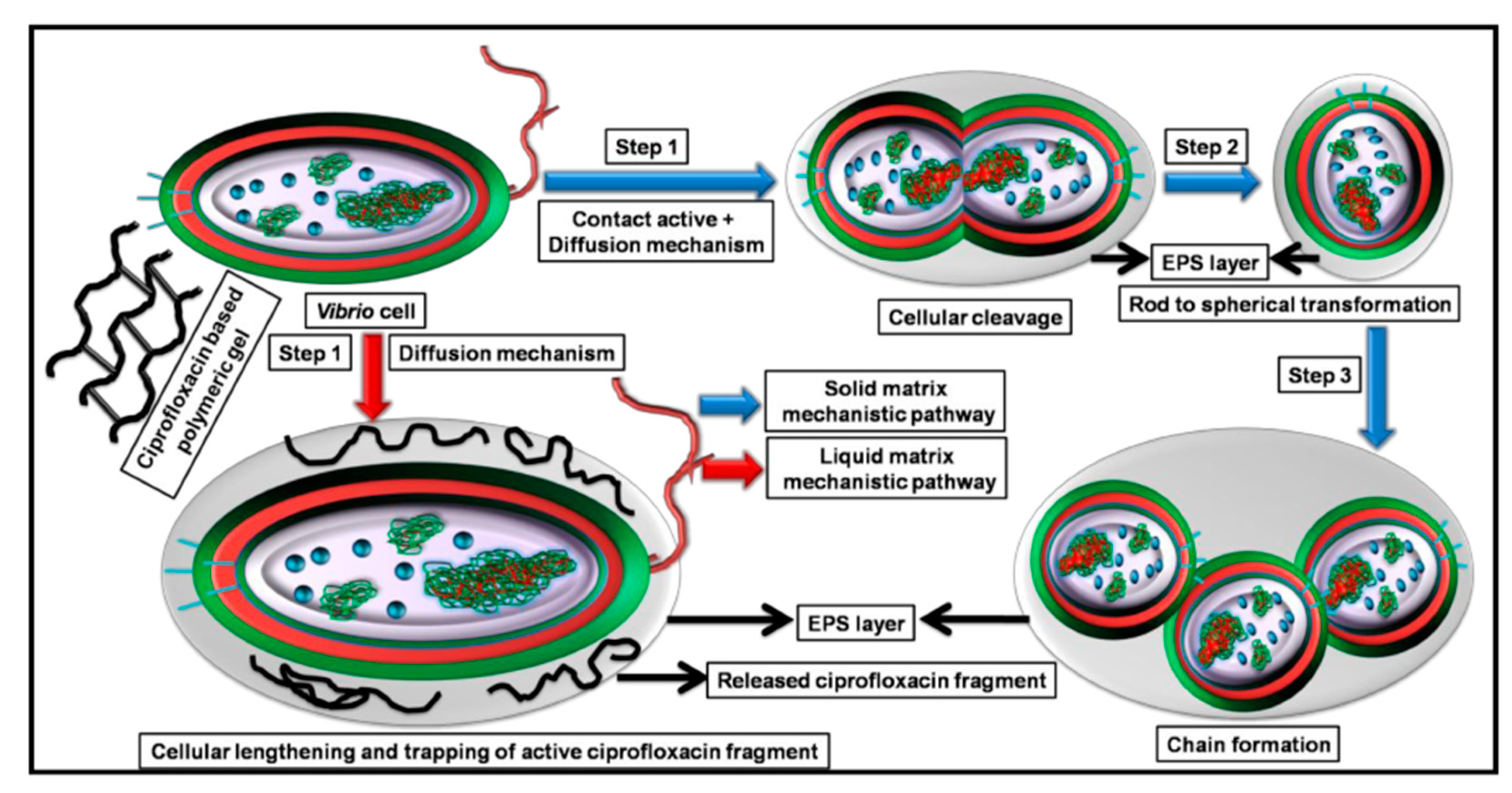
Quinolone antibiotics mainly include ciprofloxacin and levofloxacin. Among them, ciprofloxacin has an antibacterial effect on broad-spectrum Gram-positive bacteria and Gram-negative bacteria. The antibacterial mechanism of ciprofloxacin relies on the blocking of bacterial DNA replication by binding itself to the DNA rotating-enzyme, which causes bacterial chromosome double-strand breaks, so bacterial resistance to this drug develops slowly [137]. Mukherjee developed an amphoteric hydrogel loaded with ciprofloxacin antibiotics. Ciprofloxacin was covalently linked to a polymer system consisting of hydrophilic polyethylene glycol methyl ether methacrylate (PEGMA) and sulfobetaine methacrylate (SBMA). Ciprofloxacin attached to polymers enables the preparation of highly potent polymeric antibiotic conjugations (PACs) with linear and hyperbranched structures. Its bactericidal mechanism is shown in Figure 8. PACs showed antibacterial activity against both solid and liquid substrates of the strain. After that, through the continuous removal of thiol fragments at the end of ciprofloxacin and the balance of swelling and deswelling process, the hydrogel is transformed into polymeric gel through the “double” contact activity release mechanism, which makes the hydrogel show more potent antibacterial activity than the liquid matrix and achieve a complete bactericidal effect [138].
4.3. Hydrogels with Inherent Antibacterial Activity
4.3.1. Antimicrobial Peptide Hydrogel
Antimicrobial peptides (AMPs) have many antibacterial activities and exist widely in many animal and plant tissues and cells. AMPs are strongly alkaline, have thermal stability, and have vigorous antibacterial activity against microorganisms such as bacteria, fungi, and viruses [139,140]. As a new type of non-antibiotic antibacterial agent, antimicrobial peptides are considered the most likely drugs to replace antibiotics [141,142,143]. At present, many studies believe that the antimicrobial mechanism of AMPs is that antimicrobial peptides are adsorbed on the lipid membrane through static electricity, which destroys the internal and external barrier of bacterial cells, leading to the increase of membrane permeability and the death of bacteria due to the inability to maintain normal osmotic pressure. This mechanism of action is less likely to lead to bacterial resistance [144,145,146]. Some studies believe that antimicrobial peptides inhibit bacterial metabolic behavior by inhibiting bacterial membrane synthesis, DNA synthesis, and enzyme activity, thus leading to bacterial inactivation or death [147,148]. The antibacterial mechanism of antimicrobial peptides is shown in Figure 9. In this figure, Escherichia coli is shown as the target microorganisms.
In recent years, many antibacterial hydrogels with antibacterial peptides have been developed. Aye et al. use a simple method based on electrostatic interaction to prepare antibacterial silk hydrogels containing antimicrobial peptides (AMP) [150]. Baral et al. prepared an antibacterial dipeptide, which showed excellent antibacterial activity against G− bacteria (E. coli and P. aeruginosa), as well as high biocompatibility with human red blood cells and human fibroblast cells [151]. Zhou et al. reported that a popular AMP polylysine had been applied in photopolymerized antibacterial hydrogels, which generated good coatings for medical devices and implants [152]. Bai et al. designed an amphiphilic peptide A9K2 that could effectively inhibit both G+ and G− bacterial strains [153]. To produce antibacterial hydrogel composite wound dressing, Gunes and Ziylan mixed carboxymethyl chitosan (CMCht)-sodium alginate-streptococcus lactic into cotton and modified it without damaging its beneficial properties. The modified wound dressing retained the porous structure and produced adequate WVTR [154]. Atefyekta et al. studied antimicrobial peptide-functionalized mesoporous hydrogels; amp-hydrogel has high antibacterial activity against Staphylococcus epidermidis, Staphylococcus aureus, Pseudomonas aeruginosa, methicillin-resistant Staphylococcus aureus (MRSA), and multidrug-resistant Escherichia coli, and has potential hemostatic activity [155].
4.3.2. Chitosan Antibacterial Hydrogel
Chitosan is a kind of typical antibacterial cationic polymer. The amino groups in its molecular chain can be protonated in an acidic environment and have positive electric properties [156]. The modification of chitosan molecules can improve its water solubility, and its derivatives, such as chitosan quaternary ammonium salt, have good solubility in neutral pH, which significantly enhances its antibacterial activity in a neutral environment. Introducing chitosan and its derivatives has become a standard method to imbibe materials with inherent antibacterial properties. Hu et al. used Ca2+ as a physical crosslinking agent to design a natural polysaccharide hydrogel with double physical crosslinking. Cationic polymer chitosan (CS) forms ionic coordination with polyanionic polymer sodium alginate (SA), and Ca2+ forms ionic coordination with the carboxyl group on the SA molecular chain. The polysaccharide hydrogel synthesized by this method can be a suitable carrier of epidermal growth factors to promote cell proliferation and wound healing [157]. Yu et al. used positively charged CS and negatively charged carrageenan (κ-CG) to prepare a polysaccharide-based hydrogel film with good toughness and anti-cell adhesion properties. In addition to the ionic coordination between the CS and κ-CG molecular chain, hydrogen bonds can also form between the CS and κ-CG molecular chain. Through the synergistic effect of ionic coordination and hydrogen bonds, the hydrogel film has excellent self-healing properties, and the self-healing efficiency can reach 90% after the initial stretching and standing for two h [158]. Wang reported a biodegradable chitosan hydrogel system with excellent injectable, antibacterial activity, biocompatibility, and self-repairing properties. The primary raw materials for the hydrogel preparation are chitosan and aldehyde chitosan. Due to the antibacterial activity of chitosan and aldehyde chitosan in the hydrogel network, the hydrogel system can significantly inhibit the growth of bacteria upon contact, making it an excellent candidate for antibacterial wound dressing [159]. Zhang et al. developed a new antibacterial hydrogel wound dressing composed of poly(aminoethyl) modified chitosan (PAEMCS). Experiments showed that the hydrogel based on PAEMCS had excellent hygroscopicity; antibacterial activity against Escherichia coli, Staphylococcus aureus, and Staphylococcus epidermidis; and good cytocompatibility against L929 cells or HUVEC [160]. Chen et al. immobilized biodegradable antibacterial polymer chitosan (CS) onto the surface of poly(N-isopropyl acrylamide) (PNIPAAm)gel/polypropylene(PP) nonwoven composites for wound dressing applications. The results showed that the product had the antibacterial ability to Escherichia coli and Staphylococcus aureus and had good biocompatibility with fibroblasts [161].
4.3.3. Amphoteric Ion Antibacterial Hydrogel
The antibacterial mechanism of amphoteric ionic hydrogels is similar to that of AMPs. The molecular chain of cationic polymers contains many positively charged groups. The positively charged polymer molecular chain is bound to the negatively charged bacterial cell membrane through ion coordination, forming a polymer film on the bacterial surface and hindering the transport of internal and external substances. The structure of the bacterial cell membrane is destroyed, which affects the osmotic balance of bacteria inside and outside and causes the outflow of the intracellular matrix, resulting in bacterial death [162,163]. Panini et al. prepared a new type of poly hydroxyethyl methacrylate-PAM/PVA/CS interpenetrating polymer network (IPN) hydrogel by two-step radical polymerization. CS and polycations interact with negatively charged surfaces of bacteria, resulting in membrane loss of permeability, cell leakage, and eventual death. The antibacterial hydrogel has higher swelling, mechanical strength, and antibacterial activity than chitosan, and has good antibacterial activity against Gram-negative bacteria. It is an ideal material for preparing low-cost wound dressings [164]. Wang et al. designed and introduced a novel structure of amphoteric ionic poly[3-(dimethyl (4-vinyl benzyl) ammonium) propyl sulfonate] (SVBA) into the polyAAm network to obtain a novel salt-responsive conductive hydrogel. Due to the addition of SVBA, PAS-2 hydrogel has good electrical activity and significant mechanical properties, as well as good cellular compatibility, stable rheological properties, effective antibacterial properties, strong adsorption, and high permeability, which can effectively promote wound healing and can be used as a medical dressing [165]. Dai et al. developed zwitterionic sulfobetaine acrylamide antibacterial hydrogel combined with laponite (LAP) nanoplatelets and methyl acrylamide dopamine (DMA) for wound dressings. LAP nanoplatelets and DMA impart the hydrogel-enhanced mechanical strength and material adhesion, respectively. The multifunctional hydrogel dressing with antifouling, antibacterial properties, and proper adhesion was successfully constructed without secondary damage to the repaired tissues [166]. Zhu et al. developed a new type of zwitterionic hydrogel, polycarboxy betaine (PCB) hydrogel, as a wet dressing to promote skin wound healing. The high water content may promote autolysis debridement, stimulate collagen deposition, and accelerate wound healing [167].
4.4. Photosensitive Antibacterial Hydrogel
Photodynamic therapy (PDT) is another effective antibacterial method. Under the influence of light of an appropriate wavelength, a photosensitive agent (PS) is stimulated to generate reactive oxygen species or singlet oxygen to destroy bacteria, resulting in the rupture of the bacterial cell membrane and the destruction and inactivation of protein structure, thus achieving the purpose of sterilization [168,169,170]. Fighting bacterial infections in this way does not lead to the emergence of resistant bacteria. The combination of photosensitizer hydrogel is an effective therapeutic strategy for the treatment of pathogen infection [171]. Three cationic photosensitizers, (Meso-tetra (4-N-methylpyridyl) porphyrinetetratosylate (TMPyP), Zn (II) meso-tetra (N-methyl-4-pyridyl) porphinetetra chloride (Zn-TMPyP), and methylene blue (MB)), were adsorbed by montmorillonite (MMT). The photosensitive MMT/PS hydrogels were prepared by UV crosslinking method in an aqueous solution of polyvinyl alcohol-styrylpyridine salt condensation (PVA-SbQ). The results showed that all three photosensitive MMT/PS hydrogels had good swelling performance and a specific killing effect on Staphylococcus aureus [172]. Truong and Forsythe studied photolabile hydrogels and found that nanocrystalline TiO2 can be used as a photoinitiator of polymer PEGDA hydrogel. TiO2 is evenly distributed in the matrix and still has photoactivity under fluorescent lamp irradiation. It can be used as a photoinitiator in the hydrogel polymerization process and as a photosensitizer in photodynamic therapy [173]. Deng et al. prepared agarose (AG)-based hydrogel containing tannic acid-Fe(III) (TA-Fe) nanoparticles by a facile and eco-friendly strategy, which has good mechanical properties, excellent photothermal effect, and good biocompatibility. And photothermal can effectively avoid the disadvantages of traditional antibiotics in killing pathogenic bacteria and treating wound infection, and it is expected to be an antibacterial wound dressing in the biomedical field [174]. Zhou Qian separated the photosensitive antibacterial agent from the lotus, using tryptophan to modify the natural photosensitive agent tribithiophenal, improve water solubility at the same time, and reduce the phototoxicity. The modified photosensitizer was filled into the blisters, and the resulting blisters were loaded on the dressing. Photosensitive intelligent antibacterial hydrogel dressing was obtained [175].
4.5. Hydrogels with Synergetic Effects
Synergistic antibacterial hydrogels refer to antibacterial hydrogels with synergistic action of two or more antibacterial modes to improve antibacterial performance [176]. There are many reports on antibacterial hydrogel products that enhance the antibacterial effect by using silver nanoparticles synergistic with antibiotics or graphene oxide (GO).
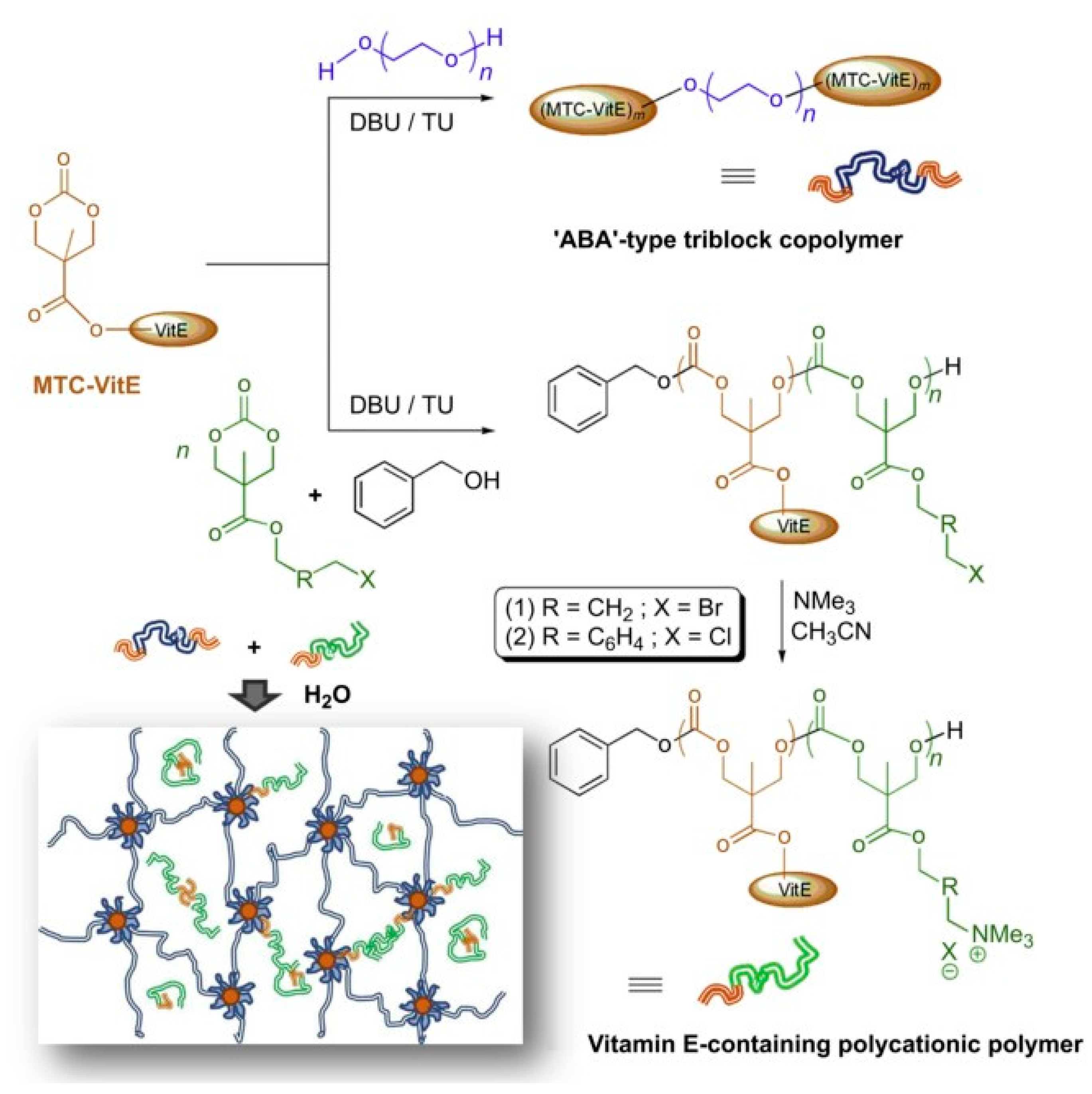
Bu studied the preparation and antibacterial properties of polymer-modified graphene oxide and its hydrogel composites; the modified GO hydrogel wound dressing was prepared by combining GO, quaternary ammonium salt (QAS), and N-halamine. The sauce has good biocompatibility and excellent antibacterial effects, promoting wound healing [177]. The nanofiber felt was prepared by blending chitosan-based silver nanoparticles with polyvinyl alcohol. Then, the nanofibers with an average diameter of 150 nm were prepared by crosslinking with glutaraldehyde and electrospinning. The results showed that the combination of chitosan and Ag-NPS had an excellent synergistic antibacterial effect. The presence of Ag NPs in the PVA/CS blend solution not only enhanced the electrospinning performance, but also enhanced the antibacterial ability of electrospinning felt, which is a good wound dressing [178]. Lee et al. developed a biodegradable antibacterial hydrogel using vitamin E functionalized polycarbonate and achieved a better synergistic antibacterial effect by combining antibacterial polycarbonate with the conventional antifungal agent fluconazole. Syntheses of the polymers and the schematic illustration of incorporating polycationic polymers into the hydrogel system are shown in Figure 10.
Yang studied the preparation and properties of antibacterial hydrogel based on polyvinyl alcohol [180]. Palantoken et al. studied UV-cured cationic quaternary ammonium polyethylenimine and embedded it with silver nitrate to prepare hydrogels with dual bacteriostatic effects [181]. Mao et al. studied the photo-inspired antibacterial activity of Ag/Ag@AgCl/ZnO nanostructured hydrogels. The hydrogel has high antibacterial efficiency against both Escherichia coli and Staphylococcus aureus. The release of Ag+ and Zn2+ stimulates the immune function to produce many white blood cells and neutrophils, thereby producing synergistic antibacterial effects and accelerated wound healing [182]. Yang et al. developed a slow-release antibacterial hydrogel based on branched polyethyleneimine (PEI), stabilized GO-AgNPs, and Pluronic F127 for a more stable and sustained antibacterial effect [183,184]. Kang et al. prepared a temperature-sensitive antibacterial hydrogel NIPOM-CG/GM with antibacterial properties based on vinyl carboxymethyl chitosan (CG), silane dispersed graphene (GM), and N-isopro polyacrylamide (NIPAM) [185].
5. Summary and Prospect
Because of its good biocompatibility and moisture absorption, traditional hydrogels have gradually become common materials in people’s daily lives. Especially in the medical field, hydrogels with antibacterial functions play an important role. They are especially suitable for biomedical applications such as wound dressing, contact lenses, and controlled release of drugs. The abuse of antibiotics in medical treatment will lead to bacterial resistance, and some traditional hydrogels are gradually withdrawn from the market. To have new biological functions, more intelligent antibacterial hydrogels need to be designed. With further research, the types of antibacterial hydrogels are gradually enriched. In this paper, according to different antibacterial modes, antibacterial hydrogels can be divided into five types: antibacterial hydrogel containing inorganic nanoparticles; antibiotic-loaded antibacterial hydrogels; hydrogels with inherent antimicrobial activity; photosensitive antibacterial hydrogels; and hydrogels with synergetic effects. The mechanism, properties, and preparation methods of these antibacterial hydrogels also expound. Experimental research shows that some antibacterial agents can also be synergies with other components; in addition, antibacterial hydrogel has a better antibacterial effect and a broader antibacterial spectrum. It also provides a diverse performance, such as water/moisture absorption, slow-release drug properties, swelling properties, permeability, ultraviolet resistance, etc. Graphene oxide (GO) and nanosilver (Ag-NPs) are considered promising antibacterial materials due to their excellent biocompatibility, broad-spectrum antibacterial properties, and non-drug resistance. Antimicrobial peptides and cationic polymers have also been used to synthesize inherently antibacterial hydrogels that effectively treat bacterial infections. The modified GO hydrogel has an excellent antibacterial effect and can promote wound healing. The combination of chitosan and Ag-NPS has uniquely promising properties and a synergistic antibacterial effect. Of course, as a wound dressing, the antibacterial hydrogel will develop into an intelligent and multi-functional tool in the future and solve the problems of mechanical strength, adhesion performance, and permeability.
Author Contributions
Conceptualization, J.L. and W.J.; methodology, J.L.; validation, J.L., W.J., and Q.X.; formal analysis, J.L. and W.J.; investigation, W.J. and Q.X.; resources, J.L.; data curation, J.L. and W.J.; writing—original draft preparation, J.L.; writing—review and editing, J.L. and Y.Z.; visualization, J.L.; supervision, J.L. and W.J.; project administration, J.L. and Y.Z.; funding acquisition, J.L. and Y.Z. All authors have read and agreed to the published version of the manuscript.
Funding
This work was financially supported by the Basic Scientific Research Funds for provincial universities in Heilongjiang Province (Industrial hemp specialty) (145109503).
Institutional Review Board Statement
Not applicable.
Informed Consent Statement
Not applicable.
Data Availability Statement
Not applicable.
Acknowledgments
Engineering Research Center for Hemp and Product in Cold Region of Ministry of Education, Qiqihar, Heilongjiang 161006, China.
Conflicts of Interest
The authors declare no conflict of interest.
References
- Hu, J.H.; Zhu, Q.G.; Sun, H.J.; Gao, W. Classification and characteristics of medical dressings. Pharm. J. Chin. People’s Lib. Army 2000, 16, 147–148, 167. [Google Scholar]
- Liu, C.; Han, H.; Ye, T.T.; Wu, D.Q. Preparation and antibacterial properties of hydrogel-based medical cotton dressing. China Text. Lead. 2021, 64–68. [Google Scholar] [CrossRef]
- Werdin, F.; Tennenhaus, M.; Schaller, H.E.; Rennekampff, H.O. Evidence-based management strategies for treatment of chronic wounds. Eplasty 2009, 9, 169–179. [Google Scholar]
- Centers for Disease Control and Prevention. National Diabetes Fact Sheet: National Estimates and General Information on Diabetes and Prediabetes in the United States, 2011; U.S. Department of Health and Human Services, Centers for Disease Control and Prevention: Atlanta, GA, USA, 2011; Volume 201, pp. 2568–2569.
- Eo, S.R.; Kim, Y.S.; Cho, S.H. Vacuum-assisted closure improves the incorporation of artificial dermis in soft tissue defects: Terudermis® and Pelnac®. Int. Wound J. 2011, 8, 261–267. [Google Scholar] [CrossRef] [PubMed]
- Smith, D.M.; Snow, D.E.; Rees, E.; Zischkau, A.M.; Hanson, J.D.; Wolcott, R.; Sun, Y.; White, J.; Kumar, S.; Dowd, S.E. Evaluation of the bacterial diversity of Pressure ulcers using b TEFAP pyrosequencing. BMC Med. Genom. 2010, 21, 41–45. [Google Scholar] [CrossRef] [PubMed]
- Selig, H.F.; Lumenta, D.B.; Giretzlehner, M.; Jeschke, M.G.; Upton, D.; Kamolz, L.P. The properties of an “ideal” burn wound dressing-What do we need in daily clinical practice? Results of a worldwide online survey among burn care specialists. Burns 2012, 38, 960–966. [Google Scholar] [CrossRef] [PubMed]
- Andreu, V.; Mendoza, G.; Arruebo, M.; Irusta, S. Smart dressings based on nanostructured fibers containing natural origin antimicrobial, anti-inflammatory, and regenerative compounds. Materials 2015, 8, 5154–5193. [Google Scholar] [CrossRef]
- Jayakumar, R.; Prabaharan, M.; Kumar, P.T.S.; Nair, S.V.; Tamura, H.V.H. Biomaterials based on chitin and chitosan in wound dressing applications. Biotechnol. Adv. 2011, 29, 322–337. [Google Scholar] [CrossRef] [PubMed]
- Phaechamud, T.; Issarayungyuen, P.; Pichayakorn, W. Gentamicin sulfate-loaded porous natural rubber films for wound dressing. Int. J. Biol. Macromol. 2016, 85, 634–644. [Google Scholar] [CrossRef]
- Borges, F.A.; Drago, B.C.; Luís, O.B.; Barros, N.; Pegorin, G.S.; Scontri, M.; Mussagy, C.U.; Ribeiro, M.C.; Milori, D.M.B.P.; Morais, C.P.; et al. Metronidazole-loaded gold nanoparticles in natural rubber latex as a potential wound dressing. Int. J. Biol. Macromol. 2022, 211, 568–579. [Google Scholar] [CrossRef]
- Watthanaphanit, A.; Rujiravanit, R. Sericin-binded-deprotenized natural rubber film containing chitin whiskers as elasto-gel dressing. Int. J. Biol. Macromol. 2017, 101, 417–426. [Google Scholar] [CrossRef] [PubMed]
- Qin, J.; Guo, J.; Xu, Q.; Zheng, Z.Q.; Mao, H.L.; Yan, F. Synthesis of pyrrolidinium-type poly(ionic liquid) membranes for antibacterial applications. ACS. Appl. Mater. Interfaces 2017, 9, 10504–10511. [Google Scholar] [CrossRef] [PubMed]
- Luo, Z.; Cui, H.; Guo, J.; Yao, J.R.; Fang, X.; Yan, F. Poly(ionic liquid)/Ce-based antimicrobial nanofibrous membrane for blocking drug-resistance dissemination from MRSA-Infected wounds. ADV Funct. Mater. 2021, 31, 2100336. [Google Scholar] [CrossRef]
- Chaiarwut, S.; Ekabutr, P.; Chuysinuan, P.; Chanamuangkon, T.; Supaphol, P. Surface immobilization of PCL electrospun nanofibers with pexiganan for wound dressing. J. Polym. Res. 2021, 28, 344. [Google Scholar] [CrossRef]
- Peng, Y.; Ma, Y.; Bao, Y.; Liu, Z.L.; Chen, L.; Dai, F.Y.; Li, Z. Electrospun PLGA/SF/artemisinin composite nanofibrous membranes for wound dressing. Int. J. Biol. Macromol. 2021, 183, 68–78. [Google Scholar] [CrossRef]
- Ghosal, K.; Agatemor, C.; Špitálsky, Z.; Thomas, S.; Kny, E. Electrospinning tissue engineering and wound dressing scaffolds from polymer-titanium dioxide nano-composites. Chem. Eng. J. 2019, 358, 1262–1278. [Google Scholar] [CrossRef]
- Wang, H.B.; Zhu, H.; Fu, W.G.; Zhang, Y.Y.; Xu, B.; Gao, F.; Cao, Z.Q.; Liu, W.G. A high strength self-healable antibacterial and anti-inflammatory supramolecular polymer hydrogel. Macromol. Rapid. Comm. 2017, 38, 1600695. [Google Scholar] [CrossRef] [PubMed]
- Wang, K.; Wang, J.; Li, L.; Xu, L.Q.; Feng, N.; Wang, Y.; Fei, X.; Tian, J.; Li, Y. Synthesis of a novel anti-freezing, non-drying antibacterial hydrogel dressing by one-pot method. Chem. Eng. J. 2019, 372, 216–225. [Google Scholar] [CrossRef]
- Wichterle, O.; Lim, D. Hydrophilic gels for biological use. Nature 1960, 185, 117–118. [Google Scholar] [CrossRef]
- Rowland, M.J.; Atgie, M.; Hoogland, D.; Scherman, O.A. Preparation and supramolecular recognition of multivalent peptide–polysaccharide conjugates by cucurbit [8]uril in hydrogel formation. Biomacromolecules 2015, 16, 2436–2443. [Google Scholar] [CrossRef]
- Liu, L.; Feng, X.; Pei, Y.; Wang, J.Z.; Ding, J.X.; Chen, L. α-Cyclodextrin concentration-controlled thermo-sensitive supramolecular hydrogels. Mat. Sci. Eng. C 2018, 82, 25–28. [Google Scholar] [CrossRef]
- El-Sherbiny, I.M.; Hamid, M.A.; Rashad, M.; Ali, A.S.M.; Azab, Y. New calcareous soil–alginate composites for efficient uptake of Fe (III), Mn (II) and As (V) from water. Carbohyd. Polym. 2013, 96, 450–459. [Google Scholar] [CrossRef]
- Hunter, A.M.; Grigson, C.; Wade, A. Influence of topically applied menthol cooling gel on soft tissue thermodynamics and arterial and cutaneous blood flow at rest. Int. J. Sports Phys. Ther. 2018, 13, 483–492. [Google Scholar] [CrossRef]
- Zhang, Y.; An, D.; Pardo, Y.; Chiu, A.; Song, W.; Liu, Q.; Zhou, F.; McDonough, S.P.; Ma, M. High-water-content and resilient PEG-containing hydrogels with low fibrotic response. Acta Biomater. 2017, 53, 100–108. [Google Scholar] [CrossRef] [PubMed]
- Patenaude, M.; Smeets, N.M.; Hoare, T. Designing injectable, covalently cross-linked hydrogels for biomedical applications. Macromol. Rapid Commun. 2014, 35, 598–617. [Google Scholar] [CrossRef]
- Ng, V.W.; Chan, J.M.; Sardon, H.; Ono, R.J.; García, J.M.; Yang, Y.Y.; Hedrick, J.L. Antimicrobial hydrogels: A new weapon in the arsenal against multidrug-resistant infections. Adv. Drug Deliv. Rev. 2014, 78, 46–62. [Google Scholar] [CrossRef]
- Noimark, S.; Dunnill, C.W.; Wilson, M.; Parkin, I.P. The role of surfaces in catheter-associated infections. Chem. Soc. Rev. 2009, 38, 3435–3448. [Google Scholar] [CrossRef]
- Hetrick, E.M.; Schoenfisch, M.H. Reducing implant-related infections: Active release strategies. Chem. Soc. Rev. 2006, 35, 780–789. [Google Scholar] [CrossRef] [PubMed]
- Shea, L.D.; Woodruff, T.K.; Shikanov, A. Bioengineering the ovarian follicle microenvironment. Annu. Rev. Biomed. Eng. 2014, 16, 29–52. [Google Scholar] [CrossRef] [PubMed]
- Liu, S.F.; Gu, Z.Y. Application of temperature-sensitive hydrogels to the development of intelligent fabrics. J. Text. Inst. 2007, 11, 139–143. [Google Scholar]
- Wu, M.Q.; Hua, J.C.; He, G.D.; Li, Z.; Gong, J.X.; Zhang, J.F. Application of hydrogels in textile industry. Dye. Finish. 2016, 42, 52–55. [Google Scholar]
- Li, H.N.; Yang, J.; Hu, X.N.; Liang, J.; Fan, Y.J.; Zhang, X.D. Superabsorbent polysaccharide hydrogels based on pullulan derivate as antibacterial release wound dressing. J. Biomed. Mater. Res. A 2011, 98, 31–39. [Google Scholar] [CrossRef]
- Kumaraswamy, S.; Francis, S.; Somashekarappa, H.M. Gamma irradiation synthesis of Ag/PVA hydrogels and its antibacterial activity. Mater. Today Proc. 2016, 3, 1792–1798. [Google Scholar]
- Xu, D.D.; Huang, J.C.; Zhao, D.; Ding, B.B.; Zhang, L.N.; Cai, J. High-flexibility, high-toughness double-cross-linked chitin hydrogels by sequential chemical and physical cross-linkings. Adv. Mater. 2016, 28, 5844–5849. [Google Scholar] [CrossRef]
- Yang, K.R.; Han, Q.; Chen, B.P.; Zheng, Y.H.; Zhang, K.S.; Li, Q.; Wang, J.C. Antimicrobial hydrogels: Promising materials for medical application. Int. J. Nanomed. 2018, 13, 2217. [Google Scholar] [CrossRef]
- Song, T.; Xi, Y.J.; Du, J.Z. Antibacterial hydrogels incoporated with poly(glutamic acid)-based vesicles. Acta Polym. Sin. 2018, 119–128. [Google Scholar] [CrossRef]
- Zhang, L.; Yuan, H.B.; Chen, L.; Hong, F. Study on properties of bacterial nanocellulose-based antibacterial hydrogel composite dressings. J. Fuel Cell Sci. Technol. 2019, 27, 31–38, 58. [Google Scholar]
- Xiao, L.Z.; Hui, F.; Tian, T.G.; Yan, R.Y.; Xin, J.W.; Zhao, X.Y.; Jiang, Y.N.; Zhang, Z.; Kuang, Y.L.; Li, N.; et al. A novel conductive antibacterial nanocomposite hydrogel dressing for healing of severely infected wounds. Front. Chem. 2021, 9, 787886. [Google Scholar] [CrossRef] [PubMed]
- Khashan, K.S.; Sulaiman, G.M.; Abdulameer, F.A.; Ibrahem, M.A.; Al-Muhimeed, T.; AlObaid, A.A. Antibacterial activity of TiO2 nanoparticles prepared by one-step laser ablation in liquid. Appl. Sci. 2021, 11, 4623. [Google Scholar] [CrossRef]
- Yan, J.; Ji, Y.; Huang, M.; Tao, L.; Liu, Y.H.; Lü, S.Y.; Liu, M.Z. Nucleobase-inspired self-adhesive and inherently antibacterial hydrogel for wound dressing. ACS Mater. Lett. 2020, 2, 1375–1380. [Google Scholar] [CrossRef]
- Wang, K.; Wang, J.; Li, L.; Xu, L.Q.; Feng, N.; Wang, Y.; Fei, X.; Tian, J.; Li, Y. Novel nonreleasing antibacterial hydrogel dressing by a one-pot method. ACS Biomater. Sci. Eng. 2020, 6, 1259–1268. [Google Scholar] [CrossRef] [PubMed]
- Caló, E.; Khutoryanskiy, V.V. Biomedical applications of hydrogels: A review of patents and commercial products. Eur. Polym. J. 2015, 65, 252–267. [Google Scholar] [CrossRef]
- Tian, R.; Qiu, X.Y.; Yuan, P.Y.; Lin, K.; Bai, K.Y.; Liu, S.Y.; Chen, X. Fabrication of self-healing hydrogels with on-demand antimicrobial activity and sustained biomolecule release for infected skin regeneration. ACS Appl. Mater. Interfaces 2018, 10, 17018–17027. [Google Scholar] [CrossRef] [PubMed]
- Ullah, F.; Othman, M.B.H.; Javed, F.; Ahmad, Z.; Akil, H.M. Classification, processing and application of hydrogels: A review. Mater. Sci. Eng. C 2015, 57, 414–433. [Google Scholar] [CrossRef]
- Li, X.Q.; Jiang, Y.J.; Wang, F.; Fan, Z.J.; Wang, H.N.; Tao, C.H.; Wang, Z.F. Preparation of polyurethane/polyvinyl alcohol hydrogel and its performance enhancement via compositing with silver particles. RSC Adv. 2017, 7, 46480–46485. [Google Scholar] [CrossRef]
- Shin, B.; Kim, J.J.; Vales, T.P.; Yang, S.K.; Kim, J.K.; Sohn, H.; Kim, H.J. Thermoresponsive drug controlled release from chitosan-based hydrogel embedded with poly(N-isopropylacrylamide) nanogels. J. Polym. Sci. Polym. Chem. 2018, 56, 1907–1914. [Google Scholar] [CrossRef]
- Fajardo, A.R.; Fávaro, S.L.; Rubira, A.F.; Muniz, E.C. Dual-network hydrogels based on chemically and physically crosslinked chitosan/chondroitin sulfate. React. Funct. Polym. 2013, 73, 1662–1671. [Google Scholar] [CrossRef]
- Alcântara, M.T.S.; Lincopan, N.; Santos, P.M.; Ramirez, P.A.; Brant, A.J.C.; Riella, H.G.; Lugão, A.B. Simultaneous hydrogel crosslinking and silver nanoparticle formation by using ionizing radiation to obtain antimicrobial hydrogels. Radiat. Phys. Chem. 2020, 169, 108777. [Google Scholar] [CrossRef]
- Wang, J.G.; Li, Y.Z.; Gao, Y.F.; Xie, Z.F.; Zhou, M.H.; He, Y.Y.; Wu, H.; Zhou, W.Y.; Dong, X.M.; Yang, Z.H.; et al. Cinnamon oil-loaded composite emulsion hydrogels with antibacterial activity prepared using concentrated emulsion templates. Ind. Crop. Prod. 2018, 112, 281–289. [Google Scholar] [CrossRef]
- Ma, Y.F.; Zhao, Z.H.; Tang, B.Y.; Wu, Y.G.; Zhang, H.L. Facile preparation of polymer-grafted halloysite nanotubes via a redox system: A novel approach to construct antibacterial hydrogel. Macromol. Res. 2020, 28, 948–952. [Google Scholar] [CrossRef]
- Song, T.L.; Ynag, J.; Lin, J.H.; Liu, Y.L.; Tao, H.B.; Huang, Z.B.; Huang, X.M.; Xie, X.B. Preparation and properties of antibacterial hydrogel. Ind. Microbiol. 2019, 49, 54–57. [Google Scholar]
- Zhang, F.; Yin, C.J.; Qi, X.J.; Guo, C.L.; Wu, X.C. Silk fibroin crosslinked glycyrrhizic acid and silver hydrogels for accelerated bacteria-infected wound healing. Macromol. Biosci. 2022, 22, 2100407. [Google Scholar] [CrossRef]
- Cheng, S.Y.; Wang, H.; Pan, X.H.; Zhang, C.; Zhang, K.X.; Chen, Z.L.; Dong, W.; Xie, A.M.; Qi, X.L. Dendritic hydrogels with robust inherent antibacterial properties for promoting bacteria-infected wound healing. ACS Appl. Mater. Interfaces 2022, 14, 11144–11155. [Google Scholar] [CrossRef]
- Xu, Z.P.; Chen, T.Y.; Zhang, K.Q.; Meng, K.; Zhao, H.J. Silk fibroin/chitosan hydrogel with antibacterial, hemostatic and sustained drug-release activities. Polym. Int. 2021, 70, 1741–1751. [Google Scholar] [CrossRef]
- Yadollahi, M.; Farhoudian, S.; Namazi, H. One-pot synthesis of antibacterial chitosan/silver bio-nanocomposite hydrogel beads as drug delivery systems. Int. J. Biol. Macromol. 2015, 79, 37–43. [Google Scholar] [CrossRef] [PubMed]
- Shi, C.; Yang, F.M.; Wang, H.B.; Wang, Y.X.; Wang, Z.C.; Pan, S.H.; Chen, J.D. Construction of polysaccharide based physically crosslinked double-network antibacterial hydroge. Mater. Lett. 2022, 316, 132048. [Google Scholar] [CrossRef]
- Li, D.R.; Fei, X.; Wang, K.; Xu, L.Q.; Wang, Y.; Tian, J.; Li, Y. A novel self-healing triple physical cross-linked hydrogel for antibacterial dressing. J. Mater. Chem. B 2021, 9, 6844–6855. [Google Scholar] [CrossRef]
- He, G.H.; Cao, J.L.; Wang, C.; Fan, L.H.; Yin, Y.H.; Cai, W.Q.; Tao, S.X. Physically cross-linked hydrogels with excellent self-healing, moldability, antibacterial activities and adjustable mechanical properties. J. Wuhan Univ. Technol.-Mater. Sci. Ed. 2019, 34, 1484–1494. [Google Scholar] [CrossRef]
- Li, Y.S.; Han, Y.; Qin, J.T.; Song, Z.Y.; Cai, H.H.; Du, J.F.; Sun, S.F.; Liu, Y. Photosensitive antibacterial and cytotoxicity performances of a TiO2/carboxymethyl chitosan/poly(vinyl alcohol) nanocomposite hydrogel by in situ radiation construction. J. Appl. Polym. Sci. 2016, 133, 44150. [Google Scholar]
- Kumar, A.; Behl, T.; Chadha, S. Synthesis of physically crosslinked PVA/Chitosan loaded silver nanoparticles hydrogels with tunable mechanical properties and antibacterial effects. Int. J. Biol. Macromol. 2020, 149, 1262–1274. [Google Scholar] [CrossRef]
- Waresindo, W.X.; Luthfianti, H.R.; Edikresnha, D.; Suciati, A.; Noor, F.A.; Khairurrijal, K. A freeze-thaw PVA hydrogel loaded with guava leaf extract: Physical and antibacterial properties. RSC Adv. 2021, 11, 30156–30171. [Google Scholar] [CrossRef] [PubMed]
- Dong, J.C.; Liu, W.; Chen, Y.J.; Wang, Q.Q. Prearation and characterization of photoantimicrobial PVA-SbQ/MMT composite hydrogel. J. Mater. Eng. 2017, 45, 156–159. [Google Scholar]
- Deng, W.C.; Tan, J.X.; Tian, Y.; Liang, Z.H.; Guan, Y.J.; Zhou, Z.N. Preparation of ZnO doped piezoelectric hydrogel and its antibacterial mechanism. China Surf. Eng. 2022, 35, 1–8. [Google Scholar]
- Nakan, U.; Bieerkehazhi, S.; Tolkyn, B.; Mun, G.A.; Assanov, M.; Nursultanov, M.E.; Rakhmetullayeva, R.K.; Toshtay, K.; Negim, E.; Ydyrys, A. Synthesis, characterization and antibacterial application of copolymers based on N, N-Dimethyl acrylamide and acrylic acid. Materials 2021, 14, 6191. [Google Scholar] [CrossRef]
- Staneva, D.; Vasileva-Tonkova, E.; Grabchev, I. Preparation, characterization, and antibacterial activity of composite material: Cotton fabric/hydrogel/silver nanoparticles. Int. J. Polym. Anal. Ch. 2016, 22, 104–111. [Google Scholar] [CrossRef]
- Uygun, M.; Kahveci, M.U.; Odaci, D.; Timur, S.; Yagci, Y. Antibacterial acrylamide hydrogels containing silver nanoparticles by simultaneous photoinduced free radical polymerization and electron transfer processes. Macromol. Chem. Phys. 2009, 210, 1867–1875. [Google Scholar] [CrossRef]
- Sautrot-Ba, P.; Razza, N.; Breloy, L.; Andaloussi, S.A.; Chiappone, A.; Sangermano, M.; Hélary, C.; Belbeckouche, S.; Coradin, T.; Versace, D.-L. Photoinduced chitosan-PEG hydrogels with long-term antibacterial properties. J. Mater. Chem. B 2019, 7, 6526–6538. [Google Scholar] [CrossRef]
- Guo, X.W.; Li, Y.Y.; Wang, X.L.; Yang, Z.X.; Chen, N.C.; Xie, Q.L. Prearation of pH-sensitivity hydrogel microspheres and potassium difformate sustained-release and antibacterial performance analysis. J. Mater. Eng. 2021, 49, 142–149. [Google Scholar]
- Zhang, H. Preparation of Zinc-Based/Polyquaternary Ammonium Antibacterial Hydrogel and Its Application as Wound Dressing; Qingdao University: Qingdao, China, 2019. [Google Scholar]
- Liu, D.; Qiu, J.J.; Xu, R.; Liu, J.Y.; Ouyang, L.P.; Qian, S.; Qiao, Y.Q.; Liu, X.Y. β-CD/PEI/PVA composite hydrogels with superior self-healing ability and antibacterial activity for wound healing. Compos. Part B-Eng. 2022, 238, 109921. [Google Scholar] [CrossRef]
- Xu, C.; Liu, L.Y.; Renneckar, S.; Jiang, F. Chemically and physically crosslinked lignin hydrogels with antifouling and antimicrobial properties. Ind. Crop. Prod. 2021, 170, 113759. [Google Scholar] [CrossRef]
- Suflet, D.M.; Popescu, I.; Pelin, I.M.; Ichim, D.L.; Daraba, O.M.; Constantin, M.; Fundueanu, G. Dual cross-linked chitosan/pva hydrogels containing silver nanoparticles with antimicrobial properties. Pharmaceutics 2021, 13, 1461. [Google Scholar] [CrossRef] [PubMed]
- Wei, Q.B.; Fu, F.; Zhang, Y.Q.; Tang, L. Preparation, characterization, and antibacterial properties of pH-responsive P(MMA-co-MAA)/silver nanocomposite hydrogels. J. Polym. Res. 2014, 21, 349. [Google Scholar] [CrossRef]
- Sabah, K.C.; Yalçın, B.; Şimşek, C.; Gürsel, Y.H.; Erbil, C. Improved mechanical properties of antimicrobial poly(N-[3-(dimethylaminopropyl)] methacrylamide) hydrogels prepared by free radical polymerization in the presence of cetyltrimethylammonium bromide as a lyotropic liquid crystal template. Soft Matter 2022, 18, 4156–4166. [Google Scholar] [CrossRef] [PubMed]
- Jayaramudu, T.; Varaprasad, K.; Raghavendra, G.M.; Sadiku, E.R.; Raju, K.M. Green synthesis of tea Ag nanocomposite hydrogels via mint leaf extraction for effective antibacterial activity. J. Biomat. Sci.-Polym. E 2017, 28, 1588–1602. [Google Scholar] [CrossRef] [PubMed]
- Virych, P.; Nadtoka, O.; Doroschuk, V.; Lelyushok, S.; Chumachenko, V.; Bezugla, T.; Kutsevol, N. Cefuroxime-loaded hydrogels for prevention and treatment of bacterial contamination of open wounds. Int. J. Polym. Sci. 2021, 2021, 4935642. [Google Scholar] [CrossRef]
- Jeong, J.; Park, J.S.; Kim, E.J.; Jeong, S.I.; Lee, J.Y.; Lim, Y.M. Preparation of radiation cross-linked poly(acrylic acid) hydrogel containing metronidazole with enhanced antibacterial activity. Int. J. Mol. Sci. 2019, 21, 187. [Google Scholar] [CrossRef] [PubMed]
- El-Mohdy, A.; Aly, H.M. Characterization, properties and antimicrobial activity of radiation induced phosphorus-containing PVA hydrogels. Arab. J. Sci. Eng. 2022, 1–11. [Google Scholar] [CrossRef]
- Bustamante-Torres, M.; Pino-Ramos, V.H.; Romero-Fierro, D.; Hidalgo-Bonilla, S.P.; Magaña, H.; Bucio, E. Synthesis and antimicrobial properties of highly cross-linked pH-sensitive hydrogels through gamma radiation. Polymers 2021, 13, 2223. [Google Scholar] [CrossRef]
- Singh, R.; Singh, A. Radiation synthesis of hydrogels with silver nanoparticles for use as an antimicrobial burn wound dressing. Polym. Sci. Ser. B 2022, 64, 188–197. [Google Scholar] [CrossRef]
- Salamatipour, N.; Hemmatinejad, N.; Bashari, A. Synthesis of redox-light responsive alginate nano hydrogel to produce smart textile. Fibers Polym. 2019, 20, 690–697. [Google Scholar] [CrossRef]
- Chang, C.; Chen, S.J.; Duan, S.X.; Zhang, Z.N.; Han, X.H. Research status and application prospects of biomedical hydrogel dressings. China Text. Lead. 2018, 5, 47–51. [Google Scholar]
- Gharibi, R.; Shaker, A.; Rezapour, A.; Agarwal, S. Antibacterial and biocompatible hydrogel dressing based on gelatin-and castor-oil-derived biocidal agent. ACS Biomater. Sci. Eng. 2021, 7, 3633–3647. [Google Scholar] [CrossRef] [PubMed]
- Sun, X.Q.; Ma, C.; Gong, W.L.; Ma, Y.A.; Ding, Y.H.; Liu, L. Biological properties of sulfanilamide-loaded alginate hydrogel fibers based on ionic and chemical crosslinking for wound dressings. Int. J. Biol. Macromol. 2020, 157, 522–529. [Google Scholar] [CrossRef] [PubMed]
- Withanage, S.; Savin, A.; Nikolaeva, V.; Kiseleva, A.; Dukhinova, M.; Krivoshapkin, P.; Krivoshapkina, E. Native spider silk-based antimicrobial hydrogels for biomedical applications. Polymers 2021, 13, 1796. [Google Scholar] [CrossRef] [PubMed]
- Zhang, F.; Han, B.; Guo, C.L.; Yang, H.; Wang, J.; Wu, X.C. Fibrous aramid hydrogel supported antibacterial agents for accelerating bacterial-infected wound healing. Mater. Sci. Eng. C 2020, 121, 111833. [Google Scholar] [CrossRef] [PubMed]
- Khan, W.S.; Pyarasani, S.; Asmatulu, R. Reinforcing antibacterial hydrogels through electrospun nanofiber layers for soft tissue engineering. J. Polym. Res. 2020, 27, 380. [Google Scholar] [CrossRef]
- Tao, G.; Wang, Y.J.; Rui, C.; Chang, H.P.; Song, K.; Zuo, H.; Zhao, P.; Xia, Q.Y.; He, H.W. Design and performance of sericin/poly(vinyl alcohol) hydrogel as a drug delivery carrier for potential wound dressing application. Mater. Sci. Eng. C 2019, 101, 341–351. [Google Scholar] [CrossRef] [PubMed]
- Shemesh, M.; Zilberman, M. Structure-property effects of novel bioresorbable hybrid structures with controlled release of analgesic drugs for wound healing applications. Acta Biomater. 2014, 10, 1380–1391. [Google Scholar] [CrossRef]
- Atar-Froyman, L.; Sharon, A.; Weiss, E.I.; Houri-Haddad, Y.; Shvero, D.K.; Domb, A.J.; Pilo, R.; Beyth, N. Anti-biofilm properties of wound dressing incorporating nonrelease polycationic antimicrobials. Biomaterials 2015, 46, 141–148. [Google Scholar] [CrossRef]
- Erdagi, S.I.; Fahanwi, A.N.; Yildiz, U. Genipin crosslinked gelatin-diosgenin-nanocellulose hydrogels for potential wound dressing and healing applications. Int. J. Biol. Macromol. 2020, 149, 651–663. [Google Scholar] [CrossRef]
- Kumar, A.; Kaur, B. Sprayed in-situ synthesis of polyvinyl alcohol/chitosan loaded silver nanocomposite hydrogel for improved antibacterial effects. Int. J. Biol. Macromol. 2020, 145, 950–964. [Google Scholar] [CrossRef] [PubMed]
- Guan, S.M.; Cao, Y.; Xu, Y.Q.; Wang, Y. Preparation and antibacterial property of Ag/AgO/CMCS hydrogel. New Chem. Mater. 2019, 47, 167–171. [Google Scholar]
- Singh, B.; Kumar, A. Graft and crosslinked polymerization of polysaccharide gum to form hydrogel wound dressings for drug delivery applications. Carbohyd. Res. 2020, 489, 107949. [Google Scholar] [CrossRef] [PubMed]
- Wang, W.L.; Yu, W.D. Preparation and characterization of CS-g-PNIPAAm microgels and application in a water vapour-permeable fabric. Carbohyd. Polym. 2015, 127, 11–18. [Google Scholar] [CrossRef] [PubMed]
- Yan, J. Preparation and Applications of Adhesive Antibacterial Hydrogel; Lanzhou University: Lanzhou, China, 2020. [Google Scholar]
- Zhu, X.H.; Huang, C.L.; Wu, L.H.; Li, X.J. Preparation and properties of medical silver ion antibacterial hydrogel dressing. Contemp. Chem. Ind. 2019, 48, 2257–2260. [Google Scholar]
- Chen, L.L.; Qin, Y.; Cheng, J.; Cheng, Y.; Lu, X.L.; Yang, S.X.; Lu, S.H.; Zheng, L.Y.; Cao, Q. A biocompatible PAA-Cu-MOP hydrogel for wound healing. RSC Adv. 2020, 10, 36212–36218. [Google Scholar] [CrossRef] [PubMed]
- Boeva, O.; Antonov, A.; Zhavoronkova, K. Influence of the nature of IB group metals on catalytic activity in reactions of homomolecular hydrogen exchange on Cu, Ag, Au nanoparticles. Catal. Commun. 2021, 148, 106173. [Google Scholar] [CrossRef]
- Ghozali, M.; Restu, W.K.; Triwulandari, E.; Muslih, A. Effect of metal oxide as antibacterial agent on thermoplastic starch/metal oxide biocomposites properties. Polym.-Plast. Technol. Mater. 2020, 59, 1317–1325. [Google Scholar] [CrossRef]
- Velmurugan, P.; Anbalagan, K.; Manosathyadevan, M.; Lee, K.J.; Cho, M.; Lee, S.M.; Park, J.H.; Oh, S.G.; Bang, K.S.; Oh, B.T. Green synthesis of silver and gold nanoparticles using Zingiber officinale root extract and antibacterial activity of silver nanoparticles against food pathogens. Bioprocess Biosyst. Eng. 2014, 37, 1935–1943. [Google Scholar] [CrossRef]
- Rai, M.K.; Deshmukh, S.D.; Ingle, A.P.; Gade, A.K. Silver nanoparticles: The powerful nanoweapon against multidrug-resistant bacteria. J. Appl. Microbiol. 2012, 112, 841–852. [Google Scholar] [CrossRef]
- Lysakowska, M.E.; Denys, A.; Klimek, L.; Sienkiewicz, M. The activity of silver nanoparticles (axonnite) on clinical and environmental strains of Scinetobacter spp. Burns 2015, 41, 364–371. [Google Scholar] [CrossRef]
- Cheng, J.H.; Lv, X.; Pan, Y.Y.; Sun, D.W. Foodborne bacterial stress responses to exogenous reactive oxygen species (ROS) induced by cold plasma treatments. Trends Food Sci. Technol. 2020, 103, 239–247. [Google Scholar] [CrossRef]
- Park, S.B.; White, S.B.; Steadman, C.S.; Pechan, T.; Pechanova, O.; Clmente, H.; Thirumalai, R.V.K.G.; Willard, S.T.; Ryan, P.L.; Feugang, J.M. Silver-coated magnetic nanocomposites induce growth inhibition and protein changes in foodborne bacteria. Sci. Rep. 2019, 9, 17499. [Google Scholar] [CrossRef]
- Tambosi, R.; Liotenberg, S.; Bourbon, M.L.; Steunou, A.S.; Babot, M.; Durand, A.; Kebaili, N.; Ouchane, S. Silver and copper acute effects on membrane proteins and impact on photosynthetic and respiratory complexes in bacteria. mBio 2018, 9, e01535-18. [Google Scholar] [CrossRef]
- Dong, Y.S.; Sun, X.S. Antibacterial mechanism of nanosilvers. Curr. Pharmacol. Rep. 2019, 5, 401–409. [Google Scholar] [CrossRef]
- Heinlaan, M.; Ivask, A.; Blinova, I.; Dubourguier, H.-C.; Kahru, A. Toxicity of nanosized and bulk ZnO, CuO and TiO2 to bacteria Vibrio fischeri and crustaceans Daphnia magna and Thamnocephalus platyurus. Chemosphere 2008, 71, 1308–1316. [Google Scholar] [CrossRef]
- Padmavathy, N.; Vijayaraghavan, R. Enhanced bioactivity of ZnO nanoparticles-an antimicrobial study. Sci. Technol. Adv. Mater. 2008, 9, 35004. [Google Scholar] [CrossRef]
- Jones, N.; Ray, B.; Ranjit, K.T.; Manna, A.C. Antibacterial activity of ZnO nanoparticle suspensions on a broad spectrum of microorganisms. FEMS Microbiol. Lett. 2008, 279, 71–76. [Google Scholar] [CrossRef]
- NeACSu, I.A.; Melente, A.E.; Holban, A.M. Novel hydrogels based on collagen and ZnO nanoparticles with antibacterial activity for improved wound dressings. Rom. Biotechnol. Lett. 2019, 24, 317–323. [Google Scholar] [CrossRef]
- Majumder, S.; Ranjan, D.U.; Yadav, S.; Sharma, P.; Ghosh, D.; Rao, G.K.; Rawat, V. Zinc oxide nanoparticles functionalized on hydrogel grafted silk fibroin fabrics as efficient composite dressing. Biomolecules 2020, 10, 710. [Google Scholar] [CrossRef]
- Baghaie, S.; Khorasani, M.T.; Zarrabi, A.; Moshtaghian, J. Wound healing properties of PVA/starch/chitosan hydrogel membranes with nano Zinc oxide as antibacterial wound dressing material. J. Biomater. Sci. Polym. Ed. 2017, 28, 2220–2241. [Google Scholar] [CrossRef]
- Sunada, K.; Kikuchi, Y.; Hashimoto, K.; Fujishima, A. Bactericidal and detoxification effects of TiO2 thin film photocatalysts. Environ. Sci. Technol. 1998, 32, 726–728. [Google Scholar] [CrossRef]
- Li, Q.L.; Mahendra, S.; Lyon, D.; Brunet, L.; Liga, M.; Li, D.; Alvarez, P.J.J. Antimicrobial nanomaterials for water disinfection and microbial control: Potential applications and implications. Water Res. 2008, 42, 4591–4602. [Google Scholar] [CrossRef]
- Ghosh, M.; Mondal, M.; Mandal, S.; Roy, A.; Chakrabarty, S.; Chakrabarti, G.; Pradhan, S.K. Enhanced photocatalytic and antibacterial activities of mechanosynthesized TiO2-Ag nanocomposite in wastewater treatment. J. Mol. Struct. 2020, 1211, 120876–120891. [Google Scholar] [CrossRef]
- Akhavan, O.; Ghaderi, E. Photocatalytic reduction of graphene oxide nanosheets on TiO2 thin film for photoinactivation of bacteria insolar light irradiation. J. Phys. Chem. C 2009, 113, 20214–20220. [Google Scholar] [CrossRef]
- Xiong, M.H.; Bao, Y.; Yang, X.Z.; Wang, Y.C.; Sun, B.L.; Wang, J. Lipase-sensitive polymeric triple-layered nanogel for“on-demand”drug delivery. J. Am. Chem. Soc. 2012, 134, 4355–4362. [Google Scholar] [CrossRef]
- Tao, Y.; Ju, E.; Ren, J.S.; Qu, X.G. Bifunctionalized mesoporous silica-supported gold nanoparticles: Intrinsic oxidase and peroxidase catalytic activities for antibacterial applications. Adv. Mater. 2015, 27, 1097–1104. [Google Scholar] [CrossRef]
- Huang, W.C.; Tsai, P.J.; Chen, Y.C. Multifunctional Fe3O4@Au nanoeggs as photothermal agents for selective killing of nosocomial and antibiotic-resistant bacteria. Small 2009, 5, 51–56. [Google Scholar] [CrossRef]
- Gopalakrishnan, K.; Ramesh, C.; Ragunathan, V.; Thamilselvan, M. Antibacterial activity of Cu2O nanoparticles on E. coli synthesized from Tridax procumbens leaf extract and surface coating with polyaniline. Dig. J. Nanomater. Biostruct. 2012, 7, 833–839. [Google Scholar]
- Ingle, A.P.; Duran, N.; Rai, M. Bioactivity, mechanism of action, and cytotoxicity of copper-based nanoparticles: A review. Appl. Microbiol. Biot. 2014, 98, 1001–1009. [Google Scholar] [CrossRef]
- He, Y.P.; Ingudam, S.; Reed, S.; Gehring, A.; Srobaugh, T.P.J.; Irwin, P. Study on the mechanism of antibacterial action of magnesium oxide nanoparticles against foodborne pathogens. J. Nanobiotechnol. 2016, 14, 54. [Google Scholar] [CrossRef]
- Leung, Y.H.; Ng, A.M.C.; Xu, X.; Shen, Z.Y.; Gethings, L.A.; Wong, M.T.; Chan, C.M.N.; Guo, M.Y.; Ng, Y.H.; Djurisic, A.B.; et al. Mechanisms of antibacterial activity of MgO: Non-ROS mediated toxicity of MgO nanoparticles towards Escherichia coli. Small 2014, 10, 1171–1183. [Google Scholar] [CrossRef]
- Shchelik, I.S.; Sieber, S.; Gademann, K. Green algae as a drug delivery system for the controlled release of antibiotics. Chem.-Eur. J. 2020, 26, 16644–16648. [Google Scholar] [CrossRef]
- ElFeky, D.S.; Awad, A.R.; Elshobaky, M.A.; Elawady, B.A. Effect of ceftaroline, vancomycin, gentamicin, macrolides, and ciprofloxacin against methicillin-resistant Staphylococcus aureus isolates: An in vitro study. Surg. Infect. 2020, 21, 150–157. [Google Scholar] [CrossRef]
- Chattopadhyay, S.; Raines, R.T. Collagen-based biomaterials for wound healing. Biopolymers 2014, 101, 821–833. [Google Scholar] [CrossRef]
- Bal-Öztürk, A.; Özkahraman, B.; Özbaş, Z.; Yasayan, G.; Tamahkar, E.; Alarçin, E. Advancements and future directions in the antibacterial wound dressings–A review. J. Biomed. Mater. Res. B 2021, 109, 703–716. [Google Scholar] [CrossRef]
- Hu, J.J.; Zheng, Z.; Liu, C.X.; Hu, Q.Y. A pH-responsive hydrogel with potent antibacterial activity against both aerobic and anaerobic pathogens. Biomater. Sci. 2019, 7, 581–584. [Google Scholar] [CrossRef]
- Li, Y.; Cheng, C.; Gao, X.; Wang, S.Y.; Ye, H.H.; Han, X. Aminoglycoside hydrogels based on dynamic covalent bonds with pH sensitivity, biocompatibility, self-healing, and antibacterial ability. J. Appl. Polym. Sci. 2020, 137, 49250. [Google Scholar] [CrossRef]
- Liang, F.Y. Therapeutic Effect and Mechanism of Chitosan-Gentamicin Conjugate on Shrimp Infected by Vibrio parahaemolyticus; Guangdong Ocean University: Zhanjiang, China, 2020. [Google Scholar]
- Zhang, J.J.; Tan, W.Q.; Li, Q.; Liu, X.R.; Guo, Z.Y. Preparation of cross-linked chitosan quaternary ammonium salt hydrogel films loading drug of gentamicin sulfate for antibacterial wound dressing. Mar. Drugs 2021, 19, 479. [Google Scholar] [CrossRef]
- Gupta, P.; Purwar, R. Influence of cross-linkers on the properties of cotton grafted poly(acrylamide-co-acrylic acid) hydrogel composite: Swelling and drug release kinetics. Iran. Polym. J. 2021, 30, 381–391. [Google Scholar] [CrossRef]
- Liao, C.H.; Chen, C.S.; Chen, Y.C.; Jiang, N.E.; Farn, J.C.; Shen, Y.S.; Hsu, M.L.; Chang, C.H. Vancomycin-loaded oxidized hyaluronic acid and adipic acid dihydrazide hydrogel: Bio-compatibility, drug release, antimicrobial activity, and biofilm model. J. Microbiol. Immunol. 2020, 53, 525–531. [Google Scholar] [CrossRef]
- Naeimi, M.; Tajedin, R.; Farahmandfar, F.; Naeimi, M. Preparation and characterization of vancomycin-loaded chitosan/PVA/PEG hydrogels for wound dressing. Mater. Res. Express 2020, 7, 095401. [Google Scholar] [CrossRef]
- Zhou, X.H.; Wei, D.X.; Ye, H.M.; Zhang, X.C.; Meng, X.Y.; Zhou, Q. Development of poly(vinyl alcohol) porous scaffold with high strength and well ciprofloxacin release efficiency. Mater. Sci. Eng. C 2016, 326–335. [Google Scholar] [CrossRef] [PubMed]
- Mukherjee, I.; Ghosh, A.; Bhadury, P.; De, P. Matrix-assisted regulation of antimicrobial properties: Mechanistic elucidation with ciprofloxacin-based polymeric hydrogel against Vibrio species. Bioconjug. Chem. 2018, 30, 218–230. [Google Scholar] [CrossRef] [PubMed]
- Zipperer, A.; Konnerth, M.C.; Laux, C.; Berscheid, A.; Janek, D.; Weidenmaier, C.; Burian, M.; Schilling, N.A.; Slavetinsky, C.; Marschal, M.; et al. Human commensals producing a novel antibiotic impair pathogen colonization. Nature 2016, 535, 511–516. [Google Scholar] [CrossRef]
- Fry, D.E. Antimicrobial peptides. Surg. Infect. 2018, 19, 804–811. [Google Scholar] [CrossRef]
- Zasloff, M. Antimicrobial peptides of multicellular organisms. Nature 2002, 415, 389–395. [Google Scholar] [CrossRef]
- Hancock, R.E.W.; Sahl, H.G. Antimicrobial and host-defense peptides as new anti-infective therapeutic strategies. Nat. Biotechnol. 2006, 24, 1551–1557. [Google Scholar] [CrossRef]
- Mowery, B.P.; Lee, S.E.; Kissounko, D.A.; Epand, R.F.; Epand, R.M.; Weisblum, B.; Stahl, S.S.; Gellman, S.H. Mimicry of antimicrobial host-defense peptides by random copolymers. J. Am. Chem. Soc. 2007, 129, 15474–15476. [Google Scholar] [CrossRef]
- Yeaman, M.R.; Yountn, Y. Mechanisms of anti-microbial peptide action and resistance. Pharmacol. Rev. 2003, 55, 27–55. [Google Scholar] [CrossRef]
- Hancock, R.E.W. Cationic peptides: Effectors in innate immunity and novel antimicrobials. Lancet Infect. Dis. 2001, 1, 156–164. [Google Scholar] [CrossRef]
- Cleophas, R.T.C.; Sjollema, J.; Busscher, H.J.; Kruijtzer, J.A.W.; Liskamp, R.M.J. Characterization and activity of an immobilized antimicrobial peptide containing bactericidal PEG-hydrogel. Biomacromolecules 2014, 15, 3390–3395. [Google Scholar] [CrossRef]
- Dasgupta, A.; Mondal, J.H.; Das, D. Peptide hydrogels. RSC Adv. 2013, 3, 9117–9149. [Google Scholar] [CrossRef]
- Aoki, W.; Kuroda, K.; Ueda, M. Next generation of antimicrobial peptides as molecular targeted medicines. J. Biosci. Bioeng. 2012, 114, 365–370. [Google Scholar] [CrossRef]
- Brogden, K.A. Antimicrobial peptides: Pore formers or metabolic inhibitors in bacteria? Nat. Rev. Microbiol. 2005, 3, 238–250. [Google Scholar] [CrossRef]
- Aye, S.S.S.; Zhang, Z.H.; Yu, X.; Yu, H.D. Silk hydrogel electrostatically functionalized with a polycationic antimicrobial peptide: Molecular interactions, gel properties, and antimicrobial activity. Langmuir 2021, 38, 50–61. [Google Scholar] [CrossRef]
- Baral, A.; Roy, S.; Ghosh, S.; Merino, D.H.M.; Hamley, I.W.; Banerjeeet, A. A peptide-based mechano-sensitive, proteolytically stable hydrogel with remarkable antibacterial properties. Langmuir 2016, 32, 1836–1845. [Google Scholar] [CrossRef]
- Zhou, C.C.; Li, P.; Qi, X.B.; Sharif, A.R.M.; Poon, Y.F.; Cao, Y.; Chang, M.W.; Leong, S.S.J.; Chan-Park, M.B. A photopolymerized antimicrobial hydrogel coating derived from epsilon-poly-L-lysine. Biomaterials 2011, 32, 2704–2712. [Google Scholar] [CrossRef]
- Bai, J.K.; Chen, C.X.; Wang, J.X.; Zhang, Y.; Cox, H.; Zhang, J.; Wang, Y.; Penny, J.; Waigh, T.A.; Lu, J.R.; et al. Enzymatic regulation of self-assembling peptide A9K2 nanostructures and hydrogelation with highly selective antibacterial activities. ACS Appl. Mater. Interfaces 2016, 8, 15093–15102. [Google Scholar] [CrossRef]
- Gunes, O.C.; Albayrak, Z.A. Antibacterial polypeptide nisin containing cotton modified hydrogel composite wound dressings. Polym. Bull. 2021, 78, 6409–6428. [Google Scholar] [CrossRef]
- Atefyekta, S.; Blomstrand, E.; Rajasekharan, A.K.; Svensson, S.; Trobos, M.; Hong, J.; Webster, T.J.; Thomsen, P.; Andersson, M. Antimicrobial peptide-functionalized mesoporous hydrogels. ACS Biomater. Sci. Eng. 2021, 7, 1693–1702. [Google Scholar] [CrossRef]
- Marguerite, R. Chitin and chitosan: Properties and applications. Prog. Polym. Sci. 2006, 31, 603–632. [Google Scholar]
- Hu, Y.; Zhang, Z.Y.; Li, Y.; Ding, X.K.; Li, D.; Shen, C.A.; Xu, F.J. Dual-crosslinked amorphous polysaccharide hydrogels based on chitosan/alginate for wound healing applications. Macromol. Rapid Commun. 2018, 39, e1800069. [Google Scholar] [CrossRef]
- Yu, H.C.; Zhang, H.; Ren, K.F.; Ying, Z.M.; Zhu, F.B.; Qian, J.; Ji, J.; Wu, Z.L.; Zheng, Q. Ultrathin κ-carrageenan/chitosan hydrogel films with high toughness and antiadhesion property. ACS Appl. Mater. Interfaces 2018, 10, 9002–9009. [Google Scholar] [CrossRef]
- Wang, X.Y.; Song, R.J.; Johnson, M.; Aa, S.; He, Z.L.; Milne, C.; Wang, X.Q.; Lara-Sáez, I.; Xu, Q.; Wang, W.X. An injectable chitosan-based self-healable hydrogel system as an antibacterial wound dressing. Materials 2021, 14, 5956. [Google Scholar] [CrossRef]
- Zhang, Y.B.; Dang, Q.F.; Liu, C.S.; Yan, J.Q.; Cha, D.S.; Liang, S.N.; Li, X.L.; Fan, B. Synthesis, characterization, and evaluation of poly(aminoethyl) modified chitosan and its hydrogel used as antibacterial wound dressing. Int. J. Biol. Macromol. 2017, 102, 457–467. [Google Scholar] [CrossRef]
- Chen, K.S.; Ku, Y.A.; Lee, C.H.; Lin, H.R.; Ling, F.H.; Chen, T.M. Immobilization of chitosan gel with cross-linking reagent on PNIPAAm gel/PP nonwoven composites surface. Mater. Sci. Eng. C 2005, 25, 472–478. [Google Scholar] [CrossRef]
- Liu, L.H.; Huang, Y.; Riduan, S.N.; Gao, S.J.; Yang, Y.Y.; Fan, W.M.; Zhang, Y.G. Main-chain imidazolium oligomer material as a selective biomimetic antimicrobial agent. Biomaterials 2012, 33, 8625–8631. [Google Scholar] [CrossRef]
- Rabea, E.I.; Badawy, M.E.T.; Stevens, C.V.; Smagghe, G.; Steurbaut, W. Chitosan as antimicrobial agent: Applications and mode of action. Biomacromolecules 2003, 4, 1457–1465. [Google Scholar] [CrossRef]
- Panpinit, S.; Pongsomboon, S.; Keawin, T.; Saengsuwan, S. Development of multicomponent interpenetrating polymer network (IPN) hydrogel films based on 2-hydroxyethyl methacrylate (HEMA), acrylamide (AM), polyvinyl alcohol (PVA) and chitosan (CS) with enhanced mechanical strengths, water swelling and antibacterial properties. React. Funct. Polym. 2020, 156, 104739. [Google Scholar]
- Wang, F.; Wang, S.G.; Nan, L.P.; Lu, J.W.; Zhu, Z.Q.; Yang, J.T.; Zhang, D.; Liu, J.J.; Zhao, X.; Wu, D.S. Conductive adhesive and antibacterial zwitterionic hydrogel dressing for therapy of full-thickness skin wounds. Front. Bioeng. Biotechnol. 2022, 10, 833887. [Google Scholar] [CrossRef]
- Dai, Z.B.; Zhang, Y.H.; Chen, C.; Yu, F.; Tian, J.; Cai, H.B.; Jiang, X.Z.; Zhang, L.S.; Zhang, W.A. An antifouling and antimicrobial zwitterionic nanocomposite hydrogel dressing for enhanced wound healing. ACS Biomater. Sci. Eng. 2021, 7, 1621–1630. [Google Scholar] [CrossRef]
- Zhu, Y.N.; Zhang, J.M.; Yang, J.; Pan, C.; Xu, T.; Zhang, L. Zwitterionic hydrogels promote skin wound healing. J. Mater. Chem. B 2016, 4, 5105–5111. [Google Scholar] [CrossRef]
- Wang, Y.Q. Modification of Acrylamide Hydrogels and Its Antimicrobial Properties; Anhui University: Hefei, China, 2021. [Google Scholar]
- Yan, X.H.; Zhang, Y.K.; Zhang, H.; Zou, Q.L.; Xing, R.R.; Jiao, T.F. An injectable dipeptide-fullerene supramolecular hydrogel for photodynamic antibacterial therapy. J. Mater. Chem. B 2018, 6, 7335–7342. [Google Scholar]
- Li, H.F.; Yao, Y.; Shi, H.; Lei, Y.L.; Huang, Y.; Wang, K.M.; He, X.X.; Liu, J.B. A near-infrared light-responsive nanocomposite for photothermal release of H2S and suppression of cell viability. J. Mater. Chem. B 2019, 7, 5992–5997. [Google Scholar] [CrossRef]
- Glass, S.; Kühnert, M.; Lippmann, N.; Zimmer, J.; Werdehausen, R.; Abel, B.; Eulenburg, V.; Schulze, A. Photosensitizer-loaded hydrogels for photodynamic inactivation of multirestistant bacteria in wounds. RSC Adv. 2021, 11, 7600–7609. [Google Scholar] [CrossRef]
- Dong, J.C.; Ge, X.D.; Wang, Q.Q.; Wei, Q.F. Preparation and property characterization of cationic photoantimicrobial hydrogrl. J. Mater. Eng. 2019, 47, 56–61. [Google Scholar]
- Truong, V.X.; Li, F.; Forsythe, J. Photolabile hydrogels responsive to broad spectrum visible light for selective cell release. ACS Appl. Mater. Interfaces 2017, 9, 32441–32445. [Google Scholar] [CrossRef]
- Deng, H.L.; Yu, Z.P.; Chen, S.G.; Fei, L.T.; Sha, Q.Y.; Zhou, N.; Chen, Z.T.; Xu, C. Facile and eco-friendly fabrication of polysaccharides-based nanocomposite hydrogel for photothermal treatment of wound infection. Carbohydr. Polym. 2020, 230, 115565. [Google Scholar] [CrossRef]
- Zhou, Q. Research on Preparation and Application of an Intelligent Photosensitive Antibacterial Hydrogel; Guangxi University: Nanning, China, 2020. [Google Scholar]
- Tong, X.Q.; Qi, X.L.; Mao, R.T.; Pan, W.H.; Zhang, M.Y.; Wu, X.; Chen, G.; Shen, J.L.; Deng, H.; Hu, R.D. Construction of functional curdlan hydrogels with bio-inspired polydopamine for synergistic periodontal antibacterial therapeutics. Carbohyd. Polym. 2020, 245, 116585. [Google Scholar] [CrossRef]
- Bu, D.L. Preparation and Antibacterial Properties of Polymer Modified Craphene Oxide and Its Hydrogel Composites; Changchun University of Technology: Changchun, China, 2021. [Google Scholar]
- Abdelgawad, A.M.; Hudson, S.M.; Rojas, O.J. Antimicrobial wound dressing nanofiber mats from multicomponent (chitosan/silver-NPs/polyvinyl alcohol) systems. Carbohyd. Polym. 2014, 100, 166–178. [Google Scholar] [CrossRef]
- Lee, A.L.Z.; Ng, V.W.L.; Wang, W.X.; Hedrick, J.L.; Yang, Y. sericin mixtures as antimicrobial hydrogels for biofilm eradication. Biomaterials 2013, 34, 10278–10286. [Google Scholar] [CrossRef]
- Yang, X. Preparation of Antibacterial Hydrogels Based on Polyvinyl Alcohol (PVA) and Their Properties; University of Science and Technology of China: Hefei, China, 2020. [Google Scholar]
- Palantoken, A.; Sari, M.; Altikatoglu, M.; Tulunay, E.Y.; Eren, T.; Pişkin, S. Dual antimicrobial effects induced by hydrogel incorporated with UV-curable quaternary ammonium polyethyleneimine and AgNO3. Mater. Sci. Eng. C 2016, 68, 494–504. [Google Scholar] [CrossRef]
- Mao, C.Y.; Xiang, Y.M.; Liu, X.; Cui, Z.D.; Yang, X.J.; Yeung, K.; Pan, H.; Wang, X.B.; Chu, P.K.; Wu, S.L. Photo-Inspired antibacterial activity and wound healing acceleration by hydrogel embedded with Ag/Ag@AgCl/ZnO nanostructures. ACS Nano 2017, 11, 9010–9021. [Google Scholar] [CrossRef]
- Yang, S.S.; Zhou, Y.H.; Zhao, Y.Z.; Wang, D.; Luan, Y. Microwave synthesis of graphene oxide decorated with silver nanoparticles for slow-release antibacterial hydrogel. Mater. Today Commun. 2022, 31, 103663. [Google Scholar] [CrossRef]
- Kang, W.W.; Liang, J.C.; Liu, T.; Long, H.; Huang, L.H.; Shi, Q.S.; Zhang, J.X.; Deng, S.P.; Tan, S.Z. Preparation of silane-dispersed graphene crosslinked vinyl carboxymethyl chitosan temperature-responsive hydrogel with antibacterial properties. Int. J. Biol. Macromol. 2022, 200, 99–109. [Google Scholar] [CrossRef]
- Nešović, K.; Mišković-Stanković, V. Silver/poly(vinyl alcohol)/graphene hydrogels for wound dressing applications: Understanding the mechanism of silver, antibacterial agent release. J. Vinyl Addit. Technol. 2022, 28, 196–210. [Google Scholar] [CrossRef]
Figure 1.
Preparation and self-healing mechanism of HA-Fe-EDTA hydrogel. Reproduced with permission [44].
Figure 1.
Preparation and self-healing mechanism of HA-Fe-EDTA hydrogel. Reproduced with permission [44].

Figure 2.
Preparation of CS-p(NIPAAm) nanocomposite hydrogel. Reproduced with permission [47].
Figure 2.
Preparation of CS-p(NIPAAm) nanocomposite hydrogel. Reproduced with permission [47].

Figure 3.
Schematic representation of the formation of a double network hydrogel (i. Polysaccharide-methacrylated; ii. Chemically crosslinked hydrogel; iii. Hydrogel dual-network). Reproduced with permission [48].
Figure 3.
Schematic representation of the formation of a double network hydrogel (i. Polysaccharide-methacrylated; ii. Chemically crosslinked hydrogel; iii. Hydrogel dual-network). Reproduced with permission [48].

Figure 4.
Antibacterial properties of hyaluronic acid/spider silk-based hydrogels. (a) Inhibitory band test of hyaluronic acid/spider silk-based hydrogels on gram negative Escherichia coli bacteria. (b) Inhibitory band test of hyaluronic acid/spider silk-based hydrogels on gram positive Micrococcus sulfuricum bacteria. (c) Inhibitory zone length of hyaluronic acid/spider silk-based hydrogels on gram negative (Escherichia coli) and gram positive (Micrococcus sulfuricum) bacteria. Reproduced with permission [86].

Figure 5.
Water absorption and swelling behavior of SS/PVA gels. (A) Water contact angle of S50/P50; (B) Time required for SS/PVA gels to achieve swelling equilibrium; (C) Swelling ratio of SS/PVA gels after immersion into PBS buffers (pH 4, 7.4, 10) at 37 °C. n = 3 per group; * p < 0.05, ** p < 0.01, *** p < 0.001. Reproduced with permission [89].

Figure 6.
Antibacterial mechanism of Ag NPs. Reproduced with permission [108].
Figure 6.
Antibacterial mechanism of Ag NPs. Reproduced with permission [108].

Figure 7.
Schematic representation of antimicrobial activity of copper nanoparticles. Reproduced with permission [123].
Figure 7.
Schematic representation of antimicrobial activity of copper nanoparticles. Reproduced with permission [123].

Figure 8.
The bactericidal mechanism of DCHBSP gel (ciprofloxacin based polymeric gel) against V. chemaguriensis strains in solid and liquid matrices. Reproduced with permission [138].
Figure 8.
The bactericidal mechanism of DCHBSP gel (ciprofloxacin based polymeric gel) against V. chemaguriensis strains in solid and liquid matrices. Reproduced with permission [138].

Figure 9.
Mode of action for intracellular antimicrobial peptide activity. Reproduced with permission [149].
Figure 9.
Mode of action for intracellular antimicrobial peptide activity. Reproduced with permission [149].

Figure 10.
Syntheses of ‘(MTC-VE)n-PEG-(MTC-VE)n’ and vitamin E-containing polycationic polymers. Schematic illustration of incorporating polycationic polymers into hydrogel system (inset). Reproduced with permission [179].
Figure 10.
Syntheses of ‘(MTC-VE)n-PEG-(MTC-VE)n’ and vitamin E-containing polycationic polymers. Schematic illustration of incorporating polycationic polymers into hydrogel system (inset). Reproduced with permission [179].

Publisher’s Note: MDPI stays neutral with regard to jurisdictional claims in published maps and institutional affiliations. |
© 2022 by the authors. Licensee MDPI, Basel, Switzerland. This article is an open access article distributed under the terms and conditions of the Creative Commons Attribution (CC BY) license (https://creativecommons.org/licenses/by/4.0/).
Share and Cite
MDPI and ACS Style
Liu, J.; Jiang, W.; Xu, Q.; Zheng, Y. Progress in Antibacterial Hydrogel Dressing. Gels 2022, 8, 503. https://doi.org/10.3390/gels8080503
AMA Style
Liu J, Jiang W, Xu Q, Zheng Y. Progress in Antibacterial Hydrogel Dressing. Gels. 2022; 8(8):503. https://doi.org/10.3390/gels8080503
Chicago/Turabian StyleLiu, Jie, Wenqi Jiang, Qianyue Xu, and Yongjie Zheng. 2022. "Progress in Antibacterial Hydrogel Dressing" Gels 8, no. 8: 503. https://doi.org/10.3390/gels8080503
Note that from the first issue of 2016, this journal uses article numbers instead of page numbers. See further details here.